Embed Size (px)
Citation preview
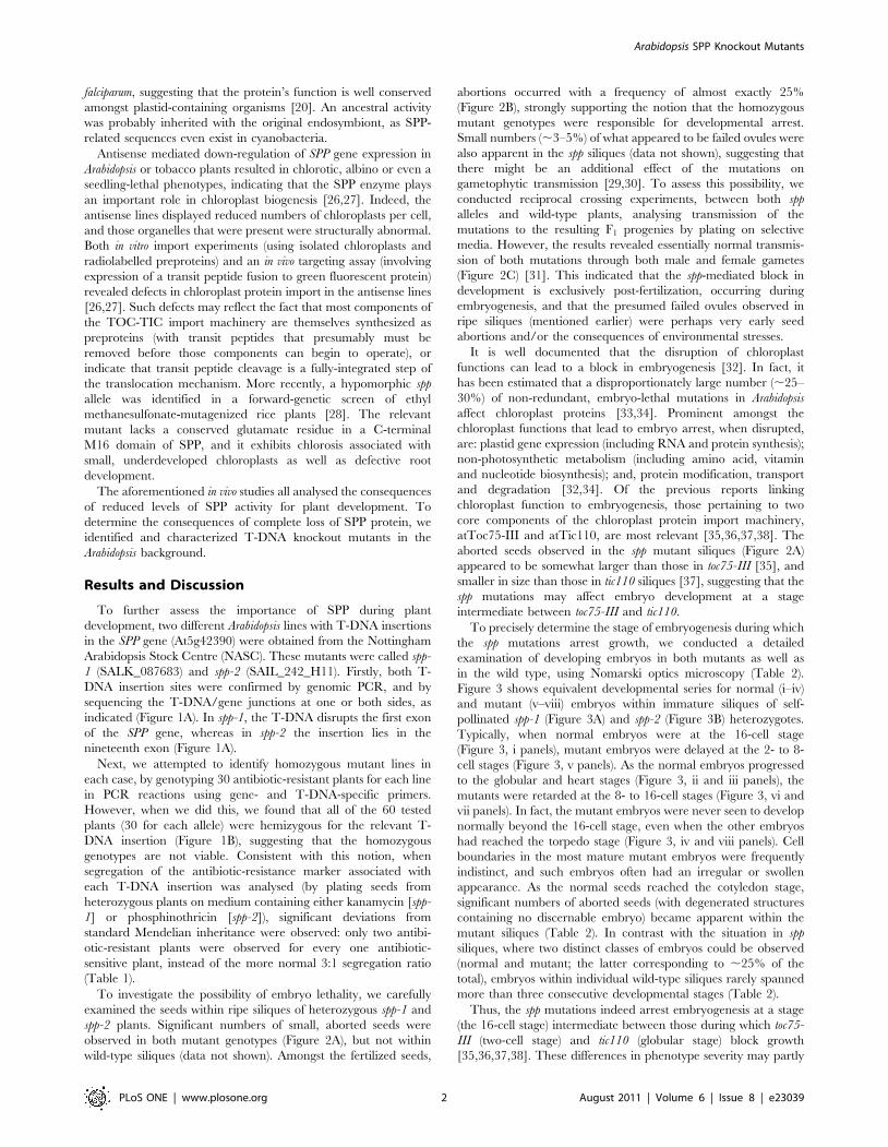
The Stromal Processing Peptidase of Chloroplasts isEssential in Arabidopsis, with Knockout MutationsCausing Embryo Arrest after the 16-Cell StageRaphael Trosch, Paul Jarvis*
Department of Biology, University of Leicester, Leicester, United Kingdom
Abstract
Stromal processing peptidase (SPP) is a metalloendopeptidase located in the stroma of chloroplasts, and it is responsible forthe cleavage of transit peptides from preproteins upon their import into the organelle. Two independent mutantArabidopsis lines with T-DNA insertions in the SPP gene were analysed (spp-1 and spp-2). For both lines, no homozygousmutant plants could be detected, and the segregating progeny of spp heterozygotes contained heterozygous and wild-typeplants in a ratio of 2:1. The siliques of heterozygous spp-1 and spp-2 plants contained many aborted seeds, at a frequency of,25%, suggesting embryo lethality. By contrast, transmission of the spp mutations through the male and female gameteswas found to be normal, and so gametophytic effects could be ruled out. To further elucidate the timing of thedevelopmental arrest, mutant and wild-type seeds were cleared and analysed by Nomarski microscopy. A significantproportion (,25%) of the seeds in mutant siliques exhibited delayed embryogenesis compared to those in wild type.Moreover, the mutant embryos never progressed normally beyond the 16-cell stage, with cell divisions not completingproperly thereafter. Heterozygous spp mutant plants were phenotypically indistinguishable from the wild type, indicatingthat the spp knockout mutations are completely recessive and suggesting that one copy of the SPP gene is able to producesufficient SPP protein for normal development under standard growth conditions.
Citation: Trosch R, Jarvis P (2011) The Stromal Processing Peptidase of Chloroplasts is Essential in Arabidopsis, with Knockout Mutations Causing Embryo Arrestafter the 16-Cell Stage. PLoS ONE 6(8): e23039. doi:10.1371/journal.pone.0023039
Editor: Miltos Tsiantis, University of Oxford, United Kingdom
Received March 21, 2011; Accepted July 5, 2011; Published August 16, 2011
Copyright: � 2011 Trosch, Jarvis. This is an open-access article distributed under the terms of the Creative Commons Attribution License, which permitsunrestricted use, distribution, and reproduction in any medium, provided the original author and source are credited.
Funding: This work was supported by a Gatsby Charitable Foundation Sainsbury PhD Studentship (http://www.gatsby.org.uk/) (to RT), and by Biotechnology andBiological Sciences Research Council (http://www.bbsrc.ac.uk/) grants BB/D016541/1, BB/F020325/1, and BB/H008039/1 (to PJ). The funders had no role in studydesign, data collection and analysis, decision to publish, or preparation of the manuscript.
Competing Interests: The authors have declared that no competing interests exist.
* E-mail: [email protected]
Introduction
The chloroplast is a unique plant cell compartment which
harbours many essential processes such as photosynthesis, starch
metabolism, and the biosynthesis of lipids and secondary
metabolites [1,2]. Like all plastids, chloroplasts are derived from
an ancient free-living cyanobacterial ancestor that was incorpo-
rated into early eukaryotic cells through endosymbiosis [3]. As a
result of this evolutionary origin, modern chloroplasts contain
DNA and are able to synthesize roughly one hundred of their own
proteins [4]. Nonetheless, the bulk of the ,3000 different proteins
in chloroplasts are encoded in the nuclear genome and must be
imported post-translationally from the cytosol [5,6].
Soon after the emergence of the signal hypothesis to account for
the translocation of ER proteins, it was suggested that nucleus-
encoded chloroplast proteins are similarly synthesized with a
targeting tag that directs them to the organelle [7,8]. This tag is an
N-terminal extension of the protein called a transit peptide, and it
is cleaved off after organellar import, producing a smaller, mature
form of the chloroplast protein [9]. Chloroplast transit peptides
vary greatly in length and amino acid sequence, and while
secondary structural features have been reported in some cases the
general significance of such observations remains uncertain
[10,11]. Thus, it is not fully understood how different preproteins
are all targeted quite specifically to the same organelle. Transit
peptides do contain slightly more hydroxylated residues and fewer
acidic residues than average, giving them a net positive charge,
and it has been suggested that a lack of a secondary structure
might be necessary for their targeting properties [12].
The transit peptide is recognized by receptor components at the
chloroplast surface, and subsequently the preprotein is guided
through pores in the outer and inner envelope membranes. The
multiprotein assemblies responsible for these recognition and
translocation events are the TOC and TIC complexes (translocon
at the outer/inner envelope membrane of chloroplasts)
[13,14,15,16,17]. Upon reaching the stromal side of the envelope,
the transit peptide is removed by the stromal processing peptidase
(SPP), a metalloendopeptidase of the M16 family (members of
which include subunit b of the mitochondrial processing peptidase,
MPP, and Escherichia coli pitrilysin) which has a high specificity for
chloroplast transit peptides [18,19,20]. The SPP enzyme recog-
nizes a stretch of basic residues with weak sequence or
physicochemical conservation at the C-terminus of the transit
peptide [21,22,23]. Following recognition, it cleaves the transit
peptide from the mature sequence using the catalytic activity of its
zinc-binding domain, and subsequently proteolyses the C-terminal
binding site of the transit peptide which facilitates release of the
peptide fragments so that they may be degraded by the
presequence protease, PreP [22,24,25]. Homologues of SPP exist
in red and green algae as well as in the malaria parasite, Plasmodium
PLoS ONE | www.plosone.org 1 August 2011 | Volume 6 | Issue 8 | e23039
falciparum, suggesting that the protein’s function is well conserved
amongst plastid-containing organisms [20]. An ancestral activity
was probably inherited with the original endosymbiont, as SPP-
related sequences even exist in cyanobacteria.
Antisense mediated down-regulation of SPP gene expression in
Arabidopsis or tobacco plants resulted in chlorotic, albino or even a
seedling-lethal phenotypes, indicating that the SPP enzyme plays
an important role in chloroplast biogenesis [26,27]. Indeed, the
antisense lines displayed reduced numbers of chloroplasts per cell,
and those organelles that were present were structurally abnormal.
Both in vitro import experiments (using isolated chloroplasts and
radiolabelled preproteins) and an in vivo targeting assay (involving
expression of a transit peptide fusion to green fluorescent protein)
revealed defects in chloroplast protein import in the antisense lines
[26,27]. Such defects may reflect the fact that most components of
the TOC-TIC import machinery are themselves synthesized as
preproteins (with transit peptides that presumably must be
removed before those components can begin to operate), or
indicate that transit peptide cleavage is a fully-integrated step of
the translocation mechanism. More recently, a hypomorphic spp
allele was identified in a forward-genetic screen of ethyl
methanesulfonate-mutagenized rice plants [28]. The relevant
mutant lacks a conserved glutamate residue in a C-terminal
M16 domain of SPP, and it exhibits chlorosis associated with
small, underdeveloped chloroplasts as well as defective root
development.
The aforementioned in vivo studies all analysed the consequences
of reduced levels of SPP activity for plant development. To
determine the consequences of complete loss of SPP protein, we
identified and characterized T-DNA knockout mutants in the
Arabidopsis background.
Results and Discussion
To further assess the importance of SPP during plant
development, two different Arabidopsis lines with T-DNA insertions
in the SPP gene (At5g42390) were obtained from the Nottingham
Arabidopsis Stock Centre (NASC). These mutants were called spp-
1 (SALK_087683) and spp-2 (SAIL_242_H11). Firstly, both T-
DNA insertion sites were confirmed by genomic PCR, and by
sequencing the T-DNA/gene junctions at one or both sides, as
indicated (Figure 1A). In spp-1, the T-DNA disrupts the first exon
of the SPP gene, whereas in spp-2 the insertion lies in the
nineteenth exon (Figure 1A).
Next, we attempted to identify homozygous mutant lines in
each case, by genotyping 30 antibiotic-resistant plants for each line
in PCR reactions using gene- and T-DNA-specific primers.
However, when we did this, we found that all of the 60 tested
plants (30 for each allele) were hemizygous for the relevant T-
DNA insertion (Figure 1B), suggesting that the homozygous
genotypes are not viable. Consistent with this notion, when
segregation of the antibiotic-resistance marker associated with
each T-DNA insertion was analysed (by plating seeds from
heterozygous plants on medium containing either kanamycin [spp-
1] or phosphinothricin [spp-2]), significant deviations from
standard Mendelian inheritance were observed: only two antibi-
otic-resistant plants were observed for every one antibiotic-
sensitive plant, instead of the more normal 3:1 segregation ratio
(Table 1).
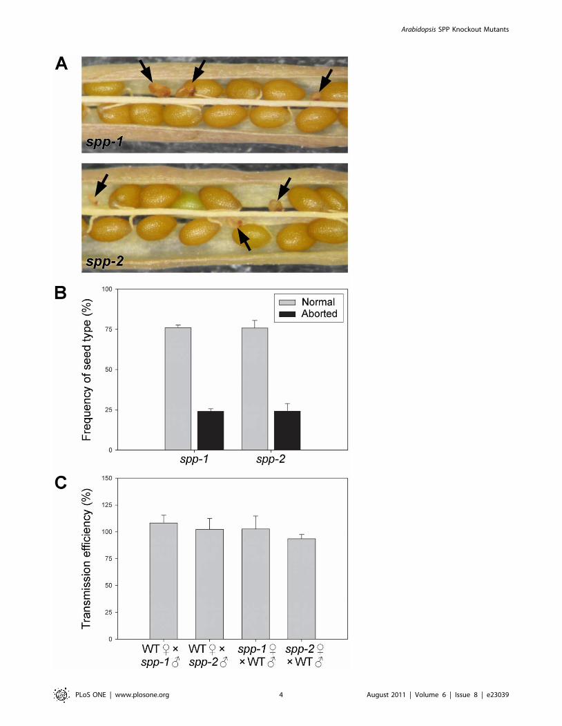
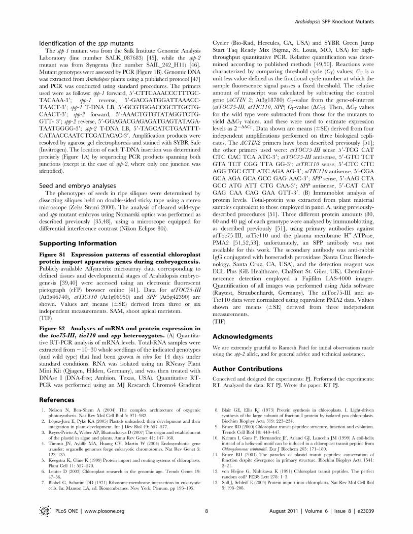
To investigate the possibility of embryo lethality, we carefully
examined the seeds within ripe siliques of heterozygous spp-1 and
spp-2 plants. Significant numbers of small, aborted seeds were
observed in both mutant genotypes (Figure 2A), but not within
wild-type siliques (data not shown). Amongst the fertilized seeds,
abortions occurred with a frequency of almost exactly 25%
(Figure 2B), strongly supporting the notion that the homozygous
mutant genotypes were responsible for developmental arrest.
Small numbers (,3–5%) of what appeared to be failed ovules were
also apparent in the spp siliques (data not shown), suggesting that
there might be an additional effect of the mutations on
gametophytic transmission [29,30]. To assess this possibility, we
conducted reciprocal crossing experiments, between both spp
alleles and wild-type plants, analysing transmission of the
mutations to the resulting F1 progenies by plating on selective
media. However, the results revealed essentially normal transmis-
sion of both mutations through both male and female gametes
(Figure 2C) [31]. This indicated that the spp-mediated block in
development is exclusively post-fertilization, occurring during
embryogenesis, and that the presumed failed ovules observed in
ripe siliques (mentioned earlier) were perhaps very early seed
abortions and/or the consequences of environmental stresses.
It is well documented that the disruption of chloroplast
functions can lead to a block in embryogenesis [32]. In fact, it
has been estimated that a disproportionately large number (,25–
30%) of non-redundant, embryo-lethal mutations in Arabidopsis
affect chloroplast proteins [33,34]. Prominent amongst the
chloroplast functions that lead to embryo arrest, when disrupted,
are: plastid gene expression (including RNA and protein synthesis);
non-photosynthetic metabolism (including amino acid, vitamin
and nucleotide biosynthesis); and, protein modification, transport
and degradation [32,34]. Of the previous reports linking
chloroplast function to embryogenesis, those pertaining to two
core components of the chloroplast protein import machinery,
atToc75-III and atTic110, are most relevant [35,36,37,38]. The
aborted seeds observed in the spp mutant siliques (Figure 2A)
appeared to be somewhat larger than those in toc75-III [35], and
smaller in size than those in tic110 siliques [37], suggesting that the
spp mutations may affect embryo development at a stage
intermediate between toc75-III and tic110.
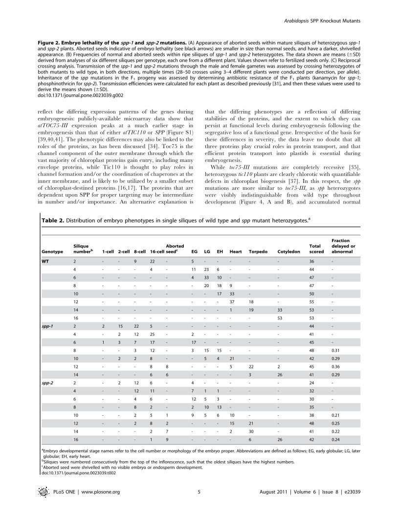
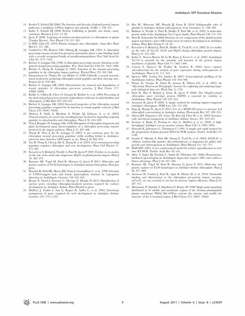
To precisely determine the stage of embryogenesis during which
the spp mutations arrest growth, we conducted a detailed
examination of developing embryos in both mutants as well as
in the wild type, using Nomarski optics microscopy (Table 2).
Figure 3 shows equivalent developmental series for normal (i–iv)
and mutant (v–viii) embryos within immature siliques of self-
pollinated spp-1 (Figure 3A) and spp-2 (Figure 3B) heterozygotes.
Typically, when normal embryos were at the 16-cell stage
(Figure 3, i panels), mutant embryos were delayed at the 2- to 8-
cell stages (Figure 3, v panels). As the normal embryos progressed
to the globular and heart stages (Figure 3, ii and iii panels), the
mutants were retarded at the 8- to 16-cell stages (Figure 3, vi and
vii panels). In fact, the mutant embryos were never seen to develop
normally beyond the 16-cell stage, even when the other embryos
had reached the torpedo stage (Figure 3, iv and viii panels). Cell
boundaries in the most mature mutant embryos were frequently
indistinct, and such embryos often had an irregular or swollen
appearance. As the normal seeds reached the cotyledon stage,
significant numbers of aborted seeds (with degenerated structures
containing no discernable embryo) became apparent within the
mutant siliques (Table 2). In contrast with the situation in spp
siliques, where two distinct classes of embryos could be observed
(normal and mutant; the latter corresponding to ,25% of the
total), embryos within individual wild-type siliques rarely spanned
more than three consecutive developmental stages (Table 2).
Thus, the spp mutations indeed arrest embryogenesis at a stage
(the 16-cell stage) intermediate between those during which toc75-
III (two-cell stage) and tic110 (globular stage) block growth
[35,36,37,38]. These differences in phenotype severity may partly
Arabidopsis SPP Knockout Mutants
PLoS ONE | www.plosone.org 2 August 2011 | Volume 6 | Issue 8 | e23039
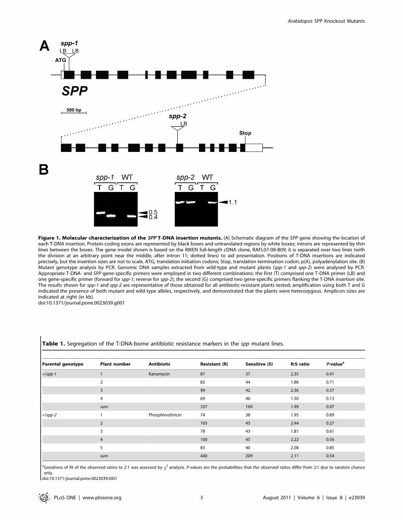
Figure 1. Molecular characterization of the SPP T-DNA insertion mutants. (A) Schematic diagram of the SPP gene showing the location ofeach T-DNA insertion. Protein-coding exons are represented by black boxes and untranslated regions by white boxes; introns are represented by thinlines between the boxes. The gene model shown is based on the RIKEN full-length cDNA clone, RAFL07-08-B09; it is separated over two lines (withthe division at an arbitrary point near the middle, after intron 11; dotted lines) to aid presentation. Positions of T-DNA insertions are indicatedprecisely, but the insertion sizes are not to scale. ATG, translation initiation codons; Stop, translation termination codon; p(A), polyadenylation site. (B)Mutant genotype analysis by PCR. Genomic DNA samples extracted from wild-type and mutant plants (spp-1 and spp-2) were analysed by PCR.Appropriate T-DNA- and SPP-gene-specific primers were employed in two different combinations: the first (T) comprised one T-DNA primer (LB) andone gene-specific primer (forward for spp-1; reverse for spp-2); the second (G) comprised two gene-specific primers flanking the T-DNA insertion site.The results shown for spp-1 and spp-2 are representative of those obtained for all antibiotic-resistant plants tested; amplification using both T and Gindicated the presence of both mutant and wild-type alleles, respectively, and demonstrated that the plants were heterozygous. Amplicon sizes areindicated at right (in kb).doi:10.1371/journal.pone.0023039.g001
Table 1. Segregation of the T-DNA-borne antibiotic resistance markers in the spp mutant lines.
Parental genotype Plant number Antibiotic Resistant (R) Sensitive (S) R:S ratio P-valuea
+/spp-1 1 Kanamycin 87 37 2.35 0.41
2 82 44 1.86 0.71
3 99 42 2.36 0.37
4 69 46 1.50 0.13
sum 337 169 1.99 0.97
+/spp-2 1 Phosphinothricin 74 38 1.95 0.89
2 105 43 2.44 0.27
3 78 43 1.81 0.61
4 100 45 2.22 0.56
5 83 40 2.08 0.85
sum 440 209 2.11 0.54
aGoodness of fit of the observed ratios to 2:1 was assessed by x2 analysis. P-values are the probabilities that the observed ratios differ from 2:1 due to random chanceonly.
doi:10.1371/journal.pone.0023039.t001
Arabidopsis SPP Knockout Mutants
PLoS ONE | www.plosone.org 3 August 2011 | Volume 6 | Issue 8 | e23039
Arabidopsis SPP Knockout Mutants
PLoS ONE | www.plosone.org 4 August 2011 | Volume 6 | Issue 8 | e23039
reflect the differing expression patterns of the genes during
embryogenesis: publicly-available microarray data show that
atTOC75-III expression peaks at a much earlier stage in
embryogenesis than that of either atTIC110 or SPP (Figure S1)
[39,40,41]. The phenotypic differences may also be linked to the
roles of the proteins, as has been discussed [34]. Toc75 is the
channel component of the outer membrane through which the
vast majority of chloroplast proteins gain entry, including many
envelope proteins, while Tic110 is thought to play roles in
channel formation and/or the coordination of chaperones at the
inner membrane, and is likely to be utilized by a smaller subset
of chloroplast-destined proteins [16,17]. The proteins that are
dependent upon SPP for proper targeting may be intermediate
in number and/or importance. An alternative explanation is
that the differing phenotypes are a reflection of differing
stabilities of the proteins, and the extent to which they can
persist at functional levels during embryogenesis following the
segregative loss of a functional gene. Irrespective of the basis for
these differences in severity, the data leave no doubt that all
three proteins play crucial roles in protein transport, and that
efficient protein transport into plastids is essential during
embryogenesis.
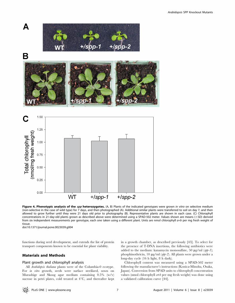
While toc75-III mutations are completely recessive [35],
heterozygous tic110 plants are clearly chlorotic with quantifiable
defects in chloroplast biogenesis [37]. In this respect, the spp
mutations are more similar to toc75-III, as spp heterozygotes
were visibly indistinguishable from wild type throughout
development (Figure 4, A and B), and accumulated normal
Figure 2. Embryo lethality of the spp-1 and spp-2 mutations. (A) Appearance of aborted seeds within mature siliques of heterozygous spp-1and spp-2 plants. Aborted seeds indicative of embryo lethality (see black arrows) are smaller in size than normal seeds, and have a darker, shrivelledappearance. (B) Frequencies of normal and aborted seeds within ripe siliques of spp-1 and spp-2 heterozygotes. The data shown are means (6SD)derived from analyses of six different siliques per genotype, each one from a different plant. Values shown refer to fertilized seeds only. (C) Reciprocalcrossing analysis. Transmission of the spp-1 and spp-2 mutations through the male and female gametes was assessed by crossing heterozygotes ofboth mutants to wild type, in both directions, multiple times (28–50 crosses using 3–4 different plants were conducted per direction, per allele).Inheritance of the spp mutations in the F1 progeny was assessed by determining antibiotic resistance of the F1 plants (kanamycin for spp-1;phosphinothricin for spp-2). Transmission efficiencies were calculated for each plant as described previously [31], and then these values were used toderive the means shown (6SD).doi:10.1371/journal.pone.0023039.g002
Table 2. Distribution of embryo phenotypes in single siliques of wild type and spp mutant heterozygotes.a
GenotypeSiliquenumberb 1-cell 2-cell 8-cell 16-cell
Abortedseedc EG LG EH Heart Torpedo Cotyledon
Totalscored
Fractiondelayed orabnormal
WT 2 - - 9 22 - 5 - - - - - 36 -
4 - - - 4 - 11 23 6 - - - 44 -
6 - - - - - 4 33 10 - - - 47 -
8 - - - - - - 20 18 9 - - 47 -
10 - - - - - - - 17 33 - - 50 -
12 - - - - - - - - 37 18 - 55 -
14 - - - - - - - - 1 19 33 53 -
16 - - - - - - - - - - 53 53 -
spp-1 2 2 15 22 5 - - - - - - - 44 -
4 - 2 12 25 - 2 - - - - - 41 -
6 1 3 7 17 - 17 - - - - - 45 -
8 - - 3 12 - 3 15 15 - - - 48 0.31
10 - 2 2 8 - - 5 4 21 - - 42 0.29
12 - - - 8 8 - - - 5 22 2 45 0.36
14 - - - 6 6 - - - - 3 26 41 0.29
spp-2 2 - 2 12 6 - 4 - - - - - 24 -
4 - - 12 11 - 7 1 1 - - - 32 -
6 - - 4 6 - 12 5 3 - - - 30 -
8 - - 8 2 - 2 10 13 - - - 35 -
10 - - 2 5 1 9 5 6 10 - - 38 0.21
12 - - 2 8 2 - - - 15 21 - 48 0.25
14 - - - 2 7 - - - 2 30 - 41 0.22
16 - - - 1 9 - - - - 6 26 42 0.24
aEmbryo developmental stage names refer to the cell number or morphology of the embryo proper. Abbreviations are defined as follows: EG, early globular; LG, laterglobular; EH, early heart.
bSiliques were numbered consecutively from the top of the inflorescence, such that the oldest siliques have the highest numbers.cAborted seed were shrivelled with no visible embryo or endosperm development.doi:10.1371/journal.pone.0023039.t002
Arabidopsis SPP Knockout Mutants
PLoS ONE | www.plosone.org 5 August 2011 | Volume 6 | Issue 8 | e23039
levels of chlorophyll pigment (Figure 4C). This indicates that a
single copy of the SPP gene is able to produce sufficient
quantities of the peptidase for normal growth under standard
conditions. The phenotypic difference between tic110 heterozy-
gotes and toc75-III or spp heterozygotes cannot easily be
explained in terms of mRNA or protein levels, as quantitative
RT-PCR and immunoblot experiments did not reveal more
severe deficiencies in +/tic110 plants (Figure S2). As was
discussed previously [42], the greater dosage dependency of
the tic110 mutation may reflect the absence of excess expression
capacity for atTic110 in the wild type.
ConclusionWhile it was evident from earlier studies that SPP is an important
chloroplast protein, the phenotype of homozygous knockout plants
had not previously been reported. In this study, we employed
Arabidopsis T-DNA insertion lines to demonstrate that SPP is
indispensable in vivo. Homozygotes were absent from the progeny of
plants carrying spp mutations, while a quarter of the seeds in the
siliques of such plants were aborted, consistent with an embryo
lethal phenotype. The mutant embryos exhibited delayed develop-
ment, with cell divisions not terminating properly after the 16-cell
stage. This work further emphasizes the importance of plastid
Figure 3. Analysis of embryo development in the spp mutants using Nomarski optics. Equivalent developmental series for normal (i–iv)and mutant (v–viii) embryos within immature heterozygous siliques of spp-1 (A) and spp-2 (B). Normal embryos: i, 16-cell stage; ii, early globular stage;iii, heart stage; iv, torpedo stage. Corresponding mutant embryos from the same siliques: v, 2- to 8-cell stages; vi, 8- to 16-cell stages; vii and viii,arrested or abnormal 16-cell stages. Embryo developmental stage names refer to the cell number or morphology of the embryo proper. Images i–iiiand v–viii are all at the same magnification (406objective); images iv are at lower magnification (206 objective). Bars = 50 mm.doi:10.1371/journal.pone.0023039.g003
Arabidopsis SPP Knockout Mutants
PLoS ONE | www.plosone.org 6 August 2011 | Volume 6 | Issue 8 | e23039
functions during seed development, and extends the list of protein
transport components known to be essential for plant viability.
Materials and Methods
Plant growth and chlorophyll analysisAll Arabidopsis thaliana plants were of the Columbia-0 ecotype.
For in vitro growth, seeds were surface sterilized, sown on
Murashige and Skoog agar medium containing 0.5% (w/v)
sucrose in petri plates, cold treated at 4uC, and thereafter kept
in a growth chamber, as described previously [43]. To select for
the presence of T-DNA insertions, the following antibiotics were
added to the medium: kanamycin monosulfate, 50 mg/ml (spp-1);
phosphinothricin, 10 mg/ml (spp-2). All plants were grown under a
long-day cycle (16 h light, 8 h dark).
Chlorophyll content was measured using a SPAD-502 meter
following the manufacturer’s instructions (Konica-Minolta, Osaka,
Japan). Conversion from SPAD units to chlorophyll concentration
values (nmol chlorophyll a+b per mg fresh weight) was done using
a validated calibration curve [44].
Figure 4. Phenotypic analysis of the spp heterozygotes. (A, B) Plants of the indicated genotypes were grown in vitro on selective medium(non-selective in the case of wild type) for 7 days, and then photographed (A). Additional similar plants were transferred to soil on day 7, and thenallowed to grow further until they were 21 days old prior to photography (B). Representative plants are shown in each case. (C) Chlorophyllconcentrations in 21-day-old plants grown as described above were determined using a SPAD-502 meter. Values shown are means (6SD) derivedfrom six independent measurements per genotype, each one taken using a different plant. Units are nmol chlorophyll a+b per mg fresh weight oftissue.doi:10.1371/journal.pone.0023039.g004
Arabidopsis SPP Knockout Mutants
PLoS ONE | www.plosone.org 7 August 2011 | Volume 6 | Issue 8 | e23039
Identification of the spp mutantsThe spp-1 mutant was from the Salk Institute Genomic Analysis
Laboratory (line number SALK_087683) [45], while the spp-2
mutant was from Syngenta (line number SAIL_242_H11) [46].
Mutant genotypes were assessed by PCR (Figure 1B). Genomic DNA
was extracted from Arabidopsis plants using a published protocol [47]
and PCR was conducted using standard procedures. The primers
used were as follows: spp-1 forward, 59-CTTCAAACCCTTTGC-
TACAAA-39; spp-1 reverse, 59-GACGATGGATTAAACC-
TAACT-39; spp-1 T-DNA LB, 59-GCGTGGACCGCTTGCTG-
CAACT-39; spp-2 forward, 59-AAACTGTGTATAGGTCTG-
GTT- 39; spp-2 reverse, 59-GGAGACGAGAGATGAGTATAGA-
TAATGGGG-39; spp-2 T-DNA LB, 59-TAGCATCTGAATTT-
CATAACCAATCTCGATACAC-39. Amplification products were
resolved by agarose gel electrophoresis and stained with SYBR Safe
(Invitrogen). The location of each T-DNA insertion was determined
precisely (Figure 1A) by sequencing PCR products spanning both
junctions (except in the case of spp-2, where only one junction was
identified).
Seed and embryo analysesThe phenotypes of seeds in ripe siliques were determined by
dissecting siliques held on double-sided sticky tape using a stereo
microscope (Zeiss Stemi 2000). The analysis of cleared wild-type
and spp mutant embryos using Nomarski optics was performed as
described previously [35,48], using a microscope equipped for
differential interference contrast (Nikon Eclipse 80i).
Supporting Information
Figure S1 Expression patterns of essential chloroplastprotein import apparatus genes during embryogenesis.Publicly-available Affymetrix microarray data corresponding to
defined tissues and developmental stages of Arabidopsis embryo-
genesis [39,40] were accessed using an electronic fluorescent
pictograph (eFP) browser online [41]. Data for atTOC75-III
(At3g46740), atTIC110 (At1g06950) and SPP (At5g42390) are
shown. Values are means (6SE) derived from three or six
independent measurements. SAM, shoot apical meristem.
(TIF)
Figure S2 Analyses of mRNA and protein expression inthe toc75-III, tic110 and spp heterozygotes. (A) Quantita-
tive RT-PCR analysis of mRNA levels. Total-RNA samples were
extracted from ,10–30 whole seedlings of the indicated genotypes
(and wild type) that had been grown in vitro for 14 days under
standard conditions. RNA was isolated using an RNeasy Plant
Mini Kit (Qiagen, Hilden, Germany), and was then treated with
DNAse I (DNA-free; Ambion, Texas, USA). Quantitative RT-
PCR was performed using an MJ Research Chromo4 Gradient
Cycler (Bio-Rad, Hercules, CA, USA) and SYBR Green Jump
Start Taq Ready Mix (Sigma, St. Louis, MO, USA) for high-
throughput quantitative PCR. Relative quantification was deter-
mined according to published methods [49,50]. Reactions were
characterized by comparing threshold cycle (CT) values; CT is a
unit-less value defined as the fractional cycle number at which the
sample fluorescence signal passes a fixed threshold. The relative
amount of transcript was calculated by subtracting the control
gene (ACTIN 2; At3g18780) CT-value from the gene-of-interest
(atTOC75-III, atTIC110, SPP) CT-value (DCT). Then, DCT values
for the wild type were subtracted from those for the mutants to
yield DDCT values, and these were used to estimate expression
levels as 2{DDCT . Data shown are means (6SE) derived from four
independent amplifications performed on three biological repli-
cates. The ACTIN2 primers have been described previously [51];
the other primers used were: atTOC75-III sense 59-TCG CAT
CTC CAC TCA ATC-39; atTOC75-III antisense, 59-GTC TCT
GTA TCT CGG TTA GG-39; atTIC110 sense, 59-CTC CTC
AGG TGC CTT ATC AGA AG-39; atTIC110 antisense, 59-CGA
GCA AGA GCA GCC GAG AAC-39; SPP sense, 59-AAG CTA
GCC ATG ATT CTG CAA-39; SPP antisense, 59-CAT CAT
GAG CAA CAG GAA GTT-39. (B) Immunoblot analysis of
protein levels. Total-protein was extracted from plant material
samples equivalent to those employed in panel A, using previously-
described procedures [51]. Three different protein amounts (80,
60 and 40 mg) of each genotype were analysed by immunoblotting,
as described previously [51], using primary antibodies against
atToc75-III, atTic110 and the plasma membrane H+-ATPase,
PMA2 [51,52,53]; unfortunately, an SPP antibody was not
available for this work. The secondary antibody was anti-rabbit
IgG conjugated with horseradish peroxidase (Santa Cruz Biotech-
nology, Santa Cruz, CA, USA), and the detection reagent was
ECL Plus (GE Healthcare, Chalfont St. Giles, UK). Chemilumi-
nescence detection employed a Fujifilm LAS-4000 imager.
Quantification of all images was performed using Aida software
(Raytest, Straubenhardt, Germany). The atToc75-III and at-
Tic110 data were normalized using equivalent PMA2 data. Values
shown are means (6SE) derived from three independent
measurements.
(TIF)
Acknowledgments
We are extremely grateful to Ramesh Patel for initial observations made
using the spp-2 allele, and for general advice and technical assistance.
Author Contributions
Conceived and designed the experiments: PJ. Performed the experiments:
RT. Analyzed the data: RT PJ. Wrote the paper: RT PJ.
References
1. Nelson N, Ben-Shem A (2004) The complex architecture of oxygenic
photosynthesis. Nat Rev Mol Cell Biol 5: 971–982.
2. Lopez-Juez E, Pyke KA (2005) Plastids unleashed: their development and their
integration in plant development. Int J Dev Biol 49: 557–577.
3. Reyes-Prieto A, Weber AP, Bhattacharya D (2007) The origin and establishment
of the plastid in algae and plants. Annu Rev Genet 41: 147–168.
4. Timmis JN, Ayliffe MA, Huang CY, Martin W (2004) Endosymbiotic gene
transfer: organelle genomes forge eukaryotic chromosomes. Nat Rev Genet 5:
123–135.
5. Keegstra K, Cline K (1999) Protein import and routing systems of chloroplasts.
Plant Cell 11: 557–570.
6. Leister D (2003) Chloroplast research in the genomic age. Trends Genet 19:
47–56.
7. Blobel G, Sabatini DD (1971) Ribosome-membrane interactions in eukaryotic
cells. In: Manson LA, ed. Biomembranes. New York: Plenum. pp 193–195.
8. Blair GE, Ellis RJ (1973) Protein synthesis in chloroplasts. I. Light-driven
synthesis of the large subunit of fraction I protein by isolated pea chloroplasts.
Biochim Biophys Acta 319: 223–234.
9. Bruce BD (2000) Chloroplast transit peptides: structure, function and evolution.
Trends Cell Biol 10: 440–447.
10. Krimm I, Gans P, Hernandez JF, Arlaud GJ, Lancelin JM (1999) A coil-helix
instead of a helix-coil motif can be induced in a chloroplast transit peptide from
Chlamydomonas reinhardtii. Eur J Biochem 265: 171–180.
11. Bruce BD (2001) The paradox of plastid transit peptides: conservation of
function despite divergence in primary structure. Biochim Biophys Acta 1541:
2–21.
12. von Heijne G, Nishikawa K (1991) Chloroplast transit peptides. The perfect
random coil? FEBS Lett 278: 1–3.
13. Soll J, Schleiff E (2004) Protein import into chloroplasts. Nat Rev Mol Cell Biol
5: 198–208.
Arabidopsis SPP Knockout Mutants
PLoS ONE | www.plosone.org 8 August 2011 | Volume 6 | Issue 8 | e23039
14. Kessler F, Schnell DJ (2006) The function and diversity of plastid protein import
pathways: a multilane GTPase highway into plastids. Traffic 7: 248–257.15. Inaba T, Schnell DJ (2008) Protein trafficking to plastids: one theme, many
variations. Biochem J 413: 15–28.
16. Jarvis P (2008) Targeting of nucleus-encoded proteins to chloroplasts in plants(Tansley Review). New Phytol 179: 257–285.
17. Li HM, Chiu CC (2010) Protein transport into chloroplasts. Annu Rev PlantBiol 61: 157–180.
18. VanderVere PS, Bennett TM, Oblong JE, Lamppa GK (1995) A chloroplast
processing enzyme involved in precursor maturation shares a zinc-binding motifwith a recently recognized family of metalloendopeptidases. Proc Natl Acad Sci
USA 92: 7177–7181.19. Richter S, Lamppa GK (1998) A chloroplast processing enzyme functions as the
general stromal processing peptidase. Proc Natl Acad Sci USA 95: 7463–7468.20. Richter S, Zhong R, Lamppa G (2005) Function of the stromal processing
peptidase in the chloroplast import pathway. Physiol Plant 123: 362–368.
21. Emanuelsson O, Nielsen H, von Heijne G (1999) ChloroP, a neural network-based method for predicting chloroplast transit peptides and their cleavage sites.
Protein Sci 8: 978–984.22. Richter S, Lamppa GK (2002) Determinants for removal and degradation of
transit peptides of chloroplast precursor proteins. J Biol Chem 277:
43888–43894.23. Rudhe C, Clifton R, Chew O, Zemam K, Richter S, et al. (2004) Processing of
the dual targeted precursor protein of glutathione reductase in mitochondria andchloroplasts. J Mol Biol 343: 639–647.
24. Richter S, Lamppa GK (2003) Structural properties of the chloroplast stromalprocessing peptidase required for its function in transit peptide removal. J Biol
Chem 278: 39497–39502.
25. Moberg P, Stahl A, Bhushan S, Wright SJ, Eriksson A, et al. (2003)Characterization of a novel zinc metalloprotease involved in degrading targeting
peptides in mitochondria and chloroplasts. Plant J 36: 616–628.26. Wan J, Bringloe D, Lamppa GK (1998) Disruption of chloroplast biogenesis and
plant development upon down-regulation of a chloroplast processing enzyme
involved in the import pathway. Plant J 15: 459–468.27. Zhong R, Wan J, Jin R, Lamppa G (2003) A pea antisense gene for the
chloroplast stromal processing peptidase yields seedling lethals in Arabidopsis:survivors show defective GFP import in vivo. Plant J 34: 802–812.
28. Yue R, Wang X, Chen J, Ma X, Zhang H, et al. (2010) A rice stromal processingpeptidase regulates chloroplast and root development. Plant Cell Physiol 51:
475–485.
29. Kovacheva S, Bedard J, Wardle A, Patel R, Jarvis P (2007) Further in vivo studieson the role of the molecular chaperone, Hsp93, in plastid protein import. Plant J
50: 364–379.30. Kasmati AR, Topel M, Patel R, Murtaza G, Jarvis P (2011) Molecular and
genetic analyses of Tic20 homologues in Arabidopsis thaliana chloroplasts. Plant J;in
press.31. Howden R, Park SK, Moore JM, Orme J, Grossniklaus U, et al. (1998) Selection
of T-DNA-tagged male and female gametophytic mutants by segregationdistortion in Arabidopsis. Genetics 149: 621–631.
32. Bryant N, Lloyd J, Sweeney C, Myouga F, Meinke D (2011) Identification ofnuclear genes encoding chloroplast-localized proteins required for embryo
development in Arabidopsis thaliana. Plant Physiol: in press.
33. McElver J, Tzafrir I, Aux G, Rogers R, Ashby C, et al. (2001) Insertionalmutagenesis of genes required for seed development in Arabidopsis thaliana.
Genetics 159: 1751–1763.
34. Hsu SC, Belmonte MF, Harada JJ, Inoue K (2010) Indispensable roles ofplastids in Arabidopsis thaliana embryogenesis. Curr Genomics 11: 338–349.
35. Baldwin A, Wardle A, Patel R, Dudley P, Park SK, et al. (2005) A molecular-
genetic study of the Arabidopsis Toc75 gene family. Plant Physiol 138: 715–733.
36. Hust B, Gutensohn M (2006) Deletion of core components of the plastid protein
import machinery causes differential arrest of embryo development in Arabidopsis
thaliana. Plant Biol (Stuttg) 8: 18–30.
37. Kovacheva S, Bedard J, Patel R, Dudley P, Twell D, et al. (2005) In vivo studies
on the roles of Tic110, Tic40 and Hsp93 during chloroplast protein import.
Plant J 41: 412–428.
38. Inaba T, Alvarez-Huerta M, Li M, Bauer J, Ewers C, et al. (2005) Arabidopsis
Tic110 is essential for the assembly and function of the protein import
machinery of plastids. Plant Cell 17: 1482–1496.
39. Casson S, Spencer M, Walker K, Lindsey K (2005) Laser capture
microdissection for the analysis of gene expression during embryogenesis of
Arabidopsis. Plant J 42: 111–123.
40. Spencer MW, Casson SA, Lindsey K (2007) Transcriptional profiling of the
Arabidopsis embryo. Plant Physiol 143: 924–940.
41. Winter D, Vinegar B, Nahal H, Ammar R, Wilson GV, et al. (2007) An‘‘Electronic Fluorescent Pictograph’’ browser for exploring and analyzing large-
scale biological data sets. PLoS One 2: e718.
42. Patel R, Hsu S, Bedard J, Inoue K, Jarvis P (2008) The Omp85-relatedchloroplast outer envelope protein OEP80 is essential for viability in
Arabidopsis. Plant Physiol 148: 235–245.
43. Aronsson H, Jarvis P (2002) A simple method for isolating import-competentArabidopsis chloroplasts. FEBS Lett 529: 215–220.
44. Ling Q, Huang W, Jarvis P (2011) Use of a SPAD-502 meter to measure leaf
chlorophyll concentration in Arabidopsis thaliana. Photosynth Res 107: 209–214.
45. Alonso JM, Stepanova AN, Leisse TJ, Kim CJ, Chen H, et al. (2003) Genome-
wide insertional mutagenesis of Arabidopsis thaliana. Science 301: 653–657.
46. Sessions A, Burke E, Presting G, Aux G, McElver J, et al. (2002) A high-
throughput Arabidopsis reverse genetics system. Plant Cell 14: 2985–2994.
47. Edwards K, Johnstone C, Thompson C (1991) A simple and rapid method forthe preparation of plant genomic DNA for PCR analysis. Nucleic Acids Res 19:
1349.
48. Goubet F, Misrahi A, Park SK, Zhang Z, Twell D, et al. (2003) AtCSLA7, acellulose synthase-like putative glycosyltransferase, is important for pollen tube
growth and embryogenesis in Arabidopsis. Plant Physiol 131: 547–557.
49. Pfaffl MW (2001) A new mathematical model for relative quantification in real-time RT-PCR. Nucleic Acids Res 29: e45.
50. Allen T, Ingles PJ, Praekelt U, Smith H, Whitelam GC (2006) Phytochrome-
mediated agravitropism in Arabidopsis hypocotyls requires GIL1 and confers afitness advantage. Plant J 46: 641–648.
51. Kasmati AR, Topel M, Patel R, Murtaza G, Jarvis P (2011) Molecular and
genetic analyses of Tic20 homologues in Arabidopsis thaliana chloroplasts. Plant J66: 877–889.
52. Aronsson H, Combe J, Patel R, Agne B, Martin M, et al. (2010) Nucleotide
binding and dimerization at the chloroplast pre-protein import receptor,atToc33, are not essential in vivo but do increase import efficiency. Plant J 63:
297–311.
53. Morsomme P, Dambly S, Maudoux O, Boutry M (1998) Single point mutationsdistributed in 10 soluble and membrane regions of the Nicotiana plumbaginifolia
plasma membrane PMA2 H+-ATPase activate the enzyme and modify the
structure of the C-terminal region. J Biol Chem 273: 34837–34842.
Arabidopsis SPP Knockout Mutants
PLoS ONE | www.plosone.org 9 August 2011 | Volume 6 | Issue 8 | e23039