Embed Size (px)
Citation preview

Amer. Zool., 24:893-909 (1984)
The Structure and Calcification of the Crustacean Cuticle1
Robert Roer and Richard Dillaman
Institute of Marine Biomedical Research, University of North Carolina at Wilmington, Wilmington, North Carolina 28403
Synopsis. The integument of decapod crustaceans consists of an outer epicuticle, an exocuticle, an endocuticle and an inner membranous layer underlain by the hypodermis. The outer three layers of the cuticle are calcified. The mineral is in the form of calcite crystals and amorphous calcium carbonate. In the epicuticle, mineral is in the form of spherulitic calcite islands surrounded by the lipid-protein matrix. In the exo- and endo- cuticles the calcite crystal aggregates are interspersed with chitin-protein fibers which are organized in lamellae. In some species, the organization of the mineral mirrors that of the organic fibers, but such is not the case in certain cuticular regions in the xanthid crabs. Thus, control of crystal organization is a complex phenomenon unrelated to the gross morphology of the matrix.
Since the cuticle is periodically molted to allow for growth, this necessitates a bidirec- tional movement of calcium into the cuticle during postmolt and out during premolt resorption of the cuticle. In two species of crabs studied to date, these movements are accomplished by active transport effected by a Ca-ATPase and Na/Ca exchange mech? anism.
The epi- and exocuticular layers of the new cuticle are elaborated during premolt but do not calcify until the old cuticle is shed. This phenomenon also occurs in vitro in cuticle devoid of living tissue and implies an alteration of the nucleating sites of the cuticle in the course of the molt.
Introduction
While the crustacean cuticle has been the subject of study for over 250 years (Reaumur, 1712, in Drach, 1939), the focus of those investigations has generally been concerned with the process of molting. Our
approach will be slightly different; we will deal with the exoskeleton of the Crustacea as a mineralized tissue that is made partic? ularly interesting by the fact that its struc? ture is affected by a cyclic molting process.
When investigating any mineralized tis?
sue, one must address some basic problems: 1) the chemical nature and crystalline form of the mineral; 2) the nature and form of the organic matrix; 3) the relationship between the organic and inorganic com?
ponents of the tissue and the influence of the matrix on crystal morphology; 4) the sources of mineral for deposition; 5) the
pathways for mineral movement into or out from the mineralized structures; 6) rates of mineral deposition; and 7) the nature and location of nucleation sites
1 From the Symposium on Mechanisms of Calcification in Biological Systems presented at the Annual Meeting ofthe American Society of Zoologists, 27-30 Decem? ber 1983, at Philadelphia, Pennsylvania.
within the matrix and mechanisms for con? trol or cessation of crystal growth. The crustacean cuticle has provided a rich source of information with respect to each of these central questions. In addition, the Crustacea offer some unique problems for those interested in biomineralization.
Since the mineralized exoskeleton ofthe Crustacea is subjected to periodic molting, bidirectional net movement of mineral
during different stages of the molt cycle is
necessary. This situation is in sharp con? trast to other calcifying systems in which net accretionary growth patterns or slightly shifting equilibria are the rule. Further?
more, crustaceans demonstrate drastic
temporal differences within the same tissue with regard to the extent of and capacity for mineralization. Such temporal differ?
ences, marked by rapid and discrete tran?
sitions, allow one to ask very specific and answerable questions about the control of nucleation and mineralization.
The subject of crustacean cuticles is as diverse as is the taxon and, hence, an ency- clopedic overview is impossible. We will restrict our review, in large part, to the
decapod crustaceans although examples from other orders will serve to remind us ofthe hazards of sweeping generalizations.
893
894 R. Roer and R. Dillaman
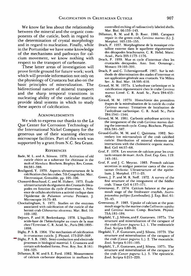
] Ep 5, im
Ex 65pm
2.2Hm/l
'V->
En 195pm
8.1 Hm/I
Fig. 1. Montage ofthe mineralized cuticular layers of Carcinus maenas carapace (from Roer 1979). En =
endocuticle, Ex = exocuticle, Ep = epicuticle. Num? bers represent the total thickness of the individual layers and the thickness of endo- and exocuticular lamellae (1).
Structure of the Decapod Integument
Basic cuticular structure
The integument of the decapod Crus? tacea is, in general, comprised of a rigid exoskeleton or cuticle underlain by the cel? lular hypodermis (Richards, 1951). The cuticle is not homogeneous, but contains four discrete layers. Despite the fact that this observation was made more than a cen?
tury ago (Williamson, 1860) and has been the subject of numerous subsequent stud?
ies, the nomenclature of the four layers of the cuticle varies from author to author. The terminology of Travis (1963), how?
ever, has come to be widely accepted and will be used exclusively in the present work. These layers from the most external to the most internal are: the epicuticle, the exo-
cuticle, the endocuticle and the membra- nous layer (Fig. 1).
The epicuticle is the outermost and thin- nest layer ofthe cuticle. It consists of tanned
lipoprotein impregnated with calcium salts
(Travis, 1955a). It is bilaminar, with the basal layer pervaded by mineral-filled canals normal to the surface (Hegdahl et al,
1977c). The exocuticle immediately underlies the
epicuticle. It is composed of chitin-protein fibers stacked in layers of continuously changing orientation (Travis, 1955a; Green and Neff, 1972). In Cancer pagurus 34% by weight of the organic component is chitin
(Welinder, 19756). The exocuticle is hard- ened by quinone tanning and calcification
(Travis, 1955a), with the mineral crystals situated between the fibers (Bouligand, 1970; Hegdahl etal, 19776).
The endocuticle is the thickest and the most heavily calcified layer of the cuticle
(Travis, 1955a, 1965). The endocuticle, like the exocuticle, is composed of hori? zontal lamellae of chitin-protein fibers with
continuously changing orientation (Green and Neff, 1972; Hegdahl etal, 1977a), the
organic material being 73% chitin by weight in Cancer pagurus (Welinder, 19756). The endocuticle is apparently not tanned, but hardened solely by Ca salts (Travis, 1955?).

Calcification in Crustacean Cuticle 895
The membranous layer is the innermost
layer of the cuticle and is in contact with the hypodermis. It consists of chitin and
protein, the chitin representing 74% ofthe
organic material in Cancer pagurus (Wel? inder, 19756), but contains no mineral
(Travis, 1955a). The mineral in the outer three layers of
the cuticle is CaC03 in the form of calcite or poorly crystalline, amorphous calcium carbonate (Travis, 1963).
The hypodermis is heterogeneous, hav?
ing numerous cell types arranged in three
principal layers (Travis, 1955a, b, 1957, 1965). The layer in contact with the cuticle and apparently responsible for its forma? tion is the outer epithelial layer or the cuti? cle secreting cells of Green and Neff (1972). The outer epithelial layer is one cell thick and may range from squamous to colum? nar as will be discussed below. Beneath the outer epithelium is the sub-epithelial con? nective tissue layer (Travis, 1955a, 6) which consists of oval reserve cells, blood sinuses with hemocytes, lipoprotein cells (Sewell, 1955) and pigment cells (Green and Neff, 1972). Proximal to the connective tissue
layer is the inner epithelium which is sim? ilar to the outer epithelium but reduced in size. The inner epithelium is bounded on its inner margin by a basement membrane with the exception of those regions cov?
ering the branchial chamber, where the inner epithelium elaborates a thin, non- calcified cuticle resembling the outer epi? cuticle in other respects (Skinner, 1962).
The cuticle is pervaded by vertically run?
ning pore canals first described by Valentin
(1837, in Richards, 1951). Further exam? ination has revealed these to be cytoplas? mic extensions of the outer epithelial cells which emanate from the apical cell bor-
ders, extend through the membranous
layer, endocuticle and exocuticle, and ter? minate at (Travis, 1963; Green and Neff, 1972) or in (Hegdahl et al, 1977'c) the epi? cuticle. The pore canals may have a typical helical or twisted ribbon morphology (Drach, 1939; Neville etal, 1969) with the
pitch of the helix being equal to the lamel- lar period (Drach, 1939). These pore canals are extremely numerous. In Cancer pagurus
there are an estimated 150,000-220,000
pore canals per mm2 (Hegdahl et al, 1977a); in Carcinus maenas there are about
950,000/mm2 (Roer, 1980); and Orconectes virilis there are 50-90 pore canals ema-
nating from each epithelial cell or about
4,000,000/mm2 (Travis, 1963). Thus, the cuticle is in close contact with the hypo? dermis at all levels and should be consid? ered living tissue.
The molt cycle Rather detailed accounts ofthe phenom?
ena related to the molt cycle of the Crus? tacea come from the last century (Vitzou, 1882), but the first method for describing the molt cycle that could be applied to the Crustacea in general did not appear until much later (Drach, 1939). According to this method, the molt cycle is divided into five stages (A-E), with further subdivisions within each stage. Thus the postmolt period corresponds to stages Al9 A2, Bb B2, Cl9 C2, and C3; intermolt is stage C4; premolt is comprised of stages D0, D/, D/', D/", D2, D3, and D4; and the act of ecdysis or
molting is stage E (Drach and Tchernigov- tzeff, 1967).
The intermolt integument Most adult decapods spend the majority
ofthe molt cycle in the intermolt condition
(stage C4), molting only once or twice yearly (Passano, 1960). The cuticle has its four
layers complete and is fully calcified (Fig. 2). The hypodermis is in its most reduced state at this time. The epithelial cells in contact with the cuticle are extremely squamous (Green and Neff, 1972) having a height of only 9 pm in Orconectes virilis
(Travis, 1965). The secretory activity of the Golgi of the epithelial cells is at a min? imum (Chassard-Bouchaud and Hubert, 1973; Hubert and Chassard-Bouchaud,
1978) and the endoplasmic reticulum is reduced (Green and Neff, 1972). The pore canals are still structurally intact and con? tain cytoplasm (Green and Neff, 1972) or
may be partially filled with calcite crystals (Travis, 1963; Travis and Friberg, 1963;
Hegdahl et al, 1977a, 6, c). The hypodermis is not completely inac-

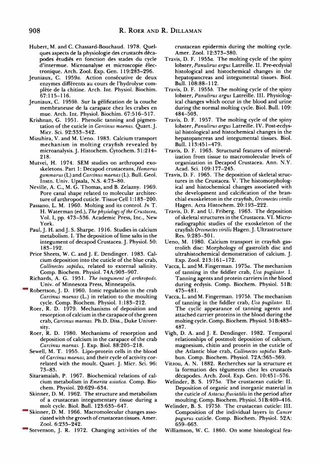
896 R. Roer and R. Dillaman
INTERMOLT C4
Membranous layer present
Cuticle complete
EARLY PREMOLT
Do-D1
Apolysis Solation of memb.
layer
LATE PREMOLT
D2-D4
POSTMOLT A,-C3 Mineral resorption
Formation of new epi- & exocuticle
Calcification of
pre-exuvial cuticle
Deposition of endocuticle
Fig. 2. Schematic representation of the cuticular events associated with progression through the molt cycle. Ep = epicuticle, Ex = exocuticle, En = endocuticle, Mb = membranous layer.
tive, however, for a peak in RNA synthesis is seen during C4 (Skinner, 1966) and the reserve cells of the highly vacuolated and reduced connective tissue are engaged in the storage of materials for the ensuing premolt period (Travis, 1957).
The premolt integument The onset of premolt (stage D0) is marked
by apolysis, or the separation of the hypo? dermis from the cuticle through the action of secreted chitinase, chitobiase and pro? tease (Jeuniaux, 1959a, 6; Bade and Stin-
son, 1978) which cause the solation ofthe membranous layer. Apolysis results in the
severing ofthe pore canals (Green and Neff,
1972) (Fig. 2). The outer epithelial cells
begin to increase in height and complexity.
Through the premolt period they change from squamous to columnar. In Orconectes
they double their height between D0 and
D2, reaching a maximum of 54 ^m (six times their height in C4) by D3 (Travis, 1965); the maximal height is about 56 jum in Car? cinus (Roer, 1980). The Golgi begin to secrete a proteinaceous paracrystalline substance and an increase in smooth endo?
plasmic reticulum associated with mito? chondria is observed (Hubert and Chas? sard-Bouchaud 1978). Mitotic activity is
apparent in the epithelial cells during stages D0, D/ and D/'; and there are peaks in 02
consumption, protein synthesis and chitin
synthesis during stage D2 (Skinner, 1962, 1966; Stevenson, 1972).
Premolt is also the period during which

Calcification in Crustacean Cuticle 897
the matrix of the new epicuticle and exo? cuticle is laid down beneath the old cuticle.
Epicuticular deposition takes place during stage D,, and exocuticular deposition begins at stage D2. Because these matrices are deposited before the molt, they are referred to as the pre-exuvial layers (Drach, 1939). Although the organic matrix ofthe
epi- and exocuticle is laid down pre-exu- vially, these layers do not calcify until after the molt (Paul and Sharpe, 1916; Travis, 1963; Travis and Friberg, 1963). The epi? cuticle is, however, tanned before the molt
(Krishnan, 1951). Concomitant with pre-exuvial deposi?
tion is the partial resorption of both the mineral and organic portions of the old cuticle (Drach, 1939; Travis, 1965; Roer,
1980). In Gecarcinus lateralis, more than
75% of the cuticle is resorbed (Skinner, 1962); while in Panuliris argus only about
20% ofthe carapace is resorbed, the endo? cuticle being completely resorbed in some areas and the exocuticle being partially resorbed in certain regions (Travis, 1955a). In Carcinus, 15-20% of the mineral is resorbed during premolt (Graf, 1978). Resorptive activity reaches a maximum
during stage D2 (Drach, 1939; Green and
Neff, 1972), and results in a rise in hemo?
lymph Ca++ concentration (Robertson, 1960) and HC03~ concentration. This causes a slight alkalosis ofthe hemolymph, the pH rising from 7.9 to 8.1 in Astacus
leptodactylus (Dejours and Beekenkamp, 1978).
The postmolt integument The initial events following the molt are
the tanning of the exocuticle and the cal? cification of the pre-exuvial layers. The exocuticle is tanned by quinone formation from dihydroxyphenols under the action of a polyphenol oxidase transported to the cuticle from the epithelial cells (Krishnan, 1951; Travis, 1957; Vacca and Fingerman, 1975a, 6). The pre-exuvial layers begin to
calcify during stage Ax. The first crystals of CaC03 are evident in Carcinus 10 hr after the molt (Drach, 1937); and in Astacus
fluviatilis the rate of Ca deposition reaches a peak two days postmolt (Welinder, 1975a). Calcification ofthe epi- and exo- cuticles begins in the most external regions
and proceeds proximally (Travis and Fri-
berg, 1963;Bouligand, 1970). Mineral
apparently reaches the outer portions of the cuticle via the pore canals. Calcium is concentrated in the distal portions of the
epithelial cells and appears to be extruded in vertical rows corresponding in position to the pore canals of the new cuticle (Tra? vis, 1957, 1963, 1965; Travis and Friberg, 1963; Chockalingham, 1971). Stage B is marked by the onset of endocuticle depo? sition; here calcification is concomitant with matrix formation, each organic lamella
being mineralized as it is laid down (Drach, 1939; Travis, 1957,1963, 1965; Travis and
Friberg, 1963). Mineral deposition continues through
postmolt. Calcification spreads throughout the exocuticle and eventually is found to
pervade the walls of the pore canals (Tra? vis, 1963; Travis and Friberg, 1963). Cal? cite crystals may finally be seen within the lumina of the pore canals as the cell pro? cesses apparently recede to be replaced with mineral (Travis, 1963; Travis and Friberg, 1963; Hegdahl etal, 1977a, 6, c). The end of postmolt is marked by the deposition of the membranous layer during stage C3 and the cessation of net calcium deposition (Passano, 1960).
The postmolt changes in the hypoder- mal cells are also marked. By one day post? molt the dedifferentiation of these epithe? lial cells has already begun (Green and Neff, 1972). In Orconectes, the epithelial cells decrease from their maximal height of 54
/tm to 21 nm within two days of ecdysis, the decrease continuing until a minimum is reached at intermolt (Travis, 1965). The
secretory activity of the Golgi and abun? dance of endoplasmic reticulum also decreases through postmolt to a virtual absence in C4 (Hubert and Chassard-Bou?
chaud, 1978).
The Relationship between Mineral and Organic Components
The epicuticle The epicuticle, as mentioned above, dif-
fers from the exo- and endocuticles in its lack of chitin and lamellar organization. This organizational difference is mani- fested in the orientation ofthe mineral. A transmission electron microscopic and
898 R. Roer and R. Dillaman
radiographic study by Hegdahl and co-
workers (1977c) on the epicuticle of Cancer
pagurus revealed that mineral was restricted
to vertical canals, 100-250 nm in diame?
ter, in the proximal layer. These canals are
presumably the distal terminations of the
pore canals. The mineral was in the form of CaC03 crystals or crystal aggregates and were distributed unevenly and surrounded
by epicuticular tissue. We have also observed a non-homoge?
neous and discontinuous distribution of mineral in the epicuticle of Carcinus maenas. While a 24 hr treatment of Carcinus cuticle with 5.25% sodium hypochlorite results in
a complete removal of the epicuticle, brief treatment renders the tissue only partially anorganic and reveals mineral clumps which are spherulitic aggregates (Figs. 3 and 4). The loss of these aggregates fol?
lowing the complete hypochlorite removal of the organic material suggests that these are both discrete from one another and from the mineral components of the
underlying exocuticle.
The exocuticle and endocuticle
The exo- and endocuticles have in com? mon a regular array of chitin-protein fibers
arranged in lamellae defined by a rotation in the orientation of parallel sheets of these fibers (Mutvei, 1974 and Figs. 6 and 16). The two layers differ in the lamellar spac? ing with the interlamellar distance being less in the exocuticle than the endocuticle
(approximately 2 fim and 8 nm respectively in Carcinus; Dillaman and Roer, 1980).
Mineral first appears as small crystals of calcite observed around the perimeters of the pore canals and then throughout the
chitin-protein fibrillar network (Travis, 1963; Yano, 1975). Calcification of the exocuticle appears to be concentrated ini?
tially in the periphery of and interstices between prisms defined by the margins of
the hypodermal cells underlying the cuti? cle (Drach, 1939; Travis, 1963; Hegdahl et al, 19776; Giraud-Guille and Quintana, 1982). When calcification is complete, however, the distribution of mineral within these layers is relatively homogeneous (Hegdahl et al, 1977a, 6; Giraud-Guille and
Quintana, 1982).
Although Bouligand (1970) claims that there is no orientation of the calcite crys? tals with respect to the chitin-protein fibers in Carcinus, other evidence points to the
contrary. The crystal aggregates clearly are
aligned with the organic fibers as deter? mined by transmission electron micros?
copy in Gaetice depressus (Yano, 1975) and in Cancer pagurus (Hegdahl et al, 1977a,
6), and by secondary ion mass spectroscopy and X radiodography in Carcinus (Giraud- Guille and Quintana, 1982). Indeed, Heg? dahl and co-workers (1977a) have clearly demonstrated rod-shaped crystal aggre? gates interspersed with the chitin-protein fibers.
We have further investigated, by scan?
ning electron microscopy, the orientation of the mineral components and the rela? tion of this orientation to the organic fibers. The organization of the cuticle is made more apparent by the comparative obser? vations of untreated cuticles, EDTA-decal- cified cuticles and those rendered anor-
ganic by sodium hypochlorite. In Carcinus
(Figs. 5-8) the composite nature of the exoskeleton is clearly seen as the inter-
spersion ofthe organic fibers with similarly oriented crystal aggregates. The orienta? tion of the chitin-protein fibers demon? strates a continuous spiral rotation with each lamella corresponding to a 180? deviation (Fig. 6). Throughout the exo- and endocuticles of Carcinus, the mineral is found as long rod-shaped elements dis-
playing the same continuously changing orientation as seen in the chitin-protein
Plate 1. Scanning electron micrographs of Carcinus maenas cuticle. Fig. 3. Partially anorganic epicuticle. x 700. Fig. 4. Partially anorganic epicuticle. x 5,500. Fig. 5. Untreated endocuticle. Note pore canals (arrow). x 10,000. Fig. 6. Decalcified endocuticle. Note pore canals (arrow). x 5,000. Fig. 7. Anorganic endocuticle. Note the pore canal space (ps). x 10,000. Fig. 8. Anorganic endocuticle. Note the individual spherulitic elements comprising rod-shaped crystal aggregates (arrow) x 40,000.
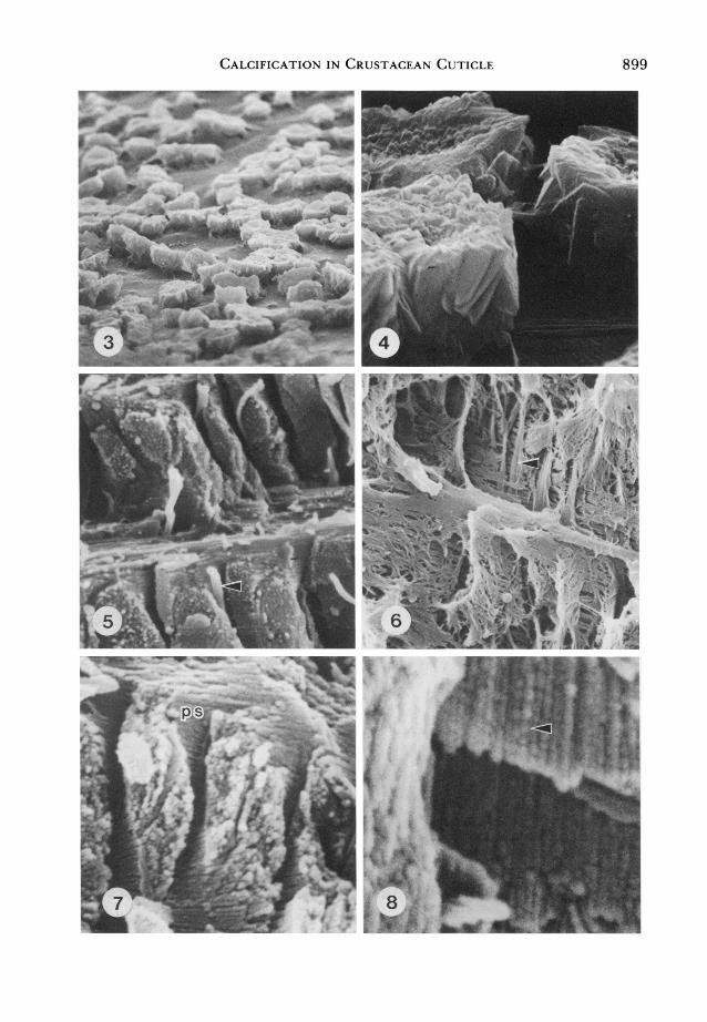
Calcification in Crustacean Cuticle 899
900 R. Roer and R. Dillaman
fiber network (Figs. 7 and 8). Inspection of the mineral rods at higher magnification reveals that these are aggregates of calcite
spherulites strung together in the orien? tation of the organic fibers (Fig. 8).
While this pattern of mineralization
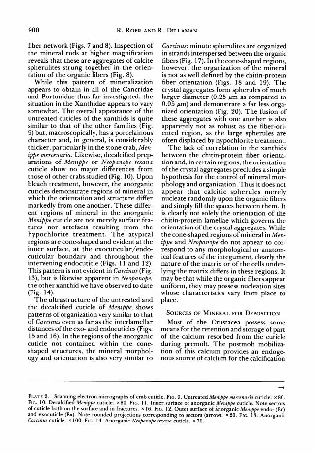
appears to obtain in all of the Cancridae and Portunidae thus far investigated, the situation in the Xanthidae appears to vary somewhat. The overall appearance of the untreated cuticles of the xanthids is quite similar to that of the other families (Fig. 9) but, macroscopically, has a porcelainous character and, in general, is considerably thicker, particularly in the stone crab, Men-
ippe mercenaria. Likewise, decalcified prep? arations of Menippe or Neopanope texana cuticle show no major differences from those of other crabs studied (Fig. 10). Upon bleach treatment, however, the anorganic cuticles demonstrate regions of mineral in which the orientation and structure differ
markedly from one another. These differ? ent regions of mineral in the anorganic Menippe cuticle are not merely surface fea? tures nor artefacts resulting from the
hypochlorite treatment. The atypical regions are cone-shaped and evident at the inner surface, at the exocuticular/endo- cuticular boundary and throughout the
intervening endocuticle (Figs. 11 and 12). This pattern is not evident in Carcinus (Fig. 13), but is likewise apparent in Neopanope, the other xanthid we have observed to date
(Fig. 14). The ultrastructure of the untreated and
the decalcified cuticle of Menippe shows
patterns of organization very similar to that of Carcinus even as far as the interlamellar distances ofthe exo- and endocuticles (Figs. 15 and 16). In the regions ofthe anorganic cuticle not contained within the cone-
shaped structures, the mineral morphol? ogy and orientation is also very similar to
Carcinus: minute spherulites are organized in strands interspersed between the organic fibers (Fig. 17). In the cone-shaped regions, however, the organization of the mineral is not as well defined by the chitin-protein fiber orientation (Figs. 18 and 19). The
crystal aggregates form spherules of much
larger diameter (0.25 /xm as compared to 0.05 /mi) and demonstrate a far less orga? nized orientation (Fig. 20). The fusion of these aggregates with one another is also
apparently not as robust as the fiber-ori- ented region, as the large spherules are often displaced by hypochlorite treatment.
The lack of correlation in the xanthids between the chitin-protein fiber orienta? tion and, in certain regions, the orientation ofthe crystal aggregates precludes a simple hypothesis for the control of mineral mor?
phology and organization. Thus it does not
appear that calcitic spherules merely nucleate randomly upon the organic fibers and simply fill the spaces between them. It is clearly not solely the orientation of the
chitin-protein lamellae which governs the orientation of the crystal aggregates. While the cone-shaped regions of mineral in Men?
ippe and Neopanope do not appear to cor?
respond to any morphological or anatom? ical features of the integument, clearly the nature of the matrix or of the cells under?
lying the matrix differs in these regions. It
may be that while the organic fibers appear uniform, they may possess nucleation sites whose characteristics vary from place to
place.
Sources of Mineral for Deposition
Most of the Crustacea possess some means for the retention and storage of part of the calcium resorbed from the cuticle
during premolt. The postmolt mobiliza? tion of this calcium provides an endoge? nous source of calcium for the calcification
Plate 2. Scanning electron micrographs of crab cuticle. Fig. 9. Untreated Menippe mercenaria cuticle. x80. Fig. 10. Decalcified Menippe cuticle. x80. Fig. 11. Inner surface of anorganic Menippe cuticle. Note sectors of cuticle both on the surface and in fractures. x 16. Fig. 12. Outer surface of anorganic Menippe endo- (En) and exocuticle (Ex). Note rounded projections corresponding to sectors (arrow). x20. Fig. 13. Anorganic Carcinus cuticle. x 100. Fig. 14. Anorganic Neopanope texana cuticle. x70.

Calcification in Crustacean Cuticle 901

902 R. Roer and R. Dillaman
r 4

Calcification in Crustacean Cuticle 903
of the new cuticle. The storage sites may be in the form of gastroliths, the calcareous concretions beneath the cuticle lining the cardiac stomach in some freshwater macrurans and terrestrial brachyurans; sternal concretions within the anterior cuticle during the biphasic molt in isopods; concretions within the posterior caeca of the midgut in amphipods; and the general deposition of calcareous spherules in the
hepatopancreas and increase in protein- bound calcium within the hemolymph (see Graf, 1978 for review). The importance of
endogenous calcium for postmolt miner? alization is dependent upon the habitat of the animal; Crustacea in sea water depend very little upon stores, as calcium is readily available from the medium while reserves are more important for freshwater and ter? restrial forms. Thus over 92% of body cal? cium is lost upon exuviation in Carcinus while only 25% and 40% are lost in the
supralittoral and terrestrial isopods Ligia and Oniscus respectively (Graf, 1978).
Uptake of calcium from the food and water is the source of exogenous calcium, but the relative importance of these two sources varies. In general, the food accounts for little of the total calcium of the new cuticle in the marine brachyura despite the
eating ofthe exuvia in many species (Drach, 1939; Graf, 1978). Such is not the case of the mole crab Emerita asiatica in which there is no storage and the food accounts for
approximately 82% ofthe calcium for min? eralization (Sitaramaiah, 1967).
In freshwater and estuarine organisms specific uptake mechanisms exist for the accumulation of calcium from the dilute media. In fact, Gammarus pulex can attain
complete calcification in media containing 0.1 mM calcium (Wright, 1980). The fresh? water crayfish, Austropotamobius pallipes, takes up calcium against an electrochemi- cal gradient by a system displaying satu-
ration kinetics with a Km = 0.13 mM Ca
(Greenaway, 1974). Active uptake of cal? cium may also be induced in euryhaline crabs in dilute media. Callinectes is able to
fully calcify its integument in 10 ppt sea?
water, although at a rate slower than that in 30 ppt seawater (Price Sheets and Den-
dinger, 1983). Both Callinectes and Carci? nus have calcium transport mechanisms with a low affinity, Km =10 mM Ca
(Greenaway, 1983). The uptake mechanism for Ca in Aus-
tropotamobius is in part dependent upon the
presence of HC03~ in the external medium
(Greenaway, 1974), however very little is known regarding the sources nor uptake pathways for the carbonate for mineraliza? tion. While a certain amount of carbon is
likely to be absorbed from consumption of the exuvium and from food, other possible sources include HC03~ from the water and metabolic C02.
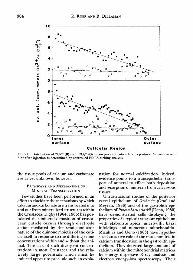
In preliminary experiments in which we
injected postmolt (B^ Carcinus with 50 /.iCi each of 45CaCl and
NaH14CO? and etched
the cuticle (according to Dillaman and
Ford, 1982) following a 6 hr incubation, we found an unequal distribution of label in the mineral throughout the cuticle (Fig. 21). The specific activity of 14C was uni? form from the inner surface out to the epi? cuticle while the specific activity of 45Ca was
highest at the inner surface and decreased
monotonically toward the epicuticle. Nowhere in the cuticle was the specific activity of 14C as high as that of 45Ca despite the fact that the specific activity of H14C03 was higher in the hemolymph than was that of45Ca.
These data imply that there is on the one hand a much larger tissue pool for carbon? ate than for calcium and, on the other hand, that equilibrium of 14C in the tissue pools and within the cuticle is much faster than that of 45Ca. The location and extent of
Plate 3. Scanning electron micrographs of Menippe mercenaria cuticle. Fig. 15. Untreated endocuticle. Note pore canals (arrow). x 5,100. Fig. 16. Decalcified endocuticle. Note pore canals (arrow). x 5,000. Fig. 17. Anorganic endocuticle from lamellar region. x 21,000. Fig. 18. Inner surface of anorganic endocuticle. Note regions of lamellate (1) and non-lamellate (n) organization. x 25. Fig. 19. Crystal organization in non-lamellate region of endocuticle. x 10,000. Fig. 20. Spherulites of non-lamellate region of endocuticle. x 20,000.
904 R. Roer and R. Dillaman
10
<MC0 o o
o + CM
o
O ? E
E a o
o (0
a CO
.a-o?o..2-0-i2. r, i^? O o O ' O
O 0 O
o o -cr-o-3>?ctfx?~flL o o o o 73
?^nO-p0-OoO-6-O-?-~-C o o o oQ oo o**> ? ??
Inner surface
Outer surface
Cuticular Region
Fig. 21. Distribution of 45Ca2+ (?) and ,4COs2~ (O) in two pieces of cuticle from a postmolt Carcinus maenas 6 hr after injection as determined by controlled EDTA-etching analysis.
the tissue pools of calcium and carbonate are as yet unknown, however.
Pathways and Mechanisms of Mineral Translocation
Few studies have been performed in an effort to elucidate the mechanisms by which calcium and carbonate are translocated into and out from mineralized structures within the Crustacea. Digby (1964, 1965) has pos- tulated that mineral deposition of crusta? cean cuticle occurs through electrode action mediated by the semi-conductor nature of the quinone moieties of the cuti? cle itself in response to the differing saline concentrations within and without the ani? mal. The lack of such divergent concen? trations in most Crustacea and the rela?
tively large potentials which must be induced appear to preclude such an expla-
nation for normal calcification. Indeed, evidence points to a transepithelial trans?
port of mineral to effect both deposition and resorption of minerals from calcareous tissues.
Ultrastructural studies of the posterior caecal epithelium of Orchestia (Graf and
Meyran, 1983) and of the gastrolith epi? thelium of Procambarus clarkii (Ueno, 1980) have demonstrated cells displaying the
properties ofa typical transport epithelium with elaborate apical microvilli, basal
infoldings and numerous mitochondria. Mizuhira and Ueno (1983) have hypothe? sized an active role of the mitochondria in calcium translocation in the gastrolith epi? thelium. They detected large amounts of calcium within the mitochondrial matrices
by energy dispersive X-ray analysis and electron energy-loss spectroscopy. Their

Calcification in Crustacean Cuticle 905
proposal is that the mitochondria effect the
transepithelial translocation of calcium
during the elaboration of the gastroliths. In the hypodermis of the shrimp, Cran?
gon crangon, electron-probe X-ray micro-
analysis has also demonstrated calcium
peaks within the epithelial cells during the
periods of maximal resorption of the car?
apace mineral (stage D) and while maximal rates of deposition are occurring (stage B)
(Hubert and Chassard-Bouchaud, 1978).
Epithelial involvement in the processes of calcium translocation during premolt resorption and postmolt deposition of car?
apace mineral was further elucidated by Roer (1979, 1980) in Carcinus. In vitro uni? directional flux studies were performed on
premolt hypodermis removed from beneath the dorso-branchial carapace and net calcium uptake experiments were con? ducted on pieces of postmolt integument from the same region. These data dem? onstrated that both resorption and depo? sition of calcium were effected by active
transport across the hypodermis. Maximal in vitro resorption rates were 22.6 x 10~8
mole/cm2-hr in stage D2, and deposition rates were 8 ? 9 x 10~8 mole/cm2-hr in
stages A{ and A2. These stages correspond to those in which maximal resorption and
deposition have been found to occur in vivo
(Drach, 1939; Green and Neff, 1972; Wel?
inder, 1975a; Vigh and Dendinger, 1982).
By employing ion substitutions and var? ious inhibitors, this calcium transport mechanism was found to involve both a Ca- ATPase and Na/Ca exchange (Roer, 1980). These results have recently been verified using another brachyuran, Calli? nectes sapidus (C. Mangum, personal com?
munication). These physiological studies in combi-
nation with morphological investigations (Travis, 1963; Bouligand, 1970; Yano,
1975) point to the importance ofthe pore canals to the calcification ofthe pre-exuvial cuticle. The epi- and exocuticles are elab? orated before the molt, but only calcify during postmolt. Mineral must reach dis? tances 70 /*m or more from the apical sur? faces of the epithelial cells for the rapid hardening of these layers following exu? viation. This transport of calcium, and pre-
sumably carbonate, is apparently effected
by the cytoplasmic extensions of the hypo- dermal cells extending up into these regions within the pore canals.
While we now possess some knowledge of the mechanisms of calcium transport associated with mineral structures in select
Crustacea, virtually nothing is known
regarding the supply of carbonate to these tissues. It has been established by histolog? ical and biochemical means, however, that carbonic anhydrase is localized within the
epithelial cells of the hypodermis and in the cuticle (pore canals?) of the anomuran Clibanarius olivaceous (Chockalingham, 1971) and of Carcinus (Giraud, 1977a, b,
1981). The amount of enzyme is highest during postmolt calcification (stages A! to
C3). The role of carbonic anhydrase in the calcification process is unclear, but inhi? bition of the enzyme by acetazolamide results in decreased rates of mineralization in vitro (Giraud, 1977a) and in vivo (Giraud, 1981) as judged by polarized light micros?
copy. Whether the enzyme acts in a trans?
port capacity or simply in the supply of bicarbonate for mineralization has not been established. Acetazolamide has no effect on in vitro calcium transport in Carcinus
hypodermis (Roer, unpublished data), so if a role in carbonate transport is established it would be unlinked to the movement of calcium.
nucleation and the control of Crystal Growth
As mentioned previously, preparation for the molt in the calcified crustaceans involves the resorption of the mineral and
organic components of the old cuticle and the simultaneous deposition of elements of the new cuticle. During premolt, the entire
organic matrix ofthe two outermost layers of the new cuticle (the epi- and exocuticle) are elaborated beneath the old cuticle. The
pre-exuvial layers do not calcify until after the molt (Drach, 1939; Travis, 1963, 1965; Travis and Friberg, 1963). In Carcinus
maenas, for example, the first crystals of
CaC03 are not observed in the epi- and exocuticle until 10 hr after ecdysis (Drach, 1937). Moreover, if a premolt crab is pre- maturely removed from its old exoskele-
906 R. Roer and R. Dillaman
ton, the new cuticle becomes leathery but does not calcify (Paul and Sharpe, 1916). Some internal conditions must therefore be met in order that deposition of mineral
may occur. Since the mineral from the old cuticle is
resorbed through the pre-exuvial layers of the new cuticle, Travis (1963, 1965) and Travis and Friberg (1963) have suggested that Ca++ and C03= are present in meta- stable solution in these layers before the molt. As calcification does not commence until after the molt, however, they pro? posed that the epithelial cells underlying the cuticle exert some control over its envi? ronment. They add that this control might be realized via the cytoplasmic extensions of the epithelial cells in the pore canals of the pre-exuvial cuticle through the regu? lation of such factors as Ca++, C03=, phos? phate or pH.
An alternative hypothesis has been put forth by Yano (1972), who favors the
unmasking of nucleation sites in the pre- exuvial cuticle as the means of controlling calcification. He suggested that an acid
mucopolysaccharide complexed to the
protein fraction of the pre-exuvial matrix inhibits premolt calcification. Postmolt
enzymatic degradation of the polysaccha- ride-protein complex would then allow calcification to proceed. While there is his? tochemical evidence for acid mucopolysac? charides in the cuticle (Yano, 1972; Giraud,
19776), no such degradative enzyme has been described.
A significant peak in phosphorylase and alkaline phosphatase activities is noted in the epicuticle and exocuticle of Clibanarius in stages A^ and A2 when calcification is
commencing (Chockalingham, 1971). It is
possible that this activity could be related to the removal of nucleation inhibitors or
crystal poisons within the matrix. The histological location of carbonic
anhydrase in the interprismatic regions of the exocuticle (Giraud, 1981) also corre?
sponds to the initial sites of mineral depo? sition (Giraud-Guille and Quintana, 1982). Thus, a possible role of carbonic anhydrase in mineral nucleation must be considered.
We are currently investigating the role of nucleating sites in the carapace of Uca
pugilator in the control of pre-exuvial cuti? cle calcification. In these experiments, pieces of pre-exuvial cuticle were removed from crabs before the molt (late stage D) and cuticle was removed from newly molted crabs (stage Ax and A2). Both types of cuti? cle were stripped of hypodermal tissue and fixed in 4% paraformaldehyde, decalcified
overnight in 0.1 M EDTA in 4% parafor? maldehyde, and the EDTA was removed
by further rinsing in the fixative alone. The cuticles were then subjected to in vitro cal? cification by successive immersions in 0.5 M solutions of CaCl2 and NaHC03. Sub?
sequent observation ofthe tissues by polar? ized light microscopy showed mineraliza? tion within the cuticles from postmolt crabs, but no crystal deposition within those of
premolt crabs (Roer and Dail, in prepa? ration). It is apparent that some factor within the matrix of the cuticle is altered in the course of the molt and the ensuing few hours; either a nucleating agent is
secreted, or an inhibitor of nucleation and/ or crystal growth is removed.
The control by the matrix of crystal mor?
phology and the cessation of crystal growth is evident in studies on carapace repair in crabs (Dillaman and Roer, 1980). Mineral
deposited within the wound scab in crabs was not formed in the context ofthe chitin-
protein network and appeared as arago- nitic granules rather than the normal cal- citic morph. Further deposition, which occurred among histologically atypical lamellae was amorphous CaC03. Repair cuticle growth showed no evidence of ces?
sation, and hence no membranous layer which normally marks the end of deposi? tion. In this context, it is possible that the secretion of the non-calcifying membra? nous layer serves to curtail further crystal growth in normal cuticle deposition.
Conclusions and Future Directions
Readdressing the basic questions of min? eralization in the Crustacea it seems we have a large body of data concerning the chemical nature and distribution of min? eral and organic components within the cuticle and we know, in certain species, the sources of the calcium for mineralization and the rates at which deposition occurs.
Calcification in Crustacean Cuticle 907
We know far less about the relationship between the mineral and the organic com?
ponents of the cuticle, both in regard to the determination of crystal morphology and in regard to nucleation. Finally, while in the Portunidae we have some knowledge of the mechanisms and pathways for cal? cium movement, we know nothing with
respect to the transport of carbonate. These latter areas of investigation will
prove fertile ground for future work; work which will provide information not only on the physiology of Crustacea but also on the basic principles of mineralization. The bidirectional nature of mineral transport and the sharp temporal transitions in
nucleating ability of the cuticular matrix
provide ideal systems in which to study these aspects of calcification.
ACKNOWLEDGMENTS
We wish to express our thanks to the La
Que Center for Corrosion Technology of the International Nickel Company for the
generous use of their scanning electron
microscope facilities. Part of this work was
supported by a grant from N.C. Sea Grant.
References
Bade, M. L. and A. Stinson. 1978. Activation of old cuticle chitin as a substrate for chitinase in the molt of Manduca. Biochem. Biophys. Res. Comm. 84:381-388.
Bouligand, Y. 1970. Aspects ultrastructuraux de la calcification chez les crabes. 7th Congres Int. Micr. Electronique, Grenoble, pp. 105-106.
Chassard-Bouchaud, C and M. Hubert. 1973. Etude ultrastructurale du tegument des Crustaces Deca- podes en fonction du cycle d'intermue. I. Pres? ence de cellules secretrices a activite cyclique dans l'epiderme de Palaemon serratus Pennant. J. Microscopie 16:75-83.
Chockalingham, S. 1971. Studies on the enzymes associated with calcification of the cuticle of the hermit crab Clibanarius olivaceous. Mar. Biol. 10: 169-182.
Dejours, P. and H. Beekenkamp. 1978. L'equilibre acide-base de l'hemolymphe au cours de la mue chez l'Ecrevisse. C R. Acad. Sc, Paris 286:1895- 1898.
Digby, P. S. B. 1964. The mechanism of calcification in crustacean cuticle. J. Physiol. 173:29P-30P.
Digby, P. S. B. 1965. Semi-conduction and electrode processes in biological material. I. Crustacea and certain soft-bodied forms. Proc. Roy. Soc. B 161: 504-525.
Dillaman, R. M. and S. E. Ford. 1982. Measurement of calcium carbonate deposition in molluscs by
controlled etching of radioactively labeled shells. Mar. Biol. 66:133-143.
Dillaman, R. M. and R. D. Roer. 1980. Carapace repair in the green crab, Carcinus maenas (L). J. Morphol. 163:135-155.
Drach, P. 1937. Morphogenese de la mosaique cris- talline externe dans le squellette tegumentaire des decapodes brachyoures. C. R. Hebd. Seanc. Acad., Paris 205:1173-1176.
Drach, P. 1939. Mue et cycle d'intermue chez les crustacees decapodes. Ann. Inst. Oceanogr., Monaco 19:103-391.
Drach, P. and C. Tchernigovtzeff. 1967. Sur la me- thode de determination des stades d'intermue et son application generale aux crustaces. Vie Milieu Ser. A: Biol. Mar. 18:595-610.
Giraud, M. M. 1977a. L'anhydrase carbonique et la calcification tegumentaire chez le crabe Carcinus maenas Linne. C. R. Acad. Sc, Paris 284:453- 456.
Giraud, M. M. 19776. Histochimie des premieres etapes de la mineralisation de la cuticle du crabe Carcinus maenas: Tentatives de localisation de l'anhydrase carbonique. C. R. Acad. Sc, Paris 284:1541-1544.
Giraud, M. M. 1981. Carbonic anhydrase activity in the integument of the crab Carcinus maenas dur? ing the intermolt cycle. Comp. Biochem. Physiol. 69A:381-387.
Giraud-Guille, M. M. and C. Quintana. 1982. Sec? ondary ion microanalysis of the crab calcified cuticle: Distribution of mineral elements and interactions with the cholesteric organic matrix. Biol. Cell 44:57-68.
Graf, F. 1978. Les sources de calcium pour les crus? taces venant de muer. Arch. Zool. Exp. Gen. 119: 143-161.
Graf, F. andj. C. Meyran. 1983. Premolt calcium secretion in midgut posterior caeca of the crus? tacean Orchestia: Ultrastructure of the epithe? lium. J. Morphol. 177:1-23.
Green, J. P. and M. R. Neff. 1972. A survey ofthe fine structure of the integument of the fiddler crab. Tissue Cell 4:137-171.
Greenaway, P. 1974. Calcium balance at the post- moult stage of the freshwater crayfish, Austro- potamobius pallipes (Lereboullet). J. Exp. Biol. 61: 35-45.
Greenaway, P. 1983. Uptake of calcium at the post- moult stage by the marine crabs Callinectes sapidus and Carcinus maenas. Comp. Biochem. Physiol. 75A:181-184.
Hegdahl, T.,J. Silness, and F. Gustavsen. 1977a. The structure and mineralization of the carapace of the crab (Cancer pagurus L.). 1. The endocuticle Zool. Scripta 6:89-99.
Hegdahl, T.,F. Gustavsen, andj. Silness. 19776. The structure and mineralization of the carapace of the crab (Cancer pagurus L.). 2. The exocuticle. Zool. Scripta 6:101-105.
Hegdahl, T.,F. Gustavsen, andj. Silness. 1977c. The structure and mineralization of the carapace of the crab (Cancer pagurus L.). 3. The epicuticle. Zool. Scripta 6:215-220.
908 R. Roer and R. Dillaman
Hubert, M. and C. Chassard-Bouchaud. 1978. Quel- ques aspects de la physiologie des crustaces deca?
podes etudies en fonction des stades du cycle d'intermue. Microanalyse et microscopie elec- tronique. Arch. Zool. Exp. Gen. 119:283-296.
Jeuniaux, C. 1959a. Action consecutive de deux enzymes differents au cours de l'hydrolyse com? plete de la chitine. Arch. Int. Physiol. Biochim. 67:115-116.
Jeuniaux, C. 19596. Sur la gelification de la couche membraneuse de la carapace chez les crabes en mue. Arch. Int. Physiol. Biochim. 67:516-517.
Krishnan, G. 1951. Phenolic tanning and pigmen? tation of the cuticle in Carcinus maenas. Quart. J. Micr. Sci. 92:333-342.
Mizuhira, V. and M. Ueno. 1983. Calcium transport mechanism in molting crayfish revealed by microanalysis.J. Histochem. Cytochem. 31:214- 218.
Mutvei, H. 1974. SEM studies on arthropod exo- skeletons. Part 1: Decapod crustaceans, Homarus gammarus (L) and Carcinus maenas (L). Bull. Geol. Instn. Univ. Upsala, N.S. 4:73-80.
Neville, A. C, M. G. Thomas, and B. Zelazny. 1969. Pore canal shape related to molecular architec? ture of arthropod cuticle. Tissue Cell 1:183-200.
Passano, L. M. 1960. Molting and its control. In T. H. Waterman (ed.), The physiology ofthe Crustacea, Vol. I, pp. 473-536. Academic Press, Inc, New York.
Paul, J. H. andj. S. Sharpe. 1916. Studies in calcium metabolism. I. The deposition of lime salts in the integument of decapod Crustacea. J. Physiol. 50: 183-192.
Price Sheets, W. C. andj. E. Dendinger. 1983. Cal? cium deposition into the cuticle of the blue crab, Callinectes sapidus, related to external salinity. Comp. Biochem. Physiol. 74A:903-907.
Richards, A. G. 1951. The integument of arthropods. Univ. of Minnesota Press, Minneapolis.
Robertson, J. D. 1960. Ionic regulation in the crab Carcinus maenas (L.) in relation to the moulting cycle. Comp. Biochem. Physiol. 1:183-212.
Roer, R. D. 1979. Mechanisms of deposition and resorption of calcium in the carapace ofthe green crab, Carcinus maenas. Ph.D. Diss., Duke Univer? sity.
Roer, R. D. 1980. Mechanisms of resorption and deposition of calcium in the carapace of the crab Carcinus maenas. J. Exp. Biol. 88:205-218.
Sewell, M. T. 1955. Lipo-protein cells in the blood of Carcinus maenas, and their cycle of activity cor? related with the moult. Quart. J. Micr. Sci. 96: 73-83.
Sitaramaiah, P. 1967. Biochemical relations of cal? cium metabolism in Emerita asiatica. Comp. Bio? chem. Physiol. 20:629-634.
Skinner, D. M. 1962. The structure and metabolism of a crustacean integumentary tissue during a molt cycle. Biol. Bull. 123:635-647.
Skinner, D. M. 1966. Macromolecular changes asso? ciated with the growth of crustacean tissues. Amer. Zool. 6:235-242.
Stevenson, J. R. 1972. Changing activities of the
crustacean epidermis during the molting cycle. Amer. Zool. 12:373-380.
Travis, D. F. 1955a. The molting cycle ofthe spiny lobster, Panulirus argus Latreille. II. Pre-ecdysial histological and histochemical changes in the hepatopancreas and integumental tissues. Biol. Bull. 108:88-112.
Travis, D. F. 19556. The molting cycle ofthe spiny lobster, Panulirus argus Latreille. III. Physiolog? ical changes which occur in the blood and urine during the normal molting cycle. Biol. Bull. 109: 484-503.
Travis, D. F. 1957. The molting cycle of the spiny lobster, Panulirus argus Latreille. IV. Post-ecdys- ial histological and histochemical changes in the
hepatopancreas and integumental tissues. Biol. Bull. 113:451-479.
Travis, D. F. 1963. Structural features of mineral? ization from tissue to macromolecular levels of
organization in Decapod Crustacea. Ann. N.Y. Acad. Sci. 109:177-245.
Travis, D. F. 1965. The deposition of skeletal struc? tures in the Crustacea. V. The histomorpholog- ical and histochemical changes associated with the development and calcification of the bran? chial exoskeleton in the crayfish, Orconectes virilis
Hagen. Acta Histochem. 20:193-222. Travis, D. F. and U. Friberg. 1963. The deposition
of skeletal structures in the Crustacea. VI. Micro-
radiographic studies of the exoskeleton of the crayfish Orconectes virilis Hagen. J. Ultrastructure Res. 9:285-301.
Ueno, M. 1980. Calcium transport in crayfish gas? trolith disc: Morphology of gastrolith disc and ultrahistochemical demonstration of calcium. J. Exp. Zool. 213:161-172.
Vacca, L. and M. Fingerman. 1975a. The mechanism of tanning in the fiddler crab, Uca pugilator. I. Tanning agents and protein carriers in the blood during ecdysis. Comp. Biochem. Physiol. 51B: 475-481.
Vacca, L. and M. Fingerman. 19756. The mechanism of tanning in the fiddler crab, Uca pugilator. II. The cyclic appearance of tanning agents and attached carrier proteins in the blood during the molting cycle. Comp. Biochem. Physiol. 51B:483- 487.
Vigh, D. A. andj. E. Dendinger. 1982. Temporal relationships of postmolt deposition of calcium, magnesium, chitin and protein in the cuticle of the Atlantic blue crab, Callinectes sapidus Rath? bun. Comp. Biochem. Physiol. 72A:365-369.
Vitzou, A. N. 1882. Recherches sur la structure et la formation des teguments chez les crustaces decapodes. Arch. Zool. Exp. Gen. 10:451-576.
Welinder, B. S. 1975a. The crustacean cuticle: II. Deposition of organic and inorganic material in the cuticle of Astacus fiuviatilis in the period after moulting. Comp. Biochem. Physiol. 51 B:409-416.
Welinder, B. S. 19756. The crustacean cuticle: III. Composition of the individual layers in Cancer pagurus cuticle. Comp. Biochem. Physiol. 52A: 659-663.
Williamson, W. C. 1860. On some histological fea-
Calcification in Crustacean Cuticle 909
tures in the shells ofthe crustacea. Quart. J. Micr. Sci. 8:35-47.
Wright, A. 1980. Calcium balance in premoult and post-moult Gammarus pulex (Amphipoda). Fresh? water Biol. 10:571-579.
Yano, I. 1972. A histochemical study on the exo-
cuticle with respect to its calcification and asso? ciated epidermal cells in a shore crab. Bull. Jap. Soc Sci. Fish. 38:733-739.
Yano, I. 1975. An electron microscope study on the calcification of the exoskeleton in a shore crab. Bull. Jap. Soc Sci. Fish. 41:1079-1082.