Embed Size (px)
Citation preview

The structure and function of ray
and axial parenchyma in woody
seed plants
Dissertation
Zur Erlangung des Doktorgrades Dr. rer. nat.
der Fakultät für Naturwissenschaften der Universität Ulm
vorgelegt von
Hugh Robert Morris
aus Irland
Ulm 2016
The structure and function of ray
and axial parenchyma in woody
seed plants
Dissertation
Zur Erlangung des Doktorgrades Dr. rer. nat.
der Fakultät für Naturwissenschaften der Universität Ulm
vorgelegt von
Hugh Robert Morris
aus Irland
Ulm 2016

Amtierender Dekan: Prof. Dr. Peter Dürre
1. Gutachter: Prof. Dr. Steven Jansen
2. Gutachter: Prof. Dr. Stefan Binder
Date of award
Dr. rer. nat. (Magna Cum Laude) awarded to Hugh Robert Morris on 20th of July,
2016
Cover picture: A stitched example I made of a transverse section of Fraxinus
excelsior stained for starch in Lugol’s solution

Table of Contents Part I ..................................................................................................................................................................... 1
General definition and function of parenchyma cells in plants .................................................................... 2
The contents of parenchyma in relation to function .................................................................................... 2
Parenchyma of the secondary xylem: an overview ...................................................................................... 3
Symplastic pathways between the secondary phloem and xylem ............................................................... 4
The longevity of living cells in the secondary xylem and the formation of heartwood .............................. 10
The functions of RAP in secondary xylem ................................................................................................... 11
Long distance water transport and RAP ..................................................................................................... 12
Defence against pathogens and RAP .......................................................................................................... 15
RAP and mechanical properties of the secondary xylem ........................................................................... 18
Aims of the thesis ........................................................................................................................................... 19
The research topics and permissions .............................................................................................................. 20
Summary of chapters ..................................................................................................................................... 21
Chapter 1: A global analysis of parenchyma tissue fractions in secondary xylem of seed plants .............. 21
Chapter 2: The amount of parenchyma and living fibres affects storage of non-structural carbohydrates
in young stems and roots of temperate trees ............................................................................................ 23
Chapter 3: Secondary xylem parenchyma – from classical terminology to functional traits ..................... 24
Conclusions and future prospects ................................................................................................................. 25
References ...................................................................................................................................................... 28
Part II .................................................................................................................................................................. 51
A global analysis of parenchyma tissue fractions in secondary xylem of seed plants .................................... 52
Abstract ...................................................................................................................................................... 53
Introduction ................................................................................................................................................ 54
Materials and Methods .............................................................................................................................. 58
Compilation of the global parenchyma dataset ..................................................................................... 58
Validation of the dataset ........................................................................................................................ 59
Climate data ............................................................................................................................................ 60
Statistical analyses .................................................................................................................................. 61
Results ........................................................................................................................................................ 62
Overview of the core dataset ................................................................................................................. 62
Validation of the dataset ........................................................................................................................ 62
Climate and RAP fractions ...................................................................................................................... 65
Discussion ................................................................................................................................................... 72

RAP fractions in relation to NSC storage capacity .................................................................................. 73
RAP fractions and drought stress ............................................................................................................ 73
RAP fractions and temperature .............................................................................................................. 74
RAP fractions as a defence system ......................................................................................................... 74
RAP and mechanical properties .............................................................................................................. 75
General Conclusion .................................................................................................................................... 76
Acknowledgements .................................................................................................................................... 76
Author contributions .................................................................................................................................. 77
References ...................................................................................................................................................... 78
Supporting Information for New phytologist.................................................................................................. 90
Part II ................................................................................................................................................................ 106
The amount of parenchyma and living fibres affects storage of non-structural carbohydrates in young
stems and roots of temperate trees ............................................................................................................. 107
Abstract .................................................................................................................................................... 108
Introduction .............................................................................................................................................. 109
Materials and Methods ............................................................................................................................ 112
Plant material ........................................................................................................................................ 112
NSC analyses ......................................................................................................................................... 114
Wood anatomy and starch distribution ................................................................................................ 115
Statistical analyses ................................................................................................................................ 118
Results ...................................................................................................................................................... 118
NSC concentrations ............................................................................................................................... 118
RAP and LFs anatomy ............................................................................................................................ 120
Starch localisation ................................................................................................................................. 121
Relationship between NSC and RAP ..................................................................................................... 123
Discussion ................................................................................................................................................. 125
Acknowledgements .............................................................................................................................. 130
References ............................................................................................................................................ 131
Part II ................................................................................................................................................................ 138
Secondary xylem parenchyma – from classical terminology to functional traits ......................................... 139
Introduction .................................................................................................................................................. 140
Parenchyma in secondary xylem .......................................................................................................... 140
Functions of RAP ................................................................................................................................... 141
Contact and isolation cells of the ray system ....................................................................................... 141

Arrangements of the axial parenchyma system ................................................................................... 148
Paratracheal AP ................................................................................................................................. 148
Apotracheal AP ................................................................................................................................. 150
From a descriptive to a functional terminology ................................................................................... 152
Conclusion and outlook ............................................................................................................................ 154
References .................................................................................................................................................... 156
Acknowledgements ...................................................................................................................................... 164
Professional profile ..................................................................................................................................... 167
Qualifications .............................................................................................................................................. 167

Trees
I think that I shall never see
A poem lovely as a tree.
A tree whose hungry mouth is prest
Against the earth’s sweet flowing breast;
A tree that looks at God all day,
And lifts her leafy arms to pray;
A tree that may in summer wear
A nest of robins in her hair;
Upon whose bosom snow has lain;
Who intimately lives with rain.
Poems are made by fools like me,
But only God can make a tree.
By Joyce Kilmer, 1913

1
Part I
-
Introduction and Summary

Introduction
2
Introduction
General definition and function of parenchyma cells in plants
In plants, parenchyma cells (Gr. para, “beside” + enchyma, “an infusion”) make up one of the
three ground tissues, alongside collenchyma and sclerenchyma cells. In general, parenchyma
cells differ in having a thin primary cell wall (rarely secondary) and were the first eukaryotic
cells to have evolved (Stewart, 1983). Early non-vascular plants, such as green algae
(Chlorophytes and Charophytes), are made up entirely of unspecialised parenchyma with the sole
function being photosynthesis, as support from other types of ground tissue is not required,
unlike with the land plants. Parenchyma cells, for the most part, resemble the undifferentiated
cells produced by division of meristematic cells (Simpson, 2006). They vary in shape from
elongate to isodiametric, although they can also be square or rectangular, as found frequently in
the ray parenchyma of secondary xylem and phloem (Panshin and Zeeuw, 1980). Parenchyma
cells are alive at maturity and have the ability to dedifferentiate (totipotency), which is
particularly important during events such as injury (Heplar and Newcomb, 1963; Wargo, 1977;
Shigo, 1984; Aloni and Plotkin, 1985). In a typical plant, this versatile tissue can be found in
every organ, from the cortex and pith of stems to the cortex of roots, the mesophyll of leaves, the
pulp of fruit, and finally the endosperm of seeds. This cell type has a very diverse suite of
functions, ranging from metabolic processes such as respiration, digestion and photosynthesis,
together with storage of NSCs, water, wound healing and regeneration (Evert, 2006).
The contents of parenchyma in relation to function
In the leaf, parenchyma forms the mesophyll (leaf middle section), which are referred to as
chlorenchyma (parenchyma with chloroplasts) owing to its special role in photosynthesis.
Chlorenchyma cells also have plentiful ribosomes and Golgi-bodies and a well-developed
endoplasmic reticulum, all crucial in a post-photosynthetic role (Evert, 2006). Where, in the
absence of light, when chloroplasts are not present, they can be replaced by amyloplasts, another
specialist organelle that accumulates starch, as found in potato tubers. Amyloplasts, unlike
chloroplasts, tend to take up most of the allotted space within the cytoplasm. Aerenchyma, which
forms channels of air-filled cavities, is another specialised parenchyma cell type. It is a spongy
tissue that makes up the mesophyll in leaves, and is also found in the roots of plants in hypoxic
soils, such as with aquatic plants (Jung et al., 2008). A study by Wang and Cao (2012)

Introduction
3
showed that flooding stimulates aerenchyma in the roots, stems and leaves of bald cypress
(Taxodium distichum) and in just the roots of the Chinese tallow tree (Triadica sebifera), which
allows for increased enhancement of O2 diffusion. Parenchyma cells also have a major role as
water reservoirs (Holbrook, 1995), especially where vacuoles are large and abundant (Evert,
2006). Specialist water conserving cells (where chloroplasts are absent) in succulent plants
typical of mesic sites (e.g. Cactaceae, Aloe, Agave, Peperonia) are notably large with thin cell
walls, and have large vacuoles with dense cytoplasm (Koller and Rost, 1988; Evert, 2006). A
study by Liese and Grover (1961) found a clear association between parenchyma amount and
water storage in the culms of bamboos, although the method used was not clear.
Mechanically, the strength of parenchyma is dependent on its turgor-pressure derived from the
cell’s hydraulic properties (Romberger et al., 1993). The thin primary cell walls of parenchyma
allow for structural changes to the cell shape, where they become rigid (gauged by stiffness and
the modulus of elasticity) upon the swelling of the central vacuole after an influx of fluids, or
flaccid when the organelle loses pressure (Virgin, 1955). Proteins referred to as aquaporins are
responsible for regulating the flow of water into and out of the vacuole, which is carried out via
active transport. When the flow of water is disrupted, plasmolysis occurs upon a significant
decline in turgor pressure, resulting in cell collapse. According to Niklas (1992), the mechanical
properties of a group of parenchyma and the density of the packing arrangement largely dictate
how effective they will be as a whole, rather than just at the individual cell level.
Parenchyma of the secondary xylem: an overview
Parenchyma is also found in the secondary xylem of “woody” plants (trees and shrubs), a
collective term for a range of tissues formed by the vascular cambium to its interior. Lignin
biosynthesis and deposition in the compound middle lamella upon completion of the secondary
cells wall of mature tracheary elements and fibres, is what differentiates the secondary xylem
from more juvenile non-woody tissues (Wardrop and Bland; 1959; Donaldson, 1992; Schuetz et
al., 2013). Lignin is a polymer that acts as a stiffening agent conceived as ‘chimney-like
Brickwork’ of ‘lignin bricks’, embedded in a cellulose matrix; these are useful analogies from a
solely mechanical perspective (Mattheck and Breloer, 1994; Schwarze et al., 2000).
Although, secondary xylem can be found in the members of the gymnosperm groups
Ginkgophyta and Gnetophyta and in most of the division Cycadophyta, the two key groups

Introduction
4
where it can be found are the Pinophyta (conifers) and the Angiospermae (angiosperms or
flowering plants). All known species of conifers have secondary xylem, whereas its presence in
the angiosperms depends on the plant’s status. Many herbaceous plants of the dicotyledons
flower and die quickly before secondary xylem can form, whereas it is seldom found in the
monocotyledon plant group (Mauseth, 1988; Dickison, 2000). The secondary xylem (known
simply as wood), is separated from the phloem and outer bark by the meristematic tissue,
referred to as the vascular cambium. It is made up of three major tissues: the fibres, the tracheary
elements and the parenchyma (ground tissue). The latter of these is the only living component,
with the exception of ‘living’ fibres, where in a number of taxa (e.g. Araliaceae, Salicaceae)
these ‘so called’ fibres can replace axial parenchyma and act as a functional substitute (Wheeler
et al., 2007). Parenchyma of the secondary xylem usually have a secondary cell wall, although
the definition has been challenged in this case, where upon lacking the complex laminar structure
belonging to the tracheary elements and fibres, it has been more likened to a thickened primary
cell wall (Panshin and de Zeeuw, 1980). Also, parenchyma of the secondary xylem are usually
lignified, a trait unique to wood parenchyma when compared to those elsewhere in the plant,
including the phloem. The lignified thickened cell walls often make it difficult to distinguish
them from typical sclerenchyma cells (i.e. the fibres) when examined in cross-section (Evert,
2006). However, not all woody plants have lignified parenchyma in their wood, including many
lianas (Carlquist, 1991; Angyalossy et al., 2012), and some tropical tree species of the Urticaceae
family; for example, Myriocarpa longipes and Urera glabriuscula (Cocoletzi et al., 2013).
Symplastic pathways between the secondary phloem and xylem
Parenchyma cells tend to intergrade with surrounding tissues of the secondary xylem, as opposed
to forming masses, as it commonly does in other regions of the plant, such as in the pith, leaf, or
cortex. The parenchyma cells of both the secondary xylem and the secondary phloem are further
divided into ray parenchyma (RP) and axial parenchyma (AP), with the former developing from
the ray initials and the latter from the fusiform initials. Both xylem and phloem are often
focussed upon in isolation, without considering the connectivity between the two regions (Spicer,
2014). In fact, literature goes as far as stating that transport in the phloem is a completely active
process involving living cells, where water and mineral transport in the xylem is a completely
passive process without any parenchyma involvement, as continues to be acknowledged by the
cohesion-tension theory (Dixon and Joly, 1894; Dixon, 1914; Askenasy, 1895; Tyree, 1997;

Introduction
5
Stiller and Sperry, 1999). Although numerous studies have been made, along with extensive
laboratory work, into the potential role of parenchyma in long distance water transport, their
considerable contribution is still not valued enough to make any alteration to the original theory
published more than 120 years ago. The considerable list of publications that are speculated to
support parenchyma participation in long-distance water uptake has a long history from 1904 to
the present day (e.g., Ursprung, 1926; Braun, 1994; Canny, 1997, 1998; Smith, 1994;
Zimmermann, 1994, Zimmermann et al., 1995; Zwieniecki et al., 2001; Clearwater and
Goldstein, 2005; Salleo et al., 2009; Brodersen et al., 2010; Jansen et al., 2011; Nardini et al.,
2011; Brodersen and McElrone, 2013; Santiago et al., 2013; Johnson et al., 2012; Trifilo et al.,
2014).
Rather than view regions of a woody plant as isolated, we should hold the vision that there is a
three-dimensional dynamic continuum of both symplastic and apoplastic pathways all the way
through the plant. For instance, RP act as a bridge between both the phloem and the xylem,
where water and metabolites must pass through the meristematic cells of the vascular cambium
to flow from one side to the other. Numerous studies show evidence of symplastic pathways
from the phloem through to the xylem (e.g., van der Schoot and van Bel, 1990; Salleo, 2004;
Sokołowska and Zagorska-Marek, 2012; Pfautsch et al., 2015). In an interesting experiment,
Salleo et al. (2004) ring-barked saplings of bay laurel (Laurus nobilis), which, in the process,
isolated the phloem from the xylem causing a lack of recovery in the xylem after stem
embolisms were induced. This showed that the phloem was integral to hydraulic recovery at the
time of embolism, speculating that phloem pressures must be maintained for continued radial
transport and hydraulic maintenance. The ray cells are connected to each other via numerous
strands of secondary plasmodesmata, symplastic (via the cytoplasm) connections that allow for
active transport of materials and communication between living cells. The outer layer of ray cells
on the xylem side of the cambium have, in turn, plasmodesmatal continuity with the AP as well
as half-bordered pit connections with the vascular conduits, where materials coming from the
phloem can pass through into both, albeit by different means (Chaffey and Barlow, 2001;
Sokołowska, 2013; Spicer, 2014).

Introduction
6
Fig. 1 TEM (transmission electron microscopy) images of the transverse sections of (a) Robinia
pseudoacacia showing two adjoining axial parenchyma cells with bordered pits (AP), showing
the plasmodesmata (PLA) connections between them, which function in symplastic movement of
metabolites (scale bar:1 μm), and (b) Acer pseudoplatanus with contact parenchyma (CP)
adjoined to a vessel (V) via half-bordered pits, which are separated by the amorphous layer (AL)
located to the inside of the contact parenchyma between the plasma membrane and the adjacent
vessel-parenchyma pit membrane.(scale bar: 2 μm).
Ray and axial parenchyma of the secondary xylem
With exceptions, RP cells are aligned horizontally where, like ‘spokes of a wheel,’ they begin at
the cambium and run centripetally towards the pith. Living cells make up the ray system in all
angiosperms with rays, while ray-tracheids, which are dead at maturity, form an integral part of
the ray system in a number of conifers (Singh et al., 2006). Commonly occurring along the
margins of the ray system, ray-tracheids can be found in the Pinaceae (except Abies, Keteleeria
and Pseudolarix), and in most of the Cupressaceae (Phillips, 1948). By contrast, AP cells are
oriented vertically and are spatially arranged in varied patterns, the latter being a characteristic
that is frequently used in the positive identification of hardwoods (IAWA Committee, 1989;
Wheeler et al., 2007); see figure 1 in chapter 1, and table 1 and 2 in chapter 3 for a thorough
assessment of AP arrangements in the secondary xylem. Conifers, by contrast, have a very
uniform pattern of tracheids, with usually uniserate rays and infrequent AP cells that are often
involved in resin synthesis. Conifers do not have the complexity of wood as can be found in the
angiosperms, especially with regard to AP spatial arrangements and conduit form, size and
patterns, with the exception of vesselless families (Wheeler et al., 1989; Wheeler and Baas,
1998).
Introduction
7
Both RP and AP form a highly interconnected three-dimensional system, a detail that we are
only truly beginning to embrace now, owing to advances in technology and computer software
(e.g., alignment and stitching tools; Huggett and Tomlinson, 2010; Brodersen, 2013). However,
past studies have shown how interconnectivity between RP, AP and vessels occurs in a range of
angiosperm species through the process of serial-sectioning (Zimmermann and Tomlinson, 1966;
Zimmermann, 1971), and through carefully crafted drawings (Kedrov, 2012).

Introduction
8
a
a
j
j
j
j
j
k
h
aa
a
A
Fig. 2 Old German drawings of cross sections from woody plants via light microscopy made
over 100 years ago showing clearly the presence or absence of parenchyma between species,
along with AP spatial distributions. Although these drawings were made without the advanced
software we have today, they are no less accurate, where RAP fractions could be measured from
these images using image J. (a) Canarium hirsutum with absent AP and narrow rays in contact
with vessels, (b) Leea angulata with AP rare and wide rays, (c) Platea latifolia with scanty
paratracheal AP around the vessels and both narrow and wide rays, (d) Micromelum pubescens,
(e) Ziziphus jujuba with aliform, confluent and narrow banded AP arrangements together with
narrow closely spaced rays AP arrangements, (f) Bouea macrophylla with wide and narrow
banded AP and narrow rays. ZG Zonengtenze (zone boundary); G Gefäbe (vessels); F Libriform
fibres; Ms Markstrahlen (medullary rays); P Holzparenchym (axial parenchyma). (Scale bar: 1
mm). Figures adapted from Moll JW and Janssonius HH, 1908. Mikrographie des Holzes: der
auf Java vorkommenden Baumarten. Leiden: EJ Brill.

Introduction
9
There are two kinds of RAP, those that have contact with conduits and those that are isolated
from the conduits. In multiseriate rays, the horizontally elongate (procumbent) cells that are
sandwiched between the layers positioned to the outside are referred to as ‘isolation cells’
(Braun, 1984), whereas those positioned to the outside, or all ray cells of uniseriate strands, are
called ‘contact cells’. Similar to the isolation cells, both in function and spatial distance from the
vessels, are the vessel-distant cells found in paratracheal (mass of cells surrounding vessels) AP.
Contact cells (of RP and AP) share half-bordered pits with the neighbouring conduits (Czaninski,
1977; Murakami et al., 1999). In angiosperms, the contact RP may be oriented vertically, and are
so-called ‘upright’ cells. The upright RP, when in contact with AP cells, share more
plasmodesmatal connections, presumably due to an increased surface area contact (Chaffey and
Barlow, 2001). Also, the upright (or ‘erect’) RP, when in association with vessels, may be more
specialised in the exchange of substances between them (Höll, 1975; Sauter and Kloth, 1986).
The composition of the cell wall is of particular interest in the contact cells of angiosperms,
where at the point of contact they have a material known as the amorphous layer (also referred to
as the protective layer, but only in reference to RP) (Chafe, 1974; Fujii et al., 1980, 1981;
Murakami et al., 1999). Science is still unclear about its functional role at the interface between
the contact cells and the vessel (Spicer, 2014). It was once considered that its function was to
protect the contact cells from high osmotic pressure coming from within the vessel (van Bel and
van der Schoot, 1988). The current thinking is that the layer can increase the surface area of the
symplast/apoplasm interface, thus allowing for enhanced efficiency of cellular exchange (Barnett
et al., 1993). Although the function of the amorphous layer in a defence role has been challenged
(Chafe 1974; Chafe and Chauret 1974), more recent research into the role of pectins in tyloses
(balloon-like extensions from contact cells that clog vessels) states otherwise (Rioux et al. 1998).
The latter study demonstrated that the pectins accumulate in the amorphous layer and extend into
the vessel, where the pectin’s are then speculated to expand the surface area of the tylosis or gel,
and consequencely blocking the vessels. Pectins may also have a role in the supercooling effect,
where pectin is thought to help form a barrier, influencing both water loss and the development
of ice crystals at freezing temperatures (Wisniewski and Davis, 1995).

Introduction
10
The longevity of living cells in the secondary xylem and the formation of heartwood
RP can occupy anywhere in the region of between 8 and 25% of total sapwood area (Ghouse and
Yunus, 1974; Gregory, 1977), whereas AP are much more variable and can range anywhere from
< 1 to 25% (Spicer, 2014), with the latter in some succulent and pachycaul species exceeding
50% (Mauseth, 1997). The longevity of RAP cells in the secondary xylem are relatively long-
lived compared to other plant regions (Evert, 2006), where they can live anywhere from two to
150 years and remain alive for up to 20 cm into the stem (Spicer and Holbrook, 2005). In
Rhododendron lapponicum, for instance, parenchyma cells were observed to live up to 200 years
old, which is extremely long-lived (Spicer and Holbrook, 2007; Schweingruber, 2013).
Interestingly, AP cells in Fraxinus were found to be alive 45+ years after vessels stopped
functioning in water uptake, where they may have changed their primary function in hydraulic
maintenance to a role in storage or defence (Spicer and Holbrook, 2007). However, a study by
Chapotin et al. (2006) showed that in the pachycaul Adansonia vessel-associated AP died before
RP, indicating that a similar trend may also be found in other species.
Heartwood is defined as an inactive but histologically similar zone of wood to sapwood and is
located to the inside of the physiologically active sapwood. As the tree ages, the parenchyma
cells die where their reserve substances are released, becoming a key ingredient in heartwood
formation, where the polyphenols give an overall stability to the tree’s core along with giving the
wood a normally darker appearance (Pinto et al., 2004). Regarded with particular importance
during this transition are the RP cells for their chemical syntheses and resulting accumulation
(Pandalai et al., 1985; Nakaba et al., 2008), and in tyloses production (Chattaway, 1949).
Parenchyma cell death during the transformation to heartwood appears to be largely under
genetic control (Nakada 2007, Bito et al., 2011, Bush et al., 2011), although studies into the
effects of seasonal changes (phenology) on heartwood formation remain limited (Imai, 2012). A
decline in sapwood respiration in a centripetal direction is suggested by many workers (e.g.,
Goodwin and Goddard, 1940; Higuchi et al., 1967; Pruyn et al., 2002; Pruyn et al., 2003; Pruyn
et al., 2005); however, this has been disputed by some, where no change was found, even at the
transition zone between sapwood and heartwood (Bowman et al., 2005; Spicer and Holbrook,
2007). Also, there may be a difference between parenchyma age and respiration rate when
comparing angiosperms to conifers, where, interestingly, no difference was found in the two
conifers studied (Pinus strobus and Tsuga canadensis), while for angiosperms the respiration

Introduction
11
rate was reduced by half in the oldest sapwood and all parenchyma of the latter remained alive
up to that point (Spicer and Holbrook, 2007). Oxygen levels also gradually deplete from the
cambium towards the inner core of the tree where oxygen deprivation has been speculated to be
the cause of parenchyma death (Panshin and Zeeuw, 1980; Eklund and Klintborg, 2000);
however, in a later study by Spicer and Holbrook (2005) the reduced amounts of O2 seem not to
affect parenchyma vitality, and therefore unlikely to cause cell death or reduce respiration at the
sapwood-heartwood boundary.
The parenchyma cells of the heartwood, although dead, still have very important posthumous
functions, which range from an increase in durability, resistance against microbes and overall
structural support (Bamber, 1976; Bamber and Fukazawa, 1985; Hillis, 1987). Aside from
extractives having fungicidal properties, there is evidence that these, along with antioxidants,
may together play a dual defensive role with the latter removing free radicals from the heartwood
(Schulz and Nicholas, 2000). Of course, the effectiveness of the heartwood as an antimicrobial
substance depends on species, moisture content, and the time of the year (Taylor et al., 2006),
with the heartwood of some species having a much higher resistance to rot than others
(Kortelainena and Viitanena, 2009).
Extractives from RAP include the following broad classifications, which all play a role in
heartwood durabilty: (1) flavonoids (Harborne, 1973; Harborne and Williams, 2000), (2)
quinones (Lukmandaru and Takahashi, 2009), (3) stilbenes (Schultz et al., 1995), and (4) tannins
(Haslam, 1989).
The functions of RAP in secondary xylem
Although the roles of RAP have been briefly discussed in all three chapters in the context of our
findings; here, the focus will be on some of the more important functions where a more detailed
insight into the role of RAP will be given. The role of non-structural carbohydrates in growth,
repair and in hydraulic maintenance will not be discussed in any great detail here, as it is
adequately covered in chapter 2 of this thesis. However, a recent and excellent review also
covers the topic in great detail (Hartmann and Trumbore, 2016).
Many of the functions of both RP and AP are shared, while others are taken over by specialised
living cell types of either the ray or axial system. For instance, the lignification of the cell walls
in RAP provides biomechanical support while also playing a role in defence against fungal

Introduction
12
pathogens, as lignin is a highly durable polymer (Eriksson et al., 1990; Lundquist and Brunow,
2004). Both ‘isolation cells’ of the radial system and vessel-distant AP cells function in the
storage of NSCs (non-structural carbohydrates), often just referred to as ‘storage cells’
(Czaninski, 1977). Smaller in size than both isolation cells and vessel-distant cells, the contact
cells (contact with the vessel in vessel-bearing angiosperms or the tracheids of conifers) of RP
and AP both share a ‘not yet’ fully understood role in maintaining the hydraulic system (Nardini
et al., 2011; Secchi and Zwieniechi, 2011) and both tend not to store NSCs on a long term basis
(Essiamah and Eschrich, 1985; Alves et al., 2001). Contact cells can also lower the pH of xylem
sap (Fromard et al., 1995), which has been found to be more acidic in early spring during bud-
burst, showing that the pH varies with the annual cycle in temperate tree species (Essimah, 1980;
Ferguson et al., 1983; Sauter, 1988). Although it is not clear what function this serves, it could,
however, facilitate the easier movement of ions and organic nutrients between contact cells and
vessels along a pH gradient (Larsson and Moller, 1990).
Storage of carbohydrates and the storage of water in RAP are also closely affiliated, meaning
that the functional role of RAP in water storage cannot be uncoupled from their role in the
storage of carbohydrate reserves, with the latter having an important function in growth (primary
metabolites) and tissue repair (secondary metabolites). Recent research into the role of NSCs
showed that stored carbohydrates contributed significantly to drought resistance in a range of
tropical and temperate tree seedlings (O’ Brien et al., 2014; O’ Brien et al., 2015). In summary,
stored NSCs may be critical for the retention of water, as inorganic and organic solutes of the
living cells acquire it from the xylem when under tension, which is in turn used to stave off
potential drought by protecting the plant from dehydration (Borchert, 1994).
Long distance water transport and RAP
The mystery of the role of RAP in long distance water transport still remains largely unsolved;
however, considering the connections between the living cells and the dead conducting cells
through a continuum expanding from the secondary phloem through to the secondary xylem,
along with the sophisticated anatomy and physiology of the contact cell-conduit pits
(Zimmermann, 1978; van Ieperen et al., 2000), the involvement of RAP in hydraulic
maintenance seems unquestionable. Where its involvement is not in dispute, the mechanisms
remain unclear (Nardini et al., 2011; Secchi and Zwieniechi, 2011). For instance, the

Introduction
13
mechanisms behind embolism repair are among the most contentious, especially in light of
recent developments over the procedures used in investigating embolisms in stems of plants
(Wheeler et al., 2013; Sperry, 2013; Torres-Ruiz et al., 2015). However, the evidence for
seasonal embolism repair remains convincing. Through X-ray tomography, visual evidence of
water droplets derived from contact parenchyma have been shown to form and expand and
coalesce in the vessels of grapevine (Brodersen et al., 2010), which confirms previous studies
using cryo-SEM (Canny, 1997; Canny et al., 2007). There is also evidence that stimulating the
activity of H+-ATPase can promote embolism repair (Salleo et al., 2004). The plasma membrane
has been reported to be activated by pressure from the conduit side (Zingarelli et al., 1999),
which might release sugars from the contact parenchyma into the embolised vessel through a
‘kind of’ active secondary transport (proton pump mechanism) (Alves et al., 2004). Where
seasonal embolism repair is likely to occur in a number of temperate species, there is no
evidence for it in tropical species (Brodersen and McElrone, 2013). As a result of increased H+-
ATPase activity in the contact cells, the sugars released into the xylem stream after hydrolysis
can directly increase the sap osmolarity and xylem pressure (Améglio et al., 2001, 2004). How
an embolism activates the living cells into repair of the conduit remains speculative. It could be
caused via wall vibrations originating from within the conduit, which mechanically trigger the
living cells into a response (Salleo et al., 2008) or through changes in osmotic concentrations
between contact cells and conduits (Secchi and Zwieniecki 2011, 2012). Another important
factor is the point at which the living cells are triggered into the repair of the conduit or,
alternatively, into the production of tyloses, where the latter is surely a last resort, as it results in
the death of the contact cell(s) and the complete blockage and dysfunction of the vessel(s)
(Klein, 1923; Zimmermann, 1978; Canny, 1997; Salleo et al., 2002; Sun et al., 2007). An
excellent review on the subject of embolism repair was written by Clearwater and Goldstein
(2005).
Hydraulic capacitance, which involves the release of stored water reserves from the bark
(including the phloem) and secondary xylem, could buffer against increasing tensions in the
xylem sap while avoiding embolisms in the process (Cruiziat et al., 2002; Meinzer et al., 2009;
McCulloh et al., 2014; Pfautsch et al., 2015). This allows photosynthesis to continue for longer
durations both diurnally and seasonally, where it has been confirmed that sapwood capacitance is
higher in species of high rainfall sites (Richards et al., 2013), indicating that this mechanism may

Introduction
14
be more common in the lowland tropics rather than, for instance, in temperate zones. Also, in
numerous reports, capacitance was shown to be higher in species with low wood density, which
are more prone to embolism formation as a consequence (Pratt et al., 2007; Sperry et al., 2008).
The parenchyma of the secondary xylem, particularly AP, are likely to play a large role in
capacitance, especially where wood density is low, which is found to be the case in succulent
growth forms (Borchert and Pockman, 2005). However, with normal growth forms, wood
density and AP fractions were found to be negatively correlated (Zheng and Martinez-Cabrera,
2013; Ziemińska et al., 2015). More work is needed to ascertain the role of AP cells in
capacitance, particularly in tropical trees, as they have not been quantified separately in available
literature, where they are generally included among a suite of other possible sources involved in
capacitance (Meinzer et al., 2009; Pfautsch et al., 2015).
Besides hydraulic capacitance, there is also evidence of the involvement of RAP in long distance
water uptake via the ionic effect, where sap flow is altered by changes in the ionic composition
and pH of the xylem sap (Fromard et al., 1995; De Boer and Volkov, 2003). Ionic solutes,
deriving from the contact cells through physico-chemical processes have been shown to increase
flow rates in the xylem by up to 2.5 times (Zwieniecki et al., 2001; Gascó et al., 2006); however,
not all solutes contribute equally with NaCl found to be significantly lower than KCl (Gascó et
al., 2006). Although the scientific basis behind the ionic effect is not challenged (Nardini et al.,
2007; Nardini et al., 2011; Nardini et al., 2012), the explanation behind the mechanism is still
largely unknown, regardless of various hypotheses put forward. For instance, the hydrogel nature
of pectins found in the pit membrane, is speculated to be involved in hydraulic conductance
(Zwienicki et al., 2001). According to this study, the pores of the pit membranes physically
change, where the cations cause the shrinkage of pectins (i.e. polysaccharidic polyelectrolytes),
thus enlarging the pores, which in turn could increase the hydraulic conductance of the xylem
(Ryden et al., 2000; Willats et al., 2001). However, available evidence shows that pectins are
lacking in intervessel pit membranes, so this mechanism has received little or no acceptance in
explaining the ionic effect (van Doorn et al., 2011, Klepsch et al. submitted).
A recent proposition to help explain long-distance water uptake and support the cohesion-tension
theory is the surfactant-coated nanobubble hypothesis (Schenk et al., 2015). Although surfactants
are thought to reduce xylem sap surface tension, which could increase the chances of embolism

Introduction
15
formation under negative pressure (Christensen-Dalsgaard et al., 2011; Domec, 2011), the
concentrations found under natural conditions are considered too low to do so. These surfactants,
along with dissolved ions such as KCl, CaCl2 and MgCl2, all originating from the contact cells,
are speculated to keep these nano-bubbles from coalescing to form large bubbles through having
a stabilising effect (Craig, 2004), which could prevent the formation of embolisms.
Defence against pathogens and RAP
The role of RAP in defence against pathogenic fungi and bacteria is considerable and greatly
underestimated, where the work carried out on this important subject has largely been the focus
of plant pathologists and not plant anatomists or physiologists per sé. However, there have been
a number of important papers where parenchyma, in particular the contact AP, has been the
focus, albeit, in many cases, indirectly, and mostly from within the primary plant tissues. These
include: in fungal-plant interactions (El Mahjoub et al., 1984; Street et al., 1986; Shi et al., 1992;
Cooper and Williams, 2004; Schwarze et al., 2003; Pouzoulet et al., 2014), and in bacterial-plant
interactions (Goodman and White, 1981; Hilaire et al., 2001; Basha et al., 2010; Schumann and
D’Arcy, 2010).
To help explain pathogen-plant interactions in the secondary xylem, the CODIT model, an
acronym for Compartmentalisation of Decay in Trees, was developed by Shigo (1970, 1976,
1979, 1984) and further developed by Boddy and Rayner (1983). It is made up of four walls
formed of two parts: wall 1-3 form the first part of the model where the walls are in place at the
time of wounding, where as wall 4, the second part of the model, forms after wounding has
occurred. A change to the model has been proposed and generally accepted, where the ‘D’ of
CODIT is replaced by ‘Damage’ or ‘Dysfunction’, simply because decay in itself is not the only
mechanism to initiate physico-chemical responses by the living cells, but rather any
biomechanical stimulus can, which could in turn pave the way for decay spread (Liese and
Dujesiefken, 1989, 1996). RAP play a large role in all four walls. In wall 1, the AP cells act to
prevent decay movement up and down the stem in an axial direction, mostly through the
production of tyloses from contact cells into the neighbouring vessels (Schmitt and Liese, 1990;
Sun et al., 2007). Wall 2 is made up of a largely static barrier composed of highly lignified
fibres at the annual ring boundary where decay is prevented from moving in a radial direction
(Shigo and Marx, 1977). However, studies show how this marginal parenchyma laid down at

Introduction
16
either side on the annual growth ring plays a role in defence, along with the intermittent RP cells
that break up the continuity of the annual ring along its circumference (Biggs, 1987; Schwarze
and Fink, 1998; Schwarze et al., 2000, 2003, 2007). Wall 3, the strongest wall in place at the
time of injury, is composed of the rays, where RP act to inhibit the movement of decay in a
lateral direction (Shigo, 1984). Wall 4, the final instalment of the CODIT model (the only wall of
part 2 of the model), also referred to as the ‘barrier zone,’ is laid down by the cambium and
completely formed of highly suberised axial parenchyma cells intermeshed among weakly
lignified vessels (Pearce and Holloway, 1984; Rademacher et al., 1984; Pearce, 1990).
Fig. 3 The CODIT model (compartmentalisation of decay in trees). Wall 1: prevents the up and
downward movement of decay through tyloses development via the contact AP. Wall 2:
prevents the radial movement of decay at the annual ring boundary (lignified fibres and marginal
apotracheal AP) in temperate species, or possibly through wide-banded AP in tropical species.
Wall 3: prevents the movement of decay laterally via the ray system (RP). Wall 4: prevents
decay from entering new wood developed by the fusiform initials of the cambia after wall 4 is
laid down. The walls run in order of ‘most effective’ with wall 4 being the strongest. Walls 1-3
are present at the time of injury (part 1), while wall 4 is formed after injury occurs (part 2).
Image sourced from: https://commons.wikimedia.org/wiki/File:CODIT_Model.svg. Licence
CC0-BY 3.0 SA https://creativecommons.org/licenses/by-sa/3.0/us/
The physico-chemical nature of RAP, along with their amount and spatial arrangement, all
together play a crucial role in the battle against pathogens. Tyloses arising from the contact
parenchyma can partially or wholly occlude the vessels, where the trigger seems to come from
the vessel side (Klein, 1923; Zimmermann, 1978; Schmitt and Liese, 1990, 1993; Sun et al.,
2008), although the mechanism behind how it occurs remains unclear. Research by Sun et al.
Introduction
17
(2007) demonstrated that wound-induced ethylene, and not embolism formation, is required to
trigger tylosis development in grapevines, which goes against previous hypotheses (Klein, 1923;
Zimmermann, 1978; Canny, 1997).
Also, when the pit aperture falls below a certain diameter to the point where it can no longer
accommodate tyloses due to a size restriction, the contact cells can secrete gels into the vessel
lumina in their place (formerly referred to as gums) (Bonsen and Kučera, 1990; Rioux et al.,
1998). In an interesting study by Rioux et al. (1998), pectin, formed in the amorphous layer of
the contact cells, is secreted into the vessel alongside the gel, where, through water-induced
expansion, can help the gel clog the vessel’s outermost peripheries. However, counterintuitive to
these findings, where the pit aperture size is critical to the release of tyloses or gels, is the study
by Hillaire et al. (2001) whom demonstrated that pathogen related proteins induce secondary
wall thickening, which in turn decreases the pit size, preventing or limiting bacterial spread from
the vessels into the contact cells. The contact cells when compared to vessel-distant parenchyma,
have high cytoplasmic activity (Sauter, 1973; Essiama and Eschrich, 1985; Shi et al., 1992;
Alves et al., 2001), thus accumulate and release several anti-microbial materials, such as lipoidal,
phenolic and terpenoid compounds (Cooper et al., 1996; Clérivet et al., 2000). Suberin, a fatty
hydrophobic sealant as well as being an anti-microbial compound, normally found in the bark
and produced by the living cells, has also been detected in the vessel lumina of numerous
angiosperm species during all stages of CODIT (Biggs, 1987).
Conifers have adapted differently to pathogenicity and herbivory owing to their contrasting
anatomical makeup, with their more uniform tracheid arrangement, mostly uniseriate rays, and
sparce AP (Sperry et al., 2006; Carnicer et al., 2013). However, similar to angiosperms, the ‘so-
called’ polyphenolic parenchyma cells (abbrev. PPC), a term associated with just conifers,
accumulate and release toxic anti-microbial compounds that act against both pathogens and
insects (Franceschi et al., 2005; Bohlmann, 2008; Kolosova and Bohlmann, 2012). Alongside
polyphenols, terpinoids are a critical defence component of conifers, where their viscosity can
disable insects and trigger the formation of specialised parenchyma known as traumatic resin
ducts in some families (Phillips and Croteau, 1999; Martin et al., 2002; Byun McKay et al.,
2003; Keeling and Bohlmann, 2006). Another factor to include here is the unique mechanism of

Introduction
18
conifers called the torus-margo bordered pit, which can localise both embolism and decay (Choat
et al., 2008; Fuhr et al., 2013; Bouche et al., 2014).
RAP and mechanical properties of the secondary xylem
A number of recent important studies have focussed on the trade-off between RAP, vessels and
fibre fractions, the three main constituents of angiosperm wood (Jacobsen et al., 2007; Martinez-
Cabrera et al., 2009; Zheng and Martinez-Cabrera, 2013; Ziemińska et al., 2013; Ziemińska et
al., 2015). A study by Jacobsen et al. (2007) found that total RAP was inversely related to the
modulus of elasticity (MoE) while Martinez-Cabrera showed in a study of 61 shrubs these two
variables to be independent of each other. This latter study also showed a trade-off between RP
and AP, where they each had opposite correlation patterns with wood density (AP – negatively,
RP – positively), demonstrating some evidence for functional partitioning between these two
parenchymatous types. For instance, rays are known to have strong biomechanical properties due
to their radial orientation and cell wall lignification (Mattheck and Kubler, 1995; Burgert et al.,
1999; Burgert and Eckstein, 2001), while the opposite was found to be true for AP, where higher
amounts are speculated to weaken stem longitudinal strength, due to the finding that AP is
negatively correlated with wood density (Zheng and Matinez-Cabrera, 2013). In contrast, a study
by Ziemińska et al. (2013) found the opposite trend where RP and AP were found to have no
correlation with each other, and where the former was negatively correlated with wood density,
AP was found to positively correlate with this variable. However, this study was carried out on
twigs rather than more mature stem wood, so caution must be taken in interpreting these results.

Summary Conclusions and future prospects
19
Aims of the thesis
The thesis is divided into three chapters (part II), with each chapter focusing on different
elements concerning the amount, spatial arrangements and functions of radial and axial
parenchyma in the secondary xylem of woody plants. Chapter 1 and 2 are research focussed,
whereas chapter 3 is an opinion paper discussing the use of terminology in describing secondary
xylem parenchyma from a historical perspective. Each of the three chapters in this thesis is an
‘already’ fully published article in the following Journals: New Phytologist, the American
Journal of Botany, and the International Association of Wood Anatomists journal, in that order.
The thesis is not laid out in the original format of the publications, due to differences in
publication layout. This decision was made in order to maintain consistency in style throughout
the text and references. The wording and graphs of the original articles have been strictly
adhered to, with the exception that the American spelling for the article in the American Journal
of Botany has been changed to British spelling, in maintaining a consistent format.

Summary Conclusions and future prospects
20
The research topics and permissions
Chapter 1 - A global analysis of parenchyma tissue fractions in secondary xylem of seed
plants
This article was made open access to allow it to be republished for my Dr.rer.nat. dissertation, by
John Wiley & Sons (manuscript number: NPH 13737) on 24/11/2015. Licence CC-BY 4.0
https://creativecommons.org/licenses/by/4.0/
Authors: Hugh Morris, Lenka Plavcová, Patrick Cvecko, Esther Fichtler, Mark A. F.
Gillingham, Hugo I. Martínez-Cabrera, Daniel J. McGlinn, Elisabeth Wheeler, Jingming Zheng,
Kasia Ziemińska, Steven Jansen
Title: A global analysis of parenchyma tissue fractions in secondary xylem of seed plants
New Phytologist, November 2015, 209: 1553-1565; doi: 10.1111/nph.13737
Chapter 2 - The amount of parenchyma and living fibres affects storage of non-structural
carbohydrates in young stems and roots of temperate trees
Full permission to use the published manuscript has been given by the publishers of the
American Journal of Botany for the non-exclusive world rights to the use of the materials as part
of my work in all languages and for all editions (print and electronic). As requested by the
Botanical Society of America, the full conventional scholarly form of acknowledgement,
including author, and title, publisher’s name and data is as follows:
Authors: Lenka Plavcová, Günter Hoch, Hugh Morris, Sara Ghiasi, and Steven Jansen
Title: The amount of parenchyma and living fibers affects storage of nonstructural carbohydrates
in young stems and roots of temperate trees
American Journal of Botany, April 2016, 103: 603-612; doi:10.3732/ajb.1500489
Chapter 3 – Secondary xylem parenchyma – from classical terminology to functional traits
Full permission to use the published manuscript has been given by the publishers of the
American Journal of Botany for the non-exclusive world rights to the use of the materials as part
of my work in all languages and for all editions (print and electronic). As requested by the
publishers, BRILL publishing group, the full conventional scholarly form of acknowledgement,
including author, and title, publisher’s name and data is as follows:
Authors: Hugh Morris and Steven Jansen
Title: Secondary xylem parenchyma – from classical terminology to functional traits
International Association of Wood Anatomists Journal, February 2016, 37: 1-15; doi:
10.1163/22941932-20160117

Summary Conclusions and future prospects
21
Summary of chapters
Chapter 1: A global analysis of parenchyma tissue fractions in secondary xylem of seed plants
Knowing the mount of parenchyma cells in the secondary xylem of woody plants, together with
their spatial distribution across a range of plant species, growth forms, and climates, is a very
valuable basis for helping scientists to underpin the functions of living cells and how they
contribute to overall plant physiology. For instance, in determining to what extent the amount of
living cells might be related to defence against herbivory and pathogens, or in hydraulic
maintenance of the stem and drought stress, or in the storage of non-structural carbohydrates
used for growth and repair, or, lastly, in the mechanical properties of the plant. Also, this study
might help to explain the degree of functional partitioning in parenchyma between plants
(between both ray and axial parenchyma separately and also total RAP together), where two
plants could have similar amounts of parenchyma, but a trade-off may exist in determining what
function(s) the parenchyma cells of each plant are used for, whether it be a more primary role in
capacitance to maintain hydraulic conductivity, or in more of a defensive role against pathogens.
This trade-off in parenchyma function would be importantly connected to the life history of that
particular species; for example, whether it is long or short-lived. This chapter only investigated
actual parenchyma percentages across a large dataset of 1727 records (1439 species) and did not
focus on their spatial arrangements, where the latter is a separate project for a follow-up study.
The primary aim of this study was to elucidate the effects of climate on ray and axial
parenchyma (RAP) fractions, where we looked at a range of climate parameters in relation to
RAP fractions, including: temperature, precipitation, latitude, altitude, etc. We also categorised
all the species into three broad climate zones: temperate, subtropical and tropical, allowing us to
explore the relationship between climate type and RAP fractions. The work also allowed us to:
(1) examine the links between parenchyma and growth form, (2) to investigate differences in
RAP between the parts of a woody plant, viz. the roots, trunks and branches, (3) to support
previous works on the anatomical divergence between the angiosperms and conifers and (4) to
explore differences between axial parenchyma (AP) and ray parenchyma (RP) fractions, which
would help to explain how conservative or versatile these two parenchyma tissues are, together
with the functional explanations behind this.
Summary Conclusions and future prospects
22
For this study, the majority of records for ray and axial parenchyma were obtained from papers
and books (55 separate literature sources). We also investigated axial parenchyma and radial
parenchyma separately where we had the data, as well as total RAP fractions between woody
plant groups (i.e. conifers vs. angiosperms) growth forms and climates. The climate data was
collected via two approaches: (1) the exact locations approach, where we had the longitudinal
and latitudinal coordinates for 411 species from 68 sampling locations, and (2) the GBIF
approach (Global Biodiversity Inventory System), which allowed us to gather climate
information for 619 species from 612 different locations.
In conclusion, the key finding showed that temperature was the biggest driver of RAP in plants,
followed by precipitation, with the latter showing a significant but negative relationship with
RAP, demonstrating an increase in RAP towards drier sites. Interestingly, AP, rather than RP,
was found to be the most versatile parenchyma type, with the sharp rise of RAP in tropical
species being mostly due to AP, while RP remained relatively constant. This was both supported
by the climate type plots and through the GAM model; however, the one anomaly was the high
RP fraction found in lianas, which is speculated to be related to the biomechanical properties of
the ray structure in this growth form. Also, regarding biomechanical properties, a ternary plot
showed by way of a visualisation how an increased RAP fraction from temperate through to
tropical climate zones occurs at the expense of fibres, the tissue type most commonly associated
with wood density. Another important finding was the difference in RAP between growth forms,
with succulents (including pachycauls) and lianas having much higher RAP fractions than non-
succulent angiosperm trees and conifers, with the latter having the lowest RAP fractions, as
expected.

Summary Conclusions and future prospects
23
Chapter 2: The amount of parenchyma and living fibres affects storage of non-structural
carbohydrates in young stems and roots of temperate trees
This important research was conducted under the premise that the amount of non-structural
carbohydrates (NSCs) in the secondary xylem of wood (total carbon), is directly related to the
amount of living cells, both radial and axial parenchyma (RAP), where RAP provides the
uppermost limit for NSC storage in plants. The role of living fibres in the storage of NSCs are
also studied here in Clematis vitalba and Acer pseudoplatanus, as they are known to be a
functional replacement in the absence of AP. NSCs include monosaccharides, disaccharides and
polysaccharides (i.e. soluble sugars and starch). NSCs are critical in plants, especially in growth
and dormancy in woody plants of seasonal climates; however, this could be due to a bias, where
far fewer studies have been conducted on NSCs in tropical climate locations where seasonality is
not a factor. Of late, much attention has been given to carbohydrates in relation to growth
limitation and drought induced tree mortality, where, with regarding the latter, it forms part of a
continuing debate with the plant hydraulics community as to which factor is the primary cause of
tree mortality under drought conditions, carbon starvation or embolism formation (Mc Dowell,
2011; Anderegg et al., 2012; Urli et al., 2013; Sevanto et al., 2014; Rowland et al., 2015;
Salmon et al., 2015).
For this study, the roots and stems of twelve temperate tree species (11 broadleaved deciduous
and one conifer) and four tropical trees species were investigated. Both NSC analyses were
carried out alongside the measurements of RAP fractions (and living fibres) and the starch
distribution using a potassium-iodine solution (Lugol’s solution). The presence of starch alone
cannot explain whether or not NSC’s are present or absent from the cell, as often it is hydrolysed
into sucrose when temperatures fall below zero, where in this form it may remain present in the
cell and unused, quickly shifting back to starch when conditions become favourable, such as
during early spring (Elle and Sauter, 2000; Hoch et al., 2002; Charrier et al., 2015). For this
reason, the tropical species used for this research make for an interesting contrast.
In conclusion, the study showed a strong relationship between RAP/living fibre fraction and
NSC concentration in the roots and stems of temperate trees and the only climber in the study, C.
vitalba, where RAP and/or living fibre fraction provides the uppermost limit for NSC storage, an
important contribution to our understanding of xylem-structure dynamics. Interestingly, the

Summary Conclusions and future prospects
24
amount of RAP was, in most cases, greater in the roots of temperate trees than in the stems,
which also meant a greater NSC accumulation in this plant part. The study also revealed, through
starch analyses, that both RP and AP/living fibres, provide equally to NSC storage in temperate
species, in that both ‘living cell’ types were observed to be full of starch grains. Also, there was
no observable difference between starch accumulations in the contact cells compared to the
vessel-distant cells (referred to as isolation cells in the ray system). Starch grains were found to
be less abundant in the RAP of tropical trees, however, where no correlation was found between
RAP amount and NSCs in these trees. Furthermore, although RAP fractions in the tropical
species were, on average, higher than in their temperate counterparts, NSC levels were even
lower in the former than in the latter. As the number of tropical species studied were low, caution
must be taken in interpreting these results, while the general trends shown in the temperate
species are a more useful indicator of the seasonal maxima for NSC concentrations in the
northern hemisphere at the onset of winter dormancy.
Chapter 3: Secondary xylem parenchyma – from classical terminology to functional traits
The chapter was written as an opinion paper to give clarity to both functional and anatomical
terms used to describe parenchyma, explaining how these terms evolved and currently relate to
functional traits and/or their use in the identification of different woods. While many terms are
still in use, other terms have become redundant due to being used less and less frequently as we
change our functional interpretation of particular types of cells overtime. This opinion paper
makes the case for a critical review of terminology, as how we use and interpret them is of
utmost importance, especially for those outside the field of anatomy, in disciplines such as plant
physiology, plant phytology, dendrochronology, and evolutionary biology, etc. Also, not only is
the consistent application of terms important, we must consider the dynamic continuum between
different anatomical tissues and refocus on the plant as a three-dimensional interconnected
system, rather than viewing it as single planes through a light microscope.

Summary Conclusions and future prospects
25
Conclusions and future prospects
The aim of this thesis was to further our understanding of the role of parenchyma in the
secondary xylem of ‘woody’ plants, a critically important facet of plants that still remains poorly
understood owing to a difficulty factor in obtaining accurate measurements until relatively
recently, and a more ‘industry focussed’ preoccupation with other cell types, such as the vessels
and fibres. This work investigated the variation in parenchyma fractions between three broad
climate types, finding significantly higher fractions of both radial and axial parenchyma (RAP)
in tropical locations, compared to both temperate and subtropical, particularly in the levels of
axial parenchyma (AP). This was supported by examining the relationship between RAP
fractions and a range of climate parameters, where temperature was found to be the main driver,
in particular AP, while ray parenchyma (RP) remained relatively unchanged. There are a number
of possible explanations for this. Cold temperatures place great energy demands on living cells,
observed by increased respiration rates (Sperling et al., 2015), and through avoidance of freezing
via the supercooling effect (Quamme, 1991; Neuner, 2014), which may explain why plants of
cooler regions have lower RAP levels. Also, pathogens are more abundant and more virulent in
the tropics (Bagchi et al., 2014), where greater RAP levels may help provide better armoury
against pathogens (Schwarze et al., 2003; Romera and Bolker, 2008).
Another interesting result is that RAP increases towards drier areas, a finding that supports the
high parenchyma levels found in low wood density pachycauls and other succulents, where the
paratracheal (surrounding the water conducting conduits) parenchyma function as a kind of
‘giant’ reservoir (Borchert and Pockman, 2005; Scholz et al., 2007; Hearn et al., 2013).
Paratracheal parenchyma around the vessels of pachycauls was found to be positively correlated
with high capacitance (water release into the vessels), where the opposite trend was found in
deciduous hardwoods with vessels enclosed by imperforate tracheary elements (Borchert and
Pockman, 2005). There are two drought strategies used by plants: drought tolerance vs. drought
avoidance, where species with large amounts of paratracheal AP fall into the latter category.
Drought avoiders, the former of the two strategies, are speculated to have few RAP cells, but are
able to tolerate low water potentials (Borchert et al., 1994; Goldstein et al., 1998). However,
more capacitance measurements of both bark and xylem are required from both temperate and
tropical species while also measuring RAP values, particularly AP, and their contact fractions
Summary Conclusions and future prospects
26
(percentage contact) with vessels. This important anatomy-function approach could reveal
important insights into plant strategies under different climatic conditions.
The high levels of RAP found in lianas, particularly RP, is of notable interest regarding two key
functions: plant biomechanics and dedifferentiation (a form of totipontency). Lianas were shown
to have increased elasticity enabling a high resistance to torsion (twisting) (Gartner, 1988; Putz
and Mooney, 1991), which is likely to be attributable to the high RP levels, with, in addition,
having often non-lignified and thin cell walls (Schnitzer et al., 2015). Also, asexual reproduction
via cloning is a common strategy deployed by lianas, where dediffertiation of RAP could allow
for rapid plantlet establishment (Putz, 1984; Yorke et al., 2014), as well as enabling fast recovery
from injury when breakage occurs (Fisher and Ewers, 1991).
We tested one of the functional hypotheses put forward in chapter one, which was to see if a
relationship existed between the levels of RAP and the amount of non-structural carbohydrates
that can be stored in the secondary xylem, and found this to be the case. Total RAP in both stems
and roots of a range of temperate species investigated provide the uppermost limit for NSC
storage. This important finding could have implications for species with low or high levels of
RAP under drought conditions, where species with the former could have more difficulty with
keeping hydrated. Studies by O’Brien et al. (2014, 2015) have built a strong case for the benefits
of stored NSC during times of short term drought in a range of both tropical and temperate tree
species. Longer drought periods, however, may trigger a decline in NSC storage resulting in a
reduction of allocation into defence compounds (Steele et al., 1995; Hartmann and Trumore,
2016), which could have a knock-on effect.
In summary, these findings demonstrate the importance of combining anatomy with function,
paving the way for many more studies, including the investigation of various functional
hypotheses. One key area that needs to be explored is the spatial arrangements of AP, where at
present only qualitative data is available through the plant database, InsideWood (InsideWood;
Wheeler et al., 2007). Also, our data was from cross-sections of wood only, where we know little
about the three-dimensional aspects of living cell patterns in wood, another key area that should
be given further exploration. Another crucial field of plant science to help support our
microscopy analyses is phylogenetics, which could be applied to the species for which we have

Summary Conclusions and future prospects
27
RAP values, thus revealing how genetically or environmentally inclined a large spectrum of
woody plants with a hugely diverse global distribution might be.

Summary References
28
References
Aloni R, Plotkin T. 1985. Wound induced and naturally occurring regenerative differentiation
of xylem in Zea mays L. Planta 163: 126-132.
Alves G, Ameglio T, Guillot A, Fleurat-Lessard G, Lacointe A, Sakr S, Petel G, Julien JL.
2004. Winter variation in xylem sap pH of walnut trees: involvement of plasma membrane
H+-ATPase of vessel-associated cells. Tree Physiology 24: 99-105.
Alves G, Sauter JJ, Julien J-L, Fleurat-Lessard P, Améglio T, Guillot A, Pétal G, Lacointe
A. 2001. Plasma membrane H+-ATPase, succinate and isocitrate dehydrogenases activities of
vessel-associated cells in walnut trees. Journal of Plant Physiology 158: 1263-1271.
Améglio T, Decourteix M, Alves G, Valentin V, Sakr S, Julien JL, et al. 2004. Temperature
effects on xylem sap osmolarity in walnut trees: evidence for a vitalistic model of winter
embolism repair. Tree Physiology 24: 785-793.
Améglio T, Ewers FW, Cochard H, Martignac M, Vandame M, Bodet C, et al. 2001. Winter
stem xylem pressure in walnut trees: effects of carbohydrates, cooling and freezing. Tree
Physiology 21: 387-394.
Anderegg WRL, Callaway E. 2012. Infestation and hydraulic consequences of induced carbon
starvation. Plant Physiology 159: 1866-1874
Angyalossy V, Angeles G, Pace MR, Lima AC, Dias-Leme CL, Lohmann LG, Madero-
Vega C. 2012. An overview on the anatomy, development and evolution of the vascular
system of lianas. Plant Ecological Diversity 5: 167-182.
Askenasy E. 1895. Ueber das Saftsteigen. Botanisches Zentralblatt 62: 237-238.
Bagchi R, Gallery RE, Gripenberg S, Gurr SJ, Narayan L, Addis CE, Freckleton
RP, Lewis OT. 2014. Pathogens and insect herbivores drive rainforest plant diversity and
composition. Nature 506: 85-88.
Summary References
29
Bamber RK, Fukazawa K 1985. Sapwood and heartwood. A review. Forestry Abstracts 46:
567-580.
Bamber RK. 1976. Heartwood function and formation. Wood Science and Technology 10: 1-8.
Barnett JR, Cooper P, Bonner LJ. 1993. The protective layer as an extension of the apoplast.
International Association of Wood Anatomists Journal 14: 163-71.
Basha S., Mazhar H., Vasanthaiah H. 2010. Proteomics approach to identify unique xylem sap
proteins in Pierce’s disease-tolerant Vitis species. Applied Biochemical Biotechnology 160:
932-944.
Biggs AR. 1987. Occurrence and location of suberin in wound reaction zones in xylem of 17 tree
species. Phytopathology 77: 718-725.
Bito N, Nakada R, Fukatsu E, Matsushita Y, Fukushima K, Imai T. 2011. Clonal variation
in heartwood norlignans of Cryptomeria japonica: evidence for independent control of
agatharesinol and sequirin C biosynthesis. Annals of Forest Science 68: 1049-1056.
Bohlmann J, Keeling CI. 2008. Terpenoid biomaterials. Plant Journal 54: 656-669.
Bonsen KJM, Kučera LJ. 1990. Vessel occlusion in plants: morphological, functional, and
evolutionary aspects. International Association of Wood Anatomists Bulletin new series 11:
393-399.
Borchert R, Pockman WT. 2005. Water storage capacitance and xylem tension in isolated
branches of temperate and tropical trees. Tree Physiology 25: 457-466.
Borchert R. 1994. Soil and stem water storage determine phenology and distribution of tropical
dry forest trees. Ecology 75: 1437–1449.
Bouche PS, Larter M, Domec JC, Burlett R, Gasson P, Jansen S, Delzon S. 2014. A broad
survey of hydraulic and mechanical safety in the xylem of conifers. Journal of Experimental
Botany 65: 4419-4431.

Summary References
30
Braun HJ. 1984. The significance of the accessory tissues of the hydrosystem for osmotic water
shifting as the second principle of water ascent, with some thoughts concerning the evolution
of trees. International Association of Wood Anatomists Bulletin 5: 275-294.
Brodersen CR, McElrone A, Choat B, Matthews M, Shackel K. 2010. Dynamics of
embolism repair in grapevine: in vivo visualisations using HRCT. Plant
Physiology 154: 1088-1095.
Brodersen CR, McElrone AJ. 2013. Maintenance of xylem network transport capacity: a
review of embolism repair in vascular plants. Frontiers in Plant Science 4: 1-11.
Brodersen CR. 2013. Visualizing wood anatomy in three dimensions with high-resolution X-ray
micro-tomography. Internation Association of Wood Anatomists Journal 34: 408-424.
Burgert I, Bernasconi A, Eckstein D. 1999. Evidence for the strength function of rays in living
trees. Holz als Roh Werkstoff 57: 397-399.
Burgert I, Eckstein D. 2001. The tensile strength of isolated wood rays of beech (Fagus
sylvatica L.) and its significance for the biomechanics of living trees. Trees 15: 168-170.
Bush D, McCarthy K, Meder R. 2011. Genetic variation of natural durability traits in
Eucalyptus cladocalyx (sugar gum). Annals of Forest Science 68: 1057-1066.
Byun McKay A, Hunter W, Goddard K, Wang S, Martin D, Bohlmann J, Plant A. 2003.
Insect attack and wounding induce traumatic resin duct development and gene expression of
(−)-pinene synthase in Sitka spruce. Plant Physiology 133: 368-378.
Canny M. 1997. Vessel contents during transpiration—embolisms and refilling. American
Journal of Botany 84: 1223-1230.
Canny M. 1998. Applications of the compensating pressure theory of water transport. American
Journal of Botany 85: 897-909.

Summary References
31
Canny MJ, Sparks JP, Huang CX, Roderick ML. 2007. Air embolisms exsolving in the
transpiration water—the effect of constrictions in the xylem pipes. Functional Plant Biology
34: 95-111.
Carlquist S. 1991. Anatomy of Vines and Liana Stems: A Review and Synthesis. In: The
Biology of Vines, Putz, FE, Mooney HA, Eds. Cambridge, UK: Cambridge University Press.
Carnicer J, Barbeta A, Sperlich D, Coll M, Peñuelas J. 2013. Contrasting trait syndromes in
angiosperms and conifers are associated with different responses of tree growth to
temperature on a large scale. Frontiers of Plant Science 4: 409. doi: 10.3389.
Chafe SC, Chauret G. 1974. Cell wall structure in the xylem parenchyma of trembling aspen.
Protoplasma 80: 129-47.
Chafe SC. 1974. Cell wall formation and ‘protective layer’ development in the xylem
parenchyma of trembling aspen. Protoplasma 80: 335-354.
Chaffey N, Barlow P. 2001. The cytoskeleton facilitates a three-dimensional symplasmic
continuum in the long-lived ray and axial parenchyma cells of angiosperm trees. Planta 213:
811-823.
Chaffey N, Barlow P. 2002. Myosin, microtubules, and microfilaments: co-operation between
cytoskeletal components during cambial cell division and secondary vascular differentiation
in trees. Planta 214: 526-36.
Chapotin SM, Razanomeharizaka JH, Holbrook NM. 2006. A biomechanical perspective on
the role of large stem volume and high water content in baobab trees (Adansonia spp.;
Bombacaceae). American Journal of Botany 93: 1251-1264.
Charrier G, Ngao J, Saudreau M, Améglio T. 2015. Effects of environmental factors and
management practices on microclimate, winter physiology, and frost resistance in trees.
Frontiers of Plant Science 6: 259.

Summary References
32
Chattaway MM. 1949. The development of tyloses and secretion of gum in heartwood
formation. Australian Journal of Science Research Series B 2: 227-40.
Choat B, Cobb AR, Jansen S. 2008. Structure and function of bordered pits: new discoveries
and impacts on whole-plant hydraulic function. New Phytologist 177: 608–626.
Choat B, Jansen S, Brodribb TJ, Cochard H, Delzon S, Bhaskar R, Bucci SJ, Feild TS,
Gleason SM, et al. 2012. Global convergence in the vulnerability of forests to drought.
Nature 491: 752-755.
Christensen-Dalsgaard KK, Tyree MT, Mussone PG. 2011. Surface tension phenomena in the
xylem sap of three diffuse porous temperate tree species. Tree Physiology 31: 361-368.
Clearwater MJ, Goldstein G. 2005. Embolism repair and long distance water transport.
In: Holbrook NM, Zwieniecki MA, eds.Vascular transport in plants. Burlington, MA, USA:
Elsevier Academic Press, 375-399.
Clérivet A, Déon V, Alami I, Lopez F, Geiger JP, Nicole M. 2000. Tyloses and gels associated
with cellulose accumulation in vessels are responses of plane tree seedlings (Platanus ×
acerifolia) to the vascular fungus Ceratocystis fimbriata f. sp platani. Trees 15: 25-31.
Cocoletzi E, Angeles G, Sosa V, Patron A. 2013. The chloroplasts and unlignified parenchyma
of two tropical pioneer forest tree species (Urticaceae). Botanical Science 91: 251-260.
Cooper R, Resende MLV, Flood J, Rowan MG, Beale MH, Potter U. 1996. Detection and
cellular localization of elemental sulphur in disease-resistant genotypes of Theobroma cacao.
Nature 379: 159-162.
Cooper RM, Williams JS. 2004. Elemental sulphur as an induced antifungal substance in plant
defence. Journal of Experimental Botany 55: 1947-53.
Craig VSJ. 2004. Bubble coalescence and specific-ion effects. Current Opinion of Colloid
Interface Science 9: 178-184.

Summary References
33
Cruiziat P, Cochard H, Ameglio T. 2002. Hydraulic architecture of trees: main concepts and
results. Annals of Forest Science 59: 723-752.
Czaninski Y. 1977. Vessel-associated cells. International Association of Wood Anatomists
Bulletin 3: 51-55.
De Boer AH, Volkov V. 2003. Logistics of water and salt transport through the plant: the
structure and functioning of the xylem. Plant, Cell & Environment 26: 87-101.
Dickison WC. 2000. Integrative plant anatomy. California, USA: Elsevier, Academic Press.
Dixon HH, Joly J. 1894. On the ascent of sap. Philosophical Transactions of the Royal Society
of London B 186: 563-576.
Dixon HH. 1914. Transpiration and the ascent of sap in plants. London, UK: Macmillan.
Domec JC. 2011. Let’s not forget the critical role of surface tension in xylem water relations.
Tree Physiology 31: 359-360.
Donaldson LA. 1992. Lignin distribution during latewood formation in Pinus radiata D. Don.
International Association of Wood Anatomists Bulletin new series 13: 381-387.
Eklund L, Klintborg A. 2000. Ethylene, oxygen and carbon dioxide in woody stems during
growth and dormancy. In: Savidge R, Barnett J, Napier R, eds. Cell and Molecular Biology of
Wood Formation BIOS. Oxford, UK: Scientific Publishers, Ltd, 43-56.
El Mahjoub M, Le Picard D, Moreau M. 1984. Origin of tyloses in melon (Cucumis melo L.)
in response to a vascular fusarium. International Association of Wood Anatomy Bulletin 5:
307-311.
Elle D, Sauter JJ. 2000. Seasonal changes of activity of a starch granule bound endoamylase
and a starch phosphorylase in poplar wood (Populus x Canadensis Moench 'robusta') and
their possible regulation by temperature and phytohormones. Journal of Plant Physiology
156: 731-740.
Summary References
34
Eriksson KEL, Blanchette RA, Ander P. 1990. Microbial and Enzymatic Degradation of
Wood and Wood Components. Heidelberg, New York: Springer-Verlag Berlin.
Essiamah S, Eschrich W. 1985. Changes of starch content in the storage tissues of deciduous
trees during winter and spring. International Association of Wood Anatomists Bulletin new
series 6: 97-10
Essiamah SK. 1980. Spring sap of trees. Berichte der Deutschen Botanischen Gesellschaft 93:
257-267.
Evert, R. 2006. Esau’s Plant Anatomy. Meristems, cells, and tissues of the plant body: their
structure, function, and development (3rd Ed). New Jersey, USA: Wiley Interscience, Inc.
Ferguson AR, Eiseman JA, Leonard JA. 1983. Xylem sap from Actinidia chinensis: seasonal
changes in composition. Annals of Botany 51: 823-833.
Fisher JB, Ewers FW. 1991. Structural responses to stem injury in vines. In: Putz FE, Mooney
HA, eds. The biology of vines. Cambridge, UK: Cambridge University Press, 99-124.
Franceschi VR, Krokene P, Christiansen E, Krekling T. 2005. Anatomical and chemical
defenses of conifer bark against bark beetles and other pests. New Phytologist 162: 353-375.
Fromard L, Babin V, Fleuratlessard P, Fromont JC, Serrano R, Bonnemain JL. 1995.
Control of vascular sap pH by the vessel-associated cells in woody species—physiological
and immunological studies. Plant Physiology 108: 913-918.
Fuhr MJ, Schbert M, Stührk C, Schwarze FWMR, Herrmann HJ. 2013. Penetration
capacity of the wood-decay fungus Physisporinus vitreus. Complex adaptive systems
modelling. Springer Open Journal 1: 1-15.
Fujii T, Harada H, Saiki H. 1980. The layered structure of secondary walls in axial
parenchyma of the wood of 51 Japanese angiosperm species. Mokuzai Gakkaishi 26: 373-
380.
Summary References
35
Fujii T, Harada H, Saiki H. 1981. Ultrastructure of ‘amorphous layer’ in xylem parenchyma
cell wall of angiosperm species. Mokuzai Gakkaishi 27: 149-156.
Gartner BL. 1988. Functional morphology and architecture in conspecific lianas and shrubs.
Bulletin of the Ecological Society of American 69: 142.
Gascó A, Nardini A, Gortan E, Salleo S. 2006. Ion-mediated increase in the hydraulic
conductivity of laurel stems: role of pits and consequences for the impact of cavitation on
water transport. Plant, Cell & Environment 29: 1946-1955.
Ghouse AKM, Yunus M. 1974. The ratio of ray and fusiform initials in some woody species of
the Ranalian complex. Bulletin of Torrey Botanical Club. 101: 363-366.
Goldstein G, Andrade JL, Meinzer FC, Holbrook NM, Cavelier J, Jackson P, Celis A. 1998.
Stem water storage and diurnal patterns of water use in tropical forest canopy trees. Plant,
Cell & Environment 21: 397-406.
Goodman RN, White JA. 1981. Xylem parenchyma plasmolysis and vessel wall disorientation
caused by Erwinia amylovora. Phytopathology 71: 844-852.
Goodwin RH, Goddard DR. 1940. The oxygen consumption of isolated woody tissues.
American Journal of Botany 27: 234-237.
Gregory RA. 1977. Cambial activity and ray cell abundance in Acer saccharum. Canadian
Journal of Botany 55: 59-64.
Harborne JB, Williams CA. 2000. Advances in flavonoid research since 1992. Phytochemistry
55: 481-504.
Harborne JB. 1973. Phytochemical Methods. 49-188, London, UK: Chapman and Hall, Ltd.
Hartmann H, Trumbore S. 2016. Understanding the roles of non-structural carbohydrates in
forest trees – from what we can measure to what we want to know. New Phytologist: doi:
10.1111/nph.13955

Summary References
36
Haslam E. 1989. Gallic Acid Derivatives and Hydrolyzable Tannins. Natural Products of
Woody Plants. Chemicals Extraneous to the Lignocellulosic Cell Wall. J. W. Rowe. Berlin-
Heidelberg-New York: Springer-Verlag, 399-433.
Hepler PK, Newcomb EH. 1963. The fine structure of tracheary xylem elements arising by re-
differentiation of parenchyma in wounded Coleus stem. Journal of Experimental Botany 14:
496-503.
Higuchi T, Shimada M, Watanabe K. 1967. Studies on the mechanism of heartwood
formation. V. Change in the pattern of glucose metabolism in heartwood formation. Mokuzai
Gakkaishi 13: 269-273.
Hilaire E, Young SA, Willard LH, McGee JD, Sweat T, Chittoor JM, Guikema G, Leach
JE 2001. Vascular defense responses in rice: peroxidase accumulation in xylem parenchyma
cells and xylem wall thickening. Molecular Plant Microbe Interactions 14: 1411–1419.
Hillis WE. 1987. Heartwood and Tree Exudates. Berlin, Germany: Springer-Verlag.
Hoch G, Richter A, Korner CH. 2003. Non-structural carbon compounds in temperate forest
trees. Plant, Cell & Environment 26: 1067-1081.
Holbrook N.M. 1995. Stem water storage. In: Gartner, BL, eds. Plant Stems: Physiology and
Functional Morphology. San Diego, CA, USA: Academic Press, 151-174.
Höll W. 1975. Radial transport in rays. In: Zimmermann MH, Milburn JA, eds. Transport in
Plants I. Phloem transport. Heidelberg, New York: Springer-Verlag, 432-450.
Huggett BA, Tomlinson PB. 2010. Aspects of vessel dimensions in the aerial roots of epiphytic
Araceae. Int. Journal of Plant Science 171: 362-369.
IAWA Committee. 1989. IAWA list of microscopic features for hardwood identification.
International Association of Wood Anatomists Bulletin new series 10: 219-332.
Summary References
37
Imai T. 2012. Chemistry of heartwood formation (in Japanese with English summary). Mokuzai
Gakkaishi 58: 11–22.
Jansen S, Gortan E, Lens F, Lo Gullo MA, Salleo S, Scholz A, Stein A, Trifilò P, Nardini
A. 2011. Do quantitative vessel and pit characters account for ion-mediated changes in the
hydraulic conductance of angiosperm xylem? New Phytologist 189: 218-228.
Johnson DM, Mcculloh KA, Woodruff DR, Meinzer FC. 2012. Hydraulic safety margins and
embolism reversal in stems and leaves: why are conifers and angiosperms so different? Plant
Science. 195: 48-53.
Jung J, Lee SC, Choi HK. 2008. Anatomical patterns of aerenchyma in aquatic and wetland
plants. Journal of Plant Biology 51: 428-439.
Kedrov GB. 2012. Function of wood. Wulfenia 19: 55-95.
Keeling CI, Bohlmann J. 2006. Diterpene resin acids in conifers. Phytochemistry 67: 2415-
2423.
Klein, G. 1923. Zur Aetiologieder Thyllen. Z. Botanik 15: 418-439.
Kolosova N, Bohlmann J. 2012. Conifer defense against insects and fungal pathogens. In:
Growth and defence in plants: resource allocation at multiple scales. Matyssek R, Schnyder
H, Oßwald W, Ernst D, Munch JC, Pretzsch H, eds Berlin Heidelberg, Germany: Springer-
Verlag, Ecological Studies 220: 85-108.
Kortelainen SM, Viitanen H. 2009. Decay resistance of sapwood and heartwood of untreated
and thermally modified Scots pine and Norway spruce compared with some other wood
species. Wood Material Science and Engineering 3: 105-114.
Larsson C, Moller IM. 1990. The Plant Plasma Membrane: Structure, Function and Molecular
Biology. New York, USA: Springer-Verlag.
Summary References
38
Liese W, Dujesiefken D. 1989. Wundreaktionen bei Bäumen. Tharandt: Tagungsbericht, 2.
Symposium, Ausgewählte Probleme der Gehölzphysiologie—Gehölze, Mikroorganismen
und Umwelt, 75–80.
Liese W, Dujesiefken D. 1996. Wound reactions of trees. In: Raychaudhuri SP, Maramorosch
K, eds. Forest trees and palms: diseases and control. Lebanon, USA: Science Publishers, 21-
35.
Liese W, Grover PN, 1961. Untersuchungen über den Wassergehalt von indischen
Bambushalmen. Berichte der Deutschen Botanischen Gesellschaft 74: 105-117.
Lukmandaru G, Takahashi K. 2009. Radial distribution of quinone in plantation teak (Tectona
grandis L.F.). Annals of Forest Science 66: 6051-6059.
Lundquist R, Brunow G. 2004. Lignins: natural products from oxidative coupling of 4-
hydroxyphenylpropanoids. Phytochemistry Reviews 3: 29-60.
Martin D, Tholl D, Gershenzon J, Bohlmann J. 2002. Methyl jasmonate induces traumatic
resin ducts, terpenoid resin biosynthesis, and terpenoid accumulation in developing xylem of
Norway spruce stems. Plant Physiology 129: 1003-1018
Martínez-Cabrera HI, Jones CS, Espino S, Schenk HJ. 2009. Wood anatomy and wood
density in shrubs: responses to varying aridity along transcontinental transects. American
Journal of Botany 96: 1388–1398.
Mattheck C, Breloer H. 1994. The Body Language of Trees: A Handbook for Failure Analysis.
London, UK: HMSO.
Mattheck C, Kubler H. 1995. Wood - The Internal Optimization of Trees. New York: Springer
Verlag.
Mauseth J, Plemons-Rodriguez B. 1997. Presence of paratracheal water storage tissue does not
alter vessel characters in cactus wood. American Journal of Botany 84: 815-815.

Summary References
39
Mauseth JD. 1988. Plant anatomy. California, USA: The Benjamin/Cummings Publishing
Company.
McCulloh, KA, DM Johnson, FC Meinzer, and DR Woodruff. 2014. The dynamic pipeline:
Hydraulic capacitance and xylem hydraulic safety in four tall conifer species. Plant, Cell &
Environment 37: 1171-1183.
McDowell NG, Beerling DJ, Breshears DD, Fisher RA, Raffa KF, Stitt M. 2011. The
interdependence of mechanisms underlying climate-driven vegetation mortality. Trends in
Ecology & Evolution 26: 523-532.
Meinzer FC, Johnson DM, Lachenbruch B, McCulloh KA, Woodruff DR. 2009. Xylem
hydraulic safety margins in woody plants: coordination of stomatal control of xylem tension
with hydraulic capacitance. Functional Ecology 23: 922-930.
Murakami Y, Funada R, Sano Y, Ohtani, J. 1999. The differentiation of contact cells and
isolation cells in the xylem ray parenchyma of Populus maximowiczii. Annals of Botany 84:
429-435.
Nakaba S, Kubo T, Funada R. 2008. Differences in patterns of cell death between ray
parenchyma cells and ray tracheids in the conifers Pinus densiflora and Pinus rigida. Trees
22: 623-630.
Nakada R. 2007. Variation of water distribution within living stems and its causal factors in
Cryptomeria japonica (L.f.) D. Don (in Japanese with English summary). Bulletin of the
National Forest Tree Breeding Center 23:121-254.
Nardini A, Dimasi F, Klepsch M, Jansen S. 2012. Ion-mediated enhancement of xylem
hydraulic conductivity in four Acer species: relationships with ecological and anatomical
features. Tree Physiology 32: 1434-1441.
Nardini A, Gascò A, Trifilò P, Lo Gullo MA, Salleo S. 2007. Ion-mediated enhancement of
xylem hydraulic conductivity is not always suppressed by the presence of Ca2+ in the sap.
Journal of Experimental Botany 58: 2609-2615.

Summary References
40
Nardini A, Salleo S, Jansen S. 2011. More than just a vulnerable pipeline: xylem physiology in
the light of ion-mediated regulation of plant water transport. Journal of Experimental Botany
62: 4701-4718.
Nardini A, Salleo S, Jansen S. 2011. More than just a vulnerable pipeline: xylem physiology in
the light of ion-mediated regulation of plant water transport. Journal of Experimental
Botany 62: 4701-4718.
Neuner G. 2014. Frost resistance in alpine woody plants. Frontiers in Plant Science 5: 1-13.
Niklas KJ. 1992. Plant biomechanics: an engineering approach to plant form and function, 1st
edn Chicago, USA: University of Chicago Press.
O’Brien MJ, Burslem DFRP, Caduff A, Tay J, Hector A. 2015. Contrasting non-structural
carbohydrate dynamics of tropical tree seedlings under water deficit and variability. New
Phytologist 205: 1083-1094.
O’Brien MJ, Leuzinger S, Philipson CD, Tay J, Hector A. 2014. Drought survival of tropical
tree seedlings enhanced by non-structural carbohydrate levels. Nature Climate Change 4:
710-714.
Pandalai RC, Nair GM, Shah JJ. 1985. Ultrastructure of ray parenchyma cells in the wood of
Melia azedarach L. (Meliaceae). Wood Science and Technology 19: 201-209.
Panshin AJ, de Zeeuw C. 1980. Textbook of wood technology. Structure, identification and uses
of the commercial woods of the United States and Canada. New York, NY, USA: McGraw-
Hill Book Company.
Pearce RB, Holloway PJ. 1984. Suberin in the sapwood of oak (Quercus robur L.): its
composition from a compartmentalization barrier and its occurrence in tyloses in undecayed
wood. Physiology Plant Pathology 24: 71-81.
Pearce RB. 1990. Occurrence of decay-associated xylem suberization in a range of woody
species. European Journal of Forest Pathology 20: 275-289.

Summary References
41
Pfautsch S, Renard J, Tjoelker MG, Salih A. 2015. Phloem as capacitor: radial transfer of
water into xylem of tree stems occurs via symplastic transport in ray parenchyma. Plant
Physiology 167: 963-971.
Pfautsch S, renard J, Tjoelker MJ, Salih A. 2015. Phloem as capacitator: Radial transfer of
water into xylem of tree stems occurs via symplastic transport in ray parenchyma. Plant
Physiology. 167: 963-971.
Phillips EWJ. 1948. Identification of softwoods by their microscopic structure. Forest Products
Research Bulletin 22: 1-56.
Phillips MA, Croteau R. 1999. Resin-based defenses in conifers. Trends in Plant Science 4:
184-190.
Pinto I, Pereira H, Usenius A. 2004. Heartwood and sapwood development within maritime
pine (Pinus pinaster Ait.) stems. Trees – Structure and Function 18: 284-294.
Pouzoulet J, Pivovaroff AL, Santiago LS, Rolshausen PE. 2014. Can vessel dimension
explain tolerance toward fungal vascular wilt diseases in woody plants? Lessons from Dutch
elm disease and esca disease in grapevine. Frontiers of Plant Science 5: 253. Doi: 10.3389.
Pratt RB, Jacobsen AL, Ewers FW, Davis SD. 2007. Relationships among xylem transport,
biomechanics and storage in stems and roots of nine Rhamnaceae species of the California
chaparral. New Phytologist 174: 787-798.
Pruyn ML, Gartner BL, Harmon ME. 2002. Within-stem variation of respiration in
Pseudotsuga menziesii (Douglas-fir) trees. New Phytologist 154: 359-372.
Pruyn ML, Gartner BL, Harmon ME. 2005. Storage versus substrate limitation to bole
respiratory potential in two coniferous tree species of contrasting sapwood width. Journal of
Experimental Botany 56: 2637-2649.
Pruyn ML, Harmon ME, Gartner BL. 2003. Stem respiratory potential in six softwood and
four hardwood tree species in the central cascades of Oregon. Oecologia 137: 10-21.
Summary References
42
Putz FE. 1984. The natural history of lianas on Barro Colorado Island, Panama. Ecology 65:
1713–1724.
Putz FE, Mooney HA. 1991. The biology of vines. Cambridge, UK: Cambridge University
Press.
Quamme HA. 1991. Application of thermal analysis to breeding fruit crops for increased cold
hardiness. HortScience 26: 513-517.
Rademacher P, Bauch J, Shigo AL. 1984. Characteristics of xylem formed after wounding in
Acer, Betula, and Fagus. International Association of Wood Anatomists Bulletin 5: 141-150.
Richards AE, Wright IJ, Lenz TI, Zanne AE. 2013. Sapwood capacitance is greater in
evergreen sclerophyll species growing in high compared to low-rainfall environments.
Functional Ecology 28: 734-744.
Rioux D, Nicole M, Simard M, Ouellette GB. 1998. Immunocytochemical evidence that
secretion of pectin occurs during gel (gum) and tylosis formation in trees. Phytopathology
88: 494-505.
Romberger JA, Hejnowicz Z, Hill JF. 1993. Plant structure: function and development. XIX
edn. Berlin, Germany: Springer.
Romero C, Bolker BM. 2008. Effects of stem anatomical and structural traits on responses to
stem damage: an experimental study in the Bolivian Amazon. Canadian Journal of Forest
Research 38: 611-618.
Rowland L, da Costa ACL, Galbraith DR, Oliveira RS, Binks OJ, Oliveira AAR, Pullen
AM, Doughty CE, Metcalfe DB, et al. 2015. Death from drought in tropical forests is
triggered by hydraulics not carbon starvation. Nature 528: 119-122.
Ryden P, MacDougall AJ, Tibbits CW, Ring SG. 2000. Hydration of pectic polysaccharides.
Biopolymers 54: 398-405.
Summary References
43
Salleo S, Lo Gullo MA, Trifilo P, Nardini A. 2004. New evidence for a role of vessel-
associated cells and phloem in the rapid xylem refilling of cavitated stems of Lauris nobilis,
L. Plant, Cell & Environment 27: 1065-1076.
Salleo S, Nardini A, Lo Gullo MA, Ghirarrdelli LA. 2002. Changes in stem and leaf
hydraulics preceding leaf shedding in Castanea sativa L. Biologia Plantarum 45: 227-234
Salleo S, Trifilò P, Lo Gullo MA. 2008. Vessel wall vibrations: trigger for embolism repair?
Functional Plant Biology 35: 289-297.
Salleo S, Trifilo P, Esposito S, Nardini A, Lo Gullo MA. 2009. Starch-to-sugar conversion in
wood parenchyma of field-growing Laurus nobilis plants: a component of the signal pathway
for embolism repair? Functional Plant Biology 36: 815-825.
Salmon Y, Torres-Ruiz JM, Poyatos R, Martinez-Vilalta J, Meir P, Cochard H, Mencuccini
M. 2015. Balancing the risks of hydraulic failure and carbon starvation: a twig scale analysis
in declining Scots pine. Plant, Cell & Environment 38: 2575-2588.
Santiago M, Pagay V, Strook AD. 2013. Impact of electroviscosity on the hydraulic
conductance of the bordered pit membrane: a theoretical investigation. Plant
Physiology 163: 999–1011.
Sauter JJ, Iten W, Zimmermann MH. 1973. Studies on the release of sugar into the vessels of
sugar maple (Acer saccharum). Canadian Journal of Botany 51: 1-8.
Sauter JJ, Kloth S. 1986. Plasmodesmal frequency and radial translocation rates in ray cells of
poplar (Populus × canadensis Moench ‘robusta’). Planta 168: 377-380.
Sauter JJ. 1988. Seasonal changes in the efflux of sugars from parenchyma cells into the
apoplast in poplar stems (Populus X canadensis "robusta"). Trees 4: 242-249.
Schmitt U, Liese W. 1990. Wound reaction of the parenchyma in Betula. International
Association of Wood Anatomists Bulletin 11: 413-420.
Summary References
44
Schmitt U, Liese W. 1993. Response of xylem parenchyma by suberization in some hardwoods
after mechanical injury. Trees—Structure and Function 8: 23-30.
Schnitzer SA, Bongers F, Burnham RJ, Putz FE. 2015. Ecology of lianas. West Sussex, UK:
John Wiley and Sons Ltd.
Schuetz M, Smith R, Ellis B. 2013. Xylem tissue specification, patterning, and differentiation
mechanisms. Journal of Experimental Botany 64: 11-31.
Schultz TP, Harms WB, Fisher TH, Mcmurtrey KD, Minn J, Nicholas DD. 1995. Durability
of angiosperm heartwood: The importance of extractives. Holzforschung 49: 29-34.
Schultz TP, Nicholas DD. 2000. Naturally durable heartwood: Evidence for a proposal dual
defensive function ofthe extractives. Phytochemistry 54: 47-52.
Schumann GL, D’Arcy CJ. 2010. Essential Plant Pathology. St. Paul, MN: APS Press.
Schwarze FWMR, Engles J, Mattheck C. 2000. Fungal Strategies of Wood Decay in Trees.
Springer-Verlag, Heidelberg.
Schwarze FWMR, Fink S, Deflorio G. 2003. Resistance of parenchyma cells in wood to
degradation by brown rot fungi. Mycological Progress 2: 267-274.
Schwarze FWMR, Fink, S. 1998. Host and cell type affect the mode of degradation by
Meripilus giganteus. New Phytologist 139: 721-731.
Schwarze FWMR. 2007. Wood decay under the microscope. Fungal Biology Reviews 21: 133-
170.
Schweingruber F, Hellmann L, Tegel W, Braun S, Nievergelt D, Buntgen U. 2013.
Evaluating the wood anatomical and dendroecological potential of Arctic dwarf shrub
communities. International Association of Wood Anatomists Bulletin 34: 485-497.
Secchi F, Zwieniecki MA. 2011. Sensing embolism in xylem vessels: the role of sucrose as a
trigger for refilling. Plant, Cell & Environment 34: 564-579.

Summary References
45
Secchi F, Zwieniecki MA. 2012. Analysis of xylem sap from functional (nonembolized) and
nonfunctional (embolized) vessels of Populus nigra: chemistry of refilling. Plant Physiology
160: 955-964.
Sevanto S, McDowell NG, Dickman LT, Pangle R, Pockman WT. 2014. How do trees die? A
test of the hydraulic failure and carbon starvation hypotheses. Plant, Cell & Environment 37:
153-161.
Shenk HJ, Steppe K, Jansen S. 2015. Nanobubbles: A new paradigm for air-seeding in xylem.
Trends in Plant Science 20: 199-205.
Shi J, Mueller WC, Beckman CH. 1992. Vessel occlusion and secretory activities of vessel
contact cells in resistant and susceptible cotton plants infected with Fusarium oxysporum
f.sp. vasinfectum. Physiological Molecular Plant Pathology 40: 133-147.
Shigo AL. 1970. An expanded concept of decay in living trees. In: Interactions of Organisms in
the Process of Decay of Forest Trees. Quebec, Canada: Laval University Bulletin, 7-9.
Shigo AL. 1976. Compartmentalization of discoloured and decayed wood in trees. Material und
Organismen, Supplement 3: 221-226.
Shigo AL. 1979. Shigo, A.L. (1979). Tree decay-an expanded concept. US Department of
Agricultural Forest Service Bulletin 419.
Shigo AL. 1984. Compartmentalisation: a conceptual framework for understanding how trees
grow and defend themselves. Annual Review Phytopathology 22: 189-214.
Shigo AL, Marx HG. 1977. CODIT (Compartmentalization of Decay in Trees). US Department
of Agricultural Forest Service Bulletin 405: 73.
Simpson MG. 2006. Plant systematics. San Diego, USA: Elsevier Academic Press.
Singh AP, Schmitt W, Möller R, Dawson BSW, Koch G. 2006. Ray tracheids in Pinus radiata
are more highly resistant to soft rot as compared to axial tracheids: Relationship to lignin
concentration. Wood Science and Technology 40: 16-25.

Summary References
46
Smith AM. 1994. The cohesive strength of water and xylem transport. Annals of Botany 74:
647-651.
Sokołowska K, Zagorska-Marek B. 2012. Symplasmic, long-distance transport in xylem and
cambial regions in branches of Acer pseudoplatanus (Aceraceae) and Populus tremula × P.
tremuloides (Salicaceae). American Journal of Botany 99: 1745-1755.
Sokołowska K. 2013. Symplasmic transport in wood: the importance of living xylem cells. In:
Sokołowska K, Sowiński P, eds. Symplastic transport in vascular plants. New York, USA:
Springer.
Sperling OR, Earles JM, Secchi F, Godfrey J, Zwieniecki MA. 2015. Frost Induces
Respiration and Accelerates Carbon Depletion in Trees. PLoS ONE 10: doi: 10.1371.
Sperry J. 2013. Cutting-edge research or cutting-edge artefact? An overdue control experiment
complicates the xylem refilling story. Plant, Cell & Environment 36: 1916-1918.
Sperry JS, Hacke UG, Pittermann J. 2006. Size and function in conifer tracheids and
angiosperm vessels. American Journal of Botany 93: 1490-1500.
Sperry JS, Meinzer FC, McCulloh KA. 2008. Safety and efficiency conflicts in hydraulic
architecture: Scaling from tissues to trees. Plant, Cell & Environment 31: 632-645.
Spicer R, Holbrook NM. 2005. Within-stem oxygen concentration and sap flow in four
temperature tree species: does long-lived xylem parenchyma experience hypoxia? Plant, Cell
& Environment. 28: 192-201.
Spicer, R, Holbrook NM. 2007. Parenchyma cell respiration and survival in secondary xylem:
Does metabolic activity decline with cell age? Plant, Cell & Environment 30: 934-943.
Spicer R. 2014. Symplastic networks in secondary vascular tissues: parenchyma distribution and
activity supporting long-distance transport. Journal of Experimental Botany 65: 1829-1848.
Stewart WN. 1983. Paleobotany and the evolution of plants. Cambridge, UK: Cambridge
University Press.

Summary References
47
Stiller V, Sperry JS. 1999. Canny's Compensating Pressure Theory fails a test. American
Journal of Botany. 86: 1082-1086.
Steele CL, Lewinsohn E, Croteau R. 1995. Induced oleoresin biosynthesis in grand fir as a
defense against bark beetles. Proceedings of the National Academy of Sciences, USA 92:
4164-4168.
Street PFS, Robb J, Ellis BE. 1986. Secretion of vascular coating components by xylem
parenchyma cells of tomatoes infected with Verticillium albo-atrum. Protoplasma 132: 1–11.
Sun Q, Rost TL, Reid MS, Matthews MA. 2007. Ethylene and not embolism is required for
wound-induced tylose development in stems of grapevines (Vitis vinifera L.). Plant
Physiology 145: 1629-1636.
Taylor AM, Gartner BL, Morrell JJ. 2006. Effects of Heartwood Extractive fractions of Thuja
plicata and Chamaecyparis nootkatensis on wood degradation by termites or Fungi. Journal
of Wood Science 52: 147-153.
Torres-Ruiz JM, Jansen S, Choat B, McElrone A, Cochard H, Brodribb TJ, Badel E,
Burlett R, Bouche PS, Brodersen C, Li S, Morris H, Delzon S. 2015. Direct X-ray
microtomography observation confirms the induction of embolism upon xylem cutting under
tension. Plant Physiology 167: 40-43.
Trifilò P, Barbera PM, Raimondo F, Nardini A, Lo Gullo MA. 2014. Coping with drought-
induced xylem cavitation: coordination of embolism repair and ionic effects in three
Mediterranean evergreens. Tree Physiology 34: 109-122.
Tyree MT. 1997. The Cohesion-Tension theory of sap ascent: current controversies. Journal of
Experimental Botany. 48: 1753-1765.
Urli M, Porte AJ, Cochard H, Guengant Y, Burlett R, Delzon S. 2013. Xylem embolism
threshold for catastrophic hydraulic failure in angiosperm trees. Tree Physiology 33: 672-
683.

Summary References
48
Ursprung, A. 1926. The osmotic quantities of plant cells. Translated and read by WILLIAM A.
BECK at the International Congress of Plant Sciences, Cornell University, Ithaca, New York.
van der Schoot C, van Bel AJE. 1990. Mapping membrane-potential differences and dye-
coupling in internodal tissues of tomato (Solanum lycopersicon L.). Planta 182: 9-21.
van Doorn WG, Hiemstra T, Fanourakis D. 2011. Hydrogel regulation of xylem water flow:
an alternative hypothesis. Plant Physiology 157: 1642-1649.
van Ieperen W, van Meeteren U, van Gelder H. 2000. Fluid ionic composition influences
hydraulic conductance of xylem conduits. Journal of Experiental Botany 51: 769-776.
Virgin HI. 1955. A new method for the determination of the turgor of plant tissues. Physiologia
Plantarum 8: 954-962.
Wang GB, Cao FL. 2012. Formation and function of aerenchyma in bald cypress (Taxodium
distichum (L.) Rich.) and Chinese tallow tree (Sapium sebiferum (L.) Roxb.) under flooding.
South African Journal of Botany 81: 71-78.
Wardrop AB, Bland, DE. 1959. The process of lignification in woody plants. In: Kratzl K,
Billek G, eds. Biochemistry of wood. New York, USA: Pergamon Press, 92–116.
Wargo MP. 1977. Wound closure in sugar maples: adverse effects of defoliation. Canadian
Journal of Forest Research 7: 410-414.
Wheeler E, Baas P. 1998. Wood identification –A review. International Association of Wood
Anatomists Journal 19: 241-264.
Wheeler EA, Baas P, Gasson PE. 1989. IAWA list of microscopic features for hardwood
identification. International Association of Wood Anatomists Bulletin new series 10: 219-
332.

Summary References
49
Wheeler EA, Bass P, Rodgers S. 2007. Variations in dicot wood anatomy: a global analysis
based on the InsideWood database. International Association of Wood Anatomists Journal
28: 229-248.
Wheeler JK, Huggett BA, Tofte AN, Rockwell FE, Holbrook NM. 2013. Cutting xylem under
tension or supersaturated with gas can generate PLC and the appearance of rapid recovery
from embolism. Plant, Cell & Environment 36: 1938-1949.
Willats WGT, Orfila C, Limberg G, Buchholt HC, van Alebeek GJ, Voragen AG, Marcus
SE, Christensen TM, Mikkelsen JD, et al. 2001. Modulation of the degree and pattern of
methyl-esterification of pectic homogalacturonan in plant cell walls. Journal of Biological
Chemistry 276: 19404-19413.
Yorke SR, Schnitzer SA, Mascaro J, Letcher S, Carson WP. 2013. Increasing liana
abundance and basal area in a tropical forest: the contribution of long-distance clonal
colonization. Biotropica 45: 317-324.
Zheng J, Martínez-Cabrera HI. 2013. Wood anatomical correlates with theoretical
conductivity and wood density across China: evolutionary evidence of the functional
differentiation of axial and radial parenchyma. Annals of Botany 112: 927-935.
Ziemińska K, Wright IJ, Westoby M. 2015. Broad anatomical variation within a narrow wood
density range – a study of twig wood across 69 Australian angiosperms. PLoS ONE 10: doi:
10.1371/journal.pone.0139496.
Ziemińska K, Butler DW, Gleason SM, Wright IJ, Westoby M. 2013. Fibre wall and lumen
fractions drive wood density variation across 24 Australian angiosperms. AoB PLANTS 5:
doi: 10.1093/aobpla/plt046.
Zimmermann M.H. 1978. Hydraulic architecture of some diffuse-porous trees. Canadian
Journal of Botany 56: 2286-2295.
Zimmermann MH, Tomlinson P. 1966. Analysis of complex vascular systems in plants:
optical shuttle method. Science 14: 530-534.

Summary References
50
Zimmermann MH. 1971. Dicotyledonous wood structure made apparent by sequential sections:
Film E 1735 (Film data and summary available as a reprint). Göttingen, Germany: Institut fur
den wissenschaftlichen Film.
Zimmermann U, Meinzer FC, Benkert R, Zhu JJ, Schneider H, Goldstein G, Kuchenbrod
E, Haase A. 1994. Xylem water transport: is the available evidence consistent with the
cohesion theory? Plant, Cell & Environment 17: 1169-1181.
Zingarelli L, Marre MT, Massardi F, Lado P. 1999. Effects of hyper-osmotic stress on K+
fluxes, H+ extrusion, transmembrane electric potential difference and comparison with the
effects of fusicoccin. Physiologia Plantarum 106: 287-295.
Zwieniecki MA, Melcher PJ, Holbrook NM. 2001. Hydrogel control of xylem hydraulic
resistance in plants. Science 291: 1059-1062.

Chapter 1 Morris et al., 2016. New Phytologist
51
Part II
-
Publications
Chapter 1

Chapter 1 Morris et al., 2016. New Phytologist
52
A global analysis of parenchyma tissue fractions in secondary xylem of seed
plants
Hugh Morris1*, Lenka Plavcová1*, Patrick Cvecko2, Esther Fichtler3, Mark A.F Gillingham2,
Hugo I. Martínez-Cabrera4, Daniel J. McGlinn5, Elisabeth Wheeler6, Jingming Zheng7, Kasia
Ziemińska8, Steven Jansen1
1Institute of Systematic Botany and Ecology, Ulm University, Albert-Einstein-Allee 11, D-89081
Ulm, Germany
2Institute of Evolutionary Ecology and Conservation Genomics, Ulm University, Albert-Einstein-
Allee 11, D-89069 Ulm, Germany
3Institute of Agronomy in the Tropics, University of Göttingen, Grisebachstrasse 6, 37077
Göttingen, Germany
4Département des Sciences Biologiques, Université du Québec à Montréal, UQÁM, CP 8888,
Succ. Centre Ville Montréal, QC, H3C 3P8, Canada
5Department of Biology, College of Charleston, SC 29424, USA
6Department of Forest Biomaterials, N.C. State University Raleigh, NC 27695-8005, USA
7Key Laboratory for Silviculture and Conservation of the Ministry of Education, Beijing Forestry
University, Beijing, 100083, China
8Department of Biological Sciences, Macquarie University, Sydney, NSW 2109, Australia
*These authors contributed equally to this work.
Published in New Phytologist 209: 1553-1565
(The original publication is available at http://onlinelibrary.wiley.com)

Chapter 1 Morris et al., 2016. New Phytologist
53
Abstract
Parenchyma is an important tissue in secondary xylem of seed plants, with functions ranging
from storage to defence and with effects on the physical and mechanical properties of wood.
Currently, we lack a large-scale quantitative analysis of ray parenchyma (RP) and axial
parenchyma (AP) tissue fractions.
Here, we use data from the literature on AP and RP fractions to investigate the potential
relationships of climate and growth form with total ray and axial parenchyma fractions (RAP).
We found a 29-fold variation in RAP fraction, which was more strongly related with temperature
than with precipitation. Stem succulents had the highest RAP values (mean ± SD: 70.2 ± 22.0%),
followed by lianas (50.1 ± 16.3%), angiosperm trees and shrubs (26.3 ± 12.4%), and conifers
(7.6 ± 2.6%). Differences in RAP fraction between temperate and tropical angiosperm trees (21.1
± 7.9% vs. 36.2 ± 13.4%, respectively) are due to differences in the AP fraction, typically three
times higher in tropical than in temperate trees, but not in RP fraction.
Our results illustrate that both temperature and growth form are important drivers of RAP
fractions. These findings should help pave the way to better understand the various functions of
RAP in plants.
Key-words: angiosperms, axial parenchyma, growth form, conifers, mean annual precipitation,
mean annual temperature, ray parenchyma, secondary xylem.

Chapter 1 Morris et al., 2016. New Phytologist
54
Introduction
Parenchyma tissue in secondary xylem is composed of living cells variable in their morphology
and physiology, which usually have thin walls and are rectangular or square in shape. They are
produced by fusiform and ray initials of the vascular cambium, which develop into axial
parenchyma (AP) strands and ray parenchyma (RP), respectively, and run perpendicular to each
other (Fig. 1). Besides the occurrence of so-called living fibres (Wolkinger 1970, 1971), ray and
axial parenchyma (RAP) tissue represents the bulk of living cells in wood. Parenchyma plays
multiple functions, as seen in Fig. 2 (green boxes). These functions range from storage and
transport of non-structural carbohydrates (NSCs) (Hoch et al., 2003; Salleo et al., 2004; O’Brien
et al., 2014; Plavcová & Jansen, 2015) to defence against pathogens (Shigo, 1984; Biggs, 1987;
Schmitt and Liese, 1993; Deflorio et al., 2008), water storage and xylem hydraulic capacitance
(Holbrook, 1995; Borchert and Pockman, 2005), storage of mineral inclusions, the transition of
functional sapwood to heartwood (Pinto et al., 2004; Spicer, 2005; Nawrot et al., 2008), and
mechanical contributions, particularly by RP (Burgert et al., 1999; Burgert & Eckstein, 2001;
Reiterer et al., 2002). Two additional functions of RAP speculated to be involved in long
distance water transport are poorly understood, the first being embolism repair (Clearwater &
Goldstein, 2005; Salleo et al. 2009; Brodersen et al. 2010), with the second involving the ion-
mediated enhancement of xylem hydraulic conductance via the release of inorganic compounds
such as K+ and Ca2+ into the transpiration stream (Zwieniecki et al., 2001; Jansen et al., 2011;
Nardini et al., 2011; Santiago et al., 2013). Radial transport via RAP also needs more
exploration. Rays provide means for interactions between phloem and xylem (Van Bell, 1990;
Spicer and Holbrook, 2007; Hearn et al., 2013; Pfautsch et al., 2015), as they stretch from the
inner bark across the cambium and into the xylem.

Chapter 1 Morris et al., 2016. New Phytologist
55
Fig. 1 Light microscopy images of transverse sections of conifer and angiosperm stem wood
with different parenchyma fractions and spatial distribution, showing a gradient from low to high
ray and axial parenchyma percentages, with a conifer (a) Picea abies, a species with no axial
parenchyma (AP) present, (b) Carpinus betulus, a diffuse-porous species with narrow bands of
AP (white arrows), (c) Fraxinus excelsior, a ring-porous species with both scanty paratracheal
AP (white arrows) and marginal bands, and (d) Crescentia cujeje, a tropical diffuse-porous
species with aliform and confluent bands of AP (white arrows). The sections were stained with a
combination of safranin and alcian blue, resulting in a red colour for strongly lignified cell walls
(tracheids (T), vessels (V), and fibres) and a blue to purple colour for both AP and RP (radial
parenchyma). All scale bars represent 100 µm.

Chapter 1 Morris et al., 2016. New Phytologist
56
Fig. 2 Diagram of variables (in blue) hypothesised to affect xylem parenchyma (including ray
and axial parenchyma) tissue fractions in wood and the functions (in green) hypothesised to be
related to parenchyma fractions. Arrows pointing upwards or downwards may indicate whether
or not particular causes may lead to a speculative increase or decrease of parenchyma fraction, or
that a functional process is either positively or negatively scaled with parenchyma fraction.
Potential variation in parenchyma quantity due to developmental changes and patterns of spatial
distribution within the xylem tissue are omitted in this framework.
RAP shows a large variability in its quantitative and qualitative anatomical characteristics across
species (e.g., Kribs, 1935, 1937; Barghoorn, 1940, 1941; Normand and Chatelet, 1951; Braun
and Wolkinger, 1970; Braun, 1984, Panshin and de Zeeuw, 1980; Koch, 1985). In fact, this
variability has been used for wood identification and systematic purposes (e.g., IAWA
Committee, 1989, 2004; Wagenführ, 2007); however, we lack a thorough understanding of the
functional meaning of this variability. Recent advances in image acquisition and analysis
techniques have made possible a more accurate and thorough examination of tissue percentages
along with the design of studies investigating patterns of variation in total parenchyma fraction
(e.g. Martínez-Cabrera et al., 2009; Ziemińska et al., 2013, 2015; Zheng and Martínez-Cabrera,
2013).

Chapter 1 Morris et al., 2016. New Phytologist
57
RAP amount responds to both phylogenetic and environmental factors (Fig. 2). Also, RAP, with
the exclusion of rayless species (Carlquist, 2001), are typically higher in angiosperm than in
conifer wood for both RP (15 to 20% compared to 4 to 8%) and AP (≤ 1% to ≥ 30% compared to
≤ 1%), respectively (Koch, 1985; Spicer, 2014). The level of RAP also depends on growth
forms, with a surprisingly high level occurring in woody succulents and lianas (Hearn, 2009).
RAP abundance might also change with climate, showing a trend for higher RAP fractions in
tropical than in temperate species (Baas, 1982; Wheeler and Baas, 1991). Interestingly, the
amount of AP seems to vary considerably between different eco-regions, both at the intra- and
interspecific level (Wheeler and Baas, 1991; Segala-Alves and Angyalossy-Alfonso, 2002),
while no general trends have been reported for RP as a whole (Fahn et al., 1986). As far as we
know, there is no published literature on actual comparisons between AP and RP amounts across
climatic regions. Different RP compositions may not have any functional advantages across a
wide range of climates (Baas, 1982). If this is so, then substantial changes in the overall RP
fraction might not be significant, as this paper aims to elucidate. However, RP in Juniperus
thurifera has been shown to vary in size and abundance annually, suggesting that RP formation
in this species is sensitive to interannual changes in precipitation and temperature (Olano et al.,
2013).
When considering the valuable contributions from earlier studies, major ecological trends in the
RAP levels remain unclear. The studies that have investigated the association between RAP and
climate have either relied on qualitatively classifying species into broadly defined categories
based on their RAP abundance (Baas, 1982; Wheeler et al., 2007) or were designed as isolated
case studies, which were limited in the number of species and sites investigated (Martinez-
Cabrera et al., 2009; Olano et al., 2013; Ziemińska et al., 2015). As far as we are aware, the
largest datasets providing detailed information on both climate-related parameters and RAP
levels encompass 61 species growing on eight different sites across South and North America
(Martínez-Cabrera et al., 2009) and 69 species collected on three sites along the east coast of
Australia (Ziemińska et al., 2015). In order to better understand the global variation in RAP
levels, which could point to various functional consequences of low vs. high RAP fraction, we
assembled an extensive dataset that includes many hundreds of species from different climatic
zones while using more rigorous quantitative methods to characterise potential trends in RAP
fractions. Our main objective was to investigate the association between climate and RAP
Chapter 1 Morris et al., 2016. New Phytologist
58
fraction across a broad range of species and environments. We expect there to be a strong
relationship between both temperature and precipitation with RAP, and potentially higher RAP
levels in tropical and subtropical species than in temperate species. Also, species that experience
drought are hypothesised to show high levels of RAP because of potential correlations of RAP
with drought resistance mechanisms such as xylem capacitance and refilling of embolised
vessels (Brodersen and McElrone, 2013; Trifilò et al., 2014).
We also aimed to elucidate the links between parenchyma fraction and growth form, and to
explore differences in RAP amount between organs viz. roots, trunks and branches. We postulate
that total RAP fractions are higher in lianas and succulents than in trees due to an increased
demand for mechanical elasticity in climbing plants and the importance of water storage in
succulents. Across different plant organs, higher RAP and especially RP levels can be expected
in roots when compared to stems because of potential differences in storage capacity and
mechanical properties (Gasson and Cutler, 1990; Stokke and Manwiller, 1994; Pratt et al., 2007).
Finally, by way of a large quantitative analysis, we aim to support previous studies (e.g., Panshin
and de Zeeuw, 1980) that total RAP fraction shows a divergence between angiosperms and
conifers. A more detailed phylogenetic investigation of RAP fractions at lower taxonomic levels
is beyond the scope of this study and was addressed recently in a range of Chinese species
(Zheng and Martínez-Cabrera, 2013).
Materials and Methods
Compilation of the global parenchyma dataset
A total of 1,727 records of wood parenchyma percentages for 1,439 separate species were
obtained from 55 different literature sources (Notes S1). The majority of these records reported
values for trunks and branches of trees and shrubs. However, data from small branches of woody
climbers and succulents together with data from woody roots were also included. If available, the
total fractions of vessels and fibres, representing the other main cell types found in wood, were
also collected.
Data on RP and AP were available for 1,268 angiosperm records, with 144 and 225 additional
records for RP and total RAP (total of 1,637 records), respectively. In conifers, only 14 out of a
total of 90 records reported values for both AP and RP. This is because AP is sparse in conifers

Chapter 1 Morris et al., 2016. New Phytologist
59
and therefore rarely measured. For this reason, it can be assumed that the total fraction of RP in
conifers is equal to the total parenchyma content without introducing substantial bias to the total
parenchyma estimate. Orthography and synonymy of species names were checked using the
Plant List (version 1.1; www.plantlist.org). In 14 instances we were unable to match the species
name reported by the author to any recognised taxon name, and these entries were omitted. Upon
pooling together data for trunks and branches reported in the same publication (see the Results
section), a core dataset of 1,727 entries (1,439 species and 127 families) was obtained and used
for our analyses. The compiled dataset and corresponding reference list is provided in Table S1,
except for the nearly 800 species from Zheng and Martínez-Cabrera (2013), which are accessible
via the TRY Plant Trait Database (https://www.try-db.org/TryWeb/Home.php; Kattge et al.,
2011).
Validation of the dataset
High data variability is inherent to large datasets compiled from different literature sources,
probably due to different methods used to quantify parenchyma. For example, RAP fractions
were based on thin transverse wood sections using light microscopy (e.g., Martínez-Cabrera et
al., 2009; Ruelle et al., 2006; Ziemińska et al., 2013, 2015), or on polished wood surfaces using
stereomicroscopy (Poorter et al., 2010; Fichtler and Worbes, 2012). The relative fraction of
parenchyma tissue can be analysed by measuring the entire area covered by the tissue or by
estimating this area using a grid overlay (Huber and Prütz, 1938; Smith, 1967). In older books
and wood atlases, the method used was not explicitly stated (Panshin and de Zeeuw, 1980; Koch,
1985; Wagenführ, 2007). Another complication is that it can be difficult to distinguish between
AP and vasicentric tracheids or thin-walled living fibres in angiosperms.
As a check of accuracy we compared data taken from the literature to our own data, where we
measured RAP fractions for 16 species, out of which 10 species and 14 genera were in our
literature-based dataset. Transverse sections of woody branches 0.5 to 1cm in diameter were
prepared with a sliding microtome, dehydrated in an ethanol series, stained with a mixture of
safranin and alcian blue and mounted in Neo Mount (Merck KGaA). Digital images were taken
with a stereo zoom microscope (Axio Zoom V16, Zeiss, Germany). A wedge-shaped region
spanning a total area of about 0.5-1.5mm2 from the cambium to the pith was outlined and
individual areas taken up by the four principal wood tissues (RP, AP, vessels, and imperforate

Chapter 1 Morris et al., 2016. New Phytologist
60
tracheary elements, i.e. fibres and tracheids) within this region were segmented manually in
Photoshop with the aid of a graphic tablet (Wacom, Cintiq Companion, model DTH-W1300).
The areas were then measured with ImageJ (Rasband, 1997–2012) and converted to percentage
proportions. For 12 temperate species, which were accessible on the Ulm University campus,
three small branches from the same individual were measured for each species. For four tropical
species, which were available in the greenhouses of the Botanical Garden of Ulm University, one
branch could be harvested for measurements, and two radial transects were measured on a
transverse section. In total, 16 species (two conifers, nine temperate angiosperms, four tropical
angiosperms and one temperate climber) were measured. Our data were then matched to data for
10 species from the compiled dataset. Another comparison was made at the generic level for 14
genera.
Climate data
To investigate correlations between climate and the amount of RAP, AP and RP, we assigned the
species into three broadly designated climatic zones: temperate, tropical, and subtropical. We
used the climatic classification system devised by Köppen (1936), where temperate includes both
maritime and continental types, with subtropical ranging from permanent wet to summer dry and
winter dry, and tropical including permanent wet, summer dry, winter dry, and monsoonal.
To complement the categorical classification we looked up the spatial coordinates for species in
our dataset to serve as proxies for species distribution. We used climate data by way of two
different approaches: (1) based on exact locations from the literature, climatic data were obtained
for 68 different sampling locations, including 461 different records and 411 species (including
both angiosperms and conifers), and (2) where exact locations were not available from the
literature, we used the Global Biodiversity Inventory Facility (GBIF), which allowed us to obtain
climatic information for 619 species from 612 different locations, covering a wide range of
latitudes, longitudes, and altitudes (Fig. S1).
For the approach based on exact locations, climatic data for each geographical location were
extracted from layers of two major climatic databases using ArcGIS (Version 10.0.4.4, ESRI,
California, USA). The layers of the mean annual temperature (MAT, °C) and mean annual
precipitation (MAP, mm) were sourced from Bioclim layers based on the World Clim Global
Climate Database (Hijmans et al. 2005) for the years 1950 to 2000. The potential
evapotranspiration (PET) dataset for each month and the aridity index (AI, which is MAP

Chapter 1 Morris et al., 2016. New Phytologist
61
divided by PET) and mean precipitation of the driest quarter (MPDQ, which is the sum of the
average precipitation in the three driest successive months) was taken from the Consortium for
Spatial Information (CGIAR CSI). For the GBIF approach, the following criteria were used: (1)
the record was not a duplicate according to the spatial coordinates of the sample, (2) we applied a
cut-off at a minimum of 10 records per species for calculating the median location and
corresponding climatic computations, and (3) the record was not located within 50 km of the
GBIF headquarters in Copenhagen (55.68°N, 12.59°E) to minimise the chance that a record was
given a coordinate that corresponded to where the data were housed, but not where the plant was
actually collected.
Statistical analyses
Potential trade-offs in angiosperm trees between total RAP fraction and the percentage of vessels
and fibre (including tracheids) fractions were analysed by plotting these three major xylem tissue
fractions on a ternary axis, including a total number of 1,302 individual specimens (394
temperate, 428 tropical, and 480 subtropical).
We used non-parametric tests due to the lack of data normality. In particular, AP fractions were
skewed towards smaller values. The paired-sample Wilcoxon signed rank test was used for
evaluating the differences in parenchyma fractions between roots and stems (i.e., any part above
soil level) within the same species. A Kruskal-Wallis and a pairwise Wilcoxon test were
performed to detect differences in RAP fractions between conifers, angiosperm trees, the two
specialized angiosperm growth forms (climbers and succulents), and between angiosperm trees
from different climatic zones. Spearman’s rank correlation coefficients were calculated to
analyse the correlation between the tissue fractions of the three main xylem cell types: RAP,
vessels, and fibres (including tracheids).
The parenchyma data for which exact locations were known was analysed separately to the data
for which only GBIF-derived climate data was known. We analysed the effect of MAT, MAP
and altitude on the proportion of parenchyma using a general additive model (GAM) with a
binomial distribution using the mgcv package (Woods 2006). No further GAM analyses were
carried out on PET, AI, and MPDQ due to a high co-linearity between these variables with MAT
and MAP (Fig. S2). Each explanatory variable was fitted with a smoother and the maximum
effective degrees of freedom (edf, which determines the amount of smoothing) were limited to

Chapter 1 Morris et al., 2016. New Phytologist
62
three partitions. All smooth terms are centred when fitting a GAM in order to ensure model
identifiability (Woods 2006). GAM models were carried out on angiosperms only since the
sample size was insufficient for conifers. All statistical analyses were performed using R (R
Development Core Team, 2010).
Results
Overview of the core dataset
Within the core dataset of 1,727 entries, there were 36 records for woody roots and 1,691 records
for woody stems. The latter can be further subdivided into 1,520 records of trunks or branches of
angiosperm trees, 89 records of trunks or branches of conifer trees, 32 records of stems from
woody climbers, and 50 records of stems from woody succulents (see Table 1 for an overview).
In general, there was a 29-fold variation in RAP fractions, with total fractions varying from 3.4%
in Thuja occidentalis (a coniferous tree) to 99% in Adenia glauca (a pachycaul succulent from
the Passifloraceae family).
Validation of the dataset
A close agreement between the literature data and our measurements was found when comparing
10 tree species (r2 = 0.571) and 14 genera (r2 = 0.920) for total RAP percentages (Fig. S3). The
agreement at the genus level was lower for the individual RP and AP data, but still significant (r2
= 0.39, P < 0.05, n = 15, and r2 = 0.777, P < 0.0001, n = 12 for RP and AP, respectively).
However, no significant correlation occurred when comparing the RP and AP fraction data from
our measurements with literature data for the same species (n = 14 and 8 for RP and AP,
respectively), indicating that there were either potentially important concerns with AP and RP
fractions reported in literature for any given species due to varying methodologies, intraspecific
differences, or interspecific variation. The latter two could be due to developmental age, the
organ, or sampling position. AP in particular seems to be the most problematic to quantify
because identifying AP on transverse sections can be difficult due to anatomical similarities with
thin-walled living fibres or tracheids (Stokke and Manwiller, 1994; Carlquist, 2014). Therefore,
most of our analyses focussed on the more robust RAP data, while conclusions about the relative
contribution of RP and AP should be interpreted with caution.
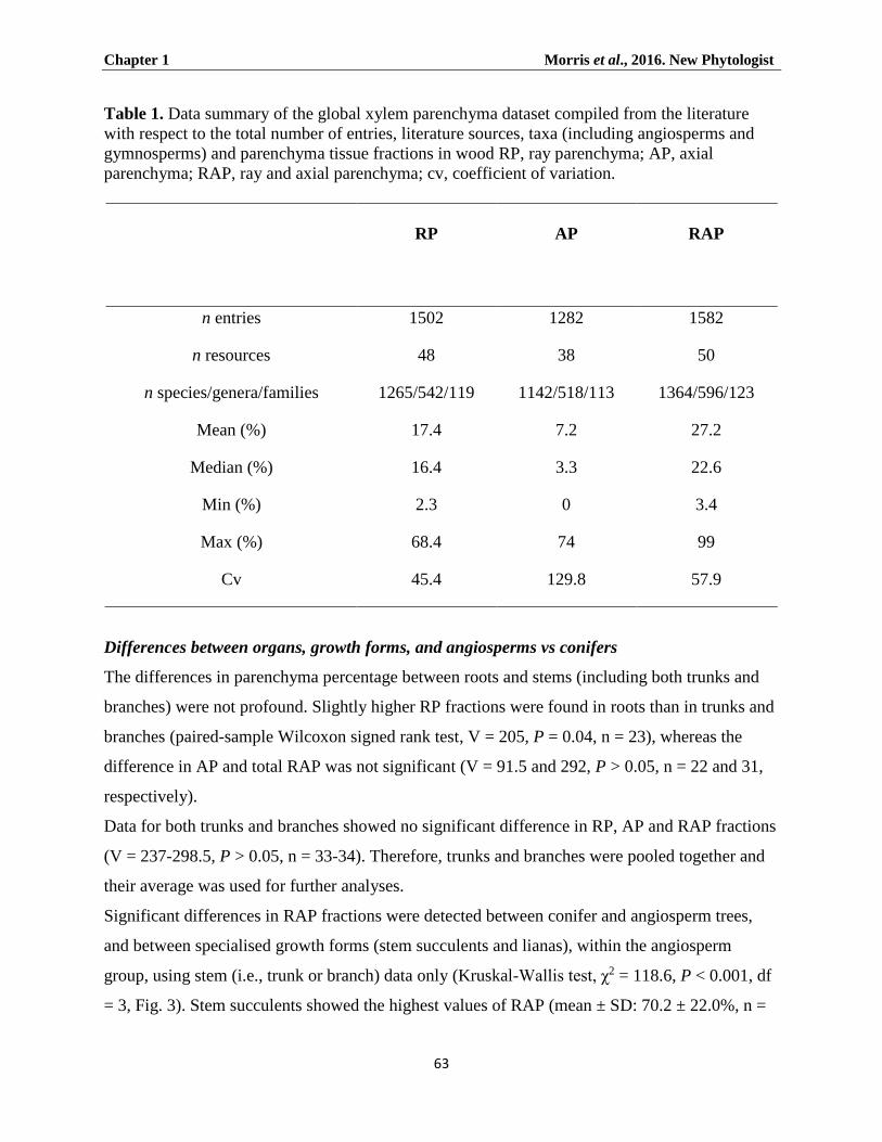
Chapter 1 Morris et al., 2016. New Phytologist
63
Table 1. Data summary of the global xylem parenchyma dataset compiled from the literature
with respect to the total number of entries, literature sources, taxa (including angiosperms and
gymnosperms) and parenchyma tissue fractions in wood RP, ray parenchyma; AP, axial
parenchyma; RAP, ray and axial parenchyma; cv, coefficient of variation.
RP AP RAP
n entries 1502 1282 1582
n resources 48 38 50
n species/genera/families 1265/542/119 1142/518/113 1364/596/123
Mean (%) 17.4 7.2 27.2
Median (%) 16.4 3.3 22.6
Min (%) 2.3 0 3.4
Max (%) 68.4 74 99
Cv 45.4 129.8 57.9
Differences between organs, growth forms, and angiosperms vs conifers
The differences in parenchyma percentage between roots and stems (including both trunks and
branches) were not profound. Slightly higher RP fractions were found in roots than in trunks and
branches (paired-sample Wilcoxon signed rank test, V = 205, P = 0.04, n = 23), whereas the
difference in AP and total RAP was not significant (V = 91.5 and 292, P > 0.05, n = 22 and 31,
respectively).
Data for both trunks and branches showed no significant difference in RP, AP and RAP fractions
(V = 237-298.5, P > 0.05, n = 33-34). Therefore, trunks and branches were pooled together and
their average was used for further analyses.
Significant differences in RAP fractions were detected between conifer and angiosperm trees,
and between specialised growth forms (stem succulents and lianas), within the angiosperm
group, using stem (i.e., trunk or branch) data only (Kruskal-Wallis test, χ2 = 118.6, P < 0.001, df
= 3, Fig. 3). Stem succulents showed the highest values of RAP (mean ± SD: 70.2 ± 22.0%, n =

Chapter 1 Morris et al., 2016. New Phytologist
64
50), followed by lianas (50.1 ± 16.3%, n = 28), and angiosperm trees and shrubs (26.3 ± 12.4%,
n = 1,384), whereas conifers exhibited the lowest fraction of RAP (7.63 ± 2.6%, n = 89, Fig. 3a).
In angiosperm trees, there were many entries with rather high RAP, e.g. 136 entries (118 species)
showed total RAP fractions above 50%.
Fig. 3 Parenchyma tissue fractions (%) across different woody plant groups and growth forms.
Because of inherent differences in wood anatomy between conifer and angiosperm trees, both
groups are analysed separately. The lowest parenchyma fractions are found in conifers, followed
by angiosperm trees (including some shrubs), climbers and succulents. The percentage of total
parenchyma (including axial and ray parenchyma) is shown in Fig. 3a, with the ray and axial
parenchyma contribution for the same groups given in Fig. 3b. The box plot in Fig. 3a shows the
median, 25th and 75th percentiles, error bars show 10th and 90th percentiles, and open circles
show outliers. Different letters above boxes indicate statistical significance between groups
(Kruskal-Wallis test, P≤0.05). Bars in Fig. 3b represent mean values ± SD. The number of
specimens for Fig. 3a (with the number of specimens for Fig. 3b between brackets) is: n gymno
tree = 89 (14), n angio tree =1,384 (1,205), n climber = 28 (9), and n succulent = 50 (32).

Chapter 1 Morris et al., 2016. New Phytologist
65
In addition to the total RAP percentage, the contribution of RP and AP was analysed (Fig. 3b),
although these data should be interpreted with caution as mentioned above. The information on
RP and AP fractions was available for stems of 14 conifer species, 1,205 angiosperm tree
species, nine climbers, and 32 succulents. Again, conifers showed much lower fractions of both
RP and AP (RP: 8.1 ± 2.7%; AP: 1.7 ± 2.2%) than angiosperm trees (RP: 17.7 ± 6.3%; AP: 6.6 ±
8.5%, Fig. 3b). Climbers in our dataset had the highest fraction of AP (29.3 ± 10.2%), while RP
was relatively low (12.5 ± 2.7%). Succulents showed a high fraction of both RP and AP (RP:
43.5 ± 14.3%, AP: 20.8 ± 18.1%, Fig. 3b).
Climate and RAP fractions
Differences in RAP fractions in angiosperm trees (including some shrubs) growing in various
climatic zones were analysed for 399 temperate, 442 tropical and 543 subtropical specimens
(Kruskal-Wallis test, χ2 = 224.9, P < 0.001, df = 2), with mean values of 21.1% (± 7.9), 22.2% (±
9.3 and 36.2% (± 13.4) for temperate, subtropical and tropical angiosperm specimens,
respectively (Fig. 4a). The amount of AP appeared to be the main driver of this difference. The
total AP fraction was 13.8% (±11.0) in tropical trees, whereas it was between 4 to 5% in
temperate and subtropical trees (see Fig. 4b). In contrast, average RP fractions spanned a narrow
range from 16.4% (± 5) in temperate to 19.4% (± 6.8) in tropical trees.

Chapter 1 Morris et al., 2016. New Phytologist
66
Fig. 4 Parenchyma tissue fractions (%) in angiosperm trees classified according to the temperate,
subtropical and tropical biome, showing the total parenchyma fraction (Fig. 4a) and the
individual contributions of ray and axial parenchyma (Fig. 4b). The box plot in Fig. 4a shows the
median, 25th and 75th percentiles, error bars show 10th and 90th percentiles, and open circles
show outliers. Different letters above boxes indicate statistical significance for tropical species
compared to temperate and subtropical trees (Kruskal-Wallis test, P ≤ 0.05). Bars in Fig. 4b
represent mean values ± SD. The number of specimens for Fig. 4a (Fig. 4b): n temperate = 399
(399), n tropical = 442 (287), n climber = 28 (9), and n succulent = 50 (32).

Chapter 1 Morris et al., 2016. New Phytologist
67
The ternary axis (Fig. 5) revealed that a higher contribution of RAP occurred mainly at the
expense of fibres, particularly in tropical and subtropical trees, while total vessel fractions were
typically between 5 to 20%, and on average 14.6%. A strongly negative correlation was found
between the tissue fractions for RAP and fibres (including tracheids) for all biomes, especially
the tropical climate (Spearman r = -0.75, P < 0.001 for all biomes; Table 2). Fibre tissue
fractions (F) were most negatively correlated with vessel tissue fraction (V) in temperate and
subtropical species (r = -0.66, and -0.59, respectively; P < 0.001), while a negative relationship
between RAP and V was only weakly significant for the temperate and tropical climate (r = -0.21
and -0.18, respectively; P < 0.001; Table 2). In some temperate trees, the relatively high vessel
fractions represented ring-porous species with narrow growth rings that have a high proportion
of early-wood and, therefore, many wide vessels.
Fig. 5 Ternary plot showing the contribution of the three principal tissues in wood: the total
amount of ray and axial parenchyma (RAP, %), vessels (V, %), and fibres (including tracheids)
(F, %). Each dot represents a specimen. The trade-off between RAP and F tissue fractions shows
a strongly negative correlation for all data and is especially clear in the tropical biome (see Table
2). Specimens are grouped according to three major climatic zones, which follow Köppen
(1936).

Chapter 1 Morris et al., 2016. New Phytologist
68
Table 2. Spearman's rank correlation coefficients showing the trade-offs between the tissue
fractions of three major xylem cell types, which were analysed for three major biomes
(temperate, subtropical, tropical). See Fig. 5 for a visual plot. RAP, ray and axial parenchyma; F,
fibres (including tracheids); V, vessels. ***, P ≤ 0.001.
Correlation All biomes Temperate Subtropical Tropical
RAP vs F −0.752*** −0.480*** −0.688*** −0.856***
RAP vs V −0.153*** −0.210*** −0.066 −0.176***
F vs V −0.427*** −0.661*** −0.591*** −0.279***
There was a strong agreement between the climatic data derived from the sampling locations and
those derived from the GBIF locations (Fig. S4). There was a clear difference between
angiosperms and conifers, as we found only significant correlations between RAP and MAT, and
between RAP and MAP for angiosperms. Moreover, due to a low sample number (90 records, 61
species) and limited number of locations, no climatic GAM analyses were performed on
conifers.
The GAM models showed that MAT was the main driver for RAP in angiosperms (F1.94, 267 =
37.21, pseudo-R² = 21.05%, P-value < 0.001; for exact locations, and F1.95, 218 = 48.22, pseudo-R2
= 31.65%, P < 0.001 for GBIF locations, Fig. 6a). However, the effect of MAT on RAP was
non-linear, with RAP fractions increasing with MAT values in hot environments (> ± 17°C), but
not for species in colder environments. When analysing the effect of MAT on AP and RP
fractions separately, GAM models revealed significant associations for both when controlling for
altitude and precipitation (Fig. S5a, Fig. S6a; Table S2, S3).

Chapter 1 Morris et al., 2016. New Phytologist
69
Fig. 6 The effect of MAT (a), MAP (b), and altitude (c) on the proportion of ray and axial
parenchyma (RAP) in angiosperm wood based on a general additive model with a binomial
distribution for the exact location dataset (red) and the GBIF derived climate data (blue). Each
climate variable was limited to three effective degrees of freedom (e.d.f.). The solid line
represents the fitted smoother; 95% confidence intervals are shown as coloured tinted areas.
Each dot represents a specimen for which the sampling location was reported in literature, or
climate data obtained from the WorldClim database. Pseudo-R2 measures the approximate
deviance explained by each explanatory variable.

Chapter 1 Morris et al., 2016. New Phytologist
70
The relationship between MAP and RAP was also significant, although the pseudo-R² values
were very low compared to those of MAT (Fig. 6b; Table S2, S3). While significance was found
for the relationship between MAP and RP (Fig. S6b), this was not the case for AP (Fig. S5b;
Table S2, S3). When controlling for MAP and MAT, RAP increased significantly with altitude
(Fig. 6c), but these altitudinal trends were non-significant or significant for AP and RP,
depending on whether the data were based on the exact locations or GBIF (Fig. S5c, Fig. S6c).
A separate GAM model between total RAP fraction and temperature also supported a quadratic
latitudinal trend (Fig. 7), for both exact locations (F1.95, 267 = 27.01, pseudo-R2 = 18%, P < 0.001)
and species from the GBIF locations (F1.96, 218 = 31.81, pseudo-R2 = 24.1%, P < 0.001). The latter
suggests that RAP increased significantly the closer the sampling location was to the equator for
both datasets.

Chapter 1 Morris et al., 2016. New Phytologist
71
Fig. 7 The effect of latitude on the proportion of ray and axial parenchyma (RAP) in angiosperm
wood based on a general additive model with a binomial distribution for the exact location
dataset (red) and the GBIF derived climate data (blue). The latitude variable was limited to three
partitions. The solid line represents the fitted smoother; 95% confidence intervals are shown as
coloured tinted areas. Each dot represents a specimen for which the sampling location was
reported in literature, or climate data obtained from the WorldClim database. Pseudo-R2
measures the approximate deviance explained by latitude.

Chapter 1 Morris et al., 2016. New Phytologist
72
Discussion
The global RAP dataset compiled demonstrates that RAP tissues show a 29-fold variation in
abundance, which agrees with previous reports across seed plants (Fujiwara et al., 1991;
Martínez-Cabrera et al., 2009; Ziemińska et al., 2013, 2015; Zheng and Martínez-Cabrera,
2013). A key finding is that RAP amounts in wood, especially AP, are driven by temperature
(Fig. 6a). Albeit, precipitation also showed a significant relationship with RAP in the GAM
models when controlling for altitude and MAT, although far less so than temperature (Fig. 6b;
Table S2, 3). MAP showed a negative trend in the GAM models, with increasing amounts of
RAP towards drier environments, which is what we expected. The temperature effect is also
reflected in the latitudinal trends for RAP, especially in the northern hemisphere. However, our
analyses did not support the expected difference between subtropical and temperate species
(Fig.4b). Also, no significant difference was found in the RAP tissue fraction between tropical
wet environments and tropical, seasonally dry areas (data not shown).
Other drivers of RAP tissue fractions include growth forms. It is clear that lianas and succulents
represent two growth forms that show higher fractions of RAP than self-supporting angiosperm
trees. Within the tree and shrub growth form, tropical plants have higher levels of RAP than
temperate and subtropical ones (Fig. 4, 5), supporting previous studies based on qualitative
(Baas, 1982; Segala-Alves and Angyalossy-Alfonso, 2002; Wheeler et al., 2007) and
quantitative approaches (Martinez-Cabrera et al., 2009). A novel finding is the fact that RAP
levels in tropical plants are mainly due to an increase in AP, while RP levels remain more
conservative in trees across the three major biomes analysed (Fig. 4b). This finding is not as
transparent in the GAM models (Figs. S4, S5), as both RP and AP are highly associated with
temperature. However, where RP amount gradually increases with temperate, AP remains
unwavering until approximately 17ºC and then sharply rises exponentially with temperature.
This is the reason AP is so high in the tropics compared to both temperate and subtropical
regions (Fig. 4b).
Explaining why some factors may drive the RAP level while others have little or no influence
requires a more detailed understanding of RAP functions (Fig. 2), especially those related to
storage capacity, resistance to drought stress, frost resistance, defence mechanisms and
mechanical properties.

Chapter 1 Morris et al., 2016. New Phytologist
73
RAP fractions in relation to NSC storage capacity
Storage of NSCs (non-structural carbohydrates) is arguably one of the most widely accepted
functions of RAP in wood. As far as we know, however, the hypothesis that high amounts of
RAP correspond with higher storage capacity of NSCs has not been tested yet in a direct,
quantitative way. The assumption that higher amounts of RAP, and therefore a higher NSC
capacity, should occur in roots when compared to stems could only partly be confirmed. We
found no significant differences between stems and roots in total RAP and AP fractions, but did
find higher levels of RP in root wood than in stem wood. As this finding came from a small
sample set (n = 21-30), it is premature to make generalisations. A higher fraction of RP in roots
than stems has been observed previously (Gasson and Cutler, 1990; Stokke and Manwiller, 1994;
Machado et al., 2007) and may be associated with a reduced need for mechanical cells such as
fibres in addition to increased storage capacity of roots. Aside from RP fractions, different cell
dimensions of RP have been reported in roots, especially a general increase in width of the entire
ray and the individual ray cells, and a tendency towards more heterocellular rays in roots than in
stems (Patel, 1965; Gasson and Cutler, 1990; Denne and Gasson, 2008).
When considering the storage capacity of RAP we could also expect to find higher RAP
fractions in plants from temperate seasonal climates. However, we did not find much support for
this hypothesis due to lower RAP fractions in trees from temperate compared to tropical biomes.
RAP are not the only wood tissue storing NSCs; septate or living fibres also do so (Webber,
1936; Yamada et al., 2011; Carlquist, 2014).
RAP fractions and drought stress
It is possible that high RAP fractions in wood benefit plants in dry conditions by conferring high
hydraulic capacitance, which could prevent embolism formation, or facilitate embolism refilling
(Fig. 6b). Support for a drought related function of RAP is provided by the high levels of RAP in
stem succulents. With an average of 70.3% RAP in succulents, it can be speculated that wood
parenchyma not only stores a considerable amount of water but also provides symplastic
connections with bark and pith that both serve as important water reservoirs (Borchert and
Pockman, 2005; Scholz et al. 2007; Hearn, 2009, Hearn et al., 2013; Pfautsch et al., 2015).
However, the large RAP variation in dry and seasonally dry environments suggests that plants
have various strategies to survive these conditions, with stem succulents having large

Chapter 1 Morris et al., 2016. New Phytologist
74
parenchyma fractions while other species may show comparatively few RAP cells. It has been
suggested that vessel-associated RAP may be involved in the embolism refilling process by
releasing sugars and water into embolised conduits (Bucci et al., 2003; Salleo et al., 2004;
Brodersen et al., 2010; Brodersen and McElrone, 2013). While embolism repair remains
controversial and poorly understood, refilling on a daily basis may not occur in conifers, which
could be owing to their low RAP fractions (Choat et al., 2014).
RAP fractions and temperature
Temperature is associated with RAP fractions in the secondary xylem, much more so than
rainfall. This finding agrees with Moles et al. (2014), who found that 15 out of 21 plant traits are
more strongly correlated with temperature than with precipitation. The high levels of RAP in
tropical plants could be linked to the greater plant diversity in the tropics and the dominance of
various families with high RAP levels (e.g., Fabaceae, Moraceae). Alternatively, particular
functions associated with RAP (e.g., defence against pathogens, hydraulic capacitance) could be
more important in tropical environments than in temperate regions. It is possible that protection
against cold, including tolerance to extracellular freezing and freeze dehydration, or freeze
avoidance by super-cooling, is an energy-demanding process (Quamme, 1991; Neuner, 2014),
which could therefore be an important factor in reducing the RAP fraction in woody plants
exposed to frost or freezing events. Also, two conifer genera growing in the tropical/subtropical
mountains of the southern hemisphere (Podocarpus spp. and Dacrydium spp.) have higher RAP
fractions than conifers from cool temperate regions (Braun, 1984). Similarly, Pinus canariensis
from the warm islands of Tenerife and La Palma have RAP averages of 14.5%, with AP values
accounting for 3% of this (Climent et al., 1998).
RAP fractions as a defence system
RAP fractions might also play a large role in defence against the spread of decay via pathogenic
fungi, viruses, and bacteria. The presence of RP may prevent the lateral spread of fungi, while
AP does the same for axial movement (Shigo, 1984; Boddy and Rayner, 1983; Biggs, 1986).
Because both RP and AP may accumulate anti-microbial compounds such as phytoalexins,
phenolic compounds and suberin, which all act to inhibit fungal spread (Biggs, 1987), trees with
high RAP fractions might be more resistant to brown rot fungi and therefore be better overall
compartmentalisers of decay than trees with lower RAP fractions (Schwarze et al., 2003).

Chapter 1 Morris et al., 2016. New Phytologist
75
In agreement with the defence role, the amount of RAP is higher in the sapwood of trees that
have recovered from pathogenesis (Tippett and Shigo, 1981; Schmitt and Liese, 1990; Arbellay
et al., 2010, 2012). Interestingly, another study across seven tree species from the Amazon, have
found that high parenchyma abundance and wide dilating rays were associated with poor
compartmentalisation of decay, but was offset by fast wound closure (Romero and Bolker,
2008). While in angiosperms, parenchyma cells in contact with vessels can seal off conduits by
way of tyloses or gum deposits to avoid the spread of decay (Biggs, 1987; Bonsen and Kučera,
1990; Schmitt and Liese, 1992; Sun et al., 2007), defence in gymnosperms mainly lies in the
occlusion of tracheids via aspiration of the torus-margo bordered pits (Fuhr et al., 2013) and the
production of abundant polyphenolic compounds and traumatic resin ducts in Pinaceae and
Cupressaceae (Phillips, 1948, Hudgins et al., 2004). Such a strategy is therefore consistent with a
lower RAP fraction in conifers. In contrast, a higher fraction of RAP in the tropics might be
driven by a greater incidence of biotic stress when compared to temperate environments (Bagchi
et al., 2014). The evolutionary ‘arms race’ with pathogens and insect herbivores may require the
deployment of more RAP or the synthesis of a more diverse suite of secondary compounds by
RAP in order to enhance tree defence abilities. On the other hand, the trade-off between RAP
and fibre tissue fractions (Fig. 5) may also suggest that high RAP fractions could equally
decrease the defence capacity.
RAP and mechanical properties
Since the amount of RAP occurs mainly at the expense of fibres, one would also expect
important effects on wood mechanical properties. It has long been assumed that parenchyma
cells often have larger lumina and thinner cell walls than fibres, so high RAP levels should
theoretically result in lower wood density together with a reduced stiffness, but retain a higher
elasticity. However, rays in Liquidambar were found to have a higher specific gravity than
surrounding tissues because the ratio of cell wall to lumen was relatively high (Taylor, 1996).
Moreover, in a study of 69 angiosperm species it was found that the main driver of the modulus
of elasticity (defined as the ratio of tensile stress to tensile strain) was fibre wall fraction rather
than RAP fraction (Ziemińska et al., 2015). However, that study only looked at species within a
small wood density range and the results should be treated with caution.
Several authors have suggested that wide rays, which are common in lianas, allow vessel-bearing
segments to twist without rupturing, which may also explain the occurrence of non-lignified or

Chapter 1 Morris et al., 2016. New Phytologist
76
less lignified ray parenchyma cells in climbing plants (Schenck, 1893; Haberlandt, 1914; Sieber
and Kučera, 1980; Gartner, 1991; Putz and Holbrook, 1991). Additional explanations for the
high amount of RAP in lianas can be linked with dedifferentiation of parenchyma, allowing rapid
recovery from injury, especially after tree fall (Dobbins and Fisher, 1986; Fisher and Ewers,
1991; Busch et al., 2010), and their ability to clone readily when detached from the parent plant
(Putz, 1984; Yorke et al., 2013). A mechanical role of RAP dependant on their turgor pressure
has also been suggested in Adansonia (Chapotin et al., 2006), with RAP in this species
occupying as much as 90%.
General Conclusion
The 29-fold variation in the parenchyma fraction is associated with temperature-driven
differences between tropical, subtropical and temperate woody plants, as well as with different
growth forms such as succulents and lianas. The ecological trends discussed suggest ways for
further research into how RAP plays a role in woody plant function in the storage of NSCs and
water, defence against pathogens, and resilience to damage. Various functions of RAP in wood
have been suggested and it is generally not clear which function takes precedence in a given
situation. Based on the available evidence, this may depend on climate, plant organ, and the
potential partitioning in the functional roles of RP and AP (Zheng and Martínez-Cabrera, 2013).
Also, the total RAP percentage does not reflect the spatial, three-dimensional arrangement of the
parenchyma network and its connectivity to other xylem tissues. More research is also needed to
test the within tree variation of RAP %, which would involve labour intensive studies with
careful and well-planned sampling. Further research based on observational evidence is needed
to investigate the role of parenchyma in more detail, such as the spatial patterns of parenchyma
networks and, along with this, to test the hypotheses presented in this paper.
Acknowledgements
HM and SJ acknowledge financial support from the German Research Foundation (DFG, project
number 2174). LP was supported by a Postdoctoral Fellowship from the Alexander von
Humboldt Foundation and acknowledges funding from the Ulm University and Ulm University
Society (Ulmer Universitätsgesellschaft). Funding to JZ was available through a Special Fund
for Forest Scientific Research in the Public Welfare, China (No.201404303). We also wish to
thank the Botanical garden of Ulm University for supplying plant material, Amy Zanne (George

Chapter 1 Morris et al., 2016. New Phytologist
77
Washington University), and anonymous reviewers for valid comments on an earlier version of
this paper.
Author contributions
H.M., L.P. and S.J. led the initial data compilation and coordinated the writing. H.M., L.P., S.J.,
E.F., H.I.M.-C., J.Z. and K.Z. contributed data and ideas. L.P. and M.A.F.G. analysed data. K.Z.
and E.W. assisted with writing the final manuscript. H.M., P.C. and D.J.MG. extracted GBIF
locations and climate data.

Chapter 1 Morris et al., 2016. New Phytologist
78
References
Arbellay E, Stoffel M, Bollschweiler M. 2010. Wood anatomical analysis of Alnus incana and
Betula pendula injured by a debris–flow event. Tree Physiology 30: 1290-1298.
Arbellay E, Fonti P, Stoffel M. 2012. Duration and extension of anatomical changes in wood
structure after cambial injury. Journal of Experimental Botany 63: 3271-3277.
Baas P. 1982. Systematic, phylogenetic and ecological wood anatomy - history and perspectives.
In: Baas P, eds. New perspectives in wood anatomy. The Hague, Netherlands: Nijhoff/Junk,
23-58.
Bagchi R, Gallery RE, Gripenberg S, Gurr SJ, Narayan L, Addis, CE, Freckleton, RP,
Lewis OT. 2014. Pathogens and insect herbivores drive rainforest plant diversity and
composition. Nature 506: 85-88.
Barghoorn ES. 1940. The ontogenetic and phylogenetic specialisation of rays in the xylem of
dicotyledons. I. The primitive ray structure. American Journal of Botany 27: 918-928.
Barghoorn ES. 1941. The ontogenetic development and phylogenetic specialization of rays in
the xylem of dicotyledons. II. Modification of the multiseriate and uniseriate rays. American
Journal of Botany 28: 273-282.
Biggs AR. 1986. Comparative anatomy of host response of two peach cultivars inoculated with
Leucostoma cincta and L. personii. Phytopathology 76: 905-912.
Biggs AR. 1987. Occurrence and location of suberin in wound reaction zones in xylem of 17 tree
species. Phytopathology 77: 718-725.
Boddy L, ADM, Rayner ADM. 1983. Origins of decay in living deciduous trees: the role of
moisture content and a re-appraisal of the expanded concept of tree decay New Phytologist.
94: 623-641.
Bonsen KJM, Kučera LJ. 1990. Vessel occlusion in plants: morphological, functional, and
evolutionary aspects. International Association of Wood Anatomists Bulletin 11: 393-399.

Chapter 1 Morris et al., 2016. New Phytologist
79
Borchert R, Pockman WT. 2005. Water storage capacitance and xylem tension in isolated
branches of temperate and tropical trees. Tree Physiology 25: 457-466.
Braun HJ, Wolkinger F. 1970. Zur funktionellen Anatomie des axialen Holzparenchyms und
Vorschlage zur Reform seiner Terminologie. Holzforschung 24: 19-26.
Braun HJ. 1984. The significance of the accessory tissues of the hydrosystem for osmotic water
shifting as the second principle of water ascent, with some thoughts concerning the evolution
of trees. International Association of Wood Anatomists Bulletin 5: 275-294.
Brodersen CR, McElrone A, Choat B, Matthews M, Shackel K. 2010. Dynamics of
embolism repair in grapevine: in vivo visualisations using HRCT. Plant Physiology 154:
1088-1095.
Brodersen CR, McElrone AJ. 2013. Maintenance of xylem network transport capacity: a
review of embolism repair in vascular plants. Frontiers in Plant Science 4, article 108: 1-11.
Bucci SJ, Scholz FG, Goldstein G, Meinzer FC, Sternberg LDL. 2003. Dynamic changes in
hydraulic conductivity in petioles of two savanna tree species: factors and mechanisms
contributing to the refilling of embolized vessels. Plant, Cell & Environment 26: 1633-1645.
Burgert I, Bernasconi A, Eckstein D. 1999. Evidence for the strength function of rays in living
trees. Holz als Roh - und Werkstoff 57: 397-399.
Burgert I, Eckstein D. 2001. The tensile strength of isolated wood rays of beech (Fagus
sylvatica L.) and its significance for the biomechanics of living trees. Trees - Structure and
Function 15: 168-170.
Busch S, Seidel R, Speck O, Speck T. 2010. Morphological aspects of self-repair of lesions
caused by internal growth stresses in stems of Aristolochia macrophylla and Aristolochia
ringens. Proceedings of Biological Sciences 277: 2113-2120.
Carlquist S. 2001. Comparative wood anatomy. Systematic, ecological, and evolutionary
aspects of dicotyledon wood, 2nd edn. Berlin, Germany: Springer.

Chapter 1 Morris et al., 2016. New Phytologist
80
Carlquist S. 2014. Fibre dimorphism: cell type diversification as an evolutionary strategy in
angiosperm woods. Botanical Journal of the Linnean Society 174: 44-67.
Chapotin SM, Razanomeharizaka JH, Holbrook NM. 2006. A biomechanical perspective on
the role of large stem volume and high water content in baobab trees (Adansonia spp.;
Bombacaceae). American Journal of Botany 93: 1251-1264.
Choat B, Brodersen CR, McElrone AJ. 2014. Synchrotron X-ray microtomography of xylem
embolism in Sequoia sempervirens saplings during cycles of drought and recovery. New
Phytologist 205: 1095-1105.
Clearwater MJ, Goldstein G. 2005. Embolism repair and long distance water transport. In:
Holbrook NM, Zwieniecki MA, eds. Vascular transport in plants. Burlington, MA, USA:
Elsevier Academic Press, 375-399.
Climent J, Gil L, Pardos JA. 1998. Xylem anatomical traits related to resinous heartwood
formation in Pinus canariensis Sm. Trees - Structure and Function. 123: 139-145.
Deflorio G, Johnson C, Fink S, Schwarze FMWR. 2008. Decay development in living
sapwood of coniferous and deciduous trees inoculated with six wood decay fungi. Forest
Ecology and Management 255: 2373-2383.
Denne P, Gasson P. 2008. Ray structure in root- and stem-wood of Larix decidua: Implications
for root identification and function. International Association of Wood Anatomists Journal
29: 17-23.
Dobbins D, Fisher J. 1986. Wound response of girdled stems of lianas. Botanical Gazette 147:
278-289.
Fahn A, Werker E, Baas P. 1986. Wood anatomy and identification of trees and shrubs from
Israel and adjacent regions. Jerusalem, Israel: The Israel Academy of Sciences and
Humanities.
Chapter 1 Morris et al., 2016. New Phytologist
81
Fichtler E, Worbes M. 2012. Wood anatomical variables in tropical trees and their relation to
site conditions and individual tree morphology. International Association of Wood
Anatomists Journal 33: 19-40.
Fisher JB, Ewers FW. 1991. Structural responses to stem injury in vines. In: Putz FE, Mooney
HA, eds. The biology of vines. Cambridge, UK: Cambridge University Press, 99-124.
Fuhr MJ, Schbert M, Stührk C, Schwarze FWMR, Herrmann HJ. 2013. Penetration
capacity of the wood-decay fungus Physisporinus vitreus. Complex adaptive systems
modelling. Springer Open Journal 1: 6.
Fujiwara S, Sameshima K, Kuroda K, Takamura N. 1991. Anatomy and properties of
Japanese hardwoods. I. Variation of fibre dimensions and tissue proportions and their relation
to basic density. International Association of Wood Anatomists Bulletin new series 12: 419-
24.
Gartner GL. 1991. Stem hydraulic properties of vines vs. shrubs of western poison oak,
Toxicodendron diversilobum. Ecologia 87: 180-189.
Gasson PE, Cutler DF. 1990. Root anatomy of 17 genera growing in the British Isles.
International Association of Wood Anatomists Bulletin new series 11: 3-46.
Haberlandt G. 1914. Physiological Plant Anatomy. (Translated from the 4th German edition by
M. Drummond). London, UK: MacNillan & Co.
Hearn DJ. 2009. Developmental patterns in anatomy are shared among separate evolutionary
origins of stem succulent and storage root-bearing growth habits in Adenia (Passifloraceae).
American Journal of Botany 96: 1941-1956.
Hearn DJ, Poulsen T, Spicer R. 2013. The evolution of growth forms with expanded root and
shoot parenchymatous storage is correlated across the eudicots. International Journal of
Plant Sciences 174: 1049-1061.
Chapter 1 Morris et al., 2016. New Phytologist
82
Hijmans RJ, Cameron SE, Parra JL, Jones PG, Jarvis A. 2005. Very high resolution
interpolated climate surfaces for global land areas. International Journal of Climatology 25:
1965-1978.
Hoch G, Richter A, Korner, CH. 2003. Non-structural carbon compounds in temperate forest
trees. Plant, Cell & Environment 26: 1067-1081.
Holbrook NM. 1995. Stem water storage. In: Gartner BL, ed. Stems and trunks in plant form
and function. San Diego, USA: Academic Press, 151-174.
Huber B, Prütz G. 1938. Über den Anteil von Fasern, Gefässen und Parenchym am Aufbau
verschiedener hölzer. Holz als Roh- und Werkstoff 10: 377-381.
Hudgins JW, Christiansen E, Franceschi V. 2004. Induction of anatomically based defence
responses in stems of diverse conifers by methyl jasmonate: a phylogenetic perspective. Tree
Physiology 24: 251-264.
IAWA Committee. 1989. IAWA list of microscopic features for hardwood identification.
International Association of Wood Anatomists Bulletin new series 10: 219-332.
IAWA Committee. 2004. IAWA list of microscopic features for softwood identification.
International Association of Wood Anatomists Journal 25: 1-70.
Jansen S, Gortan E, Lens F, Lo Gullo MA, Salleo S, Scholz A, Stein A, Trifilò P, Nardini A.
2011. Do quantitative vessel and pit characters account for ion-mediated changes in the
hydraulic conductance of angiosperm xylem? New Phytologist 189: 218-228.
Kattge J, Díaz S, Lavorel S, Prentice IC, Leadley P, Bönisch G, Garnier E, Westoby M,
Reich PB, Wright IJ et al. 2011. TRY – a global database of plant traits. Global Change
Biology 17: 2905-2935.
Koch P. 1985. Utilization of hardwoods growing on southern pine sites. Asheville, NC: USDA-
Forest Service.

Chapter 1 Morris et al., 2016. New Phytologist
83
Köppen W. 1936. Das geographische System der Klimate. In: Köppen W, Geiger R, eds.
Handbuch der Klimatologie. Berlin, Germany: Gebrüder Borntraeger, 1-44.
Kribs DA. 1935. Salient lines of structural specialisation in the wood rays of dicotyledons.
Botanical Gazette 96: 547-557.
Kribs DA. 1937. Salient lines of structural specialization in the wood parenchyma of
dicotyledons. Bulletin Torrey Botanical Club 64: 177-186
Machado SR, Rodella RA, Angyalossy V, Marcati CR. 2007. Structural variations in root and
stemwood of Styrax (Styracaceae) from Brazilian forest and cerrado. International
Association of Wood Anatomists Journal 28: 173-188.
Martínez-Cabrera HI, Jones CS, Espino S, Schenk HJ. 2009. Wood anatomy and wood
density in shrubs: responses to varying aridity along transcontinental transects. American
Journal of Botany 96: 1388-1398.
Moles AT, Perkins SE, Laffan SW, Flores-Moreno H, Awasthy M, Tindall ML, Sack L,
Pitman A, Kattge J, Aarssen LW et al. 2014. Which is a better predictor of plant traits:
temperature or precipitation? Journal of Vegetation Science 25: 1167-1180.
Nardini A, Salleo S, Jansen S. 2011. More than just a vulnerable pipeline: xylem physiology in
the light of ion-mediated regulation of plant water transport. Journal of Experimental Botany
62: 4701-4718.
Nawrot M, Pazdrowski W, Szymanski M. 2008. Dynamics of heart-wood formation and axial
and radial distribution of sapwood and heartwood in stems of European larch (Larix decidua
Mill.). Journal of Forest Science. 54: 409-417.
Neuner G. 2014. Frost resistance in alpine woody plants. Frontiers in Plant Science 5, article
654: 1-13.
Normand D, Chatelet R. 1951. Observations taxonomiques et xylologiques sur le genre
Pinacopodium Exell et Mendoҫa. Bulletin Jardin Botanique État, Bruxelles 21: 451-463.

Chapter 1 Morris et al., 2016. New Phytologist
84
O’Brien MJ, Leuzinger S, Philipson CD, Tay J, Hector A. 2014. Drought survival of tropical
tree seedlings enhanced by non-structural carbohydrate levels. Nature Climate Change 4:
710-714.
Olano JM, Arzac A, García-Cervigón AI, von Arx G, Rozas V. 2013. New star on the stage:
amount of ray parenchyma in tree rings shows a link to climate. New Phytologist 198: 486-
495.
Panshin AJ, de Zeeuw C. 1980. Textbook of wood technology. Structure, identification and uses
of the commercial woods of the United States and Canada. New York, USA: McGraw-Hill
Book Company.
Patel RN. 1965. A comparison of the anatomy of the secondary xylem in roots and stems.
Holzforschung 19: 72-79.
Pfautsch S, Renard J, Tjoelker MG, Salih A. 2015. Phloem as capacitor: radial transfer of
water into xylem of tree stems occurs via symplastic transport in ray parenchyma. Plant
Physiology 167: 963-971.
Phillips EWJ. 1948. Identification of softwoods by their microscopic structure. Forest Products
Research Bulletin 22: 1-56.
Pinto I, Pereira H, Usenius A. 2004. Heartwood and sapwood development within maritime
pine (Pinus pinaster Ait.) stems. Trees - Structure and Function 18: 284-294.
Plavcová L, Jansen S. 2015. The role of xylem parenchyma in the storage and utilization of
non-structural carbohydrates. In: Hacke UG, ed. Functional and Ecological Xylem Anatomy.
Heidelberg, Switzerland: Springer International Publishing, 209-234.
Poorter L, McDonald I, Alarcon A, Fichtler E, Licona JC, Pena-Claros M, Sterck F,
Villegas Z, Sass-Klaassen U. 2010. The importance of wood traits and hydraulic
conductance for the performance and life history strategies of 42 rainforest tree species. New
Phytologist 185: 481-492.

Chapter 1 Morris et al., 2016. New Phytologist
85
Pratt RB, Jacobsen AL, Ewers FW, Davis SD. 2007. Relationships among xylem transport,
biomechanics and storage in stems and roots of nine Rhamnaceae species of the California
chaparral. New Phytologist 174: 787-798.
Putz FE. 1984. The natural history of lianas on Barro Colorado Island, Panama. Ecology 65:
1713-1724.
Putz FE, Holbrook NM. 1991. Biomechanical studies of vines. In: Putz FE, Mooney HA, eds.
The biology of vines. Cambridge, UK: Cambridge University Press, 73-98.
Quamme HA. 1991. Application of thermal analysis to breeding fruit crops for increased cold
hardiness. HortScience 26: 513-517.
R Development Core Team. 2010. R: A language and environment for statistical computing.
Vienna, Austria: R Foundation for Statistical Computing. [WWW document] URL
http://www.r-project.org/. [accessed 7 July 2014].
Rasband WS. 1997-2012. ImageJ, US National Institutes of Health, Bethesda, MD, USA.
[WWW document] URL http://imagej.nih.gov/ij/. [accessed 27 Aug 2014].
Reiterer A, Burgert I, Sinn G, Tschegg S. 2002. The radial reinforcement of the wood
structure and its implication on mechanical and fracture mechanical properties - a comparison
between two tree species. Journal of Materials Science 37: 935-940.
Romero C, Bolker BM. 2008. Effects of stem anatomical and structural traits on responses to
stem damage: an experimental study in the Bolivian Amazon. Canadian Journal of Forest
Research 38: 611–618.
Ruelle J, Clair B, Beauchêne J, Prévost MF, Fournier M. 2006. Tension wood and opposite
wood in 21 tropical rain forest species. 2. Comparison of some anatomical and ultrastructural
criteria. International Association of Wood Anatomists Journal 27: 341–376.
Salleo S, Lo Gullo, MA, Trifilo P, Nardini, A. 2004. New evidence for a role of vessel-
associated cells and phloem in the rapid xylem refilling of cavitated stems of Laurus nobilis
L. Plant, Cell & Environment 27: 1065-1076.

Chapter 1 Morris et al., 2016. New Phytologist
86
Salleo S, Trifilo P, Esposito S, Nardini A, Lo Gullo MA. 2009. Starch-to-sugar conversion in
wood parenchyma of field-growing Laurus nobilis plants: a component of the signal pathway
for embolism repair? Functional Plant Biology 36: 815-825.
Santiago M, Pagay V, Strook AD. 2013. Impact of electroviscosity on the hydraulic
conductance of the bordered pit membrane: a theoretical investigation. Plant Physiology 163:
999-1011.
Schenck H .1893. Beitrage zur Biologie und Anatomie der Lianen, im besonderen der in
Brasilien einheimischen Arten, II. Beitrage zur Anatomie der Lianen. In: Schimper AFW, ed.
Botanische Mitteilungen aus den Tropen. Jena, Germany: Verlag von Gustav Fischer, 1-271.
Schmitt U, Liese W. 1990. Wound reaction of the parenchyma in Betula. International
Association of Wood Anatomists Bulletin 11: 413-420.
Schmitt U, Liese W. 1992. Seasonal influences on early wound reactions in Betula and Tilia.
Wood Science and Technology 26: 405-412.
Schmitt U, Liese W. 1993. Response of xylem parenchyma to suberisation in some hardwoods
after mechanical injury. Trees - Structure and Function 8: 23-30.
Scholz FB, Bucci SJ, Goldstein G, Meinzer FC, Franco AC, Miralles-Wilhelm F. 2007.
Biophysical properties and functional significance of stem water storage tissues in
Neotropical savanna trees. Plant, Cell & Environment 30: 236-248.
Scholz FG, Bucci SJ, Goldstein G, Meinzer FC, Franco AC, Miralles-Wilhelm F. 2007.
Biophysical properties and functional significance of stem water storage tissues in neo-
tropical savannah trees. Plant, Cell & Environment. 30: 236-248.
Schwarze FWMR, Fink S, Deflorio G. 2003. Resistance of parenchyma cells in wood to
degradation by brown rot fungi. Mycological Progress 2: 267-274.
Shigo AL. 1984. Compartmentalisation: a conceptual framework for understanding how trees
grow and defend themselves. Annual Review Phytopathology 22: 189-214.

Chapter 1 Morris et al., 2016. New Phytologist
87
Sieber M, Kučera LJ. 1980. On the stem anatomy of Clematis vitalba L. International
Association of Wood Anatomists Bulletin 1: 49-54.
Smith DM. 1967. Microscopic methods for determining cross-sectional cell dimensions. U.S.
Forest Service Research Paper FPL 79, October 1967. Madison, Wisconsin, USA: U.S.
Department of Agriculture, Forest Service, Forest Products Laboratory.
Spicer R. 2005. Senescence in secondary xylem: heartwood formation as an active development
programme. In: Holbrook N, Zwieniecki M, eds. Vascular transport in plants. Burlington,
MA, USA: Elsevier Academic Press, 457-475.
Spicer R, Holbrook NM. 2007. Parenchyma cell respiration and survival in secondary xylem:
does metabolic activity decline with cell age? Plant, Cell & Environment 30: 934-943.
Spicer R. 2014. Symplastic networks in secondary vascular tissues: parenchyma distribution and
activity supporting long-distance transport. Journal of Experimental Botany 65: 1829-1848.
Stokke DD, Manwiller FG. 1994. Proportions of wood elements in stem, branch, and root wood
of Black Oak (Quercus velutina). International Association of Wood Anatomists Journal 15:
301-310.
Sun Q, Rost TL, Reid MS, Matthews MA. 2007. Ethylene and not embolism is required for
wound-induced tyloses development in stems of grapevines. Plant Physiology 145: 1629-
1636.
Taylor FW. 1969. The effect of ray tissue on the specific gravity of wood. Wood & Fiber
Science 2: 142-145.
Tippett JT, Shigo AL. 1981. Barrier zone formation: A mechanism of the tree defence against
vascular pathogens. International Association of Wood Anatomists Bulletin 2: 163-168.
Trifilò P, Barbera PM, Raimondo F, Nardini A, Lo Gullo MA .2014. Coping with drought-
induced xylem cavitation: coordination of embolism repair and ionic effects in three
Mediterranean evergreens. Tree Physiology 34:109–122.

Chapter 1 Morris et al., 2016. New Phytologist
88
Van Bell AJE. 1990. Xylem-phloem exchange via the rays: the undervalued route of transport.
Journal of Experimental Botany 41: 631-644.
Wagenführ R. 2007. Holzatlas. Leipzig, Germany: Karl Hanser Verlang.
Webber IE. 1936. Systematic anatomy of the woods of the Simarubaceae. American Journal of
Botany 23: 577-587.
Wheeler EA, Baas P. 1991. A survey of the fossil record for dicotyledonous wood and its
significance for evolutionary and ecological wood anatomy. International Association of
Wood Anatomists Journal 12: 275-332.
Wheeler EA, Baas P, Rodgers S. 2007. Variations in dicot wood anatomy: a global analysis
based on the Insidewood database. International Association of Wood Anatomists Journal 28:
229-258.
Wolkinger F. 1970. Das Vorkommen der lebenden Holzfasern in Sträuchern und Bäumen.
Phyto 14: 55-67.
Wolkinger F. 1971. Morphologie und systematische Verbreitung lebenden Holzfasern bei
Sträuchern und Bäumen. III. Systematische Verbreitung. Holzforschung 25: 29-39.
Wood SN. 2006. Generalized Additive Models: An Introduction with R. London, UK: Chapman
and Hall.
Yamada Y, Awano T, Fujita M, Takabe K. 2011. Living wood fibers act as large-capacity
‘‘single-use’’ starch storage in black locust (Robinia pseudoacacia). Trees - Structure and
Function 25: 607-616.
Yorke SR, Schnitzer SA, Mascaro J, Letcher S, Carson WP. 2013. Increasing liana
abundance and basal area in a tropical forest: the contribution of long-distance clonal
colonization. Biotropica 45: 317-324.
Chapter 1 Morris et al., 2016. New Phytologist
89
Zheng J, Martínez-Cabrera HI. 2013. Wood anatomical correlates with theoretical
conductivity and wood density across China: evolutionary evidence of the functional
differentiation of axial and radial parenchyma. Annals of Botany 112: 927-35.
Ziemińska K, Butler DW, Gleason, SM, Wright, IJ, Westoby M. 2013. Fibre wall and lumen
fractions drive wood density variation across 24 Australian angiosperms. AoB PLANTS 5:
plt046.
Ziemińska K, Wright IJ, Westoby M. 2015. Broad anatomical variation within a narrow wood
density range – a study of twig wood across 69 Australian angiosperms. PLOS ONE 10(4):
e0124892.
Zwieniecki MA, Melcher PJ, Holbrook NM. 2001. Hydrogel control of xylem hydraulic
resistance in plants. Science 291: 1059-1062.

Chapter 1 Supplementary Information, New Phytologist
90
Supporting Information for New phytologist
A global analysis of parenchyma tissue fractions in secondary xylem of seed
plants
Authors: Hugh Morris, Lenka Plavcová, Patrick Cvecko, Esther Fichtler, Mark A.F Gillingham,
Hugo I. Martínez-Cabrera, Daniel J. McGlinn, Elisabeth Wheeler, Jingming Zheng, Kasia
Ziemińska, Steven Jansen
The following Supporting Information is available for this article:
Fig. S1 Distribution map of the species for which parenchyma fraction values were compiled.
Fig. S2 Poly-co-linearity matrix for the parameters analysed in relation to wood anatomy, plant
organ, geography and climate.
Fig. S3 Comparison of total parenchyma fractions in wood based on our own measurements and
literature.
Fig. S4 Comparison of mean annual temperature and mean annual precipitation for species for
which both sampling locations and GBIF locations were available.
Fig. S5 The effect of MAT, MAP and altitude on the proportion of axial parenchyma in
angiosperms.
Fig. S6 The effect of MAT, MAP and altitude on the proportion of ray parenchyma in
angiosperms.
Table S1 The Global Wood Parenchyma Database (see separate file).
Table S2 Summary of statistics for the general additive models (GAM) based on the exact
locations dataset.
Table S3 Summary of statistics for the general additive models (GAM) based on the GBIF
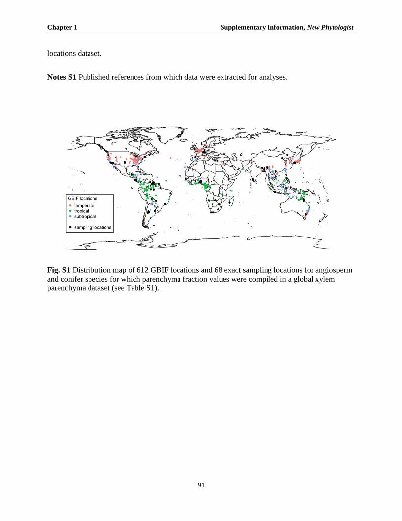
Chapter 1 Supplementary Information, New Phytologist
91
locations dataset.
Notes S1 Published references from which data were extracted for analyses.
Fig. S1 Distribution map of 612 GBIF locations and 68 exact sampling locations for angiosperm
and conifer species for which parenchyma fraction values were compiled in a global xylem
parenchyma dataset (see Table S1).
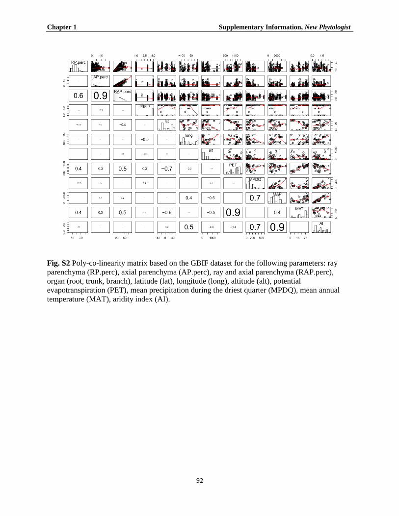
Chapter 1 Supplementary Information, New Phytologist
92
Fig. S2 Poly-co-linearity matrix based on the GBIF dataset for the following parameters: ray
parenchyma (RP.perc), axial parenchyma (AP.perc), ray and axial parenchyma (RAP.perc),
organ (root, trunk, branch), latitude (lat), longitude (long), altitude (alt), potential
evapotranspiration (PET), mean precipitation during the driest quarter (MPDQ), mean annual
temperature (MAT), aridity index (AI).

Chapter 1 Supplementary Information, New Phytologist
93
Fig. S3 Comparison of total parenchyma fractions in wood based on our own measurements and
data from literature for 10 species (a) and 14 genera (b). The following species were included in
Fig. S3a, with the number of specimens obtained from literature between brackets: Aa = Abies
alba (3), Cb = Carpinus betulus (1), Fs = Fagus sylvatica (5), Fb = Ficus benjamina (2), Fe =
Fraxinus excelsior (2), Ls = Liquidambar styraciflua (3), Pa = Picea abies (4), Qr = Quercus
robur (3), Rp = Robinia pseudoacacia (5), and Tc = Tilia cordata (1). The genera included in
Fig. S3b (with reference to the number of species/specimens from literature) are: Ab = Abies
(6/9), Ac = Acer (14/19), Ca = Carpinus (8/8), Cei = Ceiba (2/5), Fa = Fagus (4/8), Fi = Ficus
(29/31), Fr = Fraxinus (7/12), Li = Liquidambar (3/5), Picea (5/8), Pr = Prunus (9/9), Qu =
Quercus (39/60), Ro = Robinia (1/5), Te = Terminalia (9/12), and Ti = Tilia (9/9). The
regression (solid) line and the 1:1 (dashed) line are shown. *** indicates P ≤ 0.001, *indicates P
≤ 0.05.
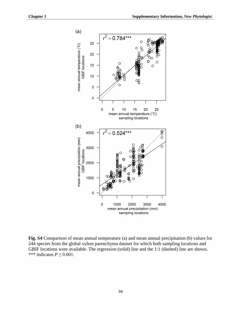
Chapter 1 Supplementary Information, New Phytologist
94
Fig. S4 Comparison of mean annual temperature (a) and mean annual precipitation (b) values for
244 species from the global xylem parenchyma dataset for which both sampling locations and
GBIF locations were available. The regression (solid) line and the 1:1 (dashed) line are shown.
*** indicates P ≤ 0.001.
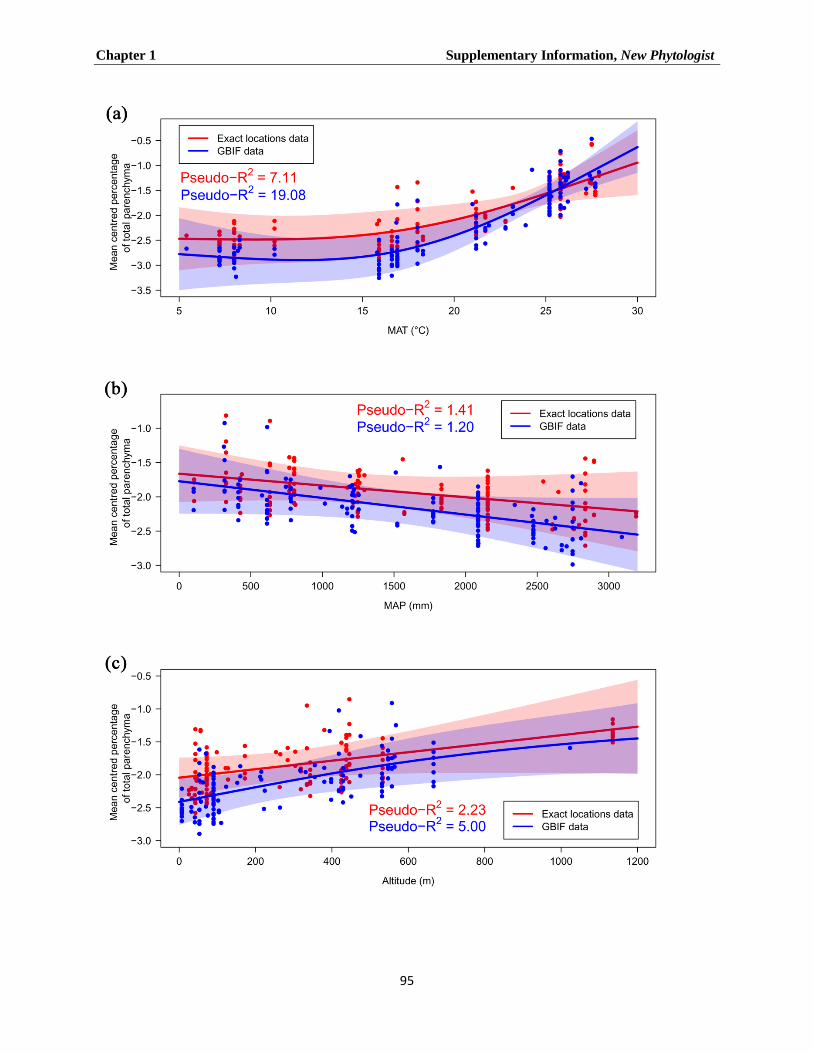
Chapter 1 Supplementary Information, New Phytologist
95

Chapter 1 Supplementary Information, New Phytologist
96
Fig. S5 The effect of MAT (a), MAP (b), and altitude (c) on the proportion of axial parenchyma
(AP) in angiosperm wood based on a general additive model (GAM) with a binomial distribution
for the exact location dataset (red) and the GBIF derived climate data (blue). Each climate
variable was limited to three partitions. The solid line represents the fitted smoother; 95%
confidence intervals are shown as coloured tinted areas. Each dot represents a specimen for
which the sampling location was reported in literature, or climate data were obtained from the
WorldClim database. Pseudo-R2 measures the approximate deviance explained by each
explanatory variable.
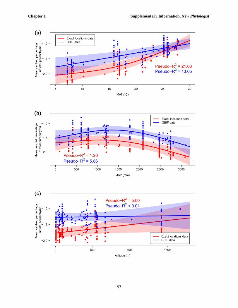
Chapter 1 Supplementary Information, New Phytologist
97

Chapter 1 Supplementary Information, New Phytologist
98
Fig. S6 The effect of MAT (a), MAP (b), and altitude (c) on the proportion of ray parenchyma
(RP) in angiosperm wood based on a general additive model with a binomial distribution for the
exact location dataset (red) and the GBIF derived climate data (blue). Each climate variable was
limited to three partitions. The solid line represents the fitted smoother; 95% confidence intervals
are shown as coloured tinted areas. Each dot represents a specimen for which the sampling
location was reported in literature, or climate data were obtained from the WorldClim database.
Pseudo-R2 measures the approximate deviance explained by each explanatory variable.
Table S1 The Global Wood Parenchyma Database, including 961 records, but excluding Zheng
and Martínez-Cabrera (2013). Data were extracted from 55 sources and include species name,
plant organ, growth form, and xylem tissue fractions (%) of ray parenchyma, axial parenchyma,
and total parenchyma. Specimens were grouped according to three major climatic zones, which
follow Köppen (1936).
Köppen W. 1936. Das geographische System der Klimate. In: Köppen W, Geiger R, eds.
Handbuch der Klimatologie. Berlin, Germany: Gebrüder Borntraeger, 1-44.
Zheng J, Martínez-Cabrera HI. 2013. Wood anatomical correlates with theoretical
conductivity and wood density across China: evolutionary evidence of the functional
differentiation of axial and radial parenchyma. Annals of Botany 112: 927-35.

Chapter 1 Supplementary Information New Phytologist
99
Table S2 Summary of statistics for the general additive models (GAM) fitted with a binomial
distribution for the exact locations dataset. The response variables were RAP, AP and RP. Each
explanatory variable was fitted with a smoother with a maximum of three effective degrees of
freedom (e.d.f). P-values are approximate (Wood, 2006). Pseudo-R2 measures the approximate
deviance explained by each explanatory variable.
Response
variable
Explanatory
variable
e.d.f F-value P-value Pseudo-
R2
RAP MAT 1.945 37.210 < 0.001 21.05%
(n = 270) MAP 1.000 5.219 0.023 1.6%
Altitude 1.000 12.933 < 0.001 3.6%
AP MAT 1.728 5.108 0.008 7.11%
(n = 137) MAP 1.000 1.471 0.227 1.41%
Altitude 1.000 2.946 0.088 2.23%
RP MAT 1.000 21.954 < 0.001 21.03%
(n = 182) MAP 1.876 4.964 0.008 1.2%
Altitude 1.000 3.584 0.060 5.4%
Wood SN. 2006. Generalized Additive Models: An Introduction with R. London, UK: Chapman
and Hall.

Chapter 1 Supplementary Information New Phytologist
100
Table S3 Summary of statistics for the general additive models (GAM) fitted with a binomial
distribution for the GBIF locations dataset. The response variables were RAP, AP and RP. Each
explanatory variable was fitted with a smoother with a maximum of three effective degrees of
freedom (e.d.f). P-values are approximate (Wood, 2006). Pseudo-R2 measures the approximate
deviance explained by each explanatory variable.
Response
variable
Explanatory
variable
e.d.f F-value P-value Pseudo-
R2
RAP MAT 1.950 48.234 <0.001 31.65%
(n = 221) MAP 1.000 8.848 0.003 2.8%
Altitude 1.000 10.433 0.001 3.1%
AP MAT 1.922 14.615 <0.001 19.05%
(n = 142) MAP 1.000 2.955 0.088 1.2%
Altitude 1.426 14.615 <0.001 5%
RP MAT 1.000 12.988 <0.001 13.05%
(n = 142) MAP 1.876 3.842 0.024 5.86%
Altitude 1.000 0.044 0.834 <0.01%
Wood SN. 2006. Generalized Additive Models: An Introduction with R. London, UK: Chapman
and Hall.
Notes S1 Published references from which data were extracted for analyses.

Chapter 1 Supplementary Information New Phytologist
100
References
Bauch J, Dunisch O. 2000. Comparison of growth dynamics and wood characteristics of
plantation-grown and primary forest Carapa guianensis in Central Amazonia. International
Association of Wood Anatomists Journal 21: 321-334.
Bauch J, Quiros L, Noldt G, Schmidt P. 2012. Study on the wood anatomy, annual wood
increment and intra-annual growth dynamics of Podocarpus oleifolius var. macrostachyus
from Costa Rica. Journal of Applied Botany and Food Quality 80: 19-24.
Bhat KM, Bhat KV, TK Dhamodaran TK, et al. 1985. Wood and bark properties of branches
of selected tree species growing in Kerala. KFRI Research Report, Kerala Forest Research
Institute 29: 1-34.
Bhat KM, Priya PB. 2004. Influence of provenance variation on wood properties of teak from
the Western Ghat region in India. International Association of Wood Anatomists Journal 25:
273-282.
Borja de la Rosa A, Machuca R, Fuentes Salinas M, Ayerde Lozada D, Fuentes López M,
Quintero Alcantar A. 2010. Caracterización tecnológica de la madera de Juniperus flaccida
var. Poblana Martínez. Revista Chapingo. Serie ciencias forestales y del ambiente 16: 261-
280.
Bravo S. 2010. Anatomical changes induced by fire-damaged cambium in two native tree
species of the Chaco region, Argentina. International Association of Wood Anatomists
Journal 31: 283-292.
Chapotin SM, Razanameharizaka JH, Holbrook NM. 2006. A biomechanical perspective on
the role of large stem volume and high water content in baobab trees (Adansonia spp.;
Bombacaceae). American Journal of Botany 93: 1251-1264.
Climent J, Gil L, Pardos JA. 1998. Xylem anatomical traits related to resinous heartwood
formation in Pinus canariensis Sm. Trees - Structure and Function 12: 139-145.

Chapter 1 Supplementary Information New Phytologist
101
da Silva LB, de Assis Ribeiro dos Santos F, Gasson P, Cutler D. 2011. Estudo comparativo da
madeira de Mimosa ophthalmocentra Mart. ex Benth e Mimosa tenuiflora (Willd.) Poir.
(Fabaceae-Mimosoideae) na caatinga nordestina1. Acta Botanica Brasilica 25: 301-314.
das Neves B, Fritz A, Barros CF. 2008. Anatomia do lenhoff de oito espécies de lianas da
familia Leguminosae ocorrentes na Floresta Atlântica. Acta Botanica Brasilica 22: 465-480.
Denne P, Gasson P. 2008. Ray structure in root-and stem-wood of Larix decidua: implications
for root identification and function. International Association of Wood Anatomists Journal
29: 17-23.
DeSmidt WJ. 1922. Studies of the distribution and volume of the wood rays in slippery elm
(Ulmus fulva Michx.). Journal of Forestry 20: 352-362.
Dias L, Luizon C, Gasson P. 2012. Anatomical comparison of original and regrowth wood from
coppiced and pollarded Poincianella pyramidalis trees in the caatinga of Pernambuco, Brazil.
International Association of Wood Anatomists Journal 33: 63-72.
Fichtler E, Worbes M. 2012. Wood anatomical variables in tropical trees and their relation to
site conditions and individual tree morphology. International Association of Wood
Anatomists Journal 33: 119.
French GE. 1923. The effect of the internal organization of the North American hardwoods
upon their more important mechanical properties. MSc Thesis, New York State College of
Forestry, Syracuse, New York.
Fujiwara S. 1992. Anatomy and properties of Japanese hardwoods II. Variation of dimensions
of ray cells and their relation to basic density. International Association of Wood Anatomists
Bulletin new series 13: 397–402.
Fujiwara S, Sameshima K, Kuroda K, Takamura N. 1991. Anatomy and properties of
Japanese hardwoods I. Variation of fibre dimensions and tissue proportions and their relation
to basic density. International Association of Wood Anatomists Bulletin new series 12: 419–
424.
Chapter 1 Supplementary Information New Phytologist
102
Gartner BL, Baker DC, Spicer R. 2000. Distribution and vitality of xylem rays in relation to
tree leaf area in Douglas-fir. International Association of Wood Anatomists Journal 21: 389-
401
Hearn DJ. 2009. Descriptive anatomy and evolutionary patterns of anatomical diversification in
Adenia (Passifloraceae). Aliso: A Journal of Systematic and Evolutionary Botany 27: 13–38.
Huber B, Prütz G. 1938. Über den Anteil von Fasern, Gefässen und Parenchym am Aufbau
verschiedener Hӧlzer. European Journal of Wood and Wood Products 1: 377–381.
Ishiguri F, Hiraiwa T, Iizuka K, Yokota S, Priadi D, Sumiasri N, Yoshizawa N, et al. 2009.
Radial variation of anatomical characteristics in Paraserianthes falcataria planted in
Indonesia. International Association of Wood Anatomists Journal 30: 343–352.
Knigge W, Schulz H. 1961. Einfluß der Jahreswitterung 1959 auf Zellartverteilung, Faserlänge
und Gefäßweite verschiedener Holzarten. Holz als Roh-und Werkstoff 19: 293–303.
Koch P. 1985. Utilization of hardwoods growing on southern pine sites. Asheville, NC: USDA-
Forest Service
Lei H, Gartner BL, Milota MR. 1997. Effect of growth rate on the anatomy, specific gravity,
and bending properties of wood from 7-year-old red alder (Alnus rubra). Canadian Journal
of Forest Research 27: 80–85.
Lei H, Milota MR, Gartner BL. 1996. Between-and within-tree variation in the anatomy and
specific gravity of wood in Oregon white oak (Quercus garryana Dougl.) International
Association of Wood Anatomists Journal 17: 445-461.
Maeglin RR. 1976. Natural variation of tissue proportions and vessel and fiber lengths in mature
northern red oak. Silvae Genetica 25: 122-126.
Mauseth J, Plemons-Rodriguez B. 1997. Presence of paratracheal water storage tissue does not
alter vessel characters in cactus wood. American Journal of Botany 84: 815–815.
Myer JE. 1922. Ray volumes of the commercial woods of the United States and their
significance. Journal of Forestry 20: 337–351.

Chapter 1 Supplementary Information New Phytologist
103
Ogunkunle ATJ, Oladele FA. 2008. Structural dimensions and paper making potentials of the
wood in some Nigerian species of Ficus L. (Moraceae). Advances in Natural & Applied
Sciences 2: 103-111.
Ogunwusi AA. 2012. Characterization of wood cellular structures of five lesser used wood
species growing in Nigeria. Journal of Natural Sciences Research 2: 128-134.
Olano JM, Arzac A, Gárcia-Cervigón AI, Arx G, Rozas V. 2013. New star on the stage:
amount of ray parenchyma in tree rings shows a link to climate. New Phytologist 198: 486–
495.
Ona T, Ito K, Shibata M, Ootake Y, Ohshima J, Yokota S, Yoshizawa N, Sonoda T. 1999.
In situ determination of proportion of cell types in wood by fourier transform raman
spectroscopy. Analytical Biochemistry 268: 43–48.
Ona T, Sonoda T, Ito K, Shibata M, Tamai Y, Kojima Y, Ohshima J, Yokota S, Yoshizawa
N. 2001. Investigation of relationships between cell and pulp properties in Eucalyptus by
examination of within-tree property variations. Wood Science and Technology 35: 229–243.
Pande PK, Chauhan L, Singh M. 2005. Wood anatomical variations within the genus
Castanopsis. Journal of Tropical Forest Science 17: 366-371.
Panshin AJ, de Zeeuw C. 1980. Textbook of wood technology. Structure, identification and uses
of the commercial woods of the United States and Canada. New York, USA: McGraw-Hill
Book Company.
Petric B, Scukanec V. 1975. Ray tissue percentages in wood of Yugoslavian hardwoods.
International Association of Wood Anatomists Bulletin 3: 43-44
Poorter L, McDonald I, Alarcón A, Fichtler E, Licona JC, Peña-Claros M, Sterck F,
Villegas Z, Sass-Klaassen U. 2010. The importance of wood traits and hydraulic
conductance for the performance and life history strategies of 42 rainforest tree species. New
Phytologist 185: 481–492.
Chapter 1 Supplementary Information New Phytologist
104
Pratt RB, Jacobsen AL, Ewers FW, Davis SD. 2007. Relationships among xylem transport,
biomechanics and storage in stems and roots of nine Rhamnaceae species of the California
chaparral. New Phytologist 174: 787–798.
Ruelle J, Clair B, Beauchêne J, Prévost MF, Fournier M, et al. 2006. Tension wood and
opposite wood in 21 tropical rain forest species. 2. Comparison of some anatomical and
ultrastructural criteria. International Association of Wood Anatomists Journal 27: 341–376.
Schulz H. 1957. Der Anteil der einzelnen Zellarten an dem Holz der Rotbuche. Holz als Roh-
und Werkstoff 15: 113-118.
Spicer R, Holbrook NM. 2007. Parenchyma cell respiration and survival in secondary xylem:
does metabolic activity decline with cell age? Plant, Cell & Environment 30: 934–943.
Sreevani P, Rao RV. 2014. Variation in basic density and tissue proportions of Eucalyptus
tereticornis SM. clones. Research Journal of Recent Sciences 4: 271-274.
Süß H, Müller-Stoll WR. 1972. Zusammenhänge zwischen der Ausbildung einiger
Holzmerkmale der Rotbuche (Fagus sylvatica L.) und der Jahrringbreite. Holz als Roh-und
Werkstoff 30: 342–346.
Taylor FW. 1968. Variations in the size and proportions of wood elements in yellow-poplar
trees. Wood Science and Technology 2: 153–165.
Van Geffen KG, Poorter L, Sass-Klaassen U, Van Logtestijn RSP, Cornelissen JHC. 2010.
The trait contribution to wood decomposition rates of 15 neotropical tree species. Ecology
91: 3686–3697.
Verdaguer D, Ojeda F. 2002. Root starch storage and allocation patterns in seeder and
resprouter seedlings of two Cape Erica (Ericaceae) species. American Journal of Botany 89:
1189–1196.
Villagra PE, Roig Juñent FA. 1997. Wood structure of Prosopis alpataco and P. argentina
growing under different edaphic conditions. International Association of Wood Anatomists
Journal 18: 37–52.

Chapter 1 Supplementary Information New Phytologist
105
von Frey-Wyssling A, Aeberli H. 1942. Der Anteil von Fasern, Gefäßen und Parenchym
verschiedener Holzarten in Dreiecksdarstellung. Holz als Roh-und Werkstoff 5: 265–268.
Wagenführ R. 2007. Holzatlas. Leipzig, Germany: Carl Hanser Verlag.
Woodrum, CL, Ewers FW, Telewski FW. 2003. Hydraulic, biomechanical, and anatomical
interactions of xylem from five species of Acer (Aceraceae). American Journal of Botany 90:
693–699.
Wu YQ, Hayashi K, Liu Y, Cai Y, Sugimori M. 2006. Relationships of anatomical
characteristics versus shrinkage and collapse properties in plantation-grown eucalypt wood
from China. Journal of Wood Science 52: 187–194.
Xi, E-H, Zhao G-J. 2011. Research on differentiated xylem cells based on fractal dimension.
BioResources 6: 3066-3079.
Yamada Y, Awano T, Fujita M, Takabe K. 2011. Living wood fibers act as large-capacity
‘single-use’ starch storage in black locust (Robinia pseudoacacia). Trees - Structure and
Function 25: 607–616.
Zheng J, Martínez-Cabrera HI. 2013. Wood anatomical correlates with theoretical
conductivity and wood density across China: evolutionary evidence of the functional
differentiation of axial and radial parenchyma. Annals of Botany 112: 927-35.
Ziemińska K, Wright IJ, Westoby M. 2015. Broad anatomical variation within a narrow wood
density range – a study of twig wood across 69 Australian angiosperms. PLOS ONE 10(4):
e0124892.
Chapter 2 Pavclová et al., 2016. American Journal of Botany
106
Part II
-
Publications
Chapter 2

Chapter 2 Pavclová et al., 2016. American Journal of Botany
107
The amount of parenchyma and living fibres affects storage of non-structural
carbohydrates in young stems and roots of temperate trees
Lenka Plavcová2*☨, Günter Hoch3, Hugh Morris2, Sara Ghiasi2 & Steven Jansen2
2Institute for Systematic Botany and Ecology, Ulm University, Albert-Einstein-Allee 11, D-89081
Ulm, Germany
3Department of Environmental Sciences - Botany, University of Basel, Schönbeinstrasse 6, CH-
4056 Basel, Switzerland
Published in: The American journal of Botany 103: 603-612.
(The original publication is available at www.amjbot.org/content/early/2016/03/18/ajb)

Chapter 2 Pavclová et al., 2016. American Journal of Botany
108
Abstract
Concentrations of non-structural carbohydrates (NSC) are used as proxies for the net carbon
balance of trees and as indicators of carbon starvation resulting from environmental stress.
Woody organs are the largest NSC-storing compartments in forest ecosystems; therefore, it is
essential to understand the factors that affect the size of this important storage pool. In wood,
NSC are predominantly deposited in ray and axial parenchyma (RAP), however, direct links
between nutrient storage and RAP anatomy have not yet been established. Here, we tested if the
NSC storage capacity of wood is influenced by the amount of RAP.
We measured NSC concentrations and RAP fractions in root and stem sapwood of twelve
temperate species sampled at the onset of winter dormancy and in stem sapwood of four tropical
trees growing in an evergreen lowland rainforest. The patterns of starch distribution were
visualised by staining with Lugol’s solution.
The concentration of NSC in sapwood of temperate trees scales tightly with the amount of RAP
and living fibers (LFs), with almost all RAP and LFs being densely packed with starch grains. In
contrast, the tropical species exhibited lower NSC concentrations despite their higher RAP and
LFs fraction and showed considerable interspecific differences in starch distribution.
The differences in RAP and LFs abundance affect the ability of sapwood to store NSC in
temperate trees, whereas a more diverse set of functions of RAP might be pronounced in species
growing in a tropical environment with little seasonality.
Key-words: axial parenchyma, carbohydrates, living fibres, rays, starch, storage, wood, xylem

Chapter 2 Pavclová et al., 2016. American Journal of Botany
109
Introduction
Non-structural carbohydrates (NSC, including mono-, di-, oligo-, and polysaccharides) are the
direct products of plant photosynthesis that can be stored in various plant tissues and used as
substrate for growth and various metabolic processes. The amount of NSC available in a plant is
considered to represent an important indicator of the net carbon balance in woody plants,
although the functional role and the active or passive regulation of NSC storage is not fully
understood (Kozlowski, 1992; Hoch, 2015). NSC levels have traditionally been studied with
respect to seasonal cycles of growth and dormancy (Drossopoulos and Niavis, 1988; Saranpää
and Hӧll, 1989; Sauter and van Cleve, 1994). More recently, they have been investigated in
relation to growth limitation (Kӧrner, 2003; Sala and Hoch, 2009; Wiley and Helliker, 2012),
altitudinal boundaries for forests (Hoch et al., 2002), plant survival under limited resource
conditions, increased atmospheric CO2 levels, and drought-induced tree mortality. Although
developmental shifts are likely between seedlings and mature trees, wood provides the largest
NSC-storing compartment among all tree tissues (Hoch et al., 2003; Würth et al., 2005), with an
additional role for parenchyma cells in the pith and bark (Essiamah and Eschrich, 1985).
In wood, NSC are predominantly deposited in ray and axial parenchyma (RAP). Ray
parenchyma (RP) represents a series of procumbent, square or upright cells produced by ray
initials that extend radially from the cambium. Besides storage, RP facilitates radial conduction
of NSC, water and other substances, and enables their exchange between phloem and xylem,
although the mechanisms of this trafficking remain poorly understood (Van Bel, 1990;
Sokolowska and Zagórska-Marek, 2012; Pfautsch et al., 2015). Axial parenchyma (AP) is made
up of axially elongated cells, which are often organized in strands. AP has been suggested to be
involved in NSC storage and transport, similar to RP (Braun and Wolkinger, 1970; Baas, 1982;
Braun, 1984; Carlquist, 2001). Abundant AP may not only increase the physical space available
for storage but could also enhance connectivity between RP and AP.
While NSC storage and dynamics appear tightly coupled to RAP anatomy and physiology
(reviewed in Plavcová and Jansen, 2015), direct links between structural and functional RAP
traits have not yet been established. Elucidating firm connections between structural and
functional traits has a long tradition in the field of functional and ecological wood anatomy.
While most research to date focuses on the functional anatomy of xylem conduits, providing us
Chapter 2 Pavclová et al., 2016. American Journal of Botany
110
with a better understanding of xylem water transport (Baas, 1982; Carlquist, 1984; Hacke and
Sperry, 2001; Jansen et al., 2009), the current study uses a similar approach to shed more light
on the role of RAP in NSC storage. More specifically, we test whether the amount of RAP found
in wood is a structural proxy for the NSC storage capacity of wood.
As most of the wood biomass consists of dead, protoplasm-free fibres and conduits, the total
volume of RAP should place a limitation on the amount of NSC that can be stored. The
proportion of RAP in wood, usually expressed as the percentage area of RAP relative to the total
wood cross-sectional area, varies greatly across species. In gymnosperms, the RAP area fractions
are commonly between 5 and 10% (Panshin and Zeeuw, 1980), while in angiosperms, the RAP
proportions are higher, typically between 20 and 50% (Martínez-Cabrera et al., 2009; Fichtler
and Worbes, 2012; Zheng and Martínez-Cabrera, 2013; Ziemińska et al., 2013). Fractions higher
than 80% have also been observed in some woody succulents and climbers (Mauseth and
Plemons-Rodriguez, 1997; Hearn, 2009). Interestingly, RAP fractions tend to be higher in
tropical than temperate angiosperm trees (Morris et al., 2015). This might be surprising at first
sight when considering that the storage function is likely to be less active in tropical
environments with a weak seasonality. However, RAP has multiple functions beyond storage of
NSC, such as storage of water (capacitance), embolism refilling, symplastic connectivity, and
various functions related to defense and resilience to disturbance (for an overview, see Fig. 2 in
Morris et al., 2015). Moreover, the storage capacity provided by RAP may not always be fully
utilized. For instance, it is well known that NSC content of wood fluctuates seasonally in both
temperate (Sauter and van Cleve, 1994; Barbaroux et al., 2003) and tropical environments
(Newell et al., 2002; Würth et al., 2005). Besides seasonal variation, there can be differences in
NSC distribution between vessel-associated RAP cells and the RAP cells that are not in direct
contact with vessels (Braun and Wolkinger, 1970; Czaninski, 1977; Braun, 1984).
In many species, living fibres (LFs) accumulate starch and hence contribute to NSC storage in
addition to RAP. LFs, defined as fibres that retain living protoplasts, are either septate or non-
septate, and vary interspecifically in their lumen diameter to wall thickness ratio (Fahn and
Leshem, 1963; Wolkinger, 1970; Carlquist, 2014). The occurrence of LFs is, however, poorly
documented in woody plants and their anatomical and functional distinction can be difficult to
make due to a structure-function continuum with AP (Carlquist, 2014). Storage function has
Chapter 2 Pavclová et al., 2016. American Journal of Botany
111
been suggested for septate fibres (Carlquist, 2001), which are twice as common in tropical as in
temperate trees (Wheeler et al., 2007). Similar to AP, septate fibres often have thin secondary
walls that distinguish them from thick-walled living and non-living fibres. In contrast, non-
septate LFs, which are found, for instance, in maples, often have thick secondary walls
(Carlquist, 2001, 2014). Providing that the thicker cell wall occurs at the expense of cell lumen
volume, it is reasonable to expect that thick-walled LFs are less efficient in storage than septate
fibres or AP.
The main goal of this study was to test if the amount of NSC stored in wood is linked with RAP
and LFs abundance, and to quantify the relative contribution of RP, AP and LFs to storage
function. To achieve this goal we measured NSC concentration in root and stem sapwood of 12
temperate species, including ten angiosperm trees, one conifer tree and one woody climber.
Samples were collected at the onset of winter dormancy when the NSC concentration is expected
to be at its maximum. We measured the area fractions of RP, AP and LFs and conducted in-situ
visualisation of starch. We expected that species with a relatively high amount of RAP, such as
pedunculate oak (Quercus robur) and black locust (Robinia pseudoacacia), would show higher
concentrations of NSC in wood than species with a low RAP fraction, for example in horse
chestnut (Aesculus hippocastanum) and Norway spruce (Picea abies). We also hypothesized that
more NSC would be stored in woody roots than stems and that this pattern will be paralleled by a
higher RAP abundance in the roots (Pate et al., 1990). To complement the data obtained for
temperate trees, measurements of NSC content, the presence of starch, together with RAP
fractions, were conducted in the stems of four tropical tree species growing in a lowland
rainforest in Costa Rica. We expected to find higher RAP proportions in tropical species (Morris
et al., 2015), although with NSC levels not necessarily as high as those of temperate species, as
the former are always actively growing, whereas the latter were sampled at the onset of winter
dormancy. Nevertheless, we anticipated a positive correlation between the amount of NSC and
the amount of RAP across the four tropical species.

Chapter 2 Pavclová et al., 2016. American Journal of Botany
112
Materials and Methods
Plant material
Young woody roots and stems of 12 temperate species (Table 1) were collected from mature
trees (more than 3 m tall) growing in a forested area surrounding the Ulm University campus,
which included the arboretum of the Botanical Garden of Ulm University (48° 25’ N, 9° 58’ E),
and a forest strip along the Iller river, 2 km south of Ulm (48° 22’ N, 9° 60’ E). These areas
experience a temperate climate with a mean annual temperature of 8.9 °C and mean annual
precipitation of 726 mm (climate-data.org). The roots were collected from a depth of 10-50 cm
and a distance of between 1 to 2 m from the root collar. The stem samples were removed from
the end regions of sub-canopy lateral branches cut at a height of 2 to 3 meters with the aid of a
telescopic pruning pole. All roots and stems were between 0.5 and 1.5 cm in diameter, which
corresponded to a root or stem age of 3 to 8 years. All samples consisted of juvenile sapwood
with no heartwood formation apparent on the cross-section after visual inspection. By avoiding
heartwood, we were sure that all RAP cells in our samples were living, and therefore capable of
storing NSC. During the last week of October 2014, six root and stem samples from three
separate individuals of each species were collected and prepared for NSC analyses. Samples for
wood anatomy and wood density measurements were collected from the same population of
trees, were of a similar size, and came from the same position on the tree as those used for the
NSC analyses. For logistical reasons, the sampling of material for anatomical and wood density
measurements was conducted ad hoc throughout the year 2014, which has no effect on the
structural parameters measured. Anatomical measurements were carried out on three different
root and stem samples per species, while wood density measurements were made on six different
root and stem samples collected from three individuals of each species.

Chapter 2 Pavclová et al., 2016. American Journal of Botany
113
Table 1: List of the species studied with reference to their family and acronym.
Species Family Acronym
temperate
Aesculus hippocastanum L. Sapindaceae Ah
Acer pseudoplatanus L. Sapindaceae Ap
Carpinus betulus L. Betulaceae Cb
Clematis vitalba L. Ranunculaceae Cv
Fraxinus excelsior L. Oleaceae Fe
Fagus sylvatica L. Fagaceae Fs
Liquidambar styraciflua L. Altingiaceae Ls
Prunus avium (L.) L. Rosaceae Pa
Quercus robur L. Fagaceae Qr
Robinia pseudoacacia L. Fabaceae Rp
Tilia cordata Mill. Malvaceae Tc
Picea abies (L.) H.Karst. Pinaceae Piab
tropical
Brosimum sp. Moraceae Bros
Ceiba pentandra (L.) Gaertn. Malvaceae Cepe
Pentaclethra macroloba (Willd.) Kuntze Fabaceae Pecl
Warszewiczia coccinea (Vahl) Klotzsch Rubiaceae Waco

Chapter 2 Plavcová et al., 2016. American Journal of Botany
114
Stem samples from four tropical species (Table 1) were collected in the vicinity of the Reserva
Biológica Tirimbina, Heredia Province, Costa Rica (10° 24’ N, 84° 6’ W) following the same
protocol as described for the temperate trees. The climate of Tirimbina is described as wet
tropical showing little seasonality, with a mean annual temperature of 25.3 °C and mean annual
precipitation of 3,777 mm (Lapinski and Tschapka, 2013). All samples of tropical trees were
collected on 12th September 2014.
NSC analyses
Root and stem samples prepared for NSC analyses (n=6 roots and 6 stems for each species)
were taken to the laboratory where they were then processed immediately or kept on ice and
processed later on the same day. In the laboratory, the root and stem samples were cut into 3-cm-
long segments and their bark and pith were carefully removed. The wood samples were heated in
a microwave oven at 600 W for 40 seconds to deactivate the NSC-modifying enzymes (Popp et
al., 1996). They were then dried to a constant weight at 80 °C for two days and sent to the Plant
Ecophysiology Laboratory at the University of Basel for NSC content analyses. NSC, defined
here as the sum of starch together with the three most abundant low molecular weight sugars
(i.e., sucrose, glucose and fructose), were analysed photometrically following a slightly modified
protocol described in Hoch et al. (2002). While various protocols can be used for NSC
concentration measurements, the method of Hoch et al. (2002) is well-established and broadly
agrees with other methods based on a recent comparison among 29 different laboratories
(Quentin et al. 2015). Approximately 10 mg of plant powder was extracted with 2 ml distilled
water at 100 °C for 30 min. An aliquot of the extract was used for the determination of low
molecular carbohydrates after enzymatic conversion of fructose and sucrose to glucose. The
concentration of total free glucose was then determined photometrically after enzymatic
conversion of glucose to gluconat-6-phosphate using a glucose hexokinase (GHK) assay reagent
(G3292, Sigma-Aldrich, St. Louis, MO, USA). Following the degradation of starch to glucose
with amyloglucosidase at 49 °C overnight, NSC was determined in a separate analysis. The
concentration of starch was calculated as NSC minus the free low molecular carbohydrates.
Tissue concentrations were given as % dry matter.
The gravimetrically based concentrations (in mg g-1) were also converted to volume based values
(in mg cm-3) using data on wood density. Wood density was measured by the water displacement

Chapter 2 Plavcová et al., 2016. American Journal of Botany
115
method. Root or stem segments about 3 cm in length (n=6 roots and 6 stems per species) were
cut and debarked. Stem segments were additionally split in half and the pith was removed. Each
segment was then immersed, with the aid of a dissecting needle, into a water-filled beaker resting
on an electronic balance, where upon the weight of the displaced water was recorded. All
segments were then oven-dried at 80 °C to a constant weight and wood density was calculated by
dividing the wood dry weight by the volume of the water displaced.
Wood anatomy and starch distribution
Root and stem segments (n=3 roots and 3 stems for each species) collected in the field were cut
into 1-cm-long pieces and stored in 70% ethanol at 4 °C until sectioned for anatomical
observations. Upon rehydration in distilled water, transverse sections about 40 µm thick were
prepared with a sliding microtome. The sections were immersed in a mixture of
0.35% safranin and 0.65% alcian blue for 2 min, thoroughly washed with distilled water, then
gradually dehydrated through an ethanol series before being mounted on a slide in Neo-Mount
(Merck Millipore, Germany). Digital images were captured at a 150x magnification using an
Axio Zoom V16 microscope (Carl Zeiss Microscopy GmbH, Jena, Germany). The relative
proportions of ray and axial parenchyma (RP + AP = RAP) and living fibres (LFs) were
measured on wedge-shaped transects spanning from the root or stem center to the cambium.
Axially oriented thick-walled cells with apparent cellular content were typically considered LFs,
while axial cells with a protoplasm and a thin secondary wall were always identified as AP. To
aid with distinguishing between LFs and thick-walled AP, radial sections of wood were also
inspected to assess if the axial cells have a spindle-shaped morphology typical for fibres or are
rectangular and arranged in strands as typical for AP. The different cell types were outlined
manually with the aid of a graphical tablet (Cintiq Companion, Wacom Co Ltd, Japan) in
Photoshop Elements (v12.1, Adobe Systems Incorporated, San Jose, CA, USA) and their area
proportions were measured in Fiji/ImageJ (Schindelin et al., 2012). To obtain the relative
proportions of RAP and LFs, the total surface area taken up by each cell type was divided by the
total surface area of the entire transect measured, which typically was about 1 mm2.
Chapter 2 Plavcová et al., 2016. American Journal of Botany
116
To visualise the distribution of starch, wood sections were incubated in a drop of 5% Lugol’s
iodine for 2 min. The samples were then briefly washed in distilled water, mounted in glycerol
and observed immediately with the Axio Zoom V16 microscope.

Chapter 2 Plavcová et al., 2016. American Journal of Botany
117
Table 2: Fixed effects estimates (means ± SE) for organ (root and stem) and climate (temperate and tropical) on concentrations of
total non-structural carbohydrates (NSC), starch and soluble sugars and on the amount of ray and axial parenchyma (RP + AP = RAP)
and living fibres (LFs). Statistical significance was tested by comparing full and reduced models using the likelihood ratio test. Test
statistics (L), degrees of freedom (df) and corresponding P values are shown. Statistically significant results (P ≤ 0.05) are indicated in
bold. To corroborate the results of the likelihood ratio tests, ΔAIC values were calculated, which represent the difference in Akaike
information criteria values between the reduced and full model.
Organ
(stem relative to root)
Climate
(tropical relative to temperate)
mean ± SE L df P value ΔAIC mean ± SE L df P value ΔAIC
NSC -10.5 ± 2.2 13.1 1 <10-3 11.1 -6.3 ± 1.7 10 1 0.002 4
starch -9.8 ± 2.1 12.2 1 <10-3 10.2 -6.7 ± 1.7 10.5 1 0.001 4.9
sugars -0.7 ± 0.2 12 1 <10-3 10 0.4 ± 0.4 0.7 1 0.391 -1.2
RAP + LFs -11.1 ± 1.8 17.2 1 0.007 15.2 9.5 ± 6.7 2 1 0.158 0.3
RAP -8.9 ± 1.8 13.8 1 <10-3 11.8 12.9 ± 5.2 5.4 1 0.020 3.8
RP -5 ± 0.9 24.9 1 <10-3 22.9 3.8 ± 2.5 2.2 1 0.136 2
AP -3.9 ± 1.9 3.6 1 0.058 1.6 9.1 ± 5.6 2.6 1 0.110 0.6

Chapter 2 Plavcová et al., 2016. American Journal of Botany
118
Statistical analyses
Mean differences in NSC and RAP between organs (roots and stems) and between climate
(temperate or tropical) were tested using linear mixed effects models, where organ and
climate represented the fixed factors and species were treated as a random factor. The models
were constructed in R using the lme function available in the nlme package (Pinheiro et al.
2015). The maximal likelihood method was used to fit the models. Statistical significance of
fixed factors was assessed using the likelihood ratio tests by comparing the full models to
reduced models from which the fixed factors were eliminated. The validity of the models was
checked by visual inspection of residual plots. The Akaike information criteria for the full
and reduced models were also calculated to confirm the appropriateness of the model
selection. Relationships between RAP + LFs and NSC were analysed by ordinary least
squares regression on species- and organ-level means. All analyses were conducted in R (R
Core Team 2013). Results were considered statistically significant at P ≤ 0.05.
Results
NSC concentrations
The concentrations of NSC and their two main fractions (i.e. starch and soluble sugars) were
significantly higher in roots than stems (Table 2, Fig. 1A). Overall, the highest NSC
concentration of 39.1 ± 2.7% (mean ± SD) was measured in the roots of Acer
pseudoplatanus, while the lowest concentration (0.9 ± 0.1%) was observed in the stems of
Picea abies. In all species and organs, NSC were present mainly in the form of starch,
whereas the soluble sugar fraction was less abundant, typically falling between 1 and 3%.
The NSC and starch concentrations were not significantly linearly correlated between roots
and stems of the same species, largely because of the relatively low NSC and starch
concentrations in Robinia pseudoacacia roots (Fig. 2A). In contrast, there was a significant
correlation between the concentrations of soluble sugars in both organs, which was
principally driven by the high soluble sugar fraction found in Tilia cordata (Fig. 2B).
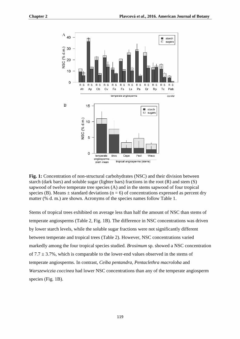
Chapter 2 Plavcová et al., 2016. American Journal of Botany
119
Fig. 1: Concentration of non-structural carbohydrates (NSC) and their division between
starch (dark bars) and soluble sugar (lighter bars) fractions in the root (R) and stem (S)
sapwood of twelve temperate tree species (A) and in the stems sapwood of four tropical
species (B). Means ± standard deviations (n = 6) of concentrations expressed as percent dry
matter (% d. m.) are shown. Acronyms of the species names follow Table 1.
Stems of tropical trees exhibited on average less than half the amount of NSC than stems of
temperate angiosperms (Table 2, Fig. 1B). The difference in NSC concentrations was driven
by lower starch levels, while the soluble sugar fractions were not significantly different
between temperate and tropical trees (Table 2). However, NSC concentrations varied
markedly among the four tropical species studied. Brosimum sp. showed a NSC concentration
of 7.7 ± 3.7%, which is comparable to the lower-end values observed in the stems of
temperate angiosperms. In contrast, Ceiba pentandra, Pentaclethra macroloba and
Warszewiczia coccinea had lower NSC concentrations than any of the temperate angiosperm
species (Fig. 1B).

Chapter 2 Plavcová et al., 2016. American Journal of Botany
120
Fig. 2: Correlation between root and stem concentrations of starch (A) and soluble sugars (B)
in twelve temperate species. Means ± standard deviations (n = 6) are shown. Abbreviations of
the species names are listed in Table 1. ns = non-significant relationship (P > 0.05), ** =
significant relationship with P ≥ 0.01. Acronyms of the species names follow Table 1.
Reference lines showing a theoretical 1:1 relationship are plotted as dashed lines. Solid line
represents the linear regression to the data.
RAP and LFs anatomy
RP was present in all species investigated with a mean area fraction of 15.9 ± 5.4%. AP was
found in all specimens (mean area fraction 10.0 ± 9.0%) except in the stems of Picea abies
and Warszewiczia coccinea. Non-septate LFs occurred in Clematis vitalba and Acer
pseudoplatanus, while septate LFs were found in Warszewiczia coccinea. In all three species,
sparse simple pitting was observed on the walls of LFs. Overall, roots of temperate species
exhibited significantly higher fractions of storage cells (i.e. RAP + LFs) than stems (34.9 ±

Chapter 2 Plavcová et al., 2016. American Journal of Botany
121
14.8% vs. 23.8 ± 11.9%, Table 2) with the exception of Robinia pseudoacacia and Quercus
robur where the amount of RAP was comparable between roots and stems, averaging around
33% (Fig. 3A). In most species, both types of wood parenchyma (i.e., RP and AP) were
higher in roots than in stems (Table 2). However, two ring-porous species, Quercus robur
and Robinia pseudoacacia, exhibited lower AP area fractions in roots compared to stems, and
Clematis vitalba and Fraxinus excelsior showed an increase in AP only (Fig. 3A). RP
represented the dominant fraction of the total RAP, with the contribution of AP being
approximately one-third of the total RAP. On average, the relative contribution of AP to the
total RAP was about 5% higher in roots than stems. The presence of LFs was particularly
noticeable in Clematis vitalba, resulting in very high LFs area fractions of 49.1% and 34.4%
in roots and stems, respectively, and virtually all fibres were alive and filled with starch (Fig.
4A, E).
The amount of living cells (RAP + LFs) tended to be higher in tropical than in temperate
trees (means 34.9 vs. 25.4%, Table 2), although the difference was not statistically
significant, presumably due to the high LFs fraction in Clematis vitalba. Nevertheless, the
four tropical species showed significantly higher fractions of RAP than those measured in the
stems of temperate angiosperms (mean difference ± SE: 12.9 ± 5.2, Table 2). Ceiba
pentandra had the highest amount of RAP reported in this study. The RAP area fraction for
the latter reached 56 ± 8.6% and was driven mainly by a high AP content (AP fraction of 38.4
± 5.3%; Fig. 3B). In contrast, Warszewiczia coccinea exhibited a particularly high RP
fraction of 25.9 ± 1.8% (Fig. 3B). This value is almost twice as high as the mean RP fraction
of all other stems measured in this study.
Starch localisation
Staining with Lugol’s solution was used to visualise the starch distribution in RP, AP and
LFs (Fig. 4). In temperate species, almost all RAP + LFs cells were filled with starch that
completely occupied the cell lumen with tightly packed grains (Fig. 4 A-C, E-G). Tilia
cordata and Picea abies (Fig. 4 D, H) were two exceptions to this pattern, with starch grains
being packed more loosely. In most species, starch grains were present in both vessel-
associated RAP cells and the cells that were not in contact with vessels, although a tendency
for lower starch accumulation in vessel-associated RAP cells was sometimes apparent
(Appendix S1, see Supplemental Data with online version of this article). In the case of
Clematis vitalba, a positive staining reaction was found in the lumina of all fibres, leaving
vessels and tracheids as the only cell types void of starch (Fig. 4A, E, Appendix S1). Besides

Chapter 2 Plavcová et al., 2016. American Journal of Botany
122
RAP and LFs, starch grains were also abundant in parenchyma cells of the perimedullary
region (i.e. the region surrounding the pith) and in some species (Quercus robur, Carpinus
betulus and Liquidambar styraciflua) were directly within the pith. In these species, the
amount of starch found in the pith tended to decline as the branch age increased. In roots, the
central region with primary xylem appeared sometimes enriched in starch (e.g. Carpinus
betulus, Aesculus hippocastanum). In Picea abies, starch was also present in the epithelial
cells associated with the resin ducts (Fig. 4D). While starch was generally less abundant in
tropical (Fig. 4I-L) than temperate trees (Fig. 4E-H), interspecific differences in starch
distribution were observed among the tropical species. In Ceiba pentandra starch grains were
present predominantly in RP (Fig. 4I), which was in contrast to Pentaclethra macroloba
where they were mostly present in the AP (Fig. 5K). Starch grains were also present in the
LFs of Warszewiczia coccinea (Fig. 4L), while in Brosimum sp. they appeared abundant in
both RP and AP, similar to that of temperate species (Fig. 4J).

Chapter 2 Plavcová et al., 2016. American Journal of Botany
123
Fig. 3: Percent area (% a.) of ray and axial parenchyma (RP + AP = RAP) and living fibres
(LFs) in root (R) and stem (S) wood of twelve temperate tree species (A) and in stem wood of
four tropical species (B). Means ± standard deviations (n = 3) are shown. Acronyms of the
species names follow Table 1.
Relationship between NSC and RAP
There was a strong positive correlation (P < 0.001, r2 = 0.64) between the NSC concentration
and the RAP + LFs fraction in temperate species when the data for Clematis vitalba were
excluded (Fig. 5). The contrasting relationship between NSC and RAP + LFs in C. vitalba
compared to the temperate trees was likely due to the lower storage capacity of thick-walled
LFs that were particularly prominent in C. vitalba (Fig. 5C). The relationship between NSC
and RAP + LFs was significant with r2 > 0.5 when either the data for roots and stems were
analysed together or when analysed separately (Fig. 5A). This relationship became even

Chapter 2 Plavcová et al., 2016. American Journal of Botany
124
stronger when the NSC concentrations were expressed per unit wood volume rather than on a
gravimetric basis, i.e., when the differences in wood density between species and organs were
taken into account (Fig. 5B). In contrast to temperate trees, NSC concentrations in the stems
of the four tropical species were independent of the respective RAP + LFs content (Fig. 5C,
white squares).

Chapter 2 Plavcová et al., 2016. American Journal of Botany
125
Discussion
In this study, we showed that the differences in NSC concentrations observed in the young
roots and stems of temperate species at the onset of winter dormancy were strongly related to
the abundance of RAP (Fig. 5), with LFs contributing to storage in Clematis vitalba and Acer
pseudoplatanus. Thus, our data provide evidence that the amount of RAP can serve as a
suitable proxy for the capacity of wood to store NSC. In this regard, our results expand on the
list of apparent structure-function relationships identified in xylem, which help us to better
understand the vast diversity in wood anatomical traits that evolved in plants.
Fig. 4: Transverse sections of roots (A-D) and stems (E-L) stained with Lugol’s solution
showing the spatial distribution of starch in wood (stained black). Four temperate species,
Clematis vitalba (A, E), Acer pseudoplatanus (B, F), Aesculus hippocastanum (C, G) and
Picea abies (D, H), and four tropical species, Ceiba pentandra (I), Brosimum sp. (J),
Pentaclethra macroloba (K) and Warszewiczia coccinea (L) are shown. Starch was present in
ray and axial parenchyma of all species, although the staining intensity varied from high (A,
B, E, F, J) to low (D, G, H, I, K, L). In some species, starch was also present in living fibres
(A, B, E, F, L). Scale bars = 500µm.

Chapter 2 Plavcová et al., 2016. American Journal of Botany
126
The visual assessment of starch distribution revealed that virtually all RAP and LFs of
temperate trees were filled with starch (Fig. 4A-H), indicating that the entire space available
for storage in wood was utilized at the onset of winter dormancy. Contrary to earlier
observations (Braun and Wolkinger, 1970; Braun, 1984), there was no clear distinction in
starch accumulation between vessel-associated RAP cells and the RAP cells that are not in
direct contact with vessels (Appendix S1). Our results also showed that both types of
interconnected wood parenchyma, i.e. ray (RP) and axial parenchyma (AP) as a whole,
contribute greatly to NSC storage. While RP represented the dominant part of the total RAP
in most of the temperate species investigated in this study, abundant AP resulted in a
substantial increase in the total RAP of some species, for example in Quercus robur and
Robinia pseudoacacia. AP fractions, often accounting for more than 40% of the total RAP,
were higher in the roots than in the stems of the temperate species (Fig. 3A). It is possible
that an increased need for mechanical support requires a strong fiber matrix that may limit the
occurrence of thin-walled AP in self-supporting stems.
Alternatively, thick-walled LFs could take on the storage function without compromising
wood mechanical strength (Carlquist, 2014, 2015). Among temperate species included in this
study, Clematis vitalba showed particularly high (up to 50% of the total cross-sectional area)
proportions of LFs in both roots and stems (Fig. 3A, Fig. 4 A, E, Appendix S1). Considering
its high RAP + LFs fraction, the NSC levels measured in Clematis vitalba were relatively low
(Fig. 5C). The lower NSC storage efficiency (defined as the NSC concentration per unit RAP
+ LFs area) in this species might be attributed to a lower ratio between cell lumen and cell
wall fractions in storage tissue composed of LFs instead of AP. On the other hand, the storage
capacity of LFs in Acer pseudoplatanus appeared to be in line with that of the other temperate
species relying solely on parenchyma (Fig. 5). Whether or not LFs may provide a storage
potential comparable to AP should be tested for a larger number of species. There is evidently
a continuum in the dimensions and wall-to-lumen ratios of AP and LFs across species
(Carlquist, 2014) and the anatomical variability of these cells, including their pit dimensions
(especially pit size and density), should be more thoroughly documented in order to better
understand its influence on the storage and mechanical properties of wood. Investments in
thicker cell walls together with the occurrence of less extensive pitting in LFs, thus resulting
in a lower storage capacity and lower accessibility and mobility of stored nutrients, might be
factors that favor the occurrence of the typically thin-walled AP.

Chapter 2 Plavcová et al., 2016. American Journal of Botany
127
Due to the higher RAP fraction, roots were able to store more NSC than stems (Fig. 1A,
Table 2), with the difference being about 2.5-fold on average and 6.5-fold at maximum. This
finding highlights the important contribution of below-ground biomass to the overall NSC
storage pool. The NSC concentrations of roots and stems of the same species were only
partially correlated (Fig. 2), showing that the division of NSC reserves between below- and
above-ground woody biomass is variable across species. The variation observed might relate
to species- and organ-specific differences in phenology and subsequent growth dynamics.
While little is known about the coordination between root and shoot phenology (Steinaker et
al., 2009), large intraspecific variation in the timing and rates of root growth has recently
been observed in a dozen temperate trees (McCormack et al., 2014). It is possible that the
availability of NSC reserves could play a major role in driving these differences in root
growth.

Chapter 2 Plavcová et al., 2016. American Journal of Botany
128
Fig 5: Relationship between the amount of wood parenchyma (RAP) and living fibres (LFs)
and the concentration of non-structural carbohydrates (NSC) in root (black circles) and stem
(grey circles) sapwood of temperate trees (A, B). Sapwood NSC concentrations were
expressed on a gravimetric (A) or a volumetric (B) basis. Means ± standard deviations (n=3-
6) are shown. Linear regression lines through all (solid black line), root (dashed dark grey
line) and stem (dotted light grey line) data and the corresponding regression coefficients (r2)
are indicated. *, **, or *** denotes that the linear relationship was significant at P ≤ 0.05,
0.01 and 0.001, respectively. Acronyms of the species names follow Table 1. The same data
as in (A) are presented in (C) together with the data for the temperate liana, Clematis vitalba
(Cv) and four tropical species (white squares). Both Clematis vitalba and the tropical species
are excluded from the correlation because they show lower NSC concentrations than the
temperate trees in spite of their high amount of RAP and LFs.

Chapter 2 Plavcová et al., 2016. American Journal of Botany
129
In this study, we focused on NSC storage in young woody organs that consist of juvenile
sapwood located in close proximity to the apical meristems. Mature wood in tree trunks also
represents important sites of NSC storage and it can be assumed that the NSC stored in trunks
provide the primary source of energy and material for the resumption of cambial activity at
the beginning of the growing season (Hill et al., 1995). In tree trunks, not only the RAP
content determines the total NSC storage potential, but so does the amount of sapwood, as the
death of RAP cells associated with heartwood formation makes these cells unavailable for
NSC storage (Magel et al., 1994). Mature wood from tree trunks may also show quantitative
differences in RAP as compared to juvenile wood in branches, although no significant
differences were found in AP, RP, and RAP between trunks and branches of 34 tree species
(Morris et al., 2015).
Compared to temperate trees, the NSC levels of the four tropical species included in this
study were significantly lower (Fig. 1B) in spite of their on average higher RAP + LFs
content (Fig. 3B). This finding is not surprising considering that the wood tissue of the
tropical trees used for NSC analyses was collected during active growth in the case of
tropical trees, whereas the samples of the temperate trees were taken at the onset of winter
dormancy. In fact, the tropical trees studied here never experience dormancy in their native
habitat and thus may not require large reserves for the resumption of growth.
The finding that NSC and RAP data were not correlated in the tropical trees (Fig. 5C) may
indicate that, in some cases, RAP plays a less important role in NSC accumulation, but might
have various alternative functions. Interestingly, the abundant AP in Ceiba pentandra was
essentially void of starch grains (Fig. 4I), and the soluble sugar fraction was not noticeably
higher either (Fig. 1B). This suggests that the accumulation of NSC is not the primary
function of AP in C. pentandra. It is possible that AP serves as an important water-storage
reservoir in this fast-growing tropical pioneer that often colonizes forest gaps and copes with
increased evaporative demands. In support of this idea, the role of RAP in stem water storage
has already been proposed (Borchert and Pockman, 2005) and a high sapwood hydraulic
capacitance was recently measured in another representative of the same genus (C. speciosa;
Carrasco et al., 2015). Besides water storage, RAP is also known to play a part in defense
against pathogens (Schwarze, 2007), which is a function that might be particularly important
in diverse tropical ecosystems (Bagchi et al., 2014). An additional explanation for the higher
levels of RAP in tropical compared to temperate species may be that the tropical trees do not
need to protect their living cells from cold-induced damage. Since protection against cold is

Chapter 2 Plavcová et al., 2016. American Journal of Botany
130
energetically costly, it may limit the RAP abundance in environments where frost frequently
occurs (Morris et al., 2015). However, more research across a broader selection of tropical
species is needed to establish firm links between NSC accumulation, functional traits of RAP
and plant ecological strategies.
Conclusion
In conclusion, the results of this study demonstrate that the amount of RAP and LFs is
strongly related to the NSC storage capacity in the wood of temperate-deciduous trees.
Because virtually all RAP and LFs of temperate trees were filled with starch, the values of
NSC concentrations measured here at the onset of winter dormancy provide estimates of the
seasonal maxima. In contrast to temperate trees, a more diverse set of functions of RAP
might be inherent in species growing in a tropical environment with little or no seasonality.
Future research should move beyond a static approach of studying NSC and focus on the
functional questions related to NSC transport, conversion and use (Richardson et al., 2013,
2015; Klein and Hoch, 2015). It can be expected that these processes are tightly coupled to
RAP connectivity and the activity of various carbohydrate-modifying enzymes and
transporters within the RAP cells (Sauter et al., 1973; Decourteix et al., 2006, 2008).
Acknowledgements
The authors thank the Botanical garden of Ulm University and the Tirimbina field station in
Costa Rica for access to plant material. The authors also wish to thank Jan Plavec and Hana
Plavcová for their help with the field work and Gaby Wiest-Danner and Mohammad
Khatamirad for technical assistance in the lab. L.P. was supported by a Postdoctoral
Fellowship from the Alexander von Humboldt Foundation and acknowledges funding from
the Ulm University and Ulm University Society (Ulmer Universitätsgesellschaft). H.M. and
S.J. acknowledge financial support from the German Research Foundation (DFG, project
number
Chapter 2 Plavcová et al., 2016. American Journal of Botany
131
References
Baas P. 1982. Systematic, phylogenetic, and ecological wood anatomy—history and
perspectives. In: New Perspectives in Wood Anatomy. Dordrecht, The Netherlands:
Springer, 23-58.
Bagchi R, Gallery RE, Gripenberg S, Gurr SJ, Narayan L, Addis CE, Freckleton RP,
Lewis OT. 2014. Pathogens and insect herbivores drive rainforest plant diversity and
composition. Nature 506: 85-88.
Barbaroux C, Bréda N, Dufrêne E. 2003. Distribution of above-ground and below-ground
carbohydrate reserves in adult trees of two contrasting broad-leaved species (Quercus
petraea and Fagus sylvatica). New Phytologist 157: 605-615.
Van Bel AJ. 1990. Xylem-phloem exchange via the rays: the undervalued route of transport.
Journal of Experimental Botany 41: 631-644.
Borchert R, Pockman WT. 2005. Water storage capacitance and xylem tension in isolated
branches of temperate and tropical trees. Tree Physiology 25: 457-466.
Braun H. 1984. The significance of the accessory tissues of the hydrosystem for osmotic
water shifting as the second principle of water ascent, with some thoughts concerning the
evolution of trees. International Association of Wood Anatomists Bulletin 5: 275-294.
Braun H, Wolkinger F. 1970. Zur funktionellen Anatomie des axialen Holzparenchyms und
Vorschläge zur Reform seiner Terminologie. Holzforschung-International Journal of the
Biology, Chemistry, Physics and Technology of Wood 24: 19–26.
Carlquist S. 2001. Comparative wood anatomy: systematic, ecological, and evolutionary
aspects of dicotyledon wood. Springer Science & Business Media.
Carlquist S. 2014. Fibre dimorphism: cell type diversification as an evolutionary strategy in
angiosperm woods. Botanical Journal of the Linnean Society 174: 44-67.
Carlquist S. 2015. Living cells in wood. 1. Absence, scarcity and histology of axial
parenchyma as keys to function. Botanical Journal of the Linnean Society 177: 291-321.

Chapter 2 Plavcová et al., 2016. American Journal of Botany
132
Carlquist S. 1984. Vessel grouping in dicotyledon wood: significance and relationship to
imperforate tracheary elements. Aliso: A Journal of Systematic and Evolutionary Botany
10: 505-525.
Carrasco LO, Bucci SJ, Di Francescantonio D, Lezcano OA, Campanello PI, Scholz FG,
Rodríguez S, et al. 2015. Water storage dynamics in the main stem of subtropical tree
species differing in wood density, growth rate and life history traits. Tree Physiology 35:
354-365.
Czaninski, Y. 1977. Vessel-associated cells. International Association of Wood Anatomists
Bulletin 3: 51–55.
Decourteix M, Alves G, Bonhomme M, Peuch M, Baaziz KB, Brunel B, Guilliot A,
Rageau R, Améglio T, et al. 2008. Sucrose (JrSUT1) and hexose (JrHT1 and JrHT2)
transporters in walnut xylem parenchyma cells: their potential role in early events of
growth resumption. Tree Physiology 28: 215-224.
Decourteix M, Alves G, Brunel N, Améglio T, Guilliot A, Lemoine L, Petel P, Sakr S.
2006. JrSUT1, a putative xylem sucrose transporter, could mediate sucrose influx into
xylem parenchyma cells and be up-regulated by freeze-thaw cycles over the autumn-
winter period in walnut tree (Juglans regia L.). Plant, Cell & Environment 29: 36-47.
Drossopoulos J, Niavis C. 1988. Seasonal changes of the metabolites in the leaves, bark and
xylem tissues of olive tree (Olea europaea L.) II. Carbohydrates. Annals of Botany 62:
321-327.
Essiamah S, Eschrich W. 1985. Changes of starch content in the storage tissues of
deciduous trees during winter and spring. International Association of Wood Anatomists
Bulletin 6: 97-106.
Fahn A, Leshem B. 1963. Wood fibres with living protoplasts. New Phytologist 62: 91-98.
Fichtler E, Worbes W. 2012. Wood anatomical variables in tropical trees and their relation
to site conditions and individual tree morphology. International Association of Wood
Anatomists Journal 33: 119-140.
Hacke UG, Sperry JS. 2001. Functional and ecological xylem anatomy. Perspectives in
Plant Ecology, Evolution and Systematics 4: 97-115.

Chapter 2 Plavcová et al., 2016. American Journal of Botany
133
Hearn DJ. 2009. Descriptive anatomy and evolutionary patterns of anatomical
diversification in Adenia (Passifloraceae). Aliso: A Journal of Systematic and
Evolutionary Botany 27: 13–38.
Hill S, Waterhouse J, Field E, Switsur V, Ap Rees T. 1995. Rapid recycling of triose
phosphates in oak stem tissue. Plant, Cell & Environment 18: 931-936.
Hoch G. 2015. Carbon reserves as indicators for carbon limitation in trees. In Progress in
Botany, 321–346. Springer.
Hoch G, Popp M, Kӧrner C. 2002. Altitudinal increase of mobile carbon pools in Pinus
cembra suggests sink limitation of growth at the Swiss treeline. Oikos 98: 361-374.
Hoch G, Richter A, Kӧrner C. 2003. Non-structural carbon compounds in temperate forest
trees. Plant, Cell & Environment 26: 1067-1081.
Jansen S, Choat B, Pletsers A. 2009. Morphological variation of intervessel pit membranes
and implications to xylem function in angiosperms. American Journal of Botany 96: 409-
419.
Klein T, Hoch G. 2015. Tree carbon allocation dynamics determined using a carbon mass
balance approach. New Phytologist 205: 147-159.
Kozlowski T. 1992. Carbohydrate sources and sinks in woody plants. The Botanical Review
58: 107-222.
Kӧrner C. 2003. Carbon limitation in trees. Journal of Ecology 91: 4–17.
Lapinski W, Tschapka M. 2013. Habitat use in an assemblage of Central American
wandering spiders. The Journal of Arachnology 41: 151-159.
Magel E, Jay-Allemand C, Ziegler H. 1994. Formation of heartwood substances in the
stemwood of Robinia pseudoacacia L. II. Distribution of nonstructural carbohydrates and
wood extractives across the trunk. Trees 8: 165-171.
Martínez-Cabrera HI, Jones CS, Espino S, Schenk HJ. 2009. Wood anatomy and wood
density in shrubs: responses to varying aridity along transcontinental transects. American
Journal of Botany 96: 1388-1398.

Chapter 2 Plavcová et al., 2016. American Journal of Botany
134
Mauseth J, Plemons-Rodriguez B. 1997. Presence of paratracheal water storage tissue does
not alter vessel characters in cactus wood. American Journal of Botany 84: 815-815.
McCormack ML, Adams TS, Smithwick EA, Eissenstat DM. 2014. Variability in root
production, phenology, and turnover rate among 12 temperate tree species. Ecology 95:
2224-2235.
Morris H, Plavcová L, Cvecko P, Fichtler E, Gillingham MAF, Martínez-Cabrera HI,
McGlinn D, Wheeler E, Zheng J, Ziemińska K, Jansen S. 2016. A global analysis of
parenchyma tissue fractions in secondary xylem of seed plants. New Phytologist 206:
1553-1565.
Newell EA, Mulkey SS, Wright JS. 2002. Seasonal patterns of carbohydrate storage in four
tropical tree species. Oecologia 131: 333-342.
Panshin AJ, de Zeeuw C. 1980. Textbook of wood technology: structure, identification,
properties and uses of the commercial woods of the United States and Canada. McGraw-
Hill Book Co.
Pate JS, Froend RH, Bowen BJ, Hansen A, Kuo J. 1990. Seedling growth and storage
characteristics of seeder and resprouter species of Mediterranean-type ecosystems of SW
Australia. Annals of Botany 65: 585-601.
Pfautsch, S, Renard J, Tjoelker M, Salih A. 2015. Phloem as capacitor: radial transfer of
water into xylem of tree stems seems to occur via symplastic transport in ray parenchyma.
Plant Physiology 167: 963-971.
Pinheiro J, Bates D, Debroy S, Sarkar D, R Core Team. 2015. nlme: Linear and nonlinear
mixed effects models. R package version 3.1-119, http://CRAN.R-
project.org/package=nlme.
Plavcová L, Jansen S. 2015. The role of xylem parenchyma in the storage and utilization of
non-structural carbohydrates. In: Hacke UG, eds. Functional and Ecological Xylem
Anatomy. Springer International Publishing. 209–234.
Popp M, Lied W, Meyer AJ, Richter A, Schiller S, Schwitte H. 1996. Sample preservation
for determination of organic compounds: microwave versus freeze-drying. Journal of
Experimental Botany 47: 1469-1473.

Chapter 2 Plavcová et al., 2016. American Journal of Botany
135
Quentin AG, Pinkard EA, Ryan MG, Tissue DT, Baggett LS, Adams HD, Maillard P, et
al. 2015. Non-structural carbohydrates in woody plants compared among laboratories.
Tree Physiology 35: 1146-1165.
R Core Team. 2013. R: A language and environment for statistical computing. R Foundation
for Statistical Computing, Vienna, Austria. http://www.R-project.org/.
Richardson AD, Carbone MS, Keenan TF, Czimczik CI, Hollinger DY, Murakami P,
Schaberg PG, Xu X. 2013. Seasonal dynamics and age of stemwood nonstructural
carbohydrates in temperate forest trees. New Phytologist 197: 850-861.
Richardson AD, Carbone MS, Huggett BA, Furze ME, Czimczik CI, Walker JC, Xu X,
et al. 2015. Distribution and mixing of old and new nonstructural carbon in two temperate
trees. New Phytologist 206: 590-597.
Sala A, Hoch G. 2009. Height-related growth declines in ponderosa pine are not due to
carbon limitation. Plant, Cell & Environment 32: 22-30.
Saranpää, P., and W. Hӧll. 1989. Soluble carbohydrates of Pinus sylvestris L. sapwood and
heartwood. Trees 3: 138-143.
Sauter JJ, van Cleve B. 1994. Storage, mobilization and interrelations of starch, sugars,
protein and fat in the ray storage tissue of poplar trees. Trees 8: 297-304.
Sauter JJ, Iten W, Zimmermann MH. 1973. Studies on the release of sugar into the vessels
of sugar maple (Acer saccharum). Canadian Journal of Botany 51: 1-8.
Schindelin J, Arganda-Carreras I, Frise E, Kaynig V, Longair M, Pietzsch T, Preibisch
S, et al. 2012. Fiji: an open-source platform for biological-image analysis. Nature
Methods 9: 676-682.
Schwarze FW. 2007. Wood decay under the microscope. Fungal Biology Reviews 21: 133-
170.
Sokolowska K, Zagórska-Marek B. 2012. Symplasmic, long-distance transport in xylem
and cambial regions in branches of Acer pseudoplatanus (Aceraceae) and Populus
tremula × P. tremuloides (Salicaceae). American Journal of Botany 99: 1745-1755.

Chapter 2 Plavcová et al., 2016. American Journal of Botany
136
Steinaker DF, Wilson SD, Peltzer Da. 2009. Asynchronicity in root and shoot phenology in
grasses and woody plants. Global Change Biology 16: 2241-2251.
Wheeler E, Baas P, Rodgers S. 2007. Variations in dicot wood anatomy: a global analysis
based on the InsideWood database. International Association of wood Anatomists Journal
28: 229-258.
Wiley E, Helliker B. 2012. A re-evaluation of carbon storage in trees lends greater support
for carbon limitation to growth. New Phytologist 195: 285-289.
Wolkinger F. 1970. Das Vorkommen lebender Holzfasern in Sträuchern und Bäumen.
Phyton 14: 55-67.
Würth MK, Pelaez-Riedl S, Wright SJ, Kӧrner C. 2005. Non-structural carbohydrate
pools in a tropical forest. Oecologia 143: 11-24.
Zheng J, Martínez-Cabrera HI. 2013. Wood anatomical correlates with theoretical
conductivity and wood density across China: evolutionary evidence of the functional
differentiation of axial and radial parenchyma. Annals of Botany 112: 927-935.
Ziemińska K, Butler DW, Gleason SM, Wright IJ, Westoby M. 2013. Fibre wall and
lumen fractions drive wood density variation across 24 Australian angiosperms. AoB
Plants 5: plt046.
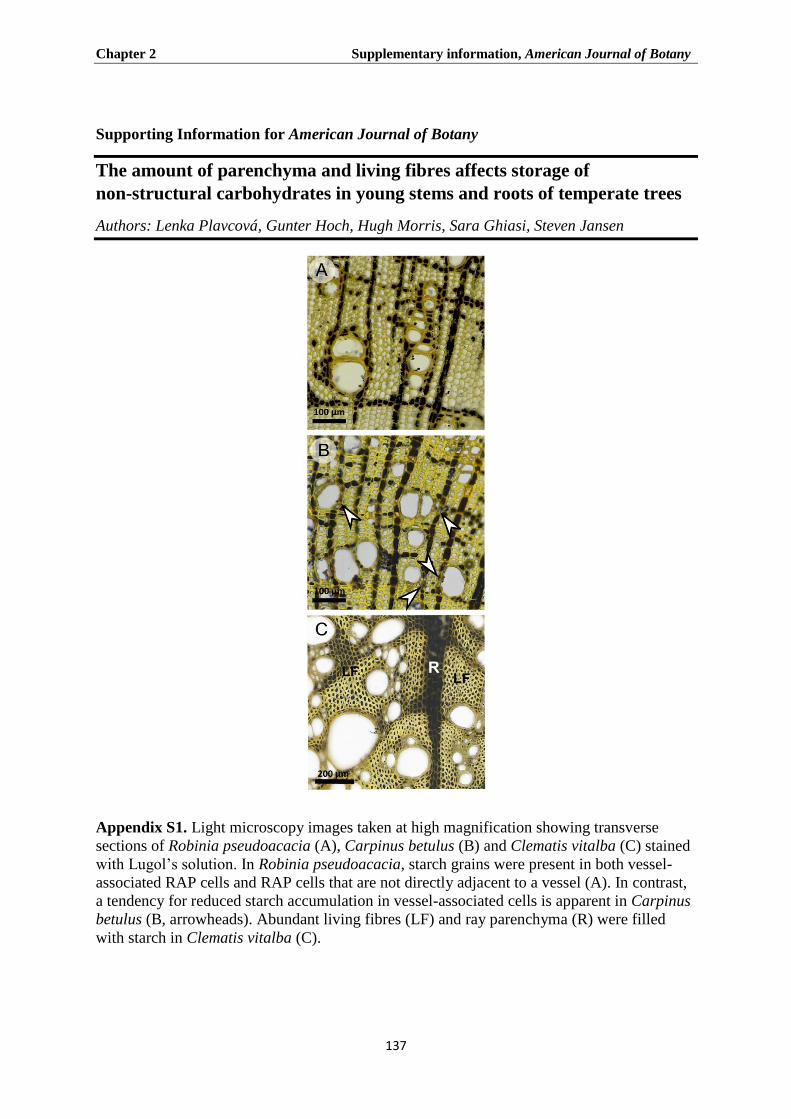
Chapter 2 Supplementary information, American Journal of Botany
137
Supporting Information for American Journal of Botany
The amount of parenchyma and living fibres affects storage of
non-structural carbohydrates in young stems and roots of temperate trees
Authors: Lenka Plavcová, Gunter Hoch, Hugh Morris, Sara Ghiasi, Steven Jansen
Appendix S1. Light microscopy images taken at high magnification showing transverse
sections of Robinia pseudoacacia (A), Carpinus betulus (B) and Clematis vitalba (C) stained
with Lugol’s solution. In Robinia pseudoacacia, starch grains were present in both vessel-
associated RAP cells and RAP cells that are not directly adjacent to a vessel (A). In contrast,
a tendency for reduced starch accumulation in vessel-associated cells is apparent in Carpinus
betulus (B, arrowheads). Abundant living fibres (LF) and ray parenchyma (R) were filled
with starch in Clematis vitalba (C).

Chapter 3 Morris and Jansen, 2016. International Association of Wood Anatomists Journal
138
Part II
-
Publications
Chapter 3

Chapter 3 Morris and Jansen, 2016. International Association of Wood Anatomists Journal
139
Opinion Paper
Secondary xylem parenchyma – from classical terminology to functional
traits
Hugh Morris1, Steven Jansen1
1Institute of Systematic Botany and Ecology, Ulm University, Albert-Einstein-Allee 11, D-
89081 Ulm, Germany
Published in: The International Association of Wood Anatomists Journal 37: 1-15.
(The original publication is available at
http://booksandjournals.brillonline.com/content/journals)

Chapter 3 Morris and Jansen, 2016. International Association of Wood Anatomists Journal
140
Introduction
Terminology plays a crucial role in describing and understanding the morphological and
anatomical variation in living organisms. The huge diversity in plant species and their many
morphological and physiological adaptations to a wide range of ecosystems is reflected in an
enormous anatomical variation of woody tissues. This is perhaps somewhat surprising given
that angiosperm wood consists of essentially three cell types only: imperforate tracheary
elements (fibres and tracheids), vessel elements, and living parenchyma cells. However,
variation in the dimensions and the arrangement of these cells provide a challenge to anyone
who aims to describe and understand quantitative and qualitative differences between wood
samples. The challenge lies not only in the consistent application and interpretation of terms
(Lens et al., 2012), but also in how we deal with a dynamic continuum (i.e., fuzzy
morphology sensu Agnes Arber and Rolf Sattler) that includes intergradations, intermediate
forms, analogous and homologous features (Sattler and Rutishauser, 1997).
While the International Association of Wood Anatomy (IAWA) lists (IAWA Committee
1933, 1964, 1989, 2004) have been successful for identification and classification of
angiosperm and gymnosperm wood, there is a lack of an anatomical glossary that goes
beyond identification, covering the broad fields related to wood anatomy such as functional
and ecological xylem anatomy, evolutionary and developmental wood anatomy,
dendrochronology, etc. It is clear that achieving such a general glossary will never be perfect
and will require a collaborative effort from many experts in various wood-related disciplines.
This opinion paper attempts to provide a critical review of terminology for wood
parenchyma. It is especially concerned with the overlap between the descriptive terms used in
systematic wood anatomical treatments and terms with functional implications. Most
terminology for ray and axial parenchyma (RAP) has been defined based on the microscopy
of transverse and longitudinal sections for wood identification purposes. A number of terms,
especially for axial parenchyma, have changed in their usage or gone out of fashion; some of
these are discussed here.
Parenchyma in secondary xylem
Parenchyma tissue in the secondary xylem of woody plants represents the living symplastic
tissue that is intermeshed with the apoplast (the dead tissues, which include the fibres [except
living, septate ones] and conductive elements). It plays a crucial but perhaps underrated role
in plant physiology. The interest in the subject from a functional perspective has been
variable, with a surge of interest during the mid to late 20th century (Braun, 1964, 1984;

Chapter 3 Morris and Jansen, 2016. International Association of Wood Anatomists Journal
141
Braun and Wolkinger, 1970; Czaninski, 1977; Sauter and Kloth, 1986) followed by a decline
and a resurgence of late (Salleo et al. 2004, Martinez-Cabrera et al., 2009, Zheng and
Martinez-Cabrera, 2013; Ziemińska et al., 2013, 2015, Spicer, 2014; Morris et al., 2015).
This renewed interest in ray and axial parenchyma (RAP) coincides with advances in image
analysis and measurement techniques. Up to this point, a far greater focus has been on the
dead conducting cells of wood (vessel elements and tracheids) and the mechanically
important fibres of angiospermous wood (Carlquist, 2015). As more papers report on wood, it
is important to address the terminology used for RAP, as there is a tendency by those outside
the discipline of wood anatomy to misuse the terms and provide incorrect information. An
overview of the terms related to axial and ray parenchyma are presented in Table 1 and 2.
Functions of RAP
The terminology for describing RAP is important because disciplines outside of traditional
wood anatomy are now paying more attention to their function e.g., plant physiology,
functional ecology, plant-ecophysiology, plant pathology, and plant evolutionary-biology.
The functions of RAP include: (1) storage and transport of non-structural carbohydrates
(NSCs) (e.g., Hoch et al. 2003; Salleo et al., 2004; O’ Brien et al., 2014; Plavcová and
Jansen, 2015), (2) defence against pathogens (e.g., Shigo, 1984; Biggs, 1987; Schmitt and
Liese, 1993; Deflorio et al., 2008), (3) water storage and capacitance (e.g. Holbrook, 1995;
Borchert and Pockman, 2005; Pfautsch et al., 2015), (4) storage of minerals, such as calcium
oxalate (Trockenbrodt, 1995), (5) the transition of sapwood into heartwood (Pinto et al.,
2004; Spicer, 2005; Nawrot et al., 2008), and (6) biomechanical contributions, particularly by
RP (e.g. Mattheck and Kubler, 1995; Burgert and Eckstein, 2001; Reiterer et al. 2002).
Contact and isolation cells of the ray system
Secondary xylem parenchyma are living cells and are composed of two major cell types, ray
parenchyma (RP) and axial parenchyma (AP), which are oriented perpendicular to each other.
RP is aligned centripetally and formed from the ray initials of the cambium; AP is aligned
axially and formed from the fusiform initials of the cambium (Metcalfe and Chalk, 1983).
The cells in wide rays of angiosperms have been further categorised into two sub-types,
‘contact’ cells and ‘isolation’ cells, terms used by various authors to describe differing
functions between the two (Braun, 1964, 1984; Braun and Wolkinger, 1970; Sauter and
Kloth, 1986, Murikami et al., 1999; Spicer, 2014). Contact cells are in direct association
with non-living conducting cells and have the ability to produce tyloses or gums. Isolation
cells, which do not make any contact with conductive tissue, might be more specialised for

Chapter 3 Morris and Jansen, 2016. International Association of Wood Anatomists Journal
142
radial transport (Sauter and Kloth, 1986). Apart from their function, they also differ in form.
Isolation cells are in many cases procumbent, and often have ray cells along their side, which
are frequently ‘upright cells’. The ‘upright’ cells, or ‘erect’ ray cells, have the same
orientation as the axial parenchyma, and may perform the same function as diffuse axial
parenchyma in forming contacts between conduits and parenchyma, or simply contact
between other parenchyma only (Höll, 1975; Carlquist, 2001). Conifers, on the other hand,
have predominantly uniseriate rays, and therefore have contact cells only in their ray
structure, where all ray parenchyma cells have some contact with tracheids.

Chapter 3 Morris and Jansen, 2016. International Association of Wood Anatomists Journal
143
Table 1. Anatomical descriptions for terms related to axial parenchyma (AP), along with the definition and source(s) of the term.
Anatomical (descriptive)
terms for AP
Definition/comment Sources for terms
Apotracheal: where AP is
distant or does not appear
to be in contact with a
vessel
Subcategories
Diffuse AP scattered randomly, often sparse (e.g. Carpinus spp, Crataegus
spp., Cornus mas).
IAWA Committee,
1933
Diffuse-in-
aggregates
AP grouped in short tangential lines. No more than 2 cells wide (e.g.
Tilia spp.).
Kribs 1937
Reticulate This is a term for very regular networks of parenchyma bands and
multiseriate rays as seen in transverse section.
Record, 1944
IAWA Committee
1989
Banded AP From one to many cells wide; up to three cells wide – narrow banded,
more than three – wide banded. This refers to apotracheal banded AP,
as you can also have banded AP that is paratracheal.
Kribs, 1937
Concentric - Many bands. Term no longer in use. Jane, 1956

Chapter 3 Morris and Jansen, 2016. International Association of Wood Anatomists Journal
144
Scalariform - Successive fine lines or bands arranged horizontally or in
arcs. Important point is that the distance between the rays is greater
than the distance between bands (e.g. in Annonaceae), and that the rays
are wider than the parenchyma bands.
Wagenführ 1961;
IAWA Committee,
1989
Marginal AP Bands of AP at a growth ring boundary. These are mainly apotracheal,
though can be paratracheal.
Jane, 1934; Hess,
1950
Initial - rarer. Occurs at the beginning of a growth ring (examples:
Cedrela odorata. Tectona grandis).
Terminal - most common type. Occurs at the end of a growth ring (e.g.
Magnolia spp.).
Ray-adjacent
AP
Diffuse AP clustered along ray margins (e.g. Staphylea spp.). Hess, 1950;
Carlquist &
Hoekman, 1985
Paratracheal: where AP is
in association with a vessel
AP Scanty
Paratracheal
A term agreed upon by the IAWA committee (1964) as preferred to
vasicentric scanty (Kribs, 1937). An incomplete sheath of AP
surrounding a vessel or occasional AP cells touching the vessel (e.g.,
Fraxinus spp., Laurus nobilis).
IAWA Committee,
1964, 1989

Chapter 3 Morris and Jansen, 2016. International Association of Wood Anatomists Journal
145
AP Vasicentric Where paratracheal parenchyma form a complete oval or circular
sheath around a vessel or vessel group.
Jane, 1934; IAWA
Committee, 1964,
1989.
AP Aliform Paratracheal AP with lateral extensions, often wing-like, forming a
diamond shaped outline referred to as lozenge-aliform. It can either
completely surround the vessel or can be to one side (e.g., Inga spp.
and Brosimum spp.). Both winged-aliform and lozenge-aliform were
designated as sub-types by the IAWA Committee, 1989.
Kribs, 1937; IAWA
Committee, 1989
AP confluent Where vasicentric or aliform arragements coalesce. It can completely
surround or form to one side of a vessel or vessel group, forming
irregular tangential or diagonal bands (e.g., Parkia pendula,
Chlorophora tinctoria). A special case is banded-confluent, where
many vessels are united by tangentially confluent parenchyma.
Kribs, 1937;
Metcalfe & Chalk,
1950; IAWA
Committee, 1964
AP absent/extremely rare AP absent or extremely rare intergrades with both apotracheal (diffuse)
or scanty paratracheal. In order to make sure that the AP is absent or
extremely rare it is necessary to view longitudinal as well as transverse
sections (e.g. Salix, Populus, Scottellia). Septate fibres are often
present where AP is rare or absent (Wheeler et al. 2007).
IAWA Committee,
1989

Chapter 3 Morris and Jansen, 2016. International Association of Wood Anatomists Journal
146
Table 2. Functional descriptions for terms related to ray and axial parenchyma (RAP), along with the definition and source(s) of the term.
Functional
terms for RAP
Definition/comment Sources for
terms
Subcategories
Accessory tissues Parenchymatous tissues in association with the conductive system (vessels and
tracheids) via pit membranes (half-bordered). Through these close pit contacts a
functional unity exists between the accessory tissues and the conductive system.
Concerns both angiosperms and gymnosperms.
Braun, 1984
Vessel-associated
cells
Specialised RAP that are in contact with the vessels, as per term. Czaninski (1977)
coined the term in preference to contact cells; however, both are used
interchangeably today.
Czaninski,
1977
Paratracheal
contact
Concerns only AP in actual contact with the vessels. Concerns angiosperms only.
Braun (1984) coined this term as a means of specifically referring to angiosperms,
where the term paratracheidal was coined in reference to only conifers. Not in use
today.
Braun, 1970,
1984
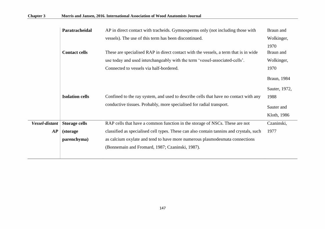
Chapter 3 Morris and Jansen, 2016. International Association of Wood Anatomists Journal
147
Paratracheidal AP in direct contact with tracheids. Gymnosperms only (not including those with
vessels). The use of this term has been discontinued.
Braun and
Wolkinger,
1970
Contact cells These are specialised RAP in direct contact with the vessels, a term that is in wide
use today and used interchangeably with the term ‘vessel-associated-cells’.
Connected to vessels via half-bordered.
Braun and
Wolkinger,
1970
Braun, 1984
Sauter, 1972,
1988
Sauter and
Kloth, 1986
Marakami et
al. 1999
Isolation cells Confined to the ray system, and used to describe cells that have no contact with any
conductive tissues. Probably, more specialised for radial transport.
Vessel-distant
AP
Storage cells
(storage
parenchyma)
RAP cells that have a common function in the storage of NSCs. These are not
classified as specialised cell types. These can also contain tannins and crystals, such
as calcium oxylate and tend to have more numerous plasmodesmata connections
(Bonnemain and Fromard, 1987; Czaninski, 1987).
Czaninski,
1977
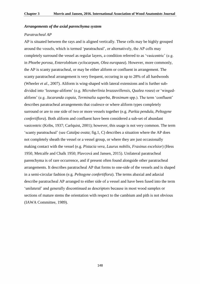
Chapter 3 Morris and Jansen, 2016. International Association of Wood Anatomists Journal
148
Arrangements of the axial parenchyma system
Paratracheal AP
AP is situated between the rays and is aligned vertically. These cells may be highly grouped
around the vessels, which is termed ‘paratracheal’, or alternatively, the AP cells may
completely surround the vessel as regular layers, a condition referred to as ‘vasicentric’ (e.g.
in Phoebe porosa, Enterolobium cyclocarpum, Olea europaea). However, more commonly,
the AP is scanty paratracheal, or may be either aliform or confluent in arrangement. The
scanty paratracheal arrangement is very frequent, occuring in up to 28% of all hardwoods
(Wheeler et al., 2007). Aliform is wing-shaped with lateral extensions and is further sub-
divided into ‘lozenge-aliform’ (e.g. Microberlinia brazzavillensis, Qualea rosea) or ‘winged-
aliform’ (e.g. Jacaranda copaia, Terminalia superba, Brosimum spp.). The term ‘confluent’
describes paratracheal arrangements that coalesce or where aliform types completely
surround or are to one side of two or more vessels together (e.g. Parkia pendula, Peltogyne
confertiflora). Both aliform and confluent have been considered a sub-set of abundant
vasicentric (Kribs, 1937; Carlquist, 2001); however, this usage is not very common. The term
‘scanty paratracheal’ (see Catalpa ovata; fig.1, C) describes a situation where the AP does
not completely sheath the vessel or a vessel group, or where they are just occasionally
making contact with the vessel (e.g. Pistacia vera, Laurus nobilis, Fraxinus excelsior) (Hess
1950, Metcalfe and Chalk 1950; Plavcová and Jansen, 2015). Unilateral paratracheal
parenchyma is of rare occurrence, and if present often found alongside other paratracheal
arrangements. It describes paratracheal AP that forms to one-side of the vessels and is shaped
in a semi-circular fashion (e.g. Peltogyne confertiflora). The terms abaxial and adaxial
describe paratracheal AP arranged to either side of a vessel and have been fused into the term
‘unilateral’ and generally discontinued as descriptors because in most wood samples or
sections of mature stems the orientation with respect to the cambium and pith is not obvious
(IAWA Committee, 1989).
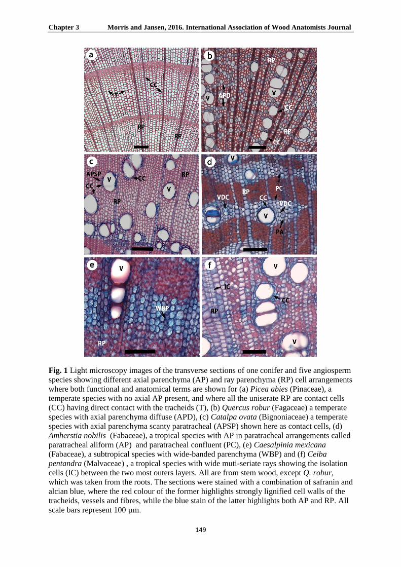
Chapter 3 Morris and Jansen, 2016. International Association of Wood Anatomists Journal
149
Fig. 1 Light microscopy images of the transverse sections of one conifer and five angiosperm
species showing different axial parenchyma (AP) and ray parenchyma (RP) cell arrangements
where both functional and anatomical terms are shown for (a) Picea abies (Pinaceae), a
temperate species with no axial AP present, and where all the uniserate RP are contact cells
(CC) having direct contact with the tracheids (T), (b) Quercus robur (Fagaceae) a temperate
species with axial parenchyma diffuse (APD), (c) Catalpa ovata (Bignoniaceae) a temperate
species with axial parenchyma scanty paratracheal (APSP) shown here as contact cells, (d)
Amherstia nobilis (Fabaceae), a tropical species with AP in paratracheal arrangements called
paratracheal aliform (AP) and paratracheal confluent (PC), (e) Caesalpinia mexicana
(Fabaceae), a subtropical species with wide-banded parenchyma (WBP) and (f) Ceiba
pentandra (Malvaceae) , a tropical species with wide muti-seriate rays showing the isolation
cells (IC) between the two most outers layers. All are from stem wood, except Q. robur,
which was taken from the roots. The sections were stained with a combination of safranin and
alcian blue, where the red colour of the former highlights strongly lignified cell walls of the
tracheids, vessels and fibres, while the blue stain of the latter highlights both AP and RP. All
scale bars represent 100 µm.
Chapter 3 Morris and Jansen. 2016. International Association of Wood Anatomists Journal
150
There is a complication between the commonly used anatomical definition of paratracheal
(Metcalfe and Chalk, 1983) and Braun’s (1984) functional definition. From a functional point
of view, it is essential to have such a term that describes AP in immediate contact with
vessels rather than also including vessel-distant AP that is part of a complete paratracheal
arrangement. Braun termed vessel-distant paratracheal parenchyma ‘interfibrous
parenchyma’, and devised the more specific term paratracheal-contact cells for the
“accessory tissue” of his “hydrosystem”, in analogy with the contact cells of the rays. The use
of the term ‘interfibrous parenchyma’ was unfortunate, as it encompassed an array of
arrangements that are in themselves quite distinct
Terms can be essential for the identification of wood; however, from a functional perspective,
many of the terms are less valuable because of a morphological continuum. For instance, the
difference between a unilateral and a lozenge-aliform arrangement may be functionally
negligible, as the only separation between the two is that the latter has a diamond-shaped
outline and is present around the whole vessel, an important difference for wood
identification purposes. However, the broader terms, such as apotracheal, paratracheal and
even scanty paratracheal are certainly important terms for functional studies, as these
arrangements have an important association with the vessel in relation to capacitance and
defence (Brodersen et al., 2010; Plavcová and Jansen, 2015; Morris et al. 2015). For instance,
the lack of such AP arrangements in most conifer species may explain why they require
narrow, “safe” tracheids as conductive tissue (Choat et al. 2012).
Apotracheal AP
The term ‘apotracheal’ describes AP cells that are not associated or contiguous with vessels
in transverse section. Some random contacts may exist, although these are infrequent (Evert,
2006). Diffuse, diffuse-in-aggregates, and marginal AP essentially describes three
subcategories of AP that are not in association with vessels. The descriptive terms for AP are
when the AP appears distributed in transverse sections. For instance, in the case of diffuse
there are a number of sources that describe AP as being isolated and irregularly dispersed,
which is true when viewed as a single transverse section. However, from a functional point of
view, this is not the case, as such cells cannot be isolated from surrounding symplastic tissue
because all parenchyma cells are to some degree interconnected with each other
(Zimmermann and Tomlinson, 1966; Zimmermann, 1971). All diffuse parenchyma types are
networking with both the vessels and the ray system. This was demonstrated by Zimmerman

Chapter 3 Morris and Jansen. 2016. International Association of Wood Anatomists Journal
151
(1971) who showed that an isolated group of AP will sooner or later contact other AP and/or
RP.
Alternatively, AP might act as a transport bridge between rays, thus by-passing the vessels
altogether, as is the case with the AP pattern diffuse-in-aggregates or the narrow bands of
axial parenchyma. The latter term ‘diffuse-in-aggregates’, describes where they form
tangential lines, forming no more than one cell wide between rays, although in transverse
section one may or may not see diffuse-in-aggregates contacting rays (Metcalfe and Chalk,
1950; Carlquist, 2001). Examples of diffuse-in-aggregates include Tilia spp. (Malvaceae) and
Dalbergia stevensonii (Fabaceae). Diffuse AP are more commonly associated with temperate
species. In tropical tree species where AP is absent or rare, there is a higher incidence of
septate fibres (Wheeler et al., 2007). Here, septate fibres are presumably taking over the
functional role of AP, where examples include: Acharaceae, Araliaceae, Burseraceae and
Salicaceae. Both Araliaceae and Salicaceae are found in temperate and tropical biomes.
Kalopanax septemlobus (Araliaceae) is an excellent example of a temperate species with
absent or extremely rare AP, but with an abundance of septate fibres taking over the
functional role.
The term ‘banded’ applies to AP that is one to many cells wide, and this is further subdivided
into narrow-banded and wide-banded apotracheal AP (see Caesalpinia Mexicana; fig.1, E).
Although banded arrangements are mostly apotracheal, they can be paratracheal, where they
are clearly associated with the vessels. Under the IAWA Committee (1989) it forms a
separate group of arrangements that includes the arbitrary, overlapping sub-categories (1)
axial parenchyma bands more than three cells wide, (2) axial parenchyma in narrow bands or
lines up to three cells wide, (3) axial parenchyma reticulate, (4) axial parenchyma scalariform
and (5) axial parenchyma in marginal or in apparently marginal bands. Wide bands (more
than three cells wide) are much more common in tropical trees, perhaps acting as a dynamic
barrier against the inward advance of decay, thus replacing the growth ring of temperate
species, a static barrier where highly lignified fibres slow the spread of decay; this is Wall 2
of the CODIT model, an acronym for Compartmentalisation of Decay in Trees (Shigo, 1984).
Although wide banded AP are more common in the trees of the tropics than in temperate
regions (e.g. Meliaceae and Moraceae), they still have a rare overall occurrence with only a
9% of the worlds’ woods having this characteristic (Wheeler et al. 2007). A quick check of
834 species with wide bands (InsideWood); Wheeler et al., 2007) showed a much higher
incidence of wide parenchyma bands with growth rings absent or indistinct (748 species out

Chapter 3 Morris and Jansen. 2016. International Association of Wood Anatomists Journal
152
of the total) indicating that in the absence of growth rings, parenchyma may be taking on a
defensive role. When the wide bands run via vessels, they intergrade with confluent AP, a
pattern under the umbrella of paratracheal. A term was in use to describe this as ‘banded
confluent’ (Jane, 1970), which acknowledged that bands are not always circumferentially
continuous, but can be as a result of there being a considerable amount of confluent
parenchyma that form together to appear as bands. There are, of course, gradations in the
continuity of the bands and they can appear independent of vessels or extend from vessel to
vessel where then the distribution of the bands appears related to the position of the vessel.
Marginal AP, coined by Hess (1950) is banded, but laid out at the beginning or at the end of a
growth ring, termed, respectively, initial and terminal. Usually only one type is present in a
species, although some species have both, for example Robinia pseudoacacia; however, this
rarely occurs (Carlquist, 1980). We do know that in a number of temperate species,
apotracheal terminal parenchyma participate actively in defence, particularly against brown
rot fungi (Schwarze and Fink, 1998, Schwarze et al. 2000, 2003, 2007). However, this
anatomical trait has a worldwide distribution, from temperate to subtropical and tropical
biomes, and studies conducted were biased to species of temperate origin. The InsideWood
database (Wheeler et al., 2007) reports 1782 from a total of over 5000 species to have
marginal parenchyma.
From a descriptive to a functional terminology
Braun (1970, 1984) tried to include both gymnosperms (with the exception of Gnetales) and
angiosperms (not including vesselless genera) by referring to RAP in association with either
tracheids or vessels as ‘accessory tissues’ of his hydrosystem, a term now mostly
discontinued in modern publications. This was a very specific functional term used by Braun
(1984), where he makes a distinction between tropical and temperate trees. Braun suggested
that in tropical trees accessory tissue tends to have little or no starch because the sugars are in
a state of constant remobilisation. By contrast, temperate trees have starch in the accessory
tissues, but it is broken down earlier than in surrounding parenchyma (Sauter, 1966; Braun,
1984; Essiamah and Essiamah, 1985). The accessory tissues further included the functional
terms ‘paratracheidal parenchyma’ and ‘paratracheal contact parenchyma’, two terms
referring specifically to AP of both gymnosperms and angiosperms, respectively. Braun’s
(1984) usage of the term ‘paratracheidal’ was perhaps to make a distinction between the
tracheids of conifers and the vessels of angiosperms, particularly for tropical conifers, which
have, on average, more AP than their temperate counterparts, such as in many genera of the
Chapter 3 Morris and Jansen. 2016. International Association of Wood Anatomists Journal
153
Podocarpaceae (Morris et al. 2015). The contact cells of the rays were also included in
Braun’s accessory tissues (Braun 1964, 1970, 1984).
The term ‘contact cell’ (see fig. 1), was also coined for any parenchymatous cell in contact
with a vessel (Sauter, 1966, 1973). More recent papers have reverted back to the original
Sauter (1966) application of the term ‘contact cell’ (Bonsen and Kučera 1990, Améglio and
Cruiziat, 1992; Sokołowska, 2013; Zwieniecki and Holbrook, 2009; Secchi and Zwieniecki,
2012). Isolation cells, from a functional and spatial perspective, not only apply to the ray
structure; vessel-distant cells (see Amherstia nobilis; fig. 2, A) within a large group of
paratracheal AP (surrounding a vessel) are in effect isolated to the same degree as those of
the ray system, and may have similar functions.
Irrespective of the difference in cambial origin between RP and AP, there is a striking
analogy between RP and AP as both have contact cells and vessel-distant cells, and many
common functions are shared. For instance, contact cells from RP and AP both have (1)
different pits, which are either half-bordered or simple, possibly functioning in the
hypothetical release of sugar into vessels as opposed to those that just store NSCs (Sauter
1972, 1973; Braun 1984), (2) both can form tyloses during heartwood formation when the pit
aperture exceeds 10 μm, or gums when the width falls below this figure, although tyloses
occur largely from the RP during the transition to heartwood (Chattaway, 1949), while
elsewhere in the sapwood, in response to pathogens etc., tyloses can occur where pits exceed
3 μm (Bonsen and Kučera 1990), (3) both have a protective layer (Schmid, 1965), also
referred to as the amorphous layer when referring to vessel-associated RP and AP (Fujii et
al., 1980, 1981), and (4) both have similar enzymatic activity marked by high mitochondrial
counts, and carbohydrate storage/mobilisation cycles (Sauter, 1973; Essiamah and Eschrich,
1985; Alves et al., 2001). While, on the other hand, isolation cells (see Ceiba pentandra; fig.
1, F) of the ray structure, and paratracheal vessel-distant AP have: (1) no pit-mediated direct
contact associations with vessel elements, (2) high plasmodesmatal densities, as no
plasmodesmata are at the interface between a contact cell and a vessel (Bonnemain and
Fromard, 1987; Czaninski, 1987), and (3) long term storage capacities (large vacuoles) with
generally slow mobilisation cycles, most evidently in temperate species (Braun, 1984). In
fact, during autumn, RAP that does not have contact with vessels accumulate oxalate,
tannins, or large quantities of starch, a trend not shown in contact cells (Buvat, 1989).

Chapter 3 Morris and Jansen. 2016. International Association of Wood Anatomists Journal
154
Czaninski (1964, 1977, 1987) in the 1960s coined the term ‘vessel-associated cells’ (VACs)
as a term for AP and RP in contact with vessels that share common functions. It was first
used in the case of Robinia pseudoacacia for the small ‘specialised cells’ surrounding the
vessels, and represented a more precise description than just ‘contact cells’. The VACs were
used as a means to further divide cells from those with a common function, such as storage of
NSCs, into those with more ‘specialised’ functions, like the production of tyloses, or the
secretion of defensive compounds into the vessel. Czaninski (1970) also coined the term
‘storage cells’, for vessel-distant AP, which are distinguished from VACs (or contact cells)
by their larger lumen and vacuole size.
Another term ‘transfer cells’ as coined by Gunning et al. (1968, 1974) and normally used in
unison with the phloem, had its use extended to xylem parenchyma in association with
vessels. Chafe (1974) stated that the contact cells might function as transfer cells owing to a
similar structure as those in the phloem. While as useful an analogy as it may have sounded,
the term used in this context seems to have expired, because vessel-contact cells lack the
elaborate wall invaginations typical of true transfer cells. These days, the terms VACs and
contact cells can be used interchangeably for angiosperms, while contact cells is a suitable
term when also referring to conifers.
Conclusion and outlook
Terms we coin and define for RAP are critically important for how we interpret them, and
therefore use them in the right context, especially in relation to xylem function. Rather than a
revision of terms, the main message here is that it is important to be mindful of what term to
use and where correctly to use it, whether it be from a functional or structural perspective.
More attention should be paid to the three dimensional structure and arrangement of RAP,
which remains poorly explored despite the development of novel techniques such as high
resolution computed tomography and image analysis tools (e.g., alignment and stitching
tools; Huggett and Tomlinson, 2010; Brodersen, 2013). Moreover, precise descriptions in
combination with exact quantification are highly desired to make progress in the field of
functional and ecological wood anatomy (Scholz et al., 2013; von Arx et al., 2013, 2015).
We also hope that training of new students will be offered in such a way that they will
develop a special eye for detail and accuracy, which is required to capture the maximum
information available in wood. Many of us would indeed be surprised by how many details
are presented in old literature such as the works by Moll and Janssonius (1909 – 1936).

Chapter 3 Morris and Jansen. 2016. International Association of Wood Anatomists Journal
155
Acknowledgements
HM acknowledges Elisabeth Wheeler and Pieter Baas for comments on an earlier draft. SJ
acknowledges financial support from the German Science Foundation (DFG).

Chapter 3 Morris and Jansen. 2016. International Association of Wood Anatomists Journal
156
References
Alves G, Sauter JJ, Julien JL, Fleurat-Lessard P, Améglio T, Guillot A, et al. 2001.
Plasma membrane H + -ATPase, succinate and isocitrate dehydrogenases activities of
vessel-associated cells in walnut trees. Journal of Plant Physiology 158: 1263-1271.
Améglio T, Cruiziat P. 1992. Alternance tension/pression de la sève dans le xylème chez le
noyer pendant l’hiver: rôle des températures. Comptes Rendus de l'Académie des Sciences
Séries. III. 315: 429-435.
Biggs AR. 1987. Occurrence and location of suberin in wound reaction zones in xylem of 17
tree species. Phytopathology 77: 718-725.
Burgert I, Eckstein D. 2001. The tensile strength of isolated wood rays of beech (Fagus
sylvatica L.) and its significance for the biomechanics of living trees. Trees 15: 168-170.
Bonnemain J, Fromard L. 1987. Physiologie comparée des cellules compagnes du phloème
et des cellules associées aux vaisseaux. Bulletin de la Société botanique de France
134: 27-37.
Bonsen KJM, Kučera LJ. 1990. Vessel occlusion in plants: morphological, functional, and
evolutionary aspects. International Association of Wood Anatomists Bulletin 11: 393-399.
Borchert R, Pockman WT. 2005. Water storage capacitance and xylem tension in isolated
branches of temperate and tropical trees. Tree Physiology 25: 457-466.
Braun HJ. 1964. Zelldifferenzierung im Holzstrahl. Berichte der Deutschen Botanischen
Gesellschaft 77: 355-376.
Braun HJ. 1970. Funktionelle Histologie der Sekundären Sprossachse I. Das Holz.
Encyclopaedia of Plant Anatomy IC, part 1: Borntraeger, Berlin.
Braun HJ, Wolkinger F. 1970. Zur funktionellen Anatomie des axialen Holzparenchyms
und Vorschlage zur Reform seiner Terminologie. Holzforschung 24: 19-26.
Braun HJ. 1984. The significance of the accessory tissues of the hydrosystem for osmotic
water shifting as the second principle of water ascent, with some thoughts concerning the
evolution of trees. International Association of Wood Anatomists Bulletin new series 5:
275-294.

Chapter 3 Morris and Jansen. 2016. International Association of Wood Anatomists Journal
157
Brodersen CR, McElrone A, Choat B, Matthews M, Shackel K. 2010. Dynamics of
embolism repair in grapevine: in vivo visualisations using HRCT. Plant Physiology 154:
1088-1095.
Brodersen CR. 2013. Visualizing wood anatomy in three dimensions with high-resolution
X-ray micro-tomography. International Association of Wood Anatomists Journal 34: 408-
424.
Buvat R. 1989. Ontogeny, cell differentiation, and structure of vascular plants. Berlin,
Germany: Springer-Verlag.
Carlquist S. 1980. Further concepts in ecological wood anatomy with comments on recent
work in wood anatomy and evolution. Aliso: A Journal of Systematic and Evolutionary
Botany 9: 499-535.
Carlquist S. 2001. Comparative wood anatomy. Systematic, ecological, and evolutionary
aspects of dicotyledon wood, 2nd edn. Berlin, Germany: Springer-Verlag.
Carlquist S. 2015. Living cells in wood. 1. Absence, scarcity and histology of axial
parenchyma as keys to function. Botanical journal of the Linnean Society 177: 291-321.
Carlquist S, Hoekman D. 1985. Wood Anatomy of Staphyleaceae: Ecology, Statistical
correlations and systematics. Flora 177: 195-216.
Chafe SC. 1974. Cell wall formation and ‘protective layer’ development in the xylem
parenchyma of trembling aspen. Protoplasma 80: 335-354.
Chattaway MM. 1949. The development of tyloses and secretion of gum in heartwood
formation. Australian Journal of Scientific Research 2: 227-240.
Choat B, Jansen S, Brodribb TJ, et al. 2012. Global convergence in the vulnerability of
forests to drought. Nature 491: 752-755.
Czaninski Y. 1964. Variations saisonnières du chondriome dans les cellules du parenchyme
ligneux vertical de Robinia pseudoacacia. Comptes Rendus de l'Académie des Sciences
258: 697-682.
Czaninski Y. 1977. Vessel-associated cells. International Association of Wood Anatomists
Bulletin 3: 51-55.
Czaninski Y. 1987. Généralité et diversité des cellules associées aux éléments conducteurs
(cellules de contact sensu lato). Bulletin de la Société botanique de France 134: 19-26.

Chapter 3 Morris and Jansen. 2016. International Association of Wood Anatomists Journal
158
Deflorio G, Johnson C, Fink S, Schwarze FMWR. 2008. Decay development in living
sapwood of coniferous and deciduous trees inoculated with six wood decay fungi. Forest
Ecology and Management 255: 2373-2383.
Essiamah S, Eschrich W. 1985. Changes of starch content in the storage tissues of
deciduous trees during winter and spring. International Association of Wood Anatomists
Bulletin new series 6: 97-106.
Evert RF. 2006. Esau's Plant anatomy: meristems, cells, and tissues of the plant body: their
structure, function and development. 3rd ed., Wiley, Hoboken, New Jersey, USA.
Fujii T, Harada H, Saiki H. 1980. The layered structure of secondary walls in axial
parenchyma of the wood of 51 Japanese angiosperm species. Mokuzai Gakkaishi 26: 373-
380.
Fujii T, Harada H, Saiki H. 1981. Ultrastructure of ‘amorphous layer’ in xylem
parenchyma cell wall of angiosperm species. Mokuzai Gakkaishi 27: 149-156.
Gunning BES, Pate JS, Briarty LG. 1968. Specialized "transfer cells" in minor veins of
leaves and their possible significance in phloem translocation. Journal of Cell Biology 37:
7-12.
Gunning BES, Pate JS. 1974. Transfer cells. In: Dynamic Aspects of Plant Ultrastructure.
London, Uk: McGraw-Hill, 441-479.
Hess, RW. 1950. Tropical wood: Classification of wood parenchyma in dicotyledons.
Tropical Woods 96: 1-20.
Hoch G, Richter A, Korner CH. 2003. Non-structural carbon compounds in temperate
forest trees. Plant, Cell & Environment 26: 1067-1081.
Höll W. 1975. Radial transport in rays. In: Zimmermann MH, Milburn JA, eds. Transport in
Plants I. Phloem transport. Heidelberg, New York: Springer-Verlag, Berlin, 432-450.
Holbrook NM. 1995. Stem water storage. In: Gartner BL, eds. Stems and trunks in plant
form and function. Academic Press, San Diego, USA, 151-174.
Huggett BA, Tomlinson PB. 2010. Aspects of vessel dimensions in the aerial roots of
epiphytic Araceae. International Journal of Plant Science 171: 362-369.
IAWA Committee. 1933. Glossary of terms used in describing wood. Tropical Woods 36: 1-
12.

Chapter 3 Morris and Jansen. 2016. International Association of Wood Anatomists Journal
159
IAWA Committee on Nomenclature. 1964. Multilingual glossary of terms used in wood
anatomy. Konkordia, Winterthur, Switzerland.
IAWA Committee. 1989. IAWA list of microscopic features for hardwood identification.
International Association of Wood Anatomists Bulletin new series 10: 219-332.
IAWA Committee. 2004. IAWA list of microscopic features for softwood identification.
International Association of Wood Anatomists Journal 25: 1-70.
Jane FW. 1934. Terminal and initial parenchyma in wood. Nature 133: 534-534.
Jane FW. 1970. The Structure of Wood. (2nd ed. Revised by K. Wilson and D.J.B White).
London, UK: A. and C. Black.
Kribs DA. 1937. Salient lines of structural specialization in the wood parenchyma of
dicotyledons. Bulletin of the Torrey Botanical Club 64: 177-186.
Lens F, Cooper L, Gandolfo MA, Groover P, Jaiswal P, Lachenbruch R, Spicer R,
Staton D, Stevenson DW, Walls RL, et al. 2012. An extension of the Plant Ontology
project supporting wood anatomy and development research. International Association of
Wood Anatomists Journal 33: 113-117.
Martínez-Cabrera HI, Jones CS, Espino S, Schenk HJ. 2009. Wood anatomy and wood
density in shrubs: responses to varying aridity along transcontinental transects. American
Journal of Botany 96: 1388-1398.
Mattheck C, Kubler H. 1995. Wood – the internal optimization of trees. Berlin, Germany:
Springer-Verlag.
Metcalfe CR, Chalk L. 1950. Anatomy of the dicotyledons. Oxford, UK: Clarendon Press.
Metcalfe CR, Chalk L. 1983. Anatomy of the dicotyledons, 2nd edn. Vol. II. Wood structure
and conclusion of the general introduction. Oxford, UK: Clarendon Press.
Murakami Y, Funada R, Sano Y, Ohtani, J. 1999. The differentiation of contact cells and
isolation cells in the xylem ray parenchyma of Populus maximowiczii. Annals of Botany
84: 429-435.
Moll JW, Janssonius HH. 1909-1936. Mikrographie des Holzes der auf Java
vorkommenden Baumarten, 6 vols. Leiden, Germany: Brill.
Morris H, Plavcová L, Cvecko P, Fichtler E, Gillingham MAF, Martínez-Cabrera HI,
McGlinn D, Wheeler E, Zheng J, Ziemińska K, Jansen S. 2016. A global analysis of

Chapter 3 Morris and Jansen. 2016. International Association of Wood Anatomists Journal
160
parenchyma tissue fractions in secondary xylem of seed plants. New Phytologist 206:
1553-1565.
Nawrot M, Pazdrowski W, Szymanski M. 2008. Dynamics of heart-wood formation and
axial and radial distribution of sapwood and heartwood in stems of European larch (Larix
decidua Mill.). Journal of Forest Science 54: 409-417.
O’Brien MJ, Leuzinger S, Philipson CD, Tay J, Hector A. 2014. Drought survival of
tropical tree seedlings enhanced by non-structural carbohydrate levels. Nature Climate
Change 4: 710-714.
Pfautsch S, Renard J, Tjoelker MG, Salih A. 2015. Phloem as capacitor: radial transfer of
water into xylem of tree stems occurs via symplastic transport in ray parenchyma. Plant
Physiology 167: 963-971.
Pinto I, Pereira H, Usenius A. 2004. Heartwood and sapwood development within maritime
pine (Pinus pinaster Ait.) stems. Trees-Structure and Function 18: 284-294.
Plavcová L, Jansen S. 2015. The role of xylem parenchyma in the storage and utilization of
non-structural carbohydrates. In: Hacke UG, eds. Functional and Ecological Xylem
Anatomy. Switzerland: Springer, 209-234.
Record SJ. 1944. Keys to American Hardwood. XII. Parenchyma Reticulate. Tropical
Woods 74: 31-43.
Reiterer A, Burgert I, Sinn G, Tschegg S. 2002. The radial reinforcement of the wood
structure and its implication on mechanical and fracture mechanical properties - a
comparison between two tree species. Journal of Material Sciences 37: 935-940.
Salleo S, Lo Gullo MA, Trifilo P & Nardini A. 2004. New evidence for a role of vessel-
associated cells and phloem in the rapid xylem refilling of cavitated stems of Laurus
nobilis L. Plant, Cell & Environment 27: 1065-1076.
Sattler R, Rutishauser R. 1997. The fundamental relevance of plant morphology and
morphogenesis to plant research. Annals of Botany 80: 571-582.
Sauter JJ. 1966. Untersuchungen zur Physiologie der Pappelholzstrahlen. I.
Jahresperiodischer Verlauf der Starkespeicherung im Holzstrahlparenchym. Zeitschrift für
Pflanzenphysiologie 55: 246-258.

Chapter 3 Morris and Jansen. 2016. International Association of Wood Anatomists Journal
161
Sauter JJ. 1972. Respiratory and phosphate activities in contact cells of wood rays and their
possible role in sugar secretion. Zeitschrift für Pflanzenphysiologie 67: 135-145.
Sauter JJ. 1988. Seasonal changes in the efflux of sugars from parenchyma cells into the
apoplast in poplar stems (Populus x canadensis "robusta"). Trees 4: 242-249.
Sauter JJ, Kloth S. 1986. Plasmodesmal frequency and radial translocation rates in ray cells
of poplar (Populus×canadensis Moench ‘Robusta’). Planta 168: 377-380.
Schmid R. 1965. The fine structure of pits in hardwoods. In: Côté WA, eds. Cellular
ultrastructure of woody plants. New York, USA: Syracuse University Press, 291-304.
Schmitt U, Liese W. 1993. Response of xylem parenchyma to suberisation in some
hardwoods after mechanical injury. Trees – Structure and Function 8: 23-30.
Scholz A, Klepsch M, Karimi Z, Jansen S. 2013. How to quantify conduits in wood?
Frontiers of Plant Sciences 4: 1-11.
Schwarze FWMR, Engels J, Mattheck C. 2000. Fungal strategies of wood decay in trees.
Berlin Heidelberg, Germany: Springer-Verlag
Schwarze FWMR, Fink S, Deflorio G. 2003. Resistance of parenchyma cells in wood
degraded by brown rot fungi. Mycological Progress 2: 26-74.
Schwarze FWMR. 2007. Wood decay under the microscope. Fungal Biological Reviews 21:
133-170.
Secchi F, Zwieniecki MA. 2012. Analysis of xylem sap from functional (non-embolized)
and non-functional (embolized) vessels of Populus nigra: chemistry of refilling. Plant
Physiology 160: 955-964.
Shigo AL. 1984. Compartmentalisation: a conceptual framework for understanding how trees
grow and defend themselves. Annual Reviews of Phytopathology 22: 189-214.
Sokołowska K. 2013. Symplasmic transport in wood: the importance of living xylem cells.
In: Sokołowska K, Sowiński P, eds. Symplasmic Transport in Vascular Plants. New
York: USA, Springer, 101-132.
Spicer R. 2005. Senescence in secondary xylem: heartwood formation as an active
development programme. In: Holbrook N, Zwieniecki M, eds. Vascular transport in
plants. MA, USA, Elsevier Academic Press, Burlington 457-475.

Chapter 3 Morris and Jansen. 2016. International Association of Wood Anatomists Journal
162
Spicer R. 2014. Symplastic networks in secondary vascular tissues: parenchyma distribution
and activity supporting long-distance transport. Journal of Experimental Botany 65: 1829-
1848.
Trockenbrodt M. 1995. Calcium oxalate crystals in the bark of Quercus robur, Ulmus
glabra, Populus tremula and Betula pendula. Annals of Botany 75: 281-284.
von Arx G, Kueffer C, Fonti P. 2013. Quantifying plasticity in vessel grouping – added
value from the image analysis tool ROXAS. International Society of Wood Anatomists
Journal 34: 433-445.
von Arx G, Arzac A, Olano JM, Fonti, P. 2015. Assessing conifer ray parenchyma for
ecological studies: pitfalls and guidelines. Frontiers of Plant Science doi: 10.3389.
Wagenführ R. 1961. Die Klassifizierung des Holz-Parenchyms – ein Beitrag zur
Bestimmung der Holzarten. Holztechnologie 8: 281-282.
Wagenführ R. 2007. Holzatlas. Leipzig, Germany: Karl Hanser Verlang,.
Wheeler EA, Baas P, Rodgers S. 2007. Variations in dicot anatomy: a global analysis based
on the InsideWood database. International Society of Wood Anatomists journal 28: 229-
258.
Zheng J, Martínez-Cabrera HI. 2013. Wood anatomical correlates with theoretical
conductivity and wood density across China: evolutionary evidence of the functional
differentiation of axial and radial parenchyma. Annals of Botany 112: 927-35.
Ziemińska K, Butler DW, Gleason SM, Wright IJ, Westoby M. 2013. Fibre wall and
lumen fractions drive wood density variation across 24 Australian angiosperms. AoB
Plants 5: doi: 10.1093/aobpla/plt046.
Ziemińska K, Wright IJ, Westoby M. 2015. Broad anatomical variation within a narrow
wood density range – a study of twig wood across 69 Australian angiosperms. PLoS ONE
10: e0124892.
Zimmermann MH. 1971. Dicotyledonous wood structure made apparent by sequential
sections: Film E 1735 (Film data and summary available as a reprint). Goettingen,
Germany: Instituts für den wissenschaftlichen Film.
Zimmermann MH, Tomlinson P. 1966. Analysis of complex vascular systems in plants:
optical shuttle method. Science 152: 72-73.

Chapter 3 Morris and Jansen. 2016. International Association of Wood Anatomists Journal
163
Zwieniecki MA, Holbrook NM. 2009. Confronting Maxwell’s demon: biophysics of xylem
embolism repair. Trends in Plant Science 14: 530-534.

Acknowlegements
164
Acknowledgements
This is like the Oscar ceremony, where you know you must remember everyone’s name, though you
are sure not to. For this reason, a collective thank you to all who helped me along the way to Dr. rer.
nat stardom.
The first major thank you goes to my Doktorvater Prof. Dr. Steven Jansen, whom supervised my MSc
while at Kew Gardens, then offered me a doctorate after he moved to Ulm as Junior Professor, which
I regrettably turned down for various personal reasons. However, Steven put the notion into my head
again during March of 2013, while I was lecturing in Brighton, when on this occasion I packed my
bags and left for Ulm. Steven is the ambassador of quan, and I would not have done a doctorate under
the supervision of anyone else. He bestowed upon me the virtues of working in a research
environment, and is, above all else, a good friend. One important thing I learned from Steven is the
value of a knowledge of the history of botany – finding those old texts and incorporating them into a
modern manuscript, shows an appreciation of the workers of the past, and prevents us from
reinventing the wheel. I would also like to thank Steven’s wife, An, who hosted a few great parties
and a person who always carries a smile. Those days of painting the house with Steven, and An
bringing the well earned beers, with chats about England and the class system and the rowing and
friends, Great times.
Secondly, I would like to thank Prof. Dr. Marian Kazda, the head of the institute, for making me feel
welcome and for his excellent beer and humorous conversation provided during the many barbeques
and Christmas parties I attended, and lest we forget, his excellent ball potting skills and mind control
games while at the pool table.
A very special thanks to the entire team at our institute, the Institute for Systematic Botany and
Ecology, in particular the technicians, Hans and Ellen. I know how hard they both work to keep the
place a float. I also wish Hans the very best with his health and treatment, and that he makes a speedy
recovery. He is needed and missed here. And to Evelin. Where would be we without Evelin sorting
out all our affairs including finding me a house to live in and teaching me how to swim well? I was
saddened upon hearing the news of Evelin’s departure from the institute, as she was more more than a
secretary to people.She was chief and commander and a good friend. I now have no one to fight with
over form filling and the general formalities of German life!! I would also like to thank all the Doctor
students, my co-workers, past and present (Marco, Pauline, Shan, Pauline, Matthias, Zohreh, Ya,
Alex), as only the emotions of doing a doctorate can be shared with those that are doing it also, and
sometimes one must unleash the fury within when things don’t quite go according to plan, as often
was the case. And I must not and would not forget Martin Werth for our conversations, our regular
coffees, and for our time together on the tropical ecology course, as well as pilates, the latter being a
saviour since coming here.
A special thanks to the team of the Botanical Garden, Ulm, in particular the Gottsbergers, Prof. Dr.
Gerhard Gottsberger and Dr. Ilse Gottsberger, both of whom are good friends of Jimmy Ratter from
their times together in Brazil. Gerhard gave me the book, a translation from Mark Twain’s travels in
Europe, called ‘the awful German language, oder auf Deutsch, ‘Die schreckliche deutsche Sprache’.
Thank you for that, a real treasure. A special thanks also to Jutta, whom I worked with closely over all
the three years I was here, mainly to do with tropical ecology. We made a good team and thank you
Jutta for all your very hard work in helping with plant identification tests, etc.
A special thanks to Lenka for our conversations on the subject of parenchyma and her general
motivation and guidelines on how to be structured ha! She is producing great work and will produce
great work in the future! We had great times in the department back then, Lenka, Brendan, Steph,
Zohreh, Pauline, Alex etc. – all the departed The good old days! I will never forget our night out in
Leuven when we painted the town red and woke up to serious heads the next morning, and Matthias

Acknowlegements
165
finally finding his hotel keys ;).Thanks to the students I worked with, including Pia, whom was my
Masters student and whom worked tirelessly counting and measuring parenchyma in the quiet solitude
of a then lonely lab. Also, a big thanks to Sara and Mohammed for looking up the plant heights of
some 4000 species. This could only be done by dedicated and meticulous people, whom Sara and
Mohammed are.
An overdue thank you to Annelies Pletsers, my friend and co-worker while I was at Kew. I never
acknowledged her in my master thesis, and of course, naturally, she raised the subject and my guilt
killed me for years! Now is my chance to make amends. She is an amazing woman and one of a kind,
so thank you, for carrying that illegal liquid nitrogen, and all because I asked you too ;).
Thank you to both Alex and Martin a social life outside of the institute. This was and is a very
important part of my time here, and to be able to discuss the in’s and out’s with those on the outside is
always refreshing while consuming copious amounts of German beer! We climbed mountains
togethers, ran marathons and ultratrails together and went through physical and mental hardships
together. That is what life is about.
Thanks also to outside friends: John Gallager the Magnolia man, and general all round great
plantsman. His excellent meals, fine wines and taste in classical music will never leave me, along with
his anecdotes about his amazing life and the amazing people he knew from the world of plants. The
classical music just plays in the background, and I too now play it in the background as a reminder, a
reminder of the great county of Dorset and civilised life and that old house with its books upon books.
Dr Jimmy Ratter, my mentor and teacher while I was doing my Masters at RBG, Edinburgh, and to
whom I have the utmost respect. Jimmy is not just a role model but a great friend and is the first
person I want to see when ever I visit Edinburgh, another home, the most wonderful of cities.
Jimmy’s travels of the world and his knowledge of the Cerrado and the rainforests is legendary, and I
could listen all day to his stories of jaguars etc., and his special sleep walk while working. A special
thanks to Sandrina Cocksman, the extremely interesting Italian lady I met while having a coffee at
Nero in Hove. A once off encounter led to a great friendship, and she has a way of pulling me
together when things are going a stray, even when I resented it. Also, a special thanks to Tim and
Countess Jane Whitely, the two finest people, and old school gentry. Tim and Jane took me under
their wing and taught me so much. I visit them regularly, and I walk away feeling great. Also, my
good friend and artistic genius, Jessica Shephard., thank you and for all those letters. She calls me
Cuthbert, and that name stuck, for whatever reason, God knows. Jessica is one of those interesting and
seriously talented people you come across so seldomly. Her charisma and attention to her art is quite
spellbounding. She is also a brilliant botanist. Thanks also to my many other friends, Derek Halpin,
Michael mcTigue, Danny Lally, Cormac Murphy, Colm, Neill, Steve and Graham, etc. Also, a special
mention of my old friend Robert McKibben, who died in Aghanistan too young.
A very special thanks to Gerry Wilson, the film man, and not just a film man, but a doctor of
exploring geology,a general polymath, a man who escaped home at 15 from his professional wrestler
father, and man who learned to be a ship navigator, a man who learned how to hunt and live in the
wild of the far north of British Columbia from the Indian, Stumbling Bear, a man who raised a wolf
cub while abandoned in the Arctic and releasing him back to the wild when he came of age, a man
who knew all the greats of old Hollywood, including John Husten, Marlon Brando, John Ford, Paul
Newman, to name a few, a man who thought me science and the value of science from the age of
eight. When I was a child, he would casually introduce me to famous directors and writers and into
my teens I was convinced an artistic route might develop, but I was more interested in our
conversations about travel and nature and the world and how things work, from communism to
fascism, from art to humanity, and all that jazz, literally. His life has and is legendary and I am a
better person for knowing him. I would sit in that room, sometimes, in Gerry’s writing chair in the

Acknowlegements
166
west of Ireland and listen to that Jazz, the great jazz musicans, many of whom he knew, and all my
cares would just drift away, just for that moment. Thanks you Gerry.
My family, where would I be without my family. From my aunts and uncles to my cousins and to my
siblings and dad, through thick and thin we stand together as a unit, sometimes the molecules become
unsteady, but always a unit. A special thanks to my aunts, Deirdre and Ann of Bray, who kept me
sane with their loving advice and the notion that things will always be all right, no matter what. A
special thanks in memory of my Uncle Jarlath, who passed away a month before I came to Ulm. Life
will never be the same without him. He was quite literally a genius; an antrepreneur, a scientist, and
great plantsman. Hugh Jarlath did his BSc (hons) horticulture at UCD in the early 70s, and passed that
bug onto me, along with my late mother. My siblings are Gillian, Catherine, Micheal and Rosaleen,
two of whom climbed the Zugspitze with me last year. Great times. Gillian busy with the kids,
Micheal busy with his books and teaching HE students, Rosaleen busy with her new job as specialist
cancer pharmacist in London, and Catherine, who broke barriers conquering the Zugspitze, busy as a
scientist in the hospital. Life goes on. A special thanks to my dad who really must be wondering what
will become of me. Even I don’t know the answer to that. Dad has been alone now for 6 years, since
mum died, and he has had to learn everything from stratch, from cooking to cleaning, from tending to
the animals, from having to deal with us in place of a mother who is irreplaceable, etc., and he has
done it all admirably, beyond admirably. We planted 50 odd Hydrangea in the garden, back home
west, and got our bags of bark mulch with coffees in between, and those were great times. Dad and I
have done a lot over the years and visited many places, including America. Great times. Oh, and
thanks for a love of music and sport!
And last but not least, never least, my mother, whose soul moved on to a new place 6 years ago, the
bedrock of our family, the epicentre that bound us, that once parted left us all broken. We are all
strong, but life will never be the same without that stability we took for granted. I remember when I
moved to England for the first time back in 2000, I said to mum during one of our lengthy phone
conversations from Ms Eaves house in Hampshire that some day I would do a PhD. She said, “You
will”. She would be proud now and her love of nature and the practicalities of life truly rubbed off on
me. Among many things, she had a real passion for gardening, and digging that earth, sowing that
seed, and watching that plant grow, whether a potato or a rose, it didn’t matter. May God bless you
where ever you are and keep a watchful eye over us all.

Curriculum Vitae
167
Hugh Morris
address: Institute of Systematic Botany and Ecology
Albert Einstein Allee, 11
89081 Ulm, Germany
mobile: 0176 85688349
email: [email protected]
Date of Birth 25.01.1977
Professional profile
A dynamic, enthusiastic, and personable individual with a strong educational background along with a
very varied experience throughout a range of disciplines, including, horticulture, arboriculture, and
botany. Currently I am a doctor student at the Ulm University, Germany, investigating the role of
xylem parenchyma in long distance water transport, embolism repair, defence against fungi,
capacitance etc., in woody plants, under the supervision of Prof. Dr Steven Jansen. My role includes
teaching on practical’s, namely tropical ecology, and tropical field work supervision, including in
places such as Costa Rica.
Qualifications
2013- 2016 PhD (expected) in plant biology at Ulm University, Germany
2011- 2012 Post Graduate in Higher Education, at the University of Brighton
2006 – 2007 MSc in the Biodiversity and Taxonomy of Plants, The University of
Edinburgh/Royal Botanic Gardens Edinburgh (Researched at the Royal
Botanic Gardens, Kew)
2001 - 2005 BSc (Hons) Arboriculture, Forestry Department, University of Central
Lancashire/Myerscough, Preston, Lancs, (OPM 68.27%)
2001 – 2003 NPTC (chainsaw use and tree climbing) (CS 30, CS 31, CS 31a, CS 31b,
CS 36, CS39, CS 41), Myerscough/RHS
2002 – 2003 Certificate in Arboriculture and Estate Management, Royal
Horticultural Society, Wisley
1998 – 2001 Diploma in Amenity Horticulture, An Grianan, Termonfeckon, Droheda,
Co Louth
1996 Leaving certificate, St Jarlath’s College, Co Galway.
Previous occupations

Curriculum Vitae
168
2013 – 2016 PhD student in plant biology at the Ulm University (current employment)
2010 – 2013 Associate lecturer, Forestry and Arboriculture Department University of Brighton
2007 – 2010 Tree officer of open spaces, Test Valley Borough Council, Andover, Hampshire
2005 - 2006 Assistant manager of Evenley Wood Garden (plant collection), Evenley, Northants
2003 – 2003 Consultant to the Royal Horticultural Society to set up a tree hazard evaluation system
2002 – 2003 Student worker at the Wisley Gardens RHS in Arboriculture and Estate Management
1999 - 2000 Student worker at Hillier Tree Nursery, Hillier Tree Farm, Andler’s Ash, Liss, Hants
Current projects
Currently managing a large project which brings parenchyma and vessels together using a
multidisciplinary approach, in particular the relationship between axial parenchyma and vessel
diameter. Here, we also focus on climate influences on vessels together with height and DBH of woody
plants. We are also preparing a review paper to investigate the role of living cells in response to
pathogenic fungi.
Memberships include: International Society of Arboriculture, the Arboricultural Society, the Institute
of Chartered Foresters, and the International Dendrology Society, Arnoldia, Linnean Society (Fellow),
IAWA (International Association of Wood Anatomy). Higher education Academy, UK (fellow).
Bibliography
I. Papers published in international peer reviewed journals
1. Morris H, Plavcová L, Cvecko P, Fichtler E, Martinez-Cabrera HI, McGlynn D, Wheeler E, Zheng
J, Ziemińska K, Jansen. S. 2015. A global analysis of parenchyma tissue fractions in secondary
xylem of seed plants. New Phytologist 209: 1553-1565.
2. Bouche P, Delzon S, Choat B, Badel E, Brodribb TJ, Burlett R, Charra-Vaskou K, Cochard H,
Lavigne B, Li S, Mayr S, Morris H, Torres-Ruiz JM, Zufferey V, Jansen S. 2015. Are pine needles
more vulnerable to xylem cavitation than branches? New insights from x-ray computed
tomography. Plant, Cell & Environment 39: 860-870.
3. Gleason SM, Westoby M, Jansen S, Choat B, Hacke UG, Pratt RB, Bhaskar, R, Brodribb, TJ,
Bucci SJ, Cao, KF, Cochard H, Delzon S, Domec JC, Fan ZX, Field TS, Jacobsen AL, Johnson
DM, Lens F, Maherali H, Martinex-Vilalta J, Mayr S, McCullah KA, Mencuccini M, Mitchell PJ,
Morris H, Nardini A, Pittermann J, Plavcová L, Schreiber SG, Sperry JS, Wright J, Zanne AE.
2015. Weak tradeoff between xylem safety and xylem-specific hydraulic efficiency across the
world’s woody plant species. New Phytologist 209: 123-136.
4. Torres-Ruiz JM, Jansen S, Choat B, McElrone A, Cochard H, Brodribb TJ, Badel E, Burlett R,
Bouche PS, Brodersen C, Li S, Morris H, Delzon S. 2014. Direct micro-CT observation confirms
the induction of embolism upon xylem cutting under tension. Plant Physiology 167: 40-43.
Curriculum Vitae
169
5. Morris H. 2013. The biological responses of trees to pruning. (In: International Society Yearbook,
2013). International Dendrology Society 1: 217-225.
6. Sano Y, Morris H, Shimada H, Ronse De Craene LP, Jansen S. 2011. Anatomical features
associated with water transport in imperforate tracheary elements of vessel-bearing angiosperms.
Annals of Botany 107: 953-964.
7. Morris H. 2010. Tree Pruning: A modern approach. (In: International Dendrology Society
Yearbook, 2010) International Dendrology Society. 1: 177-196.
8. Morris H, Jansen, S. 2016. Secondary xylem parenchyma – From classical terminology to
functional traits. International Association of Wood Anatomy Journal 37: 1-15.
9. Plavcová L, Hoch G, Morris H, Ghiasi S, Jansen J. 2016. The amount of ray and axial
parenchyma drives storage of non-structural carbohydrates in wood of temperate trees. American
Journal of Botany 103: 1-10.
10. Gleason SM, Westoby M, Jansen S, Choat B, Brodribb TJ, Cochard H, Delzon S, Hacke UG,
Jacobsen AL, Johnson DM, Lens F, Maherali H, Martínez-Vilalta J, Mayr S, McCulloh KA,
Morris H, Nardini A, Plavcová L, Pratt RB, Schreiber SG, Zanne AE. 2016. Research priorities
concerning the function and evolution of xylem. A response to Bittencourt et al. (On xylem
hydraulic efficiencies, wood space-use and the safety-efficiency tradeoff). New Phytologist, in
press.
II. Papers presented at national conferences – only published as Abstract
Morris H, , Plavcová L, Jansen S. 2016. New insights into the functional anatomy of wood
parenchyma. . BSA International Xylem Conference, Savannah, Georgia, USA. 1-3 August, 2016.
Morris H, Schiele S, Klepsch M, Jansen S. 2016. The structure and multiple functions of vessel-
associated cells in xylem. BSA International Botany Conference, Savannah, Georgia, USA. 1-3
August, 2016.
Morris H, Plavcová L, Jansen S. 2015. A global analysis of parenchyma tissue fractions in
secondary xylem of seed plants. Bordeaux, France. 7-9 September, 2015.
Morris H, Plavcová L, Jansen S. 2015. A global analysis of parenchyma tissue fractions in
secondary xylem of seed plants. Wood Science Symposium, Tervuren, Belgium, 26-29 May, 2015.
Rancic D, Ilinka P, Quarrie, SP, Morris H, Bouche P, Jansen S. 2014. Benefits of a bilateral
project between Serbia and Germany on xylem anatomy and stem hydraulic conductivity in
wheat. EU project collaborations: challenges for research improvement in agriculture.
Rancic D, Ilinka P, Quarrie, SP, Morris H, Bouche P, Jansen S. 2014. Xylem anatomy and stem
hydraulic conductivity in wheat. Faculty of Agriculture, University of Belgrade. 2-4 June, 2014.

Curriculum Vitae
170
Morris H. 2013. Castlewellan Arboretum, the last great garden restoration? Talk title:
Castlewellan’s Tree Collection and its Management. Seminar series. 28, Barcham Trees. Royal
Botanic Gardens Kew, 17 April, 2013.
Morris H. 2013. Castlewellan Arboretum, the last great garden restoration? Talk title:
Castlewellan’s Tree Collection and its Management. Seminar series. 29, Barcham Trees. National
Botanic Gardens, Glasnevin, Dublin, 23 April, 2013.
Morris HR, Cottam M, Martin JH. 2005. Wind impacts on the biomechanical attributes of Betula
pubescens and Quercus petraea. British Ecological Society Annual Meeting, University of
Hertfordshire.

Erklärung
171
Erklärung
Ich versichere hiermit, daß ich die Arbeit selbständig angefertigt habe und keine anderen als
die angegebenen Quellen und Hilfsmittel benutzt sowie die wörtlich oder inhaltlich
übernommenen Stellen als solche kenntlich gemacht habe.
Ulm, den 21.April. 2016.