Embed Size (px)
Citation preview

THE SULFHYDRYL GROUPS OF CRYSTALLINE PROTEINS
I. SOME ALBUMINS, ENZYMES, AND HEMOGLOBINS
BY RUTH E. BENESCH, HENRY A. LARDY, AND REINHOLD BENESCH
(From the Department of Biochemistry, State University of Iowa, Iowa City, Iowa, and the Institute for Enzyme Research, University of Wisconsh,
Madison, Wisconsin)
(Received for publication, February 21,1955)
The amperometric titration of sulfhydryl groups in compounds of bio- logical interest with silver in ammoniacal solution (l-5) has given some useful results.. Ita application to protein -SH groups, however, ha;8 been of limited value, because of three major drawbacks: (1) The high pH of the NHhOH-NHINO~ buffer used tends to denature proteitis and accelerate the rate of oxidation of -SH groups. (2) The presence of ammonium ions is undesirable per se, as w&3 pointed out previously (l), @rice they can cause the total disappearance of some protein -SH ‘goups (6, 7). (3) Alcohol greatly improves the sharptiess of the end-point &d this haa made it necessary to use this denaturing’ solvent in the titration of most proteins. All these factors combine to make the results obtained by this method difficult to interpret and are likely to lead to artifacts.
It haa now been found that these objections can be overcome by the use of Tris’ instead of ammonia as the agent to f&m the silver complex. This makes it possible to’ titrate proteie -SH groups in neutral, aq~ous,, buffered solutions, with a buffer which, unlike the ammonia buffer, has not been shown to have any drs&ic effects on proteins. It will be shown that in this system the -SH groups of many proteins can be studied in the native state and that their relative accessibility and the effect of denaturing agents thereon are brought out very clearly by this procedure.
EXPERIMENTAL
Stability of Silver-Tris Complex and Its Reduction at Rotating Platinum E&ctrode
Ammonia has been used as a complex-forming agent in the titration of -SH groups in order to prevent the precipitation of silver by anions such &s chloride. Moreover, the specificity of the method was thereby en- hanced by preventing the reaction of silver with possible interfering sub- stances such as purines and pyrimidines (2). An investigation of the stability of the silver-Tris complex has shown that it is of the same order
1 In this paper tris(hydroxymethyl)aminomethane will be referred to as Tris. 663
by guest on September 5, 2018
http://ww
w.jbc.org/
Dow
nloaded from

664 SULFHPDRYL GROUPS. I
of magnitude as that of the silver-ammonia complex and a hypothesis has been proposed to account for the relatively high reactivity of Tris towards silver (8). From this point of view Tris appears to be just as suitable as ammonia for the titration of -SH groups.
One consequence of the somewhat higher dissociation of the silver-Tris complex is the fact that its reduction at the rotating platinum electrode occurs at more positive pot,entials than that of the silver-ammonia complex. This difference in potential is made even larger by t,he fact that the concen- tration of Tris in the RNH, form used in the present method is only 0.02 M
as compared wibh 0.25 M ammonia used previously. As can be seen from
E (volts) FIQ. I . . Current-voltage curves of silver at the rotating
Curve A, silver wave in ammonia; Curve B, silver wave in Tris. platinum electrode.
Fig. 1, this haa the considerable advantage of extending the diffusion plateau over a much wider range of potentials (Curve B) and consequently much more satisfactory titrations are obtained. It should be emphasized that the curves in Fig. 1 were obtained in complet,ely aqueous solutions and in t.he presence of oxygen and that under t,hese conditions the diffusion plat,eau of the silver-ammonia complex (Curve A) is very ill defined.
Reference Electrode
It is convenient t.o carry out these amperomekic titrations without t,he application of an external potential. This can be done by choosing a reference eleckode which permits the reduction of the silver complex at zero-applied potential, so that the potential of the reference electrode coin- cides as nearly as possible with the middle of the diffusion plateau. In the case of the ammoniacal silver titration the Hg-HgIz electrode, with a potential of -0.23 volt versus the saturated calomel electrode, was chosen
by guest on September 5, 2018
http://ww
w.jbc.org/
Dow
nloaded from

R. E. BENESCH, H. A. LARDY, ANR R. BENESCH 665
for this purpose by Kolthoff and Harris. (9). hs can be seen from Fig. 1, a more positive electrode is, of course, required for amperometric silver titrations in Tris. It was found that the Hg-HgO-saturated Ba(OH)2 electrode of Samuelson and Browri (lo), which has a potential of -0.10 volt Yerm.s the saturated calomel electrode, fulfilled this purpose admirably. It is prepared by shaking an excess of solid red mercuric oxide and solid barium hydroxide with water for a few minutes. The electrode is made up in a vessel of the type described by Kolthoff and Harris (9). A layer of mercury is placed in the bottom of the flask, this is covered with a slurry of the solid HgO and Ba(OH)z, and the vessel is filled up with the saturated supernatant solution. This electrode, when protected from light, was found to be stable for several months.
Apparatus and Procedure
The titrations were carried out in 100 ml. beakers containing a total volume of 30 ml. In this solution were immersed the rotating platinum electrode and the bridge from the reference electrode. This consisted of a short piece of glass tubing fitted wit,h a rolled filter paper plug (Whatman No. 1) soaked in saturat.ed KCl. The glass tubing was attached to a fXl cm. long piece of Tygon tubing which led t,o a No. 2 3-way st.op-cock which, in turn, was connected to the reference electrode by a piece of Ty- gon tubing. The bridge wa+s filled with the filtered electrode solution as far as the 3-way stop-cock and with saturated KC1 from t,he stop-cock to t,he filter paper plug. By means of a funnel mounted on t,he stop-cock, t.he part of t.he bridge distal t,o the reference electrode could be washed out with saturated KC1 and fitted with a new filter paper plug every day. The platinum electrode was rotated by a Sargent synchronous rotator. The galvanometer sensit,ivity was of t.he order of 0.1 t,o 0.2 @a. per mm. The titrat,ing solution (1 or 2 X IV M ,4gNO3) was added from a syringe micro buret,te (Micro-Jlet,ric Instrument Company, Cleveland, Ohio).
Most of the work reported below was carried out in solut,ions which were 0.02 M in Tris in the RNHz form, 0.113 M in Tris in the RNH,+ form, and 0.01 M in KC1.2 This titration mixture, of which the final pH was 7.4 at 25”, was prepared by mixing 4.0 ml. of 1.0 M Tris with 3.4 ml. of 1.0 M
HNOa and 0.3 ml. of 1.0 III KCl. After the addition of a suitable concen- t,ration of the t.hiol (about 1 pmole), the solution was made up to 30 ml. and titrated. For the titration of non-protein -SH groups the presence of 0.01 per cent gelatin was found to give more reproducible current read- ings, since it promotes the deposition of silver in a finely divided form (11). This was unnecessary for the titration of proteins.
* KC1 was added, since it wm found to accelerate the rate of return of the current to zero, or nearly zero, at the beginning of the titration.
by guest on September 5, 2018
http://ww
w.jbc.org/
Dow
nloaded from

666 SULFHYDRYL GROUPS. I
Sau~ce, Pmpa?‘ation, and Stundardizatim of Materials
All chemicals, unless otherwise stated, were analytical reagent grade. The Tris was Sigma “1‘21,” obtained from the Sigma Chemical Com-
pany, St. Louis, Missouri. AgNOa was Merck primary standard. 0.1 M solutions were made up by direct weighing and were diluted daily to give the desired concentration. Urea was Mallinckrodt analytical reagent which was further purified as described previously (12), i.e. by treating a concentrated solution at 50” with Amberlite MB-l cation and anion ex- change resin and allowing it to crystallize from the warm filtrate. This purification, as well as the use of deionized water for the preparation of all solutions, was found to be absolutely essential. Knox’s unflavored No. 1 gelatin gave the most satisfactory results of several brands tried. p-Chloromercuribenzoic acid was purchased from the Sigma Chemical Com- paw.
Glutathione was purchased from the Schwarz Laboratories, Inc., Mt. Vernon, New York, and tert-dodecyl mercaptan from Phillips Petroleum Company, Bartlesville, Oklahoma (manufacturer’s assay = 98 per cent -SH) .
Crystallized bovine plasma albumin was obtained from the Armour Laboratories, Chicago, Illinois, and human serum albumin, Cohn Fraction V, was donated by the American Bed Cross.
Crystalline ovalbumin was prepared by the method of Kekwick and Cannan (13). The concentration of ovalbumin in the dialyzed solutions was determined by Kjeldahl nitrogen.
Canine and human oxyhemoglobin were crystallized as described by Drabkin (14). Excellent yields of highly crystalline hemoglobin were ob- tained by this method. Sheep hemoglobin was crystallized by a modifi- cation of the procedure of Marshall and Welker (15). The crystalline hemoglobin preparations were stored in their mother liquor. Before use, aliquots were removed by centrifugation and immediately dissolved in water. The resulting hemoglobin solutions were standardized both on the basis of their iron content (16) and their extinction at 540 and 576 rnp (17).
Creatine transphosphorylase (18) was a twice recrystallized, lyophilized ample kindly donated by Dr. L. Noda. Its activity was 51 units per mg. and the e~,Je26~ ratio was 137. The protein concentration was determined from the absorbancy at 230 rnp (J$& = 8.4 (18)).
Crystalline n-glyceraldehyde-3-phosphate dehydrogenase was prepared from rabbit muscle as described by Cori, Rein, and Cori (19). Various preparations were recrystallized three to five times in the presence of lo-’ M Versene and 1O-4 M cysteine ethyl ester at each stage. The last crop was dissolved in lo-4 M Tris buffer, pH 7.4, and wae dialyzed against three changes of lo-4 M Versene and lo”’ M Tris buffer. This W~EI followed by
by guest on September 5, 2018
http://ww
w.jbc.org/
Dow
nloaded from

R. E. BENESCH, H. A. LARDY, AND R. BENESCH 667
dialysis for 10 hours against the Tris buffer alone. The protein concen- tration of the solutions was calculated from the absorbancy at 280 rnp and the extinction coefficient of Ef&. = 10.2?
Rabbit muscle aldolaee was prepared by the method of Taylor, Green, and Cori (20). Various preparations were recrystallized two to four times from the required ammonium sulfate concentration with the addition of Versene and cysteine ethyl ester aa described above. The protein con- centration was calculated from the absorbancy at 280 rnp and the extinc- tion coefficient of Et&,. = 9.10.’
cy-Chymotrypsin (21) and chymotrypsin B (22), each recrystallized three times, were kindly donated by Dr. M. Laskowski.
TABLE I Titration of Simple Thiols
The conditions employed were 0.133 M Tris, pH 7.4, and 0.01 per cent gelatin.
Compound No. of deter- minations -SH Maximal errm
tert-Dodecyl mercaptnn.. 4 Glutsthione............................... 12
“ in8nrurea................... 2
fir cnct gsr cm;
97.3 fl.O 97.7 f1.5 98.0 0
Results
Spec@city and Accuracy of Method
In order to test the specificity of the method, the following compounds were titrated with silver under the conditions described above in concen- trations 10 t.imes greater than those used for the titration of thiols: histi- dine, leucine, arginine, phenylalanine, serine, isoleucine, lysine, threonine, proline, tryptophan, aspartic acid, valine, cystine, alanine, glutamic acid, glycine, tyrosine, methionine, diphosphopyridine nucleotide, adenosine tri- phosphate, hemin chloride, ascorbic acid, and oxidized glutathione. All these substances gave a zero titration. In addition, 14 mg. samples of ribonucleic acid and deoxyribonucleic acid also gave a zero titration. Fur- thermore, 30 mg. of a-chymotrypsin and 22 mg. of chymotrypsin B were found to give titrations of less than 0.02 I.teq.
The procedure was checked with tert-dodecyl mercaptan and glutathione under the conditions described above with the results shown in Table I. In addition, glutathione was titrated over the range pH 7.17 to 7.75.and with a total Tris concentration from 0.1 to 0.25 M with identical results. The concentration of the RNHZ form of Tris was always at least 0.09 M in order to provide enough complex-forming agent for the silver.
* We are indebted to Dr. Sidney Velick for these values.
by guest on September 5, 2018
http://ww
w.jbc.org/
Dow
nloaded from

668 SULFHYDRYL GROUPS. I
Proteins
General
The application of this method to proteins showed that, in addition to the determination of the total number of -SH groups per mole of protein, this procedure also gave clear cut evidence of the reactivity of the -SH
I I I I I I I t I I I
0.1 02 03 0.4 05 06 07 0.8 OS I.0 I4 ml 0.002 M AgNOa
t I I I I I, II I I
I 2 3 4 5 6- -II 12 13 14 I5 time in minutes
FIG. 2. Amperometric titration of hemoglobins. Curve A, sheep hemoglobin in Tris; Curve l3, human hemoglobin in Tris; Curve C, human hemoglobin in Tris and 8 M urea.
groups under different conditions, in contrast to the ammoniacal silver ti- tration, for which the drastic conditions employed precluded observations of this kind.
These differences are illustrated in Fig. 2, where silver concentration (in terms of current) is plotted against time, each point corresponding to the addition of an equal increment of silver nitrate. Curve A is an example of -SH groups which react so rapidly with silver that the current does not rise at all upon the addition of silver before the end-point, whereas beyond
by guest on September 5, 2018
http://ww
w.jbc.org/
Dow
nloaded from

R. E. BENESCH, H. A. LARDY, AND R. BENESCH 669
the end-point each silver addition leads to an immediate rise in current which does not change with time. This is in marked contrast to the tibra- tion curves obtained with proteins in which the -SH groups are “masked” and therefore react only slowly with silver. In these cases, an extreme example of which is shown in Fig. 2, Curve B, the addition of silver even before t,he end-point produces an initial rise in current which gradually decreases towards an equilibrium value, showing t,hat silver is being re- moved slowly by the -SH groups. As would be expected from mass ac- tion considerations, the rate of removal of silver increases with the silver concentration. After enough silver has been added and sufficient time has elapsed for complete reaction, further increments of silver nitrate produce a rise in current which no longer changes perceptibly u4th time. The difficulty of reaching complete reaction, as well as the longer ext.ra-
TABLE II Sulfhydryl Content of Albumins and Hemoglobins
Protein
[n Tris, pH 7.4, and urea, 8.0 Y In Tris, pH 7.4 1: - - No. of deter- moles --SH h To. of deter moles -SH minations per mole minstions per mole
_-
Bovine serum albumin. 8 0.67 f 0.04 1.04 l 0.04 Ovalbumin. . . . . . . . -
i 5.0 f 0.1 4.3
Sheep HbOs.. . . . . . . . . 7.6 f 0.3 7.8 Human “ . 7 9.5 f 0.3 8.0 f 0.2 Canine “ _. 9 11.7 f 0.4 10.3 f 0.1
-_ - -
polation to the end-point, makes the results obtained with proteins con- taining such unreactive -SH groups less exact.
The prot,eins with unreactive -SH groups were t.herefore titrated in 8 M urea, having stood in t,he presence of t.his denaturing agent for about 30 minutes. This resulted in all cases in a dramatic increase in the reactivity of the -SH groups t.owards silver (compare Curve C with Curve B of Fig. 2). It should be emphasized that this high concentrat,ion of urea does not interfere with the titration per se, since identical results are ob- tained in water and in urea with glutat.hione (Table I) and with sheep hemoglobin, the -SH groups of which are fully reactive in water (Ta- ble II).
It will be seen from Table II that, with three of the proteins which had the least reactive -SH groups (ovalbumin, canine, and human hemo- globin), the result in water is actually 10 to 15 per cent higher than that in urea. This is probably due to the inaccuracy of the titration in water, the reasons for which have been discussed above.
by guest on September 5, 2018
http://ww
w.jbc.org/
Dow
nloaded from

670 SULFHYDRYL GROUPS. I
In summary, the most accurate end-points with unreactive --SH groups are obtained by adding a slight excess of silver nitrate to the protein in 8 M urea, allowing about 30 minutes for complete reaction, and then com- pleting the titration. The end-point is obtained by plotting current ver- sus ml. of AgNOs and extrapolating to zero current.
Individual Proteins
The results obtained on bovine serum albumin and ovalbumin are in good agreement with previous findings (1, 23). Moreover, the -SH groups of bovine serum albumin were found to be very reactive and those of ovalbumin highly unreactive towards silver in water, which was also to be expected from earlier work. The increase in absolute -SH titer upon treatment of serum albumin with 8 M urea was, however, somewhat sur- prising, since Hughes’ discovery of mercaptalbumin (24) has led to the as- sumption that serum albumin contains two-thirds of a protein containing one -SH group per mole, i.e. mercaptalbumin, and one-third of a non- SH-containing protein. The present results suggest, however, that t.he latter fraction also possesses one -SH group per mole, but of a far less re- active variety. In view of the unexpected nature of this result, the nitro- prusside color of bovine serum albumin was compared in 8 pd urea and in water and was indeed found to be much stronger in the former medium. In addition, excellent recoveries of glutathione from mixtures with bovine serum albumin in both media increase our confidence in the reality of these results. Similar increases in the -SH titer of human serum albumin were also observed in 8 M urea.
The hemoglobins showed a striking variation in the reactivity of the -SH groups among different species, since the -SH groups of sheep HbOn were found to be fully reactive, whereas those of dog, and particu- larly human HbOz, were quite unreactive (cf. Fig. 2, Curves A and B). This reactivity did not change appreciably on conversion to the CO de- rivatives. In regard to the actual values of moles of -SH per mole of protein, those obtained in the presence of urea are considered the more reliable in the case of the unreactive hemoglobins of dog and man for the reasons discussed above. The figures of 8 moles of -SK per mole for sheep and human Hb and 10 moles of -SH per mole for canine Hb are, of course, much higher than any values previously reported, including those by Benesch and Benesch (25, 26). Amino acid analyses for cy&ine on hydrolysates of these proteins are of little value for purposes of comparison, since these figures are very uncertain, in view of the losses during hy- drolysis (27). More confidence can be placed, however, in the total sulfur values reported for different species. Table III shows that, when the methionine values found by Brand and Grant’ham (28) are subtracted from
by guest on September 5, 2018
http://ww
w.jbc.org/
Dow
nloaded from
R. E. BENESCH, H. A. LARDY, AND FL BENESCH 671
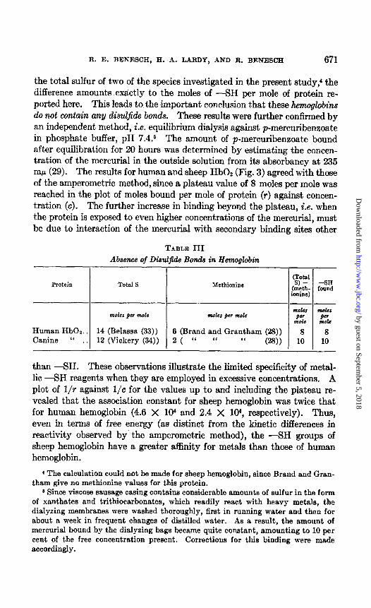
the total sulfur of t,wo of the species investigated in the present study: the difference amounts exactly to the moles of -SH per mole of protein re- ported here. This leads to the important conclusion that these hemoglobins do not contain any disul$o!e bonds. These results were further confirmed by an independent method, i.e. equilibrium dialysis against p-mercuribenzoate in phosphate buffer, pH 7.4.6 The amount of p-mercuribenzoate bound after equilibration for 20 hours was determined by estimating t.he concen- tration of the mercurial in the outside solution from its absorbancy at 235 rnh (29). The results for human and sheep HbOz (Fig. 3) agreed with those of the amperometric method, since a plateau value of 8 moles per mole was reached in the plot of moles bound per mole of prot,ein (r) against concen- tration (c). The further increase in binding beyond the plateau, i.e. when the protein is exposed to even higher concentrations of the mercurial, must be due to interaction of the mercurial with secondary binding sites other
TABLE III Absence of Diszczfide Bonds in Hewwglobin
Protein Total S Me&mine
m&s pm mole moles per mole
Human HbOl.. 14 (Belassa (33)) 6 (Brand and Grantham (23)) Canine “ , 12 (Vickery (34)) 2 ( “ “ “ cw)
8 8 I I 10 10
than -SH. These observations illustrate the limited specificity of metal- lic -SH reagents when, they are employed in excessive concentrations. A plot of l/r against l/c for the values up to and including the plateau re- vealed that the association constant for sheep hemoglobin was twice that for human hemoglobin (4.6 X lo” and 2.4 X 10⁴, respectively). Thus, even in terms of free energy (as distinct from the kinetic differences in reactivity observed by the amperometric met.hod), the -SH groups of sheep hemoglobin have a greater affinity for metals than those of human hemoglobin.
4 The calculation could not be made for sheep hemoglobin, since Brand and Gran- than-r give no methionine values for this protein.
6 Since viscose sausage casing contains considerable amounts of sulfur in the form of xanthates and trithiocarbonates, which readily react with heavy metals, the diaIyzing membranes were washed thoroughly, first in running water and then for about a week in frequent changes of distilled water. As a result, the amount of mercurial bound by the dialyzing bags became quite constant, amounting to 10 per cent of the free concentration present. Corrections for this binding were made accordingly.
by guest on September 5, 2018
http://ww
w.jbc.org/
Dow
nloaded from

672 SULFHYDRYL GROUPS. I
Some reports on direct -SH determinations of hemoglobin require further comment. Hughes (24) reported two -SH groups per mole of human hemoglobin. It is probable that this represents only the two most reactive -SH groups and that the others escaped detection, owing to t,he slow equilibration commented on by the author himself. Ingbar and Kass (3) found 2 t,o 5 moles of -SH per mole of human hemoglobin by the ammoniacal silver titration. Two main factors would tend to produce low results by this procedure: (1) slow reaction with the silver and there-
t 12 .- al
C 0
2i
$8 E
\
p-Mercuri benzoate c x I O4 FIG. 3. Reaction of hemoglobins with p-mercuribenzoate
fore incomplete equilibration and (2) the specific effect of ammonium ions in causing -SH groups to “disappear” in some proteins, a fact first re- ported with myosin by Greenstein and Edsall (6, 7). In view of these re- sults, the possibility of artifacts in the ammoniacal silver titration was al- ready pointed out, earlier (1). These factors are also responsible for an earlier report of four -SH groups per mole of dog hemoglobin (25). Tit,ra- Cons wit,h the Tris method show clearly t,hat the “ammonium effect” is operative wit.h hemoglobins. This is best illustrated with sheep hemo- globin in which slow equilibration does not const,itut,e a complicating fact,or. The data in Table IV demonstrak to what extent the presence of ammonia can affect the -SH titer.
Creative transphosphorylase presents a part,icularly clear cut case from
by guest on September 5, 2018
http://ww
w.jbc.org/
Dow
nloaded from

K. E. BENESCH, H, .A. LARDY, AND K. BENESCH 673
the point of view of the reactivity of its ---SH groups, since the first two -SH groups were found to be fully reactive, while the rest are fairly un- reactive (Table V). This is reminiscent of the a and b groups of urease described by Hellerman et al. (30), but it remains t.o be tested how enzy-
T-ABLE Iv
Eflect of Ammonium Ions on -SH Groups of Sheep Hemoglobin
Conditions moles -SH per mole
Tris 0.133 -M, final pH 7.4. ................................... 7.6 “ 0.133 “ NH4N0, 1.7 Y, final pH 7.4.. .................... 6.3 “ 0.133 “ pH10.2 ......................................... 7.4
NH,OH 0.25 M, SHIN03 0.05 M, pH 10.1, NaCl 0.85%. ........ 5.0
TABLE V
Sulfhydryl Groups of Crystalline Enzymes
The values, expressed in equivalents, are averages of at least triplicate titrations in each case. -_
Times crystallized
Creatine transphosphorylase (81,000 pm.)
Ag+ bound rapidly.. Total ag+ bound.
I -7 ii.5 I
Aldolase (147,000 gm.)
Ag+ bound in Tris. . . . . . ‘I “ I‘ ‘I + SMurea...............
n-Glyceraldehyde&phosphate dehydrogenase (120,000 gm.) - --.-.-- -._
Ag+bound..................................... / 11 1 11 1 11 / 11
mat~ic activity might be correlated with these two sets of -SH groups. It is noteworthy that the -SH titration of this enzyme is unaffected by ammonium salts.
u-Glyceraldehyde-S-phosphate dehydrogenase was found (Table V) to con- tain eleven -SH groups per mole, two of which were less reactive than the rest. The total number of -SH groups found (which remained t,he same in urea) is in good agreement with the value reported by Boyer and Segal (31) using p-mercuribenzoate. Three separate preparations of enzyme,
by guest on September 5, 2018
http://ww
w.jbc.org/
Dow
nloaded from

674 SULFRYDRYL GROUPS. I
cry&al&d four times, gave values of 10.9, 11.0, and 10.9 equivalents of -SH per 120,000 gm. of protein.
AZcZolase was found to behave much like serum albumin in that a stable end-point was reached in Tris when approximately 20 equivalents of Ag+ had been bound, but denaturation with 8 M urea made available an addi- tional 9 equivalents (Table V). The number of apparently readily avsil- able -SH groups varied somewhat, but the total number of -SH groups was remarkably consistent from preparation to preparation. Results nearly identical with those shown in Table V were obtained with a second preparation. The silver complex of aldolase was the only one found to precipitate in the titration mixture after the theoretical amount of silver had been added. This is consistent with the extremely high concentration of -SH groups found in this protein. The precipitate did not form in solutions containing 8 M urea.
Work is in progress on the relation of silver binding to catalytic activity of these proteins.
DISCUSSION
A consideration of some of the special features which combine to make the method used for this work especially suitable for the study of -SH groups in proteins seems appropriate.
The titrations are carried out under extremely mild conditions, Le. in neutral, aqueous solution, in which denaturation of proteins is kept to a minimum. On the other hand, the effect of urea denaturation can be fol- lowed under controlled conditions without intrinsic effect on the accuracy of the method.
The procedure yields information not only on the concentration of -SH groups in proteins, but also on their relative reactivity.
The specificity of the method for -SH groups is of a high order for a number of reasons: Owing to the high sensitivity of the rotating platinum electrode, the absolute concentration of protein, and therefore of silver, is extremely low (about 2 X lO+ M). This may be contrasted, for example, with equilibrium dialysis against p-mercuribenzoate, when concentrations 10 times higher had to be employed to obtain significant results. More- over, only a comparatively small excess of silver is necessary for an ac- curate determination of the end-point. Finally, since the silver is used in the form of a relatively stable complex, it will have little tendency per se to react with groups other than -SH groups.
SUMMARY
1. A new amperometric method is described for the titration of -SH groups in neutral, aqueous, buffered solution, with Ag(Tris)z+ as the titrat-
by guest on September 5, 2018
http://ww
w.jbc.org/
Dow
nloaded from

Ft. E. BENESCH, H. A. LARDY, AND R. BENESCH 675
ing agent. It is shown to be sensitive, accurate, and highly specific for -SH groups. It is especially useful for the study of proteins, since both the number and the reactivity of the -SH groups can be determined in native and denatured proteins.
2. Crystallized bovine serum albumin contains two-thirds of an -SH group per mole, but upon urea denaturation one -SH group per mole is found. This suggests the presence of one -SH group per mole in both fractions of serum albumin, an easily accessible one in mercaptalbumin, and a much less reactive one in the remaining fraction.
3. Titmtions of crystalline hemoglobins show the presence of 8 moles of -SH per mole in sheep and human hemoglobin and 10 moles of -SH per mole in canine hemoglobin. The -SH groups of sheep hemoglobin are fully reactive, while a major proportion of those of canine and human hemoglobin is highly inaccessible. It is concluded that the hemoglobins of dog and man do not contain any disulfide bonds.
4. Creatine transphosphorylase contains two reactive and four unreac- tive -SH groups. n-Glyceraldehyde-3-phosphate dehydrogenase contains eleven -SH groups per mole, two of which are somewhat less reactive than the rest. Aldolase contains approximately twenty reactive -SH groups and an additional number, making a total of twenty-nine, become avail- able only upon denaturation with urea.
This work was supported by grants from the National Science Founda- tion and the National Heart Institute and the National Institute of Arthri- tis and Metabolic Diseases of the National Institutes of Health, Public Health Service.
Addendum-Since the submission of this paper, Ingram (32) has reported a com- prehensive study of the -SH groups of hemoglobins from several different species. There is a remarkable agreement between his results and those reported here, in spite of the different methods used.
BIBLIOGRAPHY
1. Benesch, R., and Benesch, R. E., Arch. Biochem., 19, 35 (1943). 2. Benesch, R. E., and Benesch, R., Arch. Biochem., 28, 43 (1950). 3. Ingbar, S. H., and Kass, E. H., Proc. Sot. Ezp. Biol. and Med., 77, 74 (1951). 4. Wald, G., and Brown, P. K., J. Gen. Physiol., 36, 797 (1952). 5. Mazia, D., in Colowick, S. P., Glutathione, New York, 269 (1954). 6. Greenstein, J. P., and Edsall, J. T., J. Biol. Chem., 133, 397 (1949). 7. Edsall, J. T., Greenstein, J. P., and Mehi, J. W., J. Am. Chem. SOL, 61, 1613
(1939). 8. Benesch, R. E., and Benesch, R., J. Am. Chem. Sot., 77,2749 (1955). 9. Kolthoff, I. M., and Harris, W. E., Ind. and Eng. C&m., Anal. Ed., 18, 161 (1946).
10. Samuelson, G. J., and Brown, D. J., J. Am. Ckm. Sot., 67, 2711 (1935). 11. Laitinen, H. A., and Kolthoff, I. M., J. Phys. C&m., 46, 1961 (1941).
by guest on September 5, 2018
http://ww
w.jbc.org/
Dow
nloaded from

676 SULFHYDRYL GROUPS. I
12. Benesch, R., Benesch, R. E., and Rogers, W. I., in Colowick, S. P., Glutathione, New York, 31 (1954).
13. Kekwick, R. A., and Cannan, R. K., Biochem. J., 30, 227 (1936). 14. Drabkin, D. I,., J. BioE. Chem., 164, 703 (1946). 15. Haurk, I-‘. B., Oser, B. L., and Summerson, W. II., Practical physiological chemis-
try, Philadelphia, 12th edition, 441 (1947). 16. Wong, S. Y., J. Biol. Chem., 77, 409 (1928). 17. Lemberg, R., and Legge, J. W., Hematin compounds and bile pigments, New
York, 228 (1949). 18. Kuby, S. A., Noda, L., and Lardy, H. A., J. Biol. Chem., 209, 191 (1954). Noda,
L., Kuby, S. A., and Lardy, H. A., J. Biol. Chem., 209, 203 (1954). 19. Cori, G. T., Slein, M. W., and Cori, C. F., J. Biol. Chem., 173, 605 (1948). 20. Taylor, J. F., Green, A. A., and Cori, G. T., J. Biol. Chem., 173, 591 (1948). 21. Kunita, M., and Northrop, J. H., J. Gen. PhysioZ., 18,433 (1935). 22. Brown, K. D., Shupe, R. E., and Laskowski, M., J. BioZ. Chem., 173,99 (1948). 23. Fevold, H. L., Advances in Protein C&m., 6, 187 (1951). 24. Hughes, W. I,., Jr., CoZd Spring Harbor Symposia Qua&. Biol., 14, 79 (1949). 25. Beneseh, R., Cold Spring Harbor Symposia Quant. Biol., 14, 84 (1949). 26. Benesch, R. E., and Benesch, R., Arch. Biochem. and Biophys., 46, 38 (1954). 27. Halwer, M., and Nutting, G. C., J. BioZ. Chem., 166, 521 (1946). 28. Brand, E., and Grantham, J., J. Am. Chem. Sot., 66, 724 (1946). 29. Boyer, P. D., J. Am. Chem. Sot., 76, 4331 (1954). 30. Heherman, L., Chinard, F. P., and Deit,z, V. R., J. Riot Chem., 147, 443 (1943). 31. Boyer, P. D., and Segal, H. L., in McElroy, W. D., and Glass, B., The mechanism
of enzyme action, Baltimore, 520 (1954). 32. Ingram, V. M., Biochem. J., 69, 653 (1955). 33. Belassa, G., Biochem. Z., 283, 222 (1936). 34. Vickery, H. B., Proc. Sot. Exp. BioZ. and Med., 31, 6 (1933).
by guest on September 5, 2018
http://ww
w.jbc.org/
Dow
nloaded from

Reinhold BeneschRuth E. Benesch, Henry A. Lardy and
HEMOGLOBINSALBUMINS, ENZYMES, AND
CRYSTALLINE PROTEINS: I. SOME THE SULFHYDRYL GROUPS OF
1955, 216:663-676.J. Biol. Chem.
http://www.jbc.org/content/216/2/663.citation
Access the most updated version of this article at
Alerts:
When a correction for this article is posted•
When this article is cited•
alerts to choose from all of JBC's e-mailClick here
tml#ref-list-1
http://www.jbc.org/content/216/2/663.citation.full.haccessed free atThis article cites 0 references, 0 of which can be
by guest on September 5, 2018
http://ww
w.jbc.org/
Dow
nloaded from