Embed Size (px)
Citation preview
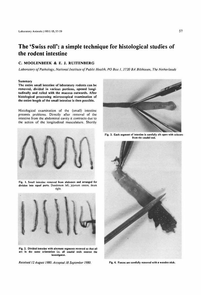
Laboratory Animals (1981) 15,57-59 57
The 'Swiss roll': a simple technique for histological studies ofthe rodent intestineC. MOOLENBEEK & E. J. RUITENBERGLaboratory of Pathology, National Institute of Public Health, PO Box 1, 3720 BA Bi/thoven. The Netherlallds
SummaryThe entire small intestine of laboratory rodents can beremoved, divided in various portions. opened longi-tudinally and rolled with the mucosa outwards. Afterhistological processing microscopical examination ofthe entire length of the small intestine is then possible.
Histological examination of the (small) intestinepresents problems. Directly after removal of theintestine from the abdominal cavity it contracts due tothe action of the longitudinal musculature. Shortly
Fig. I. Small intestine removed from abdomen and arranged fordivision into equal parts. Duodenum left, jejunum centre, ileum
right.
Fig. 3. Each segment of intestine is carefully slit open with scissorsfrom the caudal end.
Fig. 2. Divided intestine with alternate segments reversed so that allare in the same orientation i.e. all caudal ends nearest the
Investigator.
Received 12 August 1980. Accepted 18 September 1980. Fig. 4. Faeces are carefully removed with a wooden slick.
58
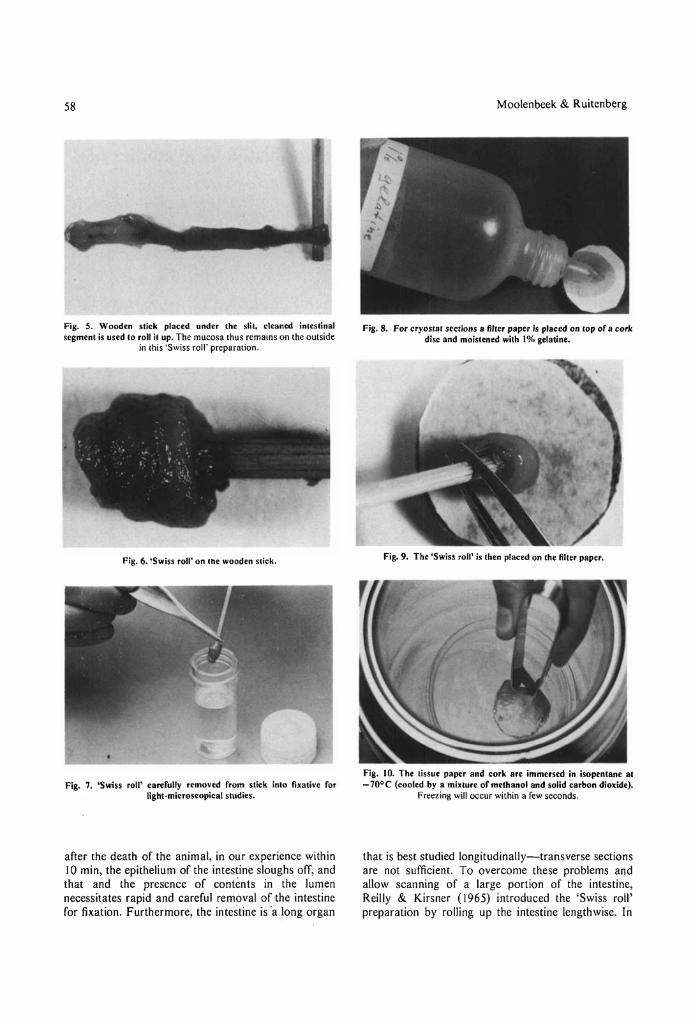
Fig. 5. Wooden stick placed under the slit, cleaned intestinalsegment is used to roll it up. The mucosa thus remains on the outside
in this 'Swiss roIl' preparation.
Fig. 6. 'Swiss roll' on Ihe wooden Slick.
Fig. 7. 'Swiss roll' carerully removed rrom stick into fixative rorlight-microscopical studies.
after the death of the animal, in our experience within10 min, the epithelium of the intestine sloughs otT, andthat and the presence of contents in the lumennecessitates rapid and careful removal of the intestinefor fixation. Furthermore, the intestine is 'a long organ
Moolenbeek & Ruitenberg
Fig. 8. For cryostat sections a filter paper is placed on top or a corkdisc and moistened with 1% gelatine.
Fig.9. The 'Swiss roll' is then placed on the filter paper.
Fig. 10. The tissue paper and cork are immersed in isopentane al-70oe (cooled by a mixlure or methanol and solid carbon dioxide).
Freezing will occur within a rew seconds.
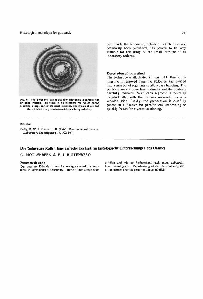
that is best studied longitudinally-transverse sectionsare not sufficient. To overcome these problems andallow scanning of a large portion of the intestine,Reilly & Kirsner (1965) introduced the 'Swiss roll'preparation by rolling up the intestine lengthwise. In
Histological technique for gut study
Fig. 11. The 'Swiss roll' can be cui after embedding in paraffin waxor after freezing. The result is an intestinal roll which allowsscanning a large part of the small intestine. The intestinal villi and
the epithelial lining remain intact despite being rolled up.
Reference
Reilly, R. W. & Kirsner,J. B. (1965). Runt intestinal disease.Laboratory Investigation 14, 102-107.
59
our hands the technique, details of which have notpreviously been published, has proved to be verysuitable for the study of the small intestine of alllaboratory rodents.
Description of the methodThe technique is illustrated in Figs 1-11. Briefly, theintestine is removed from the abdomen and dividedinto a number of segments to allow easy handling. Theportions are slit open longitudinally and the contentscarefully removed. Next, each segment is rolled uplongitudinally, with the mucosa outwards, using awooden stick. Finally, the preparation is carefullyplaced in a fixative for paraffin-wax embedding orquickly frozen for cryostat sectioning.
Die 'Schweizer Rolle': Eine einfache Technik rur histologische Untersuchungen des Darmes
C. MOOLENBEEK & E. j. RUITENBERG
lusammenrassungDer gesamte Diinndarm von Labornagern wurde entnom-men, in verschiedene Abschnitte unterteilt, der Lange nach
ero1Tnet und mit der Schleimhaut nach auBen aufgerollt.Nach histologischer Verarbeitung ist die Untersuchung desDiinndarmes iiber die gesamte Lange moglich