Embed Size (px)
Citation preview

SPECIAL ARTICLE
From th
Seatt
nell M
of Su
Brook
tiona
stetri
Roan
Palo
Toled
cine,
of Ca
the D
Colum
cine,
of Ra
Durh
With th
Amer
Socie
of Eu
the In
ology
Fundin
(AVLS
568
Editors’ Choice
The Symptoms-Varices-Pathophysiology classification of pelvic
venous disorders: A report of the American Vein & Lymphatic
Society International Working Group on Pelvic Venous Disorders
Mark H. Meissner, MD,a Neil M. Khilnani, MD,b Nicos Labropoulos, PhD,c Antonios P. Gasparis, MD,c
Kathleen Gibson, MD,d Milka Greiner, MD, PhD,e Lee A. Learman, MD, PhD,f Diana Atashroo, MD,g
Fedor Lurie, MD, PhD,h Marc A. Passman, MD,i Antonio Basile, MD,j Zaza Lazarshvilli, MD,k Joann Lohr, MD,l
Man-Deuk Kim, MD, PhD,m Philippe H. Nicolini, MD,n Waleska M. Pabon-Ramos, MD, MPH,o and
Melvin Rosenblatt, MD,p Seattle, Wash; New York, and Stony Brook, NY; Bellevue, Wash; Paris and Lyon, France;
Roanoke, Va; Palo Alto, Calif; Toledo, Ohio; Birmingham, Ala; Catania, Italy; Tbilisi, Georgia; Columbia, SC; Seoul, South
Korea; Durham, NC; Fairfield, Conn
ABSTRACTAs the importanceofpelvic venousdisorders (PeVD)hasbeen increasingly recognized, progress in thefieldhasbeen limitedbythe lack of a valid and reliable classification instrument. Misleading historical nomenclature, such as the May-Thurner, pelviccongestion, and nutcracker syndromes, often fails to recognize the interrelationship of many pelvic symptoms and their un-derlying pathophysiology. Based on a perceived need, the American Vein and Lymphatic Society convened an international,multidisciplinary panel chargedwith the development of a discriminative classification instrument for PeVD. This instrument,theSymptoms-Varices-Pathophysiology (“SVP”) classification forPeVD, includes threedomainsdSymptoms (S),Varices (V),andPathophysiology (P), with the pathophysiology domain encompassing the Anatomic (A), Hemodynamic (H), and Etiologic (E)features of the patient’s disease. An individual patient’s classification is designated as SVPA,H,E. For patients with pelvic originlower extremity signs or symptoms, the SVP instrument is complementary to and should be used in conjunction with theClinical-Etiologic-Anatomic-Physiologic (CEAP) classification. The SVP instrument accurately defines the diverse patientpopulations with PeVD, an important step in improving clinical decision making, developing disease-specific outcomemea-sures and identifying homogenous patient populations for clinical trials. (J Vasc Surg Venous Lymphat Disord 2021;9:568-84.)
Keywords: Venous insufficiency; Varicose veins; Pelvic pain; May Thurner syndrome; Renal nutcracker syndrome
The importance of venous disorders of the abdomen hindered by the use of historical syndromic
and pelvis has become increasingly recognized overthe past decade. Unfortunately, progress has beene Department of Surgery, University of Washington School of Medicine,
lea; the Department of Radiology (Interventional Radiology) Weill Cor-
edicine-New York Presbyterian Hospital, New Yorkb; the Department
rgery, Renaissance School of Medicine, Stony Brook University, Stonyc; the Lake Washington Vascular Associates, Bellevued; the Interven-
l Radiology, Hopital Americain de Paris, Parise; the Department of Ob-
cs and Gynecology, Virginia Tech Carilion School of Medicine,
okef; the Department of Obstetrics and Gynecology, Stanford Medicine,
Altog; the Department of Surgery, Jobst Vascular Institute, Promedica,
oh; the Department of Surgery, University of Alabama School of Medi-
Birminghami; the Department of Interventional Radiology, University
tania, Cataniaj; Chapidze Emergency Cardiovascular Center, Tbilisik;
epartment of Surgery, University of South Carolina School of Medicine,
bial; the Department of Radiology, Yonsei University School of Medi-
Seoulm; the Vascular Surgery, Clinique Du Parc, Lyonn; the Department
diology (Interventional Radiology), Duke University School of Medicine,
amo; and the Connecticut Image Guided Surgery, Fairfield.p
e support of the American College of Obstetricians and Gynecologists, the
ican Vein & Lymphatic Society, the American Venous Forum, the Canadian
ty of Phlebology, the Cardiovascular and Interventional Radiology Society
rope, the European Venous Forum, the International Pelvic Pain Society,
ternational Union of Phlebology, the Korean Society of Interventional Radi-
, theSociety of InterventionalRadiology, and theSociety forVascular Surgery.
g for this project was provided by the American Vein and Lymphatic Society
) and the Peter Gloviczki Chair of Venous and Lymphatic Disorders.
nomenclaturedfor example the May-Thurner, pelviccongestion, and nutcracker syndromesdwhich has often
Author conflict of interest: N.K. receives consulting fees fromMedtronic, Inc. N.L.
receives consulting fees from Medtronic, Inc, Cook Medical, Phillips, Bard, and
Tactile. K.G. receives consulting fees from Medtronic, Boston Scientific, Gore,
Vesper Medical and Phillips, receives research support from Medtronic, Bayer,
BARD, Vesper Medical, and the National Institutes of Health; and is a speaker
for Medtronic, Bristol Meyers Squib, Jansen Pharmaceuticals, and Boston Sci-
entific. W.P.B. receives royalties from Medtronic and NXT and nonfinancial
research support from Guerbet.
This paper has been co-published in the Journal of Vascular Surgery: Venous
and Lymphatic Disorders (DOI: 10.1016/j.jvsv.2020.12.084) and Phlebology
(DOI: 10.1177/0268355521999559). The publications are identical except for mi-
nor stylistic and spelling differences in keeping with each journal’s style.
Correspondence: Mark H. Meissner, MD, Department of Surgery, Box 356410,
University of Washington School of Medicine, 1959 NE Pacific St, Seattle,
WA 98195 (e-mail: [email protected]).
The editors and reviewers of this article have no relevant financial relationships to
disclose per the Journal policy that requires reviewers to decline review of any
manuscript for which they may have a conflict of interest.
2213-333X
Copyright � 2021 by the Society for Vascular Surgery, the American Venous
Forum and the Authors. This is an open access article under the CC BY-
NC-ND license (http://creativecommons.org/licenses/by-nc-nd/4.0/).
https://doi.org/10.1016/j.jvsv.2020.12.084

ARTICLE HIGHLIGHTSd Type of Research: Multispecialty, intersocietal devel-opment of a discriminative classification instrument
d Key Findings: The clinical presentation of patientswith pelvic venous disorders can be accurately andfully characterized by a discriminative instrumentthat includes presenting symptoms (S), the involvedvariceal reservoirs (V), and the underlying pathophys-iology (P), which includes the anatomic (A), hemody-namic (H), and etiologic (E) features of the disease. Apatient’s presentation is summarized as SVPA,H,E.
d Take Home Message: The use of historical nomencla-ture for pelvic venous disorders fails to recognize thecomplex and interrelated pelvic venous circulation,contributes to misdiagnosis and poor treatment out-comes, and hinders clinical research. In defininghomogenous patient populations, the Symptoms-Varices-Pathophysiology instrument will facilitate clin-ical communication, allow treatment to be moreprecisely directed, and facilitate the development ofpatient-reported outcomemeasures and clinical trials.
Journal of Vascular Surgery: Venous and Lymphatic Disorders Meissner et al 569
Volume 9, Number 3
confused the underlying pathophysiology and led todiagnostic errors and suboptimal treatment outcomes.Furthermore, the lack of a robust classification systemdefining homogenous patient populations limits clinicalcommunications, makes interpretation of the literaturedifficult, and hinders the development of appropriateclinical trials. The existence of pelvic venous disorders(PeVD) and their appropriate treatment has also beenquestioned owing to the lack of validated definitionsand imaging criteria as well as rigorous randomized clin-ical trials.1 There is a critical need for a classification sys-tem for PeVD that recognizes the variable, but oftenoverlapping, clinical presentations, as well as the underly-ing pathophysiology. A multidisciplinary panel hasranked the development of validated diagnostic criteriaand a discriminative classification instrument as themost important research priorities for PeVDs.1
For venous disorders of the lower extremities, theClinical-Etiologic-Anatomic-Physiologic (CEAP) classifi-cation, originally published in 19962 and revised in20043 and 2020,4 has become the international standardfor classification of these disorders. By defining patientgroups with similar clinical presentations and patho-physiologic features, the instrument has facilitated clin-ical communication regarding individual patients andis recognized as a reporting standard for clinical research.Despite its usefulness and general acceptance, the CEAPclassification system is limited to lower extremity venousdisorders. Since its original description, rapid advance-ments in diagnostic imaging and catheter-based inter-ventions have improved our understanding of disordersarising from veins other than those in the legs, particu-larly those of pelvic and abdominal origin.Venous disorders of the pelvis are associated with a
spectrum of symptoms arising from both reflux, mostcommonly involving the gonadal and internal iliac veins,and obstruction, usually of the left renal and iliac veins.These hemodynamic patterns are associated with atleast four broad clinical presentations, including (a) leftflank or abdominal pain and hematuria (left renal veincompression), (b) chronic pelvic pain (pelvic varicositiesassociated with primary reflux in the ovarian/internal iliacveins or obstruction of the left renal or common iliacveins), (c) venous claudication (iliac venous obstruction),and (d) symptomatic lower extremity varicosities ineither atypical (vulva/testicles, medial and posteriorthigh, sciatic nerve) or typical saphenous distributions,the latter frequently recurring after initial treatment.The relationship between pelvic symptoms and venous
pathology is far more complex than in the lower extremity.Multiple symptoms may be present concurrently andseveral potential pathophysiologic mechanisms, such asleft renal and iliac venous compression, may be simulta-neously present. Additionally, similar symptoms may arisefrom disparate underlying causes (eg, chronic pelvic paincan arise from primary ovarian vein reflux, left common
iliac vein compression, or left renal vein compression),and similar anatomic derangements may lead to differentsymptoms (eg, left renal vein compression may be associ-ated with either left flank pain and hematuria or chronicpelvic pain). This can lead to diagnostic errors and maybe responsible for the suboptimal results of many inter-ventions.5,6 From a research perspective, appropriate pa-tient classification is also important in ensuringhomogenous patient populations for the developmentof disease-specific outcome instruments and clinical trials.There is thus a critical need for precise classification ofPeVDs that has implications for both individual patientmanagement and future clinical research.
METHODSBased on the need for a classification instrument for
PeVD, the American Vein and Lymphatic Societyconvened an International Working Group on PelvicVenous Disorders in Chicago, Illinois, on July 27, 2018. In-ternational societies representing the broad spectrum ofspecialties involved in the care of patients with PeVD,including gynecologists, interventional radiologists,vascular surgeons, and phlebologists, were invited toparticipate either in-person or remotely. Invited societiesand their representatives are listed in Table I.The specific goal of the group was to develop a discrim-
inative classification instrument for PeVDs. Discriminativeinstruments are designed to measure cross-sectional dif-ferences between individuals at a single point in time, asopposed to evaluative instruments that measure longitu-dinal changes within people over time.7,8 Discriminativeinstruments include key components of the disease

Table I. International Working Group on Pelvic VenousDisorders (PeVDs) Participants
Contributor Affiliation
Diana Atashroo, MD International Pelvic Pain Society(IPPS)
Antonio Basile, MD Cardiovascular andInterventional RadiologicalSociety of Europe (CIRSE)
Antonio Gasparis, MD American Venous Forum (AVF)
Kathleen Gibson, MD American Vein and LymphaticSociety (AVLS)
Milka Greiner, MD, PhD European Venous Forum (EVF)
Nicos Labropoulos, PhD International Union ofPhlebology (UIP)
Zaza Lazarashvilli, MD International Union ofPhlebology (UIP)
Lee Learman, MD, PhD American College ofObstetricians andGynecologists (ACOG)
Joanne Lohr, MD American Venous Forum (AVF)
Neil Khilnani, MD Society of InterventionalRadiology (SIR)
Man-Deuk Kim, MD, PhD Korean Society of InterventionalRadiology
Fedor Lurie, MD, PhD Society for Vascular Surgery
Mark Meissner, MD American Vein and LymphaticSociety (AVLS)
Philippe Nicolini, MD European Venous Forum (EVF)
Waleska Pabon-Ramos, MD,MPH
Society of InterventionalRadiology (SIR)
Marc Passman, MD Society for Vascular Surgery
Mel Rosenblatt, MD American Vein and LymphaticSociety (AVLS)
570 Meissner et al Journal of Vascular Surgery: Venous and Lymphatic DisordersMay 2021
that are stable, at least over short periods of time, have alimited number of options and clear definitions thatenable uniform interpretation, and have large and stablebetween-subject variation.8 From a simplistic standpoint,discriminative instruments place patients into homoge-nous groups with similar clinical features, natural his-tories, and responses to treatment.At the initial meeting, the clinical, anatomic, and path-
ophysiologic aspects of PeVD were presented and dis-cussed among panel members, incorporating the viewsof the various subspecialties included on the panel. Themethodology underlying instrument development wasthen reviewed and alternative approaches discussed.Based on this discussion, it was agreed that the instru-ment should be based on the following principles.
a. The instrument should be patient-centric, that is,focused on the primary concerns of the patient ratherthan simply the underlying pathophysiology.
b. In addition to patient-important clinical features,complete characterization of a patient’s presentationrequires a precise description of the underlying anat-omy and pathophysiology.
c. Asymptomatic patients with pelvic venous diseaseshould be included in the classification, althoughamong symptomatic patients, only those with arecognized venous etiology should be included.Similar clinical presentations that are not of venousorigin (eg, chronic pelvic pain owing to other causes)are not included in this classification.
d. Several nuances of PeVD, particularly the observationthat PeVD are primarily symptom rather than signbased, preclude a purely CEAP-based approach. How-ever, because venous disorders of the pelvis and lowerextremity are a continuum, the instrument should, asmuch as feasible, follow the conventions of and becomplementary to CEAP. Accordingly, the pelvic in-strument should avoid duplication of lower extremitysigns that are included in CEAP. For example,although localized pelvic origin extrapelvic symptoms,such as tenderness associated with pelvic origin vari-cosities, should be included in the pelvic instrument,more generalized lower extremity signs, such asswelling continue to be best classified with CEAP.
Guided by these principles, the domains to be includedwere discussed and precise definitions developed,emphasizing the importance of optimizing the validityand reproducibility of the instrument. Small groupswere then formed to craft an initial strategy for eachdomain, which was then discussed among the entiregroup. Based on the discussion, a draft instrument (theSVP classification) was developed and three rounds ofsimulated patient classification performed by the writinggroup (M.H.M., N.K., N.L., A.G., K.G., and M.G.) to identifypotential problems with the definitions and ensurereproducibility of the instrument. Definitions werefurther refined based on the simulated classification ex-ercises and review of the literature, striving to makethem as evidence based as possible. The final draft wasthen circulated to all participants for revision.
RESULTS: THE CLASSIFICATION OF PeVD
DefinitionsMinimizing interobserver variability through precise
definitions is critical to the reproducibility of a discrimi-native instrument. The following definitions were devel-oped and should be utilized for the purpose of pelvicvenous classification. When possible, efforts were madeto make these definitions congruent with lower extrem-ity CEAP.Symptoms.
PeVDdThe spectrum of symptoms andsigns arising from the veins of the pelvis(the gonadal veins, the internal iliac veinsand their tributaries, and the venous plex-uses of the pelvis) and their primarydrainage pathways (the left renal vein, theiliac veins, and the pelvic escape points).

Journal of Vascular Surgery: Venous and Lymphatic Disorders Meissner et al 571
Volume 9, Number 3
This includes symptoms historically ascribed tothe May-Thurner, nutcracker, and pelvic conges-tion syndromes. Given their imprecise and overlap-ping nature, these historical terms should nolonger be used.1
Venous origin renal symptomsdSymptoms arising fromrenal venous hypertension secondary to left renal veinobstruction.
These include microhematuria or macrohematu-ria and left flank or abdominal pain that is wors-ened by activities such as standing, sitting, orwalking.9
Chronic pelvic paindPain symptoms perceived to origi-nate from pelvic organs/structures typically lastingmore than 6 months. It is often associated with negativecognitive, behavioral, sexual, and emotional conse-quences as well as with symptoms suggestive of lowerurinary tract, sexual, bowel, pelvic floor, myofascial, or gy-necologic dysfunction.10
Although there has historically been a lack ofconsensus11 regarding the definition of chronicpelvic pain, we have adopted that proposed bythe American College of Obstetricians and Gyne-cologists. Causes of chronic pelvic pain include awide range of disorders of the reproductive, uri-nary, gastrointestinal, neurologic, and musculo-skeletal systems,12 often with overlappingsymptoms in an individual patient.13 PeVD areincluded in the range of somatic, visceral andneurologic pain generators that are often associ-ated with chronic pelvic pain.Data regarding the demographics and symptom-atology of women with venous origin pelvic painare largely derived from small case series of thosepresenting for treatment and there is a clear needfor larger studies comparing women with chronicpelvic pain of venous and nonvenous origin. Suchlimited case series suggest that venous origin pel-vic pain most commonly occurs in multiparouswomen of reproductive age.12,14-16 Despite thisgeneral observation, a somewhat older populationwith iliac venous obstruction has recently beendescribed in which pelvic pain often occurs inconjunction with leg symptoms,17,18 implying thatpatient demographics and associated symptomsmay depend on the underlying etiology.Because chronic pelvic pain includes a spectrumof symptoms, there is substantial overlap betweenwomen with pain secondary to venous and nonve-nous causes. Descriptions of the typical character-istics of venous origin pelvic pain come largelyfrom a single dated but well-done studycomparing women with pelvic pain and variceson transuterine venography to women with eitherpelvic pain owing to other pathology or withoutpelvic pain undergoing elective sterilization.15
Most of the signs and symptoms associated withvenous-origin pelvic pain have been found to berelatively sensitive, but nonspecific.19 Pelvic painof venous origin is often characterized as dull
unilateral or bilateral pain with occasional sharpflares. Bimanual examination, demonstrating focaladnexal tenderness, often reproduces the pain.Symptoms are often worse with activities such aswalking and prolonged standing, and improvewith lying down. Although deep dyspareunia iscommon among women with pelvic pain from avariety of causes, venous origin pain is more likelyto be associated with prolonged postcoitalache.12,15,19 The combination of postcoital acheand tenderness over the ovarian point (the junc-tion of the upper andmiddle thirds of a line drawnfrom the umbilicus to the anterior superior iliacspine) has been reported to be 94% sensitive and77% specific for distinguishing a venous originfrom other causes of pelvic pain.15
Although chronic pelvic pain also occurs inmales,20,21 there is currently little evidence to sug-gest that pelvic venous disease is an importantcontributing factor. This is likely due to both differ-ences in venous anatomy as well as the role ofpregnancy in PeVDs in women. The gonadal veinsfollow an extrapelvic course in males and thearrangement of the visceral pelvic venous plexusesare substantially different.
Pelvic origin extrapelvic symptomsdSymptoms local-ized to the external genitalia or lower extremities thatarise from either reflux through recognized escapepoints in the pelvic floor22 or from iliocaval venousobstruction.
In females, reflux-related symptoms may includepain, discomfort, tenderness, itching, bleeding,and superficial venous thrombosis associatedwith nonsaphenous varicosities. These may belocalized to the vulva or the posteromedial thighin the distribution of the perineal and inferiorgluteal escape points. In males, these includetesticular discomfort and infertility related to avaricocele. Extrapelvic reflux arising from the infe-rior gluteal vein may also rarely be associated withsciatic or tibial nerve symptoms. Symptoms associ-ated with sciatic nerve varices include pain radi-ating from the buttock to the lateral aspect ofthe leg, often worsened with sitting.23,24 Anecdotalreports suggest tibial nerve symptoms are milder,often including only paresthesias on compressionof the nerve. Obstruction-related extrapelvicsymptoms include venous claudication.
Venous claudicationdExertional pain in the lower ex-tremities frequently described as a tight, "bursting"pain, in the thigh, buttock, or leg not associated with aspecific walking distance or confined to specific musclegroups, but relieved by rest and elevation of the legs.25-28
Symptoms of venous claudication are most commonlyassociated with iliocaval venous obstruction.HASTI (Provensis, Uxbridge, UK) symptomsdNonspecificsymptoms typically associated with lower extremityvenous disease including heaviness (H), aching (A),swelling (S), throbbing (T), and itching (I).27,29
Such symptoms are usually generalized to thelower extremity rather than localized to any pelvic

572 Meissner et al Journal of Vascular Surgery: Venous and Lymphatic DisordersMay 2021
origin extrapelvic lower extremity varices.Although the responsible pathology may arise inthe pelvis, generalized signs of lower extremityvenous disease are not included in the SVP classi-fication and should be accounted for by the con-current use of CEAP.
Signs.Left renal vein obstructiondCompression of theleft renal vein at the crossing of the abdominalaorta associated with symptoms related eitherto (a) renal venous hypertension (hematuriaand/or abdominal/flank pain) or (b) if decom-pressed by collaterals, pelvic varices and chronicpelvic pain or a left-sided varicocele.
Symptomatic obstruction of the left renal vein isusually attributed to compression of the renalvein between the abdominal aorta and superiormesenteric artery (anterior nutcracker syndrome),although compression may also arise from a retro-aortic course of the left renal vein (posteriornutcracker syndrome) or stretching of the renalvein over the abdominal aorta.9 Symptoms offlank pain and hematuria are presumed second-ary to renal venous hypertension, often definedas a transrenal pressure gradient of 3 or moremm Hg at the time of venography.30-33 Hematuriain such cases is often attributed to renal varices,which are often asymptomatic, effect predomi-nantly the left kidney, and have been identifiedin 10% of left renal venograms performed for a va-riety of indications.34 However, such a gradientmay be absent it there is significant decompres-sion via refluxing collaterals including the leftgonadal, ascending lumbar, adrenal, periureteral,capsular, or intrarenal veins.9,31 In such cases, pelvicvarices or a varicocele may be associated with sec-ondary gonadal vein reflux.A variety of imaging modalities including ultra-sound, venography (with or without intravascularultrasound [IVUS] and measurement of pressuregradients), computed tomography (CT), and mag-netic resonance imaging (MRI) have been used inthe evaluation of left renal vein compression.Although mean renal vein diameter reduction byCT is significantly higher in patients with symp-toms related to renal venous hypertension(74.5 6 1.9%) than in controls (25.4 6 2.4%)35 anda transrenal pressure gradient of 3 or more mmHg has been associated with hematuria,30-32 defin-itive diagnostic criteria and cut-points are lackingand may vary between patients. Furthermore,asymptomatic 50% or greater compression ofthe left renal vein (nutcracker phenomenon) isseen in 51% to 72% of CT angiograms.32 Giventhe lack of definitive anatomic and hemodynamiccriteria across a variety of clinical settings, we havenot included them in the definition, which insteadrelies on correlating the patient’s symptoms andimaging studies.
Pelvic varicose veinsdTortuous, dilated veins 5 mm ormore in diameter around the ovary and uterus.36
Pelvic varices may involve both the ovarian (pam-piniform) and uterovaginal venous plexuses, whichcommunicate through the broad ligament.12,22,37-39
There may also be extensive communication withthe vesicular and external rectal plexus.22 Althoughvenography has historically been the referencestandard for the diagnosis of pelvic varices,14,37,39 itremains an invasive study associated with the risksof ionizing radiation and is now often limited todefinitive imaging at the time of planned interven-tion. Several noninvasive imaging studies,37,40 moresuitable for initial evaluation, have been suggestedincluding transabdominal ultrasonography, trans-vaginal ultrasonography, CT, and MR imaging.Among these, pelvic ultrasound, either transabdo-minal or transvaginal, is the most widely available,has been the most extensively investigated, and al-lows an evaluation of both venous diameter andreflux. We have accordingly defined pelvic varicesbased on commonly cited ultrasound criteria.36
Other diagnostic criteria have been proposed,including greater than 4 tortuous, dilated veinsgreater than 4 mm in diameter surrounding theovaries and uterus,41 the appearance of dilatedtransuterine veins (arcuate and/or myometrialveins) connecting the left and right uterine veins,37
and reversed flow direction or disappearance offlow with Valsalva.37,40,42 However, Park et al36
found transuterine crossing veins in only 25% of pa-tients with symptomatic pelvic varicosities in com-parison with 8.6% of controls. Similarly, reversal ofDoppler flow direction during a Valsalva maneuverwas identified in only 26.9% of symptomatic pa-tients, in comparison with 8.8% of controls.36
Position does influence the ability to detect pelvicvenous pathology. Investigators have reported ul-trasound evaluation in the supine,36 30� to 45�
reverse Trendelenburg position,42,43 semi-erect,44
and upright positions.43 CT and MR imaging areobligatorily performed in the supine position.Because there is no consensus regarding posi-tioning for noninvasive examinations, it has notbeen included in the definitions of pelvic varicoseveins or reflux. However, clinicians should beaware of the role that position may have in theinterpretation of all imaging studies.
Gonadal vein refluxdRetrograde flow in either gonadalvein, spontaneously or in response to a Valsalva’s maneu-ver, as documented by ultrasound, venography, or timeresolved magnetic resonance angiography.
Retrograde flow is the primary criteria for the defi-nition of venous reflux and in the left ovarian vein,has been identified in 100% of patients with symp-tomatic pelvic varices in comparison with 25% ofcontrols.41 Although some investigators45 havedefined pelvic reflux as retrograde flow greaterthan 1 second in duration and persisting until theend of the maneuver, other investigators41,46 have

Journal of Vascular Surgery: Venous and Lymphatic Disorders Meissner et al 573
Volume 9, Number 3
noted no validated cut-point for pathologic dura-tion of reflux in the ovarian veins. Still other inves-tigators have noted variable reflux patterns,including spontaneous, intermittent retrogradeflow; retrograde flow only during a Valsalva ma-neuver; and continuous retrograde flow.47 Giventhe conflicting evidence, we have chosen not toinclude reflux duration in the definition.Gonadal vein diameter, in the presence of pelvicvarices is often used as a surrogate for retrogradeflow. Although some investigators44,45,48 have re-ported ovarian vein diameter to be an insensitivemaker of reflux, other investigators36 have re-ported positive predictive values of 71.2%, 83.3%,81.8%, and 75.8% for diameters of 5, 6, 7, and8 mm, respectively. Other investigators41 havesimilarly found pelvic varices to be present in allpatients with a left ovarian vein diameter ofmore than 6 mm by ultrasound assessment.Diameter criteria have also been reported for CTand MR.40 However, in view of the conflicting evi-dence, we have not included diameter as a criteriafor gonadal vein reflux.
Iliac venous obstructiondGreater than 50% cross-sectional area reduction by IVUS or a 50% or greaterdiameter reduction by multiplanar venography of thecommon or external iliac veins in association with appro-priate lower extremity or pelvic symptoms.
This definition was derived from those commonlyused in the literature, although it must beacknowledged that there is currently no validatedmethod of defining a clinically or hemodynamical-ly significant venous stenosis49-51 and that thisvalue may differ between patients.52 In evaluatingthe predictors of clinical improvement after iliacvenous stenting, a cross sectional area reductionof more than 54% by IVUS examination had thehighest sensitivity (83% sensitivity, 47% specificity),whereas a greater than 52% diameter decrease bymultiplanar venography had the highest speci-ficity (50% sensitivity, 71% specificity).49 Notably,the thresholds for clinical improvement afterstenting were somewhat higher for nonthrom-botic lesions. However, because a 50% or greateriliac stenosis may be present in one-quarter toone-third of the general population,52,53 it is criticalthat anatomic stenosis alone not be considered acriterion for intervention and that anymeasurement of stenosis be interpreted in thecontext of the patient’s clinical presentation.Both cross-sectional imaging and transabdominalultrasound examination have been used in theinitial evaluation of iliac obstruction and a numberof ultrasound criteria for detection of a 50% orgreater iliac venous obstruction have beendeveloped.51,53
Internal iliac venous refluxdRetrograde flow in the inter-nal iliac vein or its tributaries, either spontaneously or inresponse to a provocative Valsalva’s maneuver.
Reflux can be demonstrated by antegrade or se-lective descending venography, transabdominal/
transperineal ultrasound,43,47 or transvaginal ultra-sound.42,44 Pathologic flow patterns observed withultrasound include retrograde flow isolated to themain internal iliac trunk, cephalad flow in themain trunk and reflux in the tributaries, or retro-grade flow in both the main trunk and tributaries.
Pelvic origin extrapelvic varicesdRetrograde flow inextrapelvic veins arising from reflux exiting the pelvisthrough recognized escape points.22
Pelvic origin extrapelvic varices include refluxingveins in either atypical locations (vulva in femalesand pampiniform plexus in males, perineum,gluteal cleft, and posterior thighs), or, throughcommunication with saphenous tributaries, in atypical saphenous distribution. Extrapelvic varicesalso include intra/perineural (sciatic and tibial)varices arising from the inferior gluteal tributaryof the internal iliac vein.22,54
As elsewhere, this is an ultrasound-derived defini-tion that includes both visible varicosities as wellas refluxing pelvic-origin tributaries that are seenonly with ultrasound. Protocols for visualization ofthese refluxing tributaries are well-definedelsewhere.43
Pelvic origin extrapelvic varices may arise fromeither pelvic reflux or obstruction. However, bydefinition, collateral veins from the lower extrem-ity to the pelvis that demonstrate antegrade flowat rest and function to bypass an iliocaval venousobstruction are not pelvic origin extrapelvicvarices.
Lower extremity varicesdAs defined in CEAP,3 subcu-taneous, dilated veins $3 mm in diameter whichdemonstrate reflux in the upright position and involvethe named saphenous and accessory saphenous trunks,their tributaries and nonsaphenous superficial leg veins.
CLASSIFICATION OF PeVDsDTHE SVPINSTRUMENTDiscriminative instruments for venous disorders consist
of descriptive domains or categories, such as the clinical(C), etiologic (E), anatomic (A), and pathophysiologic (P)domains of CEAP, with precisely defined responses withineach domain. The proposed classification for PeVDs hasbeendesignated the SVPclassification and includes threedomains: symptoms (S), varices (V), the primary sign ofPeVD, and a composite anatomic-pathophysiologicdomain (P). ThecompositePdomain is composedof threesubdomains, including the anatomy of the involvedabdominal and pelvic veins (A), the associated hemody-namic abnormalities (H), and the underlying etiology (E),which are listed as subscripts after the P domain (PA,H,E).An individual patient’s pelvic classification is thus desig-nated as SVPA,H,E.Symptoms (S) and varices (V) associated with PeVD are
considered to occur in 4 anatomic zones extending in adescending fashion from the renal veins to the lower ex-tremities (Fig 1). Three of these zonesd(1) the left renal

Fig 1. The symptoms, signs (varices), and pathophysiologic manifestations of pelvis venous disorders (PeVD) occurin four anatomic zones of the abdomen and pelvis. These are arranged in descending order from the renal veinsto the lower extremities and include symptoms and varices associated with (1) the left renal vein, (2) the gonadal,internal iliac, and pelvic veins, (3) the pelvic origin extrapelvic veins arising in the pelvis and refluxing through thepelvic escape points to the genitalia and lower extremity veins, and (4) the lower extremity veins. The first threezones are included in the Symptoms-Varices-Pathophysiology (SVP) classification while the fourth zone, associ-ated with the superficial and deep veins of the lower extremity and their tributaries, is optimally classified withCEAP and is not included. L, left; R, right.
574 Meissner et al Journal of Vascular Surgery: Venous and Lymphatic DisordersMay 2021
vein, (2) the gonadal and internal iliac veins and associ-ated pelvic venous plexuses, and (3) the pelvic originextrapelvic transitional veins arising from reflux exitingthe pelvis through recognized escape pointsdareincluded in the SVP classification. Although oftencommunicating with zone 3, the fourth zone, the super-ficial and deep veins of the lower extremity and their trib-utaries, is optimally classified with CEAP and is notincluded in the SVP instrument.Each of the three primary domainsdsymptoms (S), vari-
ces (V), and pathophysiology (P) with its 3 subdomainsdis discussed in this section.
Symptoms (S). Pelvic venous classification begins withthe patient’s clinical symptoms (S) designated by
subscripts from 0 through 3 (Table II). As discussedelsewhere in this article, responses are arranged indescending anatomic zones from the renal veins to thelower extremities. Although some complaints may occurin either sex, others such as pelvic pain and varicoceleoccur predominantly or exclusively in one sex. Venousorigin extrapelvic symptoms (S3) are further subdividedinto those involving the external genitalia, those relatedto pelvic origin nonsaphenous varicosities of the leg(posteromedial thigh and sciatic/tibial nerve), and thoseof venous claudication.The pelvic origin extrapelvic veins of the thigh may
communicate with the superficial and deep veins ofthe lower extremity and be associated with any of themanifestations of C2 through C6 disease. Although

Table II. Symptoms (“S”)
S0
No symptoms of a PeVD (no renal, pelvic, or extrap-elvic symptoms)
S1 Renal symptoms of venous origin
S2 Chronic pelvic pain of venous origin
S3 Extrapelvic symptoms of venous origin
a Localized symptoms (pain, discomfort, tenderness,itching, bleeding and superficial venousthrombosis) associated with veins of the externalgenitalia (vulva and scrotum)
b Localized symptoms associated with pelvic originnonsaphenous veins of the leg. These include thoserelated to pelvic origin varices of the posteromedialthigh (pain, discomfort, tenderness, itching,superficial venous thrombosis) as well as thoserelated to sciatic/tibial nerve varices (pain,paresthesias). More generalized lower extremitysymptoms and signs, such as heaviness andswelling, are classified with CEAP not SVP.a
c Venous claudication.a
PeVD, Pelvic venous disorder; SVP, symptoms-Varices-Pathophysiology.aMust include CEAP classification for full characterization of lowerextremity symptoms.
Table III. Varices (“V”)
V0
No abdominal, pelvic, or pelvic origin extrapelvicvarices on clinical or imaging examination
V1 Renal hilar varices
V2 Pelvic varices
V3 Pelvic origin extrapelvic varices.
a Genital varices (vulvar varices and varicocele)
b Pelvic origin lower extremity varicose veins arisingfrom the pelvic escape points and extending intothe thigh. Includes visible varicosities, typically overthe posteromedial thigh, as well as sciatic varicesand other refluxing veins transitioning the pelvicfloor which are visualized only with ultrasound.a
aMust include CEAP classification for full characterization of lowerextremity varices.
Journal of Vascular Surgery: Venous and Lymphatic Disorders Meissner et al 575
Volume 9, Number 3
localized symptoms such as discomfort, pruritis,bleeding, and superficial thrombosis are included inS3a and S3b, to avoid redundancy and potentiallycompromised reproducibility, generalized lower extrem-ity signs (eg, swelling) and symptoms (eg, HASTI symp-toms associated with C2S) are not specifically includedin SVP and must be further classified using CEAP. Pa-tients presenting with more than one clinical symptomshould have all presenting features included as sub-scripts, separated by commas, following the Sdesignation.
Varices (V). The venous system of the pelvis can beconsidered to consist of 3 reservoirs where varices maydevelopd(1) the renal hilum, (2) the venous plexuses ofthe pelvis, and (3) the pelvic origin extrapelvic veins. Thelower extremity veins comprise a fourth reservoir, whichmay communicate with pelvic origin extrapelvic varices.However, as with symptoms, the lower extremity reser-voir is optimally defined with CEAP and is not includedin SVP.Increased venous pressures, arising from proximal
reflux or obstruction, are transmitted to these reservoirs,where symptoms related to either varices or increasedvenous pressure may develop. Most therapeutic interven-tions are directed toward decreasing venous pressure inthese reservoirs. The variceal reservoirs of the pelvis aredesignated V and are again denoted in a descendingfashion by the subscripts 0 to 3 (Fig 1; Table III).Although some varices (eg, pelvic origin varices of the
vulva or posteromedial thigh) may be apparent on phys-ical examination, others (renal hilar, pelvic, and some pel-vic origin extrapelvic varices) are identified only throughimaging studies. The V classification should, therefore,include the full extent of varices defined by both physicalexamination and imaging studies. As with symptoms,patients presenting with varices in more than one reser-voir should have all of their presenting features includedas multiple subscripts, separated by commas, to V.Finally, because the pelvic and lower extremity venoussystems are in continuity, patients with lower extremitysigns and symptoms arising in the pelvis should bedescribed using both SVP and CEAP as complementaryinstruments.
Pathophysiology (P). The pathophysiology domain (P)is a composite of the anatomic (A), hemodynamic (H),and etiologic (E) subdomains. Involved anatomic seg-ments in the abdomen and pelvis are designated byanatomic abbreviations that include laterality (Table IV).As in CEAP, the underlying hemodynamic (H)
derangementsdreflux (R), obstruction O), or both (R,O)dare designated by a subscript to the P category(Table V). Obstruction, which may be thrombotic or non-thrombotic in origin, primarily involves the left renal,common iliac, and external iliac veins. Reflux occurs
most commonly in the gonadal veins, internal iliac veins,and pelvic escape points with their associated pelvicorigin extrapelvic veins. By convention, the hemody-namic subscript should immediately follow the designa-tion of each involved anatomic segment. In contrast withthe lower extremities, concurrent reflux and obstructionin a single pelvic venous segment is unusual but, if pre-sent, should be designated by both the R and O sub-scripts. Also, some congenital malformations, may notbe associated with either reflux or obstruction, in whichcase the H subscript should be omitted.The etiology (E) of pelvic venous pathology is defined as
being thrombotic (T), nonthrombotic (NT), or congenital(C) (Table VI). Venous obstruction can arise from eithera previous episode of deep venous thrombosis

Table IV. Anatomy
Abbreviation Expansions
IVC Inferior vena cava
LRV Left renal vein
GV Gonadal (testicular, ovarian) veins
LGV Left gonadal vein
RGV Right gonadal vein
BGV Bilateral gonadal veins
CIV Common iliac veins
LCIV Left common iliac vein
RCIV Right common iliac vein
BCIV Bilateral common iliac veins
EIV External iliac veins
LEIV Left external iliac vein
REIV Right external iliac vein
BEIV Bilateral external iliac veins
IIV Internal iliac veins
LIIV Left internal iliac vein and tributaries
RIIV Right internal iliac vein and tributaries
BIIV Bilateral internal iliac veins and tributaries
PELV Pelvic escape veins22 (“escape points”); inguinal,obturator, pudendal, and/or gluteal
Table V. Hemodynamics
Obstruction(O)
Thrombotic or nonthrombotic (venouscompression) venous obstruction
Reflux (R) Thrombotic or nonthrombotic reflux
Table VI. Etiology (E)
Thrombotic (T)Venous reflux or obstruction arising
from a previous episode of DVT
Nonthrombotic (NT) Reflux arising from a degenerativeprocess of the vein wall or proximalobstruction; Obstruction arisingfrom extrinsic compression
Congenital (C) Congenital venous or mixed vascularmalformations
DVT, Deep vein thrombosis.
576 Meissner et al Journal of Vascular Surgery: Venous and Lymphatic DisordersMay 2021
(thrombotic) or extrinsic compression by adjacent arte-rial structures or mass lesions (nonthrombotic). Throm-botic reflux can similarly develop after an episode ofdeep venous thrombosis, whereas nonthrombotic refluxis presumed to represent a degenerative process of thevein wall leading to venous dilation and valvular incom-petence. Congenital etiologies include vascular malfor-mations, either venous or mixed. The designatedetiology (E) should be denoted by a subscript to the Pcategory, immediately after the designation of theinvolved anatomic segments and the hemodynamicderangements.
Using the SVP classification. For the purposes of docu-menting reproducibility of the instrument and forrecording data in clinical studies, all five domains and sub-domains of SVPdS, V, A, H, andEdshould bedocumentedindependently. However, such a system is overly compli-cated for routine clinical use and communication. Forsuch purposes, the A, H, and E subdomains are collapsedinto a single anatomic-pathophysiological domain P. Byconvention, this single term should include the anatomicsegment(s) involved, the underlying hemodynamics, andthe etiology in this order. That is, notation for the P domainshould be P anatomic segment, hemodynamics, etiology. If multipleanatomic segments are involved, each venous segmentafter the P should be specified in this fashion, separatingthe full anatomic-pathophysiologic description of eachsegment with a semicolon. In such cases, the anatomic
segments and associated pathology should be listedbeginning at the inferior vena cava and proceedingcaudally. For example, nonthrombotic obstruction of theleft common iliac vein associated with internal iliac refluxshould be designated as PLCIV,O,NT; LIIV,R,NT. The historicsyndromes of the abdomen and pelvis would be now bedesignated as follows in the SVP classification,
d Pelvic congestion syndrome with chronic pelvic paindue to bilateral ovarian reflux: S2V2PBGV,R,NT
d Nutcracker syndrome with flank pain and hematuria:S1V1PLRV,O,NT
d May-Thurner syndrome with left lower extremityedema: S0V0PLCIV,O,NT; Left C3sEseAdPo(CIV)
Clinical examples of the SVP classification are shown inFigs 2 to 9. The use of a scoring sheet as shown inTable VII may aid in early application of the instrument.Smart phone applications to assist in classification areavailable on the AVLS website (https://myavls.org/svp).All components of the instrument, that is S, V, and PA,H,E
are to be used in designating a patient’s final SVP classi-fication. This presumes imaging (abdominal/transperi-neal ultrasound, transvaginal ultrasonography, cross-sectional imaging, venography/IVUS, laparoscopy) hasbeen done as part of the classification, recognizing thatsome components of the classification may change asthe evaluation progresses from noninvasive to moredefinitive imaging such as venography. It is acceptableto use an interim designation (x) as a subscript for thosedomains where evaluation is not yet complete (eg, S0-
3VxPx).
DISCUSSIONDespite technical advances, progress in the diagnosis
and management of PeVDs has been hampered by theuse of historic nomenclaturedthe May-Thurner, pelviccongestion, and nutcracker syndromesdto describe

Fig 2. Left renal vein compression associated with symptoms of left flank pain and hematuria. A, Computedtomography (CT) demonstrates compression of the left renal vein (white arrow) over the abdominal aorta. B,Venography demonstrates contrast attenuation over the abdominal aorta (black arrow), renal hilar varices (whitearrow), and ascending collaterals (dashed white arrow) consistent with renal vein compression. The Symptoms-Varices-Pathophysiology (SVP) classification is S1V1PLRV,O,NT.
Journal of Vascular Surgery: Venous and Lymphatic Disorders Meissner et al 577
Volume 9, Number 3
underlying anatomic lesions that often have variable clin-ical presentations. The use of these terms ignores thecomplex and interrelated abdominal and pelvic venouscirculation, as well as the observation that similar clinicalpresentations may have different underlying pathophys-iologies while identical pathology may have differentclinical presentations. Inaccuracy in precisely character-izing a patient’s clinical presentation has often led tomisdiagnosis and suboptimal treatment outcomes andhas hindered progress in the field. The use of the histor-ical syndromic terms should be abandoned in favor of amore precise characterization of the patient’s clinical
Fig 3. Chronic pelvic pain due to compression of the left reSelective renal venography demonstrates compressive ob(black arrow) associated with renal hilar varices. The left renstar) and a refluxing left ovarian vein (white star). B, Selecpelvic varices, myometrial veins (red star) and smallPathophysiology (SVP) classification is S2V1,2PLRV,O,NT; LGV,R,N
presentation, including symptoms, signs (varices), andthe underlying venous anatomy and pathophysiology.1
Although incomplete, our understanding has progressedto the point that a discriminative instrument is neededto characterize patients with PeVD.Discriminative instruments characterize a patient’s clin-
ical presentation at a particular point in time. From apragmatic standpoint, such instruments place patientsinto categories with similar clinical features, natural his-tories, and responses to treatment. By virtue of theirfundamental features (large between subject variability),these instruments are not designed to quantitatively
nal vein with secondary reflux in the left ovarian vein. A,struction (white arrow) of the central left renal veinal vein is drained through the renal-azygous trunk (redtive left ovarian venography demonstrates associatedarcuate veins (red arrow). The Symptoms-Varices-T.
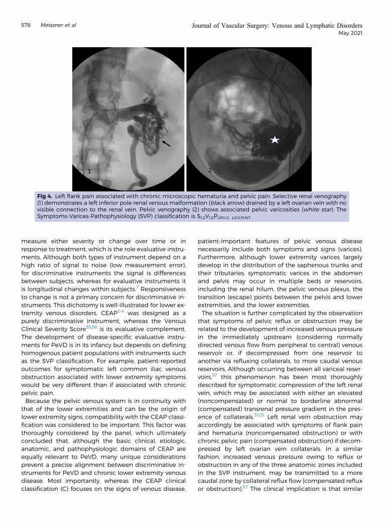
Fig 4. Left flank pain associated with chronic microscopic hematuria and pelvic pain. Selective renal venography(1) demonstrates a left inferior pole renal venous malformation (black arrow) drained by a left ovarian vein with novisible connection to the renal vein. Pelvic venography (2) shows associated pelvic varicosities (white star). TheSymptoms-Varices-Pathophysiology (SVP) classification is S1,2V1,2PLRV,C; LGV,R,NT.
578 Meissner et al Journal of Vascular Surgery: Venous and Lymphatic DisordersMay 2021
measure either severity or change over time or inresponse to treatment, which is the role evaluative instru-ments. Although both types of instrument depend on ahigh ratio of signal to noise (low measurement error),for discriminative instruments the signal is differencesbetween subjects, whereas for evaluative instruments itis longitudinal changes within subjects.7 Responsivenessto change is not a primary concern for discriminative in-struments. This dichotomy is well-illustrated for lower ex-tremity venous disorders. CEAP2-4 was designed as apurely discriminative instrument, whereas the VenousClinical Severity Score55,56 is its evaluative complement.The development of disease-specific evaluative instru-ments for PeVD is in its infancy but depends on defininghomogenous patient populations with instruments suchas the SVP classification. For example, patient-reportedoutcomes for symptomatic left common iliac venousobstruction associated with lower extremity symptomswould be very different than if associated with chronicpelvic pain.Because the pelvic venous system is in continuity with
that of the lower extremities and can be the origin oflower extremity signs, compatibility with the CEAP classi-fication was considered to be important. This factor wasthoroughly considered by the panel, which ultimatelyconcluded that, although the basic clinical, etiologic,anatomic, and pathophysiologic domains of CEAP areequally relevant to PeVD, many unique considerationsprevent a precise alignment between discriminative in-struments for PeVD and chronic lower extremity venousdisease. Most importantly, whereas the CEAP clinicalclassification (C) focuses on the signs of venous disease,
patient-important features of pelvic venous diseasenecessarily include both symptoms and signs (varices).Furthermore, although lower extremity varices largelydevelop in the distribution of the saphenous trunks andtheir tributaries, symptomatic varices in the abdomenand pelvis may occur in multiple beds or reservoirs,including the renal hilum, the pelvic venous plexus, thetransition (escape) points between the pelvis and lowerextremities, and the lower extremities.The situation is further complicated by the observation
that symptoms of pelvic reflux or obstruction may berelated to the development of increased venous pressurein the immediately upstream (considering normallydirected venous flow from peripheral to central) venousreservoir or, if decompressed from one reservoir toanother via refluxing collaterals, to more caudal venousreservoirs. Although occurring between all variceal reser-voirs,57 this phenomenon has been most thoroughlydescribed for symptomatic compression of the left renalvein, which may be associated with either an elevated(noncompensated) or normal to borderline abnormal(compensated) transrenal pressure gradient in the pres-ence of collaterals.30,31 Left renal vein obstruction mayaccordingly be associated with symptoms of flank painand hematuria (noncompensated obstruction) or withchronic pelvic pain (compensated obstruction) if decom-pressed by left ovarian vein collaterals. In a similarfashion, increased venous pressure owing to reflux orobstruction in any of the three anatomic zones includedin the SVP instrument, may be transmitted to a morecaudal zone by collateral reflux flow (compensated refluxor obstruction).57 The clinical implication is that similar

Fig 5. Chronic pelvic pain due to bilateral primary ovarianvein reflux. A dilated, refluxing left ovarian vein (black ar-row) is associated with multiple pelvic varicosities (whitearrow). Right ovarian vein reflux is also present, but notdemonstrated in this image. No obstruction of the leftrenal or common iliac veins or internal iliac reflux is pre-sent by ultrasound examination. The Symptoms-Varices-Pathophysiology (SVP) classification is S2V2PBGV,R,NT.
Fig 6. Chronic pelvic pain due to left common iliaccompression. The patient has no lower extremity symp-toms. Transabdominal ultrasound examination (notshown) demonstrates >50% compression of the leftcommon iliac vein, retrograde flow in the left internal iliacvein, and periuterine varices. Intravascular ultrasound(IVUS) (not shown) demonstrates 70% cross-sectional areareduction of the left common iliac vein at the crossing ofthe right common iliac artery. Antegrade venographydemonstrates flattening of the left common iliac vein withcontrast attenuation at the arterial crossing (black arrow)and left internal iliac reflux (white arrow). Associated pelvicvarices are better seen on delayed imaging (not shown).The Symptoms-Varices-Pathophysiology (SVP) classifica-tion is S2V2PLCIV,O,NT; LIIV,R,NT.
Journal of Vascular Surgery: Venous and Lymphatic Disorders Meissner et al 579
Volume 9, Number 3
symptoms, such as venous origin chronic pelvic pain,may arise from diverse anatomic-pathophysiologic pat-terns, whereas, depending on the degree of collateraliza-tion, similar anatomic-pathophysiologic lesions may beassociated with variable symptoms.Despite these differences, the manifestations of pelvic
and lower extremity venous disease are a continuumthat frequently coexist and there is a clear need to useCEAP as a complement to any proposed pelvic venousclassification. The SVP classification has the granularityneeded to account for the complex and interrelated na-ture of pelvic symptoms and pathophysiology, whereasCEAP accurately characterizes the signs of lower extrem-ity venous disease, even if the pathophysiologic derange-ments arise in the pelvis. Reasonable attempts havebeen made to make the instruments congruent by
incorporating the anatomic and physiologic conventionsthat are familiar to users of CEAP. The overlap betweenthe two instruments are (a) refluxing veins traversingthe pelvic escape points and (b) the transmission ofincreased venous pressure from iliocaval venous obstruc-tion to the lower extremities. These veins, as well as theirpathophysiologic origins are precisely described in SVP(eg, V3bPPELV,R,NT) and more generally in the recent revi-sion of CEAP (eg, P(r)Pelv).
4 In contrast, CEAP more pre-cisely defines the subsequent communications andclinical manifestations of these veins in the legs. The in-struments are, therefore, to be used together in limbswith pelvic origin lower extremity symptoms (S3b andS3c) and signs (V3b).

Fig 7. Symptomatic vulvar varicosities with associatedpelvic pain due to bilateral ovarian and internal iliacvenous reflux. There are no associated lower extremityvarices. Transabdominal ultrasound (not shown) showsperiuterine varices with bilateral ovarian and internal iliacreflux and no evidence of left renal or common iliacvenous obstruction. Balloon occlusion venography per-formed from a left internal iliac injection demonstratingvulvar varicosities associated with the internal (black ar-row) and external (white arrow) pudendal veins. Similarreflux through the pudendal veins is present on the right.Ovarian and right internal iliac vein injections not shown.The Symptoms-Varices-Pathophysiology (SVP) classifica-tion is S2,3aV2,3aPBGV,R,NT; BIIV,R,NT; BPELV,R,NT.
Fig 8. Post-thrombotic venous claudication and left lowerextremity swelling without visible lower extremity varices.Ultrasound (not shown) demonstrates post-thromboticreflux with partial obstruction in the left commonfemoral, femoral, and popliteal veins, and no superficialvenous reflux. The figure shows post-thrombotic changesin the left common and external iliac veins (black arrows)with large obturator collaterals (dashed white arrow)draining into the left internal iliac vein (solid white arrow).Collateral veins with antegrade flow bypassing anobstruction are not considered varices by the Symptoms-Varices-Pathophysiology (SVP) instrument. Because thepresentation involves lower extremity symptoms andsigns, the SVP classification should be used in conjunctionwith the CEAP classification. The SVP classification isS3cV0PLCIV,O,T; LEIV,O,T; Left C3sEsiAdP(o)CIV, EIV; (r,o)CFV,FV,POPV.
580 Meissner et al Journal of Vascular Surgery: Venous and Lymphatic DisordersMay 2021
The SVP instrument characterizes a patient’s present-ing features in terms of signs, symptoms, and the under-lying pathophysiology. However, there are some caveatsto be considered in using the instrument. The instrumentis a purely discriminative instrument and carries noimplication of disease severity. As with CEAP, the re-sponses within each domain are categorical variablesthat should be described by absolute numbers and per-centages rather than by a mean score. Furthermore, theSVP presumes an underlying venous etiology to the pa-tient’s clinical presentation and does not include similar

Fig 9. Locally painful, recurrent, left medial thigh varicos-ities in 56-year-old G3P3 female 21 years after greatsaphenous stripping. She has no pelvic symptoms. Ultra-sound examination (not shown) demonstrates reflux inthe bilateral ovarian and left internal iliac veins associatedwith pelvic varices communicating with the extrapelvicvarices over the left medial thigh. No right internal iliac orsuperficial or deep lower extremity reflux is seen on ul-trasound. Venography demonstrates pelvic origin varicesover the medial thigh communicating with pudendal(black arrow) and inguinal (red arrow) tributaries of the leftinternal iliac vein. The Symptoms-Varices-Pathophysiology(SVP) classification is S3bV2,3bPBGV,R,NT; LIIV,R,NT; LPELV,R,NT ;Left C2s,rEpAs,dP(r) IIV,Pelvic,NSV.
Journal of Vascular Surgery: Venous and Lymphatic Disorders Meissner et al 581
Volume 9, Number 3
clinical presentations that are nonvenous in origin.Finally, although interim designations are allowed, com-plete classification will usually only be possible onceinitial diagnostic studies are completed. Abbreviatedforms of SVP were considered, similar to basic CEAP,3
but truncating the full anatomic-pathophysiologicdescription of a patient’s presentation resulted in poten-tially misleading overlaps in classification. For example, ifthe classification was abbreviated to SVPH, chronic pelvicpain due to either left renal vein or iliac vein compressionwould be identically classified as S2V2PR,O.
The SVP instrument attempts to comprehensivelydescribe a patient’s clinical presentation. The inclusionof additional descriptive subdivisions beneath the ele-ments of some domains was considered, but ultimatelydeferred due to concerns of making the instrumentoverly complicated and limiting initial adoption. Addi-tional subdivisions that were considered included thefollowing.
a) Subcategorization of S1 (venous origin renal symp-toms) to include separate designations for flank painand hematuria.
b) Subcategorization of S2 (chronic pelvic pain) toinclude sexual, menstrual, urinary, and defacatorysymptoms.
c) Subcategorization of S3 to include hemorrhoids.Some investigators have reported a relationship be-tween PeVDs and hemorrhoids. For example, hemor-rhoids on transvaginal ultrasound have been notedin 36.3% of women presenting with pelvic origin lowerextremity reflux.58 Although the internal rectal (hem-orrhoidal) plexus drains primarily through the inferiormesenteric vein via the superior rectal vein, there issome contribution from the middle rectal tributaryof the internal iliac vein. The external rectal plexusdrains through the middle and inferior rectal tribu-taries of the internal iliac vein. However, there arecommunications between all three rectal veins, allow-ing drainage into both the portal and systemic circu-lation.22,59 There are also anecdotal reports ofimprovement in hemorrhoidal symptoms after pelvicvenous embolization,60 although the effectiveness ofphlebotonic agents, such as micronized purified flavo-noid fraction, has been inconsistent.61,62 Despite theseobservations, the pathophysiology of hemorrhoids ismore complex than simple venous dilation59,61,63 andtheir relationship to other PeVDs is not clear. Althoughat present there is insufficient evidence to support astrong relationship between hemorrhoids and PeVDs,this area warrants further investigation.
d) More precisely characterizing lower extremity venoussymptoms and signs, beyond those of pelvic originextrapelvic varices (S3b, V3b), by adding additional sub-divisions of each. That is, more precisely defining signsand symptoms arising from each of the pelvic escapepoints.
The strengths of the SVP instrument include its collab-orative multidisciplinary development, ensuring that thespectrum of clinical presentations encountered by multi-ple specialties is well-represented. In addition to accu-rately describing and classifying the spectrum ofclinical presentations, the other goals of instrumentdevelopment were to ensure that it included patientimportant domains and that it had high reproducibility.The instrument’s domains and responses are, therefore,precisely defined with minimal overlap between groups
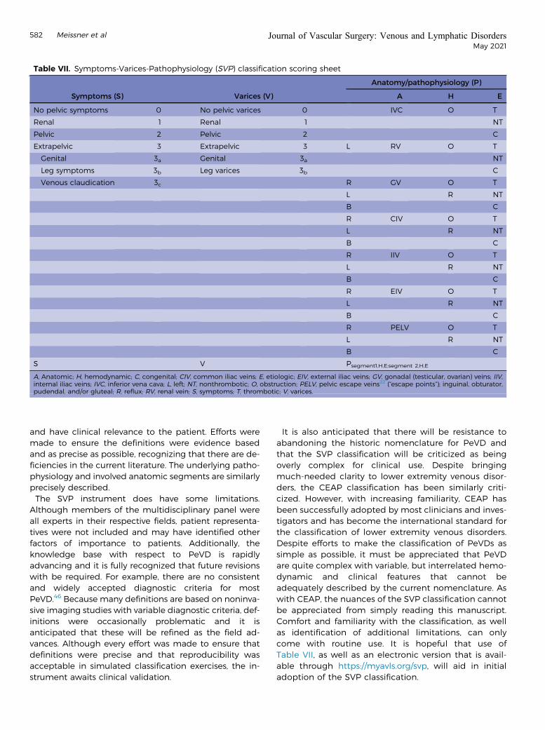
Table VII. Symptoms-Varices-Pathophysiology (SVP) classification scoring sheet
Symptoms (S) Varices (V)
Anatomy/pathophysiology (P)
A H E
No pelvic symptoms 0 No pelvic varices 0 IVC O T
Renal 1 Renal 1 NT
Pelvic 2 Pelvic 2 C
Extrapelvic 3 Extrapelvic 3 L RV O T
Genital 3a Genital 3a NT
Leg symptoms 3b Leg varices 3b C
Venous claudication 3c R GV O T
L R NT
B C
R CIV O T
L R NT
B C
R IIV O T
L R NT
B C
R EIV O T
L R NT
B C
R PELV O T
L R NT
B C
S V Psegment1,H,E;segment 2,H,E
A, Anatomic; H, hemodynamic; C, congenital; CIV, common iliac veins; E, etiologic; EIV, external iliac veins; GV, gonadal (testicular, ovarian) veins; IIV,internal iliac veins; IVC, inferior vena cava; L, left; NT, nonthrombotic; O, obstruction; PELV, pelvic escape veins22 (“escape points”); inguinal, obturator,pudendal, and/or gluteal; R, reflux; RV, renal vein; S, symptoms; T, thrombotic; V, varices.
582 Meissner et al Journal of Vascular Surgery: Venous and Lymphatic DisordersMay 2021
and have clinical relevance to the patient. Efforts weremade to ensure the definitions were evidence basedand as precise as possible, recognizing that there are de-ficiencies in the current literature. The underlying patho-physiology and involved anatomic segments are similarlyprecisely described.The SVP instrument does have some limitations.
Although members of the multidisciplinary panel wereall experts in their respective fields, patient representa-tives were not included and may have identified otherfactors of importance to patients. Additionally, theknowledge base with respect to PeVD is rapidlyadvancing and it is fully recognized that future revisionswith be required. For example, there are no consistentand widely accepted diagnostic criteria for mostPeVD.46 Because many definitions are based on noninva-sive imaging studies with variable diagnostic criteria, def-initions were occasionally problematic and it isanticipated that these will be refined as the field ad-vances. Although every effort was made to ensure thatdefinitions were precise and that reproducibility wasacceptable in simulated classification exercises, the in-strument awaits clinical validation.
It is also anticipated that there will be resistance toabandoning the historic nomenclature for PeVD andthat the SVP classification will be criticized as beingoverly complex for clinical use. Despite bringingmuch-needed clarity to lower extremity venous disor-ders, the CEAP classification has been similarly criti-cized. However, with increasing familiarity, CEAP hasbeen successfully adopted by most clinicians and inves-tigators and has become the international standard forthe classification of lower extremity venous disorders.Despite efforts to make the classification of PeVDs assimple as possible, it must be appreciated that PeVDare quite complex with variable, but interrelated hemo-dynamic and clinical features that cannot beadequately described by the current nomenclature. Aswith CEAP, the nuances of the SVP classification cannotbe appreciated from simply reading this manuscript.Comfort and familiarity with the classification, as wellas identification of additional limitations, can onlycome with routine use. It is hopeful that use ofTable VII, as well as an electronic version that is avail-able through https://myavls.org/svp, will aid in initialadoption of the SVP classification.

Journal of Vascular Surgery: Venous and Lymphatic Disorders Meissner et al 583
Volume 9, Number 3
The SVP instrument is a starting point in bringinggreater scientific rigor to PeVDs. It is presumed that,much like lower extremity CEAP, the instrument will becarefully studied and any deficiencies addressed infuture revisions. However, it is only through the precisedefinition of homogenous patient populations that clin-ical care can be optimized, appropriate outcome instru-ments developed, and rigorous clinical trials conducted.
REFERENCES1. Khilnani NM, Meissner MH, Learman LA, Gibson KD, Daniels JP,
Winokur RS, et al. Research priorities in pelvic venous disorders inwomen: recommendations from a multidisciplinary researchconsensus panel. J Vasc Interv Radiol 2019;30:781-9.
2. Beebe HG, Bergan JJ, Bergqvist D, Eklof B, Eriksson I, Goldman MP,et al. Classification and grading of chronic venous disease in thelower limbs. A consensus statement. Eur J Vasc Endovasc Surg1996;12:487-91.
3. Eklof B, Rutherford RB, Bergan JJ, Carpentier PH, Gloviczki P,Kistner RL, et al. Revision of the CEAP classification for chronicvenous disorders: consensus statement. J Vasc Surg 2004;40:1248-52.
4. Lurie F, Passman M, Meisner M, Dalsing M, Masuda E, Welch H, et al.The 2020 update of the CEAP classification system and reportingstandards. J Vasc Surg Venous Lymphat Disord 2020;8:342-52.
5. Meissner MH, Gibson K. Clinical outcome after treatment of pelviccongestion syndrome: sense and nonsense. Phlebology 2015;30:73-80.
6. Greiner M, Dadon M, Lemasle P, Cluzel P. How does the patho-physiology influence the treatment of pelvic congestion syndromeand is the result long-lasting? Phlebology 2012;27(Suppl 1):58-64.
7. Guyatt GH, Kirshner B, Jaeschke R. A methodologic framework forhealth status measures: clarity or oversimplification? J Clin Epidemiol1992;45:1353-5.
8. Kirshner B, Guyat G. A methodological framework for assessinghealth indices. J Chron Dis 1985;38:27-36.
9. Kurklinsky AK, Rooke TW. Nutcracker phenomenon and nutcrackersyndrome. Mayo Clin Proc 2010;85:552-9.
10. American College of Obstetricians and Gynecologists. ReVITALize.Gynecology data definitions (version 1.0) Washington, D.C.: Amer-ican College of Obstetricians and Gynecologists; 2018. Available at:www.acog.org/-/media/project/acog/acogorg/files/pdfs/publications/revitalize-gyn.pdf. Accessed March 10, 2021.
11. Williams RE, Hartmann KE, Steege JF. Documenting the currentdefinitions of chronic pelvic pain: implications for research. ObstetGynecol 2004;103:686-91.
12. Phillips D, Deipolyi AR, Hesketh RL, Midia M, Oklu R. Pelvic conges-tion syndrome: etiology of pain, diagnosis, and clinical management.J Vasc Interv Radiol 2014;25:725-33.
13. Zondervan KT, Yudkin PL, Vessey MP, Jenkinson CP, Dawes MG,Barlow DH, et al. Chronic pelvic pain in the community–symptoms,investigations, and diagnoses. Am J Obstet Gynecol 2001;184:1149-55.
14. Beard RW, Highman JH, Pearce S, Reginald PW. Diagnosis of pelvicvaricosities in women with chronic pelvic pain. Lancet 1984;2:946-9.
15. Beard RW, Reginald PW, Wadsworth J. Clinical features of womenwith chronic lower abdominal pain and pelvic congestion. Br JObstet Gynaecol 1988;95:153-61.
16. Scultetus AH, Villavicencio JL, Gillespie DL, Kao TC, Rich NM. Thepelvic venous syndromes: analysis of our experience with 57 patients.J Vasc Surg 2002;36:881-8.
17. Santoshi RKN, Lakhanpal S, Satwah V, Lakhanpal G, Malone M,Pappas PJ. Iliac vein stenosis is an underdiagnosed cause of pelvicvenous insufficiency. J Vasc Surg Venous Lymphat Disord 2018;6:202-11.
18. Daugherty SF, Gillespie DL. Venous angioplasty and stenting improvepelvic congestion syndrome caused by venous outflow obstruction.J Vasc Surg Venous Lymphat Disord 2015;3:283-9.
19. Herrera-Betancourt AL, Villegas-Echeverri JD, Lopez-Jaramillo JD,Lopez-Isanoa JD, Estrada-Alvarez JM. Sensitivity and specificity of
clinical findings for the diagnosis of pelvic congestion syndrome inwomen with chronic pelvic pain. Phlebology 2018;33:303-8.
20. RanaN,DrakeMJ, RinkoR,DawsonM,WhitmoreKE. The fundamentalsof chronic pelvic pain assessment, based on international continencesociety recommendations. Neurourol Urodyn 2018;37:S32-8.
21. Potts JM. Chronic pelvic pain syndrome: a non-prostatocentricperspective. World J Urol 2003;21:54-6.
22. Kachlik D, Pechacek V, Musil V, Baca V. The venous system of thepelvis: new nomenclature. Phlebology 2010;25:162-73.
23. Labropoulos N, Tassiopoulos AK, Gasparis AP, Phillips B, Pappas PJ.Veins along the course of the sciatic nerve. J Vasc Surg 2009;49:690-6.
24. Ricci S, Georgiev M, Jawien A, Zamboni P. Sciatic nerve varices. Eur JVasc Endovasc Surg 2005;29:83-7.
25. Meissner MH, Eklof B, Smith PC, Dalsing MC, DePalma RG,Gloviczki P, et al. Secondary chronic venous disorders. J Vasc Surg2007;46(Suppl S):68S-83S.
26. Delis KT, Bountouroglou D, Mansfield AO. Venous claudication iniliofemoral thrombosis: long-term effects on venous hemodynamics,clinical status, and quality of life. Ann Surg 2004;239:118-26.
27. Perrin M, Eklof B, van Rij A, Labropoulos N, Vasquez M, Nicolaides A,et al. Venous symptoms: the SYM Vein Consensus statement devel-oped under the auspices of the European Venous Forum. Int Angiol2016;35:374-98.
28. Gloviczki P, Cho J-S. Surgical treatment of chronic occlusions of theiliac veins and inferior vena cava. In: Rutherford RB, editor. VascularSurgery. 6th ed. Philadelphia (PA): Elsevier, Inc; 2005. p. 2303-20.
29. Paty J, Elash CA, Turner-Bowker DM. Content validity for theVVSymQ(�) Instrument: a new patient-reported outcome measurefor the assessment of varicose veins symptoms. Patient 2017;10:51-63.
30. Kim KW, Cho JY, Kim SH, Yoon JH, Kim DS, Chung JW, et al. Diag-nostic value of computed tomographic findings of nutcracker syn-drome: correlation with renal venography and renocaval pressuregradients. Eur J Radiol 2011;80:648-54.
31. Takebayashi S, Ueki T, Ikeda N, Fujikawa A. Diagnosis of thenutcracker syndrome with color Doppler sonography: correlationwith flow patterns on retrograde left renal venography. AJR Am JRoentgenol 1999;172:39-43.
32. Kim SH. Doppler US and CT diagnosis of Nutcracker Syndrome.Korean J Radiol 2019;20:1627-37.
33. Beinart C, Sniderman KW, Tamura S, Vaughan ED, Sos TA. Left renalvein to inferior vena cava pressure relationship in humans. J Urol1982;127:1070-1.
34. Beckmann CF, Abrams HL. Idiopathic renal vein varices: incidenceand significance. Radiology 1982;143:649-52.
35. Hangge PT, Gupta N, Khurana A, Quencer KB, Albadawi H,Alzubaidi SJ, et al. Degree of left renal vein compression predictsNutcracker Syndrome. J Clin Med 2018;7:1-8.
36. Park SJ, Lim JW, Ko YT, Lee DH, Yoon Y, Oh JH, et al. Diagnosis ofpelvic congestion syndrome using transabdominal and transvaginalsonography. AJR Am J Roentgenol 2004;182:683-8.
37. Steenbeek MP, van der Vleuten CJM, Schultze Kool LJ, Nieboer TE.Noninvasive diagnostic tools for pelvic congestion syndrome: a sys-tematic review. Acta Obstet Gynecol Scand 2018;97:776-86.
38. Gray HR In: Pick TP, Howden R, editors. Gray’s Anatomy. 15th ed. NewYork (NY): Barnes & Noble; 2010.
39. Kauppila A. Uterine phlebography with venous compression. Aclinical and roentgenological study. Acta Obstet Gynecol Scand1970;3(Suppl 3):1-66.
40. Arnoldussen CW, de Wolf MA, Wittens CH. Diagnostic imaging ofpelvic congestive syndrome. Phlebology 2015;30:67-72.
41. Malgor RD, Adrahtas D, Spentzouris G, Gasparis AP, Tassiopoulos AK,Labropoulos N. The role of duplex ultrasound in the workup of pelviccongestion syndrome. J Vasc Surg Venous Lymphat Disord 2014;2:34-8.
42. Whiteley MS, Dos Santos SJ, Harrison CC, Holdstock JM, Lopez AJ.Transvaginal duplex ultrasonography appears to be the gold stan-dard investigation for the haemodynamic evaluation of pelvicvenous reflux in the ovarian and internal iliac veins in women.Phlebology 2015;30:706-13.
43. Labropoulos N, Jasinski PT, Adrahtas D, Gasparis AP, Meissner MH.A standardized ultrasound approach to pelvic congestion syndrome.Phlebology 2017;32:608-19.

584 Meissner et al Journal of Vascular Surgery: Venous and Lymphatic DisordersMay 2021
44. Hansrani V, Dhorat Z, McCollum CN. Diagnosing of pelvic veinincompetence using minimally invasive ultrasound techniques.Vascular 2017;25:253-9.
45. Dos Santos SJ, Holdstock JM, Harrison CC, Lopez AJ, Whiteley MS.Ovarian vein diameter cannot be used as an indicator of ovarianvenous reflux. Eur J Vasc Endovasc Surg 2015;49:90-4.
46. Champaneria R, Shah L, Moss J, Gupta JK, Birch J, Middleton LJ, et al.The relationship between pelvic vein incompetence and chronicpelvic pain in women: systematic reviews of diagnosis and treatmenteffectiveness. Health Technol Assess 2016;20:1-108.
47. Lemasle P, Greiner M. Duplex ultrasound investigation in pelviccongestion syndrome: technique and results. Phlebolymphology2017;24:79-87.
48. Black CM, Thorpe K, Venrbux A, Kim HS, Millward SF, Clark TW, et al.Research reporting standards for endovascular treatment of pelvicvenous insufficiency. J Vasc Interv Radiol 2010;21:796-803.
49. Gagne PJ, Gasparis A, Black S, Thorpe P, Passman M, Vedantham S,et al. Analysis of threshold stenosis by multiplanar venogram andintravascular ultrasound examination for predicting clinicalimprovement after iliofemoral vein stenting in the VIDIO trial. J VascSurg Venous Lymphat Disord 2018;6:48-56e41.
50. Neglen P, Raju S. Intravascular ultrasound scan evaluation of theobstructed vein. J Vasc Surg 2002;35:694-700.
51. Labropoulos N, Borge M, Pierce K, Pappas PJ. Criteria for definingsignificant central vein stenosis with duplex ultrasound. J Vasc Surg2007;46:101-7.
52. Raju S, Kirk O, Davis M, Olivier J. Hemodynamics of "critical" venousstenosis. J Vasc Surg Venous Lymphat Disord 2014;2:52-9.
53. Metzger PB, Rossi FH, Kambara AM, Izukawa NM, Saleh MH, Pinto IM,et al. Criteria for detecting significant chronic iliac venous obstruc-tions with duplex ultrasound. J Vasc Surg Venous Lymphat Disord2016;4:18-27.
54. Choudur HN, Joshi R, Munk PL. Inferior gluteal vein varicosities: a rarecause of sciatica. J Clin Rheumatol 2009;15:387-8.
55. Rutherford RB, Padberg FT, Comerota AJ, Kistner RL, Meissner MH,Moneta GL. Venous severity scoring: An adjunct to venous outcomeassessment. J Vasc Surg 2000;31:1307-12.
56. Vasquez MA, Rabe E, McLafferty RB, Shortell CK, Marston WA,Gillespie D, et al. Revision of the venous clinical severity score: venousoutcomes consensus statement: special communication of theAmerican Venous Forum Ad Hoc Outcomes Working Group. J VascSurg 2010;52:1387-96.
57. Meissner MH, Gloviczki P. Pelvic venous disorders. In: Almeida JI,editor. Atlas of endovascular venous surgery. 2nd ed. Philadelphi(PA): Elsevier; 2019. p. 567-99.
58. Holdstock JM, Dos Santos SJ, Harrison CC, Price BA, Whiteley MS.Haemorrhoids are associated with internal iliac vein reflux in up toone-third of women presenting with varicose veins associated withpelvic vein reflux. Phlebology 2015;30:133-9.
59. Margetis N. Pathophysiology of internal hemorrhoids. Ann Gastro-enterol 2019;32:264-72.
60. van der Vleuten CJ, van Kempen JA, Schultze-Kool LJ. Embolizationto treat pelvic congestion syndrome and vulval varicose veins. Int JGynaecol Obstet 2012;118:227-30.
61. Zagriadskii EA, Bogomazov AM, Golovko EB. Conservative treatmentof hemorrhoids: results of an observational multicenter study. AdvTher 2018;35:1979-92.
62. Aziz Z, Huin WK, Badrul Hisham MD, Tang WL, Yaacob S. Effi-cacy and tolerability of micronized purified flavonoid fractions(MPFF) for haemorrhoids: a systematic review and meta-anal-ysis. Complement Ther Med 2018;39:49-55.
63. Pata F, Sgro A, Ferrara F, Vigorita V, Gallo G, Pellino G. Anatomy,physiology and pathophysiology of haemorrhoids. Rev Recent ClinTrials 2020 April 6. [Epub ahead of print].
Submitted Nov 28, 2020; accepted Dec 5, 2020.