Embed Size (px)
Citation preview

The Taenia solium Genome Project
Universidad Nacional Autónoma de México

Institute of Biotechnology:E Morett, X Soberón, A Garcíarrubio, P. Gaytan, J. Yañez
Center of Genomic Sciences:MA Cevallos, VM González,
School of Medicine:A. Landa, L Jiménez
School of Sciences:V. Valdés
Institute of Biomedical Research:G. Fragoso, C Larralde, J Morales-Montor, E Sciutto, JC Carrero, JP Laclette,
M. José, P. de la Torre, R. Bobes.
The ConsortiumThe Consortium

• Virginia Walbot, Stanford University, USA
• Bruce Roe, Oklahoma University, USA
• Luis Herrera-Estrella, CINVESTAV-Irapuato, MEX
• Charles, B. Shoemaker, Tufts University, USA
• Klaus Brehm, University of Wurzburg, GER
Advisory BoardAdvisory Board

1. Taenia solium is the causal agent of human and porcine cysticercosis; a diseasethat still is a public health problem of considerable relevance in México and inseveral other countries.
2. This parasite/disease has been studied by multiple groups in Mexico during atleast three decades. A considerable number of contributions on the understandingof the parasite and disease have been made by Mexican scientists. T. solium isan organism that the Mexican scientific community can justifiably appropriate.
3. A genomic project of this magnitude (estimated genome size 120 ~ 270 Mb) willpromote the organization of a human team able to approach this and otherprojects in genomic sciences, by networking current capabilities in severalresearch centers at UNAM. The project requires a considerable capability on DNAsequencing and a parallel capability on bioinformatics.
4. The project will contribute to the knowledge of an organism with an interestingphylogenetic position for studies of comparative genomics, etc.
5. A project of this magnitude will unite groups with diverse disciplinarybackgrounds: immunology, molecular biology, cell biology, bioinformatics,among others.
Justification of the ProjectJustification of the Project

6. The complete sequence of the T. solium genome will provide us with a much betterunderstanding of the parasite’s physiology, life cycle and metabolism. Also, theknowledge of the complete enzymatic repertoire will help us to identify essentialgenes, which will be good candidates to be targeted by newly designed drugs.

Life cycle of Taenia solium
• Man is the definitive host and harbors the adulttapeworm in the upper small intestine.
• Three stages: larval, embryo, and adult. Caninfect man both as larva or as adult.
• Infection with adult occurs through eatinguncooked pork. (Embryo eaten and turns into adultin the intestine).
• Infection with the larva causes cysticercosis.Occurs when animals or humans become theintermediate host of the larval form. Occurs byinjesting eggs. The eggs hatch in the intestine andthe larvae burrow through the intestinal walls anddisseminate into the soft tissues.
MAN (adult tapeworm in upper small intestine)Eggs intermittently eliminated in fecesConsumed by PIGS and MANOva develop into larvae which infect the tissuesMAN consumes pork

The adult tapeworm

Definition• A parasitic infection involving the CNS caused by the larval stage of the pork tapeworm, taenia solium, which has a markedpredilection for neural tissue.• Infection caused by Cysticercus cellulose which is the larval form.
Epidemiology• Cysticercosis is the most common parasitic infection involving the CNS.• Endemic in areas of Mexico, eastern Europe, Asia, Central and South America, and Africa.• Incidence of Neurocysticercosis (CNS involvement) may reach 4% in some areas.• 80% of infected individuals show symptoms within 7 years of infection
• Man is the definitive host and harbors the adult tapeworm in the upper small intestine. The worm attaches to the wall of thejeujunum by 4 suckers and 2 hooklets, where it absorbs food directly. Man is the only permanent host of the adult form.Proglottids (mature segments, each containing reproductive organs) ladened with eggs are eliminated intermittently in the feces. Ifconsumed by the intermediate host, the hog, the ova will develop into larvae or onchospheres that penetrate the intestinal wall,then invade the lymphatics and veins, and then disseminates into skeletal muscles and other tissues. Man becomes infected whenhe ingests poorly cooked, infected pork. The consumed cysticercus develops an evaginated scolex that attaches to the jejunalmucosa and develops into an adult worm in the human intestine, completing its life cycle.
• Acquisition of larval forms producing cysticercosis occurs after ingestion of T. solium eggs in food or water contaminated withinfected feces. The ova can also be acquired by oral transmission from unclean hands of carriers of the adult tapeworm. Internalautoinfection is also possible by regurgitation of eggs from the jejunum into the stomach through reverse peristalsis. Within 2months, the larvae develop a cyst wall. Within 4 months, they mature into embryos. Embryos can survive 5-7 yrs. Those whichdie, calcify. When the pigs are eaten, the embryos remain viable and are ingested with the life cycle being repeated.
• Onchospheres once outside the GI tract invade other tissues and become mature cysticerci which are oval, translucent cysts,containing a single scolex bearing four suckers. Within 12 weeks, the cysticerci mature and are primarily observed in skeletalmuscle; other infected sites include the brain, eyes, liver, lung and subcutaneous tissues
Cysticercosis

Pathophysiology Cisticercosis (infection with the larva) involves the following common sites and symptoms are referrable to these sites.i) Brain (60-90%)ii) Skeletal muscle, eye, subcutaneous tissueGross• Two types of cysts develop in the brain:1. Cysticercus cysts. Thin walled cysts 3-20mm in diameter forming in the parenchyma or subarachnoid spaces. They contain thescolex (head), and are usually static, producing little inflammation.2. Racemose cysts. Large (4-12cm), grow actively and produce grape-like clusters in the basal subarachnoid space with intenseinflammation. They do not contain larvae.Microscopic• Should identify scolex with four suckers and anterior hooklets. Live cysticerci evoke little inflammatory response. Deadcysticerci evoke intense responses, monocytes, macrophages.
CNS Manifestations of Cysts1. Diffuse parenchymatous disease. Without focal mass; disseminated larval death; inflammatory reaction, toxic encephalopathyand meningitis. This probably represents the acute encephalitic phase described in some reports (J of Child Neurology, 10:177,1995). This form is uncommon but more frequently reported in children. A subacute or chronic cysticercotic encephalitis alsoexists, caused by degeneration of multiple cysticerci.2. Cysts (classified by location).i) Intraparenchymatous cysts as mass lesions. Solitary or part of multifocal disease. Tend to present with seizures.ii) Subarachnoid and cisternal cysts.The commonest location for extraventricular, nonparenchymatous CNS involvement is thebasal cisterns(found in almost all cisterns). Often forms a basal meningitis in the cisterns. These are usually of the racemosevariety. Dosolateral subarachnoid space can be involved by the cysticerci type and usually cause minimal symptoms.iii) Intraventricular cysts. May be solitary or multiple.iv) Spinal cystsv) Mixed lesions3. Basilar adhesive and racemose form. Obliterative arachnoiditis; hydrocephalus; mixed types with cisternal cysts or spinaldisease.4. Ventriculitis. Arachnoiditis/meningitis; hydrocephalus; mixed types with intraventricular or cisternal cysts.5. Calcified larval form with seizures.
Pathology

Clinical• Manifestations are the consequence of the organism lodging in the meninges, parenchyma, subarachnoid space,etc. This can cause blockage of CSF flow, a mass lesion, chronic meningitis, cranial neuropathy, and/or seizures.
Laboratory Investigations• Diagnosis is by:1. Identification of probable epidemiologic exposure.2. Origin of patient from endemic region3. History and physical4. Radiological data5. Serologyi) Eosinophilia. Suggestive of parasitic infection but is inconsistent and unreliable.ii) Serology of serum and CSF. Cysticercosis antibody titers determined by ELISA. Indirect hemagglutinins orindirect immuofluorescence.iii) CSF may be normal. Pleocytosis (eosinophils) seen in 15%.(NB. T. solium ova only present in the stool in 1/3 of cases.
Imaging• Soft tissue X-rays may show calcified nodules.• On CT scans, ring-enhancing lesions represent living cysticerci. Little edema as long as larva is alive. Centralpunctate high density probably represents scolex. Calcified (dead) forms of larvae are identified best by CT.Usually without surrounding edema.• Contrast enhancement generally does not identify the walls of live cysts in the CSF spaces. Water solublepositive contrast ventriculography or cisternography historically was the best method for detecting lesions in theCSF spaces. MRI is the preferred imaging modality for these lesions. A T2 weighted image (or FLAIR) will oftenreveal multiple areas of signal prolongation. Following gadolinium, the cysts with the scolex can be seenclearly.(248k)
Clinical Manifestations

Medical1. Steroids: indicated during acute neurological deterioration or while on other therapy.2. Antihelminthics. A single dose will kill the tapeworm.i) Praziquantel. May be used in basilar adhesive arachnoiditis, cisternal cystic lesions, and active parenchymatous disease.Given as 50 mg/kg in three divided doses for 15 days. Drug of choice for intestinal infestation.ii) Albendazole. A newer agent which is superior to praziquantel. 15 mg/kg divided into 2-3 doses for 3 months.iii) Niclosamide. May be used to treat GI tapeworms.
Surgical• Indications:1. Establishing the diagnosis (by biopsy, stereotactic or open)2. Treatmenti) Palliation of hydrocephalus.ii) Excision of intracisternal or intraparenchymatous cysts because of mass effect.iii) Excision of intraventricular cysts.iv) Excision of rare spinal masses.
• Total surgical excision is necessary; the wall of the larval form may continue to cause difficulties even in the absence ofa persistent scolex.• Little danger of disseminating cysts through CSF diversion.• Intraventricular cysts should be removed surgically whenever possible expecially when they cause obstruction of the IVthventricle.IVth ventricular cysts are the commonest• Treat patients with steroids perioperatively
Treatment & Results

Cysticercosis in the world

Sequencing the Taenia solium genome
May 2007 report

Sequences are produced at 3 sequencing centers.
CINVESTAV (Irapuato).Centro de Ciencias Genómicas (Cuernavaca).Instituto de Biotecnología (Cuernavaca).
All sequences are collected and analyzed at IBT.

There are 2 kinds of projects:1 ESTs2 Genomic shotgun
Two technologies are used:
Sanger:Pros: long reads, higher quality, easier assemblyCons: more expensive, some genome regions could bepoorly represented
454 pyrosequencing:Pros: cheaper, high throughput, no cloning required,unbiased representation.Cons: short reads, lower quality, homo-nucleotidecompression, difficult to assemble.

DNA library inSephadex beads
Water-oilemulsion
microreactors
Clonal amplification by PCR
Break of microreactors
a) PCR emulsion and clonal PCR
b) Pyrosequencing
loading in chamber
DNA synthesis coupled to light emissionCCD detection
454 Pyrosequencing

The libraries
Up to now, 8 "libraries" have been built:
An adult cDNA library (cd1)Two larva cDNA libraries (cd2 and cd3)A genomic library with 2-5 Kb inserts (sg1)A genomic library with 1-3 Kb inserts (sg2)A genomic library with 7-10 Kb inserts (sg3, startingsequencing)A genomic fosmid library (sg4)Several anonymous 454 libraries (454)
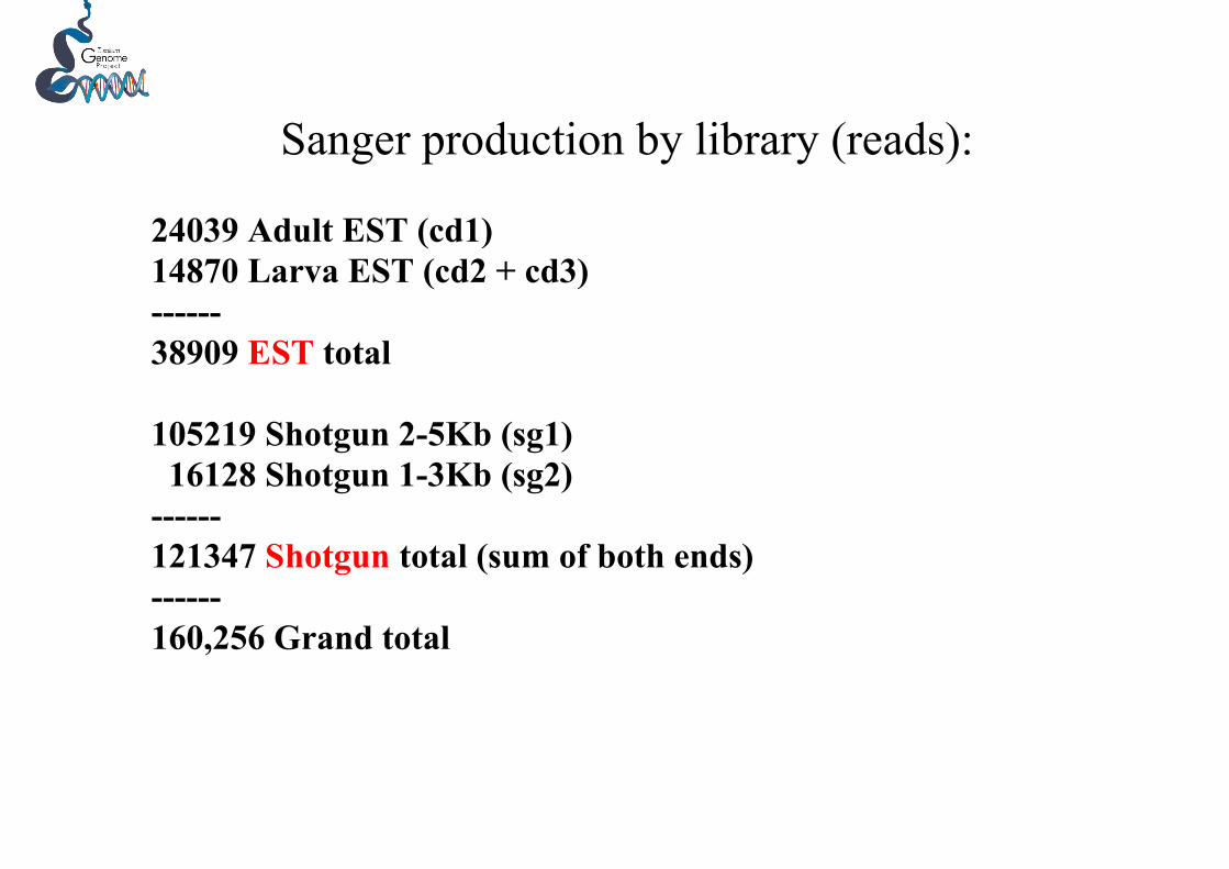
Sanger production by library (reads):
24039 Adult EST (cd1)14870 Larva EST (cd2 + cd3)------38909 EST total
105219 Shotgun 2-5Kb (sg1) 16128 Shotgun 1-3Kb (sg2)------121347 Shotgun total (sum of both ends)------160,256 Grand total
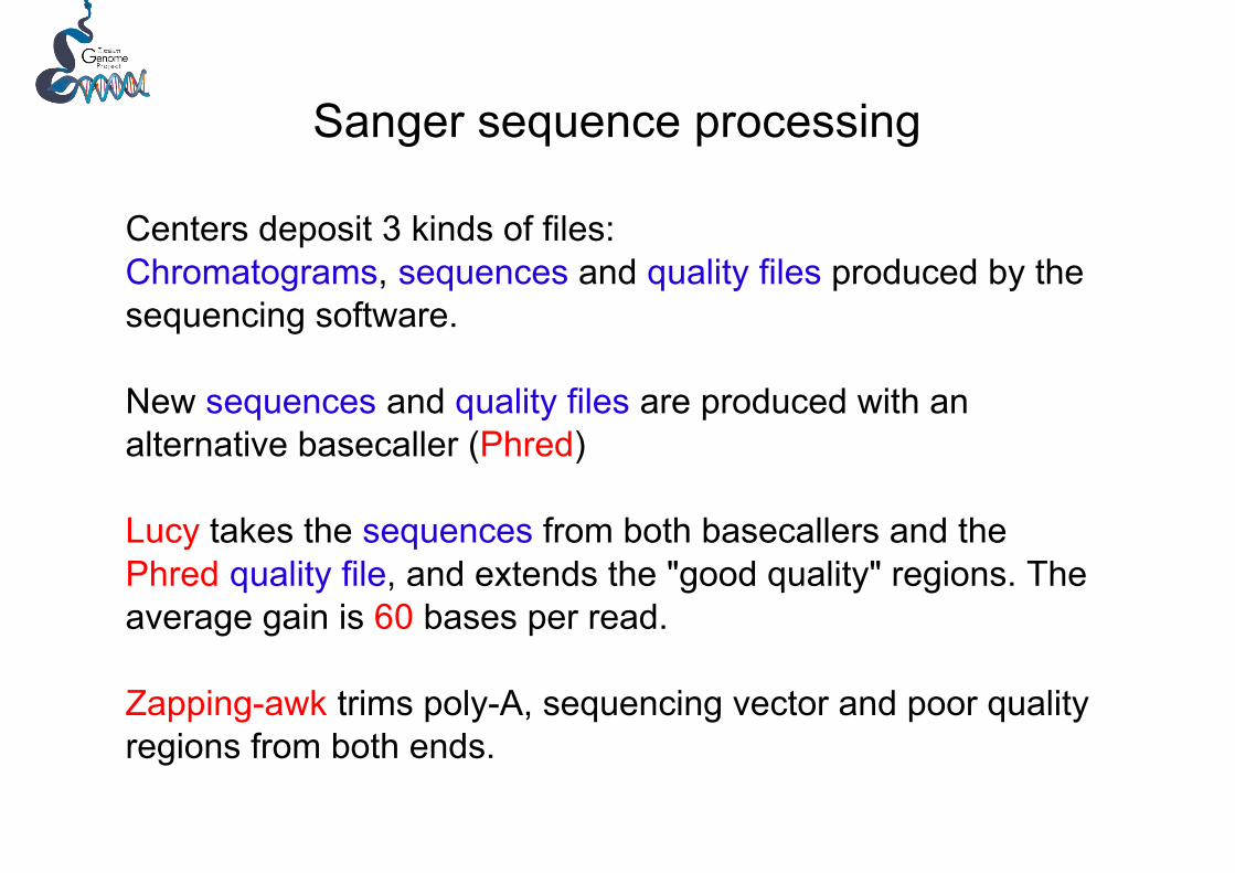
Sanger sequence processing
Centers deposit 3 kinds of files:Chromatograms, sequences and quality files produced by thesequencing software.
New sequences and quality files are produced with analternative basecaller (Phred)
Lucy takes the sequences from both basecallers and thePhred quality file, and extends the "good quality" regions. Theaverage gain is 60 bases per read.
Zapping-awk trims poly-A, sequencing vector and poor qualityregions from both ends.

Sequence number and average length aftertrimming
(initial/ final /bases)
cd1 24039 17529 568cd2 12470 9132 582cd3 2400 1409 531
sg1 55219 50405 665sg2 16128 14130 640

Sequence cleaning
Trimmed sequences (zap.seq) are masked against thecloning vector, the sequencing vector, the NCBI UniVect database, Lambda phage, Eschericia coli genome, and the T.solium mitochondrion.
Chimerism is detected by "wrong place" poly-A presence andby internal vector-insert splice sites.
Chimeras are cleaved judiciously and all masked regions areremoved. The resulting fragments longer than 150bp arekept (split.seq).
Chimeras are frequent in the EST libraries

Size distributions of clean (split.seq) ESTs

Size distributions of clean (split.seq) shotgun traces

454 sequencingThere have been 56 runs, each producing ~20Mb.
Read are assembled at the flowgram level by the 454 “denovo” assembler (Newbler).
28 runs is the maximum that Newbler can handle, so thereads have been assembled in two batches, with ~560 Mbeach.
Both batches behaved very similarly: they produced ~79 Mbof assembled sequence, incorporating >95% of the reads.Assemblies included 362 and 382 thousand contigs, withaverage contig lengths of 217 and 203 bases.

454 sequence reassembly
Contigs from both batches were assembled at the sequencelevel with PCAP. The number of contigs was reduced from744 K to 215 K, while average length almost doubled (from210 to 382 bases).
N50 (the smallest size in the collection of the longestcontigs which hold 50% of the assembled sequence)changed from 239 to 448 bases.
There are 130 contigs longer than 5Kb. The longest one has16336 bases.

Whole genes in the 454 contigsNCBI has 95 complete CDS T. solium sequences. After eliminatingvery similar sequences (nr90) we made a collection of 45 wholegenes.
Using MegaBlast, four genes did not match the 454 contigs, and only2 genes were entirely contained in a single contig. Aprox. 65% of thegenes were covered by contigs.
Most genes matched 2 or more contigs, which indicates that gapsizes in the 454 assembly are within the range of gene sizes.
If the sequences of the genes are taken as correct, the 454 sequenceshave an error rate slightly over 1% (1.08%)
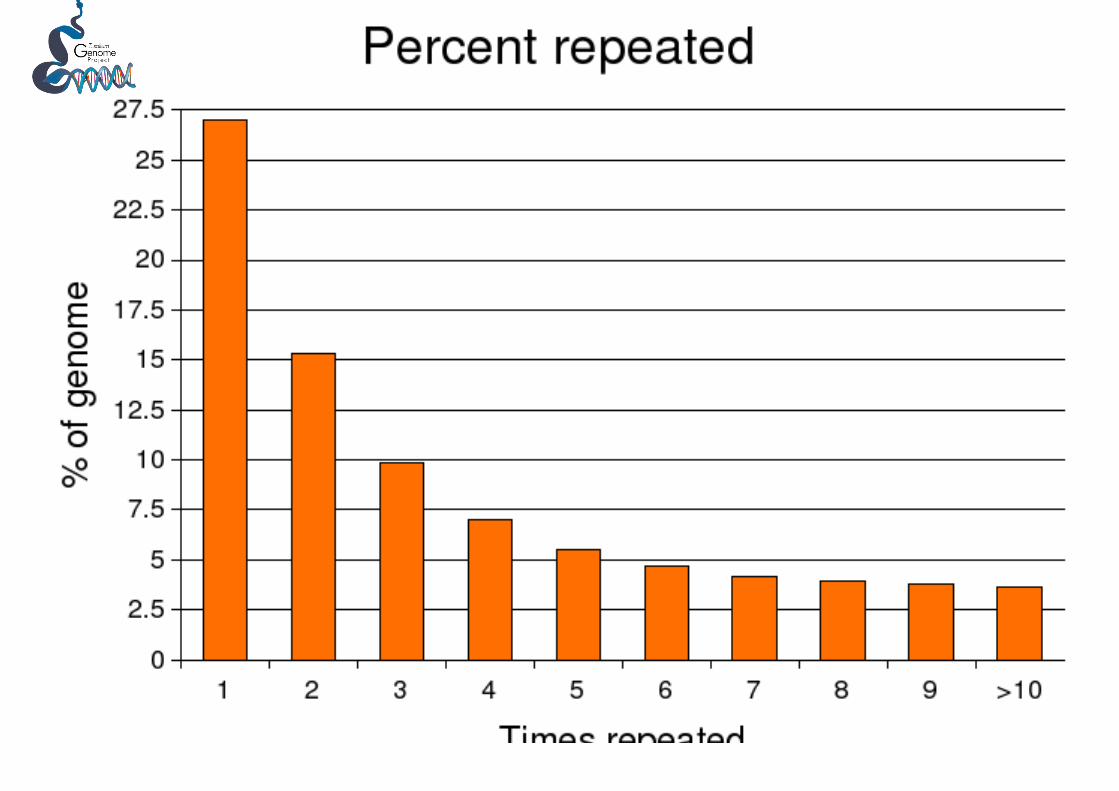
What is the amount of repeated sequences in thegenome?
ReapeatMasker suggests it is ~6.8%
The naïve approach of comparing all sequences vs all sequences,and taking as repeated any segment with at least 4 hits, gives asimilar value: 7.0%
The following small (53 bp) tandem repeat represents 0.5% of thegenome:
CGCTCTCACTGAATGCGATTTCGTATGAGTGATC-TTCACCAGACTGCAGATTT (Pst I)
Different tetranucleotide repeats of the form (Txxx)n amount to4.5% of the genome.

The genome sizeThe “footprint” calculation
Presently, the sum of 454 contigs is 82Mb.
Those 82Mb should be 65% of the genome, because 65% of NCBIcomplete CDS sequences find a match in those contigs, and ,similarly, 65% of ‘unique’ (non repeated) sequences from the sangershotgun (10.2 out of 15.9 Mb) find a match in those contigs.
Thus, the genome size must be:
82Mb /0.65 = 126Mb

The “coverage” calculation.
At 560 Mb of 454 sequence (half the amount we have now) weestimated that the distribution of reads per contig was consistentwith a 4-5X coverage. The calculation did not require assuming agenome size.
From this data we inferred that the genome size should be in therange:
560Mb/5 =112 Mb to 560Mb/4=140Mb
So this and the footprint calculation are in good agreement

The Lander-Waterman corollary
According to the Lander-Waterman formula of random nucleotidesampling, for any genome size, there should be a 5% decrease ingaps (a 5% increase in sequence) when going from 3X to 6Xcoverage.
That is nearly what happened when we assembled the two 560Mbbatches; so this is consistent with a 560MB batch being 3X thegenome.
However, that same formula shows that at 6X, 99.7% of thesequence should be contained. Thus, the genome size must be 82Mb.
This is consistent with the saturation of the curves we have observed,but contradicts the fact that the contigs are missing ~35% of allsearched query sequences, as shown in the footprint calculation.

Tota
l len
gth
of c
ontig
s sho
rter t
han
X
X= Contig length
Saturation of the 454 assembly

From ESTs to genes
Unique genes are identified by clustering allEST-fragments with an assembler (minimus).
Out of the initial 23290 ESTs, 19067 wereincorporated into 2564 genes (contigs).
We have ~6800 “genes”, including the 2564contigs, plus 2592 larva and 1611 adult ESTsthat remained as solitons.
Highly expressed genes.
There are 349 “genes” with 10 or more sequences.
They account for 50% of all transcripts. Many aredifferentially expressed.(Note: because we have more adult than larvasequences, a 1.5X normalization factor should beapplied for proper comparison).

Larva/adult ESTs in 3 categories of high expression contigs
HE LV AD

Homology annotation
With a Blast cutoff ( expect < 1e-10 ), aprox 1/3 (2038) of the6800 genes have a significant match in SwissProt, so someannotation can be inferred.
Gene Ontology (GO) and Gene Ontology broad categories (GoSlim)can be asigned to 36% (2448 genes) of our genes.
Comparison of GoSlim annotations to the GoSlim annotations of C.elegans leads to contradictions, indicating that very little realinformation is being transferred.
Aprox. 27% of the genes do not have a match (expect< 1e-3) inSProt + TREMBL, so they could be new genes.


High expression genes with good SProt homologues

?

PolymorphismPolymorphism in the contigs can hint to assembly errors,sequencing errors or poor alignments. The 3 possibilities canfrequently be distinguished by close examination of thealignments.
We are analyzing correlated polymorphism: sequences thatshare non-consensus nucleotides at different positions. Thesecan hint to heterozygosis, gene duplications, and alternativesplicing.
Around 20% of the contigs that can be analyzed ( those with 4sequences or more) show correlated polymorphism. In general,this is not associated to a larva/adult division.

Strategies for the classification of polymorphisms
Heterozygosis can only explain dimorphism, and the divergentsequences tend to be aligned along their full lengths.
If the alignment is tri-morphic, or the divergent sequenceshave in common end positions that are distinct from the othersequences, duplication can be assumed.
Shared indels, close to highly divergent regions, can indicatealternative splicing.
The work is in progress, but a relevant data is that genes withparalogues should be less than 20%, probably ~10%.

Heterozygosity

Different length paralogues

Tri-morphic contig
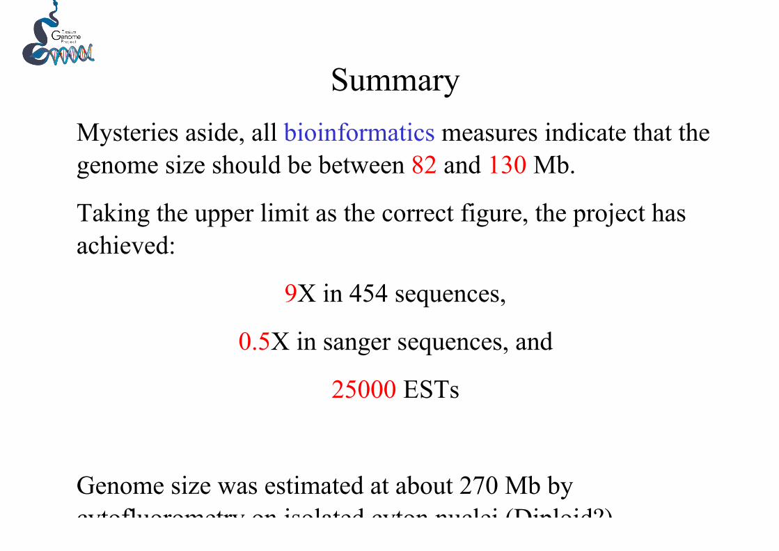
SummaryMysteries aside, all bioinformatics measures indicate that thegenome size should be between 82 and 130 Mb.
Taking the upper limit as the correct figure, the project hasachieved:
9X in 454 sequences,
0.5X in sanger sequences, and
25000 ESTs
Genome size was estimated at about 270 Mb bycytofluorometry on isolated cyton nuclei (Diploid?)

Other cousins (flatworms) being sequenced:
Schmidtea mediterranea (480Mb); WU; (3 million reads) 7Xcoverage + 50000 ESTs
Schistosoma mansoni (270Mb/8Chr); Sanger + TIGR; (370Kreads)
Isodiametra pulchra (Acoela); WU; 10,000-20,000 shotgun, planed
Clonorchis sinensis; Seoul National Univ.
Fasciola hepatica; Sanger; 15000 ESTs, finished
Echinococcus granulosus (150Mb); Sanger 10000 EST
Echinococcus multilocularis (150Mb); Sanger 10000 EST
Schistosoma haematobium (270 Mb); Sanger; 15000 ESTs

Analysis of Taenia solium adult and larvae cDNA libraries
14,000 adult clones 9,000 larvae clones
2,564 contigs 1,611 adult singleton clones 2,592 larvae singleton clones
Larvae has ca 60% fewer sequences but ca 60% more singletonsAdult has many genes with high level of expression.
Blast analysis with SwissProt to identify cDNAs with putative function.About half of the contigs showed identity with SwissProt entries

Exp_rank contig_id category exp_total exp_larva exp_adult Swiss_id
1 878 HE 422 30 392: Q9BMK3:
Swiss_descriptionFatty acid-binding protein.SIMILARITY: Belongs to the fatty-acid binding protein (FABP)
Fatty acid-binding protein
From higher to lower expression

Are fatty acids the main food source of T. solium?
Biochim Biophys Acta. 2000The fatty acid transport function of fatty acid-binding proteins.Storch J, Thumser AE.
The intracellular fatty acid-binding proteins (FABPs) comprise a family of 14-15 kDa proteins whichbind long-chain fatty acids... Collectively, data from these studies have provided strong support fordefining the FABPs as fatty acid transport proteins.

14 1559 AD 138 0 138: Q10442: Putative mitochondrial carrierMitochondrial aspartate-glutamate transporterSUBCELLULAR LOCATION: Mitochondrion; mitochondrial inner membrane;
Transport of glutamate in mitochondria is required for mitochondrialtransamination reactions and ornithine synthesis. Plays also a role inmalate-aspartate NADH shuttle, which is critical for growth on acetateand fatty acids!!!

Is the Fatty Acid Binding Protein of T. solium a goodcandidate for a vaccine?
Proc Natl Acad Sci U S A. 1996A Schistosoma mansoni fatty acid-binding protein, Sm14, is thepotential basis of a dual-purpose anti-helminth vaccineMiriam Tendler et alMolecular cloning of components of protective antigenic preparations has suggested that relatedparasite fatty acid-binding proteins could form the basis of the protective immune crossreactivitybetween the parasitic trematode worms Fasciola hepatica and Schistosoma mansoni... A recombinantform of the S. mansoni antigen, rSm14, protected outbred Swiss mice by up to 67% against challengewith S. mansoni cercariae...The same antigen also provided complete protection against challenge withF. hepatica metacercariae in the same animal model. The results suggest that it may be possible toproduce a single vaccine that would be effective against at least two parasites, F. hepatica and S.mansoni, of veterinary and human importance, respectively.

19: 213 HE 103 40 63: Q6P378: ActinFUNCTION: Actins are highly conserved proteins that are involved in various types of cell motility and areubiquitously expressed in all eukaryotic cells.
68: 1350 HE 40 11 29: P35432: Actin
110: 968 HE 27 10 17: P53456: Actin-2
37: 409 HE 65 12 53: P53456: Actin-2
122: 2236 HE 25 9 16: P53456: Actin
232: 365 HE 13 3 10: Q2KI95: Actin-binding LIM proteinSUBUNIT: Interacts with ZNF638 and TTN/titin
36: 266 HE 67 12 55: Q24800: SeverinFUNCTION: Severin blocks the ends of F-actin and causes the fragmentation and depolymerization of actinfilaments. This severin binds stably with actin both in a Ca(2+) dependent and a Ca(2+) independent manner.
42: 124 HE 57 15 42: Q24800: Severin192: 1971 AD 16 1 15: Q24800: Severin
322: 636 HE 10 4 6: P45594: Cofilin/actin-depolymerizing factor326: 2442 HE 10 4 6: P45594: Cofilin/actin-depolymerizing factor
348: 1852 HE 10 1 9: Q3SYZ8: PDZ and LIM domain protein 3FUNCTION: May play a role in the organization of actin filament arrays within muscle cells
Actin and actin related functions

60: 665 HE 43 14 29: P41383: Tubulin alpha chainFUNCTION: Tubulin is the major constituent of microtubules. It binds two moles of GTP, one at anexchangeable site on the beta chain and one at a non-exchangeable site on the alpha-chain.SUBUNIT: Dimer of alpha and beta chains.
124: 136 HE 24 7 17: O17449: Tubulin beta-1 chain
156: 1614 AD 19 0 19: Q68FR8: Tubulin alpha-3 chain
216: 1886 HE 14 2 12: Q9BQE3: Tubulin alpha-6 chain
237: 593 HE 13 4 9: Q6P9T8: Tubulin beta-2C chain
Tubulin

20: 1169 HE 103 7 96: Q22799: Dynein light chainFUNCTION: May be involved in some aspects of dynein-related intracellular transport andmotility. May play a role in changing or maintaining the spatial distribution of cytoskeletalstructures
87: 145 LV 34 34 0: Q24117: Dynein light chain
200: 1334 LV 15 15 0: P63170: Dynein light chain
234: 1506 AD 13 0 13: Q39580: Dynein 8 kDa light chain
242: 2524 AD 13 0 13: O02414: Dynein light chain LC6
275: 1645 AD 12 1 11: Q39580: Dynein
DyneinDynein is a motor protein (also called molecular motor or motor molecule) in cells whichconverts the chemical energy contained in ATP into the mechanical energy of movement.Dynein transports various cellular cargo by "walking" along cytoskeletal microtubules towardsthe minus-end of the microtubule, which is usually oriented towards the cell center. Thus, theyare called "minus-end directed motors," while kinesins, motor proteins that move toward themicrotubules' plus end, are called plus-end directed motors.

25: 375 HE 92 24 68: P02612: Myosin regulatory light chain 2FUNCTION: Plays an important role in regulation of both smooth muscle and nonmuscle cellcontractile activity. ENZYME REGULATION: Phosphorylation of MLC-2 by the enzyme MLCkinase in the presence of calcium and calmodulin increases the actin-activated myosin ATPaseactivity and thereby regulates the contractile activity.
128: 1515 HE 24 11 13: Q24756: Myosin light chain (also P27166 Calmodulin)
112: 1356 HE 27 2 25: Q95PU1: TropomyosinFUNCTION: Tropomyosin, in association with the troponin complex, plays a central role in thecalcium dependent regulation of muscle contraction.
210: 1739 HE 15 3 12: P43689: Tropomyosin-2
255: 766 HE 12 9 3: Q08093: Calponin-2FUNCTION: Thin filament-associated protein that is implicated in the regulation and modulationof smooth muscle contraction. It is capable of binding to actin, calmodulin, troponin C andtropomyosin. The interaction of calponin with actin inhibits the actomyosin Mg-ATPase activity.
297: 919 HE 11 6 5: P46150: Moesin/ezrin/radixin homologFUNCTION: Involved in connections of major cytoskeletal structures to the plasma membrane.305: 1639 HE 11 2 9: Q8T305: ParamyosinFUNCTION: Paramyosin is a major structural component of many thick filaments isolated frominvertebrate muscles.
Myosin and related proteins

100: 782 HE 29 11 18: P21251: Calmodulin (CaM)FUNCTION: Calmodulin mediates the control of a large number of enzymes and otherproteins by Ca(2+). Among the enzymes to bestimulated by the calmodulin-Ca(2+) complexare a number ofprotein kinases and phosphatases
Calmodulin
Collagen
63: 1262 HE 42 8 34: O42350: Collagen alphaFUNCTION: Type I collagen is a member of group I collagen (fibrillar forming collagen).
94: 184 HE 32 7 25: P08123: Collagen alpha-2(I)
137: 1503 AD 21 1 20: O46392: Collagen alpha-2(I) chain precursorFUNCTION: Type I collagen is a member of group I collagen (fibrillar forming collagen).

23: 1455 AD 95 0 95: Q8K450: Sperm-associated antigenFUNCTION: Necessary for sperm flagellar function.SUBUNIT: Interacts with SPAG6.
Does Taenia has sperm flagella?
Sperm protein

30: 2240 AD 71 0 71: P23398: UbiquitinFUNCTION: Protein modifier which can be covalently attached to target lysines either as a monomer or as a lysine-linked polymer. Attachment to proteins as a Lys-48-linked polymer usually leads to their degradation by proteasome.Attachment to proteins as a monomer or as an alternatively linked polymer does not lead to proteasomal degradationand may be required for numerous fonctions, including maintenance of chromatin structure, regulation of geneexpression, stress response, ribosome biogenesis and DNA repair (By similarity).
123: 223 HE 25 14 11: P23398: Ubiquitin180: 42 HE 17 3 14: P23398: Ubiquitin269: 2475 HE 12 3 9: P23398: Ubiquitin66: 1452 AD 41 1 40: Q41365: 26S protease regulatory subunit(ATPase 6)FUNCTION: The 26S protease is involved in the ATP-dependent degradation of ubiquitinated proteins. Theregulatory (or ATPase) complex confers ATP dependency and substrate specificity to the 26S complex
191: 1307 LV 16 16 0: Q965X6: E3 ubiquitin-protein ligaseFUNCTION: E3 ubiquitin-protein ligase that mediates ubiquitination and subsequent proteasomal degradation oftarget proteins. E3 ubiquitin ligases accept ubiquitin from an E2 ubiquitin-conjugating enzyme in the form of athioester and then directly transfers the ubiquitin to targeted substrates. It probably triggers the ubiquitin-mediateddegradation of different substrates
244: 2026 AD 13 0 13: P21670: Proteasome subunit alpha type 4FUNCTION: The proteasome is a multicatalytic proteinase complex which is characterized by its ability to cleavepeptides with Arg, Phe, Tyr, Leu, and Glu adjacent to the leaving group at neutral or slightly basic pH. Theproteasome has an ATP-dependent proteolytic activity.
303: 627 HE 11 9 2: Q06AA9: Ubiquitin-conjugating enzyme338: 1508 HE 10 3 7: O73817: Proteasome subunit beta
Protein degradation

33: 1809 AD 69 1 68: Q9D020: Cytosolic 5'-nucleotidase IIICATALYTIC ACTIVITY: A 5'-ribonucleotide + H(2)O = a ribonucleoside+ phosphate.
Nucleotidase
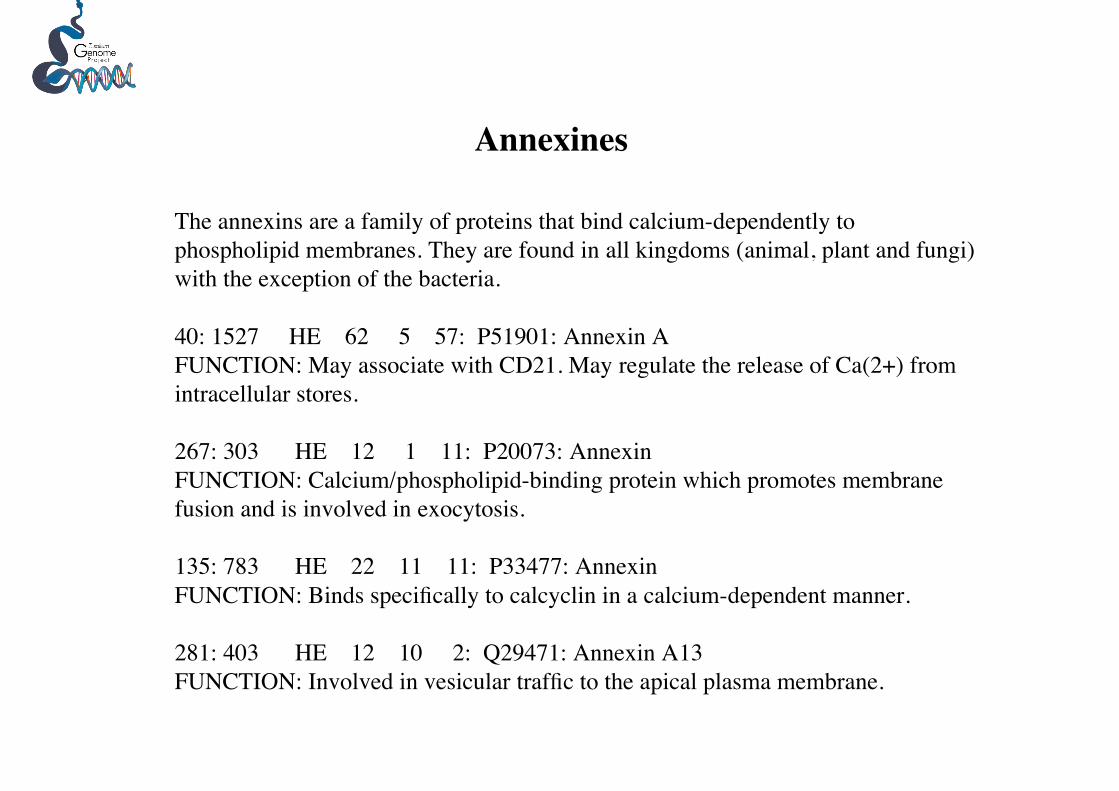
The annexins are a family of proteins that bind calcium-dependently tophospholipid membranes. They are found in all kingdoms (animal, plant and fungi)with the exception of the bacteria.
40: 1527 HE 62 5 57: P51901: Annexin AFUNCTION: May associate with CD21. May regulate the release of Ca(2+) fromintracellular stores.
267: 303 HE 12 1 11: P20073: AnnexinFUNCTION: Calcium/phospholipid-binding protein which promotes membranefusion and is involved in exocytosis.
135: 783 HE 22 11 11: P33477: AnnexinFUNCTION: Binds specifically to calcyclin in a calcium-dependent manner.
281: 403 HE 12 10 2: Q29471: Annexin A13FUNCTION: Involved in vesicular traffic to the apical plasma membrane.
Annexines

49: 1077 AD 51 1 50: Q06543: Eukaryotic Transcription factor 5SIMILARITY: Belongs to the FET5 family.
296: 762 HE 11 8 3: O43474: Krueppel-like factor 4FUNCTION: May act as a transcriptional activator. Binds the CACCC core sequence. May beinvolved in the differentiation of epithelial cells and may also function in the development of theskeleton and kidney.
Transcription factors

51: 821 HE 48 19 29: P51469: Glyceraldehyde-3-phosphate dehydrogenaseCATALYTIC ACTIVITY: D-glyceraldehyde 3-phosphate + phosphate + NAD(+) = 3-phospho-D-glyceroyl phosphate + NADH. PATHWAY: Carbohydrate degradation; glycolysis; pyruvatefrom D-glyceraldehyde 3-phosphate: step 1.55: 732 HE 48 31 17: P53442: Fructose-bisphosphate aldolaseCATALYTIC ACTIVITY: D-fructose 1,6-bisphosphate = glycerone phosphate + D-glyceraldehyde 3-phosphate. PATHWAY: Carbohydrate degradation; glycolysis; D-glyceraldehyde 3-phosphate and glycerone phosphate from D-glucose: step 4.82: 959 HE 36 23 13: Q27655: EnolaseCATALYTIC ACTIVITY: 2-phospho-D-glycerate = phosphoenolpyruvate + H(2)O.99: 2111 AD 30 0 30: Q7VS43: 2,3-bisphosphoglycerate-dependent phosphoglyceratemutaseFUNCTION: Catalyzes the interconversion of 2-phosphoglycerate and 3-phosphoglycerate.151: 1394 HE 20 7 13: P27443: NAD-dependent malic enzymeCATALYTIC ACTIVITY: (S)-malate + NAD(+) = pyruvate + CO(2) + NADH.164: 711 HE 18 12 6: P53442: Fructose-bisphosphate aldolaseCATALYTIC ACTIVITY: D-fructose 1,6-bisphosphate = glycerone phosphate + D-glyceraldehyde 3-phosphate.188: 1954 AD 16 0 16: P06745: Glucose-6-phosphate isomeraseCATALYTIC ACTIVITY: D-glucose 6-phosphate = D-fructose 6- phosphate.252: 413 HE 12 3 9: Q589R5: Triosephosphate isomerase263: 243 HE 12 8 4: Q60HD8: Phosphoglycerate kinase
Carbohydrate degradation; glycolysis

67: 490 HE 40 26 14: P07379: Phosphoenolpyruvate carboxykinaseCATALYTIC ACTIVITY: GTP + oxaloacetate = GDP + phosphoenolpyruvate + CO(2).ENZYME REGULATION: Activity is affected by a number of hormones regulating thismetabolic process (such as glucagon, insulin, or glucocorticoids).PATHWAY: Carbohydrate biosynthesis; gluconeogenesis.
Carbohydrate biosynthesis; gluconeogenesis.
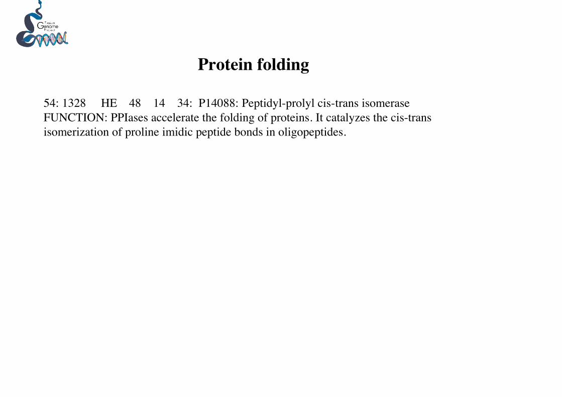
54: 1328 HE 48 14 34: P14088: Peptidyl-prolyl cis-trans isomeraseFUNCTION: PPIases accelerate the folding of proteins. It catalyzes the cis-transisomerization of proline imidic peptide bonds in oligopeptides.
Protein folding

62: 1454 AD 42 0 42: P75925: Cytochrome b561COFACTOR: Binds 2 heme B (iron-protoporphyrin IX) groups permolecule.
Cytochromes

50: 1451 AD 51 0 51: P49154: 40S ribosomal protein S2 64: 2228 HE 42 8 34: P55935: 40S ribosomal protein S9 75: 1516 HE 37 7 30: O57592: 60S ribosomal protein L7a 97: 2056 HE 30 5 25: Q90YU6: 60S ribosomal protein L22103: 750 HE 29 8 21: Q5E973: 60S ribosomal protein L18130: 329 HE 23 7 16: Q4R5P3: 60S ribosomal protein L10a147: 1161 HE 20 6 14: Q3SYR7: 60S ribosomal protein L9152: 1967 AD 20 1 19: Q29361: 60S ribosomal protein L35154: 721 HE 20 6 14: Q2I0I6: 60S ribosomal protein L26173: 2357 HE 18 11 7: Q9NB34: 60S ribosomal protein L34223: 345 HE 14 10 4: Q2YGT9: 60S ribosomal protein L6224: 22 HE 14 6 8: P48162: 60S ribosomal protein225: 894 HE 14 11 3: Q9GT45: 40S ribosomal protein S26226: 1166 HE 14 9 5: P39018: 40S ribosomal protein S19230: 217 HE 13 4 9: P23403: 40S ribosomal protein S20235: 595 LV 13 12 1: O61231: 60S ribosomal protein L10250: 1790 HE 13 1 12: P47826: 60S acidic ribosomal protein P0258: 653 HE 12 7 5: P52812: 40S ribosomal protein S11264: 454 HE 12 7 5: Q5RC11: 60S ribosomal protein L11278: 16 HE 12 8 4: Q3T0B7: 40S ribosomal protein S27279: 849 HE 12 6 6: Q7KR04: 40S ribosomal protein S15Ab282: 536 HE 11 8 3: O17445: 60S ribosomal protein L15284: 432 HE 11 9 2: Q90YR6: 40S ribosomal protein S8289: 1246 HE 11 3 8: P47840: 40S ribosomal protein S12306: 2521 HE 11 3 8: P20280: 60S ribosomal protein307: 312 HE 11 6 5: Q5R7Y8: 60S ribosomal protein L8
Ribosomal proteins

190: 328 LV 16 16 0: P61220: translation initiation factor 1bFUNCTION: Probably involved in translation.
207: 1823 HE 15 4 11: O55135: Eukaryotic translation initiation factor 6 (eIF-6)FUNCTION: Binds to the 60S ribosomal subunit and prevents its association with the 40Sribosomal subunit to form the 80S initiation complex
243: 980 HE 13 4 9: Q0ULD0: Nascent polypeptide-associated complex subunit betaFUNCTION: Component of the nascent polypeptide-associated complex (NAC), a dynamiccomponent of the ribosomal exit tunnel, protecting the emerging polypeptides from interactionwith other cytoplasmic proteins to ensure appropriate nascent protein targeting (By similarity).The NAC complex also promotes mitochondrial protein import by enhancing productive ribosomeinteractions with the outer mitochondrial membrane and blocks the inappropriate interaction ofribosomes translating non-secretory nascent polypeptides with translocation sites in the membraneof the endoplasmic reticulum (By similarity). EGD1 may act as a transcription factor that exert anegative effect on the expression of several genes that are transcribed by RNA polymerase
261: 1738 HE 12 1 11: P13549: Elongation factorFUNCTION: This protein promotes the GTP-dependent binding of aminoacyl-tRNA to the A-siteof ribosomes during protein biosynthesis.
90: 1488 HE 34 4 30: Q8AWF2: Nascent polypeptide-associated complex subunit
Translation factors

70: 964 HE 39 4 35: Q5RAP9: ATP synthase lipid-binding proteinFUNCTION: This protein is one of the chains of the nonenzymatic membrane component (F0) ofmitochondrial ATPase.CATALYTIC ACTIVITY: ATP + H(2)O + H(+)(In) = ADP + phosphate + H(+)(Out).
329: 99 LV 10 9 1: P34546: Vacuolar ATP synthase
340: 2217 AD 10 0 10: P05630: ATP synthase delta chain
ATP synthesis

84: 910 HE 35 22 13: O14463: ThioredoxinFUNCTION: Participates in various redox reactions through the reversible oxidation of itsactive center dithiol to a disulfide and catalyzes dithiol-disulfide exchange reactions.
115: 2484 HE 27 10 17: Q8T6C4: Thioredoxin peroxidaseFUNCTION: Reduces peroxides with reducing equivalents provided through the thioredoxinsystem. It is not able to receive electrons from glutaredoxin. May play an important role ineliminating peroxides generated during metabolism. Might participate in the signalingcascades of growth factors and tumor necrosis factor-alpha by regulating the intracellularconcentrations of H(2)O(2) (By similarity).CATALYTIC ACTIVITY: 2 R'-SH + ROOH = R'-S-S-R' + H(2)O + ROH.
249: 582 HE 13 4 9: P35705: Thioredoxin-dependent peroxide reductase(Peroxiredoxin)FUNCTION: Involved in redox regulation of the cell. Protects radical-sensitive enzymesfrom oxidative damage by a radical- generating system.
321: 1179 HE 10 4 6: Q17770: Protein disulfide-isomeraseCATALYTIC ACTIVITY: Catalyzes the rearrangement of -S-S- bonds inCC proteins.
-S-S- redox proteins

96: 1524 AD 32 2 30: O35660: Glutathione S-transferaseFUNCTION: Conjugation of reduced glutathione to a wide number of exogenous andendogenous hydrophobic electrophiles
157: 1245 AD 19 1 18: O35660: Glutathione S-transferase
168: 1543 AD 18 0 18: Q9N0V4: Glutathione S-transferase
215: 772 LV 14 14 0: P10299: Glutathione S-transferase P
Glutathione S-transferase

108: 468 HE 27 17 10: O02705: Heat shock protein HSP 90-alphaFUNCTION: Molecular chaperone. Has ATPase activity
146: 71 HE 20 16 4: P27541: Heat shock protein 70 kDa protein
162: 112 HE 18 5 13: Q5ZM98: Stress-70 protein, mitochondrial precursorFUNCTION: Implicated in the control of cell proliferation and cellular aging. May also act as achaperone
245: 84 HE 13 10 3: P31689: DnaJ (Chaperone protein)FUNCTION: Co-chaperone of Hsc70. Seems to play a role in proteinCC import into mitochondria.
Heat shock proteins
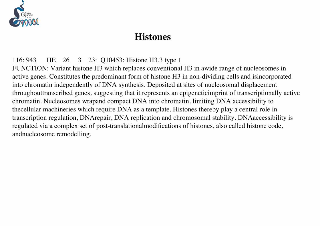
116: 943 HE 26 3 23: Q10453: Histone H3.3 type 1FUNCTION: Variant histone H3 which replaces conventional H3 in awide range of nucleosomes inactive genes. Constitutes the predominant form of histone H3 in non-dividing cells and isincorporatedinto chromatin independently of DNA synthesis. Deposited at sites of nucleosomal displacementthroughouttranscribed genes, suggesting that it represents an epigeneticimprint of transcriptionally activechromatin. Nucleosomes wrapand compact DNA into chromatin, limiting DNA accessibility tothecellular machineries which require DNA as a template. Histones thereby play a central role intranscription regulation, DNArepair, DNA replication and chromosomal stability. DNAaccessibility isregulated via a complex set of post-translationalmodifications of histones, also called histone code,andnucleosome remodelling.
Histones

165: 344 HE 18 2 16: Q5E971: 21 kDa transmembrane-trafficking proteinFUNCTION: Involved in vesicular protein trafficking
260: 960 HE 12 6 6: P51823: ADP-ribosylation factorFUNCTION: GTP-binding protein that functions as an allosteric activator of the choleratoxin catalytic subunit, an ADP-ribosyltransferase. Involved in protein trafficking; maymodulate vesicle budding and uncoating within the Golgi apparatus.
Protein trafficking

171: 455 HE 18 12 6: Q26537: 14-3-3 protein homologSIMILARITY: Belongs to the 14-3-3 family.
290: 60 HE 11 7 4: O49998: 14-3-3-like protein F
347: 857 HE 10 6 4: Q5ZKC9: 14-3-3 protein zetaFUNCTION: Adapter protein implicated in the regulation of a large spectrum of both general andspecialized signaling pathway. Binds to a large number of partners, usually by recognition of aphosphoserine or phosphothreonine motif. Binding generally results in the modulation of the activityof the binding partner
181: 1969 AD 16 0 16: P92177: 14-3-3 protein epsilonFUNCTION: Positively regulates Ras-mediated pathways. Acts downstream or parallel to Raf, butupstream of nuclear factors in Ras signaling. Three mutants have been isolated, that suppress therough eye phenotype caused by mutated Ras1
Signaling

81: 138 HE 36 19 17: Q04820: Malate dehydrogenaseCATALYTIC ACTIVITY: (S)-malate + NAD(+) = oxaloacetate + NADH.
246: 1425 AD 13 0 13: O87840: Succinyl-CoA synthetase beta chainCATALYTIC ACTIVITY: ATP + succinate + CoA = ADP + phosphate + succinyl-CoA.
270: 220 HE 12 7 5: P21912: Succinate dehydrogenase [ubiquinone] iron-sulfurproteinCATALYTIC ACTIVITY: Succinate + ubiquinone = fumarate + ubiquinol.
324: 570 HE 10 3 7: P21912: Succinate dehydrogenase
Tricarboxylic acid cycle

251: 96 HE 12 9 3: P24406: Transforming protein RhoA precursor (Rho1)FUNCTION: Regulates a signal transduction pathway linking plasma membrane receptors to theassembly of focal adhesions and actin stress fibers. May be an activator of PLCE1.
Signal transduction pathways

268: 576 HE 12 3 9: P21128: Placental protein 11 precursorFUNCTION: Probable serine protease.
Placental proteins!!

304: 720 HE 11 2 9: Q9EQS0: Transaldolase (EC 2.2.1.2)FUNCTION: Transaldolase is important for the balance of metabolites in thepentose-phosphate pathway.CATALYTIC ACTIVITY: Sedoheptulose 7-phosphate + D-glyceraldehyde3-phosphate = D-erythrose 4-phosphate + D-fructose 6-phosphate.PATHWAY: Carbohydrate degradation; pentose phosphate pathway; D-fructose 6-phosphate and D-glyceraldehyde 3-phosphate from D-ribose 5-phosphate and D-xylulose 5-phosphate (non-oxidative stage): step 2.
Pentose-phosphate pathway

319: 681 HE 10 6 4: O35547: Long-chain-fatty-acid--CoA ligaseFUNCTION: Activation of long-chain fatty acids for both synthesis of cellular lipids, anddegradation via beta-oxidation.
Lipid synthesis

334: 146 HE 10 3 7: P17248: Tryptophanyl-tRNA synthetase
tRNA synthetases

72: 2334 AD 39 0 39: Q8CBW7: Cysteine-rich hydrophobic domain 1 protein
78: 1467 AD 36 0 36: O75828: Carbonyl reductase [NADPH] 3CATALYTIC ACTIVITY: R-CHOH-R' + NADP(+) = R-CO-R' + NADPH.
92: 215 HE 33 10 23: P07943: Aldose reductaseFUNCTION: Catalyzes the NADPH-dependent reduction of a wide variety of carbonyl-containing compounds
101: 1775 AD 29 0 29: Q99LM2: CDK5 regulatory subunit-associated protein 3FUNCTION: Potential regulator of CDK5 activity. May be involved in cell proliferation.
118: 1972 HE 26 6 20: P97315: Cysteine and glycine-rich proteinFUNCTION: Could play a role in neuronal development.
121: 2034 AD 25 0 25: P37111: Aminoacylase-1FUNCTION: Involved in the hydrolysis of N-acylated or N-acetylated amino acids (except L-aspartate).
136: 1754 HE 21 2 19: Q5RCU5: Carbonyl reductaseFUNCTION: Catalyzes the reduction of a wide variety of carbonyl compounds.
159: 76 HE 19 11 8: P18238: ADP,ATP carrier proteinFUNCTION: Catalyzes the exchange of ADP and ATP across the mitochondrial inner membrane.
160: 219 HE 19 12 7: P37805: Transgelin-3. Abundant and ubiquitous expression in neurons.
Miscellaneous functions
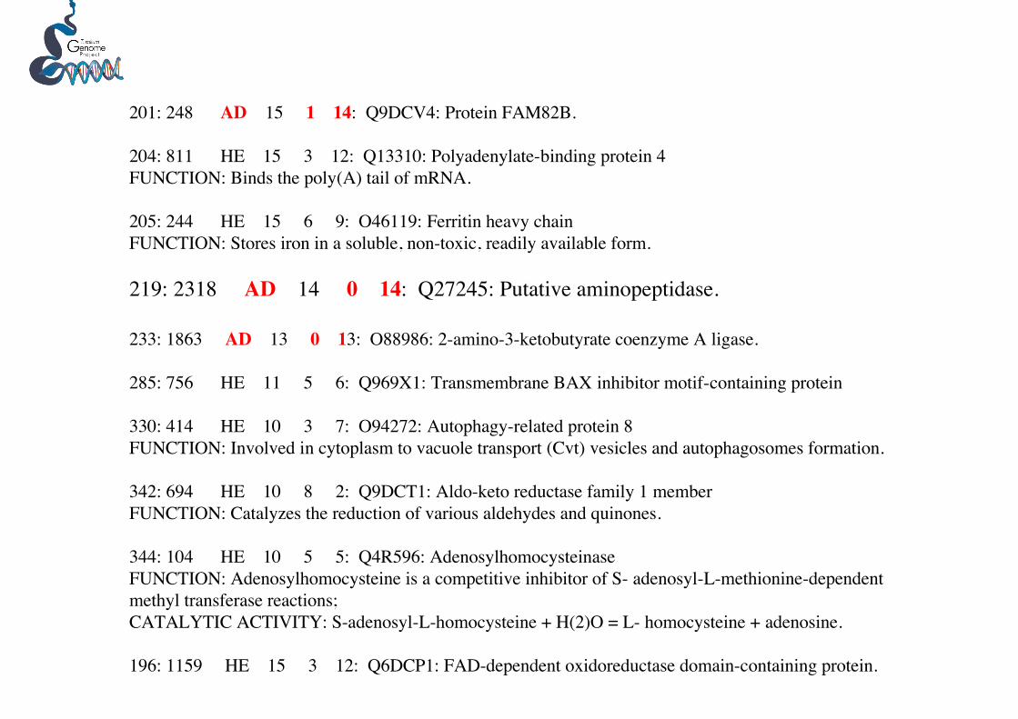
201: 248 AD 15 1 14: Q9DCV4: Protein FAM82B.
204: 811 HE 15 3 12: Q13310: Polyadenylate-binding protein 4FUNCTION: Binds the poly(A) tail of mRNA.
205: 244 HE 15 6 9: O46119: Ferritin heavy chainFUNCTION: Stores iron in a soluble, non-toxic, readily available form.
219: 2318 AD 14 0 14: Q27245: Putative aminopeptidase.
233: 1863 AD 13 0 13: O88986: 2-amino-3-ketobutyrate coenzyme A ligase.
285: 756 HE 11 5 6: Q969X1: Transmembrane BAX inhibitor motif-containing protein
330: 414 HE 10 3 7: O94272: Autophagy-related protein 8FUNCTION: Involved in cytoplasm to vacuole transport (Cvt) vesicles and autophagosomes formation.
342: 694 HE 10 8 2: Q9DCT1: Aldo-keto reductase family 1 memberFUNCTION: Catalyzes the reduction of various aldehydes and quinones.
344: 104 HE 10 5 5: Q4R596: AdenosylhomocysteinaseFUNCTION: Adenosylhomocysteine is a competitive inhibitor of S- adenosyl-L-methionine-dependentmethyl transferase reactions;CATALYTIC ACTIVITY: S-adenosyl-L-homocysteine + H(2)O = L- homocysteine + adenosine.
196: 1159 HE 15 3 12: Q6DCP1: FAD-dependent oxidoreductase domain-containing protein.