Embed Size (px)
Citation preview
The Thymus Gland
Diagnosis and Surgical Management
Bearbeitet vonKyriakos Anastasiadis, Chandi Ratnatunga
1. Auflage 2007. Buch. ix, 112 S. HardcoverISBN 978 3 540 33425 5
Format (B x L): 19,3 x 27 cmGewicht: 490 g
Weitere Fachgebiete > Medizin > Sonstige Medizinische Fachgebiete > Radiologie,Bildgebende Verfahren
Zu Inhaltsverzeichnis
schnell und portofrei erhältlich bei
Die Online-Fachbuchhandlung beck-shop.de ist spezialisiert auf Fachbücher, insbesondere Recht, Steuern und Wirtschaft.Im Sortiment finden Sie alle Medien (Bücher, Zeitschriften, CDs, eBooks, etc.) aller Verlage. Ergänzt wird das Programmdurch Services wie Neuerscheinungsdienst oder Zusammenstellungen von Büchern zu Sonderpreisen. Der Shop führt mehr
als 8 Millionen Produkte.
Chapter
Location
The thymus gland is located in the anterosuperior me-diastinum. It usually extends from the thyroid gland to the level of the fourth costal cartilage. It lies posterior to the pretracheal fascia, the sternohyoid and sternothyroid muscles and the sternum (mostly behind the manubrium and the upper part of its body). It is located anteriorly to the innominate vein and is found between the pari-etal pleura and extrapleural fat and central to the phrenic nerves. It lies on the pericardium, with the ascending aorta and aortic arch behind it, while in the neck it lies over the trachea. Parallel to the gland on each side lie the phrenic nerves, which converge towards the gland at its middle segment (particularly important issue in thymec-tomy procedures). The gland consists classically of two lobes, even though other lobular structures may be pres-ent (Figs. 1.1, 1.2). The thyrothymic ligament connects the upper parts of its lobes to the thyroid gland. A variety
of extensions of the upper lobes, as well as relationships to the innominate vein, have been described (Figs. 1.3). Thus, rather than being located in its classical anterior po-sition, one or both of the upper lobe thymus may even lie behind the innominate vein. Moreover, it has to be noted that besides the classical location of the gland, ectopic thymic tissue could be found in the mediastinal fat of the majority of patients. This is now accepted as the normal
Chapter
1 Anatomy
Kyriakos Anastasiadis and Chandi Ratnatunga
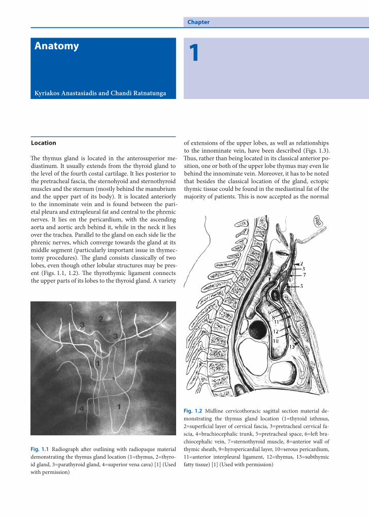
Fig. 1.1 Radiograph after outlining with radiopaque material demonstrating the thymus gland location (1=thymus, 2=thyro-id gland, 3=parathyroid gland, 4=superior vena cava) [1] (Used with permission)
Fig. 1.2 Midline cervicothoracic sagittal section material de-monstrating the thymus gland location (1=thyroid isthmus, 2=superficial layer of cervical fascia, 3=pretracheal cervical fa-scia, 4=brachiocephalic trunk, 5=pretracheal space, 6=left bra-chiocephalic vein, 7=sternothyroid muscle, 8=anterior wall of thymic sheath, 9=hyropericardial layer, 10=serous pericardium, 11=anterior interpleural ligament, 12=thymus, 13=subthymic fatty tissue) [1] (Used with permission)
eral defect in the normal embryonic development of the thymus may involve anomalies of the parathyroids (i.e. DiGeorge anomaly) or major thoracic vessels or both. The stromal cell component of the gland that comprises of epithelial cells originates from the endoderm and be-comes the thymic corpuscles (Hassal’s) or the epithelial reticular cells. The connective tissue component of the gland is derived from surrounding mesoderm. The bone marrow donates the colonizing lymphocytes to it.
By the end of ninth week of development (see Chap. 3) the primordial thymus becomes competent to attract these lymphoid stem cells from the bloodstream, and to provide an epithelial microenvironment within which thymocytes can become mature T cells. The primary endodermal thy-mic cells are now infiltrated by lymphocytes of bone mar-row origin, which undergo numerous mitotic divisions within the thymus and become the most numerous cell type in the organ. Appropriate development of the thymic epithelium during this period is important, as impaired development following neural crest ablation leads to loss of the gland’s capacity to attract lymphoid stem cells.
During embryogenesis, failure of the gland to descend into the mediastinum or maldescent can lead to a par-tially (one lobe) or fully ectopic or to aberrant thymic tissue sequestration in the neck. This is not uncommon, and in only 2% of Jaretzki’s and Wolff ’s patients was all thymic tissue confined to the thymic capsule. Ectopic thymic tissue in the mediastinum and in the neck has been described in up to 98% of the myasthenic popula-tion. This has important implications to thymectomy, as will be discussed in later chapters. Aberrant thymic tis-sue may, therefore, be found in the neck in up to one third of the population, as well as being dispersed in the mediastinum; common sites include lateral to the pleuro-
surgical anatomy of the thymus, defining the findings as “variation” and not “ectopic” [see chapter 9 – Fig 9.13, where a composite anatomy of the thymus as described by Jaretzki is illustrated, based on surgical-anatomi-cal studies in 50 consecutive transcervical-transsternal maximal thymectomies for MG; thymic tissue was found outside the confines of the classical cervical-mediastinal lobes (A and B) in 32% of the specimens in the neck and in 98% of the specimens in the mediastinum].
Embryology
The thymus develops from the epithelium of the ventral diverticulum. It rises at the sixth gestational week from the third pharyngeal pouch and branchial cleft on each side as precursor to its ultimate bilobar structure, with pos-sible minor contribution from the fourth pouch (Fig. 1.4). The thymic masses from each side move towards each other in the midline and come into direct contact, but do not fuse, remaining from the eighth week onwards as two connected thymic lobes. The gland then descends during the eighth week from the neck into anterior mediastinum of the thorax, to take up its final position.
The gland shares a common origin with the inferior parathyroids and the major thoracic vessels. Thus, para-thyroid tissue can be embedded in the thymus, and a gen-
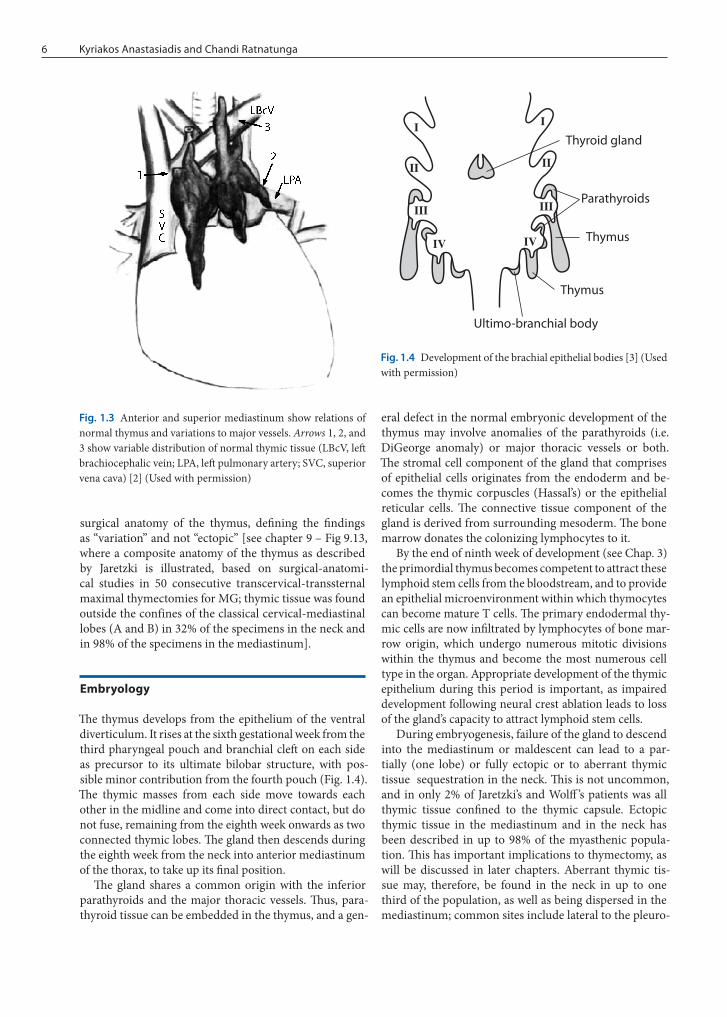
Fig. 1.3 Anterior and superior mediastinum show relations of normal thymus and variations to major vessels. Arrows 1, 2, and 3 show variable distribution of normal thymic tissue (LBcV, left brachiocephalic vein; LPA, left pulmonary artery; SVC, superior vena cava) [2] (Used with permission)
Fig. 1.4 Development of the brachial epithelial bodies [3] (Used with permission)
� Kyriakos Anastasiadis and Chandi Ratnatunga
pericardial surface in close proximity to the phrenic nerves, inferiorly adjacent to the diaphragm and the car-diophrenic fat, around the inferior pulmonary ligaments and also the pulmonary hilum or even the lungs. It can also be found in the sub-mandibular and paratracheal re-gion, as well as in the aortopulmonary window or in the posterior mediastinum. Most of this ectopic cervical and mediastinal tissue, however, is cystic and non-functional.
Structural Anatomy
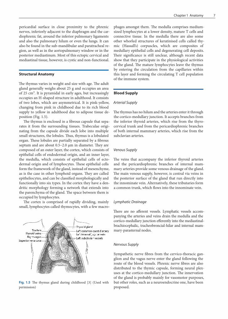
The thymus varies in weight and size with age. The adult gland generally weighs about 25 g and occupies an area of 25 cm3. It is pyramidal in early ages, but increasingly occupies an H-shaped structure in adulthood. It consists of two lobes, which are asymmetrical. It is pink-yellow, changing from pink in childhood due to its rich blood supply to yellow in adulthood due to adipose tissue de-position (Fig. 1.5).
The thymus is enclosed in a fibrous capsule that sepa-rates it from the surrounding tissues. Trabeculae origi-nating from the capsule divide each lobe into multiple small structures, the lobules. Thus, thymus is a lobulated organ. These lobules are partially separated by a fibrous septum and are about 0.5–2.0 μm in diameter. They are composed of an outer layer, the cortex, which consists of epithelial cells of endodermal origin, and an inner layer, the medulla, which consists of epithelial cells of ecto-dermal origin and of lymphocytes. These epithelial cells form the framework of the gland, instead of mesenchyme, as is the case in other lymphoid organs. They are called epitheliocytes, and can be classified morphologically and functionally into six types. In the cortex they have a den-dritic morphology forming a network that extends into the parenchyma of the gland. The space between them is occupied by lymphocytes.
The cortex is comprised of rapidly dividing, mainly small, lymphocytes called thymocytes, with a few macro-
phages amongst them. The medulla comprises medium-sized lymphocytes at a lower density, mature T cells and connective tissue. In the medulla there are also some other whorled structures of keratinised cells called thy-mic (Hassall’s) corpuscles, which are composites of medullary epithelial cells and degenerating cell deposits. Their significance is still unclear, although recent data show that they participate in the physiological activities of the gland. The mature lymphocytes leave the thymus by entering the circulation from the capillaries within this layer and forming the circulating T cell population of the immune system.
Blood Supply
Arterial Supply
The thymus has no hilum and the arteries enter it through the cortico-medullary junction. It accepts branches from the inferior thyroid arteries, which rise from the thyro-cervical trunk and from the pericardiophrenic branches of both internal mammary arteries, which rise from the subclavian arteries.
Venous Supply
The veins that accompany the inferior thyroid arteries and the pericardiophrenic branches of internal mam-mary arteries provide some venous drainage of the gland. The main venous supply, however, is central via veins in the posterior surface of the gland that run directly into the innominate vein. Alternatively, these tributaries form a common trunk, which flows into the innominate vein.
Lymphatic Drainage
There are no afferent vessels. Lymphatic vessels accom-panying the arteries and veins drain the medulla and the cortico-medullary junction efferently into the mediastinal-brachiocephalic, tracheobroncial-hilar and internal mam-mary-parasternal nodes.
Nervous Supply
Sympathetic nerve fibres from the cervico-thoracic gan-glion and the vagus nerve enter the gland following the route of the blood vessels. Phrenic nerve fibres are also distributed to the thymic capsule, forming neural plex-uses at the cortico-medullary junction. The innervation of the gland is probably mainly for vasomotor purposes, but other roles, such as a neuroendocrine one, have been proposed.
Fig. 1.5 The thymus gland during childhood [3] (Used with permission)
�Chapter 1 Anatomy
Suggested Bibliography
Banister LH. Haemolymphoid system. In Banister LH: Gray’s anatomy. 38th edn. New York: Churchill Livingston, 1995, pp 1423–1429.
Bockman DE. Development of the thymus. Micr Res Tech 1997;38:209–215.
Bodey B, Bodey B Jr, Siegel SE, Kaiser HE. Novel insights into the function of the thymic Hassall’s bodies. In Vivo 2000;14:407–418.
Jaretzki A III, Wolff M. “Maximal’’ thymectomy for myasthenia gravis. Surgical anatomy and operative results. J Thorac Cardiovasc Surg 1988;96:711–776.
Jaretzki A III. Thymectomy for myasthenia gravis; analysis of controversies regarding technique and results. Neurology 1997;48(Suppl 5):S52–S63.
Lele SM, Lele MS, Anderson VM. The thymus in infancy and childhood. Embryonic, anatomic and pathologic conside-rations. Chest Surg Clin N Am 2001;11:233–253.
Lumley JSP, Craven JL, Aitken JT. Thorax. In Lumley JSP, Cra-ven JL, Aitken JT: Essential anatomy. 5th edn. New York: Churchill Livingston, 1995, pp 77–78.
Marieb EN. The lymphatic system. In Marieb EN: Human ana-tomy and physiology. 4th Edn. California: The Benjamin/Cummings, 1998, pp 751–752.
Martini F. The lymphatic system and immunity. In Martini F: Fundamentals of anatomy and physiology. 5th edn. New Jersey: Prentice-Hall, 2001, pp 760–762.
Moore KL, Dalley A. Thorax. In Moore KL, Dalley A: Clinical oriented anatomy. 4th edn. Philadelphia: Lippincott, Wil-liams and Wilkins, 1999, pp 142–143.
Schurman HJ, Kuper F, Kendall M. Thymic microenviron-ment at the light microscopic level. Micr Res Tech 1997;38:216–226.
Sinnatamby CS. The thorax – Pt 5: anterior mediastinum. In Sinnatamby CS: Last’s anatomy. Regional and applied. 10th edn. London: Churchill-Livingstone, 1999, pp 189–190.
Suster S, Rosai J. Histology of the normal thymus. Am J Surg Pathol 1990;14:284–303.
Van Wynsberghe D, Noback CR, Carola R. The endocrine sys-tem. In Van Wynsberghe D, Noback CR, Carola R: Human anatomy and physiology. 3rd edn. Boston: McGraw-Hill, 1995, pp 571.
Von Gaudecker B. Functional histology of the human thymus. Anat Embyol (Berlin) 1991;183:1–15.
References
1. Di Marino V, Argeme M, Brunet C, Coopens R, Bonnoit J. Macroscopic study of the adult thymus. Surg Radiol Anat. 1987;9:51–62.
2. Sone S, Higashihara T, Morimoto S, et al. Normal anatomy of thymus and anterior mediastinum by pneumomediasti-nography. Am J Roentgenol. 1980;134:81–89.
3. Gray’s Anatomy of the Human Body. 20th ed. Philadelphia: Lea & Febiger, 1918; New York: bartleby.com, 2000.
� Kyriakos Anastasiadis and Chandi Ratnatunga