Embed Size (px)
Citation preview
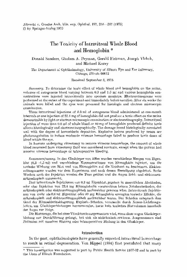
Albrecht v. Graefes Arch. klin. exp. Ophthal. 197, 255--267 (1975) �9 by Springer-Verlag 1975
The Toxicity of Intravitreal Whole Blood and Hemoglobin *
Donald Sanders, Gholam A. Peyman, Gerald Fishman, Joseph Vlehek, and Michael Korey
The Department of Ophthalmology, University of Illinois Eye and Ear Infirmary, Chicago, Illinois 60612
Received September 3, 1975
Summary. To determine the toxic effect of whole blood and hemoglobin on the retina, volumes of autogenous blood varying between 0.3 and 1.5 ml and various hemoglobin con- centrations were injected intravitreally into speciosa monkeys. Electroretinograms were performed at the outset of the experiment and immediately before sacrifice. After six weeks the animals were killed and the eyes were processed for histologie and electron microscopic examination.
Three intravitreal injections of 0.3 mt of autogenous blood administered at one-month intervals or one injection of 22.4 mg of hemoglobin did not produce a toxic effect on the retina demonstrable by light or electron microscopic examination or electroretinography. Intravitreal injection of more than 0.6 ml of whole blood or 40 mg of hemoglobin produced definite toxic effects histologically and electroretinographically. The damage found histologically correlated well with the degree of hemosiderin deposition. Explosive lesions produced by xenon arc photocoagulation to induce moderate vitreous hemorrhage failed to produce toxic doses of blood within the eye.
In humans undergoing vitrectomy to remove vitreous hemorrhage, the amount of whole blood recovered from vitrectomy fluid was considered nontoxic, except when the patient had massive vitreous hemorrhage or intraoperative bleeding.
Zusammen/assung. In den Glask6rper yon Affen wurden verschiedene Mengen yon Eigen- blur (0,3--1,5 ml) und verschiedene Konzentrationen yon H~moglobin injiziert, um die toxische Wirkung yon Blur und von Hi~moglobin auf die Netzhaut zu bestimmen. Elektro- retinogramme wurden vor dem Experiment und nach dessen Beendigung abgeleitet. Sechs Wochen nach der Injektion wurden die Tiere get6tet und die Augen lieht- und elektronen- mikroskopisch untersucht.
Drei intravitreale Injektionen yon 0,3 ml Eigenblut, gegeben in monatlichen Abst~nden, oder eine Injektion yon 22,4 mg H~moglobulin verursachten keinen Netzhautschaden, der mikroskopisch oder elektroretinographisch nachweisbar gewesen wiire. Intravitreale Injektio- ~len yon mehr als 0,6 ml Eigenblut oder 40 mg H~moglobin erzengten toxische Effekte, die mikroskopisch und elektroretinographisch nachweisbar waren. Der Schaden entsprach dem Grad der H~mosiderinablagernng. Exptosiv-Schaden, verursacht durch Xenon-Liehtkoagu- l.ation, um Glask6rperblutungen hervorzurufen, hatte kein toxisches Blutvolumen innerhalb des Auges zur Folge.
Die Blutmenge, die bei einer Vitrektomie ausgewaschen wird, wenn diese wegen Glaskhrper- blutung zur Durchftihrung gelangt, hat sich als nichttoxisch erwiesen. Ausgenommen sind Patienten mit massiver Blutung oder intraoperativer Blutung in den Glaskhrper.
Introduction
I n the past, ophthalmologists have general ly expected in t rav i t rea l hemorrhage to result in re t inal degenerat ion. Von Hippel (1894) first postula ted tha t m a n y
* This investigation was supported in part by Public Health Service 1107-02 and in part by the Lions of Illinois Foundation.
256 D. Sanders et al.
re t ina l changes encountered are due to the toxic effects of i ron re leased in red blood cell b reakdown, and s imi lar conclusions have been d rawn from clinical and expe r imen ta l s tudies (0guchi , 1913; Samuels and Fuchs, 1952; Duke, 1958; Squire and MeEwen, 1958; Cibis and Yamash i t a , 1959; Franco is and Hanssens, 1962 ; Ho lm and Bennet t , 1962 ; Babel , 1964; Regnaul t , 1966 ; Masciulli , Anderson, and Charles, 1972).
Surpris ingly, in m a n y cases, a pa t i en t ' s vis ion has recovered af ter v i t reous surgery even though the re t ina was exposed to pro longed or r epea ted episodes of bleeding.
Many inves t iga tors (Oguchi, 1913 ; Duke, 1958 ; Squire and McEwen, 1958 ; Cibis and Yamash i t a , 1959 ; Babel , 1964) have t r i ed to s imula te the condi t ions of v i t reous hemorrhage b y the in t rav i t reM in jec t ion of whole b lood or hemoglobin. g e g n a u l t (1966, 1970) has shown t h a t hemorrhages spon taneous ly occurr ing in h u m a n eyes are s t r ik ing ly s imilar to those expe r imen ta l l y induced in r abb i t eyes.
Therefore, i t appears t h a t in t r av i t r ea l in jec t ion of b lood in an imals is a useful mode l for s tudy ing the t ox i c i t y of b lood in a cl inical set t ing.
The purposes of the present s t u d y were (1) to es tabl ish the toxic level of in t rav i t reM hemoglobin and whole b lood for the re t ina in pr imates , (2) to eva lua te the usefulness of br ight- f lash e lec t ro re t inography (ERG) in p red ic t ing the toxic effect of b lood and hemoglobin on the re t ina in the presence of opaque media , (3) to produce expe r imen ta l v i t reous hemorrhage and measure the amoun t of b lood and hemoglobin found in the vi treous, and (4) to measure the amoun t of hemoglobin and b lood ex t r ac t ed from the v i t reous in pa t i en t s undergoing v i t r e e tomy to remove v i t reous hemorrhage.
Materials and Methods
Part 1: Intravitreal Blood and Hemoglobin
Speciosa monkeys were anesthetized with an intravenously administered mixture of 0.2 Inl sodium pentobarbitM, 0.2 ml of sodium thiamylal, and 0.4 ml sterile normal saline. Then 2 ml of venous blood was drawn from the femoral vein by means of a 3-ml syringe containing 0.1 ml of sodium citrate. Both eyes of each monkey were dilated with 1% cyclopentolate and 10 % phenylephrine, and two drops of proparacMne hydrochloride (OphthMne) were placed on each eye. After stabilizing the eye with a toothed forceps, a 25-gauge needle on a 3-ml syringe penetrated the eye at the pars plana in the inferior temporal quadrant and aspirated 0.3 ml of vitreous. Care was taken on needle insertion to avoid the lens and the retina. Immediately after this procedure, each eye was penetrated 3 to 5 mm superior to the original site of vitreous removal, and varying concentrations of hemoglobin or eitrated whole blood were injected.
Three monkeys received bilateral injections of 0.3 ml of whole blood once a month for three successive months, and three monkeys received a single bilateral injection of either 0.6, 1.0, or 1.5 ml. The animals receiving repeated injections of 0.3 ml of blood were killed six months after the last injection. Those receiving the greater volumes of blood were killed six weeks after injection. As a control one monkey received bilateral intravitreal injection of 1 ml of saline containing 0.05 ml of sodium citrate and was killed six weeks after injection.
For hemoglobin injection, the 2 ml of blood drawn from the femoral vein was lysed by sonication and dilution with distilled water, centrifuged at 10000 I~PM for ten minutes in a Sorvall centrifuge, and the hemoglobin-containing supernatant was drawn off.
Four eyes of two monkeys received different amounts of hemoglobin: (1) 0.2 ml of 11.2 gin/ 100 ml hemoglobin (a total of 22.4 mg of hemoglobin injected); (2) 0.4 ml of 11.2 gm/100 ml hemoglobin (a total of 44.8 mg) ; (3) 0.5 ml of 8 gin/100 ml hemoglobin (a total of 40 rag) ; and (4) 1.0 ml of 8 gm/100 ml hemoglobin (a total of 80 rag). The animals were killed six weeks after the hemoglobin injection.
Int ravi t rea l Toxicity 257
In bo th hemoglobin-injected and whole-blood-injected monkeys, ERGs were taken just before and one hour and one week after injection as well as at the t ime of sacrifice. For pre- injection ERGs, a xenon gas discharge tube coupled to a Grass PS- I I photost imulator set a t I~6 served as the stimulus. For postinjeetion ERGs, a tIoneywell strobe replaced the xenon gas discharge tube.
Each eye was examined daily for the first week after every injection by direct and indirect ophthalmoscopy. After the first week, each eye was observed three times a week for the remainder of the experiment. Before enucleation, the intraocular pressures were recorded with Schi6tz tonometers.
After enucleation, the anterior segments were removed and the eyes immersed in a 1% glutaraldehyde/1% formaldehyde mixture in phosphate buffer, pH 7.4. Eye cups were agitated in a fresh formaldehyde/glutaraldehyde solution for 20 minutes on an Eberbach Clinical Rota tor a t 5~ and were refrigerated overnight.
After overnight fixation, the posterior hemisphere was cut in half bisecting the macula and cutt ing either above or below the disk. The half containing the disk was processed for light microscopy. Slides were stained for hemosiderin by means of Gomori's ferrocyanide method. The other half of the eye was prepared for electron microscopy by dissecting the sclera from the choroid and retina., which were then washed in Sorenson's phosphate buffer, pH 7.35, post- fixed with Dalton 's chrome osmium for two hours, again washed in phosphate buffer, de- hydra ted in graded ethanols and propylene oxide, and embedded in Epon.
Thick (0.5 ~m) sections were cut on a SorvM1 MT2-B ultramierotome and stained with Mallory's stain for five minutes. Selected areas were thin-sectioned and stained with uranyl acetate and lead citrate, and subsequently examined with a JEM 100B electron microscope.
Part 2: Experimental Vitreous Hemorrhage Produced by Xenon Are Photo- coagulation
Between 30 and 40 explosive lesions were produced in bo th eyes of three monkeys, by means of a Zeiss xenon arc photoeoagulator set a t red I I I with a field diaphragm of 6. The animals were killed 24 hours later, ~nd the eyes enucleated and frozen in liquid freon. The selera, choroid, retina, lens, and anterior chamber contents could be peeled away leaving a ball of frozen vitreous containing blood products. After the vitreous from each eye was homogenized, the vitreous volmne was measured and ~n equal volume of distilled water was added to lyse the reamining red blood cells. The material was centrifuged at 10 000 RPM for ten minutes in a Sorvall centrifuge, and the supernatant and a sample of whole blood from each animal were tested for hemoglobin concentration with a Coulter hemoglobinometer. The volume of blood in the vitreous was calculated by the following formula:
Vitreous blood (ml )=
Supernate volume (ml) • supernate Hb concentration (gm/lO0 7nl) Blood t t b concentration (gm/100 ml)
Part 3: Human Vitreous Hemorrhage Careful history-taking and physical examinat ion were performed on each pat ient to
undergo vitrectomy, including direct and indirect ophthalmoscopy by a t least two ophthal- mologists. All pat ients had preoperative ul trasonography and bright-flash ERG. The pat ients ' blood hemoglobin concentration was determined and assumed constant for the durat ion of vitreous hemorrhage. A description of the operative procedure and any complications were recorded.
All products aspirated during vitrectomy, including vitreous, hemorrhage, and infusion solution, were collected by suction in sterile containers. This solution was centrifuged a t 118 000 I~PS[ for 20 minutes with an Internat ional Centrifuge, model UV, and the total volume of superna tan t recorded. The precipitate was made up to a volume of 5 ml with distilled water to lyse all the intact red blood cells, then diluted with 0.9% sodium chloride into solutions of 1:10, 1:20, 1:40, and 1:80 concentrations. Samples of the original lysed precipitate, and the t :10 , 1:20, 1:40, and 1:80 dilutions and supernatant solutions were tested by a Coulter
6 Albrech t v. Graefes Arch. klin. exp. Oph~hal.
258 D. Sanders et al.
hemoglobinometer, and the readings recorded. The total blood volume in the vitreous aspirate was determined as follows:
Precipitate: The Coulter hemogtobinometer was calibrated to 1:500 dilution of blood; thus, readings of the original lysed precipitate and the dilutions were multiplied by appropriate factors to adjust values to a 1 : 500 dilution. These adjusted values of grams Hb/100 ml fluid were averaged to determine the average Hb concentration (gm/100 ml) with the following formula:
Whole blood equivalent= (Solution Hb concentration) (solution volume)
Blood Hb concentration Supernatant: The supernata.nt fluid level was read directly off the hemoglobinometer, and
the whole blood equivalent was determined with the above formula. The total whole blood equivalent was determined by adding the amount of blood in the precipitate to that in the supernatant.
Results
Part 1; Whole Blood and Hemoglobin Toxicity
Ophtha lmoscopy
Whole Blood; All concent ra t ions of b lood obscured the fundus. I n those eyes in jec ted wi th 0.3 ml of b lood r epea ted ly , grayish-whi te membranous opaci t ies were no ted th ree to four weeks af ter the f irst i n t r av i t r ea l inject ion. The clinical appea rance was s imi lar to t h a t of a mode ra t e v i t reous hemorrhage in a human. Bo th eyes in jec ted wi th 1.5 ml of whole b lood became phthis ica l , and none d e m o n s t r a t e d an increase in in t raoeu la r pressure.
Hemoglobin Twenty- four hours af ter hemoglob in in ject ion, diffuse red blood produc ts obscured the fundus view. W i t h i n th ree to four weeks th is diffuse red ref lex was rep laced b y a g ray-ye l low reflex obscuring the fundus view.
E lee t ro re t inography , His to logic F indings , and Ul t r a s t ruc tu re
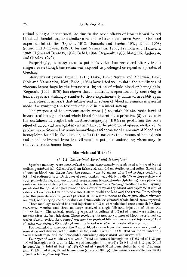
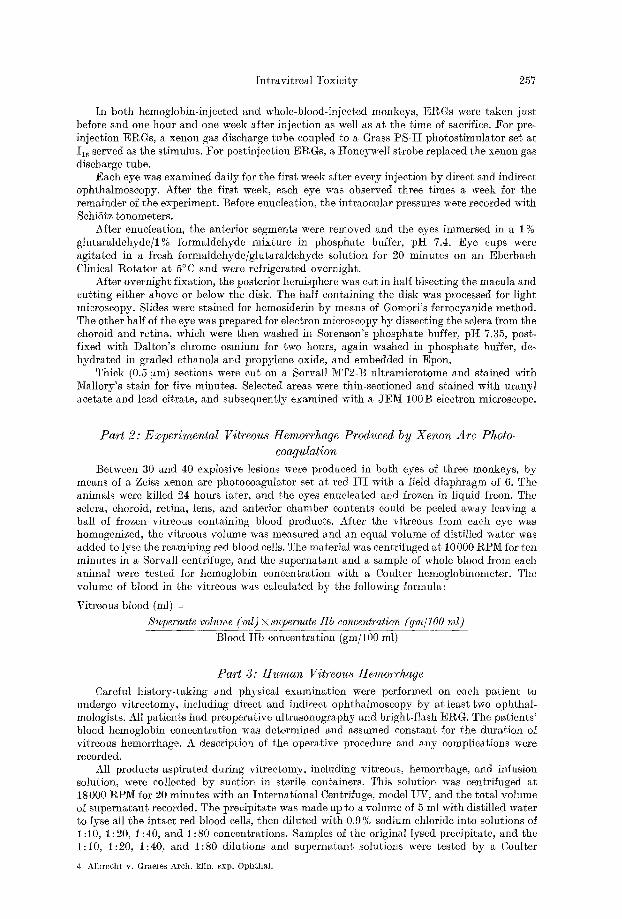
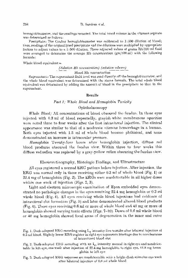
All eyes regis tered a normal E R G p a t t e r n before inject ion. Af ter inject ion, the E g G was normal only in those receiv ing e i ther 0.3 ml of whole blood (Fig. 1) or 22.4 mg of hemoglobin (Fig. 2). The E R G s were nonde tec t ab l e in all h igher doses
wi th in one week of in jec t ion ( f igs . 2, 3). L igh t and e lec t ron microscopic examina t i on of Epon e m b e d d e d eyes demon-
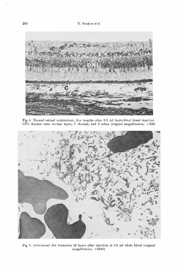
s t r a t ed no pa thologic changes in the eyes receiving 22.4 mg hemoglobin or 0.3 ml whole blood (Fig. 4). Al l eyes receiving whole blood in ject ions had evidence of in t r av i t r ea l clot fo rmat ion ( f ig . 5) and la te r d e m o n s t r a t e d a l t e red blood products (Fig. 6). Those eyes receiving 0.6 ml or more of whole blood and 40 mg or more of hemoglobin showed va ry ing toxic effects (Figs. 7-10). Doses of 0.6 ml whole b lood or 40 mg hemoglobin showed focal areas of degenera t ion in the inner and outer
Fig. 1. Dark-adapted E g G recording using I~a intensity five months after bilateral injection of 0.3 ml blood. Slightly lower ERG register in right eye represents blockage due to nonelearanee
of intravitreal blood clot
Fig. 2. Dark-adapted ERG recording with an I16 intensity normal in right eye and nondetec- table in left eye, one week after injection of 22.4 mg hemoglobin to right eye, 44.8 mg hemo-
globin to left eye
Fig. 3. Dark-adapted ERG responses are nondeteetable with a bright-flash stimulus one week after bilateral injection of 0.6 ml whole blood

Intravitreal Toxicity 259
260 D. Sanders et al.
Fig. 4. Normal retinal architecture, five months after 0.3 ml intravitreal blood injection. O N L denotes outer nuclear layer; C choroid; and S selera (original magnification, •
Fig. 5. Intravitreal clot formation 24 hours after injection of 0.3 ml whole blood (original magnification, •
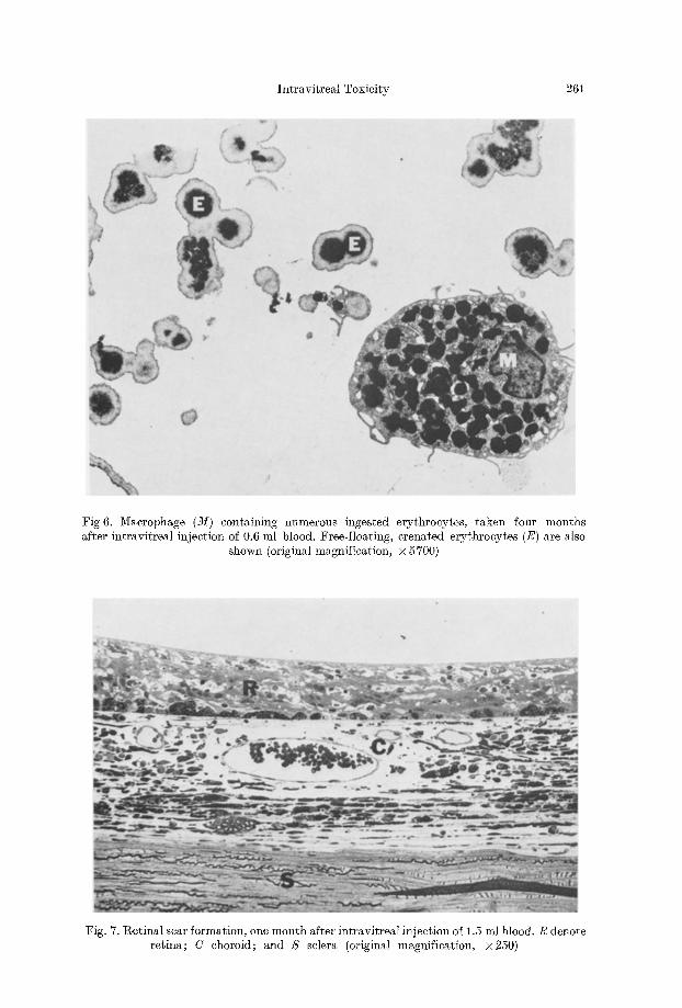
Intravitreal Toxicity 261
Fig 6. Macrophage (21i) containing numerous ingested erythrocytes, taken four months after intravitreal injection of 0.6 ml blood. Free-floating, crenated erythrocytes (E) are also
shown (original magnification, • 5700)
Fig. 7. Retinal scar formation, one month after intravitreal injection of 1.5 ml blood. R denote retina; C choroid; and S sclera (original magnification, •
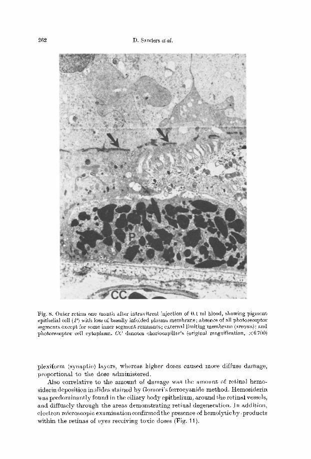
262 D. Sanders et al.
Fig. 8. Outer retina one month after intravitreal injection of 0.1 ml blood, showing pigment epithelial cell (P) with loss of basally iMotded plasma membrane; absence of all photoreceptor segments except for some inner segment remnants; external limiting membrane (arrows) ; and photoreceptor cell cytoplasm. CC denotes chorioeapillaris (original magnification, •
plexiform (synaptie) layers, whereas higher doses caused more diffuse damage, proportional to the dose administered.
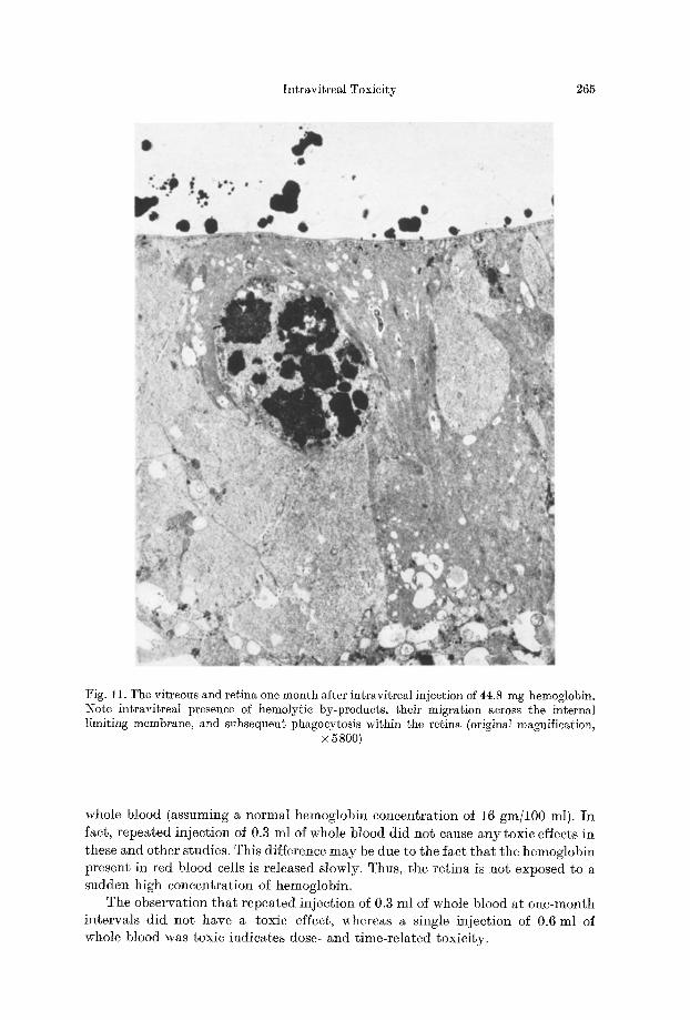
Also correlative to the amount of damage was the amount of retinal hemo- siderin deposition in slides stained by Gomori's ferrocyanide method. Hemosiderin was predominantly found in the ciliary body epithelium, around the retinal vessels, and diffusely through the areas demonstrating retinal degeneration. In addition, electron microscopic examinationconfirraedthe presence of hemolytic by-products within the retinas of eyes receiving toxic doses (Fig. 11).
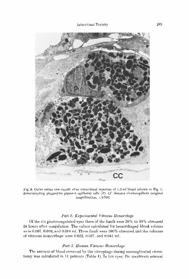
Intrgvitreal Toxicity 263
Fig. 9. Outer retina one month after intravitreM injection of 1.5 ml blood (shown in Fig. 1) demonstrating phagocytic pigment epithelial cells (P). CC denotes choriocapillaris (original
magnification, •
Part 2: Experimental Vitreous Hemorrhage
Of the six photocoagnlated eyes three of the fundi were 30 % to 50 % obscured 24 hours after coagulation. The values calculated for hemorrhaged blood volume were 0.007, 0.010, and 0.014 ml. Three fundi were 100% obscured and the volumes of vitreous hemorrhage were 0.022, 0.027, and 0.041 ml.
Part 3: Human Vitreous Hemorrhage
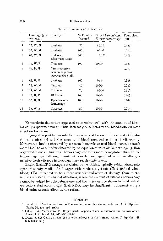
The amount of blood removed by the vitrophage during uncomplicated vitrec- tomy was calculated in 11 patients (Table 1). In ten eyes, the maximum amount
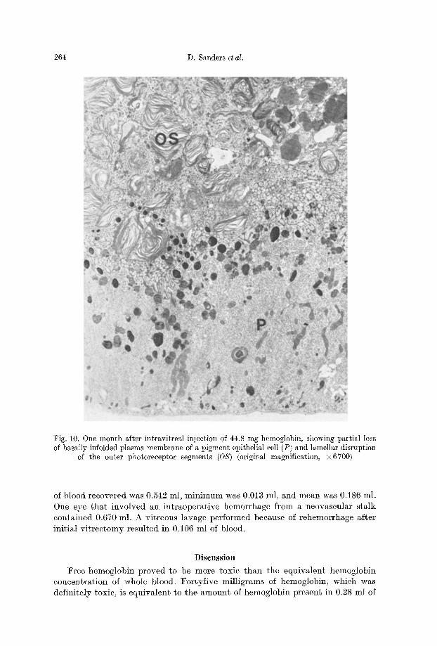
264 D. Sanders et al.
Fig. 10. One month after intravitreal injection of 44.8 mg hemoglobin, showing partial loss of basally infolded plasma membrane of a pigment epithelial cell (P) and lamellar disruption
of the outer photoreeeptor segments (OS) (original magnification, •
of blood recovered was 0.542 ml, minimum was 0.013 ml, and mean was 0.186 ml. One eye that involved an intraoperative hemorrhage from a neovascular stalk contained 0.670 ml. A vitreous lavage performed because of rehemorrhage after initial vitreetomy resulted in 0.106 ml of blood.
Diseussion
Free hemoglobin proved to be more toxic than the equivalent hemoglobin concentration of whole blood, l~'ortyfive milligrams of hemoglobin, which was definitely toxic, is equivalent to the amount of hemoglobin present in 0.28 ml of
Intravitreal Toxicity 265
Fig. 11. The vitreous and retina one month after intravitreal injection of 44.8 mg hemoglobin. Note intravitreal presence of hemolytic by-products, their migration across the internal limiting membrane, and subsequent phagoeytosis within the retina (original magnification,
• 5800)
whole blood (assuming a normal hemoglobin concentration of 16 gm/100 ml). In fact, repeated injection of 0.3 ml of whole blood did not cause any toxic effects in these and other studies. This difference may be due to the fact that the hemoglobin present in red blood cells is released slowly. Thus, the retina is not exposed to a sudden high concentration of hemoglobin.
The observation that repeated injection of 0.3 ml of whole blood at one-month intervals did not have a toxic effect, whereas a single injection of 0.6 ml of whole blood was toxic indicates dose- and time-related toxicity.
266 D. Sanders et al.
Table 1. Summary of clinical data
Case, age (yr), I-Iistory % Fundus % Old hemorrhage/ Total blood sex, race obscured % new hemorrhage (ml)
1 22, W, M Diabetes 70 80/20 0.150
2 37, W, M Diabetes 100 60/40 0.542
3 62, W, M 1%ebleed 100 0/100 0.106 after vitrectomy
4 71, W, F Diabetes 100 100/0 0.383
5 51, B, M Intraoperative - - - - 0.670 hemorrhage from neovascular stalk
6 62, B, M Diabetes 100 95/5 0.268
7 73, W, IV[ Trauma 40 100/0 0.037
8 28, W, M Diabetes 70 80/20 0.113
9 26, B, F Sickle cell 100 100/0 0.145
10 30, B, M Spontaneous 100 100/0 0.109 hemorrage
11 55, W, F Diabetes 20 100/0 0.013
t temosider in deposit ion appeared to correlate well with the amoun t of histo- logically apparen t damage. Thus, i ron may be a factor in the blood-induced toxic
effect on the ret ina. In general, a positive correlation was observed between the amount of fundus
clinically obscured and the amount of blood removed at time of vitrectomy. Moreover, a fundus obscured by a recent hemorrhage (red blood) contains much more blood than a fundus obscured by an equal amount of old hemorrhage (yellow organized blood). Thus fresh hemorrhage contains more hemoglobin than an old hemorrhage, and although most vitreous hemorrhages had no toxic effect, a massive h'esh vitreous hemorrhage may reach toxic levels.
Bright-flash ERG changes correlated well with histologically evident damage in spite of cloudy media. At dosages with moderately toxic effect (0.6 ml whole blood) EI%G appeared to be a more sensitive indicator of damage than micro- scopic evaluation. In clinical situations, where the amount of vitreous hemorrhage cannot he judged by ophthalmoscopy and the retina can be shown to be attached, we believe that serial bright-flash EI%Gs may be singificant in demonstrating a blood-induced toxic effect on the retina.
Relerences 1. Babel, J.: L'action ~oxique de l'hemosiderine sur les tissue oculaires. Arch. OphthM.
(Paris) 24, 405-416 (1964) 2. Cibis, P. A., Yamashita, T. : ExperimentM aspects of ocular siderosis and hemosiderosis.
Amer. J. Ophthal. 48, 465-480 (1959) 3. Duke, J. 1%. : Ocular effects of systemic siderosis in the human. Amer. J. Ophthal. 48,
628-633 (1958)
lntravitreal Toxicity 267
4. Francois, J., Hanssens, M. : Histopathologieal examination of a case of Eales' disease. Bull. Soe. belge Ophthal. 131, 358-370 (1962)
5. Holm, A. G., Bennett, W. : Hemosiderosis bulbi following trauma. Amer. J. Ophthal. 53 (1962)
6. l~[asciulli, L., Anderson, D. R., Charles, S. : Experimental ocular siderosis in the squirrel monkey. Amer. J. OphthM. 74, 638-661 (1972)
7. Oguchi, C. : ~ber die Wirkung yon Blutinjektionen in den GlaskSrper nebst Bemerkungen fiber die sog. Retinitis proliferans. Albrecht v. Graefes Arch. OphthM. 84, 446-520 (1913)
8. gegnault, F. R. : L'hemorragie intravitr~ene; thesis. Paris: Polyeopie Gilanne 1966 9. t~egnault, F. R. : Vitreous hemorrhage, an experimental study. Arch. OphthM. 83, 458-
474 (1970) 10. Samuels, B., Fuehs, A. : Clinical Pathology of the Eye. New York: Paul B. Hoeber, Inc.
1952 11. Squire, C., McEwen, W. K.: The effect of iron compounds on rabbit vitreous. Amer. J.
J. OphthM. 46, 356-358 (1958) 12. yon Hippel, E. : Uber Siderosis Bulbi und die Beziehungen zwisehen Siderotischer und
HS~matogener Pigmentierung. Albrecht v. Graefes Arch. OphthM. 40, 123-279 (1894)
Gholam A. Peyman, M. D. University of Illinois Eye and Ear Infirmary 1855 West Taylor Street Chicago, Illinois 60612, USA'