Embed Size (px)
Citation preview
Stancu Adrian Medicamentul Veterinar / Veterinary Drug
Vol. 11(1) May- June 2017
66
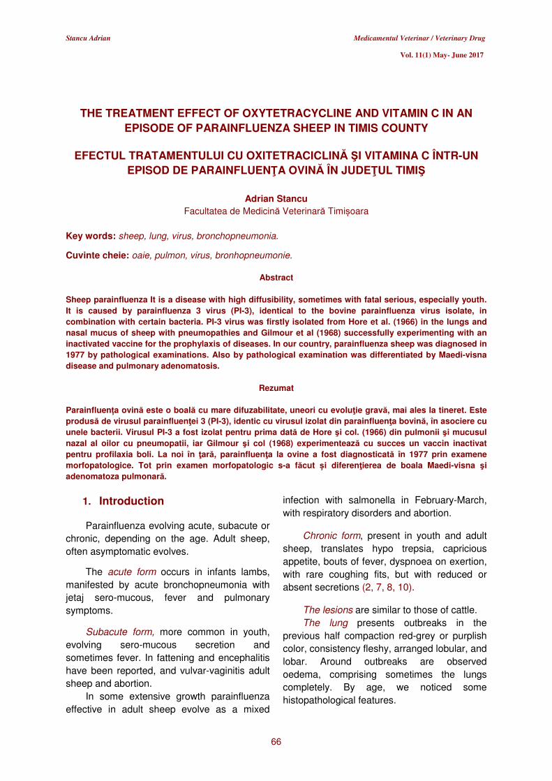
THE TREATMENT EFFECT OF OXYTETRACYCLINE AND VITAMIN C IN AN EPISODE OF PARAINFLUENZA SHEEP IN TIMIS COUNTY
EFECTUL TRATAMENTULUI CU OXITETRACICLINĂ ŞI VITAMINA C ÎNTR-UN
EPISOD DE PARAINFLUENŢA OVINĂ ÎN JUDEŢUL TIMIŞ
Adrian Stancu Facultatea de Medicină Veterinară Timișoara
Key words: sheep, lung, virus, bronchopneumonia.
Cuvinte cheie: oaie, pulmon, virus, bronhopneumonie.
Abstract
Sheep parainfluenza It is a disease with high diffusibility, sometimes with fatal serious, especially youth. It is caused by parainfluenza 3 virus (PI-3), identical to the bovine parainfluenza virus isolate, in combination with certain bacteria. PI-3 virus was firstly isolated from Hore et al. (1966) in the lungs and nasal mucus of sheep with pneumopathies and Gilmour et al (1968) successfully experimenting with an inactivated vaccine for the prophylaxis of diseases. In our country, parainfluenza sheep was diagnosed in 1977 by pathological examinations. Also by pathological examination was differentiated by Maedi-visna disease and pulmonary adenomatosis.
Rezumat
Parainfluența ovină este o boală cu mare difuzabilitate, uneori cu evoluţie gravă, mai ales la tineret. Este produsă de virusul parainfluenţei 3 (PI-3), identic cu virusul izolat din parainfluenţa bovină, în asociere cu unele bacterii. Virusul PI-3 a fost izolat pentru prima dată de Hore şi col. (1966) din pulmonii şi mucusul nazal al oilor cu pneumopatii, iar Gilmour şi col (1968) experimentează cu succes un vaccin inactivat pentru profilaxia boli. La noi în ţară, parainfluenţa la ovine a fost diagnosticată în 1977 prin examene morfopatologice. Tot prin examen morfopatologic s-a făcut și diferenţierea de boala Maedi-visna şi adenomatoza pulmonară.
1. Introduction Parainfluenza evolving acute, subacute or
chronic, depending on the age. Adult sheep,
often asymptomatic evolves.
The acute form occurs in infants lambs,
manifested by acute bronchopneumonia with
jetaj sero-mucous, fever and pulmonary
symptoms.
Subacute form, more common in youth,
evolving sero-mucous secretion and
sometimes fever. In fattening and encephalitis
have been reported, and vulvar-vaginitis adult
sheep and abortion.
In some extensive growth parainfluenza
effective in adult sheep evolve as a mixed
infection with salmonella in February-March,
with respiratory disorders and abortion.
Chronic form, present in youth and adult
sheep, translates hypo trepsia, capricious
appetite, bouts of fever, dyspnoea on exertion,
with rare coughing fits, but with reduced or
absent secretions (2, 7, 8, 10).
The lesions are similar to those of cattle.
The lung presents outbreaks in the
previous half compaction red-grey or purplish
color, consistency fleshy, arranged lobular, and
lobar. Around outbreaks are observed
oedema, comprising sometimes the lungs
completely. By age, we noticed some
histopathological features.
Stancu Adrian Medicamentul Veterinar / Veterinary Drug
Vol. 11(1) May- June 2017
67
Thus, the lambs of 1-4 weeks bronchitis
and bronchiolitis are seen proliferating, with
swelling becoming cubic pneumocytes and
alveolar epithelium pseudo-acinary take issue
with pronounced exudation, epithelial
desquamation and sincitialization (3, 5, 6).
Unvaccinated lambs infected in the first
days of life, histological changes of lung
epithelial advocates type of pneumonia.
Alveolar septs giant cells present in the
epithelium and alveolar among cells exfoliate,
is in full phagocytic activity.
In these cells, the epithelial and
macrophage intra nuclear and intra
cytoplasmic inclusions stands, small or large
oxyphyls and basophils.
Hyperaemia of regional lymph nodes and
lymphoid hypoplasia (4, 9, 11).
Over the age of lambs and adult sheep a
month is recorded, in addition, activation and
proliferation of mesenchymal alveolar septs,
per bronchial and perivascular space.
The presence of "hyaline membranes" and
giant cells are constant elements encountered
in influenza infections in humans and calves.
In regional lymph nodes, in addition to
redness and follicular hyperplasia, highlights
macrophages and giant cells, 2-5 nuclei,
sometimes with inclusions.
Blast aspects of lymphocytes and plasma
cell differentiation in the lungs and lymph
nodes are specific immune reactions (3, 5, 8).
Aim Research has aimed diagnosis of
parainfluenza based on macroscopic and
histopathological lesions.
2. Materials and methods
The research was conducted during
March 2016 - April 2016 through necropsy 5
sheep cadavers, aged 1-3 months, the
household Merinos from 12 sheep with clinical
signs of respiratory disease in a herd of 120
heads.
Necropsy was performed by specific
technique mammals.
Suspected parainfluenza emerged from
the necropsy examination of the first body
when the lungs was observed purplish-red
coloration of both its surface and the section,
the consistent being fleshy.
In cutting the trachea was observed a
foamed liquid beaten egg white appearance
characteristic of pulmonary oedema.
Regional lymph nodes also were
hyperaemia. These issues were observed
macroscopic later at the other corpses.
Macroscopic examination covered the
structural peculiarities modified record (shape,
size, color, lobullation, consistency, exam
section) and sampling in order to perform
microscopic examination.
The samples preparation was carried out
as follows: 24 h alcohol fixation at room
temperature (prevent the tissue alteration due
to the enzymes activity; preserve the tissue
texture; improves the optical differentiation),
alcohol dehydration (five steps: 70, 80, 90,
100% and 100% alcohol, each step for two
hours), clearing with benzene, paraffin wax at
56°C, embedding tissues into paraffin blocks,
trimming of paraffin blocks (6 µm), sections
mounting on the glass slides (using Meyer
albumin), haematoxylin - eosin- metal-blau
staining was performed as follows:
• deparaffination of tissue sections in
benzene,
• rehydration using decreasing
concentrations of alcohol,
• rinsing in distilled water,
• haematoxylin staining,
• alcohol,
• eosin staining water removal using
increasing concentrations of alcohol,
cover slide mounting.
Haematoxylin will stain the nuclei in blue
and the mucins in light blue.
Eosin will stain the cytoplasm in pink,
collagenin pale pink, red blood cells in bright
red, and colloid in red.
The microscopic examination is useful as
differentiating diagnosis method only if
chemical preparation of samples is applied (1,
3, 4).
Stancu Adrian Medicamentul Veterinar / Veterinary Drug
Vol. 11(1) May- June 2017
68
In animals with clinical signs of disease
were oxytetracycline and vitamin C was
administered by the parenteral route.
3. Results and discussions External examination of the bodies
revealed a small amount of sero mucous
secretion around the nasal cavities.
Lung macroscopic examination, it was
found that it presents compaction outbreaks of
red-violet coloration throughout its surface,
consists of meaty arranged lobular, docimasy
is positive, lobar bronchopneumonia (figures 1,
2), pulmonary oedema (Figure 3). pulmonary
congestion (6, 7).
Retropharyngeal lymph nodes, bronchial
and mediastinal were hyperaemia. Microscopic
were found hyperplasia limfohistiocitary the
septa (figures 4, 5) congestion, bleeding,
serous exudation or serofibrinoase interlobular
and form "pockets per arterial" peeling
epithelium broncho-alveolar and sincitialisation
of the formation of multinucleated giant cells
(lesion pathognomonic) viral intra cytoplasmic
inclusions in both transient and in
multinucleated giant cells mononuclear
macrophages (figures 8, 9, 10).
The sick animals’ clinical signs
disappeared after 3-4 days after treatment.
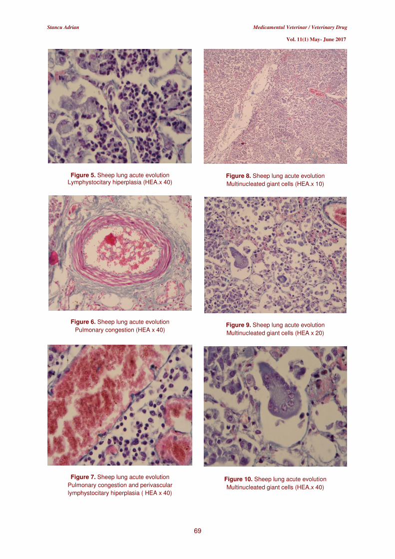
Figure 1. Sheep lung acute evolution
Lobular bronchopneumonia (surface examination)
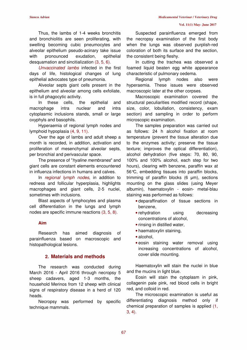
Figure 2. Sheep lung acute evolution.
Lobular bronchopneumonia (section examination)
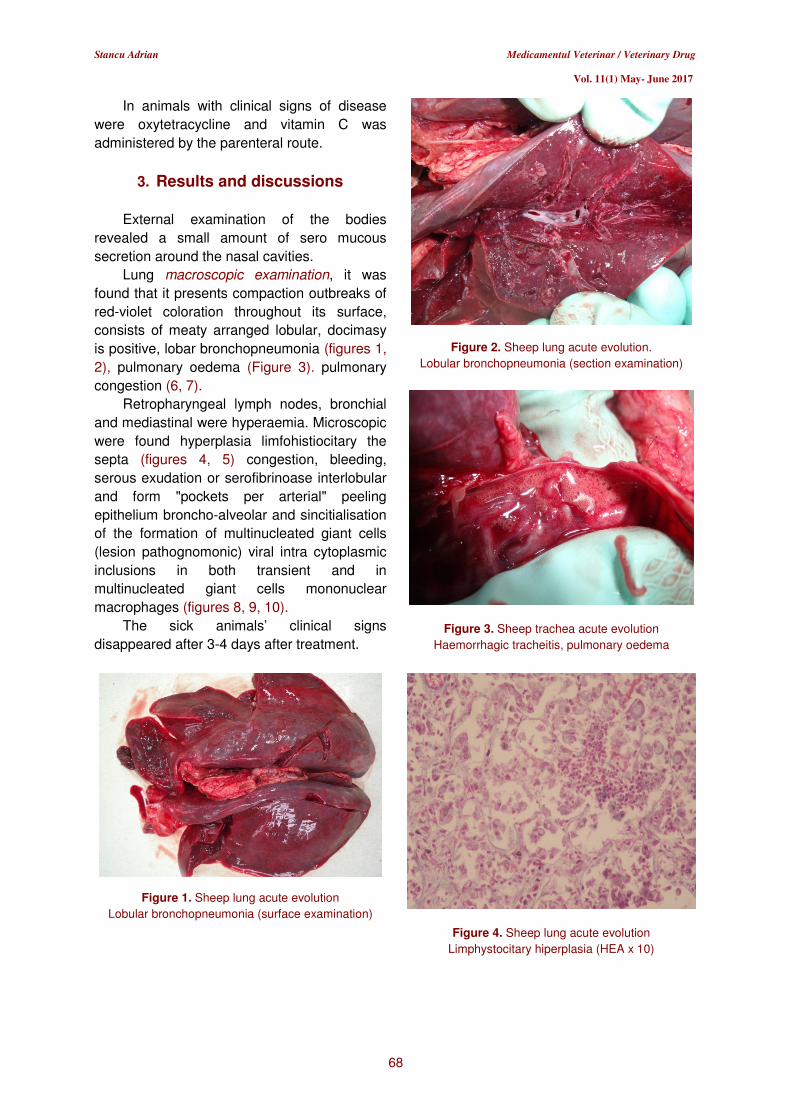
Figure 3. Sheep trachea acute evolution
Haemorrhagic tracheitis, pulmonary oedema
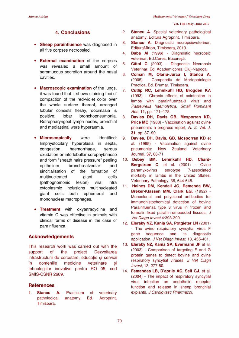
Figure 4. Sheep lung acute evolution
Limphystocitary hiperplasia (HEA x 10)
Stancu Adrian Medicamentul Veterinar / Veterinary Drug
Vol. 11(1) May- June 2017
69
Figure 5. Sheep lung acute evolution Lymphystocitary hiperplasia (HEA.x 40)
Figure 6. Sheep lung acute evolution
Pulmonary congestion (HEA x 40)
Figure 7. Sheep lung acute evolution
Pulmonary congestion and perivascular
lymphystocitary hiperplasia ( HEA x 40)
Figure 8. Sheep lung acute evolution
Multinucleated giant cells (HEA.x 10)
Figure 9. Sheep lung acute evolution
Multinucleated giant cells (HEA x 20)
Figure 10. Sheep lung acute evolution
Multinucleated giant cells (HEA.x 40)
Stancu Adrian Medicamentul Veterinar / Veterinary Drug
Vol. 11(1) May- June 2017
70
4. Conclusions
• Sheep parainfluence was diagnosed in
all five corpses necropsied.
• External examination of the corpses
was revealed a small amount of
seromucous secretion around the nasal
cavities.
• Macroscopic examination of the lungs,
it was found that it shows staining foci of
compaction of the red-violet color over
the whole surface thereof, arranged
lobular consists fleshy, docimasia is
positive, lobar bronchopneumonia.
Retropharyngeal lymph nodes, bronchial
and mediastinal were hyperaemia.
• Microscopically were identified:
limphystocitary hyperplasia in septa,
congestion, haemorrhage, serous
exudation or interlobullar serophybrinose
and form "sheath hairs pressure" peeling
epithelium broncho-alveolar and
sincitialisation of the formation of
multinucleated giant cells
(pathognomonic lesion) viral intra
cytoplasmic inclusions multinucleated
giant cells both ephemeral and
mononuclear macrophages.
• Treatment with oxytetracycline and
vitamin C was effective in animals with
clinical forms of disease in the case of
parainfluenza.
Acknowledgements This research work was carried out with the
support of the project Dezvoltarea
infrastructurii de cercetare, educaţie şi servicii
în domeniile medicine veterinare şi
tehnologiilor inovative pentru RO 05, cod
SMIS-CSNR 2669.
References
1. Stancu A. Practicum of veterinary
pathological anatomy Ed. Agroprint,
Timisoara.
2. Stancu A. Special veterinary pathological
anatomy, Editura Agroprint, Timisoara.
3. Stancu A. Diagnostic necropsicveterinar,
EdituraMirton, Timisoara, 2013.
4. Baba AI (1996) - Diagnostic necropsic
veterinar, Ed.Ceres, Bucureşti.
5. Cătoi C (2003) - Diagnostic Necropsic
Veterinar, Ed. Academicpres, Cluj-Napoca.
6. Coman M, Olariu-Jurca I, Stancu A, (2005) - Compendiu de Morfopatologie
Practică, Ed. Brumar, Timişoara.
7. Cutlip RC, Lehmkuhl HD, Brogden KA (1993) - Chronic effects of coinfection in
lambs with parainfluenza-3 virus and
Pasteurella haemolytica, Small Ruminant
Res. 11, pp. 171–178.
8. Davies DH, Davis GB, Mcsporran KD, Price MC (1983) - Vaccination against ovine
pneumonia: a progress report, N. Z. Vet. J.
31, pp. 87–90.
9. Davies, DH, Davis, GB, Mcsporran KD et
al. (1985) - Vaccination against ovine
pneumonia: New Zealand Veterinary
Journal, 37, 66-71.
10. Debey BM, Lehmkuhl HD, Chard-Bergstrom C. et al. (2001) - Ovine
paramyxovirus serotype 7-associated
mortality in lambs in the United States.
Veterinary Pathology, 38, 644-648.
11. Haines DM, Kendall JC, Remenda BW, Breker-Klassen MM, Clark EG. (1992) -
Monoclonal and polyclonal antibodies for
immunohistochemical detection of bovine
Parainfluenza type 3 virus in frozen and
formalin-fixed paraffin-embedded tissues, J
Vet Diagn Invest 4:393-399.
12. Eleraky NZ, Kania SA, Potgieter LN (2001)
- The ovine respiratory syncytial virus F
gene sequence and its diagnostic
application. J Vet Diagn Invest, 13, 455-461.
13. Eleraky NZ, Kania SA, Evermann JF et al.
(2003) - Comparison of targeting F and G
protein genes to detect bovine and ovine
respiratory syncytial viruses. J Vet Diagn
Invest, 13, 277-80.
14. Femandes LB, D'aprile AC, Seif GJ. et al.
(2004) - The impact of respiratory syncytial
virus infection on endothelin receptor
function and release in sheep bronchial
explants. J Cardiovasc Pharmacol.
Stancu Adrian Medicamentul Veterinar / Veterinary Drug
Vol. 11(1) May- June 2017
71
15. Fenner E, Paul JG, Frederick A, Rott RM., (1996) Veterinary Virology. 2nd Edition.,
Academic Press, New York.
16. Jehan G, Hussein H.A., Reda I.M., (2009) -
Isolation and characterization of PI-3 virus
from sheep and goats, Int J Virol, 5(1), 28-
35.
17. Goodwin KA, Jackson R, Brown C, Davies PR, Morris RS, Perkins NR (2004)
- Pneumonic lesions in lambs in New
Zealand: patterns of prevalence and effects
on production, N. Z. Vet. J. 52, pp. 175–179.
18. Grubbs ST, Kania SA, Potgieter LN. (2001)- Prevalence of ovine and bovine
respiratory syncytial virus infections in cattle
deter-mined with a synthetic peptide based
immunoassay. J Vet Diagn Invest, 13, 128-
131.
19. Jubb KVF, Kennedy PC, Palmer N. (1993)
- Palhology of domestic animals, Third
Edition, voi. 1-3, Academic Press, New York
- London.
20. Kingsbury DW (1991) - The
Paramyxoviruses. Plenum Press, New York.
21. Martin WB. (1996) - Respiratory infections
of sheep, Comp. Immunol. Microbiol. Infect.
Dis.19, pp. 171–179.
22. Moga-Mânzat R (2005) - Boli Virotice şi
prionice ale animalelor, Ed. Brumar,
Timişoara.
23. Radostits OM, Blood DC, Gay CC (1994) -
Veterinary medicine, 8th Edition, Bailliere
Tindall, London, UK.
24. Răpuntean G, Boldizar E. (2002) -
Virusologie special veterinară, Editura
Academic Press, Cluj-Napoca.