Embed Size (px)
Citation preview
The two dimeric forms of RNase A
Salvatore Sorrentinoa, Roberto Baronea, Enrico Buccib, Giovanni Gottec, Nello Russod,Massimo Libonatic, Giuseppe D'Alessioa;*
aDipartimento di Chimica Organica e Biologica, Universita© di Napoli Federico II, Via Mezzocannone 16, 80134 Naples, ItalybCentro di Studio di Biocristallogra¢a del CNR, Via Mezzocannone 4, 80134 Naples, Italy
cDipartimento di Scienze Neurologiche e della Visione, Sezione di Chimica Biologica, Universita© di Verona, Strada le Grazie 8, 37134 Verona, ItalydDipartimento di Scienze della Vita, Seconda Universita© di Napoli, Via Arena 18, 81100 Caserta, Italy
Received 27 November 1999
Edited by Pierre Jolles
Abstract In 1965 Fruchter and Crestfield (J. Biol. Chem. 240,2868^3874) observed that dimeric RNase A prepared bylyophilization from acetic acid could be separated into twoforms. Surprisingly, no other structural or functional differencescould be detected between the two forms. In 1998 a structure fordimeric RNase A was determined by X-ray crystallography byLiu et al. (Proc. Natl. Acad. Sci. USA 95, 3437^3442). Wefound that the two forms of dimeric RNase A have indeeddifferent structural and functional properties, and suggest thatthe dimer whose structure was investigated by Liu and coworkersmay be identified with the lesser form of dimeric RNase A.z 2000 Federation of European Biochemical Societies.
Key words: Ribonuclease; RNase A; Protein oligomer
1. Introduction
In 1962 Crest¢eld et al. discovered that up to 20% of bovinepancreatic RNase A associated into dimers and higher aggre-gates upon lyophilization from concentrated solutions of ace-tic acid [1]. They then proposed an unusual structure for thesedimers, based on an elegant, now historic experiment [2]. Byassociating two di¡erent types of inactive monomers, one in-activated by alkylation at His-12, located on the N-terminalhelix, the other alkylated at His-119, located in the main pro-tein body, enzymatic activity was restored; upon dissociationinto monomers, activity was lost again. These results couldonly be explained by the formation upon dimerization of twocomposite active sites, each comprising His residues from dif-ferent subunits. Their conclusion was that the dimeric struc-ture was based on the interchange between protomers of theirN-terminal helices.
This type of quaternary structure has been given increasingattention in the last few years, for its incidence in both arti-¢cial and natural oligomers [3], and for its evolutionary [4^6]and pathological [3] implications.
A few years after their initial observation on dimeric RNaseA, Fruchter and Crest¢eld reported that upon ion-exchangechromatography on sulfo-ethyl Sephadex dimeric RNase Acould be separated into two fractions [7], with the lesser onerepresenting 20^25% of the dimeric protein. Surprisingly, afterseveral tests, no structural or functional di¡erences were de-tected between these two fractions.
Recently, the structure of dimeric RNase A was determinedby X-ray crystallography [8]. It was found that indeed the twoRNase A protomers associate in the dimeric structure throughthe exchange, or swap, of their N-terminal helices. However,it was not determined whether the investigated dimeric struc-ture was that of the larger or of the lesser dimeric fraction.
The observation of the existence of two isoforms of dimericRNase A has been recently con¢rmed and expanded, as iso-forms were observed also for the higher aggregates of RNaseA [9]. We report here that there are signi¢cant structural andfunctional di¡erences between the two dimers of RNase A,and propose that the recently determined three-dimensionalstructure is that of the lesser dimeric isoform of the protein.
2. Materials and methods
2.1. ProteinsBovine pancreatic RNase A (type XII-A) was purchased from Sig-
ma. Bovine seminal RNase (BS-RNase) was puri¢ed as previouslydescribed [10]. Protein concentration was determined spectrophoto-metrically using for RNase A an absorbance coe¤cient of 7.3 at280 nm for a 1% solution [11] and for BS-RNase an absorbancecoe¤cient of 4.65 at 278 nm for a 1% solution.
2.2. SubstratesYeast RNA, double-stranded poly(A)Wpoly(U), poly(U) and cytidyl-
yl-(3P,5P)-adenosine (CpA) were Sigma products. The cytosolic RNaseinhibitor (CRI) was purchased from Promega.
2.3. RNase assaysRibonuclease activity toward yeast RNA was measured at 25³C
following the Kunitz spectrophotometric assay [12] with 0.6 mg/mlof RNA in 0.1 M sodium acetate/acetic acid bu¡er, pH 5.0. Degra-dation of poly(U) and poly(A)Wpoly(U) at 25³C was followed spectro-photometrically at 260 nm in 0.1 M MOPS, pH 7.5 containing 0.1 MNaCl. Substrate concentration was 0.1 mM in phosphodiester groups(about 40 Wg/ml). An enzyme unit was de¢ned as the fraction ofabsorbance change per minute over the total measurable change[13]. Enzyme activity toward CpA was measured as described [14]in the absence or presence of RNase inhibitor with 50 WM substratein 0.1 M MES at pH 6.0 containing 0.1 M NaCl and RNase freebovine serum albumin (10 Wg/ml).
2.4. Cross-linking assayThe procedure described by Ciglic et al. [15] was followed. About
40 Wg of each protein in 300 Wl of 50 mM sodium acetate at pH 5.0
0014-5793 / 00 / $20.00 ß 2000 Federation of European Biochemical Societies. All rights reserved.PII: S 0 0 1 4 - 5 7 9 3 ( 9 9 ) 0 1 7 4 2 - 1
*Corresponding author. Fax: (39)-81-5521217.E-mail: [email protected]
Abbreviations: RNase A, bovine pancreatic ribonuclease; D-I, theprevalent dimer-I fraction of dimeric RNase A; D-II, the dimer-IIlesser fraction of dimeric RNase A; BS-RNase, bovine seminalRNase; DVS, divinyl sulfone; CRI, cytosolic RNase inhibitor
FEBS 23169 14-1-00
FEBS 23169 FEBS Letters 466 (2000) 35^39
containing 0.1% divinyl sulfone (DVS) was incubated at 30³C for 72 h.The reaction was stopped by addition of L-mercaptoethanol(200 mM). After 20 min, aliquots of 10 Wl were analyzed with 15%SDS-PAGE under reducing conditions [16].
2.5. Preparation of RNase A dimersAggregates of ribonuclease A were obtained by lyophilization of the
enzyme from 40% acetic acid solutions according to the procedure ofCrest¢eld et al. [1]. The lyophilized material was dissolved in 1.2 ml of0.2 M sodium phosphate bu¡er pH 6.55 and applied to a SephadexG-75 column (1.5U105 cm) equilibrated with the same bu¡er andcalibrated with monomeric RNase A and dimeric BS-RNase. Elutionwas performed at room temperature at a £ow rate of 6 ml/h. Thedimeric fraction was concentrated and dialyzed against bu¡er A(40 mM sodium phosphate bu¡er, pH 6.55) with Centriprep concen-trators (Amicon), and applied to a Source 15 S HR 10/10 columnequilibrated with bu¡er A on a Pharmacia FPLC system. The prep-aration of dimeric RNase A was resolved into two components by aphosphate gradient using 200 mM sodium phosphate, pH 6.55 asbu¡er B (see Fig. 1). Elution was performed at 25³C at a £ow rateof 1 ml/min and fractions were manually collected.
2.6. Dissociation kineticsPreparations of RNase A dimers (0.42 mg/ml), equilibrated in
50 mM Tris^HCl pH 7.5 containing 130 mM NaCl by extensivedialysis at 4³C, were incubated at 37³C. At appropriate time intervals,aliquots were analyzed by gel ¢ltration with a £ow rate of 0.6 ml/minon a Pharmacia FPLC system equipped with a Superdex 75 10/30column equilibrated with 0.1 M Tris^HCl at pH 7.5 containing0.3 M NaCl. The amounts of monomer and dimer were estimatedby measuring the areas of their absorbance pro¢les at 278 nm.
2.7. Circular dichroism (CD) measurementsThe CD measurements were made at 25³C on a 715 Jasco spectro-
polarimeter equipped with PTC-348 WI thermostat, under a constantnitrogen £ow. Hellma quartz cells of 0.1 cm path length and a proteinconcentration of about 0.13 mg/ml were used in the far-UV region(190^240 nm). In the near-UV region (240^320 nm) protein concen-tration was 0.8 mg/ml with a path length of 1 cm. All samples weredissolved in 100 mM sodium phosphate bu¡er, pH 6.55. The reducedmean residue ellipticities ([a]res) were calculated taking in account thatthe mean residue molecular weight for pancreatic RNase A is 110.5[17].
3. Results and discussion
3.1. The two forms of dimeric RNase ADimeric RNase A was prepared by gel ¢ltration and frac-
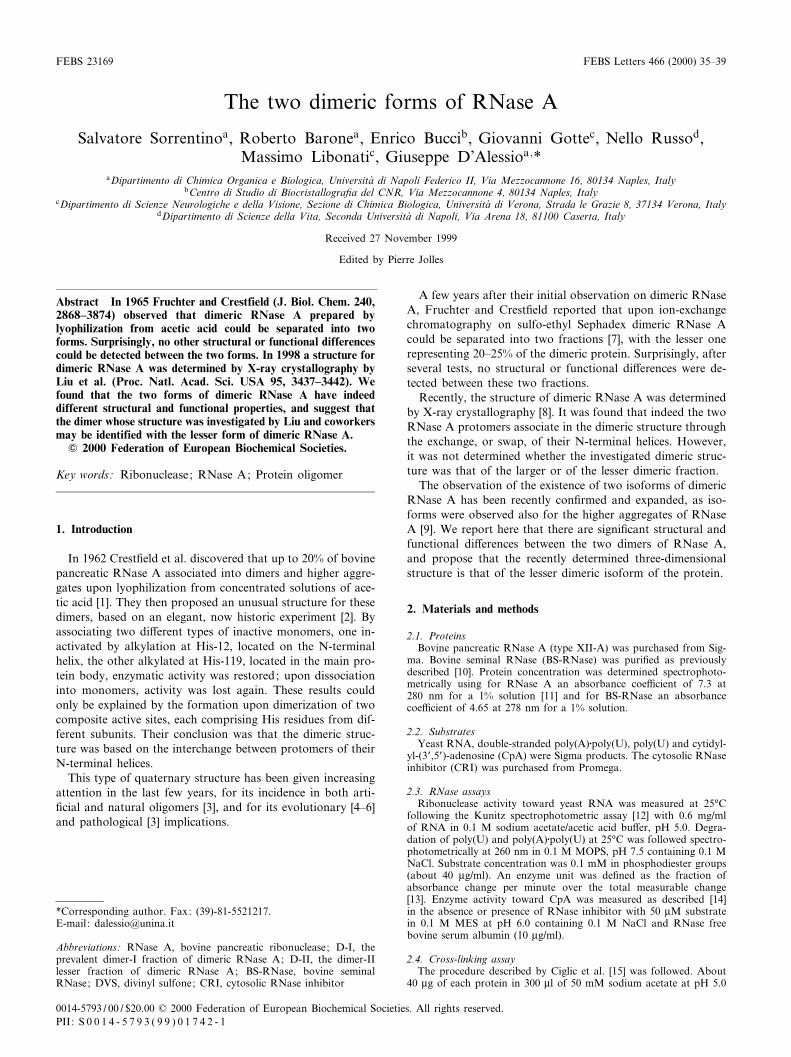
tionated by chromatography on a cation exchanger as de-scribed in Section 2. Two main fractions were separated andcollected (see Fig. 1). The main component, named dimer-I orD-I [7], appeared to have a higher positive net charge, as iteluted 7 min later than the minor one, named dimer-II orD-II. The latter represented 20^25% of the whole dimericfraction of RNase A.
The homogeneity of each dimeric form was demonstratedby re-chromatography under the same conditions of suitablealiquots of puri¢ed D-I and D-II fractions. Each type ofdimer eluted as a single peak with the same elution time asin Fig. 1. Upon dissociation by heating at 65³C for 30 minand re-chromatography, both D-I and D-II coeluted withmonomeric RNase A. This con¢rms that D-I and D-II aredistinct dimeric associations of a single monomeric form ofRNase A.
The two small fractions preceding D-I and D-II, respec-tively, in the chromatographic pattern (see Fig. 1) werefound to be dimeric by gel ¢ltration (data not shown).After heating and dissociation, and upon re-chromatographyas above, each eluted as a more negatively charged monomer,
1 min earlier than native RNase A. This suggests that they arethe respective deamidation products of the two main dimersD-I and D-II. Possibly, as found for naturally dimeric BS-RNase when compared to its monomeric counterpart [18],deamidation is faster in dimeric than in monomeric RNaseA.
Based on these results, we can conclude that the two dimer-ic forms D-I and D-II are those originally described byFruchter and Crest¢eld [7] and recently investigated by Gotteet al. [9].
3.2. Catalytic and structural properties of D-I and D-II ofRNase A
The catalytic activities of D-I and D-II were compared inside-by-side assays performed with single- and double-stranded polyribonucleotides. Native monomeric RNase Awas included as a standard for comparison. The results aresummarized in Table 1. On yeast RNA and on poly(U) assubstrates D-I and D-II displayed similar activity values,somewhat lower than that measured for RNase A. On thedouble-stranded substrate poly(A)Wpoly(U) instead the twodimers were both more active than the monomer, with D-Itwice as active as D-II.
Fig. 1. Ion-exchange chromatography of RNase A dimers. An ali-quot (about 2 mg) of RNase A dimers from Sephadex G-75 was ap-plied to a Source 15 S HR 10/10 column (Pharmacia FPLC system)equilibrated with 40 mM sodium phosphate bu¡er, pH 6.55 (bu¡erA). Elution was performed with a gradient as indicated (dashedline), using 200 mM sodium phosphate, pH 6.55 as bu¡er B, at a£ow rate of 1 ml/min at 25³C. The elution time (23 min) of nativeRNase A (M) is indicated by an arrow.
Table 1Activity of RNase A and its dimeric forms on single- and double-stranded polyribonucleotides
Speci¢c activity (U/mg of protein)a toward
Yeast RNAb Poly(U)c Poly(A)WPoly(U)c
RNase A 112 þ 6 395 þ 20 1.3 þ 0.1D-I 88 þ 4 382 þ 13 6.2 þ 0.3D-II 85 þ 4 387 þ 15 2.5 þ 0.1aSpectrophotometric assays performed at 25³C. Each activity valueis the mean ( þ S.D.) of four measurements. Units are de¢ned inSection 2.bKunitz assays performed at pH 5.0 in sodium acetate bu¡er.cAssays performed at pH 7.5 in 0.1 M MOPS containing 0.1 MNaCl.
FEBS 23169 14-1-00
S. Sorrentino et al./FEBS Letters 466 (2000) 35^3936
These results are in line with those recently obtained withdi¡erent assay procedures [9]. Taken together with the obser-vations reported above that both D-I and D-II have a positivenet charge higher than that of monomeric RNase A, and thatD-I is more basic than D-II, they support the proposal [13]that the action of pancreatic-type RNases on double-strandedsubstrates correlates with positive charge densities located indiscrete regions of the protein.
The structures of the CRI [19] and of its complexes withRNase A [20] or angiogenin [21] have been determined. Whenthe structure of dimeric RNase A [8] was modeled onto thatof CRI (data not shown) it was apparent that dimeric RNaseA cannot bind to the inhibitor. As the structure determinedfor dimeric RNase A [8] must be clearly assigned to at leastone of the two dimers D-I or D-II, their sensitivity to CRIwas tested by assaying their enzymatic activity in the presenceof CRI.
With CpA as a substrate both D-I and D-II dimers werefound to be fully susceptible to inhibition by CRI. Based onthe considerations expressed above, that at least one of thedimeric forms cannot bind to the inhibitor, and given themetastability of the two dimers (see below), these results sug-gest that both dimeric isoforms dissociate upon interactingwith CRI, and bind to the inhibitor as monomers. Yet, it
cannot be excluded that the dimeric form distinct from thatdescribed by Liu et al. [8] can also ¢t as a dimer in the com-plex with CRI.
It has been shown that cross-linking with DVS is a conven-ient tool for determining whether RNase dimers exchangetheir N-terminal arms as they do in seminal RNase [15].The assay is based on the selective simultaneous reactivitywith DVS of both His-12, located on the N-terminal arm ofpancreatic-type RNases, and His-119, located on the mainprotein body. When in an RNase dimer there is exchange ofthe N-terminal arms between protomers, the two His residuesare contributed one from each protomer, and the result of thereaction with DVS is a covalent link between protomers. Inthe absence of arm exchange, just as in a monomer, His-12and -119 belonging to the same protomer are instead cross-linked.
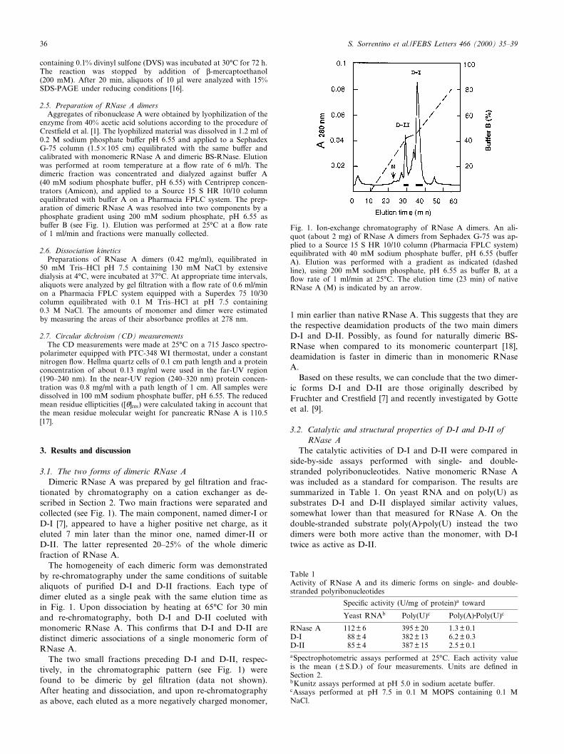
D-I and D-II were treated for 72 h at 30³C with DVS asdescribed [15]. Cross-linking was tested by SDS-PAGE underreducing conditions. Positive (BS-RNase), and negative(RNase A) controls were included in the experiment. Asshown in Fig. 2, about 60% of D-I was found to migrate asa cross-linked dimer, whereas D-II was almost completelycross-linked. These results lead to the conclusion that inboth D-I and D-II the protomers exchange their N-terminalhelices, as found for the dimer investigated by Liu et al. [8].The observed fragmentations, with band splitting (see Fig. 2),are side e¡ects of the reaction [15].
Given the metastable nature of RNase A dimers (see be-low), it is not surprising that both D-I and D-II in part dis-sociate and react with DVS as monomers, as has already beenreported for the unresolved mixture of dimers [15]. Hence thisdi¡erence in reactivity with DVS between D-I and D-II can beexplained by a di¡erent degree of dissociation of the twodimers in the prolonged incubation with DVS at 30³C.
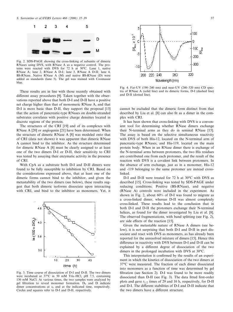
This interpretation is con¢rmed by the results of an experi-ment in which the kinetics of dissociation of the two dimers at37³C were measured. The fraction of each dimer dissociatedinto monomers as a function of time was determined by gel¢ltration (see Section 2). D-I was found to be more readilydissociated than D-II (see Fig. 3). The data ¢tted ¢rst-orderplots and gave t1=2 times of 29 and 16 h, respectively, for D-IIand D-I. The di¡erent stabilities of D-I and D-II indicate thatthe two dimers have a di¡erent structure.
Fig. 2. SDS-PAGE showing the cross-linking of subunits of dimericRNases using DVS, with RNase A as a negative control. The pro-teins were reacted with DVS for 72 h at 30³C. Lane 1, nativeRNase A; lane 2, RNase A D-I; lane 3, RNase A D-II; lane 4,BS-RNase. Native RNase A (M) and native BS-RNase (D) wereadded as standards (lane 5). The gel was stained with Coomassieblue.
Fig. 3. Time course of dissociation of D-I and D-II. The two dimerswere incubated at 37³C in 50 mM Tris^HCl, pH 7.5, containing130 mM NaCl. At various times, the two samples were analyzed bygel ¢ltration to reveal monomer formation. D0 and D indicatedimer concentrations at t0 and at the indicated time, respectively.Circles and squares refer to D-I and D-II, respectively.
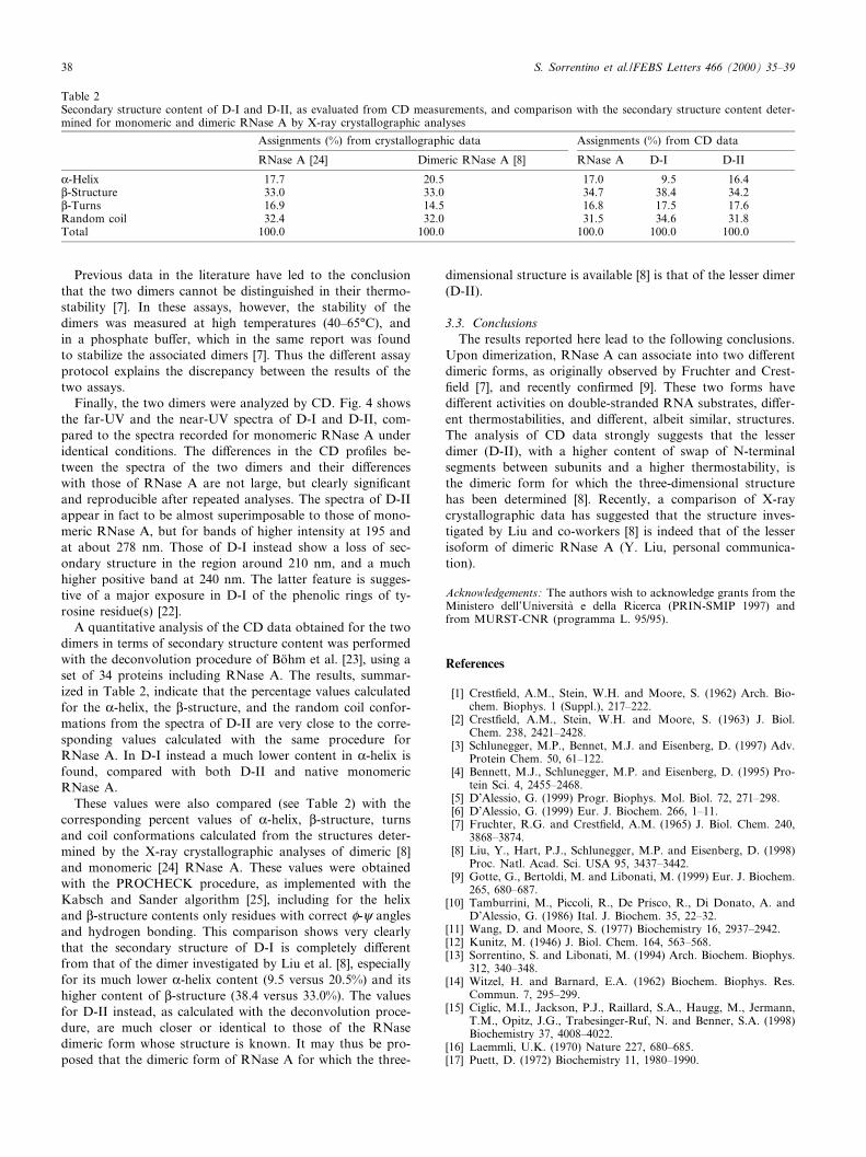
Fig. 4. Far-UV (190^240 nm) and near-UV (240^320 nm) CD spec-tra of RNase A (solid line) and its dimeric forms, D-I (dashed line)and D-II (dotted line).
FEBS 23169 14-1-00
S. Sorrentino et al./FEBS Letters 466 (2000) 35^39 37
Previous data in the literature have led to the conclusionthat the two dimers cannot be distinguished in their thermo-stability [7]. In these assays, however, the stability of thedimers was measured at high temperatures (40^65³C), andin a phosphate bu¡er, which in the same report was foundto stabilize the associated dimers [7]. Thus the di¡erent assayprotocol explains the discrepancy between the results of thetwo assays.
Finally, the two dimers were analyzed by CD. Fig. 4 showsthe far-UV and the near-UV spectra of D-I and D-II, com-pared to the spectra recorded for monomeric RNase A underidentical conditions. The di¡erences in the CD pro¢les be-tween the spectra of the two dimers and their di¡erenceswith those of RNase A are not large, but clearly signi¢cantand reproducible after repeated analyses. The spectra of D-IIappear in fact to be almost superimposable to those of mono-meric RNase A, but for bands of higher intensity at 195 andat about 278 nm. Those of D-I instead show a loss of sec-ondary structure in the region around 210 nm, and a muchhigher positive band at 240 nm. The latter feature is sugges-tive of a major exposure in D-I of the phenolic rings of ty-rosine residue(s) [22].
A quantitative analysis of the CD data obtained for the twodimers in terms of secondary structure content was performedwith the deconvolution procedure of Bo«hm et al. [23], using aset of 34 proteins including RNase A. The results, summar-ized in Table 2, indicate that the percentage values calculatedfor the K-helix, the L-structure, and the random coil confor-mations from the spectra of D-II are very close to the corre-sponding values calculated with the same procedure forRNase A. In D-I instead a much lower content in K-helix isfound, compared with both D-II and native monomericRNase A.
These values were also compared (see Table 2) with thecorresponding percent values of K-helix, L-structure, turnsand coil conformations calculated from the structures deter-mined by the X-ray crystallographic analyses of dimeric [8]and monomeric [24] RNase A. These values were obtainedwith the PROCHECK procedure, as implemented with theKabsch and Sander algorithm [25], including for the helixand L-structure contents only residues with correct P-i anglesand hydrogen bonding. This comparison shows very clearlythat the secondary structure of D-I is completely di¡erentfrom that of the dimer investigated by Liu et al. [8], especiallyfor its much lower K-helix content (9.5 versus 20.5%) and itshigher content of L-structure (38.4 versus 33.0%). The valuesfor D-II instead, as calculated with the deconvolution proce-dure, are much closer or identical to those of the RNasedimeric form whose structure is known. It may thus be pro-posed that the dimeric form of RNase A for which the three-
dimensional structure is available [8] is that of the lesser dimer(D-II).
3.3. ConclusionsThe results reported here lead to the following conclusions.
Upon dimerization, RNase A can associate into two di¡erentdimeric forms, as originally observed by Fruchter and Crest-¢eld [7], and recently con¢rmed [9]. These two forms havedi¡erent activities on double-stranded RNA substrates, di¡er-ent thermostabilities, and di¡erent, albeit similar, structures.The analysis of CD data strongly suggests that the lesserdimer (D-II), with a higher content of swap of N-terminalsegments between subunits and a higher thermostability, isthe dimeric form for which the three-dimensional structurehas been determined [8]. Recently, a comparison of X-raycrystallographic data has suggested that the structure inves-tigated by Liu and co-workers [8] is indeed that of the lesserisoform of dimeric RNase A (Y. Liu, personal communica-tion).
Acknowledgements: The authors wish to acknowledge grants from theMinistero dell'Universita© e della Ricerca (PRIN-SMIP 1997) andfrom MURST-CNR (programma L. 95/95).
References
[1] Crest¢eld, A.M., Stein, W.H. and Moore, S. (1962) Arch. Bio-chem. Biophys. 1 (Suppl.), 217^222.
[2] Crest¢eld, A.M., Stein, W.H. and Moore, S. (1963) J. Biol.Chem. 238, 2421^2428.
[3] Schlunegger, M.P., Bennet, M.J. and Eisenberg, D. (1997) Adv.Protein Chem. 50, 61^122.
[4] Bennett, M.J., Schlunegger, M.P. and Eisenberg, D. (1995) Pro-tein Sci. 4, 2455^2468.
[5] D'Alessio, G. (1999) Progr. Biophys. Mol. Biol. 72, 271^298.[6] D'Alessio, G. (1999) Eur. J. Biochem. 266, 1^11.[7] Fruchter, R.G. and Crest¢eld, A.M. (1965) J. Biol. Chem. 240,
3868^3874.[8] Liu, Y., Hart, P.J., Schlunegger, M.P. and Eisenberg, D. (1998)
Proc. Natl. Acad. Sci. USA 95, 3437^3442.[9] Gotte, G., Bertoldi, M. and Libonati, M. (1999) Eur. J. Biochem.
265, 680^687.[10] Tamburrini, M., Piccoli, R., De Prisco, R., Di Donato, A. and
D'Alessio, G. (1986) Ital. J. Biochem. 35, 22^32.[11] Wang, D. and Moore, S. (1977) Biochemistry 16, 2937^2942.[12] Kunitz, M. (1946) J. Biol. Chem. 164, 563^568.[13] Sorrentino, S. and Libonati, M. (1994) Arch. Biochem. Biophys.
312, 340^348.[14] Witzel, H. and Barnard, E.A. (1962) Biochem. Biophys. Res.
Commun. 7, 295^299.[15] Ciglic, M.I., Jackson, P.J., Raillard, S.A., Haugg, M., Jermann,
T.M., Opitz, J.G., Trabesinger-Ruf, N. and Benner, S.A. (1998)Biochemistry 37, 4008^4022.
[16] Laemmli, U.K. (1970) Nature 227, 680^685.[17] Puett, D. (1972) Biochemistry 11, 1980^1990.
Table 2Secondary structure content of D-I and D-II, as evaluated from CD measurements, and comparison with the secondary structure content deter-mined for monomeric and dimeric RNase A by X-ray crystallographic analyses
Assignments (%) from crystallographic data Assignments (%) from CD data
RNase A [24] Dimeric RNase A [8] RNase A D-I D-II
K-Helix 17.7 20.5 17.0 9.5 16.4L-Structure 33.0 33.0 34.7 38.4 34.2L-Turns 16.9 14.5 16.8 17.5 17.6Random coil 32.4 32.0 31.5 34.6 31.8Total 100.0 100.0 100.0 100.0 100.0
FEBS 23169 14-1-00
S. Sorrentino et al./FEBS Letters 466 (2000) 35^3938
[18] Di Donato, A., Ciardiello, M.A., de Nigris, M., Piccoli, R.,Mazzarella, L. and D'Alessio, G. (1993) J. Biol. Chem. 268,4745^4751.
[19] Kobe, B. and Deisenhofer, J. (1993) Nature 366, 751^756.[20] Kobe, B. and Deisenhofer, J. (1996) J. Mol. Biol. 264, 1028^
1043.[21] Acharya, K.R., Papageorgiou, A.C. and Shapiro, R. (1997)
EMBO J. 16, 5162^5177.
[22] Grandi, C., D'Alessio, G. and Fontana, A. (1979) Biochemistry18, 3413^3420.
[23] Bo«hm, G., Muhr, R. and Jaenicke, R. (1992) Protein Eng. 5,191^195.
[24] Wlodawer, A., Svensson, L.A., Sjolin, L. and Gilliland, G.L.(1988) Biochemistry 27, 2705^2717.
[25] Kabsh, W. and Sander, C. (1983) Biopolymers 22, 2577.
FEBS 23169 14-1-00
S. Sorrentino et al./FEBS Letters 466 (2000) 35^39 39


![Designing Dimeric Lanthanide(III)-Containing Ionic liquids › ws › files › 158240242 › ...COMMUNICATION Designing Dimeric Lanthanide(III)-Containing Ionic liquids Éadaoin McCourt,[a]](https://img.pdfslide.net/doc/110x75/60b904bbc8cfbf6cfb110109/designing-dimeric-lanthanideiii-containing-ionic-liquids-a-ws-a-files-a.jpg)
![lncRNAs: function and mechanism in cartilage development ......ticle RNase MRP. RNase MRP is the source of two short RNA designated RMRP-S1 and RMRP-S2 [58]. Mutations in RNase MRP](https://img.pdfslide.net/doc/110x75/60dc29d704644d4b965001ed/lncrnas-function-and-mechanism-in-cartilage-development-ticle-rnase-mrp.jpg)