Embed Size (px)
Citation preview

The use of bone cement in difficult distal radius fractures
Mithun Neral & Mario Solari & Chad Purnell &Ronit Wollstein
Published online: 2 October 2013# American Association for Hand Surgery 2013
AbstractBackground The lack of structural support remains a challengein the treatment of comminuted distal radius fractures. Calciumphosphate and calcium sulfate bone cement has been used inother fracture locations in addition to fixation and has beenshown to allow for retention of reduction in difficult cases.Methods A case-control retrospective review of 34 consecu-tive distal radius fractures treated with surgery was performedwith the patients classified by Arbeitsgemeinschaft fürOsteosynthesefragen (AO) classification. Complications andpostoperative radiographs were evaluated.Results Cement was used in the most difficult cases. Radialheight was retained in both groups. Volar tilt was significantlybetter in the cement group. There were no significant differ-ences between the case and control groups for any complica-tion. No complications related to the use of the cement werefound.Conclusions The use of bone cement as an adjunct to fixationof distal radius fractures seems to include minimal risks andmay afford a technical advantage in maintaining reductionduring surgery for difficult fractures. Since there is an aspectof fracture difficulty that we cannot control for by usingradiographic assessment alone, cement may provide an ad-vantage over fixation without cement, despite similar out-comes. Bone cement can be part of the "tool box" for difficultdistal radius fractures. Further study is necessary to define thetechnical advantages and limitations of each particular cementproduct.
Keywords Bone cement . Distal radius fractures .
Arbeitsgemeinschaft für OsteosynthesefragenOsteosynthesefragen (AO) classification
Background and Purpose
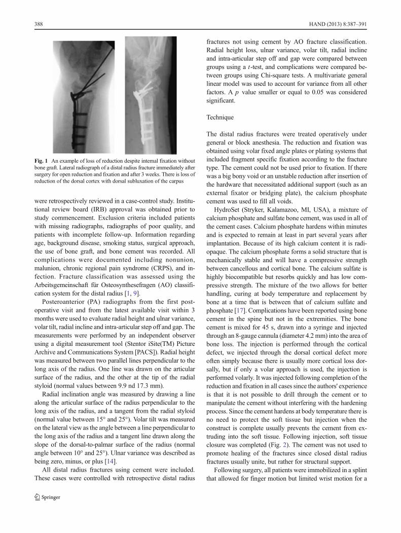
The treatment of comminuted, intra-articular distal radiusfractures has been evolving to include a multitude of tech-niques for internal fixation [2, 3]. While long-term outcomesand complications are still being evaluated, a significant chal-lenge in the treatment of these fractures is structural support inthe presence of soft and sometimes osteoporotic metaphysealbone. This is especially true for already healing fractureswhere an osteotomy is necessary prior to reduction (Fig. 1).Current techniques utilized to overcome this problem haveincluded the use of adjunct external fixation, locking, andbridging plates. Bone grafting has been used for support, butrequires cortical bone, which may not provide immediate ordelayed maintenance during the healing process [12]. Further-more, bone grafting includes donor site morbidity in autolo-gous grafts and potential infection in allografts [5]. Bonecement has been used for scaffolding in spine surgery, butalso in metaphyseal and intra-articular fractures such as in thetibial plateau, proximal humerus, and distal radius [4, 8, 11,13, 16].
We retrospectively compared distal radius fractures treatedwith open reduction and internal fixation using bone cementto those without the use of cement in an attempt to define therole of bone cement in the surgery of distal radius fractures.
Materials and Methods
All distal radius fractures treated over a period of 30 monthsby a single surgeon with open reduction and internal fixation
M. Neral :M. Solari :C. Purnell : R. WollsteinDepartment of Plastic Surgery, University of Pittsburgh MedicalCenter, M.D. 3550 Terrace Street, Pittsburgh, PA 15261, USA
M. Neral :M. Solari :C. Purnell : R. Wollstein (*)Department of Orthopedic Surgery, University of PittsburghMedicalCenter, M.D. 3550 Terrace Street, Pittsburgh, PA 15261, USAe-mail: [email protected]
HAND (2013) 8:387–391DOI 10.1007/s11552-013-9548-z
were retrospectively reviewed in a case-control study. Institu-tional review board (IRB) approval was obtained prior tostudy commencement. Exclusion criteria included patientswith missing radiographs, radiographs of poor quality, andpatients with incomplete follow-up. Information regardingage, background disease, smoking status, surgical approach,the use of bone graft, and bone cement was recorded. Allcomplications were documented including nonunion,malunion, chronic regional pain syndrome (CRPS), and in-fection. Fracture classification was assessed using theArbeitsgemeinschaft für Osteosynthesefragen (AO) classifi-cation system for the distal radius [1, 9].
Posteroanterior (PA) radiographs from the first post-operative visit and from the latest available visit within 3months were used to evaluate radial height and ulnar variance,volar tilt, radial incline and intra-articular step off and gap. Themeasurements were performed by an independent observerusing a digital measurement tool (Stentor iSite(TM) PictureArchive and Communications System [PACS]). Radial heightwas measured between two parallel lines perpendicular to thelong axis of the radius. One line was drawn on the articularsurface of the radius, and the other at the tip of the radialstyloid (normal values between 9.9 nd 17.3 mm).
Radial inclination angle was measured by drawing a linealong the articular surface of the radius perpendicular to thelong axis of the radius, and a tangent from the radial styloid(normal value between 15° and 25°). Volar tilt was measuredon the lateral view as the angle between a line perpendicular tothe long axis of the radius and a tangent line drawn along theslope of the dorsal-to-palmar surface of the radius (normalangle between 10° and 25°). Ulnar variance was described asbeing zero, minus, or plus [14].
All distal radius fractures using cement were included.These cases were controlled with retrospective distal radius
fractures not using cement by AO fracture classification.Radial height loss, ulnar variance, volar tilt, radial inclineand intra-articular step off and gap were compared betweengroups using a t-test, and complications were compared be-tween groups using Chi-square tests. A multivariate generallinear model was used to account for variance from all otherfactors. A p value smaller or equal to 0.05 was consideredsignificant.
Technique
The distal radius fractures were treated operatively undergeneral or block anesthesia. The reduction and fixation wasobtained using volar fixed angle plates or plating systems thatincluded fragment specific fixation according to the fracturetype. The cement could not be used prior to fixation. If therewas a big bony void or an unstable reduction after insertion ofthe hardware that necessitated additional support (such as anexternal fixator or bridging plate), the calcium phosphatecement was used to fill all voids.
HydroSet (Stryker, Kalamazoo, MI, USA), a mixture ofcalcium phosphate and sulfate bone cement, was used in all ofthe cement cases. Calcium phosphate hardens within minutesand is expected to remain at least in part several years afterimplantation. Because of its high calcium content it is radi-opaque. The calcium phosphate forms a solid structure that ismechanically stable and will have a compressive strengthbetween cancellous and cortical bone. The calcium sulfate ishighly biocompatible but resorbs quickly and has low com-pressive strength. The mixture of the two allows for betterhandling, curing at body temperature and replacement bybone at a time that is between that of calcium sulfate andphosphate [17]. Complications have been reported using bonecement in the spine but not in the extremities. The bonecement is mixed for 45 s, drawn into a syringe and injectedthrough an 8-gauge cannula (diameter 4.2 mm) into the area ofbone loss. The injection is performed through the corticaldefect, we injected through the dorsal cortical defect moreoften simply because there is usually more cortical loss dor-sally, but if only a volar approach is used, the injection isperformed volarly. It was injected following completion of thereduction and fixation in all cases since the authors' experienceis that it is not possible to drill through the cement or tomanipulate the cement without interfering with the hardeningprocess. Since the cement hardens at body temperature there isno need to protect the soft tissue but injection when theconstruct is complete usually prevents the cement from ex-truding into the soft tissue. Following injection, soft tissueclosure was completed (Fig. 2). The cement was not used topromote healing of the fractures since closed distal radiusfractures usually unite, but rather for structural support.
Following surgery, all patients were immobilized in a splintthat allowed for finger motion but limited wrist motion for a
Fig. 1 An example of loss of reduction despite internal fixation withoutbone graft. Lateral radiograph of a distal radius fracture immediately aftersurgery for open reduction and fixation and after 3 weeks. There is loss ofreduction of the dorsal cortex with dorsal subluxation of the carpus
388 HAND (2013) 8:387–391

period of 6 weeks. Hand therapy was instituted for range ofmotion (ROM) and gradual strengthening. Patients werefollowed in clinic every 2 weeks for the first 3 months. At 6weeks, radiographs were obtained to evaluate healing of thefractures.
Results
The population consisted of 36 distal radius fractures thatwere treated with open reduction and internal fixation usingcement. Of these, 34 patients had adequate follow-up. There-fore, 34 cases were controlled with 26 retrospective distalradius fractures not using cement by AO fracture classifica-tion. The controls were cases in the last 30 months that wereoperated on without the use of cement and were matched tocases by AO classification. We were unable to control foreight C3 fracture cases. One patient in the cement group wastreated with an external fixator in addition to internal fixationand cement (Table 1).
The case and control populations were not shown to besignificantly different and could be compared (Table 2). Theaverage follow-up period was 56.4 (67.7) days with no sig-nificant difference between the cement and control groups.Fifteen cases and nine control patients had follow-up radio-graphs of sufficient quality to assess radial height measure-ments. Volar tilt, radial inclination, and ulnar variance weremeasured on radiographs of 30 cases and 23 controls. Bothcement and non-cement patients averaged less than 1 mm in
radial height loss. The difference between the groups was notsignificant. Volar tilt measurements differed significantly be-tween the two groups (Table 3). Articular gapping and step-offwere not apparent on any of the post- operative radiographsand therefore the results in the cement and control groups didnot differ.
There was one nonunion in the cement group at 6 weekspost-surgery and one malunion in the same group. Overallcomplications, pin site infection, nonunion, malunion, chronicpain, and CRPS did not significantly differ between the caseand control groups for any complication. The cement groupinitially had a significant occurrence of chronic pain andCRPS but when controlled for AO grade, the associationwas not significant (Table 4).
Discussion
Kim et al. concluded that adding bone cement does not changethe amount of collapse in distal radial fractures treated withinternal fixation [6]. We did find a significant differencebetween the groups in the measurement of volar tilt. Overall,there was an average difference of 6° between the groups. It isnot clear if this difference is clinically significant. However,since height retention was similar in both groups, and volar tiltwas improved, it is possible that the use of cement preventsheight loss in fractures that otherwise would have collapsed.
While minimal complications did exist in the cement groupsuch as infection, nonunion, and malunion, we were unable tofind a clear association between the use of cement and thesecomplications. These complications could be accounted for bythe greater complexity of cases for which cement was used(e.g., the nonunionwas in a patient who fell from a height withsignificant associated soft tissue injury due to the high energyof the injury). We could not control for eight fractures, whichwere all C3 fractures. This is logical since we used the cementonly in fractures with significant intra-articular or metaphyseal
Table 1 The distribution of fractures by AO stage in the two populations
AO stage A2 B1 B2 C1 C2 C3 Total
No cement 1 2 2 3 13 5 26
Cement 1 2 2 3 13 13 34
Total 2 4 4 6 26 18 60
Fig. 2 Before and afterradiographs of a distal radiusfracture treated with openreduction internal fixation andcement bone graft
HAND (2013) 8:387–391 389
bone loss. Since a higher fracture grade is associated withcomplications such as chronic pain and CRPS, it was neces-sary to correct for this bias in our analysis when evaluatingcomplications by comparing the cases and controls withineach individual AO classification for each complication.
Since our study is a retrospective review with a smallpopulation, the conclusions from the results are limited. Themeasurement of radial height, ulnar variance, articular step-off, gapping, and radial inclination are all dependent on thequality of the radiograph and the precision of the PA view.Many of these radiographs were taken before the patientsregained maximal ROM, and therefore many of the viewswere suboptimal and were excluded from the study. Moreoverthe differences in angle and height were small. Radial heightmeasurements were all <1mm, and possibly within the rangeof our measurement error. The study by Knirk and Jupiter [7]that was also performed on similar radiographs found that theclinically significant step-off was 2 mm, so it is probably validto say there was no clinically significant loss of height in thepopulation as a whole.
A limitation of the study is that we were unable to extractsignificant data regarding the patients' clinical outcomes. Thisis likely due to the fact that this was a retrospective studyperformed in a clinic in a level I trauma center with poorfollow up. Further study to correlate the measurements withclinical outcome is needed. Furthermore, our relatively short
follow up period does not provide long-term information.However, radiographs were evaluated after healing of thefractures and the beginning of mobilization, after which thefracture configuration should remain unchanged [10].
In general the cases that included the use of cement werethe more difficult ones. Even thoughwe controlled for fracturecomminution using the AO radiographic system, this proba-bly does not completely control for actual instability andtechnical difficulty encountered during surgery. More specif-ically, not all fractures in a certain radiographic category willbe equally difficult/easy to stabilize, and sometimes duringactual surgery we encounter more bone loss or instability thanwe expect from looking at the radiographs.
When dealing with difficult distal radius fractures andsignificant articular comminution and bone loss, it is helpfulto have a variety of technical options so that the fixationmethods can be tailored to the fracture. Different platingsystems, Kirschner wires, and external fixators all includeinherent advantages and disadvantages. After reviewing ourresults, we found that the use of calcium phosphate/calciumsulfate bone cement as an adjunct to internal fixation incomminuted distal radius fractures allows for retention ofreduction in difficult cases and includes no significant addi-tional adverse effects as found in previous similar studies [15].Though cement use does not seem to confer a clear advantagein the final outcome of surgery, we feel that there is an aspectof fracture difficulty and instability that we are not able controlfor using radiographic assessment alone. It is our opinion thatbone cement should be available since it provides immediatestability thereby enabling the surgeon in certain cases toreduce the need for adjunct fixators while accumulating min-imal complications. It seems to be most helpful in the verydifficult subset of fractures including "old" partially healedfractures and those with the most significant articular involve-ment. The cost of cement use needs to be taken into accountbut may be compensated for in those cases where its useallows the surgeon to abandon using an external fixator orother adjunct stabilization. Further study is necessary to definethe technical limitations of each particular cement product,and to better delineate the indications for cement use infractures of the distal radius.
Table 3 Measurement results; comparison between cement and controlsmean (standard deviation)
Radial heightloss
Radialinclination (°s)
Volar tilt(°)
Ulnar variance(mm)
Cement 0.8 (0.3) 22.4 (1.2) 3.5 (0.6) 0.5 (0.6)
No cement 0.7 (0.8) 23.6 (1.0) −2.7 (2.0) 2.6 (0.4)
p value 0.8 0.5 0.002 0.3
Radial height loss was the measured change in radial height between theimmediate post-fixation radiographs and the final films once healed. Asignificant difference was found for volar tilt between the cemented andnon-cemented groups
Table 4 Comparison of complication rates between the groups
Cement (n=34) No cement (n=26)
Complications — overall 5 (15%) 3 (12%)
Pin site infection 1 (3%) 0 (0%)
Nonunion 1 (3%) 0 (0%)
Malunion 1 (3%) 0 (0%)
Chronic pain 3 (9%) 3 (12%)
RSD 2 (6%) 1 (4%)
Table 2 Factors that may have an effect on fracture healing; comparisonbetween the two groups
Hydroset (n=34) No hydroset (n=26)
Age, years 46.9 (18.5) 42.6 (16.4)
Males 59% 54%
Background disease 24% 0%
smoking 32% 35%
Diabetes 18% 4%
Immunocompromised 9% 0%
Dominant=Injured 50% 58.3%
390 HAND (2013) 8:387–391

The study was approved by the IRB and was conducted in accordancewith the Helsinki declaration.
Conflict of interest The authors have no conflict of interest to discloseThe need for informed consent was waived by the IRB since this was a
retrospective study.
References
1. Andersen DJ, Blair WF, Steyers Jr CM, et al. Classification of distalradius fractures: an analysis of interobserver reliability andintraobserver reproducibility. J Hand Surg [Am]. 1996;21(4):574–82.
2. Cherubino P, Bini A, Marcolli D. Management of distal radiusfractures: treatment protocol and functional results. Injury.2010;41(11):1120–6.
3. Gruber G, Zacherl M, Giessauf C, et al. Quality of life after volarplate fixation of articular fractures of the distal part of the radius. JBone Joint Surg Am. 2010;92(5):1170–8.
4. Handoll HH, Watts AC. Bone grafts and bone substitutes for treatingdistal radial fractures in adults. Cochrane Database Syst Rev. 2008;2:CD006836.
5. Heneghan HM, McCabe JP. Use of autologous bone graft in anteriorcervical decompression: morbidity and quality of life analysis. BMCMusculoskelet Disord. 2009;10:158.
6. Kim JK, Koh YD, Kook SH. Effect of calcium phosphate bonecement augmentation on volar plate fixation of unstable distal radialfractures in the elderly. J Bone Joint Surg Am. 2011;93(7):609–14.
7. Knirk JL, Jupiter JB. Intra-articular fractures of the distal end of theradius in young adults. J Bone Joint Surg Am. 1986;68(5):647–59.
8. Kopylov P, Jonsson K, Thorngren KG, Aspenberg P. Injectablecalcium phosphate in the treatment of distal radial fractures. J HandSurg (Br). 1996;21(6):768–71.
9. Kreder HJ, Hanel DP, McKee M, et al. Consistency of AO fractureclassification for the distal radius. J Bone Joint Surg Br. 1996;78(5):726–31.
10. Lozano-Calderón SA, Brouwer KM, Doornberg JN, GoslingsJC, Kloen P, Jupiter JB. Long-term outcomes of correctiveosteotomy for the treatment of distal radius malunion. J HandSurg Eur. 2010;35(5):370–80.
11. Robinson CM, Page RS. Severely impacted valgus proximal humeralfractures. Results of operative treatment. J Bone Joint Surg Am.2003;85-A(9):1647–55.
12. Schatzker J, Haeri GB, Chapman M. Methylmethacrylate as anadjunct in the internal fixation of intertrochanteric fractures of thefemur. J Trauma. 1978;18:732–5.
13. Simpson D, Keating JF. Outcome of tibial plateau fractures managedwith calcium phosphate cement. Injury. 2004;35(9):913–8.
14. Spence LD, Savenor A, Nwachuku I. MRI of fractures of the distalradius: comparison with conventional radiographs. Skeletal Radiol.1998;27(5):244–9.
15. Suhm N, Gisep A. Injectable bone cement augmentation for thetreatment of distal radius fractures: a review. J Orthop Trauma.2008;22(8 Suppl):S121–5.
16. Vlad MD, Sindilar EV, Marinoso ML, et al. Osteogenic biphasiccalcium sulphate dihydrate/iron-modified alpha-tricalcium phosphatebone cement for spinal applications: In vivo study. Acta Biomater2009.
17. Yetkinler DN, Ladd AL, Poser R, Constantz BR, Carter D. Biome-chanical evaluation of fixation of intraarticular fractures of the distalpart of the radius in cadavera: Kirschner wires compared with calcium-phosphate bone cement. J Bone Joint Surg. 1999;81-B(3):391–9.
HAND (2013) 8:387–391 391