Embed Size (px)
Citation preview
Review Article
The Use of Unilateral Deep Plane Neck Lifting to Improve the Aesthetic Appearance of the Neck Dissection Deformity
Yadranko Ducic, MD, FRCS(C), * and Peter A. Hilger, MD, FACS~-
The primary concerns of head and neck cancer surgeons are, and should remain, the complete extirpation of tumor and the prevention of tumor recurrence. In recent years, numerous advances have been made in the reconstruction of this patient population, significantly improving their functional and aesthetic outcomes. In this article, with an illustrative case example, we present our technique of unilateral deep plane neck lift that may be considered when one is attempting to achieve better symmetry in patients after radical neck dissection. (Am J Otolaryngol 2000;21:202-206. Copyright © 2000 by W.B. Saunders Company)
Multidisciplinary efforts directed at the treat- ment of various head and neck cancers have led to major advances in the rehabilitation of this patient population. In particular, microsur- gica] free flaps and osseointegration have revo- lutionized the capabilities of modern recon- struction, shortening hospital stays and improving the overall therapeutic, functional, and aesthetic results achieved. 1 After com- plete tumor removal or regression, the major goal of any reconstructive effort is directed at the expeditious return of the patient into all aspects of society, both productive and social. However, there is no doubt that the necessary disfigurement associated with the complete removal of many head and neck tumors, may have a profound impact on the patient's psy-
chological well-being and socialization capa- bilities.2, 3
The physical implications for the patient after neck dissection, have, appropriately, re- ceived a substantial amount of attention in the literature. Physiotherapy and preservation of cranial nerve 11, when appropriate, have sig- nificantly improved the range of motion and strength of patients' shoulder girdle move- ments and neck excursion capabilities postop- eratively. 4-6 However, the aesthetic rehabilita- tion of this patient population after neck dissection has largely been ignored. In this article, we use an illustrative case example to show the major improvement in physical ap- pearance that may be achieved with a simple unilateral deep plane neck lift.
From the Department of Otolaryngology at the Univer- sity of Texas SouthWestern Medical Center in Dallas, TX* and the Division of Otolaryngology and Facial Plastic Surgery at John Peter Smith Hospital in Fort Worth, TX,I- and from the Department of Otolaryngology at the University of Minnesota, Minneapolis, MN.:I:
Address reprint requests to Yadranko Ducic, MD, FRCS(C), Director, Otolaryngology and Facial Plastic Surgery, 1500 S Main Street, John Peter Smith Hospital, Fort Worth, TX 76104.
Copyright © 2000 by W.B. Saunders Company 0196-0709/00/2103-0010510.00/0 doi: 10.1053/AJOT.2000.6605
202
TECHNIQUE
A 60-year-old woman presented twenty years after a right radical neck dissection and a full course of postoperative radiotherapy for squa- mous cell carcinoma of the floor of mouth that had metastasized to the cervical lymph nodes of the right side of the neck. At the time of presentation, she was free of disease and in good overall health. She had been quite un- happy with the appearance of her neck since
American Journal of Otolaryngology, Vo121, No 3 (May-June), 2000: pp 202-206
DEEP PLANE NECK LIFTING AFTER NECK DISSECTION 203
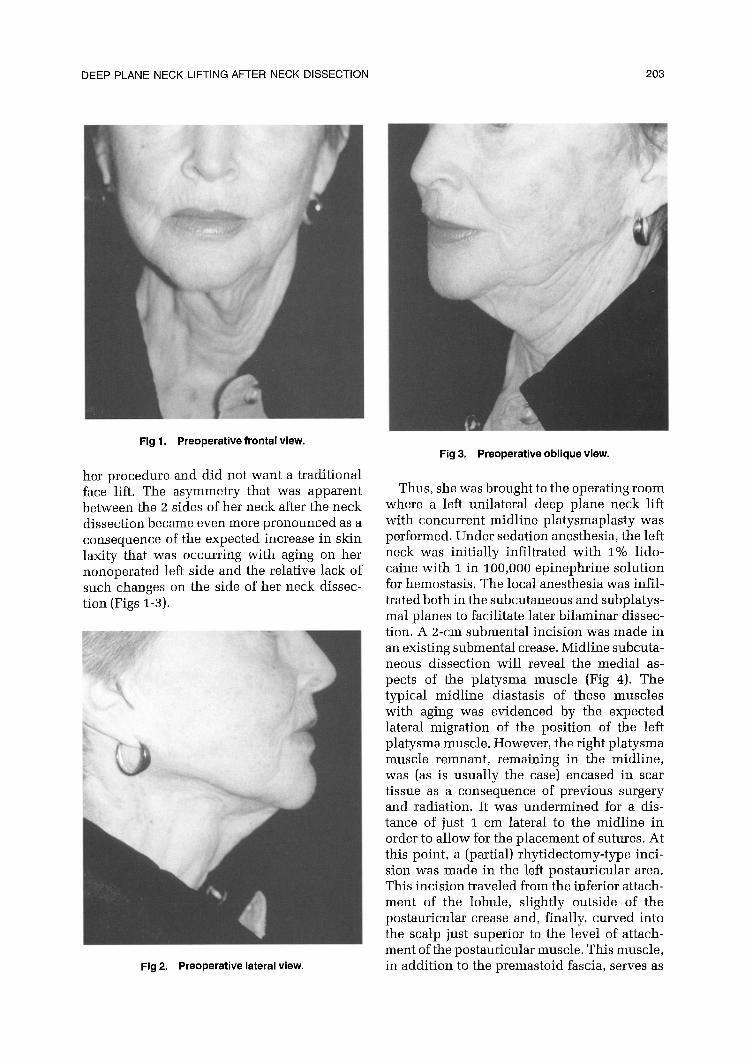
Fig 1. Preoperative frontal view.
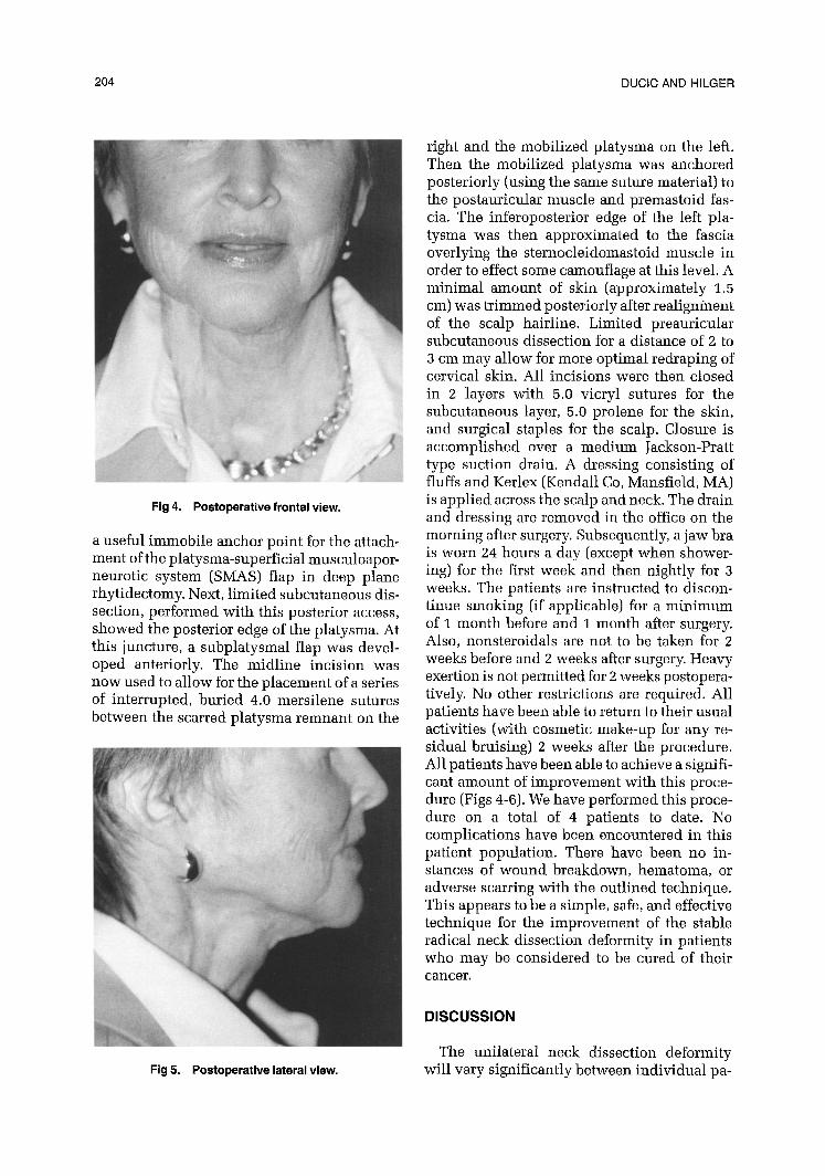
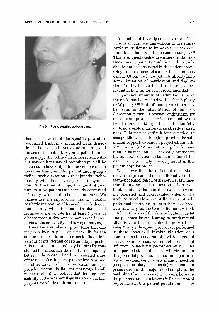
her procedure and did not want a traditional face lift. The asymmetry that was apparent between the 2 sides of her neck after the neck dissection became even more pronounced as a consequence of the expected increase in skin laxity that was occurring with aging on her nonoperated left side and the relative lack of such changes on the side of her neck dissec- tion (Figs 1-3).
Fig 2. Preoperative lateral view.
Fig 3. Preoperative oblique view.
Thus, she was brought to the operating room where a left unilateral deep plane neck lift with concurrent midline platysmaplasty was performed. Under sedation anesthesia, the left neck was initially infiltrated with 1% lido- caiue with 1 in 100,000 epinephrine solution for hemostasis. The local anesthesia was infil- trated both in the subcutaneous and subplatys- mal planes to facilitate later bilaminar dissec- tion. A 2-cm submental incision was made in an existing submental crease. Midline subcuta- neous dissection will reveal the medial as- pects of the platysma muscle (Fig 4). The typical midline diastasis of these muscles with aging was evidenced by the expected lateral migration of the position of the left platysma muscle. However, the right platysma muscle remnant, remaining in the midline, was (as is usually the case) encased in scar tissue as a consequence of previous surgery and radiation. It was undermined for a dis- tance of just 1 cm lateral to the midline in order to allow for the placement of sutures. At this point, a (partial) rhytidectomy-type inci- sion was made in the left postauricular area. This incision traveled from the inferior attach- ment of the tobule, slightly outside of the postauricular crease and, finally, curved into the scalp just superior to the level of attach- ment of the postauricular muscle. This muscle, in addition to the premastoid fascia, serves as
204 DUCIC AND HILGER
Fig 4. Postoperative frontal view.
a useful immobile anchor point for the attach- ment of the platysma-superficial musculoapor- neurotic system (SMAS) flap in deep plane rhytidectomy. Next, limited subcutaneous dis- section, performed with this posterior access, showed the posterior edge of the platysma. At this juncture, a subplatysmal flap was devel- oped anteriorly. The midline incision was now used to allow for the placement of a series of interrupted, buried 4.0 mersilene sutures between the scarred platysma remnant on the
i -
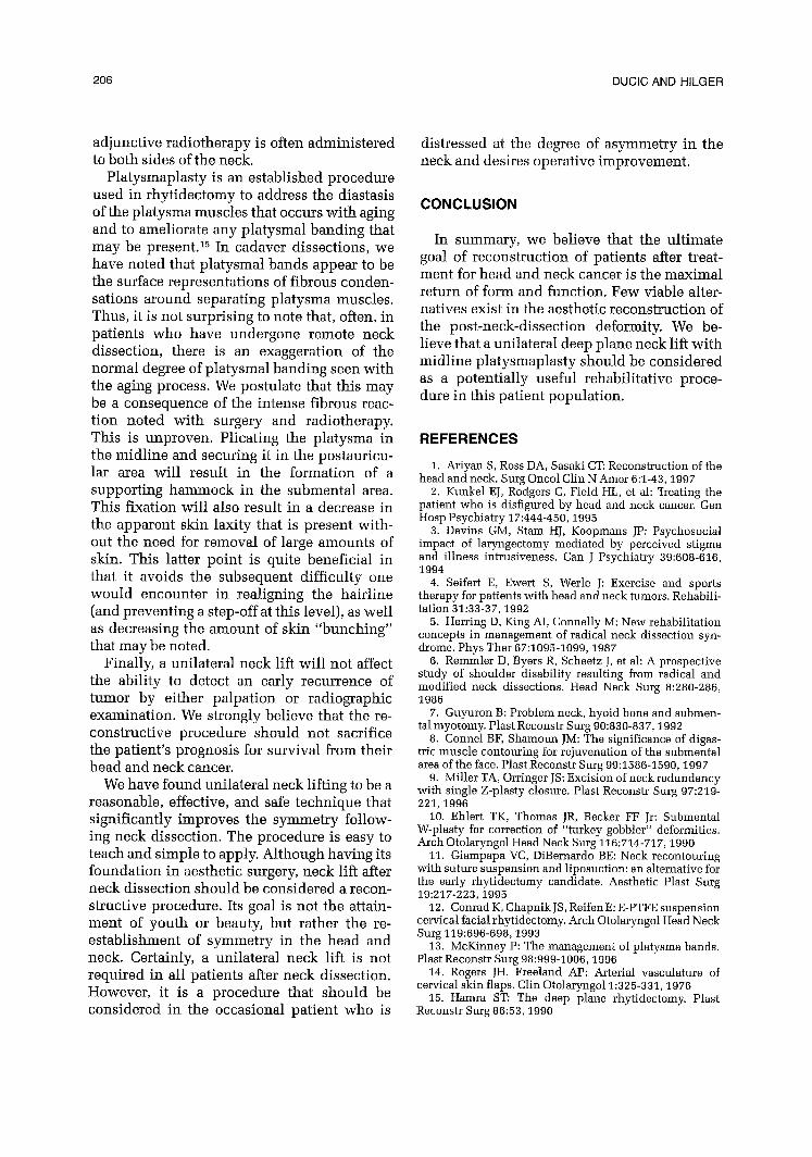
Fig 5. Postoperative lateral view.
right and the mobilized platysma on the left. Then the mobilized platysma was anchored posteriorly (using the same suture material) to the postauricular muscle and premastoid fas- cia. The inferoposterior edge of the left pla- tysma was then approximated to the fascia overlying the sternocleidomastoid muscle in order to effect some camouflage at this level. A minimal amount of skin (approximately 1.5 cm) was tr immed posteriorly after realignment of the scalp hairline. Limited preauricular subcutaneous dissection for a distance of 2 to 3 cm may allow for more optimal redraping of cervical skin. All incisions were then closed in 2 layers with 5.0 vicryl sutures for the subcutaneous layer, 5.0 prolene for the skin, and surgical staples for the scalp. Closure is accomplished over a medium Jackson-Pratt type suction drain. A dressing consisting of fluffs and Kerlex (Kendall Co, Mansfield, MA) is applied across the scalp and neck. The drain and dressing are removed in the office on the morning after surgery. Subsequently, a jaw bra is worn 24 hours a day (except when shower- ing) for the first week and then nightly for 3 weeks. The patients are instructed to discon- tinue smoking (if applicable} for a min imum of I month before and 1 month after surgery. Also, nonsteroidals are not to be taken for 2 weeks before and 2 weeks after surgery. Heavy exertion is not permitted for 2 weeks postopera- tively. No other restrictions are required. All patients have been able to return to their usual activities (with cosmetic make-up for any re- sidual bruising) 2 weeks after the procedure. All patients have been able to achieve a signifi- cant amount of improvement with this proce- dure (Figs 4-6). We have performed this proce- dure on a total of 4 patients to date. No complications have been encountered in this patient population. There have been no in- stances of wound breakdown, hematoma, or adverse scarring with the outlined technique. This appears to be a simple, safe, and effective technique for the improvement of the stable radical neck dissection deformity in patients who may be considered to be cured of their cancer .
D I S C U S S I O N
The unilateral neck dissection deformity will vary significantly between individual pa-
DEEP PLANE NECK LIFTING AFTER NECK DISSECTION 205
Fig 6. Postoperative oblique view.
tients as a result of the specific procedure performed (radical v modified neck dissec- tions), the use of adjunctive radiotherapy, and the age of the patient. A young patient under- going a type III modified neck dissection with- out concomitant use of radiotherapy will be expected to have only minor asymmetries. On the other hand, an older patient undergoing a radical neck dissection with adjunctive radio- therapy wilt often have significant asymme- tries. At the time of surgical removal of their tumors, most patients are correctly concerned primarily with their chances for cure. We believe that the appropriate time to consider aesthetic restoration of form after neck dissec- tion is only when the patient's chances of recurrence are remote (ie, at least 5 years of disease-flee survival after squamous cell carci- noma of the oral cavity and laryngopharynx).
There are a number of procedures that one may consider in place of a neck lift for the amelioration of form after neck dissection. Various grafts (dermal or fat) and flaps (pecto- ralis major or trapezius) may be initially con- sidered to camouflage the relative discrepancy between the operated and nonoperated sides of the neck. For the most part, unless required for other head and neck reconstruction (eg, pedicled pectoralis flap for pharyngeal wall reconstruction), we believe that the long-term stability of these camouflage materials, for this purpose, preclude their routine use.
A number of investigators have described various incomplete transections of the supra- hyoid musculature to improve the neck con- tours in patients seeking cosmetic surgery. 7-a This is of questionable usefulness in the rou- tine cosmetic patient population and certainly should not be considered in the patient recov- ering from treatment of a major head and neck cancer. Often, the latter patients already have some limitation of mastication and degluti- tion. Adding further insult to these systems, no matter how minor, is not recommended.
Significant amounts of redundant skin in the neck may be resected with either Z-plasty or W-plasty. 9-1° Both of these procedures may be useful in the rehabilitation of the neck dissection patient. However, enthusiasm for these techniques needs to be tempered by the fact that one is adding further and potentially quite noticeable incisions to an already scarred neck. This may be difficult for the patient to accept. Likewise, although improving the sub- mental support, expanded polytetrafluoroeth- ylene suture (or other suture type) inframan- dibular suspension can severely exacerbate the apparent degree of skeletonization of the neck that is routinely already present in this patient population. 1~-13
We believe that the unilateral deep plane neck lift represents the best alternative in the aesthetic rehabilitation of the cervical asymme- tries following neck dissection. There is a fundamental difference that exists between the operated and nonoperated sides of the neck. Surgical elevation of flaps as routinely performed to provide access to the neck dissec- tion and any adjunctive radiotherapy both result in fibrosis of the skin, subcutaneous fat and platysma layers, leading to fundamental alterations in the normal blood supply to these areas. 14 Any subsequent procedures performed in these areas will involve violation of a compromised blood supply with attendant risks of skin necrosis, wound dehiscence, and infection. A neck lift performed only on the nonoperated side of the neck, will circumvent this potential problem. Furthermore, perform- ing a predominantly deep plane dissection (deep to the platysma muscle) will result in preservation of the major blood supply to the neck skin (forms a vascular network between the platysma and skin layers)2 4 This may be of importance in this patient population, as any
206 DUCIC AND HILGER
adjunctive radiotherapy is often administered to both sides of the neck.
Platysmaplasty is an established procedure used in rhytidectomy to address the diastasis of the platysma muscles that occurs with aging and to ameliorate any platysmal banding that may be present. 15 In cadaver dissections, we have noted that platysmal bands appear to be the surface representations of fibrous conden- sations around separating platysma muscles. Thus, it is not surprising to note that, often, in patients who have undergone remote neck dissection, there is an exaggeration of the normal degree of platysmal banding seen with the aging process. We postulate that this may be a consequence of the intense fibrous reac- tion noted with surgery and radiotherapy. This is unproven. Plicating the platysma in the midline and securing it in the postauricu- lar area will result in the formation of a supporting hammock in the submental area. This fixation wilt also result in a decrease in the apparent skin laxity that is present with- out the need for removal of large amounts of skin. This latter point is quite beneficial in that it avoids the subsequent difficulty one would encounter in realigning the hairline (and preventing a step-off at this level), as well as decreasing the amount of skin "bunching" that may be noted.
Finally, a unilateral neck lift will not affect the ability to detect an early recurrence of tumor by either palpation or radiographic examination. We strongly believe that the re- constructive procedure should not sacrifice the patient's prognosis for survival from their head and neck cancer.
We have found unilateral neck lifting to be a reasonable, effective, and safe technique that significantly improves the symmetry follow- ing neck dissection. The procedure is easy to teach and simple to apply. Although having its foundation in aesthetic surgery, neck lift after neck dissection should be considered a recon- structive procedure. Its goal is not the attain- ment of youth or beauty, but rather the re- establishment of symmetry in the head and neck. Certainly, a unilateral neck lift is not required in all patients after neck dissection. However, it is a procedure that should be considered in the occasional patient who is
distressed at the degree of asymmetry in the neck and desires operative improvement.
CONCLUSION
In summary, we believe that the ultimate goal of reconstruction of patients after treat- ment for head and neck cancer is the maximal return of form and function. Few viable alter- natives exist in the aesthetic reconstruction of the post-neck-dissection deformity. We be- lieve that a unilateral deep plane neck lift with midline platysmaplasty should be considered as a potentially useful rehabilitative proce- dure in this patient population.
REFERENCES
1. Ariyan S, Ross DA, Sasaki CT: Reconstruction of the head and neck. Surg Oncol Clin NAmer 6:1-43, 1997
2. Kunkel EJ, Rodgers C, Field HL, et ah Treating the patient who is disfigured by head and neck cancer. Gen Hosp Psychiatry 17:444-450, 1995
3. Devins GM, Stare HJ, Koopmans JP: Psychosocial impact of laryngectomy mediated by perceived stigma and illness intrusiveness. Can J Psychiatry 39:608-616, 1994
4. Seifert E, Ewert S, Werle J: Exercise and sports therapy for patients with head and neck tumors. Rehabili- tation 31:33-37, 1992
5. Herring D, King AI, Connelly M: New rehabilitation concepts in management of radical neck dissection syn- drome. Phys Ther 67:1095-1099, 1987
6. Remmler D, Byers R, Scheetz J, et ah A prospective study of shoulder disability resulting from radical and modified neck dissections. Head Neck Surg 8:280-286, 1986
7. Guyuron B: Problem neck, hyoid bone and submen- tal myotomy. Plast Reconstr Surg 90:830-837, 1992
8. Connel BF, Shamoun JM: The significance of digas- tric muscle contouring for rejuvenation of the submental area of the face. Plast Reconstr Surg 99:1586-1590, 1997
9. Miller TA, Orringer JS: Excision of neck redundancy with single Z-plasty closure. Plast Reconstr Surg 97:219- 221, 1996
10. Ehlert TK, Thomas JR, Becker FF Jr: Submental W-plasty for correction of "turkey gobbler" deformities. Arch Otolaryngol Head Neck Surg 116:714-717, 1990
11. Giampapa VC, DiBernardo BE: Neck recontouring with suture suspension and liposuction: an alternative for the early rhytidectomy candidate. Aesthetic Plast Surg 19:217-223, 1995
12. Conrad K, Chapnik JS, Reifen E: E-PTFE suspension cervical facial rhytidectomy. Arch Otolaryngol Head Neck Surg 119:696-698, 1993
13. McKinney P: The management of platysma bands. Plast Reconstr Surg 98:999-1006, 1996
14. Rogers JH, Freeland AP: Arterial vasculature of cervical skin flaps. Clin Otolaryngol 1:325-331, 1976
15. Hamra ST: The deep plane rhytidectomy. Plast Reconstr Surg 86:53, 1990