Embed Size (px)
Citation preview
THE VENOUS CIRCULATION IN TELEOST FISH
RESPONSES TO EXERCISE,
TEMPERATURE AND HYPOXIA
AKADEMISK AVHANDLING
för filosofie doktorsexamen i zoofysiologi som enligt naturvetenskapliga fakultetens beslut kommer att försvaras offentligt fredagen den 27 april 2007,
kl. 10.00 i föreläsningssalen, Zoologiska institutionen, Medicinaregatan 18, Göteborg
ERIK SANDBLOM
Department of Zoology/Zoophysiology
2007

Published by the Department of Zoology/Zoophysiology Göteborg University, Sweden Published papers are used with permission from the publisher: I. The Company of Biologists II. and IV. Elsevier III. and V. The American Physiological Society Printed by Vasastadens Bokbinderi AB, Göteborg 2007 © Erik Sandblom 2007 ISBN 978-91-628-7131-4

DISSERTATION ABSTRACT Sandblom, Erik (2007). The venous circulation in teleost fish -Responses to exercise, temperature and hypoxia. Department of Zoology/Zoophysiology, Göteborg University, Box 463, SE-405 30 Göteborg, Sweden. In fish and other vertebrates, venous capacitance changes have important implications on venous return and cardiac filling pressure. The main objective of this thesis was to gather information on venous haemodynamic responses and neurohumoral control mechanisms in two teleost species; the sea bass, Dicentrarchus labrax, and the rainbow trout, Oncorhynchus mykiss. As previous studies of venous function in fish have primarily focused on the pharmacology of the venous vasculature, special attention was paid to venous responses elicited by exercise, acute temperature changes and environmental hypoxia, which represent natural cardiovascular challenges in aquatic environments.
Methods: Cardiac output (Q), central venous (Pven) and dorsal aortic (Pda) blood pressures were recorded in vivo. The mean circulatory filling pressure (MCFP), a measure of vascular capacitance, was measured as the venous plateau pressure during ventral aortic occlusion. In one study, vascular capacitance curves were also constructed by measuring MCFP at different blood volumes (between 80-120% of the assumed blood volume), to investigate changes in vascular compliance (C) and unstressed blood volume (USBV) during normoxia and hypoxia. In another study, blood volume was measured using dilution of 51Cr-labelled red blood cells. Drugs were administered systemically to elucidate the role of adrenergic control systems and the renin-angiotensin system (RAS) in the observed cardiovascular responses.
Results and conclusions: Exercise, in both sea bass and rainbow trout, results in increased Q and increased MCFP. Although Pven increases during exercise in both species, cardiac stroke volume (SV) only increases in rainbow trout, whereas increased heart rate (fH) is exclusively responsible for the increased blood flow in sea bass. When ambient temperature was raised acutely from 10 to 13 and 16°C, rainbow trout respond with a significantly elevated Q which, in contrast to the exercise response, is exclusively mediated by tachycardia with an unchanged Pven and SV. Similarly, however, MCFP increases which indicates an actively reduced vascular capacitance, especially since the blood volume does not change between 10 and 16°C. In both species, blockade of α-adrenoceptors delays the increase in Pven during exercise, and in rainbow trout, additional blockade of angiotensin converting enzyme abolishes all venous exercise responses. Environmental hypoxia typically elicits bradycardia that is associated with reduced vascular capacitance and an increased Pven. Q is unchanged or increased during hypoxia due to an increased SV. The capacitance responses during hypoxia are mainly due to changes in USBV that are mediated by both nervous and humoral α-adrenergic mechanisms.
In summary, it is shown that vascular capacitance decreases during exercise, acute temperature increase and hypoxia. This mobilizes blood to the central venous compartment which, depending on the heart rate response, results in maintained or increased Pven, SV and Q. It is also suggested that the decrease in capacitance during exercise and acute temperature increase prevents blood from passively pooling in the venous periphery as blood flow increases. RAS is activated during exercise after α-blockade to increase Pven and MCFP. Thus, RAS affects venous capacitance in fish and not only arterial tone as previously suggested.
Keywords: blood volume, catecholamines, central venous pressure, exercise, hypoxia, mean circulatory filling pressure, preload, renin-angiotensin, temperature, vascular capacitance.


LIST OF PAPERS This thesis is based on the following papers which are referred to in the text by their Roman numbers: I. Sandblom E., Farrell A. P., Altimiras J., Axelsson M. and
Claireaux G. (2005). Cardiac preload and venous return in swimming sea bass (Dicentrarchus labrax). J Exp Biol 208, 1927-1935.
II. Sandblom E., Axelsson M. and McKenzie D. J. (2006) Venous
responses during exercise in rainbow trout, Oncorhynchus mykiss: α-adrenergic control and the antihypotensive function of the renin-angiotensin system. Comp Biochem Physiol 144, 401-409.
III. Sandblom E. and Axelsson M. (2007) Venous hemodynamic
responses to acute temperature increase in the rainbow trout (Oncorhynchus mykiss). Am J Physiol, (In Press).
IV. Sandblom E. and Axelsson M. (2005) Effects of hypoxia on the
venous circulation in rainbow trout (Oncorhynchus mykiss). Comp Biochem Physiol 140, 233-239.
V. Sandblom E. and Axelsson M. (2006) Adrenergic control of venous
capacitance during moderate hypoxia in the rainbow trout (Oncorhynchus mykiss): role of neural and circulating catecholamines. Am J Physiol 291, 711-718.

TABLE OF CONTENTS
ABBREVIATIONS ..................................................................................2
1. INTRODUCTION...............................................................................4
1.1 GENERAL OVERVIEW OF FISH CIRCULATORY SYSTEMS ........5 1.1.1 Adrenergic and cholinergic control of the heart ................................................6 1.1.2 Adrenergic and cholinergic control of the vasculature .......................................7 1.1.3 The renin-angiotensin system..........................................................................8
1.2 THE VENOUS CIRCULATION.................................................................9 1.2.1 Gravitational forces in an aquatic environment ...............................................9 1.2.2 Central venous pressure and the Frank-Starling mechanism .........................10 1.2.3 Mean circulatory filling pressure and venous capacitance................................13 1.2.4 Coupling of cardiac output and venous return ...............................................16 1.2.5 Passive and active responses .........................................................................17 1.2.6 Control of the venous circulation...................................................................18
1.3 INTEGRATED CARDIOVASCULAR RESPONSES..........................21 1.3.1 Exercise......................................................................................................21 1.3.2 Temperature................................................................................................23 1.3.3 Hypoxia .....................................................................................................24
2. AIM ..................................................................................................... 28
3. RESULTS AND DISCUSSION ......................................................... 30
3.1 METHODOLOGICAL CONSIDERATIONS.......................................30 3.1.1 Mean circulatory filling pressure ...................................................................30 3.1.2 Stressed blood volume and vascular compliance .............................................35 3.1.3 Blood volume...............................................................................................37 3.1.4 Future perspectives.......................................................................................37
3.2 INTEGRATED CARDIOVASCULAR RESPONSES..........................38 3.2.1 Exercise......................................................................................................38 3.2.2 Temperature................................................................................................41 3.2.3 Hypoxia .....................................................................................................43
4. CONCLUSIONS ................................................................................ 46
5. ACKNOWLEDGEMENTS ............................................................... 48
6. REFERENCES .................................................................................. 50

1

ABBREVIATIONS ACE angiotensin converting enzyme Ang I angiotensin I Ang II angiotensin II APP arterial plateau pressure BL s-1 body lengths per second C vascular compliance CaO2 arterial oxygen content CvO2 mixed venous oxygen content fH heart rate Mb body mass MCFP mean circulatory filling pressure MO2 oxygen consumption Pda or PDA dorsal aortic blood pressure PO2 oxygen partial pressure Pven or PCV central venous blood pressure ΔPven or ΔPV venous pressure difference Q cardiac output RAS renin-angiotensin system Rsys systemic vascular resistance Rven or Rv venous vascular resistance SBV stressed blood volume SV or Vs cardiac stroke volume VPP venous plateau pressure VR venous return Ucrit critical swimming speed USBV unstressed blood volume
2

3

1. INTRODUCTION
The cardiovascular system is one of the most central components in the physiological machinery which maintains homeostasis of the animal body. The role of the cardiovascular system is diverse and ranges from being a carrier of information in the form of circulating hormones, to being involved in the regulation of temperature and hydromineral balance. However, its most fundamental role is possibly to supply O2 and nutrients and to remove CO2 and metabolic waste products produced by cellular metabolic processes throughout the body. The main driving force to circulate the blood is provided by the arterial blood pressure which is generated by the beating heart, while local blood flow is regulated by changes in arteriolar resistance. In most vertebrates, the rate and force of cardiac contraction is directly controlled by the autonomic nervous system and various humoral control systems. However, secondary factors like arterial and venous blood pressure also affect cardiac performance. The venous circulation, which contains a large portion of the total blood volume, and is responsible for carrying oxygen-depleted blood back to the heart, is particularly interesting in this regard. An early conclusion made by the Danish Nobel prize-winning zoophysiologist August Krogh may serve to emphasize the importance of the venous vasculature for cardiac performance:
“The heart cannot do more than send out what it gets” (Krogh, 1912)
In the large and diverse vertebrate group represented by the fishes, the
importance and function of the cardiovascular system for the exchange of gases, nutrients and metabolites does not represent any major exceptions from the general picture outlined above. However, as fish are ectothermic, breathe water and spend their entire life in water which has a density (very) similar to their own body fluids, the aquatic lifestyle presents a number of cardiovascular challenges which differ considerably from those experienced by terrestrial animals. Thus, before land was colonized by vertebrate life, the selection pressures behind the evolution of the vertebrate cardiovascular system were those of an aquatic environment. Understanding the form and function of the cardiovascular system of our evolutionary predecessors, including the fishes, is
4

therefore ultimately of fundamental importance in order to fully understand and appreciate the form and function of the human cardiovascular system.
In general, this thesis focuses on the cardiovascular responses of teleost fish to exercise, acute temperature changes and environmental hypoxia, which all represent naturally occurring events in their aquatic environment. More specifically, the neurohumoral control of the venous circulation during these cardiovascular challenges is the central theme.
1.1 GENERAL OVERVIEW OF FISH CIRCULATORY SYSTEMS
The typical teleost circulation is comprised of a single heart connected in series with the gills. The heart is a four-chambered structure with the sinus venosus, atrium, ventricle and bulbus arteriosus serially connected and enclosed in a more or less rigid pericardial cavity (Farrell, 1991; Farrell and Jones, 1992). Venous blood returns from the periphery and enters the sinus venosus, via the paired ducts of Cuvier before entering the atrium. The ventricle, which is the main pressure generating component of the heart, is filled by atrial contraction, but also by direct inflow of blood from the central veins during diastole (Lai et al., 1998). Both the atrio-ventricular and sino-atrial junctions are guarded by valves, whereas the connections between the sinus venosus and ducts of Cuvier are not (Farrell and Jones, 1992). The ventricle pumps blood via the highly compliant bulbus arteriosus into the ventral aorta, which splits into four pairs of afferent branchial arteries that perfuse the gills where gas exchange takes place. There are two major pathways in the gills. In the arterio-venous pathway, blood flows directly over to the central venous compartment and supplies the gill tissues with oxygen and nutrients, whereas oxygenated blood in the arterio-arterial pathway leaves the gills via four pairs of efferent branchial arteries (Nilsson and Sundin, 1998). The main portion of this blood enters the dorsal aorta, but some is also directed to the cephalic region via the carotid arteries. In unfed fish, 30-40 percent of cardiac output (Q) is typically diverted to the gut circulation (stomach, intestine and liver) via the coeliaco-mesenteric artery which in many species branches directly from the dorsal aorta (Farrell et al., 2001; Thorarensen et al., 1991). After having passed through the capillary beds, blood is returned to the heart via the venous circulation. Blood from the stomach and intestine is collected by the hepatic portal vein carrying blood to the liver, which in turn is drained by the hepatic veins directly into the sinus venosus. The hepatic veins are typically
5

short and differ in number among species. It has been suggested that active control of sphincters in these veins is a mechanism by which blood can be rapidly mobilized from the splanchnic circulation to the central venous circulation (Johansen and Hanson, 1967). Paired posterior and anterior cardinal veins drain the caudal and cranial portions of the body, respectively. They fuse with the ducts of Cuvier dorsal to the heart. Dorsal, lateral and ventral cutaneous veins primarily drain the skin, but also the buccal and opercular cavities (Satchell, 1991; Satchell, 1992). Valves are present in fish veins, but only at the junction of tributary vessels (ostial valves), and not along the length of the vessels (parietal valves) as in mammals (Fig. 1). A more detailed description on the control and function of the venous circulation is given in section 1.2.
i.
ii.
i.
ii.
i.
ii.
Figure 1. Schematic illustration of the location of valves in a venous segment. Mammals have both ostial (i.) and parietal (ii.) valves, whereas only the former seem to be present in fish veins. Arrows indicate direction of blood flow. After Satchell, 1991.
1.1.1 Adrenergic and cholinergic control of the heart
The teleost heart receives a dual autonomic innervation from exitatory adrenergic fibers, as well as inhibitory cholinergic fibers (Nilsson, 1983, 1997; Taylor, 1985; Taylor et al., 1999). One notable exception to this pattern among the teleosts is the Pleuronectids, which lack adrenergic innervation (Donald and
6
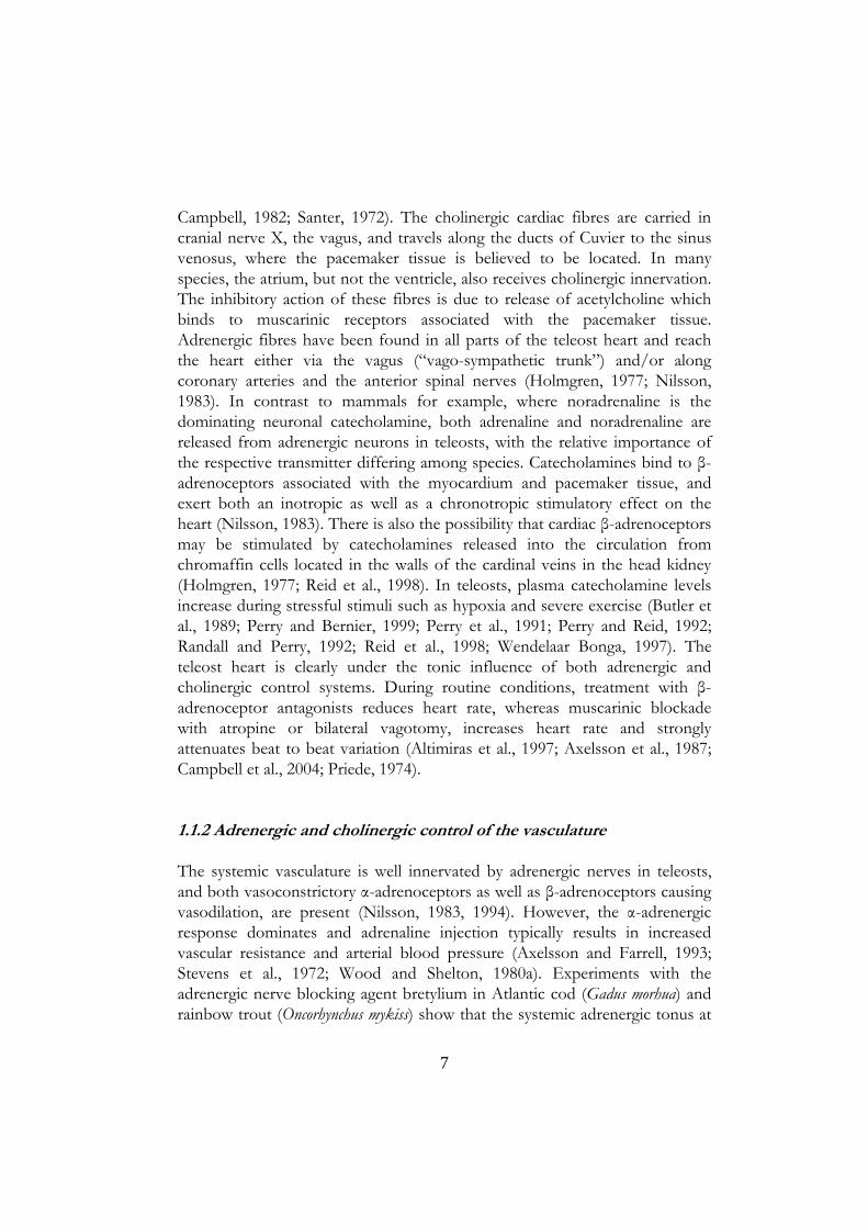
Campbell, 1982; Santer, 1972). The cholinergic cardiac fibres are carried in cranial nerve X, the vagus, and travels along the ducts of Cuvier to the sinus venosus, where the pacemaker tissue is believed to be located. In many species, the atrium, but not the ventricle, also receives cholinergic innervation. The inhibitory action of these fibres is due to release of acetylcholine which binds to muscarinic receptors associated with the pacemaker tissue. Adrenergic fibres have been found in all parts of the teleost heart and reach the heart either via the vagus (“vago-sympathetic trunk”) and/or along coronary arteries and the anterior spinal nerves (Holmgren, 1977; Nilsson, 1983). In contrast to mammals for example, where noradrenaline is the dominating neuronal catecholamine, both adrenaline and noradrenaline are released from adrenergic neurons in teleosts, with the relative importance of the respective transmitter differing among species. Catecholamines bind to β-adrenoceptors associated with the myocardium and pacemaker tissue, and exert both an inotropic as well as a chronotropic stimulatory effect on the heart (Nilsson, 1983). There is also the possibility that cardiac β-adrenoceptors may be stimulated by catecholamines released into the circulation from chromaffin cells located in the walls of the cardinal veins in the head kidney (Holmgren, 1977; Reid et al., 1998). In teleosts, plasma catecholamine levels increase during stressful stimuli such as hypoxia and severe exercise (Butler et al., 1989; Perry and Bernier, 1999; Perry et al., 1991; Perry and Reid, 1992; Randall and Perry, 1992; Reid et al., 1998; Wendelaar Bonga, 1997). The teleost heart is clearly under the tonic influence of both adrenergic and cholinergic control systems. During routine conditions, treatment with β-adrenoceptor antagonists reduces heart rate, whereas muscarinic blockade with atropine or bilateral vagotomy, increases heart rate and strongly attenuates beat to beat variation (Altimiras et al., 1997; Axelsson et al., 1987; Campbell et al., 2004; Priede, 1974).
1.1.2 Adrenergic and cholinergic control of the vasculature
The systemic vasculature is well innervated by adrenergic nerves in teleosts, and both vasoconstrictory α-adrenoceptors as well as β-adrenoceptors causing vasodilation, are present (Nilsson, 1983, 1994). However, the α-adrenergic response dominates and adrenaline injection typically results in increased vascular resistance and arterial blood pressure (Axelsson and Farrell, 1993; Stevens et al., 1972; Wood and Shelton, 1980a). Experiments with the adrenergic nerve blocking agent bretylium in Atlantic cod (Gadus morhua) and rainbow trout (Oncorhynchus mykiss) show that the systemic adrenergic tonus at
7

rest, as well as during moderate exercise and hypoxia, is primarily mediated by neural mechanisms and not by circulating catecholamines (Axelsson and Fritsche, 1991; Axelsson and Nilsson, 1986; Fritsche and Nilsson, 1990; Smith, 1978; Smith et al., 1985). It is possible, however, that in response to severely stressful stimuli plasma catecholamine levels may reach levels high enough to increase systemic vascular resistance and arterial blood pressure (Bernier and Perry, 1999; Perry and Bernier, 1999; Randall and Perry, 1992). In addition, circulating catecholamines have been suggested to be an important source for neuronal uptake, which may be a prerequisite for sustained neuronally mediated vasoconstriction (Xu and Olson, 1993).
The control of the branchial vasculature is complex and involves neural and circulating catecholamines, as well as cholinergic nerves (Nilsson and Sundin, 1998; Sundin and Nilsson, 2002). A relatively sparse adrenergic innervation projects to the arterio-venous pathways where α-adrenoceptors dominate, but also to the afferent filamental artery and the sphincter region of the efferent filamental artery where β-adrenoceptors dominate. It seems that circulating catecholamines may be of greater importance for the control of the gill circulation than the systemic circulation (Nilsson and Sundin, 1998; Sundin and Nilsson, 2002). Injected catecholamines (adrenaline) typically reduce overall branchial resistance (Nilsson, 1983). Cholinergic efferents constrict the efferent filamental artery sphincter by stimulation of muscarinic receptors (Nilsson and Sundin, 1998; Sundin and Nilsson, 2002).
1.1.3 The renin-angiotensin system
The renin-angiotensin system (RAS) is often regarded as an endocrine “anti-drop” factor for arterial blood pressure in fish (Olson et al., 1994; Olson, 1992; Platzack, 1995; Platzack et al., 1993; Russell et al., 2001; Zhang et al., 1995). During hypotension and/or hypovolemia, the proteolytic enzyme renin is released into the circulation, mainly from juxtaglomerular cells in the kidney (Nishimura, 1978; Nishimura et al., 1979; Olson, 1992). Renin converts the α2-globulin angiotensinogen to the decapeptide angiotensin I (Ang I), which is further hydrolyzed to the octapeptide angiotensin II (Ang II) by angiotensin converting enzyme (ACE) (Olson, 1992). The gills are believed to be the primary site for ACE activity in teleosts (Olson, 1998; Olson et al., 1989). Ang II is the biologically active molecule in the RAS and has intrinsic properties, but also exerts some of its vasoactive properties through activation of adrenergic control systems (Bernier et al., 1999; Bernier and Perry, 1999; Carroll and Opdyke, 1982; Oudit and Butler, 1995). At present, the general
8

consensus is that the primary vasoactive site for RAS in fish is the systemic resistance vasculature (Olson et al., 1994; Olson, 1992; Russell et al., 2001; Zhang et al., 1995).
1.2 THE VENOUS CIRCULATION
Our knowledge regarding the control and function of the venous circulation in fish, and all non-mammalian animals for that matter, is still fragmentary. The following account is an attempt to summarize the literature on venous haemodynamics and its control in fish. In many places, specific information for fish is still missing, and here, references to the more abundant mammalian literature are made. It should be kept in mind, however, that extrapolations from studies on mammals to other animals need to be done cautiously. Despite the general paucity of data for fish in many places, it is my hope that the following sections will serve as a useful background for future, more precise, studies on the venous circulation in fish.
1.2.1 Gravitational forces in an aquatic environment
The cardiovascular systems of land-living animals are constantly challenged by gravitational forces. This implies that blood tends to pool in the lower parts of the body (i.e. below the heart), and the taller the animal, the greater is the gravitational impact. In mammals, a number of homeostatic mechanisms have evolved to prevent orthostatic blood pooling. Compression of veins by the surrounding skeletal muscles (“the muscle pump”), in combination with active and passive changes in venous capacitance, are the most important mechanisms by which this is achieved (Pang, 2001). These mechanisms serve to prevent formation of oedema and ensure that blood is returned to the heart.
In water-living animals such as fish, the gravitational impact on the cardiovascular system is small due to the fact that blood has a density similar to water and the hydrostatic water pressure counteracts the gravitational forces acting on the blood in the circulatory system (Fig. 2). Thus, orthostatic blood pooling is unlikely to be a major concern for fish (Satchell, 1991, 1992).
9
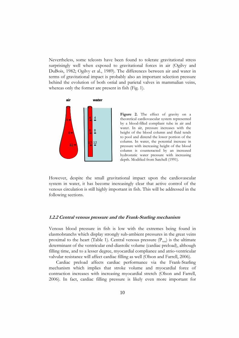
Nevertheless, some teleosts have been found to tolerate gravitational stress surprisingly well when exposed to gravitational forces in air (Ogilvy and DuBois, 1982; Ogilvy et al., 1989). The differences between air and water in terms of gravitational impact is probably also an important selection pressure behind the evolution of both ostial and parietal valves in mammalian veins, whereas only the former are present in fish (Fig. 1).
air water
4
8
12
4
8
12
4
8
12
air water
4
8
12
4
8
12
4
8
12
Figure 2. The effect of gravity on a theoretical cardiovascular system represented by a blood-filled compliant tube in air and water. In air, pressure increases with the height of the blood column and fluid tends to pool and distend the lower portion of the column. In water, the potential increase in pressure with increasing height of the blood column is counteracted by an increased hydrostatic water pressure with increasing depth. Modified from Satchell (1991).
However, despite the small gravitational impact upon the cardiovascular system in water, it has become increasingly clear that active control of the venous circulation is still highly important in fish. This will be addressed in the following sections.
1.2.2 Central venous pressure and the Frank-Starling mechanism
Venous blood pressure in fish is low with the extremes being found in elasmobranchs which display strongly sub-ambient pressures in the great veins proximal to the heart (Table 1). Central venous pressure (Pven) is the ultimate determinant of the ventricular end-diastolic volume (cardiac preload), although filling time, and to a lesser degree, myocardial compliance and atrio-ventricular valvular resistance will affect cardiac filling as well (Olson and Farrell, 2006).
Cardiac preload affects cardiac performance via the Frank-Starling mechanism which implies that stroke volume and myocardial force of contraction increases with increasing myocardial stretch (Olson and Farrell, 2006). In fact, cardiac filling pressure is likely even more important for
10

determining SV in fish than in mammals, because the ejection fraction for fish hearts is high (80-100 %). This leaves little scope for increasing SV by reducing the end-systolic volume (Coucelo et al., 2000; Farrell and Jones, 1992; Forster and Farrell, 1994; Franklin and Davie, 1992; Lai et al., 1990). However, this conclusion is so far based on experiments on relatively few species and it is possible that exceptions from this pattern may emerge. Both atrial and ventricular muscle in fish responds in accordance with the Frank-Starling mechanism (Farrell and Jones, 1992).
Heterod
SpeciesSV/DCPCS/VC* PCV CV HPV/SIV
PV* Source
CyclostomesLampetra tridentata -0.4 - -0.1 Johansen et al., 1973
Myxine glutinosa 0.10 Johnsson et al., 1996Eptatretus cirrhatus 0.01 - 0.07 0.02 - 0.16 Foster and Forster., 2007
- 0.04 Johnsson et al., 1996Elasmobranchs
Scyliorhinus canicula -0.45 Short et al., 1977Squalus acanthias -0.08 - -0.07 Sandblom et al., 2006
- -0.15 -0.10 - 0.00 Johansen and Hanson., 1967Triakis semifasciata 0.20 - 0.26 Lai et al., 1990
ontus portusjacksoni -0.28 0.21 Birch et al., 1969Squalus acanthias 0.1 - 0.4 Capra and Satchell., 1977
Cephaloscyllium isabella 0.21 Satchell and Weber.., 1987Dipnoans
Protopterus aethiopicus ~0 ~0.25 - 0.5 Johansen et al., 1968Teleosts
Anguilla anguilla -0.53 - 0.66 Mott., 1951Dicentrarchus labrax 0.11 Paper IOncorhynchus mykiss ~0.40 Zhang et al., 1995
- 0.27 Conklin et al., 1997- 0.47 Olson et al., 1997- 0.40 - 0.47 Zhang et al., 1998- ~0.20 Perry et al., 1999- 0.40 Hoagland et al., 2000- -0.06 Altimiras and Axelsson., 2004- ~0.02 Paper II- 0.01 Paper III- 0.06 - 0.07 Paper IV- 0.06 Paper V
Synbranchus marmoratus ~0.25 - 0.47 Skals et al., 2006Anguilla japonica 0.13 0.12 Chan and Chow., 1976
Oncorhynchus mykiss 0.19 Kiceniuk and Jones., 1977- 0.74 Wood and Shelton., 1980
Pseudopleuronectes americanus 0.40 - 0.53 Cech et al., 1976- 0.40 Cech et al., 1977
Platichtys stellatus 0.28 Wood et al., 1979Oncorhynchus mykiss 1.06 - 1.20 Stevens and Randall., 1967
Table 1. Summary of literature values for routine venous pressures in fish
Values are obtained from both anaesthetized and unanaesthetized animals. SV (sinus venosus); DC (duct of Cuvier); PCS (posterior cardinal sinus); VC (vena cava); PCV (posterior cardinal vein); CV (caudal vein); HPV (hepatic portal vein); SIV (supraintestinalvein) and PV (pulmonary vein) * only in Protopterus.
SpeciesSV/DCPCS/VC* PCV CV HPV/SIV
PV* Source
CyclostomesLampetra tridentata -0.4 - -0.1 Johansen et al., 1973
Myxine glutinosa 0.10 Johnsson et al., 1996Eptatretus cirrhatus 0.01 - 0.07 0.02 - 0.16 Foster and Forster., 2007
- 0.04 Johnsson et al., 1996Elasmobranchs
Scyliorhinus canicula -0.45 Short et al., 1977Squalus acanthias -0.08 - -0.07 Sandblom et al., 2006
- -0.15 -0.10 - 0.00 Johansen and Hanson., 1967Triakis semifasciata 0.20 - 0.26 Lai et al., 1990ontus portusjacksoni -0.28 0.21 Birch et al., 1969
Squalus acanthias 0.1 - 0.4 Capra and Satchell., 1977Cephaloscyllium isabella 0.21 Satchell and Weber.., 1987
DipnoansProtopterus aethiopicus ~0 ~0.25 - 0.5 Johansen et al., 1968
TeleostsAnguilla anguilla -0.53 - 0.66 Mott., 1951
Dicentrarchus labrax 0.11 Paper IOncorhynchus mykiss ~0.40 Zhang et al., 1995
- 0.27 Conklin et al., 1997- 0.47 Olson et al., 1997- 0.40 - 0.47 Zhang et al., 1998- ~0.20 Perry et al., 1999- 0.40 Hoagland et al., 2000- -0.06 Altimiras and Axelsson., 2004- ~0.02 Paper II- 0.01 Paper III- 0.06 - 0.07 Paper IV- 0.06 Paper V
Synbranchus marmoratus ~0.25 - 0.47 Skals et al., 2006Anguilla japonica 0.13 0.12 Chan and Chow., 1976
Oncorhynchus mykiss 0.19 Kiceniuk and Jones., 1977- 0.74 Wood and Shelton., 1980
Pseudopleuronectes americanus 0.40 - 0.53 Cech et al., 1976- 0.40 Cech et al., 1977
Platichtys stellatus 0.28 Wood et al., 1979Oncorhynchus mykiss 1.06 - 1.20 Stevens and Randall., 1967
Heterod
Table 1. Summary of literature values for routine venous pressures in fish
Values are obtained from both anaesthetized and unanaesthetized animals. SV (sinus venosus); DC (duct of Cuvier); PCS (posterior cardinal sinus); VC (vena cava); PCV (posterior cardinal vein); CV (caudal vein); HPV (hepatic portal vein); SIV (supraintestinalvein) and PV (pulmonary vein) * only in Protopterus.
11

Thus, an increased filling pressure may therefore have substantial effects on Q by increasing atrial stroke volume and contraction force. As a large portion of the ventricular filling is mediated by atrial contraction in fish, this will in turn significantly affect filling and performance of the ventricle (Farrell, 1984, 1991; Farrell and Jones, 1992). Atrial contraction is not the only mechanism by which the ventricle fills. Similar to the situation in mammals, direct inflow to the ventricle from the central veins occurs during diastole, but likely to a much lesser extent than in mammals (Lai et al., 1998; Olson and Farrell, 2006). The pressure in the central veins will therefore to some extent directly influence ventricular filling and performance.
The cardiac responses to filling pressure can conveniently be studied using in situ perfused heart preparations. This has been done on several groups of fish including teleosts (Blank et al., 2002, 2004; Davie et al., 1992; Farrell et al., 1982, 1983, 1989; Icardo et al., 2005; Stuart et al., 1983), elasmobranchs (Davie and Farrell, 1991; Franklin and Davie, 1993) and hagfish (Forster, 1989; Forster et al., 1991; Johnsson et al., 1996). It seems that all hearts examined respond to increased filling pressure with an increased Q in accordance with the Frank-Starling mechanism. Cardiac filling patterns The term vis a tergo is used to describe the pressure that fills the heart from behind, while the opposite, vis a fronte, describes the cardiac suction force created by the contracting heart inside a more or less rigid pericardial cavity. The vis a fronte mechanism allows the heart to generate flow even at sub-ambient filling pressures (Farrell, 1984, 1991; Farrell and Jones, 1992; Olson and Farrell, 2006). In the intact animal, vis a tergo is equivalent to the central venous pressure (filling pressure) and both cardiac as well as vascular factors and blood volume determine this pressure. The cardiac effect on filling pressure is primarily related to heart rate changes, such that Pven and SV are inversely proportional to fH (Altimiras and Axelsson, 2004; Farrell et al., 1989; Short et al., 1977; Taylor et al., 1977). In other words, when fH decreases, blood tends to pool in the central veins and Pven and SV increases. In contrast, if fH increases, the diastolic filling time is shortened and Pven and SV are reduced. In rainbow trout, this mechanically coupled mechanism keeps Q more or less constant if heart rate is pharmacologically manipulated over a relatively broad range of heart rates (Altimiras and Axelsson, 2004). The vascular factors dictating cardiac filling pressure are related to capacitance changes of the venous vasculature as will be discussed in detail below.
A rigid pericardium is undoubtedly an important prerequisite for vis a fronte filling (Farrell and Jones, 1992; Johansen, 1971). In elasmobranchs, where the
12

pericardium is particularly rigid, the vis a fronte mechanism is pronounced and most likely explains the strongly sub-ambient central venous pressures frequently observed in this group (Table 1). However, also teleosts, such as the rainbow trout, can generate routine cardiac outputs at sub-ambient filling pressures (Farrell et al., 1988) and Pven can be negative in rainbow trout in vivo (Altimiras and Axelsson, 2004; Erik Sandblom, unpublished observation). In situ perfused rainbow trout hearts can generate up to around 50% of maximum Q at sub-ambient filling pressures but, positive filling pressures are required to raise Q further (Farrell et al., 1988; Farrell and Jones, 1992). This strongly indicates that the vis a fronte mechanism is important in teleosts as well, and it has been suggested that a switch from vis a fronte to vis a tergo occurs when the circulatory system is challenged, such as during exercise (Farrell and Jones, 1992).
1.2.3 Mean circulatory filling pressure and venous capacitance
The vascular factors dictating venous return and cardiac filling pressure are primarily determined by the capacitance of the venous vasculature. The mean circulatory filling pressure (MCFP) is often used as an index of venous capacitance (Pang, 2001; Rothe, 1993). MCFP is the pressure in the circulation when blood flow is zero. It is dependent on vascular tone, vascular compliance (C) and blood volume. MCFP is much lower than arterial pressure, lower than capillary pressure and higher than central venous pressure (Pang, 2001; Rothe, 1993). Given that blood volume is not altered, MCFP measurements can be used to provide an estimate of active changes in the venous capacitance vasculature. This has been done for mammals, fish and reptiles (Conklin et al., 1997; Hoagland et al., 2000; Olson et al., 1997; Pang, 2001; Rothe, 1993; Sandblom and Axelsson, 2005; Sandblom et al., 2006 Skals et al., 2005; Skals et al., 2006; Zhang et al., 1995; Zhang et al., 1998). In mammals, it is generally assumed that MCFP is equivalent to the peripheral venous pressure at the level of the small veins and venules and MCFP may thus provide an estimate of the upstream driving pressure for venous return (VR; Guyton, 1955; Guyton et al., 1955; Pang, 2001; Rothe, 1993). Hence, the pressure difference that drives VR (ΔPven) is described by the equation:
(1) ΔPven= MCFP-Pven
As VR equals Q at steady state, the resistance to venous return (Rven) is calculated as:
13

(2) Rven= (MCFP-Pven)/Q By using the above equations and assuming that MCFP correctly estimates venular blood pressure, Rven has been estimated to account for 2% of the total systemic vascular resistance in rainbow trout (Zhang et al., 1995). Compliance and stressed blood volume From a change in MCFP it is not possible to directly distinguish if a response results from changes in compliance and/or unstressed blood volume. This information can be obtained by measuring MCFP at different blood volumes and construct vascular capacitance curves (Fig. 3).
60
80
100
120
140
bloodvolume (%)
0
routine
increased smoothmuscle tone
decreased compliance
SBV
USBV
0.0 0.2 0.4 0.6 0.8MCFP (kPa)
60
80
100
120
140
bloodvolume (%)
0
routine
increased smoothmuscle tone
decreased compliance
SBV
USBV
0.0 0.2 0.4 0.6 0.8MCFP (kPa)
Figure 3. Schematic illustration of vascular capacitance curves. The slope of the curves represents vascular compliance (C) and the intercept of the y-axis at zero MCFP is the unstressed blood volume (USBV) which does not create pressure by stretching the vasculature. The remainder of the blood volume, which creates pressure, is the stressed blood volume (SBV). An increased vascular tone produces a right-ward parallel displacement of the curve and consequently a reduction in USBV. A decreased C rotates the curve clockwise without changing USBV. Thus, both increased tone and decreased C can result in an identical change in MCFP at 100% blood volume (vertical arrows), but through entirely different mechanisms.
14

These curves describe the relationship between contained blood volume and transmural pressure in the circulation. As vascular capacitance is the relationship between pressure and contained volume, it cannot be described by a single number, but rather in terms of vascular capacitance curves (Pang, 2001; Rothe, 1993). Two vascular factors determine vascular capacitance namely: unstressed blood volume (USBV) and C. USBV is the blood volume which is required to fill the residual vascular space up to the point where pressure starts to increase. Thus, USBV can be thought of as being haemodynamically inert. The remaining part of the blood volume, which stretches the vasculature and creates pressure, is the stressed blood volume (SBV). The USBV is illustrated on the vascular capacitance curve as the extrapolated intercept of the y-axis, i.e. the blood volume at zero MCFP (Fig. 3). The slope of the vascular capacitance curve equals C. As compliance is a measure of vascular elasticity it is described by the ratio of a change in distending pressure (ΔP) to the resultant change in volume (ΔV) according to the equation:
(3) C= (ΔV)/ (ΔP)
Several studies have determined vascular capacitance in vivo for rainbow trout by measuring MCFP at different blood volumes. Unstressed blood volumes in the range of 13.3 to 26.0 ml kg body mass-1 (Mb
-1) and compliances of 12.8 to 25.5 ml kPa-1 kg Mb
-1 have been obtained (Conklin et al., 1997; Hoagland et al., 2000; Olson et al., 1997; Zhang et al., 1995, 1998).
Although the in vivo vascular capacitance curve is in fact a measure of the entire circulatory capacitance, it is generally assumed to primarily reflect venous capacitance (Pang, 2001; Rothe, 1993). In mammals, this assumption is based on the fact that approximately 70% of the total blood volume is contained within the venous circulation, with the major portion being restricted to small veins and venules. Furthermore, the compliance of the venous compartment is significantly much higher compared with the arterial compliance (Greenway and Lautt, 1986; Hainsworth, 1986; Pang, 2001; Rothe, 1993).
15

1.2.4 Coupling of cardiac output and venous return
The relationship between cardiac output and venous return can be described by cardiac and vascular function curves (Fig. 4). This concept was first developed by Arthur C. Guyton and co-workers in the early 1950’s (Guyton, 1955; Guyton et al., 1954). The cardiac function curve is determined by the cardiac filling pressure, such that Q increases with increasing Pven in accordance with the Frank-Starling mechanism. Venous return depends on the pressure difference between the venous periphery (MCFP) and the central venous pressure at the level of the heart (Pven). In Figure 4, MCFP equals Pven at zero flow and during steady state conditions, Q and VR are equal and equilibrium at a specific Pven will be reached. During conditions when Q increases, such as exercise, both the cardiac and the vascular curves are presumably affected.
card
iac
outp
ut /
veno
usre
turn
central venous pressure
routine cardiac output
routine venous return
adrenergic stimulation
adrenergic stimulation
=MCFP
1.
3.2.
4.
card
iac
outp
ut /
veno
usre
turn
central venous pressure
routine cardiac output
routine venous return
adrenergic stimulation
adrenergic stimulation
=MCFP
1.
3.2.
4.
F
igure 4. Schematic illustration of cardiac and vascular function curves. Cardiac output increases with increasing filling pressure (central venous pressure), whereas venous return decreases. The intersection of the curves indicates where steady-state equilibrium between cardiac output and venous return is established (1). General adrenergic stimulation of heart rotates the cardiac function curve counter-clockwise as heart rate increases and the heart becomes more sensitive to filling pressure. Adrenergic stimulation of the vasculature results in a right-shift of the vascular function curve due to a decreased vascular capacitance. In this example, a new equilibrium is attained at a higher flow and a lower vascular capacitance, but at the same central venous pressure (2). Point (3) demonstrates a hypothetical equilibrium when only the vasculature is stimulated and (4) demonstrates a hypothetical equilibrium when only the heart is stimulated. The intercept of the vascular function curve at the x-axis (i.e. at zero flow), is the mean circulatory filling pressure. Modified from Guyton (1963).
16
For example, adrenergic stimulation of the heart increases the sensitivity to changes in filling pressure and rotates the curve counter-clockwise.
Furthermore, increased vascular adrenergic tone increases MCFP and the pressure gradient for VR becomes steeper. This is reflected as a right shift of the vascular function curve. Thus, from Figure 4 it is clear that an increase in venous tone need not necessarily result in any significant increase in Pven if Q increases concomitantly (i.e. point 2). This may seem paradoxical, but serves to emphasize the complex interrelationship between the venous circulation and the heart. It also illustrates that measurement of only Pven does not provide enough information to draw detailed conclusions about venous responses in vivo. Although Guyton’s curves nicely illustrate the complex interrelationship between the heart and the peripheral venous circulation, they have been criticised for presenting an over-simplified view of the circulatory system where the possible effects of heart rate and afterload on cardiac output are not fully taken into account (Rothe, 1993).
As previously mentioned, the effects of changes in cardiac filling pressure and adrenergic stimulation on the cardiac function curve have been investigated in detail in fish by using perfused heart preparations. The vascular function curve is somewhat more technically difficult to study and this has not been done for fish, although the individual factors that affect vascular capacitance, such as USBV and C, have been studied in some detail as outlined above. In both Guyton’s model and when the driving pressure for venous return and venous resistance is calculated (equations (1) and (2)), MCFP is assumed to equal the pressure in the small veins and venules. This assumption may seem plausible for fish as well, but direct experimental evidence supporting this is lacking (see also Results and Discussion).
1.2.5 Passive and active responses
If a change in contained blood volume of a vascular bed is mediated by changes in vascular, and in particular venous, compliance and/or unstressed blood volume, it is generally referred to as an active change. However, blood volume can also change passively when the flow rate through the vasculature changes (Hainsworth, 1986; Rothe, 1993). This occurs because the pressure drop along a vascular segment decreases when flow decreases, according to Pouiseille’s law (4) Q= πΔPr4/8 μl
17

where π = 3.14, ΔP = the pressure difference, r = vessel radius, μ = viscosity and l = vessel length. Hence, if the arterial resistance to a specific vascular bed increases (r becomes smaller), inflow (Q) to the venous circulation downstream of the arterioles will decrease. Given that all other variables remain unchanged, ΔP along the downstream vasculature will then decrease. Primarily, this is the result of a decreased upstream distending pressure which causes the venous vessels to passively recoil and transfer blood away from that tissue (Hainsworth, 1986; Rothe, 1986; Rothe et al., 2006). This mechanism makes it difficult to determine if changes in contained blood volume are due to altered upstream arterial resistance or from active changes in the capacitance vasculature, especially as venous and arterial tone often change simultaneously. In mammals, it seems that the relative importance of passive and active responses for blood volume mobilization differs considerably among different organs and vascular beds (Hainsworth, 1986). In fact, the relative importance of active and passive blood volume changes in overall haemodynamics is still a matter of considerable debate (Mitzner et al., 2006; Rothe et al., 2006). Clearly, this represents an area in cardiovascular physiology where our present knowledge is limited and will be a challenging future research task.
1.2.6 Control of the venous circulation
In this thesis, the role of neural and humoral adrenergic control systems and the renin-angiotensin system during exercise, acute temperature changes and environmental hypoxia have received special attention. The following account summarizes what is known about these two control systems with regard to the venous circulation in fish. Catecholamines Venous vascular control by means of adrenergic mechanisms is probably the most extensively studied and best understood control system to date. Early investigators reported that adrenaline increases venous blood pressure in fish. Capra and Satchell (1977) injected boluses of various adrenergic agonists in dogfish (Squalus acanthias) and noted that central and caudal venous pressures increase in response to adrenaline and decrease in response to noradrenaline. The β-adrenoceptor agonist isoprenaline decreases central venous pressure, but has a variable effect on caudal venous pressure. In the Japanese eel (Anguilla japonica), pressures in the cardinal vein and sinus venosus increase in a dose-dependent manner after both adrenaline and noradrenaline, whereas
18

isoprenaline decreases the same variables (Chan and Chow, 1976). Similarly, in rainbow trout caudal venous pressure increase dose-dependently in response to boluses of both adrenaline and noradrenaline, whereas isoproterenol and phenylephrine has no significant effect (Wood and Shelton, 1980a). Unfortunately, no attempts were made to estimate venous capacitance responses in these early studies which makes it difficult to separate the relative contribution of cardiac effects, passive flow effects and active veno-specific events.
Later studies have more specifically sought to investigate the catecholaminergic control of venous capacitance in fish. Both adrenaline and noradrenaline dose-dependently increase tension, but do not affect C, in isolated rainbow trout vascular segments from the anterior cardinal vein, ductus of Cuvier and intestinal vein. Posterior cardinal vein segments appear refractory to both agonists (Conklin and Olson, 1994b). The responses are blocked by phentolamine, but unaffected by propranolol, revealing an α-adrenoceptor mediated mechanism.
In rainbow trout in vivo, infusion of adrenaline at 3.3 nmol min-1 kg Mb-1
increases Pven and ventral and dorsal aortic pressures, whereas noradrenaline at 3.3 nmol min-1 kg Mb
-1 only increases the arterial pressures (Zhang et al., 1998). The adrenaline-induced change in Pven is associated with a significant reduction of venous capacitance as infusion of adrenaline (1.0 nmol min-1 kg Mb
-1) results in reductions of both C and USBV. Interestingly, noradrenaline infusion at 2.6 and 10.4 nmol min-1 kg Mb
-1 has no effect on C or USBV in rainbow trout (Zhang et al., 1998). In the air-breathing teleost, Synbranchus marmoratus, injection of adrenaline and the specific α-adrenoceptor agonist phenylephrine increases Pven and MCFP, whereas the β-adrenoceptor agonist isoproterenol has the opposite effect (Skals et al., 2006). Elasmobranchs also have the capability to alter venous capacitance by means of adrenergic mechanisms. Bolus injections of adrenaline and phenylephrine increase Pven and MCFP in dogfish, whereas isoproterenol decreases the same variables (Sandblom et al., 2006).
The relative importance of humoral and neural catecholamines in venous control has not been investigated. An immunohistochemical examination of the innervation pattern of various large veins from Atlantic cod and rainbow trout failed to demonstrate adrenergic nerves in any of the vessels examined and it was suggested that this may have been due to a non-functional antibody (Johnsson et al., 2001). I am not aware of any imunohistochemical studies were the innervation pattern of the venous microcirculation (where vascular capacitance primarily is believed to be regulated) has been examined in fish. However, the notion that the venous vasculature rapidly constricts within 8-10
19

s due to baroreflex stimulation during transient stoppage of cardiac output in rainbow trout (Sandblom and Axelsson, 2005; Zhang et al., 1995), but not in dogfish (Sandblom et al., 2006), may suggest that adrenergic nervous reflex control of venous capacitance is well developed in some teleosts, but not in elasmobranchs.
The renin-angiotensin system Available, albeit limited, data suggest that routine venous tone from the renin-angiotensin system is limited in fish. Blockade of ACE with lisinopril decreases Pda, but does not affect routine venous capacitance in rainbow trout as measured by vascular capacitance curves (Olson et al., 1997; Zhang et al., 1995). Furthermore, isolated segments of the intestinal vein and the posterior cardinal vein from rainbow trout only contract modestly in response to Ang II. The anterior cardinal vein and the ductus of Cuvier are refractory to the peptide and compliance is not affected in any of the vessels (Conklin and Olson, 1994b). Paradoxically, precontracted strips from the anterior cardinal vein and ductus of Cuvier relax in response to Ang II. This response seems to be mediated via an endothelium-dependent prostanoid-mediated mechanism, of which the physiological function in vivo is uncertain (Conklin and Olson, 1994a).
The above findings have lead to the suggestion that RAS in fish primarily regulate arterial/arteriolar resistance and not venous capacitance (Olson et al., 1994; Russell et al., 2001). However, it is possible that the apparent lack of response in routine venous capacitance to ACE inhibitors in vivo could be explained by concomitant changes in blood volume that mask the vascular effect of the antagonist. It is also possible that the pharmacological responses of segments of large isolated veins do not accurately reflect the whole-body capacitance response in vivo. In mammals, angiotensins are potent venopressors (Pang and Tabrizchi, 1986; Rothe and Maass-Moreno, 2000; Tabrizchi et al., 1992; Tabrizchi and Pang, 1992) and in American eel (Anguilla rostrata) and Antarctic borch (Pagothenia borchgrevinki), injection of Ang II increases Q through an increased SV, which has been suggested to be mediated by an increased cardiac filling pressure (Axelsson et al., 1994; Oudit and Butler, 1995). These responses are attenuated, but not blocked, by α-adrenoceptor blockade which could indicate active constriction of the venous capacitance vasculature from Ang II. Clearly, more work is required to resolve the importance of the RAS for venous control in fish.
20

1.3 INTEGRATED CARDIOVASCULAR RESPONSES
Catching prey, escaping predators, migrating to spawning grounds or undertaking vertical migrations, are examples of naturally occurring events that are associated with exercise in fish. In fact, some pelagic species such as the tunas are obligate ram-ventilators and thus need to swim constantly in order to ventilate their gills (Graham and Dickson, 2004). Furthermore, many aquatic environments are highly heterogeneous in terms of temperature and oxygen availability. Fish may frequently encounter large variations in ambient temperature. This occurs both on a long term (seasonal) scale, but also on a much shorter scale, for example, in salmonids that may swim through thermoclines when foraging in surface waters. Hypoxia is much more common in water than air, because less oxygen can be dissolved in water and the diffusion rate for oxygen in water is only a fraction of that in air, and the solubility for oxygen decreases with increasing temperature and salinity (Dejours, 1975). In other words, water contains relatively little oxygen and when it is consumed it is slowly replaced. Hypoxic conditions therefore occur naturally, or due to anthropogenic impact, on a regular basis in many aquatic environments. (Brauner and Val, 2006; Nilsson and Renshaw, 2004; Val et al., 2006; Wu, 2002).
The following account summarizes what is known about the cardiovascular responses in fish to exercise, acute temperature changes and environmental hypoxia, which all represent naturally occurring events in aquatic environments.
1.3.1 Exercise
Swimming can be broadly classified into burst, prolonged or sustained, depending on the duration of the swimming period (Beamish, 1978). Burst swimming is a rapid and mainly anaerobic event, whereas the other two require an increased oxygen uptake and delivery to match the increased tissue oxygen demand. Different variables can be altered to ensure adequate supply of oxygen to the working muscles. This is summarized by the Fick equation: (5) MO2= fH * SV (CaO2-CaO2) where MO2 is oxygen consumption per unit time and CaO2 and CvO2 are arterial and mixed venous oxygen contents, respectively. Hence, an increased
21

oxygen demand can be met either by an increased Q through an increased fH or SV, or by an increased blood oxygen extraction such that the difference in arterio-venous oxygen content (CaO2-CvO2) increases. The relative contribution of these different factors seems to vary among species and type of cardiovascular challenge. In this thesis, the hearts ability to increase tissue oxygen delivery by increasing fH or SV has been the primary focus. Cardiovascular responses Previously it was generally thought that fish primarily increase SV to increase Q during exercise (Butler, 1985, 1986; Farrell, 1991; Farrell and Jones, 1992; Jones and Randall, 1978; Kiceniuk and Jones, 1977; Randall and Daxboeck, 1982; Stevens and Randall, 1967b). These assumptions, however, were largely based on studies of salmonids, more or less heavily instrumented. It has since become increasingly clear that changes in fH may be equally or even more important in salmonids, as well as in other species (Altimiras and Larsen, 2000; Axelsson et al., 1992; Axelsson and Nilsson, 1986; Chatelier et al., 2005; Clark et al., 2005; Cooke et al., 2003; Joaquim et al., 2004; Kolok et al., 1993; Korsmeyer et al., 1997). The cardiac chronotropic and inotropic responses during exercise are mediated by various combinations of both intrinsic mechanisms, as well as by the influence of various neurohumoral control systems (Farrell and Jones, 1992).
The changes in arterial blood pressure and vascular resistance associated with exercise are quite variable and result from opposing effects of metabolite-induced vasodilation and increased vasomotor tone in the somatic, gastrointestinal and branchial circuits (Bushnell et al., 1992). In Atlantic cod and rainbow trout, the increased systemic vasomotor tone during exercise is mediated by an increased adrenergic nervous tone (Axelsson and Fritsche, 1991; Axelsson and Nilsson, 1986; Smith, 1978). In fact, it is doubtful whether plasma catecholamine levels normally increase at all during non-exhaustive exercise in teleosts (Axelsson and Nilsson, 1986; Butler, 1986; Butler et al., 1986; Primmett et al., 1986). A metabolite- or β-adrenoceptor-mediated relaxation of the systemic circulation is typically unmasked after treatment with the adrenergic nerve-blocking agent bretylium and/or α-adrenoceptor antagonists. In Atlantic cod, RAS is activated during swimming after α-adrenoceptor blockade and counteracts the typical hypotension and results in a post-exercise hypertension (Platzack et al., 1993). At least in unfed fish, blood flow to the gastrointestinal circulation decreases during exercise to prioritize perfusion of the swimming musculature (Axelsson et al., 1989; Axelsson and Fritsche, 1991; Farrell et al., 2001; Thorarensen et al., 1993).
22

Information about venous blood pressure changes during exercise is scarce for fish. Kiceniuk and Jones (1977) recorded pressure in the right common cardinal vein of swimming rainbow trout. Despite an almost doubling of SV and relatively small changes in fH at the critical swimming speed (Ucrit), no significant increase in venous pressure was observed. Conversely, in the leopard shark (Triakis semifasciata) pressure in the cardinal sinus increases significantly from 0.20-0.26 to 0.32-0.49 kPa (min-max values) during swimming at 0.3-0.7 BL s-1 (Lai et al., 1990). Overall, the mechanisms controlling venous function during exercise are poorly understood in fish.
1.3.2 Temperature
Many aquatic environments display a significant spatial thermal heterogeneity (Clark et al., 2005; Levy, 1990; Rodnick et al., 2004). In this thesis, special attention has been paid to the cardiovascular responses to short-term (acute) variations in temperature that can be expected to occur in fish that swims through thermoclines. Cardiovascular responses Most fish are ectothermic water-breathers and gas exchange takes place at a highly efficient counter-current arrangement between water and blood at the gills. This also means that metabolically produced heat is effectively dissipated to the surrounding water and changes in ambient temperature are also rapidly mirrored by the body temperature of the fish (Crawshaw, 1976; Reynolds, 1977; Taylor et al., 1997). Metabolic rate is directly related to temperature in fish (Brett, 1973; Farrell, 1997; Lee et al., 2003) and acute environmental temperature changes are therefore associated with a number of cardiovascular responses to meet the changes in metabolic demand. Cardiac output often increases with temperature (Brodeur et al., 2001; Cech et al., 1976; Farrell, 1984, 1997; Gollock et al., 2006; Korsmeyer et al., 1997; Lannig et al., 2004; Mark et al., 2002; Stevens et al., 1972), although an increased blood oxygen extraction, with unaltered or only slightly increased Q, may be important as well. The relative importance of changes in Q and increased blood oxygen extraction seems to vary depending on where in the animal’s “thermal window” the temperature change takes place (Cech et al., 1976; Lannig et al., 2004; Mark et al., 2002). However, when Q increases, the mechanism by which this is accomplished also vary among species. In Atlantic cod, lingcod (Ophiodon elongatus) and winter flounder (Pseudopleuronectes americanus); the increased cardiac output with increasing temperature is mediated by
23
tachycardia whereas SV is unchanged (Cech et al., 1976; Gollock et al., 2006; Stevens et al., 1972). Yet, in other species such as rainbow trout, the Antarctic bernach (Trematomus bernacchii) and yellowfin tuna (Thunnus albacares), Q also increase through tachychardia, but SV tends to drop as fH increases (Axelsson et al., 1992; Brodeur et al., 2001; Korsmeyer et al., 1997). The reduced SV in these species could be the effect of decreased cardiac filling time, reduced cardiac filling pressure, reduced cardiac contractility or a combination of these factors (Altimiras and Axelsson, 2004; Farrell et al., 1989; Shiels et al., 2002). It could be speculated that species which maintain SV at high temperatures do so by increasing cardiac filling pressure. However, even if SV is sometimes maintained in the winter flounder when temperature and heart rate increases, there is no pressure increase in the caudal vein (Cech et al., 1976). The venous capacitance response to temperature has not been investigated in fish.
The arterial blood pressure response to acute temperature increase varies among species as well (and likely experimental protocols). In rainbow trout and Japanese eel, Pda increases (Heath and Hughes, 1973; Takei and Tsukada, 2001), whereas caudal artery pressure in winter flounder is unchanged (Cech et al., 1976). According to Pouiseille’s law (equation (4)), blood pressure is not only affected by vascular resistance and cardiac output, but will also be affected by a change in the viscosity of the blood. Blood viscosity decreases with increasing temperature and shear rate (Bushnell et al., 1992; Fletcher and Haedrich, 1987; Graham and Fletcher, 1983, 1985; Graham et al., 1985; Macdonald and Wells, 1991). This implies that arterial blood pressure during an acute temperature increase will be affected by an apparently complex interaction of changes in vascular resistance, blood viscosity and cardiac output.
1.3.3 Hypoxia
A number of behavioural and physiological strategies have evolved in fish to cope with changes in ambient oxygen levels. The behavioural responses often involve escape behaviour which can be regarded as a “first line of defence” to hypoxia (Brauner and Val, 2006). Furthermore, many tropical fish species have evolved the ability to breathe air through more or less refined behavioural, morphological and physiological mechanisms (Brauner and Val, 2006; Fritsche and Nilsson, 1993; Reid et al., 2006). However, for fishes which are obligate water breathers, and when the hypoxic conditions cannot be avoided, it is essential for the fish to be able to rapidly adjust its physiology to the reduced
24

oxygen availability. The following account is a description of the general trends of the cardiovascular responses to rapidly induced environmental hypoxia in water-breathing teleosts. Oxygen sensing Due to the high solubility for CO2 in water, most water-breathing animals monitor O2-, rather than CO2-levels, to regulate breathing and cardiovascular function (Milsom, 1998). Receptors that monitor environmental O2 levels (external), as well as the O2 levels of the blood (internal), are essential for the fish to rapidly respond to altered oxygen conditions. Oxygen chemoreceptors are primarily located in the gills, including the pseudobranch, with the afferent fibres travelling in cranial nerves IX (the glossopharyngeal) and X (the vagus). Oxygen-sensitive receptors innervated by cranial nerves V and VII in the orobuccal cavity have also been demonstrated in some species (Fritsche and Nilsson, 1993; Perry and Gilmour, 2002; Sundin and Nilsson, 2002; Taylor et al., 1999). The support for centrally-located oxygen receptors in fish seems to be rather weak (Jones and Milsom, 1982; Sundin and Nilsson, 2002; Taylor et al., 1999). Recent patch-clamp studies of neuroepithelial cells from the gills of zebrafish (Danio rerio) and channel catfish (Ictalurus punctatus) have verified that these cells give rise to the afferent signal during environmental hypoxia. The cellular mechanism by which this is achieved closely resembles that of the mammalian O2-chemoreceptors in the lungs and aortic arch (Burleson et al., 2006; Jonz et al., 2004). Cardiovascular responses Both ventilation frequency and amplitude increase in most species during hypoxia, but if water PO2 becomes too low and the cost of ventilation exceeds the gain of the O2 uptake, ventilation may decrease (Fritsche and Nilsson, 1993). In contrast to exercise, hypoxia is a potent stimulus for catecholamine release in teleosts (Perry and Bernier, 1999; Randall and Perry, 1992; Reid et al., 1998; Wendelaar Bonga, 1997). It has been suggested that the hypoxic threshold for catecholamine release is set by the P50 value for the O2-haemoglobin saturation curve, as release typically occurs when haemoglobin-O2 saturation drops below 50% (Randall and Perry, 1992; Reid et al., 1998). This seems to be a highly species-dependent level, with species like tunas and salmonids having high P50 values, whereas more hypoxia tolerant species have lower P50 values.
The cardiovascular responses to hypoxia have been studied in some detail and it is clear that many of the responses vary among species and depending on experimental conditions. In most teleosts, rapidly induced hypoxia results
25

in reduced heart rate (hypoxic bradycardia) due to an increased cholinergic tone on the heart (Burleson and Smatresk, 1990; Farrell, 1982; Fritsche, 1990; Fritsche and Nilsson, 1989, 1990, 1993; Holeton and Randall, 1967; Perry et al., 1999; Randall, 1982; Smith and Jones, 1978; Wood and Shelton, 1980b). However, in the Antarctic fish Trematomus bernachii, average fH increases slightly (Axelsson et al., 1992) and in Sea raven (Hemitripterus americanus) and five-bearded rockling (Ciliata mustela) heart rate is more or less irresponsive to environmental hypoxia (Fritsche, 1990; Saunders and Sutterlin, 1971). Depending on the magnitude of the bradycardia, Q may drop slightly or remain unchanged because SV often increases and compensates for the reduced heart rate (Farrell, 1982; Fritsche and Nilsson, 1989; Perry and Desforges, 2006; Wood and Shelton, 1980b).
The vascular responses to hypoxia are also variable and depend on the interaction between altered neurohumoral vasomotor tone and on possible direct effects of the chemical composition of the blood. Therefore, arterial hypertension, hypotension, as well as unchanged arterial blood pressure, have been reported for teleosts during hypoxia (Bushnell et al., 1992). However, it seems that overall vasomotor tone generally increases. For example, when Atlantic cod is exposed to acute hypoxia (water PO2=4-5.3 kPa), both ventral and dorsal aortic blood pressure increase and this response can be blocked with bretylium, an adrenergic nerve blocking agent. Additional treatment with phentolamine further reduces the hypotensive response. This suggests that adrenergic nerves and, to a lesser extent, circulating catecholamines are responsible for the hypoxic hypertension in Atlantic cod (Fritsche and Nilsson, 1990). However, the relative importance of circulating and neural catecholamines is likely characterized by large inter-specific differences (Perry and Bernier, 1999).
Resistance of the coeliac and mesenteric arteries increases and reduces blood flow to the gut during hypoxia. Again, the response can be partly blocked with bretylium, and more or less completely with phentolamine, suggesting that both adrenergic nerves as well as circulating catecholamines mediate the response (Axelsson and Fritsche, 1991). Gut blood flow also decreases in unfed sea bass (Dicentrarchus labrax) during hypoxia (Axelsson et al., 2002). The compromised perfusion of the gastrointestinal tract likely occurs to favour perfusion of more vital organ systems.
Gill resistance in rainbow trout increases during severe hypoxia (water PO2=1.1-8.6 kPa) (Perry et al., 1999; Sundin and Nilsson, 1997). This is mainly due to an increased cholinergic tone, presumably on the efferent filamental artery sphincter (Sundin and Nilsson, 1997). In Atlantic cod, arterio-arterial resistance is unaffected by hypoxia (water PO2 = 5.3-6.5 kPa), whereas arterio-
26
venous resistance increases due to an increased α-adrenergic tone (Sundin, 1995). The resistance in perfused gills from rainbow trout and Atlantic cod increases in response to hypoxia (Pettersson and Johansen, 1982; Ristori and Laurent, 1977; Smith et al., 2001).
Reports on venous responses to hypoxia are few. Pven increases in rainbow trout during graded hypoxia (Perry et al., 1999), whereas pressure in the caudal vein does not change in winter flounder after approximately two hours of exposure to hypoxic water (Cech et al., 1977). An increase in Pven during hypoxia may well be explained by the accompanying bradycardia. It is unknown whether active changes in venous vascular tone and/or compliance also contribute in fish. In anaesthetized dogs, hypoxic stimulation of internal chemoreceptors results in increased venous smooth muscle tone (Rothe et al., 1990a, 1990b).
Overall, there is no general consensus regarding the physiological significance of the cardiovascular responses to hypoxia in fish. It has been suggested that increased blood pressure and reduced heart rate may enhance branchial gas transfer during hypoxia, but in a recent study the authors failed to detect any beneficial effect of either bradycardia or arterial hypertension on arterial blood gas levels (Perry and Desforges, 2006). As oxygenation of the heart in fish to a varying degree relies on the oxygen left in venous blood, it can be speculated that bradycardia may enhance oxygenation of the myocardium during environmental hypoxia (A. P. Farrell, personal communication).
27

2. AIM Prior to this thesis, very few studies have addressed the venous haemodynamic responses that likely occur during various natural cardiovascular challenges in fish. The general objective of this thesis is to gather basic information about control mechanisms and overall function of the venous circulation in teleost fish in vivo. More specifically, the role of the venous circulation during various natural cardiovascular challenges has been investigated and the following aspects have received special attention: 1. Control of central venous blood pressure and venous capacitance by
adrenergic mechanisms and the renin-angiotensin system during sustained exercise.
2. Venous capacitance and blood volume responses to acute temperature
changes. 3. Nervous and humoral catecholaminergic control of central venous
blood pressure and venous capacitance during environmental hypoxia.
28

29

3. RESULTS AND DISCUSSION
3.1 METHODOLOGICAL CONSIDERATIONS
In this thesis, venous responses have been measured in vivo in teleost fish during various natural cardiovascular challenges. Four of the studies were conducted on freshwater adapted rainbow trout, Oncorhynchus mykiss (i.e. Papers II, III, IV and V), while one study was conducted on the European sea bass, Dicentrarchus labrax, adapted to seawater (Paper I). With around 30 000 extant species of fish, that can be found in extremely diverse habitats ranging from polar seas to hot springs, and from several thousand meters depth to dry land, any extrapolation from these studies to a general picture for “fish”, must off course be done wisely and with a bit of caution.
Some of the experimental approaches used to quantify vascular capacitance changes in these studies are relatively novel in fish cardiovascular research. Therefore, before moving on to discuss the main findings regarding venous control and function in fish, a more comprehensive discussion will be made on the overall applicability of these methods in comparative cardiovascular research. For more detailed discussions and methodological descriptions, the reader is referred to the individual papers (Papers I-V).
3.1.1 Mean circulatory filling pressure
MCFP is measured as the central venous blood pressure during zero flow. Stoppage of cardiac output was therefore accomplished by mechanical occlusion of the ventral aorta (i.e. Papers I, II, III and V). In rainbow trout, the surgically accessible portion of the ventral aorta, between the pericardium and first pair of afferent branchial arteries, is rather short (Fig. 5). A combined Doppler flow/occlusion probe was therefore custom-made to enable recordings of MCFP and Q in the same fish (i.e. studies II, IV and V). In similarly sized sea bass, however, the accessible portion of the ventral aorta is considerably longer and in these fish it is possible to place a Doppler flow probe adjacent to a separate vascular occluder on the same vessel (Fig. 5, see also Paper I).
30

Length of occlusion In Papers I, II, III and V, MCFP is measured as an average of the venous plateau pressure between the 5th and the 7th second during a ~8-10 s ventral aortic occlusion (Fig. 6). The length of this period is a compromise between two opposing factors, namely barostatic reflexes triggered by the reduced arterial/branchial blood pressure, and possible inequalities in venous and arterial plateau pressures at the end of the occlusion.
i.
ii.
iii.
QVO
VO + Q
i.
ii.
iii.
QVO
VO + Q
Figure 5. Schematic illustrations of rainbow trout (upper) and sea bass (lower) showing placement of vascular occluder (VO) and flow probe (Q). Given the limited access to the ventral aorta in trout, a custom-made combined flow probe and vascular occluder was used. A cross-section of the probe used on trout is magnified to illustrate PE-50 catheter connected to latex-baloon (i.); inflatable latex-baloon (ii.) and doppler crystal with lead (iii.). In sea bass, a separate occluder and flow probe was placed on the same vessel. In both cases, care was taken not to damage the pericardium. Modified from Sandblom and Axelsson, 2005 and Paper I. When the length of the ventral aortic occlusion exceeds ~8-10 s, barostatic reflexes are initiated in both mammals and rainbow trout (Rothe, 1993; Sandblom and Axelsson, 2005; Zhang et al., 1995). This leads to a reflex mediated constriction of the capacitance vasculature and MCFP is consequently overestimated. However, in in vivo studies of venous function in dogfish (Sandblom et al., 2006) and South American rattlesnake (Crotalus durissus) (Skals et al., 2005), occlusion times beyond 20 s have been used without any apparent baroreflex responses.
31

0
1
2
3
4
5
-0.1
0.0
0.1
0.2
0.3
0
10 s
Q (a
rbitr
ary
units
)P
0
1
2
3
4
5
(kPa
)P
(kPa
)da
This either indicates that reflex control of the vasculature is poorly developed in these animals, or that the vasculature was constricted prior to the occlusion leaving little scope for further changes. It should be kept in mind that in most of these experiments the pericardium had to be opened to place a perivascular occluder around the heart’s outflow tract(s), and pericardioectomy may affect vascular tone as discussed below. In elasmobranchs, nervous control of the vasculature is limited (Butler and Metcalfe, 1988; Holcombe et al., 1980; Nilsson and Holmgren, 1988; Opdyke et al., 1972; Satchell, 1992). This may allow the use of longer occlusion times in elasmobranchs than in teleosts.
The opposing problem with short occlusion times is that arterial and venous plateau pressures do not equilibrate during the occlusion. However, even with longer occlusions, arterio-venous pressure equilibrium is generally not achieved, as the capillary beds collapse before full pressure equilibrium is reached (see Fig. 6). In Guyton’s pioneering studies of MCFP in dogs, an attempt was made to solve this problem by using an arterio-venous shunt to pump blood from the arterial circulation to the central venous compartment. MCFP was then taken at the point of intersection where arterial and venous pressures were equal (Guyton et al., 1954). This method is technically challenging for a number of reasons, especially if experiments are conducted
ven
-0.1
0.0
0.1
0.2
0.3
0
10 s
Q (a
rbitr
ary
units
)P
(kPa
)P
(kPa
)da
Figure 6. Original recordings of dorsal aortic pressure (Pda), central venous pressure (Pven) and cardiac output (Q) in a 680g rainbow trout during MCFP measurement. Zero flow was induced by ventral aortic occlu-sion between vertical arrows. The horizontal bar show the 2 s period where MCFP is taken. Note the slight arterial and venous hypertension following the occlusion.
ven
32

on unanaesthetized animals. Attempts to mathematically compensate for remaining arterio-venous pressure differences have also been made using the following equation
(6) MCFP=VPP + K(APP – VPP) where K is the ratio of arterial to venous compliance, VPP is the venous plateau pressure and APP is the arterial plateau pressure during zero flow (Pang, 2000).
All MCFP measurements on fish to date have assumed that the effects of a lack of full pressure equilibrium are negligible. This assumption is based on the supposedly large compliance difference between the venous and arterial compartments. For example, according to equation (6) in a hypothetical circulatory system with a venous to arterial compliance ratio of 1/25 and where the arterial and venous vascular volumes for simplicity are assumed to be equal, a remaining arterio-venous pressure difference of 1 kPa would only account for an underestimation of the measured MCFP by 0.04 kPa. Furthermore, small differences in the arterio-venous pressure difference with different treatments may thus be assumed to have a minimal effect on MCFP. For example, during exercise Rsys sometimes decreases (Papers I and II). This may lower the arterio-venous pressure difference somewhat, but it is unlikely that this could explain only but a very small fraction of the measured increase in MCFP with exercise.
The arterio-venous compliance ratio of isolated, large conducting vessels from rainbow trout has been investigated in vitro. The arterial (efferent branchial artery) to venous (anterior cardinal vein) compliance ratio is 1/21 and 1/32 for hatchery reared rainbow trout and wild steelhead trout, respectively (Conklin and Olson, 1994b). However, it is possible that the compliance of the small veins and venules may be even higher than the large conducting veins. Nevertheless, these findings render support for the assumption that any underestimation of MCFP due to remaining blood in the arterial circulation during vascular occlusion is small also in fish.
As the concept of MCFP assumes that all pressures equalize in the circulation during zero flow, not only remaining pressure differences between the arterial and the central venous circulation may affect interpretation of the measured value of MCFP. Also, remaining pressure differences within various parts of the venous compartment will affect this. In mammals, the central venous and portal venous plateau pressures have been compared during transient cardiac arrest (Gaddis et al., 1986; Tabrizchi et al., 1993). Theoretically, these pressures should be the same during the MCFP
33

manoeuvre, but following infusion with vasoactive agents (Tabrizchi et al., 1993) and blood volume depletion (Gaddis et al., 1986), the portal venous plateau pressure typically exceeds MCFP as measured in the central veins. This suggests that the central venous plateau pressure may underestimate MCFP at certain physiological states. These types of experiments have not yet been conducted for fish, but should clearly be addressed in the future to validate the accuracy of MCFP measurements for fish.
In conclusion, exercise (Papers I and II), acute temperature increase (Paper III) and environmental hypoxia (Paper V) resulted in increased MCFP (i.e. a decreased vascular capacitance). The relatively short occlusion times used in these studies may, if anything, have resulted in small underestimations of MCFP, and consequently the magnitude of the decreases in capacitance. Integrity of the pericardium Previous studies on venous pressure and capacitance in rainbow trout have involved surgical opening of the pericardium to place flow probes and vascular occluders around the bulbus arteriosus and/or electrodes for ventricular fibrillation (Conklin et al., 1997; Hoagland et al., 2000; Olson et al., 1997; Zhang et al., 1995, 1998). When MCFP was measured in the present studies, a vascular occluder was placed around the ventral aorta without opening the pericardium (see Fig. 5). Table 2 compares recorded and calculated venous variables from Paper V with previous studies of the same variables in rainbow trout. It appears that Pven is considerably higher in fish where the pericardium has been opened (see also Table 1). It can also be suggested that this may be due to an increased venous tone, as USBV is typically lower in most studies using fish with opened pericardia (Table 2). In elasmobranchs, which normally have a negative central venous pressure, pericardioectomy increases Pven to slightly positive values and MCFP is elevated (Sandblom et al., 2006; Sudak, 1965).
Experiments with perfused hearts have demonstrated that the Starling curve is right-shifted when the pericardium is cut in both teleosts (Farrell et al., 1988) and elasmobranchs (Franklin and Davie, 1993). This demonstrates that a higher filling pressure is required to produce the same cardiac output after the pericardium has been opened. Taken together, this suggests that the fish is forced to switch from vis a fronte to vis a tergo filling to maintain Q when the intrapericardial pressure is made ambient, and this is likely achieved by an increased venous smooth muscle tone. Minerick et al. (2003) estimated C in rainbow trout in vivo, using a ramp-infusion protocol, and reported that there was no difference between fish with cut and intact pericardia. USBV was not determined.
34

T
Reference
Zhang et al., (1995)
Conklin et al., (1997)
Olson et al., (1997)
Zhang et al., (1998)
H
oagland et al., (2001)
Paper V
able 2. Summary of previous studies on vascular capacitance in rainbow trout (Oncorhynchus mykiss).
USBV(ml kg Mb
-1)C
(ml kPa-1 kg Mb-1)
Pven(kPa)
Temp(ºC)
Catheter used forblood volumemanipulation
Pericardiumopened?
13.3 25.5 ~0.40 12 dorsal aortic catheter yes
20.7+0.6 12.8+1.5 0.27+0.08 12 - yes
18.8+0.519.6+0.820.6+0.6
17.2+0.718.0+3.718.0+0.7
0.47+0.03 12 - yes
18.3+0.722.0+0.523.0+0.9
22.5+1.515.0+0.716.5+1.5
0.40+0.030.40+0.030.47+0.03
15 - yes
26.0+0.5 19.5+0.7 0.40+0.03 12dorsal aorticand venous
catheteryes
25.2+2.8 21.2+4.4 0.06+0.07 15 venouscatheter
no
Summary of data for untreated Oncorhynchus mykiss including mean values (+S.E.M. or S.D. In Paper V) of routine unstressed blood volume (USBV) and vascular compliance (C) at 90-110% blood volume and routine central venous pressure (Pven). In cases where absolute values were not reported, Pven was estimated from mean pressure traces. Note, in Olson et al., (1997) and Zhang et al., (1998), cardiovascular data was reported for 3 individual series.
Reference
Zhang et al., (1995)
Conklin et al., (1997)
Olson et al., (1997)
Zhang et al., (1998)
oagland et al., (2001)
Paper V
able 2. Summary of previous studies on vascular capacitance in rainbow trout (Oncorhynchus mykiss).
USBV(ml kg Mb
-1)C
(ml kPa-1 kg Mb-1)
Pven(kPa)
Temp(ºC)
Catheter used forblood volumemanipulation
Pericardiumopened?
13.3 25.5 ~0.40 12 dorsal aortic catheter yes
20.7+0.6 12.8+1.5 0.27+0.08 12 - yes
18.8+0.519.6+0.820.6+0.6
17.2+0.718.0+3.718.0+0.7
0.47+0.03 12 - yes
18.3+0.722.0+0.523.0+0.9
22.5+1.515.0+0.716.5+1.5
0.40+0.030.40+0.030.47+0.03
15 - yes
26.0+0.5 19.5+0.7 0.40+0.03 12dorsal aorticand venous
catheteryes
25.2+2.8 21.2+4.4 0.06+0.07 15 venouscatheter
no
Summary of data for untreated Oncorhynchus mykiss including mean values (+S.E.M. or S.D. In Paper V) of routine unstressed blood volume (USBV) and vascular compliance (C) at 90-110% blood volume and routine central venous pressure (Pven). In cases where absolute values were not reported, Pven was estimated from mean pressure traces. Note, in Olson et al., (1997) and Zhang et al., (1998), cardiovascular data was reported for 3 individual series.
H
T
3.1.2 Stressed blood volume and vascular compliance
In Paper V, vascular capacitance curves were constructed (i.e. Fig. 2 and Table 1) for normoxic and hypoxic rainbow trout by measuring MCFP at different blood volumes (80-120% of the assumed blood volume). By extrapolating the curve back to zero MCFP it can be determined whether the capacitance changes are mediated by changes in unstressed blood volume (the intercept of the blood volume axis) and/or vascular compliance (slope of the line). Since the relationship is not necessarily linear over the full blood volume range, USBV and C are calculated for 80-100, 90-110 and 100-120% of the assumed blood volume (Rothe, 1993; Zhang et al., 1998). Thus, from Table 1 in Paper V, it is clear that absolute values for routine USBV and C may differ depending on what blood volume range they are calculated from. However,
35
the qualitative response to hypoxia is essentially the same, regardless of what interval is being used.
In order to be able to compare USBV and C statistically, these variables need to be calculated for individual fish (i.e. Table 1, Paper V). Thus, the capacitance curves presented in Paper V are constructed using mean values for USBV and C which are derived from individual values of USBV and C. In other studies on fish and snakes, it seems that mean MCFP values have been used for vascular capacitance curves (Conklin et al., 1997; Hoagland et al., 2000; Skals et al., 2005; Zhang et al., 1998). However, when capacitance curves were calculated from mean MCFP values in Paper V, this produced slightly different values of mean USBV and C. Therefore, I suggest that in order to construct vascular capacitance curves and to enable statistical comparisons, mean values of USBV and C should be used, rather than mean MCFP values.
Another concern when constructing capacitance curves is that blood volume needs to be manipulated. When blood volume is decreased, this will trigger baroreflexes and lead to an overestimate of MCFP as discussed above. On the other hand, when blood volume is increased, this may lead to stretch-induced release of atrial natriuretic peptide which increases C in rainbow trout (Cousins and Farrell, 1996; Cousins et al., 1997; Farrell and Olson, 2000; Olson et al., 1997). In Paper V, blood volume was altered via a large-bore (ID, 1 mm) venous catheter to change blood volume as quickly as possible (typically 3-15 s) to minimize the effect of baroreflex stimulation. However, a potential overestimate of routine C due to atrial natriuretic peptide release, as blood is injected into the sinus venosus, can clearly not be ignored.
For both fish and mammals, it seems that both USBV and C are controlled and can change independently. Stressed blood volume is believed to be determined by vascular smooth muscle tone, whereas the mechanistic basis for compliance changes is poorly understood, even in mammals (Pang, 2001). However, for fish, it has been suggested that changes in capacitance through modulation of C and USBV have different physiological functions, with changes in compliance being more important in hypervolemic states and changes in stressed volume being more important in hypovolemic states (Conklin et al., 1997; Olson et al., 1997). For example, atrial natriuretic peptide, which is released in response to atrial stretch caused by hypervolemia (Cousins and Farrell, 1996; Cousins et al., 1997; Farrell and Olson, 2000), reduces Pven by increasing C in rainbow trout (Farrell and Olson, 2000; Olson et al., 1997). Conversely, the neurohypophyseal hormone arginine vasotocin, which is released in response to hypovolemic stress, decreases USBV whereas C is unchanged in rainbow trout (Conklin et al., 1997).
36
3.1.3 Blood volume
During the course of the studies presented in this thesis, it has become increasingly clear that blood volume is an important variable to monitor during these types of experiments. In Paper V, blood volume was directly measured by calculating the dilution of 51Cr-labeled red blood cells (Duff et al., 1987; Gingerich et al., 1987). This method was used as other indicators, such as plasma dyes and radiolabelled albumins, tend to overestimate blood volume due to leakage into extra-vascular spaces (Olson, 1992). However, working with radiolabelled red blood cells is awkward in many ways, and simpler methods for reliable estimates of blood volume in fish are desirable.
In several of the studies, prazosin was used to block α-adrenoceptors (i.e. Papers I, II, IV and V). This antagonist clearly abolishes many of the venous responses to exercise and hypoxia as will be discussed below, but routine Pven increases (Papers I, II, IV and V) and routine MCFP is unchanged (Papers I, II and V). This somewhat paradoxical response may be the result of an increased blood volume due to a reduced capillary filtration pressure after the blocker. Thus, possible blood volume changes should always be considered when using blockers that will affect both arterial and venous tone.
Interestingly, in another study on rainbow trout, prazosin lowered Pven and occasionally increased USBV (Zhang et al., 1998). However, in the study by Zhang and co-workers (1998), venous variables were measured approximately 20-40 min after administration of the blocker, whereas at least 1.5 hours was allowed before recordings began in the present studies. This could indicate that 20-40 minutes is not enough time for a new pressure-volume steady state to be established.
3.1.4 Future perspectives
As previously pointed out, the calculation of the pressure gradient for venous return and venous resistance (i.e. equations (1) and (2) in Papers I and III) and the concept of vascular function curves (Fig. 4), rests on the assumption that MCFP gives an accurate estimate of the pressure in the small veins and venules. Although this assumption may seem plausible for fish as well, it needs to be emphasised again that I am not aware of any experimental evidence verifying the accuracy of this assumption for fish. Another open question is what regions in the circulatory systems have the most important capacitance function. In mammals, the splanchnic (liver, spleen and small and large intestine) venous circulation is highly compliant and contains a large portion
37

of the total blood volume (around one-fourth). Thus, the splanchnic circulation is considerer the most important blood volume reservoir in mammals (Pang, 2001). The amount of blood contained in the splanchnic circulation of fishes also seems to be quite large, although it has been suggested that a comparatively larger portion of the total blood volume is located in large conducting vessels, respiratory organs (i.e. the gills) and the heart in fish (Olson, 1992). Regional vascular capacitance control and function in fish clearly represents an important area to address in the future.
In mammals, the pressure-diameter relationships of intestinal venules have been determined using micro pressure recording devices (i.e. servo-null systems) in combination with video microscopy techniques. From these studies it has become clear that the venules of the splanchnic circulation are highly reactive to vasoactive substances and baroreflex stimulation (Haase and Shoukas, 1991, 1992; Shoukas and Bohlen, 1990). Applying these methods to fish could provide useful clues to help answer some of the questions outlined above.
The mechanistic basis for vascular compliance modulation, and its potential interaction with changes in stressed blood volume, clearly also needs to be investigated. This is a fundamental question that, to my knowledge, has not been resolved for any animal group.
3.2 INTEGRATED CARDIOVASCULAR RESPONSES
3.2.1 Exercise
The swimming speeds in the two exercise experiments were 1 and 2 BL s-1 for sea bass (Paper I) and 2/3 BL s-1 for rainbow trout (Paper II). These swimming speeds were chosen as they resulted in reasonably stable and clear cardiovascular responses and allowed us to swim fish continuously for up to 30 min, even after pharmacological treatment. However, when interpreting these results it should be kept in mind that swimming at other velocities and/or durations could produce qualitatively, as well as quantitatively, different responses.
Exercise in both sea bass and rainbow trout is associated with significant increases in Q and Pven, and a significant reduction of venous capacitance as
38

indicated by the increase in MCFP. Despite these similarities, the mechanism by which Q increases differs significantly between the two studies. The increase in sea bass is entirely mediated by tachycardia (Paper I, Fig. 3 and Table 1), whereas mainly SV increases in rainbow trout (Paper II, Fig. 3). Although SV does not increase in sea bass, the increase in Pven observed in this species is likely important to maintain SV as the cardiac filling time decreases when fH increases. Other studies on sea bass have also reported that fH modulation is the primary means of increasing Q in this species (Axelsson et al., 2002; Chatelier et al., 2005, 2006). Similarly, the SV increase in rainbow trout (Paper II) is also in agreement with previous studies of exercise performance in rainbow trout, at least under similar experimental conditions (Claireaux et al., 2005; Farrell, 1991; Farrell and Jones, 1992; Kiceniuk and Jones, 1977; Randall and Daxboeck, 1982; Taylor et al., 1996). However, especially for the rainbow trout study (Paper II), it should be noted that post-surgical stress and the experimental protocol as such, may increase resting fH and therefore reduce the available scope for increasing this variable during exercise. When fH is monitored in swimming rainbow trout using a non-invasive wireless ECG recording system and after a recovery time of at least 3 days in the swim tunnel, resting heart rate is notably lower. The scope for changes in heart rate is therefore significantly higher in these rainbow trout compared with more invasive studies (Altimiras and Larsen, 2000). Most studies of blood pressure and blood flow in unanaesthetized animals have up to recently involved surgical instrumentation and more or less rigorous confinement of the animal to make the necessary recordings. However, newly developed bio-telemetry technology will allow future studies of cardiovascular variables, including multiple pressures and flows, to be made in free-ranging animals with minimal effects of surgery and confinement stress (Axelsson et al., 2007).
Blockade of α-adrenergic receptors with prazosin has a marked effect on the haemodynamic response to exercise. The nearly instantaneous increase in Pven observed during exercise in both sea bass (Paper I, Fig. 3) and rainbow trout (Paper II, Fig. 1) is abolished after α-adrenoceptor blockade. The rapidity of these responses, even at relatively low swim speeds, may suggest that adrenergic nervous constriction of the venous capacitance vasculature is responsible for this response. Contribution from the “muscle pump” in raising Pven during swimming can, of course, not be ignored. Although this mechanism may have contributed to the increased Pven in untreated fish, it is clearly not functioning after prazosin treatment at 1 BL s-1 in sea bass (Paper I) and during the first ~10 min of swimming at 2/3 BL s-1 in rainbow trout (Paper II). Again, this finding rather points towards venous capacitance
39

changes being responsible for the increase in Pven during exercise. MCFP increases at the end of the exercise period in both Paper I and II. Although it cannot be claimed that this is also the case early in the swim period, this clearly seems reasonable to assume. Measurements of MCFP at the onset of exercise are needed to fully resolve this possibility.
The finding that Pven and MCFP also increases significantly during exercise after prazosin in sea bass at 2 BL s-1 (Paper I), led to the suggestion that other vasoactive systems may be activated during exercise to maintain venous (and arterial) tone. Previous studies have demonstrated that the renin-angiotensin system is activated in swimming fish after α-adrenoceptor blockade and this serves to restore arterial blood pressure (Platzack et al., 1993). Part of the objective of the study on rainbow trout in Paper II was therefore to investigate in more detail any potential influence from RAS in the control of venous function during exercise. In these experiments, a slowly developing increase in Pven, that is significant after about 13 min, was observed during swimming after prazosin treatment (Paper II, Fig. 1). Similarly to the sea bass, MCFP is significantly increased at the end of the exercise period suggesting a reduced venous capacitance. After an additional dosage of enalapril to block the formation of Ang II, no changes in Pven are observed during exercise and MCFP is unchanged at the end of the exercise period (Paper II, Figs. 1 and 2). These findings suggest that RAS affects venous capacitance in fish as well, and not only systemic arterial resistance as previously suggested (Conklin and Olson, 1994a, 1994b; Olson et al., 1994; Olson, 1992; Russell et al., 2001; Zhang et al., 1995). However, it remains to be elucidated what role (if any) the RAS has on venous function in fish with an intact α-adrenergic control system.
An argument can be made that blood volume may change during exercise in fish and this would in turn affect MCFP. Depending on the blood volume response, this would either overestimate (increased blood volume), or underestimate (reduced blood volume) MCFP. Gill lamellar recruitment during exercise could theoretically favour uptake of fluid in freshwater and loss of intravascular fluid in seawater (Stevens, 1968; Wood and Randall, 1973). However, exercise typically results in reduced plasma volume in both freshwater and saltwater species, presumably due to increased capillary filtration and/or accumulation of intracellular metabolites which leads to osmotic fluid shifts (Olson, 1992; Pearson and Stevens, 1991; Stevens, 1968; Wang et al., 1994; Wood and Randall, 1973; Yamamoto and Itazawa, 1989; Yamamoto et al., 1980). The exercise studies in Paper I and II were performed on a saltwater species and a freshwater species, respectively, and yet MCFP increases in both. Taken together, it seems reasonable that, if
40

anything, the venous capacitance responses in the two exercise studies may represent small underestimations of the vascular capacitance response due to a potential decrease in plasma volume. More importantly, this means that the changes in MCFP that were observed in Paper I and II represent true vascular responses and not secondary effects due to an increased blood volume.
In Paper V, the blood volume response to acute temperature increase was measured directly in rainbow trout. An acute increase in ambient temperature from 10 to 16°C results in an increase in Q by 31% and a small increase in Pda. This response likely resulted in an increase of the perfused gill area as well, but no significant effect on blood volume was found (Paper V, Fig. 3). However, to fully appreciate the magnitude of the venous exercise responses, future studies should also monitor changes in blood volume.
The benefit of the reduction in venous capacitance during exercise is probably at least twofold. When blood flow to the swimming musculature increases during exercise it may be important to make the venous capacitance vasculature “stiffer” to prevent passive flow induced pooling of blood in the venous circulation. Furthermore, the decreased capacitance is probably also a reflection of an active blood transfer from areas such as the splanchnic circulation. This serves to increase Pven to maintain or increase stroke volume and increases the blood volume available for oxygen delivery to the swimming musculature.
3.2.2 Temperature
Relatively few studies have investigated the cardiovascular responses to acute temperature changes in fish. This is surprising given the highly heterogeneous thermal environment which many fishes inhabit. As all fishes are largely ectothermic, changes in ambient temperature will rapidly be reflected by the body temperature and the overall metabolism. When the ambient temperature is acutely elevated, cardiac output often increases to meet the increased metabolic demand (Brodeur et al., 2001; Cech et al., 1976; Farrell, 1984; Farrell, 1997; Gollock et al., 2006; Korsmeyer et al., 1997; Lannig et al., 2004; Mark et al., 2002; Stevens et al., 1972).
In Paper III, the venous responses to an acute temperature increase was investigated in rainbow trout. Increasing temperature from 10 to 13 and 16°C results in an increased Q by 20 and 31%, respectively (Paper III, Fig. 1). The presumed increase in oxygen consumption is additionally met by an increased oxygen carrying capacity, as indicated by the fact that splenic release of
41

erythrocytes results in a significantly elevated hematocrit from a routine of 21% at 10°C to 27% at 16°C. The increased hematocrit is not an effect of erythrocyte swelling, as no haemoconcentration is observed in splenectomised fish (Paper III, Fig. 3).
In contrast to the exercise response in rainbow trout, the increase in Q during acute temperature elevation is entirely the result of tachycardia. If anything, SV drops, possibly due to the associated reduction in cardiac filling time. It is worth noting that, as far as I know, no fish species examined so far seem to increase SV during acute temperature increase. This clearly contrasts with the typical cardiac response to exercise in many species.
The cardiac filling pressure, as indicated by Pven, does not change in the rainbow trout when temperature is increased (Paper III, Fig. 2). Since MCFP increases, the maintained Pven with increasing heart rate at higher temperatures is most likely mediated by a decreased venous capacitance, which mobilizes blood to the central venous compartment. It is reasonable to speculate that without changes in venous capacitance, Pven will drop as fH increases (Altimiras and Axelsson, 2004) and this will clearly compromise the heart’s ability to increase Q. In some species, such as Atlantic cod and lingcod, SV is not compromised as in the rainbow trout, but rather maintained when temperature is acutely increased over a broad range of temperatures (Gollock et al., 2006; Stevens et al., 1972). It can be speculated that this is achieved by an increased cardiac filling pressure, but at present there are no data to either support or refute this idea. Thus, inter-specific comparisons of the venous haemodynamic response to acute temperature changes represent an interesting area to explore in the future.
In mammals, vascular compliance increases passively with temperature (Green and Jackman, 1979; Rubini, 2005; Shoukas and Brunner, 1980), although compliance of isolated frog mesenteric venules appears rather insensitive to temperature (Neal and Michel, 2000). In fish, it is unknown if temperature has a direct effect on vascular compliance. However, if compliance does increase with temperature, there will be a conflict between vascular factors that dictate venous return and cardiac filling pressure (i.e. venous capacitance) and the need to increase cardiac output with increasing temperature. In other words, an increased venous compliance at high temperatures would likely reduce cardiac filling pressure and consequently compromise the heart’s ability to increase Q. This suggests that compensatory changes in venous tone and/or compliance are necessary to offset any passive effects of temperature on vascular compliance in ectothermic animals. Studies of temperature effects on compliance in isolated vessels/organs from various
42
ectotherms, in combination with studies of intact animals, would likely provide information about these possibilities.
3.2.3 Hypoxia
The involvement of the venous circulation in the cardiovascular responses to environmental hypoxia was studied in Paper IV and V. The typical response to rapidly induced hypoxia in most teleosts is a reflex bradycardia due to an increased cholinergic tone on the heart (Burleson and Smatresk, 1990; Farrell, 1982; Fritsche, 1990; Fritsche and Nilsson, 1989, 1990, 1993; Holeton and Randall, 1967; Perry et al., 1999; Randall, 1982; Smith and Jones, 1978; Wood and Shelton, 1980b). A bradycardic response was observed during severe hypoxia (water PO2 = 7.3 kPa) in Paper IV and at a water PO2 of ~9 kPa in Paper V as well. However, the mild hypoxia (water PO2 = 11.5 kPa) in Paper IV, does not evoke a bradycardic response suggesting that this level of hypoxia is above the threshold necessary to elicit this response. In both studies, the observed bradycardia is offset by an increased SV, such that Q does not change. Regardless of the heart rate response, however, SV always increased during hypoxia in the present studies. Thus, with the mild hypoxia in Paper IV, where no bradycardia occurs, this results in a significantly increased Q. In both studies, hypoxia is associated with a significantly increased cardiac filling pressure as indicated by the increase in Pven (Paper I, Figs. 1 and 2; Paper II, Fig. 3). The increased central venous pressure in response to hypoxia in the present studies is in agreement with one previous study on rainbow trout (Perry et al., 1999).
It is not possible to directly interpret an increased Pven as a response mediated by increased venous tone or decreased compliance. The bradycardia observed in Paper IV and V, and possibly also the reduction in systemic resistance observed during severe hypoxia in Paper IV, could both explain this response without an active reduction of venous capacitance. However, the increased Pven during mild hypoxia in Paper IV, without changes in fH or vascular resistance, certainly argues in favour of an active mobilization of blood to the central venous compartment. The study presented in Paper V was designed to further investigate the venous responses to hypoxia in a more detailed mechanistic manner. Vascular capacitance curves were constructed during normoxia and hypoxia (water PO2 = ~9 kPa) in one group of untreated fish, and in two additional groups of fish that had received pretreatment with either prazosin to block α-adrenoceptors or bretylium to block transmitter
43

release from adrenergic nerves. This experimental approach allowed us to address two fundamental questions. (1) Is the increase in Pven observed during hypoxia associated with an adrenergically mediated decrease in venous capacitance? (2) If so, is that response mediated by adrenergic nerves and/or catecholamines released into the blood stream?
It is clear from Table 1 in Paper V that rapidly induced hypoxia does result in an increased α-adrenergic venous tone; because USBV decreases significantly whereas C is unchanged at all blood volume intervals and prazosin treatment blocks these capacitance changes. The response to hypoxia after bretylium treatment is somewhat more equivocal as the reduction in USBV is abolished at the 90-110 and 100-120% blood volume intervals, but not at 80-100% and C decreases significantly during hypoxia at 90-110% blood volume with bretylium, a response not observed with the other two treatments. Furthermore, when mean values for MCFP at the different blood volumes are compared, there does not seem to be any major difference in response between untreated and bretylium treated rainbow trout, as MCFP increases significantly during hypoxia in both treatments (Paper V, Fig. 1). Although it is safe to conclude that an increased α-adrenergic tone can explain most (if not all) of the capacitance responses to the level of hypoxia used in Paper V, the relative involvement of circulating and neural catecholamines to these responses is less certain. As bretylium only partially blocks the capacitance response to hypoxia, it can be speculated that both circulating as well as neural catecholamines are involved.
One shortcoming of the study presented in Paper V is that plasma catecholamines were not measured. However, the level of hypoxia used has previously been shown to elicit catecholamine release in rainbow trout under similar experimental conditions (Perry and Reid, 1994; Perry and Bernier, 1999; Ristori and Laurent, 1989). It would be interesting and likely informative to repeat the study presented in Paper V at milder levels of hypoxia, where release of circulating catecholamines is less likely to occur, such as the mild hypoxia used in Paper IV (water PO2 = 11.5 kPa) where Pven increases without changes in fH or Rsys. It can be suggested that the outcome of such experiments would be that venous capacitance is mainly controlled by adrenergic nervous mechanisms at milder levels of hypoxia, while the importance of circulating catecholamines increases with deeper hypoxia.
Although major inter-specific differences in the overall hypoxic cardiovascular response exist, it has been debated whether circulating catecholamines reach levels high enough to affect systemic resistance in hypoxic teleosts in vivo (Nilsson, 1994; Perry and Bernier, 1999). The study on rainbow trout in Paper V indicates that circulating catecholamines do not
44
compensate for the hypoxia-induced reduction in vascular resistance, as Rsys and Pda decrease during hypoxia after both prazosin, as well as bretylium treatment (Paper V, Fig. 3). The decrease in venous capacitance, however, is not markedly affected by adrenergic nerve blockade, while it is almost completely blocked by general α-adrenoceptor blockade (Paper V, Fig. 1 and Table 1) Thus, it may be speculated that the venous capacitance vasculature in teleosts is a more important target for circulating catecholamines than the resistance vessels of the arterial vasculature.
Although it is clear from the above observations that venous capacitance decreases in rainbow trout during hypoxia, the most fundamental question regarding the overall benefits of the cardiovascular responses to hypoxia still remains unanswered. It seems, however, that venous capacitance decreases in order to increase cardiac filling pressure and SV. The decreased capacitance probably also reflects a mobilization of blood from less oxygen demanding tissues (presumably the gastrointestinal tract) to favour perfusion of more vital organ systems.
45
4. CONCLUSIONS Prior to this thesis, few studies have addressed the control and function of the venous circulation in fish. Although some have investigated the pharmacological responses to various vasoactive substances in vivo, very few, if any, have examined the putative venous responses that occur in fish during various natural cardiovascular challenges. The studies presented in this thesis are therefore an important contribution to the field of comparative cardiovascular physiology as they, for the first time, demonstrate that vascular capacitance is actively controlled in teleosts when their cardiovascular system is challenged by events that occur on a more or less regular basis in their natural environment.
When Q increases in sea bass and rainbow trout during exercise, a rapid increase in central venous blood pressure and MCFP is observed. This suggests that the vascular capacitance decreases and blood is mobilized to the central venous compartment. Blockade of α-adrenoceptors with prazosin abolishes the rise in Pven and MCFP in sea bass at 1, but not at 2 BL s-1. In rainbow trout, the increase in Pven develops much slower during exercise after prazosin treatment. Additional blockade of angiotensin converting enzyme with enalapril completely abolishes all changes in Pven and MCFP in rainbow trout, suggesting that activation of the renin-angiotensin system is responsible for the slowly developing responses after prazosin. Hence, this study suggests that the venous circulation in teleost fish is controlled by the RAS, a finding in agreement with previous studies on mammals, but different from other studies on fish. Despite the similar venous haemodynamic responses to exercise in sea bass and rainbow trout, the two species use completely different mechanisms for increasing Q. Sea bass only increase heart rate, while mainly stroke volume increases in the rainbow trout during swimming. However, when rainbow trout is exposed to an acute elevation of the ambient temperature, they also respond with increased Q and a decreased vascular capacitance, but in contrast to the exercise response in this species, Q only increases through tachycardia whereas SV is reduced at the high temperature.
Taken together, it may be suggested that a decreased vascular capacitance is important when Q increases in order to mobilize blood from less oxygen demanding tissues, such as the gastrointestinal tract, to the central venous compartment. However, depending on the heart rate response, Pven and SV may increase, decrease or remain unchanged. An increased venous tone or decreased compliance may also be important as a means of preventing blood
46

from passively pooling in the peripheral venous vasculature when blood flow increases.
Environmental hypoxia typically elicits bradycardia in rainbow trout, whereas SV increases and Q is unchanged which is in agreement with numerous previous studies on teleosts. These responses are associated with a significant increase in Pven and a reduced vascular capacitance. At least with the present experimental protocol, this is the result of an α-adrenoceptor-mediated reduction of the unstressed blood volume, involving both neural and humoral components.
Future studies on venous function in fish can preferably be directed at regional capacitance changes. For example, the splanchnic venous vasculature is probably the most important blood volume reservoir in mammals. More detailed studies of this part of the circulation in fishes would likely provide a more complete understanding of the importance and function of the venous capacitance vasculature in fish. Furthermore, direct studies of the small veins and venules in different vascular beds are required to resolve their role in overall circulatory homeostasis in fish.
47

5. ACKNOWLEDGEMENTS There are many people that have contributed to this thesis in different ways. I would like to thank the following people:
First of all, my dear supervisor Michael Axelsson, for your positive, generous, wise and helpful personality. Working under your supervision has made my PhD-studies extremely inspiring and a true pleasure.
Tony Farrell, for being my “external mentor”. Your impact on my training to be a scientist cannot be overestimated. While working at West Vancouver lab, you and your wife Miriam have accommodated me in your home for several weeks; I have enjoyed and learned tremendously from those stays.
Susanne Holmgren, for always backing up the PhD-students at the department and for keeping a high and professional standard on the PhD-education.
Guy Claireaux, for inviting us to do some productive work on sea bass in La Rochelle. Thank you for all the nice dinners in the backyard and for lending your bikes so Karin and I could see Ile de Ré. Jordi Altimiras, also part of this research trip, for many interesting discussions during late nights in the lab. Thank you for always showing such encouragement and genuine interest in my work. David McKenzie, for kindly lending me the swim-tunnel used in the sea bass work and for inviting me to Hirtshals to use the same swim-tunnel for some more work on rainbow trout. Thank you all for excellent and interesting collaboration.
Lena Sundin, my assistant supervisor, for always taking time to help and discuss whatever crossed my mind.
Thrandur Björnsson, for helping me with all the bureaucracy towards the end of my period as a PhD-student and for linguistic improvement of this thesis. In addition, I have been fortunate to be surrounded by people, who have not been directly involved in the work with this thesis, but have facilitated or brightened up my time at the department in various ways:
The rest of the COCA-group, including: Jenny Turesson, who took really good care of me and let me stay in “her” room when I was a fresh PhD-student. Henrik Seth, who joined the group after me and was supposed to learn fish surgery from me (now I learn from him), it’s great with someone who actually thinks that you know what you’re doing… Albin Gräns, who recently joined the group and who I already have enjoyed a fair number of good beers with. By the way, Albin, I really look forward to future “pike adventures”! Thank you all for friendship, inspiring collaboration and great company at conferences and other occasions.
Gunilla Rydgren, for helping me with odds and ends, from surgery to ordering equipment.
Stefan Nilsson and Kristina “Snuttan” Sundell for being great sources of inspiration. In the early years, your lectures opened up my eyes for the beauty of comparative physiology. Stefan, thank you for reading and providing valuable input to this thesis.
The GENI group, Jenny, Monika, Anna, Catharina, Fatemeh, Susanne and Christina, for tasty cakes and for reading and commenting on this thesis.
Snuttan and Barbro, for help with scary isotopes. The professional “TA-personnel”, Bernth, Lena, Ann-Sofie, Agneta, Erika, Lillioth, Lars-
Åke, Birgit, Mattias, Kalle, Birgitta, Peter and Bengt, for helping me with all the difficult things. Particularly, I would like to thank Bernth for bringing home all the fish used in my studies.
48

The boys in the “whisky-club”: Jon, Daniel, Andreas, Henrik and Albin, for well-deserved breaks and interesting, but not always so “scientifically relevant”, discussions.
David and Åsa, for occasionally visiting Göteborg and for accommodating me in Umeå. It’s just a pity you live so close to the polar circle.
And, of course, all other past and present PhD-students at Zoologen, from first to fourth floor (you know who you are). Thank you for all good times!
During my time at Zoologen I have had the opportunity to work on various other exciting projects, not included in this thesis. I am indebted to:
Tobias Wang, for great hospitality and dinner-parties while working on turtles in Århus, for agreeing to be my opponent and for being such an energetic advocate of “old-fashion physiology”. We should definitely publish together some day!
Maria Faldborg Steinhausen, a Danish “iron lady”, who I spent weeks together with in a tent swimming sockeye salmon. Your energy and positive attitude is just beyond imagination. I wish we can work together again sometime.
Joakim Larsson, for interesting collaboration on β-blockers. Christopher Wilson, for excellent practical assistance and food (i.e. donuts, pizza and
Asian junk food) delivery while working on dogfish in Vancouver.
I also would like to thank some old and very dear friends: Stig Jonasson, for being like no one else and for reminding me about the (other) real
world outside Zoologen. Fredrik Jutfelt and Henrik Sundh, for long-lasting friendship and many memorable
moments, both academically, but perhaps even more important, under “significantly non-academic circumstances”. Fredrik, I will never forget our first scientific and non-scientific experiments back in the “good old days” in Kalmar. They shaped me forever and you can, to a large extent, be held responsible for where I am today.
Ida Dahlberg (and Fredrik J), for friendship and a particularly welcoming and open home.
Behind successful scientists there is often a supportive family that rarely receives the appreciation it deserves. Although I do not claim to be a successful scientist, I would nevertheless like use this opportunity to thank my family, which is the most supportive and caring one could wish for.
My second family, Ingrid and Roger, Anna and Fredrik. Thank you for spoiling us in Dalsjöfors.
My first family, Mum, Dad, Anna, Karin and Fredrik and the rest of the “Sandblom clan” in Sandhult. Mum and Dad, I don’t know whether it’s the genes or the environment, but I‘m sure you made me understand that anything is possible with hard work and a curious and positive mind. Thank you for always letting me follow my dreams and “keep on playing with my fishes”, instead of trying to force me into something more conventional. Finally, unarguably the most important person:
Karin, my love and wise companion through life. I do not think you fully realize how important your contribution to this thesis is. Your company, endless support and understanding means everything to me.
My research has been financially supported by: The faculty of Science, Göteborg University and grants from Vetenskapsrådet, Helge Ax:son Johnsons stiftelse, Paul och Marie Berghaus donationsfond, Kungliga Vetenskapsakademien, Hierta-Retzius fond, Knut och Alice Wallenbergs stiftelse, Jubileumsfonden, Viktor Rydbergs minne and Society for experimental biology.
49

6. REFERENCES
Altimiras, J., Aissaoui, A., Tort, L. and Axesson, M. (1997). Cholinergic and adrenergic
tones in the control of the heart rate in teleosts. How should they be calculated? Comp Biochem Physiol 118A, 131-139.
Altimiras, J. and Axelsson, M. (2004). Intrinsic autoregulation of cardiac output in rainbow trout (Oncorhynchus mykiss) at different heart rates. J Exp Biol 207, 195-201.
Altimiras, J. and Larsen, E. (2000). Non-invasive recording of heart rate and ventilation rate in rainbow trout during rest and swimming. Fish go wireless! J Fish Biol 57, 197-209.
Axelsson, M., Altimiras, J. and Claireaux, G. (2002). Post-prandial blood flow to the gastrointestinal tract is not compromised during hypoxia in the sea bass Dicentrarchus labrax. J Exp Biol 205, 2891-2896.
Axelsson, M., Dang, Q., Pitsillides, K., Munns, S., Hicks, J. W. and Kassab, G. S. (2007). A novel fully implantable multi-channel biotelemetry system for measurement of blood flow, pressure, ECG and temperature. J Appl Physiol 102, 1220-1228.
Axelsson, M., Davison, B., Forster, M. and Nilsson, S. (1994). Blood pressure control in the antarctic fish Pagothenia borchgrevinki. J Exp Biol 190, 265-279.
Axelsson, M., Davison, W., Forster, M. E. and Farrell, A. P. (1992). Cardiovascular responses of the red-blooded antarctic fishes Pagothenia bernacchii and P. borchgrevinki. J Exp Biol 167, 179-201.
Axelsson, M., Driedzic, W. R., Farrell, A. P. and Nilsson, S. (1989). Regulation of cardiac output and gut blood flow in the searaven, Hemitripterus americanus. Fish Physiol Biochem 6, 315-326.
Axelsson, M., Ehrenström, F. and Nilsson, S. (1987). Cholinergic and adrenergic influence on the teleost heart in vivo. Exp Biol 46, 179-186.
Axelsson, M. and Farrell, A. P. (1993). Coronary blood flow in vivo in the coho salmon (Oncorhynchus kisutch). Am J Physiol 264, R963-971.
Axelsson, M. and Fritsche, R. (1991). Effects of exercise, hypoxia and feeding on the gastrointestinal blood flow in the Atlantic cod Gadus morhua. J Exp Biol 158, 181-198.
Axelsson, M. and Nilsson, S. (1986). Blood pressure control during exercise in the Atlantic cod, Gadus morhua. J Exp Biol 126, 225-236.
Beamish, F. W. H. (1978). Swimming capacity. In Fish Physiology, vol. VII (ed. W. S. Hoar and D. J. Randall) pp. 101-187. Academic Press Ltd.
Bernier, N. J., McKendry, J. E. and Perry, S. F. (1999). Blood pressure regulation during hypotension in two teleost species: Differential involvement of the renin-angiotensin and adrenergic systems. J Exp Biol 202, 1677-1690.
Bernier, N. J. and Perry, S. F. (1999). Cardiovascular effects of angiotensin-II-mediated adrenaline release in rainbow trout Oncorhynchus mykiss. J Exp Biol 202, 55-66.
Birch, M. P., Carre, C. G. and Satchell, G. H. (1969). Venous return in the trunk of the Port Jackson shark, Heterodontus portusjacksoni. J Zool 159, 31-49.
Blank, J. M., Morrissette, J. M., Davie, P. S. and Block, B. A. (2002). Effects of temperature, epinephrine and Ca(2+) on the hearts of yellowfin tuna (Thunnus albacares). J Exp Biol 205, 1881-1888.
Blank, J. M., Morrissette, J. M., Landeira-Fernandez, A. M., Blackwell, S. B., Williams, T. D. and Block, B. A. (2004). In situ cardiac performance of Pacific bluefin tuna hearts in response to acute temperature change. J Exp Biol 207, 881-890.
50
Brauner, C. J. and Val, A. L. (2006). Oxygen transfer. In The physiology of tropical fishes., vol. 21 (ed. A. L. Val, V. M. F. Almeida-Val and D. J. Randall), pp. 277-306. Academic Press Inc.
Brett, J. R. (1973). Metabolic rates and critical speed of sockeye salmon (Oncorhyncus nerka) in relation to size and temperature. J Fish Res Bd Canada 30, 379-387.
Brodeur, J. C., Dixon, D. G. and McKinley, R. S. (2001). Assessment of cardiac output as a predictor of metabolic rate in rainbow trout. J Fish Biol 58, 439-452.
Burleson, M. L., Mercer, S. E. and Wilk-Blaszczak, M. A. (2006). Isolation and characterization of putative O2 chemoreceptor cells from the gills of channel catfish (Ictalurus punctatus). Brain Res 1092, 100-107.
Burleson, M. L. and Smatresk, N. J. (1990). Effects of sectioning cranial nerves IX and X on cardiovascular and ventilatory reflex responses to hypoxia and sodium cyanide in channel catfish. J Exp Biol 154, 407-420
Bushnell, P. G., Farrell, A. P. and Jones, D. R. (1992). The arterial system. In Fish Physiology, The cardiovascular system., vol. XII (ed. W. S. Hoar D. J. Randal and A. P. Farrell), pp. 89-139. Academic Press Inc.
Butler, P. J. (1985). Exercise in non-mammalian vertebrates: a review. J R Soc Med 78, 739-747.
Butler, P. J. (1986). Exercise. In Fish physiology: recent advances, (ed. S. Nilsson and S. Holmgren), pp. 102-118. Croom Helm.
Butler, P. J., Axelsson, M., Ehrenstrom, F., Metcalfe, J. D. and Nilsson, S. (1989). Circulating catecholamines and swimming performance in the Atlantic cod Gadus morhua. J Exp Biol 141, 377-388.
Butler, P. J. and Metcalfe, J. D. (1988). Cardiovascular and respiratory systems. In Physiology of elasmobranch fishes, (ed. T. J. Shuttleworth), pp. 1-47. Springer-Verlag.
Butler, P. J., Metcalfe, J. D. and Ginley, S. A. (1986). Plasma catecholamines in the lesser spotted dogfish Scyliorhinus canicula and rainbow trout Salmo gairdneri at rest and during different levels of exercise. J Exp Biol 123, 409-422.
Campbell, H. A., Taylor, E. W. and Egginton, S. (2004). The use of power spectral analysis to determine cardiorespiratory control in the short-horned sculpin Myoxocephalus scorpius. J Exp Biol 207, 1969-1976.
Capra, M. F. and Satchell, G. H. (1977). The differential haemodynamic responses of the elasmobranch, Squalus acanthias, to the naturally occurring catecholamines, adrenaline and noradrenaline. Comp Biochem Physiol 58, 41-47.
Carroll, R. G. and Opdyke, D. F. (1982). Evolution of angiotensin II-induced catecholamine release. Am J Physiol 243, 65-69.
Cech, J. J., Jr., Bridges, D. W., Rowell, D. M. and Balzer, P. J. (1976). Cardiovascular responses of winter flounder, Pseudopleuronectes americanus (Walbaum), to acute temperature increase. Can J Zool 54, 1383-1388.
Cech, J. J., Jr., Rowell, D. M. and Glasgow, J. S. (1977). Cardiovascular responses of the winter flounder Pseudopleuronectes americanus to hypoxia. Comp Biochem Physiol 123, 123-125.
Chan, D. K. and Chow, P. H. (1976). The effects of acetylcholine, biogenic amines and other vasoactive agents on the cardiovascular functions of the eel, Anguilla japonica. J Exp Zool 196, 13-26.
Chatelier, A., McKenzie, D. J. and Claireaux, G. (2005). Effects of changes in water salinity upon exercise and cardiac performance in the European seabass (Dicentrarchus labrax). Mar Biol 147, 855-862.
Chatelier, A., McKenzie, D. J., Prinet, A., Galois, R., Robin, J., Zambonino, J. and Claireaux, G. (2006). Associations between tissue fatty acid composition and physiological traits of performance and metabolism in the seabass (Dicentrarchus labrax). J Exp Biol 209, 3429-3439.
51

Claireaux, G., McKenzie, D. J., Genge, A. G., Chatelier, A., Aubin, J. and Farrell, A. P. (2005). Linking swimming performance, cardiac pumping ability and cardiac anatomy in rainbow trout. J Exp Biol 208, 1775-1784.
Clark, T. D., Ryan, T., Ingram, B. A., Woakes, A. J., Butler, P. J. and Frappell, P. B. (2005). Factorial aerobic scope is independent of temperature and primarily modulated by heart rate in exercising Murray cod (Maccullochella peelii peelii). Physiol Biochem Zool 78, 347-355.
Conklin, D., Chavas, A., Duff, D., Weaver, L., Zhang, Y. and Olson, K. R. (1997). Cardiovascular effects of arginine vasotocin in the rainbow trout Oncorhynchus mykiss. J Exp Biol 200, 2821-2832.
Conklin, D. J. and Olson, K. R. (1994a). Angiotensin II relaxation of rainbow trout vessels in vitro. Am J Physiol 266, 1856-1860.
Conklin, D. J. and Olson, K. R. (1994b). Compliance and smooth muscle reactivity of rainbow trout (Oncorhynchus mykiss) vessels in vitro. J Comp Physiol 163, 657-663.
Cooke, S. J., Grant, E. C., Schreer, J. F., Philipp, D. P. and DeVries, A. L. (2003). Low temperature cardiac response to exhaustive exercise in fish with different levels of winter quiescence. Comp Biochem Physiol 134, 157-165.
Coucelo, J., Joaquim, N. and Coucelo, J. (2000). Calculation of volumes and systolic indices of heart ventricle from Halobatrachus didactylus: echocardiographic noninvasive method. J Exp Zool 286, 585-595.
Cousins, K. L. and Farrell, A. P. (1996). Stretch-induced release of atrial natriuretic factor from the heart of rainbow trout (Oncorhynchus mykiss). Can J Zool 74, 380-387.
Cousins, K. L., Farrell, A. P., Sweeting, R. M., Vesely, D. L. and Keen, J. E. (1997). Release of atrial natriuretic factor prohormone peptides 1-30, 31-67 and 99-126 from freshwater- and seawater-acclimated perfused trout (Oncorhynchus mykiss) hearts. J Exp Biol 200, 1351-1362.
Crawshaw, L. I. (1976). Effect of rapid temperature change on mean body temperature and gill ventilation in carp. Am J Physiol 231, 837-841.
Davie, P. S. and Farrell, A. P. (1991). Cardiac performance of an isolated heart preparation from the dogfish Squalus acanthias the effects of hypoxia and coronary artery perfusion. Can J Zool 69, 1822-1828.
Davie, P. S., Farrell, A. P. and Franklin, C. E. (1992). Cardiac performance of an isolated eel heart: effects of hypoxia and responses to coronary artery perfusion. J Exp Zool 262, 113-21.
Dejours, P. (1975). Principles of comparative respiratory physiology. North-Holland Publishing Company.
Donald, J. and Campbell, G. (1982). A comparative study of the adrenergic innervation of the teleostean heart. J Comp Physiol 147, 85-91.
Duff, D. W., Fitzgerald, D., Kullman, D., Lipke, D. W., Ward, J. and Olson, K. R. (1987). Blood volume and red cell space in tissues of the rainbow trout Salmo gairdneri. Comp Biochem Physiol 87, 393-398.
Farrell, A. P. (1982). Cardiovascular changes in the unanesthetized lingcod Ophiodon elongatus during short-term progressive hypoxia and spontaneous activity. Can J Zool 60, 933-941.
Farrell, A. P. (1984). A review of cardiac performance in the teleost heart: intrinsic and humoral regulation. Can J Zool 62, 523-536.
Farrell, A. P. (1991). From hagfish to tuna: a perspective on cardiac function in fish. Physiol Zool 64, 1137-1164.
Farrell, A. P. (1997). Effects of temperature on cardiovascular performance. In Global Warming: Implications for freshwater and marine fish. (ed. C. M. Wood and D. G. McDonald), pp. 135-158. Cambridge University Press.
52

Farrell, A. P., Johansen, J. A. and Graham, M. S. (1988). The role of the pericardium in cardiac performance of the trout (Salmo gairdneri). Physiol Zool 61(3) 213-221.
Farrell, A. P. and Jones, D. R. (1992). The heart. In Fish Physiology, The cardiovascular system., vol. XII (ed. W. S. Hoar D. J. Randal and A. P. Farrell), pp. 1-88. Academic Press Inc.
Farrell, A. P., MacLeod, K. and Driedzic, W. R. (1982). The effects of preload, after load and epinephrine on cardiac performance in the sea raven, Hemitripterus americanus. Can J Zool 60, 3165-3171.
Farrell, A. P., MacLeod, K. R., Driedzic, W. R. and Wood, S. (1983). Cardiac performance in the in situ perfused fish heart during extracellular acidosis: interactive effects of adrenaline. J Exp Biol 107, 415-429.
Farrell, A. P. and Olson, K. R. (2000). Cardiac natriuretic peptides: A physiological lineage of cardioprotective hormones? Physiol Biochem Zool 73(1), 1-11.
Farrell, A. P., Small, S. and Graham, M. S. (1989). Effect of heart rate and hypoxia on the performance of a perfused trout heart. Can J Zool 67, 274-280.
Farrell, A. P., Thorarensen, H., Axelsson, M., Crocker, C. E., Gamperl, A. K. and Cech, J. J., Jr. (2001). Gut blood flow in fish during exercise and severe hypercapnia. Comp Biochem Physiol 128A, 551-563.
Fletcher, G. L. and Haedrich, R. T. (1987). Rheological properties of rainbow trout blood. Can J Zool 65, 879-883.
Forster, M. (1989). Performance of the heart of the hagfish Eptatretus cirrhatus. Fish Physiol. Biochem. 6, 327-331.
Forster, M. E., Axelsson, M., Farrell, A. P. and Nilsson, S. (1991). Cardiac function and circulation in hagfishes. Can J Zool 69, 1985-1992.
Forster, M. E. and Farrell, A. P. (1994). The volumes of the chambers of the trout heart. Comp Biochem Physiol 109, 127-132.
Foster, J. M. and Forster, M. E. (2007). Effects of salinity manipulations on blood pressures in an osmoconforming chordate, the hagfish, Eptatretus cirrhatus. J Comp Physiol 177, 31-39.
Franklin, C. E. and Davie, P. S. (1992). Dimensional analysis of the ventricle of an in situ perfused trout heart using echocardiography. J Exp Biol 166, 47-60.
Franklin, C. E. and Davie, P. S. (1993). The role of the pericardium in cardiac function in the dogfish, Squalus acanthias. J Fish Biol 43, 213-219.
Fritsche, R. (1990). Effects of hypoxia on blood pressure and heart rate in three marine teleosts. Fish Physiol Biochem, 8, 85-92.
Fritsche, R. and Nilsson, S. (1989). Cardiovascular responses to hypoxia in the Atlantic cod Gadus morhua. Exp Biol 48, 153-160.
Fritsche, R. and Nilsson, S. (1990). Autonomic nervous control of blood pressure and heart rate during hypoxia in the cod Gadus morhua. J Comp Physiol 160, 287-292.
Fritsche, R. and Nilsson, S. (1993). Cardiovascular and ventilatory control during hypoxia. In Fish Ecophysiology (ed. J. C. Rankin and F. B. Jensen), pp. 180-206. Chapman & Hall.
Gaddis, M. L., Rothe, C. F., Tunin, R. S., Moran, M. and MacAnespie, C. L. (1986). Mean circulatory filling pressure: potential problems with measurement. Am J Physiol 251, H857-862.
Gingerich, W. H., Pityer, R. A. and Rach, J. J. (1987). Estimates of plasma packed cell and total blood volume in tissues of the rainbow trout Salmo gairdneri. Comp Biochem Physiol 87, 251-256.
Gollock, M. J., Currie, S., Petersen, L. H. and Gamperl, A. K. (2006). Cardiovascular and haematological responses of Atlantic cod (Gadus morhua) to acute temperature increase. J Exp Biol 209, 2961-2970.
Graham, J. B. and Dickson, K. A. (2004). Tuna comparative physiology. J Exp Biol 207, 4015-4024.
53

Graham, M. S. and Fletcher, G. L. (1983). Blood and plasma viscosity of winter flounder Pseudopleuronectes americanus influence of temperature red cell concentration and shear rate. Can J Zool 61, 2344-2350.
Graham, M. S. and Fletcher, G. L. (1985). On the low viscosity blood of two cold water, marine sculpins; a comparison with the winter flounder. J Comp Physiol 155, 455-459.
Graham, M. S., Fletcher, G. L. and Haedrich, R. L. (1985). Blood viscosity in Arctic fishes. J Exp Zool 234, 157-160.
Green, J. F. and Jackman, A. P. (1979). Mechanism of the increased vascular capacity produced by mild perfusion hypothermia in the dog. Circ Res 44, 411-9.
Greenway, C. V. and Lautt, W. W. (1986). Blood volume, the venous system, preload, and cardiac output. Can J Physiol Pharmacol 64, 383-387.
Guyton, A. C. (1955). Determination of cardiac output by equating venous return curves with cardiac response curves. Physiol Rev 35, 123-129.
Guyton, A. C. (1963). Circulatory Physiology: Cardiac output and its regulation. W. B. Saunders Company.
Guyton, A. C., Lindsey, A. W. and Kaufmann, B. N. (1955). Effect of mean circulatory filling pressure and other peripheral circulatory factors on cardiac output. Am J Physiol 180, 463-468.
Guyton, A. C., Polizo, D. and Armstrong, G. G. (1954). Mean circulatory filling pressure measured immediately after cessation of heart pumping. Am J Physiol 179, 261-267.
Haase, E. B. and Shoukas, A. A. (1991). Carotid sinus baroreceptor reflex control of venular pressure-diameter relations in rat intestine. Am J Physiol 260, 752-758.
Haase, E. B. and Shoukas, A. A. (1992). Blood volume changes in microcirculation of rat intestine caused by carotid sinus baroreceptor reflex. Am J Physiol 263, 1939-1945.
Hainsworth, R. (1986). Vascular capacitance: its control and importance. Rev Physiol Biochem Pharmacol 105, 101-173.
Heath, A. G. and Hughes, G. M. (1973). Cardiovascular and respiratory changes during heat stress in rainbow trout (Salmo gairdneri). J Exp Biol 59, 323-338.
Hoagland, T. M., Weaver, L., Jr., Conlon, J. M., Wang, Y. and Olson, K. R. (2000). Effects of endothelin-1 and homologous trout endothelin on cardiovascular function in rainbow trout. Am J Physiol 278, 460-468.
Holcombe, R. F., Wilde, D. W. and Opdyke, D. F. (1980). Vaso motor tone in the dogfish shark Squalus acanthias. Comp Biochem Physiol 65, 135-138.
Holeton, G. F. and Randall, D. J. (1967). Changes in blood pressure in the rainbow trout during hypoxia. J Exp Biol 46, 297-305.
Holmgren, S. (1977). Regulation of the heart of a teleost, Gadus morhua, by autonomic nerves and circulating catecholamines. Acta Physiol Scand 99, 62-74.
Icardo, J. M., Imbrogno, S., Gattuso, A., Colvee, E. and Tota, B. (2005). The heart of Sparus auratus: a reappraisal of cardiac functional morphology in teleosts. J Exp Zool 303, 665-675.
Joaquim, N., Wagner, G. N. and Gamperl, A. K. (2004). Cardiac function and critical swimming speed of the winter flounder (Pleuronectes americanus) at two temperatures. Comp Biochem Physiol 138, 277-285.
Johansen, K. (1971). Comparative physiology: gas exchange and circulation in fishes. Annu Rev Physiol 33, 569-612.
Johansen, K. and Hanson, D. (1967). Hepatic vein sphincters in elasmobranchs and their significance in controlling hepatic blood flow. J Exp Biol 46, 195-203.
Johansen, K., Lenfant, C. and Hanson, D. (1968). Cardiovascular dynamics in the lungfishes. Zeitschrift für vergleichende physiologie. 59, 157-186.
54

Johansen, K., Lenfant, C. and Hanson, D. (1973). Gas exchange in the lamprey, Entosphenus tridentatus. Comp Biochem Physiol 44, 107-119.
Johnsson, M. and Axelsson, M. (1996). Control of the systemic heart and the portal heart of Myxine glutinosa. J Exp Biol 199, 1429-1434.
Johnsson, M., Axelsson, M., Davison, W., Forster, M. and Nilsson, S. (1996). Effects of preload and afterload on the performance of the in situ perfused portal heart of the New Zealand hagfish Eptatretus cirrhatus. J Exp Biol 199, 401-405.
Johnsson, M., Axelsson, M. and Holmgren, S. (2001). Large veins in the Atlantic cod (Gadus morhua) and the rainbow trout (Oncorhynchus mykiss) are innervated by neuropeptide-containing nerves. Anat Embryol 204, 109-115.
Jones, D. R. and Milsom, W. K. (1982). Peripheral receptors affecting breathing and cardiovascular function in non-mammalian vertebrates. J Exp Biol 100, 59-91.
Jones, D. R. and Randall, D. J. (1978). The respiratory and circulatory systems during exercise. In Fish Physiology, (ed. W. S. Hoar and D. J. Randall), pp. 425-492. Academic Press Inc.
Jonz, M. G., Fearon, I. M. and Nurse, C. A. (2004). Neuroepithelial oxygen chemoreceptors of the zebrafish gill. J Physiol 560, 737-752.
Kiceniuk, J. W. and Jones, D. R. (1977). The oxygen transport system in trout (Salmo gairdneri) during sustained exercise. J Exp Biol 69, 247-260.
Kolok, A. S., Spooner, R. M. and Farrell, A. P. (1993). The effect of exercise on the cardiac output and blood flow distribution of the largescale sucker Catostomus macrocheilus. J Exp Biol 183, 301-321.
Korsmeyer, K. E., Lai, N. C., Shadwick, R. E. and Graham, J. B. (1997). Heart rate and stroke volume contributions to cardiac output in swimming yellowfin tuna: Response to exercise and temperature. J Exp Biol 20, 1975-1986.
Krogh, A. (1912). The regulation of the supply of blood to the right heart. Skand Arch Physiol 27, 227-248.
Lai, N. C., Graham, J. B., Dalton, N., Shabetai, R. and Bhargava, V. (1998). Echocardiographic and hemodynamic determinations of the ventricular filling pattern in some teleost fishes. Physiol Zool 71, 157-167.
Lai, N. C., Shabetai, R., Graham, J. B., Hoit, B. D., Sunnerhagen, K. S. and Bhargava, V. (1990). Cardiac function of the leopard shark Triakis semifasciata. J Comp Physiol 160, 259-268.
Lannig, G., Bock, C., Sartoris, F. J. and Portner, H. O. (2004). Oxygen limitation of thermal tolerance in cod, Gadus morhua L., studied by magnetic resonance imaging and on-line venous oxygen monitoring. Am J Physiol 287, 902-910.
Lee, C. G., Farrell, A. P., Lotto, A., MacNutt, M. J., Hinch, S. G. and Healey, M. C. (2003). The effect of temperature on swimming performance and oxygen consumption in adult sockeye (Oncorhynchus nerka) and coho (O. kisutch) salmon stocks. J Exp Biol 206, 3239-3251.
Levy, D. A. (1990). Sensory mechanism and selective advantage for diel vertical migration in juvenile sockeye salmon Oncorhynchus nerka. Can J Fish Aquat Sci 47, 1796-1802.
Macdonald, J. A. and Wells, R. M. G. (1991). Viscosity of body fluids from Antarctic Notothenioid fish. In Biology of Antarctic fish, (ed. G. di Prisco, B. Maresca and B. Tota), pp. 163-178. Springer-Verlag.
Mark, F. C., Bock, C. and Portner, H. O. (2002). Oxygen-limited thermal tolerance in Antarctic fish investigated by MRI and (31)P-MRS. Am J Physiol 283, 1254-1262.
Milsom, W. K. (1998). Phylogeny of respiratory chemoreceptor function in vertebrates. Zoology 101, 316-332.
55

Minerick, A. R., Chang, H. C., Hoagland, T. M. and Olson, K. R. (2003). Dynamic synchronization analysis of venous pressure-driven cardiac output in rainbow trout. Am J Physiol 285, 889-896.
Mitzner, W., Tyberg, J. V., Stickland, M. K., Robinson, V. J., Rowell, L. B., Sandblom, E., Axelsson, M., Farrell, A. P. and Shoukas, A. A. (2006). Comments on Point:Counterpoint series "Active venoconstriction is/is not important in maintaining or raising end-diastolic volume and stroke volume during exercise and orthostasis." J Appl Physiol 101, 1267-1268.
Mott, J. C. (1951). Some factors affecting the blood circulation in the common eel (Anguilla anguilla). J Physiol 114, 587-598.
Neal, C. R. and Michel, C. C. (2000). Effects of temperature on the wall strength and compliance of frog mesenteric microvessels. J Physiol 526(3), 613-622.
Nilsson, G. E. and Renshaw, G. M. (2004). Hypoxic survival strategies in two fishes: extreme anoxia tolerance in the North European crucian carp and natural hypoxic preconditioning in a coral-reef shark. J Exp Biol 207, 3131-3139.
Nilsson, S. (1983). Autonomic nerve function in the vertebrates. Springer-Verlag. Nilsson, S. (1994). Evidence for adrenergic nervous control of blood pressure in teleost fish.
Physiol Zool 67, 1347-1359. Nilsson, S. (1997). The autonomic nervous system of the dog and the dogfish: A
comparative approach. Acta Physiol. Scand. 161, 39-46. Nilsson, S. and Holmgren, S. (1988). The autonomic nervous system. In Physiology of
elasmobranch fishes (ed. T. J. Shuttleworth), pp. 143-169. Springer-Verlag. Nilsson, S. and Sundin, L. (1998). Gill blood flow control. Comp Biochem Physiol 119, 137-
147. Nishimura, H. (1978). Physiological evolution of the renin-angiotensin system. Jpn Heart J
19, 806-822. Nishimura, H., Lunde, L. G. and Zucker, A. (1979). Renin response to hemorrhage and
hypotension in the aglomerular toadfish Opsanus tau. Am J Physiol 237, 105-111. Ogilvy, C. S. and DuBois, A. B. (1982). Effects of tilting on blood pressure and fluid
pressures of bluefish and smooth dogfish. Am J Physiol 242, 70-76. Ogilvy, C. S., Fox, S. H. and Dubois, A. B. (1989). Mechanisms of cardiovascular
compensation for gravity in bluefish (Potatomus saltatrix). Biol Bull 176, 176-190. Olson, K., Chavez, A., Conklin, D., Cousins, K., Farrell, A., Ferlic, R., Keen, J., Kne,
T., Kowalski, K. and Veldman, T. (1994). Localization of angiotensin II responses in the trout cardiovascular system. J Exp Biol 194, 117-138.
Olson, K. R. (1992). Blood and extracellular fluid volume regulation: role of the renin angiotensin system, kallikrein-kinin system, and atrial natriuretic peptides. In Fish physiology, vol. XIIB, (ed. D. J. Randall. W. S. Hoar, A. P. Farrell), pp. 135-254. Academic Press Inc.
Olson, K. R. (1998). Hormone metabolism by the fish gill. Comp Biochem Physiol 119, 55-65. Olson, K. R., Conklin, D. J., Farrell, A. P., Keen, J. E., Takei, Y., Weaver, L., Jr., Smith,
M. P. and Zhang, Y. (1997). Effects of natriuretic peptides and nitroprusside on venous function in trout. Am J Physiol 273, 527-539.
Olson, K. R. and Farrell, A. P. (2006). The cardiovascular system. In The physiology of fishes (ed. D. H. Evans and J. B. Claiborne), pp. 119-152. Taylor and Francis.
Olson, K. R., Lipke, D. W., Kullman, D., Evan, A. P. and Ryan, J. W. (1989). Localization of angiotensin-converting enzyme in the trout gill. J Exp Zool 250, 109-115.
Opdyke, D. F., McGreehan, J. R., Messing, S. and Opdyke, N. E. (1972). Cardiovascular responses to spinal cord stimulation and autonomically active drugs in Squalus acanthias. Comp Biochem Physiol 42, 611-620.
56

Oudit, G. Y. and Butler, D. G. (1995). Angiotensin II and cardiovascular regulation in a freshwater teleost, Anguilla rostrata LeSueur. Am J Physiol 269, 726-735.
Pang, C. C. (2000). Measurement of body venous tone. J Pharmacol Toxicol Methods 44, 341-360.
Pang, C. C. (2001). Autonomic control of the venous system in health and disease: effects of drugs. Pharmacol Ther 90, 179-230.
Pang, C. C. and Tabrizchi, R. (1986). The effects of noradrenaline, B-HT 920, methoxamine, angiotensin II and vasopressin on mean circulatory filling pressure in conscious rats. Br J Pharmacol 89, 389-394.
Pearson, M. P. and Stevens, E. D. (1991). Size and hematological impact of the splenic erythrocyte reservoir in rainbow trout, Oncorhynchus mykiss. Fish Physiol Biochem 9, 39-50.
Perry, S. and Reid, S. (1994). The effects of acclimation temperature on the dynamics of catecholamine release during acute hypoxia in the rainbow trout Oncorhynchus mykiss. J Exp Biol 186, 289-307.
Perry, S. F. and Bernier, N. J. (1999). The acute humoral adrenergic stress response in fish: Facts and fiction. Aquaculture 177, 285-295.
Perry, S. F. and Desforges, P. R. (2006). Does bradycardia or hypertension enhance gas transfer in rainbow trout (Oncorhynchus mykiss)? Comp Biochem Physiol 144, 163-172.
Perry, S. F., Fritsche, R., Hoagland, T. M., Duff, D. W. and Olson, K. R. (1999). The control of blood pressure during external hypercapnia in the rainbow trout (Oncorhynchus mykiss). J Exp Biol 202, 2177-2190.
Perry, S. F., Fritsche, R., Kinkead, R. and Nilsson, S. (1991). Control of catecholamine release in vivo and in situ in the Atlantic cod Gadus morhua during hypoxia. J Exp Biol 155, 549-566.
Perry, S. F. and Gilmour, K. M. (2002). Sensing and transfer of respiratory gases at the fish gill. J Exp Zool 293, 249-263.
Perry, S. F. and Reid, S. D. (1992). Relationship between blood O2 content and catecholamine levels during hypoxia in rainbow trout and American eel. Am J Physiol 263, 240-249.
Pettersson, K. and Johansen, K. (1982). Hypoxic vasoconstriction and the effects of adrenaline on gas exchange efficiency in fish gills. J Exp Biol 97, 263-272.
Platzack, B. (1995). Blood pressure control by the renin-angiotensin system and its interaction with the sympathetic nervous system in fish. Braz J Med Biol Res 28, 1227-1231.
Platzack, B., Axelsson, M. and Nilsson, S. (1993). The renin-angiotensin system in blood pressure control during exercise in the cod Gadus morhua. J Exp Biol 180, 253-262.
Priede, I. G. (1974). The effect of swimming activity and section of the vagus nerves on heart rate in rainbow trout. J Exp Biol 60, 305-319.
Primmett, D. R., Randall, D. J., Mazeaud, M. and Boutilier, R. G. (1986). The role of catecholamines in erythrocyte pH regulation and oxygen transport in rainbow trout (Salmo gairdneri) during exercise. J Exp Biol 122, 139-148.
Randall, D. J. (1982). The control of respiration and circulation in fish during exercise and hypoxia. J Exp Biol 100, 275-288.
Randall, D. J. and Daxboeck, C. (1982). Cardiovascular changes in the rainbow trout (Salmo gairdneri Richardson) during exercise. Can J Zool 60, 1135-1140.
Randall, D. J. and Perry, S. F. (1992). Catecholamines. In Fish physiology, vol. XIIB, (ed. W. S. Hoar, D. J. Randall and A. P. Farrell), pp. 255-300. Academic Press Inc.
Reid, S. G., Bernier, N. J. and Perry, S. F. (1998). The adrenergic stress response in fish: control of catecholamine storage and release. Comp Biochem Physiol 120, 1-27.
57

Reid, S. G., Sundin, L. and Milsom, W. K. (2006). The cardiorespiratory system in tropical fishes: structure, function and control. In The physiology of tropical fishes, vol. 21 (ed. A. L. Val, V. M. F. Almeida-Val and D. J. Randall), pp. 225-275: Academic Press Inc.
Reynolds, W. W. (1977). Thermal equilibration rates in relation to heartbeat and ventilatory frequencies in largemouth blackbass, Micropterus salmoides. Comp Biochem Physiol 56, 195-201.
Ristori, M. T. and Laurent, P. (1977). Action de l´hypoxie sur le système vasculaire branchial de la tête perfusée de truite. C r Séanc Soc Biol 171, 809-813.
Ristori, M. T. and Laurent, P. (1989). Plasma catecholamines in rainbow trout Salmo gairdneri during hypoxia. Exp Biol 48, 285-290.
Rodnick, K. J., Gamperl, A. K., Lizars, K. R., Bennett, M. T., Rausch, R. N. and Keeley, E. R. (2004). Thermal tolerance and metabolic physiology among redband trout populations in south-eastern Oregon. J Fish Biol 64, 310-335.
Rothe, C. F. (1986). Physiology of venous return. An unappreciated boost to the heart. Arch Intern Med 146, 977-982.
Rothe, C. F. (1993). Mean circulatory filling pressure: its meaning and measurement. J Appl Physiol 74, 499-509.
Rothe, C. F., Flanagan, A. D. and Maass-Moreno, R. (1990a). Reflex control of vascular capacitance during hypoxia, hypercapnia, or hypoxic hypercapnia. Can J Physiol Pharmacol 68, 384-391.
Rothe, C. F., Hainsworth, R. and Drinkhill, M. J. (2006). Point:Counterpoint: Active venoconstriction is/is not important in maintaining or raising end-diastolic volume and stroke volume during exercise and orthostasis. J Appl Physiol 101.
Rothe, C. F. and Maass-Moreno, R. (2000). Active and passive liver microvascular responses from angiotensin, endothelin, norepinephrine, and vasopressin. Am J Physiol 279, 1147-1156.
Rothe, C. F., Maass-Moreno, R. and Flanagan, A. D. (1990b). Effects of hypercapnia and hypoxia on the cardiovascular system: vascular capacitance and aortic chemoreceptors. Am J Physiol 259, 932-939.
Rubini, A. (2005). Effect of perfusate temperature on pulmonary vascular resistance and compliance by arterial and venous occlusion in the rat. Eur J Appl Physiol 93, 435-439.
Russell, M. J., Klemmer, A. M. and Olson, K. R. (2001). Angiotensin signaling and receptor types in teleost fish. Comp Biochem Physiol 128, 41-51.
Sandblom, E. and Axelsson, M. (2005). Baroreflex mediated control of heart rate and vascular capacitance in trout. J Exp Biol 208, 821-829.
Sandblom, E., Axelsson, M. and Farrell, A. P. (2006). Central venous pressure and mean circulatory filling pressure in the dogfish Squalus acanthias: adrenergic control and role of the pericardium. Am J Physiol 291, 1465-1473.
Santer, R. M. (1972). Ultrastructural and histochemical studies on the innervation of the heart of a teleost, Pleuronectes platessa L. Z Zellforsch 131, 519-528.
Satchell, G. H. (1991). Physiology and form of fish circulation: Cambridge University Press. Satchell, G. H. (1992). The venous system. In Fish physiology, The cardiovascular system vol.
XIIA, (ed. W. S. Hoar, D. J. Randall and A. P. Farrell) pp. 141-183. Academic Press Inc. Satchell, G. H. and Weber, L. J. (1987). The caudal heart of the carpet shark Cephaloscyllium
isabella. Physiol Zool 60, 692-698. Saunders, R. L. and Sutterlin, A. M. (1971). Cardiac and respiratory responses to hypoxia in
the sea raven, Hemitripterus americanus, and an investigation of possible control mechanisms. J Fish Res Bd Canada 28, 491-503.
Shiels, H. A., Vornanen, M. and Farrell, A. P. (2002). The force-frequency relationship in fish hearts -a review. Comp Biochem Physiol 132, 811-826.
58

Short, S., Butler, P. J. and Taylor, E. W. (1977). The relative importance of nervous, humoral and intrinsic mechanisms in the regulation of heart rate and stroke volume in the dogfish (Scyliorhinus canicula). 70, 77-92.
Shoukas, A. A. and Bohlen, H. G. (1990). Rat venular pressure-diameter relationships are regulated by sympathetic activity. Am J Physiol 259, 674-680.
Shoukas, A. A. and Brunner, M. C. (1980). Epinephrine and the carotid sinus baroreceptor reflex. Influence on capacitive and resistive properties of the total systemic vascular bed of the dog. Circ Res 47, 249-257.
Skals, M., Skovgaard, N., Abe, A. S. and Wang, T. (2005). Venous tone and cardiac function in the South American rattlesnake Crotalus durissus: mean circulatory filling pressure during adrenergic stimulation in anaesthetised and fully recovered animals. J Exp Biol 208, 3747-3759.
Skals, M., Skovgaard, N., Taylor, E. W., Leite, C. A., Abe, A. S. and Wang, T. (2006). Cardiovascular changes under normoxic and hypoxic conditions in the air-breathing teleost Synbranchus marmoratus: importance of the venous system. J Exp Biol 209, 4167-4173.
Smith, D. G. (1978). Neural regulation of blood pressure in rainbow trout (Salmo gairdneri). Can J Zool 56, 1678-1683.
Smith, D. G., Nilsson, S., Wahlqvist, I. and Eriksson, B. M. (1985). Nervous control of the blood pressure in the Atlantic cod, Gadus morhua. J Exp Biol 117, 335-347.
Smith, F. M. and Jones, D. R. (1978). Localization of receptors causing hypoxic bradycardia in trout (Salmo gairdneri). Can J Zool 56, 1260-1265.
Smith, M. P., Russell, M. J., Wincko, J. T. and Olson, K. R. (2001). Effects of hypoxia on isolated vessels and perfused gills of rainbow trout. Comp Biochem Physiol 130, 171-181.
Stevens, E. D. (1968). The effect of exercise on the distribution of blood to various organs in rainbow trout. Comp Biochem Physiol 25, 615-625.
Stevens, E. D., Bennion, G. R., Randall, D. J. and Shelton, G. (1972). Factors affecting arterial pressure and blood flow from the heart in intact, unrestrained lingcod, Ophiodon elongatus. Comp Biochem Physiol 43, 681-695.
Stevens, E. D. and Randall, D. J. (1967a). Changes in blood pressure, heart rate and breathing rate during moderate swimming activity in rainbow trout. J Exp Biol 46, 307-315.
Stevens, E. D. and Randall, D. J. (1967b). Changes of gas concentrations in blood and water during moderate swimming activity in rainbow trout. J Exp Biol 46, 329-337.
Stuart, R. E., Hedtke, J. and Weber, L. J. (1983). Physiological and pharmacological investigation of the nonvascularized marine teleost heart with adrenergic and cholinergic agents. Can J Zool 61, 1944-1948.
Sudak, F. N. (1965). Some factors contributing to the development of subatmospheric pressure in the heart chambers and pericardial cavity of Mustelus canis (Mitchill). Comp Biochem Physiol 15, 199-215.
Sundin, L. and Nilsson, G. E. (1997). Neurochemical mechanisms behind gill microcirculatory responses to hypoxia in trout: in vivo microscopy study. Am J Physiol 272, 576-585.
Sundin, L. and Nilsson, S. (2002). Branchial innervation. J Exp Zool 293, 232-248. Sundin, L. I. (1995). Responses of the branchial circulation to hypoxia in the Atlantic cod,
Gadus morhua. Am J Physiol 268, 771-778. Tabrizchi, R., King, K. A. and Pang, C. C. (1992). Direct and indirect effects of
angiotensin II on venous tone in conscious rats. Eur J Pharmacol. 219, 141-145. Tabrizchi, R., Lim, S. L. and Pang, C. C. (1993). Possible equilibration of portal venous
and central venous pressures during circulatory arrest. Am J Physiol 264, 259-261. Tabrizchi, R. and Pang, C. C. (1992). Effects of drugs on body venous tone, as reflected by
mean circulatory filling pressure. Cardiovasc Res 26, 443-448.
59

Takei, Y. and Tsukada, T. (2001). Ambient temperature regulates drinking and arterial pressure in eels. Zool Sci 18, 963-967. Taylor, E. W. (1985). Control and co-ordination of gill ventilation and perfusion. Symp Soc
Exp Biol 39, 123-161. Taylor, E. W., Egginton, S., Taylor, S. E. and Butler, P. J. (1997). Factors which may
limit swimming performance at different temperatures. In Global Warming: Implications for freshwater fish. (ed. C. M. Wood and D. G. McDonald), pp. 135-158 . Cambridge University Press.
Taylor, E. W., Jordan, D. and Coote, J. H. (1999). Central control of the cardiovascular and respiratory systems and their interactions in vertebrates. Physiol Rev 79, 855-916.
Taylor, E. W., Short, S. and Butler, P. J. (1977). The role of the cardiac vagus in the response of the dogfish Scyliorhinus canicula to hypoxia. J Exp Biol 70, 57-75.
Taylor, S., Egginton, S. and Taylor, E. (1996). Seasonal temperature acclimatisation of rainbow trout: cardiovascular and morphometric influences on maximal sustainable exercise level. J Exp Biol 199, 835-845.
Thorarensen, H., Gallaugher, P. E., Kiessling, A. K. and Farrell, A. P. (1993). Intestinal blood flow in swimming chinook salmon Oncorhynchus tshawytscha and the effects of haematocrit on blood flow distribution. J Exp Biol 179, 115-129.
Thorarensen, H., McLean, E., Donaldson, E. M. and Farrell, A. P. (1991). The blood vasculature of the gastrointestinal tract in chinook Oncorhynchus tshawytscha Walbaum and Coho Oncorhynchus kisutch Walbaum salmon. J Fish Biol 38, 525-532.
Val, A. L., Almeida-Val, V. M. F. and Randal, D. J. (2006). Tropical environment. In The physiology of tropical fishes, vol. 21 (ed. A. L. Val, V. M. F. Almeida-Val and D. J. Randall), pp. 1-45. Academic Press.
Wang, Y., Heigenhauser, G. J. and Wood, C. M. (1994). Integrated responses to exhaustive exercise and recovery in rainbow trout white muscle: acid-base, phosphogen, carbohydrate, lipid, ammonia, fluid volume and electrolyte metabolism. J Exp Biol 195, 227-258.
Wendelaar Bonga, S. E. (1997). The stress response in fish. Physiol Rev 77, 591-625. Wood, C. h. M. and Shelton, G. (1980a). Cardiovascular dynamics and adrenergic responses
of the rainbow trout in vivo. J Exp Biol 87, 247-270. Wood, C. M. and Randall, D. J. (1973). The influence of swimming activity on water
balance in the rainbow trout (Salmo gairdneri). J Comp Physiol 82, 257-276. Wood, C. M. and Shelton, G. (1980b). The reflex control of heart rate and cardiac output in
the rainbow trout: interactive influences of hypoxia, haemorrhage, and systemic vasomotor tone. J Exp Biol. 87, 271-284.
Wu, R. S. (2002). Hypoxia: from molecular responses to ecosystem responses. Mar Pollut Bull 45, 35-45.
Xu, H. Y. and Olson, K. R. (1993). Significance of circulating catecholamines in regulation of trout splanchnic vascular resistance. J Exp Zool 267, 92-96.
Yamamoto, K. I. and Itazawa, Y. (1989). Erythrocyte supply from the spleen of exercised carp. Comp Biochem Physiol 92, 139-144.
Yamamoto, K. I., Itazawa, Y. and Kobayashi, H. (1980). Supply of erythrocytes into the circulating blood from the spleen of exercised fish. Comp Biochem Physiol 65, 5-12.
Zhang, Y., Jenkinson, E. and Olson, K. R. (1995). Vascular compliance and mean circulatory filling pressure in trout: effects of ACE inhibition. Am J Physiol 268, 1814-1820.
Zhang, Y., Weaver, L., Jr., Ibeawuchi, A. and Olson, K. R. (1998). Catecholaminergic regulation of venous function in the rainbow trout. Am J Physiol 274, 1195-1202.
60