Embed Size (px)
Citation preview
ROMANIAN ARCHIVES OF MICROBIOLOGY AND IMMUNOLOGY, Volume 79, Issue 2, pp 134-143, April-June 2020
THE VIRULENCE FACTORS INVOLVED IN BORDETELLA PERTUSSIS PATHOGENESIS AND THEIR GENETIC REGULATION
ABSTRACT
Bordetella pertussis causes whooping cough or pertussis. B. pertussis produces pertussis toxin, a virulence factor encoded by the ptx genes, the expression of this protein being controlled by the BvgA/S operon.
The aim of this article was to describe the most important virulence factors involved in B. pertussis pathogenesis as well as the regulation of their expression. The following databases were investigated: NCBI, PubMLST, UniProt, PGDB using the key words: B. pertussis taxonomy, gene regulation, pertussis MLST, pertussis toxin subunit 1, Tohama I strain.
Different studies demonstrated the importance of BvgA/S operon, of the induced mutations or the change of the environmental conditions in the expression of B. pertussis virulence factors. The controlled expression of the virulence factors through BvgA/S operon has been demonstrated by many researchers over the time, by inducing different mutations in the promoter region of pertussis toxin, by plasmid conjugation or changing the environmental conditions. The lack of pertactin protein expression in the isolates has been described after the introduction of the acellular vaccine, demonstrating the correlation among the evolution of the disease, the shift in the antigenic profile and the vaccine efficacy.
Keywords: pertussis toxin, pertussis vaccination, virulence factors
REZUMAT
Bordetella pertussis provoacă tusea convulsivă sau pertussis. B. pertussis produce toxina pertussis, un factor de virulență codificat de genele ptx, expresia acestei proteine fiind controlată de operonul BvgA/S.
Obiectivul articolului a fost reprezentat de descrierea principalilor factori de virulență ai B. pertussis și reglajul genetic al exprimării acestora. Au fost investigate următoarele baze de date: NCBI, PubMLST, UniProt, PGDB folosind cuvintele-cheie: taxonomia B. pertussis, reglaj genetic, pertussis MLST, subunitatea 1 a toxinei pertussis, tulpina Tohama I.
Expresia controlată a factorilor de virulență prin intermediul operonului BvgA/S a fost demonstrată de-a lungul timpului de mulți cercetători, prin inducerea diferitelor mutații în regiunea promotor a toxinei pertussis, prin conjugarea plasmidelor sau modificarea condițiilor de mediu.
Lipsa de exprimare a proteinei pertactină a fost descrisă după introducerea vaccinului acelular, fiind important să se coreleze evoluția bolii, schimbarea profilului antigenic și eficacitatea vaccinului.
Cuvinte-cheie: toxina pertussis, vaccinare pertussis, factori de virulență
* Corresponding author: Georgeta Cristina Oprea, e-mail: [email protected]
134
Cantacuzino National Medico-Military Institute for Research and Development, Bacterial Respiratory Infections Laboratory, Bucharest, Romania
Georgeta Cristina Oprea*
135
GEORGETA CRISTINA OPREA
INTRODUCTION
Bordetella pertussis is known in the literature as the etiological agent causing the whooping cough and belongs to the family Alcaligenaceae, together with B. parapertussis, B. bronchiseptica, B. holmesii,B. hinzii, B.avium, B. trematum, B. petrii and B. ansorpii.
B. pertussis is a Gram-negative bacteriumwhich grows on specific culture media, represented by Regan-Lowe (charcoal-blood agar) or Bordet-Gengou agar (supplemented with sheep or horse blood) within 3-6 days and the colonies are shaped as droplets of mercury [1].
B. pertussis, B. parapertussis and B.holmesii are described as human pathogens causing respiratory infections, while the other species of the genus are animal pathogens. B. pertussis and B. parapertussis evolved from a B. bronchiseptica ancestor and during the evolution, the species have acquired different insertion sequences, such as the IS481 insertion element for B. pertussis or IS1001 in the genome of B. parapertussis. The genome size of B. pertussis Tohama I is 4.086.189 bp [2] and the insertion sequences are responsible for the loss of some of the genes.
The reemergence of pertussis is associated with a novel allele of the pertussis toxin promoter (ptxP3) and the strains that harbor this allele have increased virulence [3].
Although pertussis is a vaccine preventable disease, it causes death or severe illness in children and newborns who do not receive the whole-cell or the acellular vaccine. The acellular vaccine contains detoxified pertussis toxin (chemically detoxified or genetically inactivated) and different purified proteins (fillamentous haemagglutinin, pertactin and/or fimbrial proteins 2 and 3). The introduction of this vaccine is hypothetically correlated with the lack of expression of pertactin for the circulating strains [4-8].
Pertussis is not considered a childhood disease anymore because it can also affect adolescents
and adults [9] and the molecular detection of the pathogen strains has increased the number of confirmed cases, which demonstrates the need to further study the molecular mechanisms involved in the expression of the virulence factors. The diagnosis of pertussis is based on serology (ELISA for the detection of anti-pertussis toxin antibodies) and the molecular identification of genus specific and species specific genes by real-time PCR or conventional PCR. Pulsed-field gel electrophoresis technique (PFGE) is used to compare the macrorestriction profiles of the circulating strains [10], but the characterization of the strains is also performed using other typing methods (MLST, MLVA, WGS) [11-12].
The Importance Of The Virulence Factors Involved In Bordetella pertussis Pathogenesis
The genome of B. pertussis Tohama I strain, which was isolated in Japan in 1950 [13], consists of 3.867 genes, which encode for 3.436 proteins; there are 16 genes encoding for surface proteins: prn (12 alleles) encoding for pertactin; tcfA (6 alleles) encoding for tracheal colonization factor; ptxA (4 alleles) encoding for the S1 subunit of pertussis toxin; ptxB encoding for S2 subunit of pertussis toxin; ptxC (2 alleles) encoding for S3 subunit of pertussis toxin; ptxD encoding for S4 subunit of pertussis toxin; ptxE encoding for S5 subunit of pertussis toxin; fim 3 (4 alleles) encoding for fimbrial antigen 3; fim2 (2 alleles) encoding for fimbrial antigen 2; OmpQ (2 alleles) encoding for outer membrane protein Q; vag8 (2 alleles) encoding for virulence activated gene 8; bapC (2 alleles) encoding for Bordetella autotransporter protein C; cyaA (2 alleles) encoding for adenylate cyclase toxin; brkA encoding for Bordetella resistance to killing gene; ompP encoding for outer membrane protein P [14].
The virulence factors for Bordetella pertussis are represented by adhesins with an important role in the adherence to the epithelial cells of the respiratory tract, and the toxins secreted by the bacteria. The adhesins are represented by filamentous haemagglutinin (FHA) - a protein with receptors on the phagocytic cells or the
epithelial cells, the fimbrial proteins constituted of the major subunits Fim 2 and Fim 3 and the minor subunit Fim D, pertactin (PRN) - an autotransporter protein and tracheal colonization factor (TCF), with rich Arg-Gly-Asp (RGD) domains which bind to the phagocytic cells [15].
There are three distinct fimbrial serotypes in B. pertussis: Fim 2, Fim 3 and Fim 2,3 and the predominant serotype in the vaccinated populations is Fim 3 [16]. Variations have been found in the genes encoding for outer membrane protein Q, virulence activated gene 8, autotransporter protein C, the adenylate cyclase toxin and FHA [17]. The mutations in the alleles for the fimbrial proteins are represented by silent mutations, due to SNPs (single nucleotide polymorphism), altering the aminoacid sequences. The mutations for the tcfA are non-silent, represented by deletions and insertions. High levels of antibodies against PRN, PTX and Fim are correlated with protection in the vaccine trials. The antibodies against PRN are very important in Bordetella pertussis phagocytosis and PRN is the only antigen that induces opsonic antibodies, involved in B. pertussis binding to the phagocytic cells [18].
Pertussis toxin (PTX) is an exotoxin produced exclusively by B. pertussis and transported across the outer membrane of the bacteria. It is constituted of five binding subunits (B) [19-20] comprising S2, S3, 2S4 and S5, which bind to the surface of the cells, and one catalytic subunit (A or S1 subunit) which enters the host cell and hydrolyses the NAD, being protected from the proteasome degradation in the cytosol [21]. Pertussis toxin is encoded by the ptx genes, and the activation of the promoter ptxP is related to the transcription of the genes and the virulence of the strains. An important variation of amino acids can occur in the PTX region, but it does not significantly modify the capacity of the anti-pertussis toxin antibodies to neutralize the toxin molecules [22]. Studies showed that the purified pertussis toxin affects the macrophages and the neutrophiles, which are important in the
protection against lung bacterial pathogens and pertussis toxin may also inhibit early neutrophil recruitment in response to B. pertussis infection in mice [23].
Another important toxin is the adenylate cyclase-hemolysin (Ac-Hly or ACT), an immunogenic protein, secreted by the Cya BDE proteins [24]. It does not remain associated with the bacterial cell surface and becomes active when it enters the cell and binds to calmodulin, a cytosolic protein that synthesizes the cyclic AMP from the cellular ATP [25].
The adenylate cyclase toxin binds the CR3 receptors [20] from the surface of the phagocytes, enters the cell and induces apoptosis of the cell; it induces synthesis of antibodies during the infection. This toxin forms pores in the cell membranes and it has hemolytic activity on erythrocytes; the glycine-aspartate repeats of the protein are called RTX domains and play an important role in the activity of the protein, binding the calcium and stabilizing the secondary and tertiary structure of the adenylate cyclase [26]. ACT induces changes to the cell through its hemolytic and enzymatic activity and can enhance the apoptotic effects of anticancer drugs. In mouse models, infected with mutant strains of B. pertussis, deficient in ACT, the bacterial cells were cleared faster from the lungs, assumed because of the bactericidal effects of the phagocytic cells [27]. Adenylate cyclase has not been included as a component of the pertussis acellular vaccines, even though the anti-ACT antibodies enhance the phagocytosis of B. pertussis [28], but there are perspectives of the use of detoxified ACT as a vaccine component.
The most important role of pertussis toxin and adenylate cyclase toxin is the suppression and modulation of the host immune responses to the infection [24].
The Genetic Regulation of the Virulence Factors in Bordetella pertussis
A very important fact is that the expression of the virulence factors in B. pertussis can be
THE VIRULENCE FACTORS INVOLVED IN BORDETELLA PERTUSSIS PATHOGENESIS AND THEIR GENETIC REGULATION
136
137
GEORGETA CRISTINA OPREA
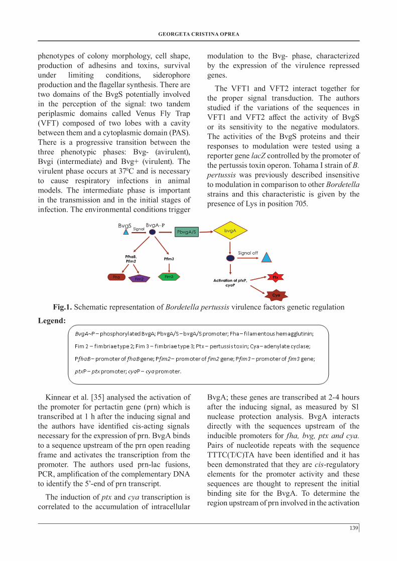
affected by the changes in the environment, and the phenomenon is called “phase modulation” or the bacteria can lose the expression of the virulence factors (“phase variation”) [15]. The control of the modulation and variation is encoded by a two-component signal transduction system represented by the BvgA/S operon. The bvg locus includes a putative sensory protein called BvgS and a positive regulator of transcription called BvgA [29]. The S part of the component is an inner membrane protein, which senses the changes in the environment, it autophosphorylates, then phosphorylates the A component from the cytosol, which becomes active. The activated BvgA binds the promoters of the genes and triggers the transcription of the genes encoding for the virulence factors, called vag genes (virulence-activated genes) [30]. When the virulence factors represented by the adhesins and toxins are expressed, the Bordetella species are in „phase I” of expression. The vag genes are not expressed when the BvgA/S operon is not activated or when there are mutations in the operon; in this case, the vrg genes are expressed (virulence-repressed genes) and the Bordetella are called „phase IV” [29]. The regulation of expression is very important for the pathogenicity of Bordetella pertussis, because if the growth conditions are changing, the expression of the virulence factors also changes.
The expression of the genes encoding for the virulence factors is influenced by environmental conditions, and the expression of the virulence-associated genes is repressed at 25°C and activated at 37°C. Other factors that mediate the expression are represented by the nicotinic acid and MgSO4 concentrations [31], which activate the genes at physiological concentrations. The BvgA and BvgS proteins share structural homology with regulatory proteins involved in gene expression.
Scarlato et al. [32] proposed a model of regulation of expression based on the theory that the signals perceived by the BvgS are transduced through the transmembrane
domain of the protein to the cytoplasmic region. In the presence of the signal, the Bvg S autophosphorylates, then it phosphorylates the BvgA, which becomes active and also activates the bvg locus as a whole. The authors constructed a plasmid with the promoter of the bvg locus, a plasmid with the promoter of the fha gene and a plasmid with both DNA fragments of the promoter regions from bvg and fha. They amplified the open reading frame of the bvgA by PCR, then they isolated the RNA, they obtained the proteins and analysed them by immunoblotting. Their conclusion was that the intergenic sequence between the bvgA and fha genes contains five promoters. P1, P2 and P3 are responsible for the transcription of the bvg locus and the P4 promoter directs the synthesis of an antisense RNA with unknown function. The fifth promoter is called PFHA and it regulates the transcription of a homologous gene. The inactivation of bvgS or the absence of signals shuts off P1, P3, P4 and PFHA, so the conclusion is that the BvgS protein is necessary for the activity of these promoters; the same promoters are inactive when MgSO4 is added to the culture medium, because this substance inactivates the bvgS gene. The researchers also raised the titer of antibodies against BvgA protein in mice and the results revealed that the protein is located in the cytoplasm because it is a DNA binding protein. The transcription of BvgA protein is inactivated in the presence of MgSO4 in the medium, inhibiting the activity of the protein, but the amount decreases in 24-36 hours. P2 promoter is active and it directs the synthesis of a low level BvgA protein for the strains in phase I or phase III. The bvgS gene becomes inactive in the presence of MgSO4, so there is a transcriptional feedback at the bvg locus of the BvgS protein [32].
The antisense RNA shuts off the translation of the complementary mRNA binding to the ribosomal binding site, located upstream from the AUG start codon. The activation of the promoter P4 is correlated to an increased amount of BvgA, so the antisense RNA could have a positive regulatory function. In B. pertussis, the bvg
THE VIRULENCE FACTORS INVOLVED IN BORDETELLA PERTUSSIS PATHOGENESIS AND THEIR GENETIC REGULATION
138
locus is important in the expression regulation of the virulence genes by activating the BvgA, starting the dimerization and transactivation of P1, P3, P4 and PFHA and all the promoters of the bvg regulon. The activation of P1, P3 and P4 promoters increases the amount of Bvg Aprotein, which activates the bvg regulated genes. In the absence of the inducing conditions, the BvgA is inactivated and stored in the cell. The level of BvgA is not enough to activate the promoters of the bvg regulon [32].
Other authors presented a model for the role of the bvg products in sensory transduction and gene regulation [33]. They used B. pertussis Bvg+ strains and constructed mutations in E. coli and transferred them to B. pertussis by shuttle mutagenesis.
There are three open reading frames called bvgA, bvgB and bvgC which may form an operon and their encoded amino acid sequences were compared to the database. Regions of homology were found between the BvgA, BvgC and some of the bacterial proteins that mediate signal transduction by a two-component system [33]. They found similarity to a factor involved in nitrogen fixation and a region of similarity to a sensor protein involved in virulence control in Agrobacterium.
The cytoplasmic region of BvgS contains three domains: the transmitter, the receiver and the C-terminal domain, separated by alanine and proline sequences. The C-terminal domain is very important in the phosphorylation from BvgS to BvgA; it is a trans-phosphorylation that requires the transmitter and the receiver. A mutation in the C-terminal domain is correlated to theinactivation of BvgS in vivo and nophosphorylation of BvgA in vitro. The site forphosphorylation is a conserved histidineresidue located in the transmitter. The BvgStransfers a phosphoryl group to theregulator; the site for phosphorylation in theregulatory proteins is an aspartic acidresidue. The result of the phosphorylation isan output signal described as a proteininteraction or the activation of
transcription. The BvgS from B. pertussis belongs to a hybrid sensor proteins family that contains an additional C-terminal domain. BvgA has an N-terminal receiver domain that contains the aspartic acids and lysine conserved residues [33].
The initial site of phosphorylation is His 729 site of the transmitter and then the phosphoryl group is transferred to the receiver [34]. The receiver might donate the phosphoryl group to the C-terminal histidine. Or, the receiver phosphorylation may relieve an auto-inhibitory effect that allows the transmitter to transfer a phosphoryl group to the C-terminal domain, which is a phosphor donor for BvgA and a mutation in BvgS prevents the BvgA phosphorylation. The binding sites of BvgA for the phosphoryl group are upstream of fhaB promoter and the phosphorylation activates the Bordetella virulence genes. There is a possibility that the BvgA/S signal transduction to be analogous to the phosphorylation cascade involved in initiating the sporulation in Bacillus subtilis [34].
The bvgS genes are different, while the bvgA genes have many similarities, with a small number of synonymous substitutions [29]. The bvgS is conserved in B. pertussis, with two non-silent single nucleotide polymorphisms. The bvgA was found almost invariant with only 15 single nucleotide polymorphisms; the bvgS has evolved differently from the housekeeping genes. A model of the histidine-conserved domain was constructed and a linker B that precedes the his-kinase domain distinguishes B. pertussis Tohama I from other isolates. Tohama I strain has a Lys residue at position 705 instead of a Glu residue found in the other 81 strains. This substitution might be involved in the change of the conformation of this region, according to signal transduction. In BvgA, there are three substitutions found for the isolates: Glu to Gly at position 64, Asp to Gly at position 137 and Val to Glu at position 197; the substitutions are not in conserved positions.
The BvgA/S operon is responsible for the
139
GEORGETA CRISTINA OPREA
phenotypes of colony morphology, cell shape, production of adhesins and toxins, survival under limiting conditions, siderophore production and the flagellar synthesis. There are two domains of the BvgS potentially involved in the perception of the signal: two tandem periplasmic domains called Venus Fly Trap (VFT) composed of two lobes with a cavity between them and a cytoplasmic domain (PAS). There is a progressive transition between the three phenotypic phases: Bvg- (avirulent), Bvgi (intermediate) and Bvg+ (virulent). The virulent phase occurs at 370C and is necessary to cause respiratory infections in animal models. The intermediate phase is important in the transmission and in the initial stages of infection. The environmental conditions trigger
modulation to the Bvg- phase, characterized by the expression of the virulence repressed genes.
The VFT1 and VFT2 interact together for the proper signal transduction. The authors studied if the variations of the sequences in VFT1 and VFT2 affect the activity of BvgS or its sensitivity to the negative modulators. The activities of the BvgS proteins and their responses to modulation were tested using a reporter gene lacZ controlled by the promoter of the pertussis toxin operon. Tohama I strain of B. pertussis was previously described insensitive to modulation in comparison to other Bordetella strains and this characteristic is given by the presence of Lys in position 705.
Kinnear et al. [35] analysed the activation of the promoter for pertactin gene (prn) which is transcribed at 1 h after the inducing signal and the authors have identified cis-acting signals necessary for the expression of prn. BvgA binds to a sequence upstream of the prn open reading frame and activates the transcription from the promoter. The authors used prn-lac fusions, PCR, amplification of the complementary DNA to identify the 5’-end of prn transcript.
The induction of ptx and cya transcription is correlated to the accumulation of intracellular
BvgA; these genes are transcribed at 2-4 hours after the inducing signal, as measured by S1 nuclease protection analysis. BvgA interacts directly with the sequences upstream of the inducible promoters for fha, bvg, ptx and cya. Pairs of nucleotide repeats with the sequence TTTC(T/C)TA have been identified and it has been demonstrated that they are cis-regulatory elements for the promoter activity and these sequences are thought to represent the initial binding site for the BvgA. To determine the region upstream of prn involved in the activation
Fig.1. Schematic representation of Bordetella pertussis virulence factors genetic regulation
Legend:
THE VIRULENCE FACTORS INVOLVED IN BORDETELLA PERTUSSIS PATHOGENESIS AND THEIR GENETIC REGULATION
140
of bvg, the authors constructed plasmids that contained prn-lac transcriptional fusions and they conjugated them with B. pertussis Tohama I strain. The results showed that there is a region of 220 bp upstream of the prn ORF that plays an important role for the promoter activation of the bvg. An AT-rich region was found and it was determined that it contains potential BvgA binding half-sites homologous to other bvg-activated promoters. They used allelic exchange to introduce the prn upstream region with mutations into Bordetella pertussis Tohama I strain, but the transcriptional activity of the prn-lac fusion was not significantly altered.
Carbonetti et al. [36] induced mutations upstream from the translational start site of the rpoA gene including the alpha subunit of RNA-polymerase and the authors thought that the mutations cause the overexpression of rpoA gene and they demonstrated that fact by overexpressing the rpoA on a plasmid in B. pertussis.
To construct the translational fusions between the wild type strains and the mutant alleles of rpoA and lacZ , the authors amplified a fragment containing a part of the rpoA from the start to the SalI site and a fragment of the upstream sequence with an EcoRI site incorporated into the primer.
They analysed BC75 and RPV3 mutants and demonstrated that the mutations of the genes encoding for α subunit of RNA-polymerase cause overexpression of this subunit that leads to down regulation of the toxin gene promoters. The mutant strain BC75 reduces transcription of pertussis toxin and adenylate cyclase toxin and RPV3 has a similar phenotype, but the reduction of the toxin expression is less pronounced than for the BC75. They amplified a segment of DNA from BC75 and RPV3 that included the start for the rpoA open reading frame and the upstream region, they cloned it and put it into pBluescript plasmid and sequenced it. They found single nucleotide substitutions in the noncoding region upstream from the rpoA open reading frame. In BC75 strain, the mutation was represented by
the substitution of adenine to guanine upstream from the ATG codon of rpoA, and for the RPV3 strain, the mutation was C to T upstream from the ATG. The effect of the substitution of A to G was enhancement of the ribosome binding correlated to the initiation of the translation, leading to an overproduction of the rpoA gene product; the mutation in RPV3 was thought to have the same effect as the one in BC75 [36].
They have also created the same mutations using the site-specific mutagenesis and introduced them into Tohama I strain by allelic exchange and the result was that the strains secreted very low levels of PTX, but produced normal levels of PRN and FIM, the same as the BC75 and RPV3 strains. The region around the start of the rpoA gene was amplified from hemolytic and non-hemolytic colonies and the authors found that the hemolytic colonies had the wild type sequence and the non-hemolytic colonies had the same mutations as in BC75 and RPV3, the results showing that these mutations are responsible for the phenotype. The hemolytic strains secreted wild-type levels of PTX into the medium; they cloned and sequenced the entire rpoA gene and the upstream regions from BC75 and RPV3 and the conclusion was that these were the only mutations involved in the phenotype of the strains. It is thought that the expression of the toxin promoters is influenced not by the activation of BvgA, but most by the concentration of free BvgA level necessary to activate the promoters.
Another possibility that could explain the overexpression of the α-unit is that this subunit might bind to sequences in the ptx and cya promoters and inhibit the transcription, but the α-binding sequence has not been established [36].
The proteins necessary for the secretion of pertussis toxin in the extracellular medium are located at the ptl locus, downstream of the ptx locus, encoding for the structural subunits of the toxin [22]. The promoter for the ptx genes has an important role in the expression of the ptl genes. The subunits of the toxin must traverse the inner
141
GEORGETA CRISTINA OPREA
and the outer membrane of the bacterium, using transport proteins, the ptl proteins. The ptl locus harbores eight open reading frames (A-H).
Kotob et al. used a vector with a cloned region from the S5 subunit and ptlC and integrated it into Bordetella pertussis chromosome by recombination [22]. If the ptx and ptl genes are parts of different operons, both genes should express and secrete the pertussis toxin. The strain they infected secreted the pertussis toxin, but it was still possible for the ptl and ptx genes to be part of the same operon. The authors made a plasmid by inserting nucleotides 930 to 4569 of the ptx-ptl region of pUW2036 into the SalI-BamHI site of pUC18 using standard techniques and then amplified by PCR the ptx-ptl region with the ptx promoter, and also the ptx-ptl region without the promoter. They demonstrated that the strains with the ptx promoter produced pertussis toxin subunits, unlike the strains which did not have the ptx promoter and did not produce pertussis toxin subunits. The results showed that the presence of the ptx promoter is necessary for the expression of the ptl genes [22]. The ptl genes are regulated by the bvg operon.
The authors also introduced the ptx promoter from B. pertussis into B. bronchiseptica strains, but those strains did not produce pertussis toxin, although the B. bronchiseptica strains have homology to the ptx and ptl regions, but they have a mutation in the promoter region [22].
The cya operon responsible for the hemolytic phenotypes has been described by Masure [37];the operon contains five genes: cyaA, cyaB, cyaC, cyaD and cyaE. The structural gene is represented by cyaA and its transcription is driven by a single promoter, which depends on the bvglocus. An additional constitutive promoter was found between cyaA and cyaB and it is involved in the transcription of cyaB, cyaD and cyaE genes.The product of the cyaA gene is an extracellular calmodulin-stimulated adenylate cyclase that confers the hemolytic phenotype and also the increasement of cAMP in the host cells. The products of the other
genes of the operon are also required for the secretion of the adenylate cyclase. The product of the cyaC confers the hemolytic and invasive properties of the toxin. The expression of the adenylate-cyclase toxin is a characteristic for BP536 strain of B. pertussis and it is controlled by the bvg locus and can be mediated by the addition of MgSO4 in the culture medium. The author constructed plasmids with copies of the cyaA gene and placed them into mutant strain BP348 containing an insertion in the cyaA. A copy of a plasmid that contained the intergenic region of the cyaA and cyaC expressed a bvg-dependent enzyme activity in response to the MgSO4. Another plasmid containing only the promoter of the cyaA, without the intergenic region, expressed very low enzyme activity. The conclusion is that the deleted region contains the cis-acting elements necessary for the mediated expression of cyaA.
CONCLUSIONS
Whooping cough is still a major public health problem in many countries, despite the detailed knowledge obtained from molecular analysis of the virulence factors involved in pertussis pathogenesis. The pertussis toxin and adenylate cyclase toxin are very important in the pathogenesis of pertussis, as well as the adhesion factors. The most important genes for encoding the adhesins and toxins are located at the bvg locus, but the complete regulation of the expression of these genes has not yet been completely elucidated. The expression of virulence genes is controlled by a two-component system named BvgA/S, an operon which is thought to have a very important role in the pathology of pertussis. The BvgS is a protein membrane sensitive to different environmental conditions which activates the BvgA, which in turn activates the promoter of different genes. Different strains of B. pertussis have been analysed and researchers have induced mutations in different sites of the genome, mostly in the promoter regions, allowing the identification of different factors involved in the expression of those genes.
THE VIRULENCE FACTORS INVOLVED IN BORDETELLA PERTUSSIS PATHOGENESIS AND THEIR GENETIC REGULATION
142
REFERENCES
1. Hegerle N and Guiso N. Epidemiology of whooping cough and typing of Bordetella pertussis. Future Microbiol. 2013;8(11):1391-403.
2. Parkhill J, Sebaihia M, Preston A, et al. Comparative analysis of the genome sequences of Bordetella pertussis, Bordetella parapertussisand Bordetella bronchiseptica. Nat Genet. 2003;35(1):32-40.
3. Bart MJ, Harris SR, Advani A, et al. Global population structure and evolution of Bordetella pertussis and their relationship with vaccination. mBio. 2014;5(2):e01074.
4. Advani A, Gustafsson L, Ahren C, et al. Appearance of Fim3 and ptxP3-Bordetella pertussis strains in two regions of Sweden with different vaccination programs. Vaccine. 2011;29(18):3438-42.
5. Barkoff AM, Mertsola J, Guillot S, et al. Appearance of Bordetella pertussis strains not expressing the vaccine antigen pertactin in Finland. Clin Vaccine Immunol. 2012;19(10):1703-4.
6. Hegerle N, Paris AS, Brun D, et al. Evolution of French Bordetella pertussis and Bordetella parapertussis isolates: increase of Bordetellae not expressing pertactin. Clin Microbiol Infect. 2012;18(9):E340-6.
7. Martin SW, Pawloski L, Williams M, et al. Pertactin-negative Bordetella pertussis strains: evidence for a possible selective advantage. Clin Infect Dis. 2015;60(2):223-7.
8. Mooi FR, van Loo IH, van Gent M, et al. Bordetella pertussis strains with increased toxin production associated with pertussis resurgence. Emerg Infect Dis. 2009;15(8):1206-13.
9. vonKonig W, Halperin CHS, Riffelman M, Guiso N. Pertussis of adults and infants. Lancet Infect Dis. 2002;2:744-750.
10. Advani A, Hallander HO, Dalby T, et al.
Pulsed-field gel electrophoresis analysis of Bordetella pertussis isolates circulating in Europe in 1998 to 2009. J Clin Microbiol. 2013;51(2):422-8.
11. Litt DJ, Jauneikaite E, Tchipeva D, et al. Direct molecular typing of B. pertussis from clinical specimens submitted for diagnostic quantitative PCR. J Med Microbiol. 2012;61(12):1662-1668.
12. Bouchez V, Guglielmini J, Dazas M, et al. Genomic sequencing of Bordetella pertussis for epidemiology and global surveillance of whooping cough. Emerg Inf Dis. 2018;24(6):988-994.
13. Caro V, Bouchez V and Guiso N. Is the sequenced Bordetella pertussis strain Tohama I representative of the species? J Clin Microbiol. 2008;46(6):2125-8.
14. van Loo IHM, Heuvelman KJ, King AJ, Mooi FR. Multilocus sequence typing of Bordetella pertussis based on surface protein genes. J Clin Microbiol. 2002;40(6):1994-2001.
15. World Health Organization. Laboratory Manual for the diagnosis of whooping cough caused by Bordetella pertussis/Bordetella parapertussis. Immunization, Vaccines and Biologicals. Geneva: WHO Document Production Services; 2014. 49 p.
16. Kallonen T, He Q. Bordetella pertussis strain variation and evolution postvaccination. Expert Rev Vaccines. 2009;8:863-75.
17. Packard ER, Parton R, Coote JG, Fry NK. Sequence variation and conservation in virulence-related genes of Bordetella pertussis isolates from the UK. J Med Microbiol. 2004;53(5):355-65.
18. Hellwig SMM, Rodriguez ME, Berbers GAM, et al. Crucial role of antibodies to pertactin in Bordetella pertussis immunity. J Infect Dis. 2003;188(5):738-42.
19. Rambow-Larsen A and Weiss A. Temporal Expression of pertussis toxin and Ptl secretion proteins by Bordetella pertussis J. Bacteriol.
143
GEORGETA CRISTINA OPREA
2004;186(1):43–50.
20. Melvin JA, Scheller EV, Miller JF, Cotter P.A. Bordetella pertussis pathogenesis: current and future challenges. Nat Rev Microbiol. 2014;12(4):274-88.
21. Worthington ZE, Carbonetti NH. Evading the proteasome: absence of lysine residues contributes to pertussis toxin activity by evasion of proteasome degradation. Infect Immun. 2007;75(6):2946-53.
22. Kotob SI, Hausman SZ, Burns DL. Localization of the promoter for the ptl genes of Bordetella pertussis, which encode proteins essential for secretion of pertussis toxin. Infect Immun. 1995;63(8):3227-30.
23. Andreasen C, Carbonetti NH. Pertussis toxin inhibits early chemokine production to delay neutrophil recruitment in response to Bordetella pertussis respiratory tract infection in mice. Infect Immun. 2008;76(11):5139-48.
24. Carbonetti NH. Pertussis toxin and adenylate cyclase toxin: key virulence factors of Bordetella pertussis and cell biology tools. Future Microbiol. 2010;5(3):455-69.
25. Ladant D, Michelson S, Sarfati R, et al. Characterization of the calmodulin-binding and of the catalytic domains of Bordetella pertussis adenylate cyclase. J Biol Chem. 1989;264(7):4015-20.
26. Chenal A, Guijarro JI, Raynal B, et al. RTX calcium binding motifs are intrinsically disordered in the absence of calcium: implication for protein secretion. J Biol Chem. 2009;284(3):1781-9.
27. Henderson MW, Inatsuka CS, Sheets AJ, et al. Contribution of Bordetella fillamentous hemagglutinin and adenylate cyclase toxin to supression and evasion of Interleukin-17- mediated inflamation. Infect Immunity. 2012;80(6):2061-75.
28. Weingart CL, Mobberley-Schuman PS, Hewlett EL, et al. Neutralizing antibodies to adenylate cyclase toxin promote phagocytosis of Bordetella pertussis by human neutrophils. Infect and Immun 2000;68(12):7152-5.
29. Herrou J, Debrie AS, Willery E, et al. Molecular evolution of the two-component system BvgAS involved in virulence regulation in Bordetella. PLoS One. 2009;4(9):e6996.
30. Moon K, Bonocora RP, Kim DD, et al. The BvgAS regulon of Bordetella pertussis. mBio 2017;8(5):e01526-17.
31. Bone MA, Wilk AJ, Perault AI, et al. Bordetella PlrSR regulatory system controls BvgAS activity and virulence in the lower respiratory tract. Proc Natl Acad Sci USA. 2017;114(8):E1519-27.
32. Scarlato V, Aricò B, Prugnola A, RappuoliR.Sequential activation and environmental regulation of virulence genes in Bordetella pertussis. EMBO Journal. 1991;10(12)3971-5.
33. Aricó B, Miller JF, Roy C, et al. Sequences required for expression of Bordetella pertussis virulence factors share homology with prokaryotic signal transduction proteins. Proc Natl Acad Sci USA. 1989;86(17):6671-5.
34. Beier D, Gross R. The BvgS/BvgAphosphorelay system of pathogenic Bordetellae: structure, function and evolution. Adv Exp Med Biology. 2008;631:149-60.
35. Kinnear SM, Boucher PE, Stibitz S, Carbonetti NH. Analysis of BvgA Activation of the Pertactin Gene Promoter in Bordetella pertussis. J Bacteriol. 1999;181(17):5234-41.
36. Carbonetti NH, Fuchs TM, Patamawenu A, et al. Effect of mutations causing overexpression of RNA Polymerase alpha subunit on regulation of virulence factors in Bordetella pertussis. J Bacteriol. 1994;176(23):7267-73.
37. Masure HR. Modulation of adenylate cyclase toxin production as Bordetella pertussis enters human macrophages. Proc Natl Acad Sci USA. 1992;89(14):6521-5.


![Bordetella Pertussis virulence factors in the continuing ... · Bordetella polysaccharides (Bps), and their significance in biofilm formation in vitro and in mice [26]. By func-tioning](https://img.pdfslide.net/doc/110x75/5ffe9ca82b08704ea77e4974/bordetella-pertussis-virulence-factors-in-the-continuing-bordetella-polysaccharides.jpg)