Embed Size (px)
Citation preview

The Y chromosome of the Okinawa spiny rat, Tokudaiamuenninki, was rescued through fusion with an autosome
Chie Murata & Fumio Yamada &
Norihiro Kawauchi & Yoichi Matsuda &
Asato Kuroiwa
Published online: 24 December 2011# Springer Science+Business Media B.V. 2011
Abstract The genus Tokudaia comprises three species,two of which have lost their Y chromosome and have anXO/XO sex chromosome constitution. Although Toku-daia muenninki (Okinawa spiny rat) retains the Y chro-mosome, both sex chromosomes are unusually large. We
conducted a molecular cytogenetic analysis to character-ize the sex chromosomes of T. muenninki. Using cross-species fluorescence in situ hybridization (Zoo-FISH),we found that both short arms of the T. muenninki sexchromosomes were painted by probes from mouse chro-mosomes 11 and 16. Comparative genomic hybridizationanalysis was unable to detect sex-specific regions in thesex chromosomes because both sex probes highlightedthe large heterochromatic blocks on the Y chromosomeas well as five autosomal pairs. We then performedcomparative FISHmapping using 29mouse complemen-tary DNA (cDNA) clones of the 22 X-linked genes andthe seven genes linked to mouse chromosome 11 (whosehomologue had fused to the sex chromosomes), andFISH mapping using two T. muenninki cDNA clones ofthe Y-linked genes. This analysis revealed that the ances-tral gene order on the long arm of the X chromosome andthe centromeric region of the short arm of the Y chromo-some were conserved. Whereas six of the mouse chro-mosome 11 genes were also mapped to Xp and Yp, inaddition, one gene, CBX2, was also mapped to Xp, Yp,and chromosome 14 in T. muenninki. CBX2 is the candi-date gene for the novel sex determination system in thetwo other species of Tokudaia, which lack a Y chromo-some and SRY gene. Overall, these results indicated thatthe Y chromosome of T. muenninki avoided a loss event,which occurred in an ancestral lineage of T. osimensis andT. tokunoshimensis, through fusion with an autosome.Despite retaining the Y chromosome, sex determination
Chromosome Res (2012) 20:111–125DOI 10.1007/s10577-011-9268-6
Responsible Editor: Tariq Ezaz and Jennifer Graves.
C. Murata :A. KuroiwaGraduate School of Life Science, Hokkaido University,Kita 10 Nishi 8, Kita-ku,Sapporo, Hokkaido 060-0810, Japan
F. YamadaForestry and Forest Products Research Institute,Tsukuba, Ibaraki 305-8687, Japan
N. KawauchiYachiyo Engineering Co., Ltd,Kokuba Bldg. 9f, 3-21-1, Kumoji,Naha, Okinawa 900-0015, Japan
Y. MatsudaLaboratory of Animal Genetics, Department of AppliedMolecular Biosciences, Graduate School of BioagriculturalSciences, Nagoya University,Furo-cho, Chikusa-ku( Nagoya 464-8601, Japan
A. Kuroiwa (*)Laboratory of Animal Cytogenetics, Faculty of Science,Hokkaido University,Kita 10 Nishi 8, Kita-ku,Sapporo, Hokkaido 060-0810, Japane-mail: [email protected]

in T. muenninki might not follow the usual mammalianpattern and deserves further investigation.
Keywords X chromosome . evolution . Zoo-FISH .
FISH .CBX2 . Spiny rat . Tokudaia
AbbreviationsASA American Standards AssociationBSA Bovine serum albumincDNA Complementary DNACGH Comparative genomic hybridizationdUTP Deoxyuridine 5-triphosphateFISH Fluorescence in situ hybridizationFITC Fluorescein isothiocyanateHMG High-mobility groupIHB Intercalary heterochromatin blockkb Kilo base pairsMMU Mus musculus chromosomeMYA Million years agoORF Open reading framePAR Pseudoautosomal regionqRT-PCR Quantitative real-time PCRRBMY1A1 RNA binding motif protein, Y
chromosome, family 1, member A1SSC Saline sodium citrateTMU Tokudaia muenninki chromosomeTTO Tokudaia tokunoshimensis chromosomeUV UltravioletZoo-FISH Cross-species fluorescence in situ
hybridization
Introduction
The genus Tokudaia belongs to the subfamilyMurinae (Muridae, Rodentia) and consists of threespecies: Tokudaia muenninki (Okinawa spiny rat),Tokudaia osimensis (Amami spiny rat), and Toku-daia tokunoshimensis (Tokunoshima spiny rat).Each species is endemic to an island in the south-ernmost part of Japan and is named after theisland on which it is found. The sex chromosomesin this genus are unusual among mammals. The diploidchromosome numbers of T. osimensis and T. tokunoshi-mensis are an odd number, 25 and 45, respectively.Neither T. osimensis nor T. tokunoshimensis has a Ychromosome and sex is determined with an XO/XOsex chromosome constitution in both species (Honda
et al. 1977, 1978; Kobayashi et al. 2007). Although thediploid chromosome number of T. muenninki is 44 withXX/XY sex chromosome constitution, both sex chro-mosomes are unusually large (Tsuchiya et al. 1989;Murata et al. 2010). The euchromatic regions of the Xand Y chromosomes occupy 8% and 4% of the haploidgenome, respectively, which suggests that an autosomaladdition has occurred (Murata et al. 2010). On the basisof molecular phylogenetic analysis, T. muenninki wasthe first species in the genus to diverge, which suggeststhat the Y chromosome was lost in the common ancestorof T. osimensis and T. tokunoshimensis after this event(Murata et al. 2010). Three stepwise events that led to thedisappearance of the Y chromosome have been sug-gested; gene transpositions from the Y chromosome toautosomes, superseding Y-linked genes by new genes onthe X chromosome or autosomes, and Y-to-X transloca-tion containing Y-linked genes (Arakawa et al. 2002;Kuroiwa et al. 2010).
The SRY gene, which is the master gene for malesex determination in placental mammals (Sinclair et al.1990; Koopman et al. 1991) and is located on the Ychromosome, has been lost completely in T. osimensisand T. tokunoshimensis (Soullier et al. 1998; Sutou etal. 2001). Other Y chromosome genes have also beenlost in T. osimensis, including RBMY1A1 (RNA bind-ing motif protein, Y chromosome, family 1, memberA1) (Kuroiwa et al. 2010), which plays a role inspermatogenesis (Mazeyrat et al. 1999; Elliott 2004).However, most proto-Y-linked genes have been con-served in both sexes of T. osimensis and T. tokunoshi-mensis through translocation from the Y chromosometo the distal region of the long arm of the X chromo-some. The absence of SRY indicates that T. osimensisand T. tokunoshimensis must have a novel sex-determining mechanism. Recently, CBX2 (chromoboxhomolog 2, synonym is M33) has emerged as a can-didate gene for sex determination in these species(Kuroiwa et al. 2011). CBX2 is a single copy genethat is located on an autosome in mice and humans.However, quantification of the copy number of CBX2in T. osimensis and T. tokunoshimensis by quantitativereal-time PCR showed that there were two or threemore copies of CBX2 per haploid genome in malesthan in females, which suggests that CBX2 might beinvolved in a novel sex-determining mechanismthrough gene dosage (Kuroiwa et al. 2011).
Although T. muenninki possesses a Y chromosome,several aspects of sex determination in this species are
112 C. Murata et al.

unusual (Tsuchiya et al. 1989; Murata et al. 2010).Multiple copies of the SRY gene (>70) are foundin T. muenninki, and these are distributed along theentire long arm of the Y chromosome (Murata etal. 2010). Most copies are not functional; however,an open reading frame (ORF) is conserved in threecopies (Murata et al. 2010). A single amino acidsubstitution that is specific to T. muenninki wasdetected in the DNA binding surface domain inthe high-mobility group (HMG)-box of all SRYcopies (Murata et al. 2010). This region is impor-tant for the binding of SRY to target DNA sequences,which suggests that the DNA-binding ability of all SRYproteins produced in T. muenninki is weakened by thismutation.
To reveal the composition of the X and Y chromo-somes in T. muenninki, we performed cross-speciesfluorescence in situ hybridization (Zoo-FISH) withchromosome-specific DNA probes constructed fromthe laboratory mouse (Mus musculus). To describe thecomposition in more detail and identify sex-specificregions in the sex chromosomes, we performed com-parative genomic hybridization (CGH) analysis.When this provided limited information, we thenconducted comparative FISH analysis using 29mouse cDNA clones of functional genes linked tothe X chromosome and chromosome 11 and FISHmapping using two T. muenninki cDNA clones ofthe Y-linked genes.
Materials and methods
Sample collection, cell culture, and DNA and RNAextraction
The three species of Tokudaia are all endangered (TheIUCN Red List of Threatened Species; http://www.iucnredlist.org/ 15/9/2011) and have been protectedby the Japanese government as natural treasures since1972. With permission from the Agency for CulturalAffairs and the Ministry of the Environment inJapan, we trapped the Okinawa spiny rat, T. muen-ninki, on Okinawa-jima Island in March 2008 andFebruary 2009 (Yamada et al. 2010). We collectedtail tissue from four males and three females for usein the study. Cell culture and DNA extraction wereperformed as described by Murata et al. (2010).Total RNA was extracted from fibroblasts derived
from tail tissue from a T. muenninki male usingTRIZOL Reagent (Invitrogen) in accordance withthe manufacturer’s protocol. The cDNAs were syn-thesized using SuperScript II Reverse Transcriptase(Invitrogen).
DNA probes
Seven mouse cDNA clones of functional geneslinked to chromosome 11 and 22 linked to the Xchromosome were used in the study. Their genenames, symbols, accession numbers, sizes, andchromosomal location in mouse (M. musculus), rat(Rattus norvegicus), and three Tokudaia species arelisted in Table 1. A 6.6-kilo base pairs (kb) mousegenomic DNA fragment was used for chromosomallocalization of the 18S–28S rRNA gene (Kominamiet al. 1982).
Clones of T. muenninki DDX3Y (AB672502;1.8 kb) and UTY (AB672501; 2.0 kb), which are bothlinked to the Y chromosome, were also used for FISHmapping. The mouse homologues referred to here areDdx3y (NM_012008) and Uty (NM_009484), and therat genes are DDX3Y (FJ775727) and UTY(FJ775728). The T. muenninki homologues of thesegenes were cloned by degenerate PCR. The primerpairs used in the PCR were as follows: DDX3Y, 5′-TGA TCG TGG AAG TGG ATC C-3′and 5′-CACAGC TGG TAA TGT ACT GCA-3′; UTY, 5′-GACGCT GTT GAA CAA GGC AA-3′ and 5′-GGA GTGGGT GGA TTC AAC TTC-3′. The PCR for DDX3Ywas carried out in 10 μl of 1× Ex Taq buffer thatcontained 0.5 μl of template cDNA, 2 mM MgCl2,0.5 μM of each primer, 0.2 mM each of the fourdNTPs, and 0.25 U of Ex Taq (TaKaRa Bio Inc.).The reaction profile was denaturation for 5 min at94°C, followed by 35 cycles of denaturation for 30 sat 94°C, annealing for 30 s at 60 °C, and extension for2 min at 72°C, and then a final extension at 72°C for7 min. The PCR for UTY was carried out in 20 μl of1× PrimeSTAR GXL buffer (Mg2+ plus) thatcontained 1.0 μl of template cDNA, 0.3 μM of eachprimer, 0.2 mM each of the four dNTPs, and 0.25 U ofPrimeSTAR GXL DNA Polymerase (TaKaRa BioInc.). The reaction profile was denaturation for 3 minat 94°C, followed by 35 cycles of denaturation for 10 sat 98°C, annealing for 15 sec at 60°C, and extensionfor 1.5 min at 68°C, and then a final extension at 72°Cfor 5 min.
Sex-autosome fusions in Tokudaia muenninki 113
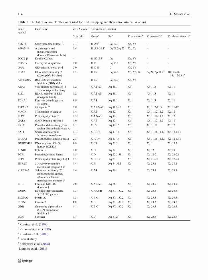
Table 1 The list of mouse cDNA clones used for FISH mapping and their chromosomal locations
Genesymbol
Gene name cDNA clone Chromosome location
Size (kb) Mousea Rata T. muenninkid T. osimensise T. tokunoshimensise
STK10 Serin/threonine kinase 10 3.1 11 A4b 10q 12.3 Xp, Yp – –
ADAM19 A disintegrin andmetalloproteinasedomain 19 (meltrin beta)
1.4 11 A5-B1.1c 10q 21.3-q 22 Xp, Yp – –
DOC2 β Double C2 beta – 11 B3-B5 10q Xp, Yp – –
COASY Coenzyme A synthase 2.0 11 D 10q 32.1 Xp, Yp – –
GAA Glucosidase, alpha, acid 2.0 11 D-E 10 Xp, Yp – –
CBX2 Chromobox homolog 2(Drosophila Pc class)
1.5 11 E2 10q 32.3 Xp, Yp, 14 3q 24, 6p 11.2f 10q 25-26,14q 12-13.1f
ARHGDIA Rho GDP dissociationinhibitor (GDI) alpha
– 11 E2 10q 32.3 Xp, Yp – –
ARAF v-raf murine sarcoma 3611viral oncogene homolog
1.2 X A2-A3.1 Xq 11.1 Xq Xp 11.3 Xq 11
ELK1 ELK1, member of ETSoncogene family
1.2 X A2-A3.1 Xq 11.1 Xq Xp 11.3 Xq 11
PDHA1 Pyruvate dehydrogenaseE1 alpha 1
0.9 X A4 Xq 11.1 Xq Xp 11.3 Xq 11
TSPAN7 tetraspanin 7 2.0 X A1.3-A2 Xq 11.2-12 Xq Xp 11.2-11.3 Xq 11-12
MAOA Monoamine oxidase A 1.4 X A2 Xq 12 Xq Xp 11.12-11.2 Xq 12
PLP2 Proteolipid protein 2 1.2 X A2-A3.1 Xq 12 Xq Xp 11.12-11.2 Xq 12
GATA1 GATA binding protein 1 1.0 X A2 Xq 12 Xq Xp 11.12-11.2 Xq 12
PIGA Phosphatidylinositol glycananchor biosynthesis, class A
1.1 X F5 Xq 12-13 Xq Xp 11.12 Xq 12
SAT1 Spermidine/spermineN1-acetyl transferase 1
1.1 X F3-F4 Xq 13-14 Xq Xp 11.11-11.12 Xq 12-13.1
PHKA2 Phosphorylase kinase alpha 2 2.3 X F3-F4 Xq 13-14 Xq Xp 11.11-11.12 Xq 12-13.1
DXHXS423 DNA segment, Chr X,human DXS423
0.8 X C3 Xq 21.3 Xq Xq 11 Xq 13.3
EFNB1 Ephrin B1 1.0 X D Xq 22.1 Xq Xq 12 Xq 21
PGK1 Phosphoglycerate kinase 1 1.5 X D Xq 22.3-31.1 Xq Xq 12-21 Xq 21-22
PLP1 Proteolipid protein (myelin) 1 1.5 X F1-F2 Xq 32 Xq Xq 21-22 Xq 22-23
HTR2C 5-Hydroxytryptamine(serotonin) receptor 2 C
1.4 X F1 Xq 34-35.1 Xq Xq 23.1 Xq 24.1
SLC25A5 Solute carrier family 25(mitochondrial carrier,adenine nucleotidetranslocator), member 5
1.4 X A4 Xq 36 Xq Xq 23.1 Xq 24.1
FHL1 Four and half LIMdomains 1
2.0 X A6-A7.1 Xq 36 Xq Xq 23.2 Xq 24.2
IDH3G Isocitrate dehydrogenase3 (NAD+) gamma
1.3 X A7.3-B Xq 37.1-37.2 Xq Xq 23.3 Xq 24.3
PLXNA3 Plexin A3 1.3 X B-C1 Xq 37.1-37.2 Xq Xq 23.3 Xq 24.3
CETN2 Centrin 2 0.8 X B Xq 37.1-37.2 Xq Xq 23.3 Xq 24.3
GDI1 Guanosine diphosphate(GDP) dissociationinhibitor 1
1.1 X B-C1 Xq 37.1-37.2 Xq Xq 23.3 Xq 24.3
BGN biglycan 1.7 X B Xq 37.2 Xq Xq 23.3 Xq 24.3
a Kuroiwa et al. (1998)b Kuramochi et al. (1999)c Kurohara et al. (2000)d Present studye Kobayashi et al. (2008)f Kuroiwa et al. (2011)
114 C. Murata et al.

Chromosome preparation and Zoo-FISH analysis
The preparation of R-banded chromosomes was per-formed as described byMatsuda et al. (1992a), Matsudaand Chapman (1995), and Kobayashi et al. (2008).
The Zoo-FISH analysis was performed usingbiotin- and Cy3-labeled chromosome-specific paintingprobes from the laboratory mouse (Cambio Ltd.).FISH was carried out as described by Nakamura etal. (2007) with slight modifications. The probes weredenatured at 75°C for 10 min and pre-annealed byincubation at 37°C for 1 h. The chromosome slideswere hybridized with the biotin- or Cy3-labeledprobes at 37°C for 5 days. After hybridization withthe biotin-labeled probes, the slides were washed in50% formamide in 2× saline sodium citrate (SSC) at37°C for 15 min, in 2× SSC for 15 min, and then in4× SSC for 5 min at room temperature. To detect thehybridization signals of the biotin-labeled probes, thechromosome slides were incubated with avidin fluo-rescein isothiocyanate (FITC) (Vector Laboratories) orstreptavidin-Cy5 (GE Healthcare UK Ltd.) diluted1:500 in 1% BSA/4× SSC at 37°C for 1 h. The slideswere washed sequentially on a shaker with 4× SSC,0.1% Nonidet P-40/4× SSC, and then 4× SSC for5 min each at room temperature. After hybridizationwith the Cy3-labeled probe, the slides were washedsequentially in 4× SSC, 0.1% Nonidet P-40/4× SSC,and then 4× SSC for 5 min each at room temperature.
CGH analysis
The CGH analysis was conducted using metaphasespreads from one male and one female of T. muen-ninki as described by Kobayashi et al. (2007) withslight modifications. The genomic DNA for theprobes was also extracted from one male and onefemale. The male and female genomic DNA probeswere labeled differently, with Cy3-deoxyuridine5-triphosphate (dUTP; GE Healthcare) and fluorescein-12-dUTP (Invitrogen), respectively, by nick translationat 15°C for 90 min. The labeled DNA probes weredenatured at 75°C in formamide and dissolved inhybridization buffer (50% formamide, 2× SSC, 10%dextran sulfate, and 2 mg/ml BSA). The cocktail ofprobes for one slide contained 700 ng of labeled DNAfrom the female and 700 ng of labeled DNA from themale. Slides were hybridized at 37°C for 4 days. Theslides were then washed sequentially in 4× SSC, 0.1%
Nonidet P-40 in 4× SSC, 4× SSC, and 2× SSC for 5 mineach at room temperature, and then rinsed in 2× SSC.
FISH analysis
The FISH analysis using cDNA clones as probes wasperformed as described by Kuroiwa et al. (2010). Theexception was the FISH analysis using the 18S–28SrRNA gene clone as a probe. In this case, the chromo-some slides were treated with RNase (1 μg/μl in 2×SSC) for 1 h, washed three times in 2× SSC for 3 mineach, and dehydrated in 70% and 100% ethanol at 4°Cfor 5 min each before the chromosomes were dena-tured in 70% formamide in 2×SSC at 69°C for 2 min.After hybridization and washing, the slides were incu-bated under parafilm for 1 h at 37°C with fluorescei-nated avidin (avidin-FITC; Vector Laboratories) diluted1:500 in 1% bovine serum albumin (BSA)/4× SSC[instead of goat anti-biotin antibody (Vector Laborato-ries)]. The slides were washed sequentially on a shakerwith 4× SSC, 0.1% Nonidet P-40 in 4× SSC, and 4×SSC for 10 min each, and then rinsed with 2× SSC andstained with 0.75 mg/ml propidium iodide for 3 min.
Microphotography and image capture
The chromosomal slides were observed under a Nikonfluorescence microscope using Nikon filter sets B-2A(450–490 nm) and ultraviolet (UV)-2A (330–380 nm).Kodak Ektachrome American Standards Association(ASA) 100 films were used for microphotography.The digital FISH images were captured using the550CW-QFISH application program of Leica Micro-systems Imaging Solution Ltd. using a cooled CCDcamera (MicroMAX 782Y, Princeton Instruments)mounted on a Leica DMRA microscope.
Results
Zoo-FISH analysis
We conducted the Zoo-FISH analysis on chromo-somes from a male and a female specimen of T.muenninki using all M. musculus chromosome(MMU)-specific painting probes except for the Ychromosome probe. Each chromosome of T. muen-ninki could be identified on the basis of the Hoechst-stained bands that were obtained by the replication
Sex-autosome fusions in Tokudaia muenninki 115

R-banding method (Fig. 1). The MMU probes for allautosomes and the X chromosome were hybridizedsuccessfully to T. muenninki chromosomes (TMU).The images for the probes MMUX, MMU11, and
MMU16, are shown in Fig. 1a–c. The patterns for allthe T. muenninki chromosomes are summarized inFig. 1f. Although the long arm of the X chromosomeof T. muenninki was covered with the MMUX probe
Fig. 1 Karyotypic characterization in T. muenninki. a–c Zoo-FISH for T. muenninki with MMU-specific probes. Hybridizationof FITC- (green) labeled mouse painting probes, MMUX, toPI-stained chromosomes of a T. muenninki female (a). Hybridiza-tion of Cy3- (yellow) and Cy5- (pink) labeled mouse paintingprobes, MMU11 (b) and MMU 16 (c), respectively, to Hoechst-stained chromosomes of a T. muenninki male. Arrows and arrow-heads indicate the hybridization signals on the sex chromosomesand autosomes [TMU10 (b) and TMU15 (c)], respectively. Thescale bar indicates 10 μm. (d, e) Mapping of T. muenninki chro-mosomes by FISH using mouse genomic DNA clones of the 18S–28S rRNA genes. Hybridization signals of the 18S–28S rRNAgenes were visualized by avidin-FITC on PI-stained chromosomes
(d). Hoechst G-banding patterns are shown in e. Arrows andarrowheads indicate the hybridization signals on the sex chromo-somes and a pair of autosomes (TMU1), respectively. The scalebar indicates 10 μm. f Comparative cytogenetic maps of T. muen-ninki. The comparative cytogenetic maps showing chromosomehomologies between mouse and T. muenninkiwere constructed byZoo-FISH analysis with mouse probes. The numbers under thechromosomes of T. muenninki indicate the chromosome numbersfor this species. The numbers inside the chromosomes indicate thechromosome numbers for mouse that correspond to the chromo-somal segments of T. muenninki indicated. Arrowheads indicatethe locations of the 18S–28S rRNA genes. Large heterochromaticregions are shown in black
116 C. Murata et al.

(Fig. 1a), the short arms of the X and Y chromosomesboth hybridized with the mouse probes MMU11 andMMU16 (Fig. 1b and c, respectively). The 18S–28Sribosomal RNA genes were localized to the distal endsof chromosome 1 and the short arms of the sex chro-mosomes (Fig. 1d, e), which corresponded to MMU9and MMU11, respectively (Fig. 1f).
Fourteen chromosomes of T. muenninki (TMU1, 2,3, 5, 7, 9, 11, 14, 15, 17, 18, 19, 20, and 21) each werehybridized with a single mouse probe; six chromo-somes (TMU4, 6, 8, 10, 12, and 13) each hybridizedwith two probes; and one chromosome (TMU16) washybridized with four probes (Fig. 1f). Thirty-four con-served segments were detected between the mouse andT. muenninki chromosomes. The hybridization pat-terns between T. muenninki and the mouse probescould be grouped into three categories: (1) 11 mouseprobes (MMU2, 3, 4, 6, 7, 9, 12, 14, 18, 19, and X)each hybridized to a single chromosome or chromo-somal segment of T. muenniki; (2) six mouse probes(MMU1, 8, 11, 13, 15, and 16) each hybridized to twochromosomes; and (3) three mouse probes (MMU5,10, and 17) each hybridized to more than three chro-mosomes (Fig. 1f).
CGH analysis
We performed CGH to identify the male-specific regionin the Y chromosome of T. muenninki. CGH signalswith differently labeled genomic DNAs from a femaleand a male were highlighted in the long arm of the Ychromosome, as well as in the heterochromatic regionsof five autosomal pairs in both sexes (Fig. 2). The longarm of the Y chromosome, Yq, was labeled intensely notonly by the male probe, which contained Y chromo-somal DNA, but also by the female probe, although theintensity of Hoechst fluorescence of Yq was not higherthan that of any of the other chromosomes (Fig. 2a–c).The short arm of the Y chromosome, Yp, which is aproto-autosomal segment that has been translocated tothe Y chromosome, as shown in Fig. 1, was stainedequally by the female- and male-derived probes(Fig. 2d). Therefore, the loss or gain of a male-specificchromosomal region was not detected in the T. muen-ninki male by CGH analysis. Furthermore, when unla-beled female DNA and mouse C0t-1 DNAwere used inthe CGH analysis as competitors, no male-specificregion was detected in the T. muenninki male (data notshown). In addition, the sex-specific region was not
Fig. 2 CGH images of a male (a–d) and a female (e–h) meta-phase spread for T. muenninki. The Hoechst G-banding patternsare shown in a and e. The CGH images (d, h) were obtained bymerging the images of metaphase spreads hybridized with
female genomic DNA (b, f) and male genomic DNA (c, g).Arrows indicate the sex chromosomes. Scale bar represents10 μm
Sex-autosome fusions in Tokudaia muenninki 117
detected in diploid mitotic complements of the T. muen-ninki female (Fig. 2e–h).
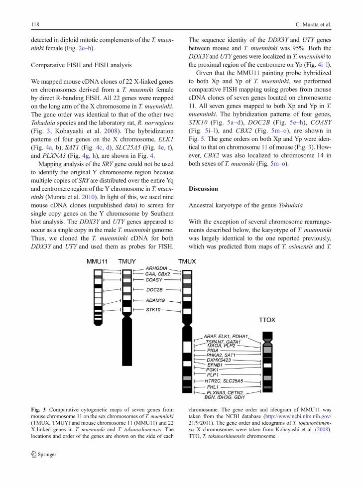
Comparative FISH and FISH analysis
We mapped mouse cDNA clones of 22 X-linked geneson chromosomes derived from a T. muenniki femaleby direct R-banding FISH. All 22 genes were mappedon the long arm of the X chromosome in T. muenninki.The gene order was identical to that of the other twoTokudaia species and the laboratory rat, R. norvegicus(Fig. 3, Kobayashi et al. 2008). The hybridizationpatterns of four genes on the X chromosome, ELK1(Fig. 4a, b), SAT1 (Fig. 4c, d), SLC25A5 (Fig. 4e, f),and PLXNA3 (Fig. 4g, h), are shown in Fig. 4.
Mapping analysis of the SRY gene could not be usedto identify the original Y chromosome region becausemultiple copies of SRY are distributed over the entire Yqand centromere region of the Y chromosome in T. muen-ninki (Murata et al. 2010). In light of this, we used ninemouse cDNA clones (unpublished data) to screen forsingle copy genes on the Y chromosome by Southernblot analysis. The DDX3Y and UTY genes appeared tooccur as a single copy in the male T. muenninki genome.Thus, we cloned the T. muenninki cDNA for bothDDX3Y and UTY and used them as probes for FISH.
The sequence identity of the DDX3Y and UTY genesbetween mouse and T. muenninki was 95%. Both theDDX3YandUTY genes were localized in T. muenninki tothe proximal region of the centromere on Yp (Fig. 4i–l).
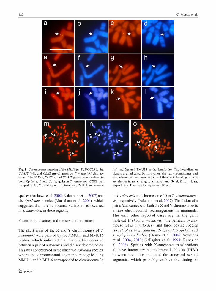
Given that the MMU11 painting probe hybridizedto both Xp and Yp of T. muenninki, we performedcomparative FISH mapping using probes from mousecDNA clones of seven genes located on chromosome11. All seven genes mapped to both Xp and Yp in T.muenninki. The hybridization patterns of four genes,STK10 (Fig. 5a–d), DOC2B (Fig. 5e–h), COASY(Fig. 5i–l), and CBX2 (Fig. 5m–o), are shown inFig. 5. The gene orders on both Xp and Yp were iden-tical to that on chromosome 11 of mouse (Fig. 3). How-ever, CBX2 was also localized to chromosome 14 inboth sexes of T. muenniki (Fig. 5m–o).
Discussion
Ancestral karyotype of the genus Tokudaia
With the exception of several chromosome rearrange-ments described below, the karyotype of T. muenninkiwas largely identical to the one reported previously,which was predicted from maps of T. osimensis and T.
Fig. 3 Comparative cytogenetic maps of seven genes frommouse chromosome 11 on the sex chromosomes of T. muenninki(TMUX, TMUY) and mouse chromosome 11 (MMU11) and 22X-linked genes in T. muenninki and T. tokunoshimensis. Thelocations and order of the genes are shown on the side of each
chromosome. The gene order and ideogram of MMU11 wastaken from the NCBI database (http://www.ncbi.nlm.nih.gov/21/9/2011). The gene order and ideograms of T. tokunoshimen-sis X chromosomes were taken from Kobayashi et al. (2008).TTO, T. tokunoshimensis chromosome
118 C. Murata et al.

tokunoshimensis generated by chromosome painting(Nakamura et al. 2007): MMU1b/17a/17e, a single seg-ment of MMU14, and the two distinct chromosomalsegments of MMU15 occurred in T. muenninki, whereasMMU1b/17a, two distinct chromosomal segments ofMMU14, and a single segment of MMU15 occurred inT. osimensis and T. tokunoshimensis. The former seg-ments found in T. muenninki are also conserved in theancestral karyotype of the genus Apodemus (Matsubaraet al. 2004), which is the most closely related group to thegenus Tokudaia in Murinae (Michaux et al. 2002; Satoand Suzuki 2004; Rowe et al. 2008), suggesting that theancestor of genus Tokudaia might have the same seg-ments as T. muenninki, MMU1b/17a/17e, two distinctchromosomal segments ofMMU14 and a single segmentof MMU15. Therefore, we have demonstrated that theancestral karyotype of the genus Tokudaia had a diploid
chromosome number of 48 and contained the followingchromosomes that are homologous to mouse: 1a, 1b/17a/17e, 2, 3, 4, 5a, 5b/11a, 5c/6, 7/19, 8a, 8b, 9, 10a, 10b/17b, 10c/17c, 11b/16a, 12/17d, 13a, 13b/15a, 14, 15b,16b, 18, X, and Y (Fig. 6). Lineage-specific chromosomerearrangements in T. muenninki, inferred from the ances-tral karyotype, were as follows: (1) fusion between seg-ments homologous to MMU11b/16a and the ancestral Yin TMUY; (2) centric fusions between segments homol-ogous to MMU11b/16a and MMUX in TMUX andbetween segments homologous to MMU10b/17b andMMU13b/15a in TMU16; and (3) two pericentric inver-sions in TMU17 and TMU19 (Fig. 6).
Furthermore, the localization of the 18S–28S ribo-somal RNA genes to two chromosomal segmentshomologous to MMU9 and MMU11 in T. muenninki(Fig. 1d–f) was also conserved in the other two Tokudaia
Fig. 4 Chromosome mapping of four X-linked genes and twoY-linked genes on T. muenninki chromosomes. The hybridizationsignals are indicated by arrows. Four X-linked genes, ELK1 (a, b),SAT1 (b, c), SLC25A5 (e, f), andPLXNA3 (g, h), weremapped fromthe central to the distal part of T. muenninki Xq in the order shown.
Two Y-linked genes, DDX3Y (i, j) and UTY (k, l) were localized tothe proximal region of the centromere in T. muenninki Yp. R- andHoechst G-banding patterns are shown in (a, c, e, g, i, k) and (b, d, f,h, j, l), respectively. The scale bar indicates 10 μm
Sex-autosome fusions in Tokudaia muenninki 119
species (Arakawa et al. 2002; Nakamura et al. 2007) andsix Apodemus species (Matsubara et al. 2004), whichsuggested that no chromosomal variation had occurredin T. muenninki in these regions.
Fusion of autosomes and the sex chromosomes
The short arms of the X and Y chromosomes of T.muenninki were painted by the MMU11 and MMU16probes, which indicated that fusions had occurredbetween a pair of autosomes and the sex chromosomes.This was not observed in the other two Tokudaia species,where the chromosomal segments recognized byMMU11 and MMU16 corresponded to chromosome 3q
in T. osimensis and chromosome 10 in T. tokunoshimen-sis, respectively (Nakamura et al. 2007). The fusion of apair of autosomes with both the X and Y chromosomes isa rare chromosomal rearrangement in mammals.The only other reported cases are in: the giantmole-rat (Fukomys mechowii), the African pygmymouse (Mus minutoides), and three bovine species(Boselaphus tragocamelus, Tragelaphus spekei, andTragelaphus imberbis) (Deuve et al. 2006; Veyruneset al. 2004, 2010; Gallagher et al. 1998; Rubes etal. 2008). Species with X-autosome translocationsall have intercalary heterochromatic blocks (IHBs)between the autosomal and the ancestral sexualsegments, which probably enables the timing of
Fig. 5 Chromosomemapping of the STK10 (a–d),DOC2B (e–h),COASY (i–l), and CBX2 (m–o) genes on T. muenninki chromo-somes. The STK10, DOC2B, and COASY genes were localized toboth Xp (a, e, i) and Yp (c, g, k) in T. muenninki. CBX2 wasmapped to Xp, Yp, and a pair of autosomes (TMU14) in the male
(m) and Xp and TMU14 in the female (o). The hybridizationsignals are indicated by arrows on the sex chromosomes andarrowheads on the autosomes. R- and Hoechst G-banding patternsare shown in (a, c, e, g, i, k, m, o) and (b, d, f, h, j, l, n),respectively. The scale bar represents 10 μm
120 C. Murata et al.

replication of the sex and autosomal segments to beregulated independently (Dobigny et al. 2004). Thecentromeric heterochromatin block of T. muenninkimight function as an IHB and an X-autosomalboundary in this species (Murata et al. 2010).
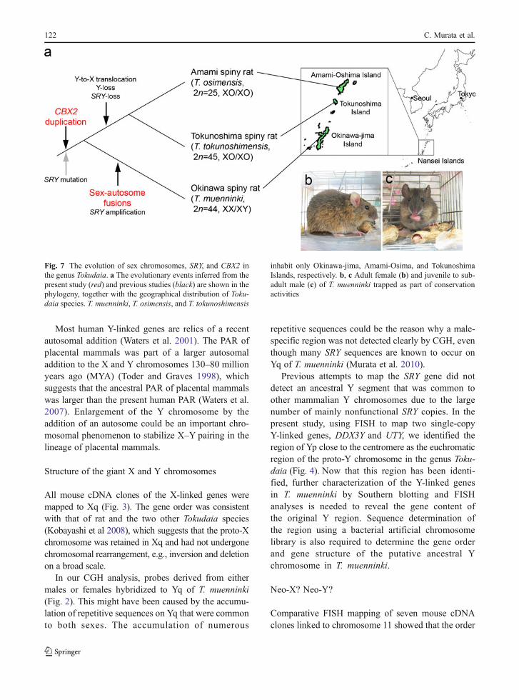
Avoidance of Y chromosome loss through fusionwith an autosome
T. muenninki is known to be the only species with a Ychromosome in the genus Tokudaia; as a consequence, itfollows the general XY pattern of mammalian sexdetermination (Tsuchiya et al. 1989). However, ourresults indicated that the Y chromosome of T. muenninkihas evolved in a unique manner. The Y chromosomemight have had instability persisting in the commonancestral species of the genus Tokudaia (Fig. 7). In anancestral lineage common to the two XO/XO spiny rats,T. osimensis and T. tokunoshimensis, most of the Y-linked genes (except SRY and RBMY1A1) escaped tothe X chromosome, and the Y chromosome was lostsubsequently (Kuroiwa et al. 2010). In contrast, in the
ancestral population of T. muenninki, the X and Ychromosomes fused with a pair of autosomes.
The enlargement of the Y chromosome through fusionwith an autosome extended the pseudoautosomal regions(PARs). PARs behave like an autosomal pair and recom-bine during male meiosis, which is thought to play acritical role in spermatogenesis (Kauppi et al. 2011;Matsuda et al. 1992b; Mohandas et al. 1992). The XYmale mice with inverted X PAR, Y PAR flanked at thedistal end by X PAR boundary together with adjacentX-specific material, or both of the variant PARs, areknown to produce XO progeny by leading to the produc-tion of unusual X and Y products and consequent sexchromosome loss (Burgoyne and Evans 2000). If variantPAR was associated with the Y chromosome loss in anancestral lineage common to the two XO/XO spiny rats,T. osimensis and T. tokunoshimensis, the Y chromosomewould be stabilized by the acquisition of new PARs in theancestral lineage of T. muenninki, and thus, loss of the Ychromosome was avoided. To test this hypothesis, futureresearch should include the identification and sequencingof an ancestral PAR in three Tokudaia species.
Fig. 6 A schematic representation of the ancestral karyotype of theTokudaia species and chromosome rearrangements that occurred inT. muenninki after its divergence from the common ancestor. Thenumbers inside the chromosomes indicate the mouse chromosomenumbers that correspond to the indicated chromosomal segments ofthe Tokudaia species. The lower chromosomes correspond to thechromosomes of T. muenninki, and the numbers underneath are thechromosome numbers for this species. The numbers above theupper chromosomes, those of the schematic ancestral karyotype,
indicate the chromosomes, chromosomal segments, or chromo-some associations that are homologous to mouse chromosomes.They were numbered following the nomenclature of Stanyon et al.(2004) and Nakamura et al. (2007). Arrows show how the chro-mosomes of T. muenninki were derived from the ancestral karyo-type. Two-pronged arrows to a single chromosome indicate thesuccessive occurrence of fusions. “Inv” indicates a pericentricinversion
Sex-autosome fusions in Tokudaia muenninki 121
Most human Y-linked genes are relics of a recentautosomal addition (Waters et al. 2001). The PAR ofplacental mammals was part of a larger autosomaladdition to the X and Y chromosomes 130–80 millionyears ago (MYA) (Toder and Graves 1998), whichsuggests that the ancestral PAR of placental mammalswas larger than the present human PAR (Waters et al.2007). Enlargement of the Y chromosome by theaddition of an autosome could be an important chro-mosomal phenomenon to stabilize X–Y pairing in thelineage of placental mammals.
Structure of the giant X and Y chromosomes
All mouse cDNA clones of the X-linked genes weremapped to Xq (Fig. 3). The gene order was consistentwith that of rat and the two other Tokudaia species(Kobayashi et al 2008), which suggests that the proto-Xchromosome was retained in Xq and had not undergonechromosomal rearrangement, e.g., inversion and deletionon a broad scale.
In our CGH analysis, probes derived from eithermales or females hybridized to Yq of T. muenninki(Fig. 2). This might have been caused by the accumu-lation of repetitive sequences on Yq that were commonto both sexes. The accumulation of numerous
repetitive sequences could be the reason why a male-specific region was not detected clearly by CGH, eventhough many SRY sequences are known to occur onYq of T. muenninki (Murata et al. 2010).
Previous attempts to map the SRY gene did notdetect an ancestral Y segment that was common toother mammalian Y chromosomes due to the largenumber of mainly nonfunctional SRY copies. In thepresent study, using FISH to map two single-copyY-linked genes, DDX3Y and UTY, we identified theregion of Yp close to the centromere as the euchromaticregion of the proto-Y chromosome in the genus Toku-daia (Fig. 4). Now that this region has been identi-fied, further characterization of the Y-linked genesin T. muenninki by Southern blotting and FISHanalyses is needed to reveal the gene content ofthe original Y region. Sequence determination ofthe region using a bacterial artificial chromosomelibrary is also required to determine the gene orderand gene structure of the putative ancestral Ychromosome in T. muenninki.
Neo-X? Neo-Y?
Comparative FISH mapping of seven mouse cDNAclones linked to chromosome 11 showed that the order
Fig. 7 The evolution of sex chromosomes, SRY, and CBX2 inthe genus Tokudaia. a The evolutionary events inferred from thepresent study (red) and previous studies (black) are shown in thephylogeny, together with the geographical distribution of Toku-daia species. T. muenninki, T. osimensis, and T. tokunoshimensis
inhabit only Okinawa-jima, Amami-Osima, and TokunoshimaIslands, respectively. b, c Adult female (b) and juvenile to sub-adult male (c) of T. muenninki trapped as part of conservationactivities
122 C. Murata et al.

of genes on T. muenninkiXp andYpwas identical to thatof mouse chromosome 11. No obvious chromosomerearrangement, e.g., inversion, was detected betweenthe X and Y chromosomes. However, it remains possi-ble that some divergence in nucleotide sequence hasoccurred between the X and Y chromosomes if theproto-autosomal segments are in the early stages ofdifferentiation. On the basis of mitochondrial sequencedata for cytochrome b, divergence times between T.muenninki and the two other species are estimated tobe around 2.5–2.7 MYA (Murata et al. 2010; on thebasis of the substitution rate of this gene inmurids, 4.8%per MY; Suzuki et al. 2003). In the black muntjac(Muntiacus crinifrons), some sequence divergence hasoccurred between neo-sex chromosomes within the past0.5 MY (Zhou et al. 2008). Therefore, there has beensufficient time for mutations to accumulate between thesex chromosomes of T. muenninki.
Duplication of CBX2 in the genus Tokudaia
Gene duplication has been proposed as a primarysource of material for the acquisition of evolutionarynovelties, e.g., new gene functions and new patterns ofexpression (Ohno 1970; Sidow 1996). Several mech-anisms have been put forward to explain how paralogsdiverge from one another. In one process, asymmetricevolution, one gene copy retains the ancestral functionthrough purifying selection, whereas the other copycan undergo neofunctionalization or pseudogenization(Lynch and Conery 2000). Such a duplication eventallowed the evolution of a sex-determining gene in amodel fish species, medaka (Oryzas latipes, Schartl2004).
Previously, we examined the copy numbers and chro-mosomal locations of ten genes, including CBX2, toidentify candidate genes for sex determination in thetwo Tokudaia species that lack SRY (Kuroiwa et al.2011). The Cbx2/CBX2 gene is a single copy gene inmice and humans and is known be involved in gonadaldifferentiation. The knockout of Cbx2 in mouse causesmale-to-female sex reversal, and that mouse with thekaryotype 40, XY has normal female external genitalia,which indicates that Cbx2/CBX2 represses ovariandevelopment (Katoh-Fukui et al. 1998; Biason-Lauberet al. 2009). CBX2 occurs at two loci in T. osimensis andT. tokunoshimensis, owing to the translocation of aduplicated copy of the gene to another autosome. Fur-thermore, two or three more copies of CBX2 are present
in males of these species as compared with females. It islikely that these extra copies of CBX2 repress ovariandevelopment and hence cause the undifferentiatedgonad to develop testes in these species. Therefore, thedifferences in gene dosage between sexes mightbe involved in a novel sex-determining mechanism(Kuroiwa et al. 2011). In the present study, we haveshown that T. muenninki also possesses multiple copiesof CBX2. This suggests that the duplication of CBX2occurred in a common ancestor of all three Tokudaiaspecies, after which the autosomal segment thatcontained the original CBX2 copy became linked tothe T. muenninki sex chromosomes through chromo-somal fusion. The comparison of the copy number ofCBX2 between T. muenninkimale and female is neededto reveal when Tokudaia species acquired the extracopies of CBX2 in male genome.
T. muenninki possesses multiple copies of the SRYgene on the Y chromosome (Murata et al. 2010). Mostof the copies are pseudogenes, with a complete ORFconserved in only three copies (Murata et al. 2010). Amutation, which results in a single amino acid substi-tution in the DNA binding surface domain of theHMG box of all SRY copies, might have weakenedthe DNA binding ability of the SRY protein in T.muenninki. If the Y-linked CBX2 in T. muenninki hasacquired a male-specific function through early differ-entiation between Xp and Yp, CBX2 might also havebecome an important gene for sex determination in T.muenninki in the presence of a weakened SRY. Futureresearch should include the sequencing of each copyof CBX2 and a functional analysis of the SRY genes inT. muenninki to test this hypothesis.
Acknowledgments The authors thank C. Nishida-Umehara, K.Matsubara, M. Tsuji. K. Sahara, and A. Yoshido for help with cellculture and FISH. This work was supported by a Grant-in-Aid forScientific Research on Priority Areas from the Ministry of Educa-tion, Culture, Sports, Science and Technology, Japan, and a grantfrom the Naito Foundation, Japan.
References
Arakawa Y, Nishida-Umehara C, Matsuda Y, Sutou S, Suzuki H(2002) X-chromosomal localization of mammalian Y-linkedgenes in two XO species of the Ryukyu spiny rat. CytogenetGenome Res 99:303–309
Biason-Lauber A, Konrad D, Meyer M, Beaufort C, SchoenleEJ (2009) Ovaries and female phenotype in a girl with 46,
Sex-autosome fusions in Tokudaia muenninki 123

XY karyotype and mutations in the CBX2 gene. Am J HumGenet 84:658–663
Burgoyne PS, Evans EP (2000) A high frequency of XO off-spring from XPaf Y* male mice: evidence that the Pafmutation involves an inversion spanning the X PARboundary. Cytogenet Cell Genet 91:57–61
Deuve JL, Bennett NC, O’Brien PCM et al (2006) Complexevolution of X and Y autosomal translocations in the giantmole-rat, Cryptomys mechowi (Bathyergidae). Chromo-some Res 14:681–691
Dobigny G, Ozouf-Costaz C, Bonillo C, Volobouev V (2004)Viability of X-autosome translocations in mammals: anepigenomic hypothesis from a rodent case-study. Chromo-soma 113:34–41
Elliott DJ (2004) The role of potential splicing factors includingRBMY, RBMX, hnRNPG-T and STAR proteins in sper-matogenesis. Int J Androl 27:328–334
Gallagher DS, Davis SK, De Donato M et al (1998) A karyo-typic analysis of nilgai, Boselaphus tragocamelus (Artio-dactyla: Bovidae). Chromosome Res 6:505–513
Honda T, Suzuki H, Itoh M (1977) An unusual sex chromosomeconstitution found in the Amami spinous country-rat, Toku-daia osimensis osimensis. Jpn J Genet 52:247–249
Honda T, Suzuki H, Itoh M, Hayashi K (1978) Karyotypicaldifferences of the Amami spinous country-rats, Tokudaiaosimensis osimensis obtained from two neighbouringislands. Jpn J Genet 53:297–299
Katoh-Fukui Y, Tsuchiya R, Shiroishi Tet al (1998)Male-to-femalesex reversal in M33 mutant mice. Nature 393:688–692
Kauppi L, Barchi M, Baudat F et al (2011) Distinct properties ofthe XY pseudoautosomal region crucial for male meiosis.Science 331:916–920
Kobayashi T, Yamada F, Hashimoto T et al (2007) Exceptionalminute sex-specific region in the X0 mammal, Ryukyuspiny rat. Chromosome Res 15:175–187
Kobayashi T, Yamada F, Hashimoto T et al (2008) Centromererepositioning in the X chromosome of XO/XO mammals,Ryukyu spiny rat. Chromosome Res 16:587–593
Kominami R,Mishima Y, Urano Y, SakaiM,MuramatsuM (1982)Cloning and determination of the transcription termination siteof ribosomal RNA gene of the mouse. Nucl Acids Res10:1963–1979
Koopman P, Gubbay J, Vivian N, Goodfellow P, Lovell-BadgeR (1991) Male development of chromosomally femalemice transgenic for Sry. Nature 351:117–121
Kuramochi S, Matsuda Y, Okamoto M et al (1999) Molecularcloning of the human gene STK10 encoding lymphocyte-oriented kinase, and comparative chromosomal mapping ofthe human, mouse, and rat homologues. Biochem BiophysRes Commun 270:522–527
Kurohara K, Matsuda Y, Nagabukuro A, Tsuji A, Amagasa T,Fujisawa-Sehara A (2000) Meltrin beta (ADAM19) gene:cloning, mapping, and analysis of the regulatory region.Immunogenetics 49:369–375
Kuroiwa A, Watanabe T, Hishigaki H, Takahashi E, Namikawa T,Matsuda Y (1998) Comparative FISH mapping of mouse andrat homologues of twenty-five human X-linked genes. Cytoge-net Cell Genet 1:208–212
Kuroiwa A, Ishiguchi Y, Yamada F, Abe S, Matsuda Y (2010)The process of a Y-loss event in an XO/XO mammal, theRyukyu spiny rat. Chromosoma 119:519–526
Kuroiwa A, Handa S, Nishiyama C et al (2011) Additionalcopies of CBX2 in the genomes of males of mammalslacking SRY, the Amami spiny rat (Tokudaia osimensis) andthe Tokunoshima spiny rat (Tokudaia tokunoshimensis).Chromosome Res 19:635–644
Lynch M, Conery JS (2000) The evolutionary fate and conse-quences of duplicate genes. Science 290:1151–1155
Matsubara K, Nishida-Umehara C, Tsuchiya K, Nukaya D,Matsuda Y (2004) Karyotypic evolution of Apodemus(Muridae, Rodentia) inferred from comparative FISH analyses.Chromosome Res 12:383–395
Matsuda Y, Chapman VM (1995) Application of fluorescence insitu hybridization in genome analysis of the mouse. Elec-trophoresis 16:261–272
Matsuda Y, Harada Y-N, Natsuume-Sakai S et al (1992a) Loca-tion of the mouse complement factor H gene (cfh) by FISHanalysis and replication R-banding. Cytogenet Cell Genet61:282–285
Matsuda Y, Moens PB, Chapman VM (1992b) Deficiency of Xand Y chromosomal pairing at meiotic prophase in sper-matocytes of sterile interspecific hybrids between labora-tory mice (Mus domesticus) andMus spretus. Chromosoma101:483–492
Mazeyrat S, Saut N, Mattei MG, Mitchell MJ (1999) RBMYevolved on the Y chromosome from a ubiquitously tran-scribed X-Y identical gene. Nat Genet 22:224–226
Michaux JR, Chevret P, Filippucci MG, Macholan M (2002)Phylogeny of the genus Apodemus with a special emphasison the subgenus Sylvaemus using the nuclear IRBP geneand two mitochondrial markers: cytochrome b and 12SrRNA. Mol Phylogenet Evol 23:123–136
Mohandas TK, Speed RM, Passage MB et al (1992) Role of thepseudoautosomal region in sex-chromosome pairing dur-ing male meiosis: meiotic studies in a man with a deletionof distal Xp. Am J Hum Genet 51:526–533
Murata C, Yamada F, Kawauchi N, Matsuda Y, Kuroiwa A(2010) Multiple copies of SRY on the large Y chromosomeof the Okinawa spiny rat, Tokudaia muenninki. Chromo-some Res 18:623–634
Nakamura T, Kuroiwa A, Nishida-Umehara C, Matsubara K,Yamada F, Matsuda Y (2007) Comparative chromosomepainting map between two Ryukyu spiny rat species, Toku-daia osimensis and Tokudaia tokunoshimensis (Muridae,Rodentia). Chromosome Res 15:799–806
Ohno S (1970) Evolution by gene duplication. Springer, Berlin–Heidelberg–New York
Rowe KC, Reno ML, Richmond DM, Adkins RM, Steppan SJ(2008) Pliocene colonization and adaptive radiations inAustralia and New Guinea (Sahul): Multilocus systematicsof the old endemic rodents (Muroidea: Murinae). MolPhylogenet Evol 47:84–101
Rubes J, Kubickova S, Pagacova E et al (2008) Phylogenomicstudy of spiral-horned antelope by cross-species chromo-some painting. Chromosome Res 16:935–947
Sato JJ, Suzuki H (2004) Phylogenetic relationships and diver-gence times of the genus Tokudaia within Murinae (Mur-idae; Rodentia) inferred from the nucleotide sequencesencoding the Cytb gene, RAG1, and IRBP. Can J Zool82:1343–1351
Schartl M (2004) Sex chromosome evolution in non-mammalianvertebrates. Curr Opin Genet Dev 14:634–641
124 C. Murata et al.
Sidow A (1996) Gen(om ) e duplications in the evolution ofearly vertebrates. Curr Opin Genet Dev 6:715–722
Sinclair AH, Berta P, PalmerMS,Hawkins JR, Griffiths BL, SmithMJ, Foster JW, Frischauf AM, Lovell-Badge R, GoodfellowPN (1990) A gene from the human sex-determining regionencodes a protein with homology to a conserved DNA-binding motif. Nature 346:240–244
Soullier S, Hanni C, Catzeflis F, Berta P, Laudet V (1998) Male sexdetermination in the spiny rat Tokudaia osimensis (Rodentia:Muridae) is not Sry dependent. Mamm Genome 9:590–592
Stanyon R, Yang F, Morescalchi AM, Galleni L (2004) Chro-mosome painting in the long-tailed field mouse providesinsights into the ancestral murid karyotype. CytogenetGenome Res 105:406–411
Sutou S, Mitsui Y, Tsuchiya K (2001) Sex determination with-out the Y chromosome in two Japanese rodents Tokudaiaosimensis osimensis and Tokudaia osimensis spp. MammGenome 12:17–21
Suzuki H, Sato JJ, Tsuchiya K et al (2003) Molecular phylogenyof wood mice (Apodemus, Muridae) in East Asia. Biol JLinn Soc 80:469–481
Toder R, Graves JA (1998) CSF2RA, ANT3, and STS are auto-somal in marsupials: implications for the origin of thepseudoautosomal region of mammalian sex chromosomes.Mamm Genome 9:373–376
Tsuchiya K, Wakana S, Suzuki H, Hattori S, Hayashi Y (1989)Taxonomic study of Tokudaia (Rodentia: Muridae): I. Geneticdifferentiation.Memoirs Nat SciMuseum. Tokyo 22:227–234
Veyrunes F, Catalan J, Sicard B et al (2004) Autosome and sexchromosome diversity among the African pygmy mice,subgenus Nannomys (Murinae; Mus). Chromosome Res12:369–382
Veyrunes F, Catalan J, Tatard C et al (2010) Mitochondrial andchromosomal insights into karyotypic evolution of thepygmy mouse, Mus minutoides, in South Africa. Chromo-some Res 18:563–574
Waters PD, Duffy B, Frost CJ, Delbridge ML, Graves JA (2001)The human Y chromosome derives largely from a singleautosomal region added to the sex chromosomes 80–130million years ago. Cytogenet Cell Genet 92:74–79
Waters PD, Wallis MC, Marshall Graves JA (2007) Mammaliansex—origin and evolution of the Y chromosome and SRY.Semin Cell Dev Biol 18:389–400
Yamada F, Kawauchi N, Nakata K et al (2010) Rediscovery afterthirty years since the last capture of the critically endangeredOkinawa spiny rat Tokudaia muenninki in the northern part ofOkinawa Island. Mammal Study 35:243–255
Zhou Q, Wang J, Huang L et al (2008) Neo-sex chromosomes inthe black muntjac recapitulate incipient evolution of mam-malian sex chromosomes. Genome Biol 9:R98
Sex-autosome fusions in Tokudaia muenninki 125