Embed Size (px)
Citation preview
The Dsl1 Tethering Complex Actively Participates in SolubleNSF (N-Ethylmaleimide-sensitive Factor) Attachment ProteinReceptor (SNARE) Complex Assembly at the EndoplasmicReticulum in Saccharomyces cerevisiae*□S
Received for publication, December 22, 2010, and in revised form, April 6, 2011 Published, JBC Papers in Press, April 11, 2011, DOI 10.1074/jbc.M110.215657
Melanie Diefenbacher, Holmfridur Thorsteinsdottir1, and Anne Spang2
From the Biozentrum, Growth & Development, University of Basel, Klingelbergstrasse 70, CH-4056 Basel, Switzerland
Intracellular transport is largely dependent on vesicles thatbud off from one compartment and fuse with the target com-partment. The first contact of an incoming vesicle with the targetmembrane is mediated by tethering factors. The tethering factorresponsible for recruiting Golgi-derived vesicles to the ER is theDsl1 tethering complex, which is comprised of the essential pro-teins Dsl1p, Dsl3p, and Tip20p. We investigated the role of theTip20p subunit at the ER by analyzing two mutants, tip20-5 andtip20-8. Both mutants contained multiple mutations that werescattered throughout the TIP20 sequence. Individual mutationscould not reproduce the temperature-sensitive phenotype oftip20-5 and tip20-8, indicating that the overall structure of Tip20pmight be altered in the mutants. Using molecular dynamics simu-lations comparing Tip20p and Tip20-8p revealed that someregions, particularly the N-terminal domain and parts of the stalkregion,weremore flexible in themutantprotein, consistentwith itsincreased susceptibility to proteolysis. Both Tip20-5p andTip20-8p mutants prevented proper ER trans-SNARE complexassembly invitro.Moreover,Tip20pmutantproteinsdisturbed theinteraction between Dsl1p and the coatomer coat complex, indi-cating that the Dsl1p-coatomer interaction could be stabilized orregulated by Tip20p. We provide evidence for a direct role of theDsl1 complex, in particularTip20p, in the formation and stabiliza-tion of ER SNARE complexes.
The correct targeting and delivery of proteins and lipids tovarious organelles, including the cell membrane, is an essentialprocess in eukaryotic cells. Vectorial transport ensures direc-tionality and provides the order in which proteins travelthrough organelles along the secretory pathway. Trafficbetween different membrane-bound compartments is medi-ated mostly by transport vesicles. The basic principles for gen-eration and consumption of COPII and COPI vesicles thatoperate in the ER-Golgi3 shuttle and within the Golgi complex
are very similar. In either case, a small GTPase (Sar1p forCOPIIand Arf1p for COPI) is recruited to the donor membrane andactivated. The GTPase in turn recruits cargo, SNAREs,which are important for the subsequent fusion event, and addi-tional coat proteins (1–3). In addition to Sar1p, the COPII coatconsists of the Sec23/34 and Sec13/31 subcomplexes, whereasCOPI vesicles require Arf1p, an ArfGAP, and the heptamericcoatomer complex (4–8). The polymerization of coat proteinsdeforms the membrane, and ultimately, transport vesicles arereleased. The exact timing for uncoating of vesicles remainselusive, but some partial uncoating may occur already duringthe budding process and could be completed after tethering tothe target membrane (3, 9–11). The first contact of the vesiclewith the acceptor membrane is mediated by tethering factorsbringing the vesicle closer to the membrane at which a vesicleSNARE (v-SNARE) and the SNAREs on the target membrane(t-SNAREs) form a four-helix bundle (trans-SNARE complex),thereby promoting fusion of the lipid bilayers (12–15). SomeSNAREs can participate in multiple fusion events at differentcompartments, and these can act redundantly to other SNAREs(16–21). Therefore, additional factors are needed to providespecificity in the fusion process. Rab/Ypt-GTPases, tetheringfactors as well as Sec1/Munc18 proteins have been shown toorchestrate, stabilize, and proofread the assembly of cognatev-t-SNARE complexes (22–29).In yeast, the SNARE complexes involved in the fusion of Gol-
gi-derived COPI vesicles with the ER are the v-SNARE Sec22pand the three t-SNAREs Sec20p, Ufe1p, andUse1p (24, 30–32).In addition, another v-SNARE, Bet1p, could also participate inthe fusion of retrograde transport carriers with the ER (33). Incontrast, the trans-SNARE complexes formed during thefusion of COPII vesicles at the Golgi contain the t-SNARESed5p and the v-SNAREs Bos1p, Bet1p, and Sec22p or Ykt6p,which seem to be functionally redundant in this process in vivo(18, 24); in this case, the v-SNAREs seem to provide three hel-ices and the t-SNARE only one.The Dsl1 tethering complex consisting of Dsl1p, Dsl3p, and
Tip20p, is essential for the fusion of retrograde Golgi-derivedCOPI vesicles with the ER (27, 34–39). TheDsl1 complex bindsto the ER through interaction of Dsl3p and Tip20p with Use1pand Sec20p, respectively, whereas Dsl1p interacts with the
* This work was supported by the Swiss National Science Foundation and theUniversity of Basel.
□S The on-line version of this article (available at http://www.jbc.org) containssupplemental Tables S1–S3 and Figs. S1–S3.
1 Present address: Novartis Pharma AG, CH-4002 Basel, Switzerland.2 To whom correspondence should be addressed: Biozentrum, Growth &
Development, University of Basel, Klingelbergstrasse 70, 4056 Basel, Swit-zerland. Fax: 416-1267-2145; E-mail: [email protected].
3 The abbreviations used are: ER, endoplasmic reticulum; v-SNARE, vesicleSNARE; t-SNARE, SNARE on the target membrane; r.m.s.d., root mean
square deviation; r.m.s.f., root mean square fluctuation; Ni-NTA, nickel-ni-trilotriacetic acid; COP, coat protein complex; 5-FOA, 5� fluoroorotic acid.
THE JOURNAL OF BIOLOGICAL CHEMISTRY VOL. 286, NO. 28, pp. 25027–25038, July 15, 2011© 2011 by The American Society for Biochemistry and Molecular Biology, Inc. Printed in the U.S.A.
JULY 15, 2011 • VOLUME 286 • NUMBER 28 JOURNAL OF BIOLOGICAL CHEMISTRY 25027
at Universitaetsbibliothek M
edizin on Novem
ber 9, 2017http://w
ww
.jbc.org/D
ownloaded from
coatomer complex (27, 34–38). Recent evidence suggests thatthe complex also accelerates the formation of ER trans-SNAREcomplexes (27). Thus, the Dsl1 complex appears to have twofunctions: one is tetheringCOPI vesicles throughDsl1p and thesecond is increasing the efficiency of the fusion process throughacceleration of SNARE complex assembly.Previously, we showed that a temperature-sensitive allele of
TIP20, tip20-8, caused the back-fusion of COPII vesicles to theER (40), a process that normally does not occur in the cell. Incontrast, another allele, tip20-5, or dsl1-1did not show this phe-notype. In this study, we aimed to understand the function ofTip20p in the Dsl1 complex.We found that tip20-8 and tip20-5contained multiple mutations that were not clustered to a spe-cific part of the protein. No single point mutation showed asignificant phenotype. However, molecular dynamics simula-tions revealed that Tip20-8p is generally more flexible than thewild-type protein. In particular, the N-terminal hinge region,which is in immediate vicinity of theDsl1p interactions site andseveral residues within the long �-helical stalk region that alsoincludes the binding site for Sec20p, showed increased fluctu-ations. The Tip20 mutant proteins were defective in Dsl1 com-plex formation, and, as a consequence, SNARE complex assem-bly was strongly reduced. In addition, Tip20p mutant proteinsinhibited binding of coatomer to Dsl1p. We provide evidencethat Tip20p plays two different regulatory roles in the Dsl1complex: first controlling the interaction with coatomer andsecond driving SNARE complex assembly at the ER.
EXPERIMENTAL PROCEDURES
Alignment and Evolutionary Conserved Residues—The align-ment and evaluation of evolutionarily conserved residues wereperformed using the ConSurf database (41). For the alignment,the algorithm used PSI-Blast to extract in total 56 relatedsequences from the UniProt database and aligned these usingstandard methods. The evolutionary conservation of eachamino acid position was calculated using the Rate4Site algo-rithm (42). The conservation scores were normalized andtranslated to nine color codes, which represent the grade ofconservation, one is maximum variability, and nine is maxi-mum conservation.Mapping ofMutations inCrystal Structure—The x-ray struc-
ture for Tip20p was downloaded from the Protein Data Bank(ProteinData Bank code 3FHN). This structure has a number ofmissing loops, which were rebuilt using the ModLoop server(43) for automatedmodeling of loops in protein structures. Thetip20-5 and the tip20-8 mutations were incorporated into thestructure using themutation tool in the Swiss-pdb Viewer (44).The side chain conformations of the mutated residues wereregenerated from the backbone structure of 3FHN using theprogram SCWRL (45).Strain Construction—Standard techniques for DNA manip-
ulation (46) and standard yeast genetic techniques and media(47) were used throughout. Yeast strains used in this study arelisted in supplemental Table S1, sequences of the primers arelisted in supplemental Table S2, and constructs are listed insupplemental Table S3.Yeast strains that express variants of Tip20p containing only
one of the mutations identified in tip20-8 or tip20(�1–81),
tip20(I10D,L28E) or tip20(V17E), were constructed as follows.Expression plasmids (kindly provided by F. M. Hughson) of thecorresponding constructs or wild-type TIP20 were subclonedinto a LEU2 plasmid carrying a fusion construct of the 5�- and3�-UTR ofTIP20. These plasmids thenwere transformed into ayeast strain in which TIP20was chromosomally deleted. TIP20is essential, therefore a URA3 plasmid with a wild-type copy ofTIP20 was present in the �tip20 strain to keep it viable. Aftertransformation of the LEU2 plasmids, the URA3 plasmid wasshuffled out of the strains using 5-fluoroorotic acid, leaving atip20 variant as the sole source of Tip20p.Antibodies—Polyclonal rabbit antibodies directed against
Tip20p, Arf1p (33), Sec61p (generous gift from M. Spiess),Dsl1p, Dsl3p (both generous gifts from H. D. Schmitt), Ykt6p(generous gift from C. Ungermann), Bos1p and coatomer (48),mouse monoclonal anti-HA (Sigma), anti-His (AbD Serotecand GE Healthcare), anti-Pgk1p (Invitrogen) antibodies, andHRP-conjugated anti-His antibody (Sigma) were used in thisstudy.Growth Assays—For growth assays, cells of the indicated
strains were grown to logarithmic phase in YPD medium,diluted to a cell density of 0.1 A600, followed by four serial dilu-tions of 10-fold each. Drops were spotted on YPD plates andincubated at indicated temperatures for appropriate times.Preparation of Yeast Total Cell Extract—Of each of the indi-
cated strains, cells from logarithmically growing cultures wereharvested (8 A600), washed once with H2O and resuspended in200 �l buffer B88 (20 mM HEPES, pH 6.8, 150 mM KAc, 5 mM
Mg(Ac)2, 250 mM sorbitol) supplemented with 1 mM DTT,aprotinin, leupeptin, and pepstatin A. About 160 �l of glassbeads were added. After vigorous vortexing for 15 min at 4 °C,cell debris and glass beads were pelleted (5 min, 300 � g, 4 °C),and the supernatant (� total cell extract) was transferred to afresh reaction tube. For subsequent analysis by SDS-PAGE andimmunoblotting, 30 ng of the total cell extracts were used.Subcellular Fractionation—Overnight cultures were diluted
to 0.1 A600 and grown at the permissive temperature (23 °C) toA600 0.4–0.6. Cells equivalent of 13–26 A600 were harvested bycentrifugation at 1,800 � g for 5 min, washed once with water,resuspended to 5 A600/ml in buffer A (100 mM Tris-Cl, pH 9.4,10 mM DTT), and incubated for 5 min at room temperature.Afterward, the cells were harvested by centrifugation at 1,800�g for 5 min and converted into spheroplasts by incubation at 5A600/ml in buffer B (0.75 � YP, 0.7 M sorbitol, 0.5% glucose, 50mM Tris-Cl, pH 7.5) containing 25 �l/ml Zymolase T20 (25mg/ml) for 30 min at 23 °C. The spheroplasts were collected bycentrifugation at 200� g for 3min, resuspended in 170–340 �lB88* buffer (20 mM HEPES, pH 6.8, 250 mM sorbitol, 150 mM
NaAc, 5 mM Mg(Ac)2) supplemented with 1 mM DTT, apro-tinin, leupeptin, and pepstatin A, transferred to a microfugetube, and disrupted with a Dounce homogenizer on ice.Unlysed spheroplasts and cell debris were removed by centri-fugation at 2000 � g for 2 min at 4 °C. The supernatantwas transferred to a fresh microfuge tube and centrifuged at13,000 � g for 10 min at 4 °C. Afterward, the supernatant wascentrifuged at 100,000 � g for 60 min at 2 °C. Pellets were sol-ubilized in the starting volume of modified B88* buffer. Sam-ples were analyzed by immunoblot.
Tip20p Regulates SNARE Complex Assembly and Coatomer Binding
25028 JOURNAL OF BIOLOGICAL CHEMISTRY VOLUME 286 • NUMBER 28 • JULY 15, 2011
at Universitaetsbibliothek M
edizin on Novem
ber 9, 2017http://w
ww
.jbc.org/D
ownloaded from
Molecular Dynamics Simulations—Themolecular dynamicssimulations for the native and mutant protein were carried outusing the software packageGROMACS (49). The protein struc-tures were immersed into a water box of dimension 112.40 Å�106.23 Å � 164.92 Å with periodic boundary conditions. Asteepest descent minimization was performed to minimize theenergy of each system and to relax the water molecules. Then,the systems were equilibrated to 300 K, and a production sim-ulation was performed for 6 ns. The program Gromacs wasused for the subsequent analysis of root mean square deviation(r.m.s.d.) and root mean square fluctuation (r.m.s.f.) with themodules g_rms and g_rmsf. For the principal component anal-ysis the g_covar module was used to calculate and diagonalizethe covariance matrix. The corresponding eigenvectors wereanalyzed with the g_anaeig module.Protein Purification—The C-terminal GST-tagged cytoplas-
mic region of Ufe1p was purified from Escherichia coli celllysates via glutathione (GSH)-agarose (Sigma-Aldrich). STEbuffer (25% (w/v) sucrose, 50 mM Tris-Cl, pH 8.0, and 40 mM
EDTA) served as lysis buffer. For washes, PBS containing 15%glycerol was used, and the elution was performed with 150 mM
Tris-Cl, pH 8.0, 120mMNaCl, 50mM glutathione, 5mMDTT, 1mMEDTA, and 1mMPMSF. TheN-terminal GST-tagged cyto-plasmic tail of Sec20p and the N-terminal GST-tagged Dsl1pwere purified via GSH-agarose. B88 buffer (20 mM HEPES, pH6.8, 150 mM KAc, 5 mM Mg(Ac)2, 250 mM sorbitol) with 0.5%Triton X-100 was used for lysis and washes. For elution, 20 mM
HEPES, pH 6.8, 150 mM KAc, 5 mM Mg(Ac)2, 250 mM sorbitol,50 mM glutathione, 0.5% Triton X-100 was used. The N-termi-nal His6-tagged Dsl1p, the N-terminal His6-tagged Tip20p, theN-terminal His6-tagged cytoplasmic region of Use1p coex-pressed with Dsl3p, and the N-terminal His6-tagged cytoplas-mic region of Sec20p were purified via nickel-nitrilotriaceticacid (Ni-NTA)-agarose (Qiagen). Lysis and washes were per-formed in 50 mM Tris-Cl, pH 8.0, 200 mM NaCl, 20 mM imid-azole, and 2 mM �-mercaptoethanol. Proteins were eluted with50 mM HEPES, pH 7.5, 150 mM KCl, 270 mM imidazole, 10%glycerol, and 2 mM �-mercaptoethanol. N-terminal His6-tagged Tip20-5p and Tip20-8p were purified via Ni-NTA-aga-rose (Qiagen). The lysis and washes were performed in 50 mM
Tris-Cl, pH 7.5, 500 mM NaCl, 20 mM imidazole, 1% TritonX-100, 10% glycerol, and 1 mM TCEP. Protein was eluted with50 mM Tris-Cl, pH 7.5, 500 mM NaCl, 300 mM imidazole, 1%Triton X-100, 10% glycerol, 1 mM TCEP. The C-terminal His6-tagged cytoplasmic regions of Sec22p, Ykt6p, Bet1p, Bos1p, andSnc1pwere purified viaNi-NTA-agarose (Qiagen) according tomanufacturer’s instructions. Coatomer was purified asdescribed previously (50).GST Pulldown Assay—GST fusion proteins (2.5 �g) were
immobilized onto 25 �l of 50% glutathione-agarose slurry (GEHealthcare) for 60 min at 4 °C. Unbound proteins wereremoved by three washes with Buffer C (25mMTris-Cl, pH 7.5,150 mM KCl, 10% glycerol, 1% Triton X-100, 2 mM �-mercap-toethanol). The beads were incubated with recombinant His6-tagged proteins (5�g or 100�g) in Buffer C at 4 °C on a rotatingwheel. The total reaction volume was 200–800 �l. After bind-ing, beadswerewashed three timeswithBufferC, transferred toa fresh tube, washed once with 25 mM Tris-Cl, pH 7.5, and
heated to 65 °C for 10 min in sample buffer. Eluted proteinswere analyzed by SDS-PAGE followed by Coomassie Bluestaining or immunoblotting.
RESULTS
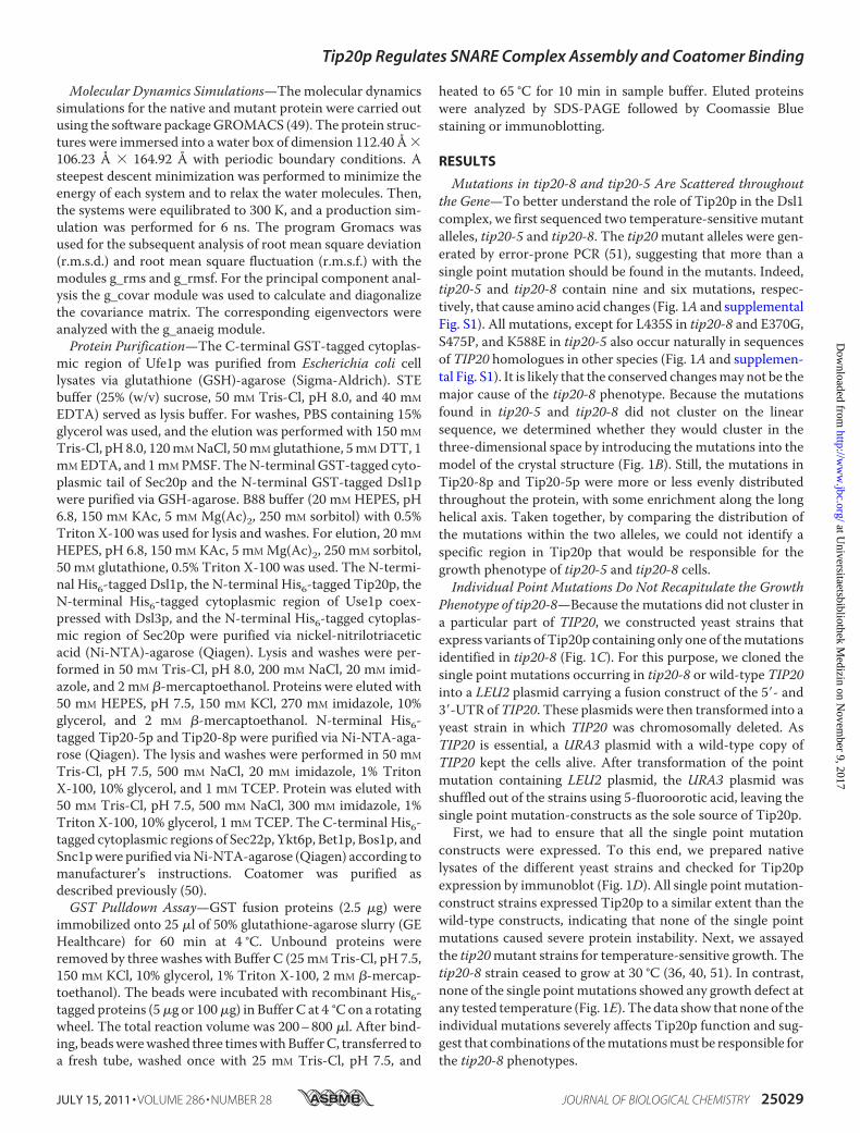
Mutations in tip20-8 and tip20-5 Are Scattered throughoutthe Gene—To better understand the role of Tip20p in the Dsl1complex, we first sequenced two temperature-sensitivemutantalleles, tip20-5 and tip20-8. The tip20mutant alleles were gen-erated by error-prone PCR (51), suggesting that more than asingle point mutation should be found in the mutants. Indeed,tip20-5 and tip20-8 contain nine and six mutations, respec-tively, that cause amino acid changes (Fig. 1A and supplementalFig. S1). All mutations, except for L435S in tip20-8 and E370G,S475P, and K588E in tip20-5 also occur naturally in sequencesof TIP20 homologues in other species (Fig. 1A and supplemen-tal Fig. S1). It is likely that the conserved changesmay not be themajor cause of the tip20-8 phenotype. Because the mutationsfound in tip20-5 and tip20-8 did not cluster on the linearsequence, we determined whether they would cluster in thethree-dimensional space by introducing the mutations into themodel of the crystal structure (Fig. 1B). Still, the mutations inTip20-8p and Tip20-5p were more or less evenly distributedthroughout the protein, with some enrichment along the longhelical axis. Taken together, by comparing the distribution ofthe mutations within the two alleles, we could not identify aspecific region in Tip20p that would be responsible for thegrowth phenotype of tip20-5 and tip20-8 cells.Individual Point Mutations Do Not Recapitulate the Growth
Phenotype of tip20-8—Because the mutations did not cluster ina particular part of TIP20, we constructed yeast strains thatexpress variants of Tip20p containing only one of themutationsidentified in tip20-8 (Fig. 1C). For this purpose, we cloned thesingle point mutations occurring in tip20-8 or wild-type TIP20into a LEU2 plasmid carrying a fusion construct of the 5�- and3�-UTR ofTIP20. These plasmids were then transformed into ayeast strain in which TIP20 was chromosomally deleted. AsTIP20 is essential, a URA3 plasmid with a wild-type copy ofTIP20 kept the cells alive. After transformation of the pointmutation containing LEU2 plasmid, the URA3 plasmid wasshuffled out of the strains using 5-fluoroorotic acid, leaving thesingle point mutation-constructs as the sole source of Tip20p.First, we had to ensure that all the single point mutation
constructs were expressed. To this end, we prepared nativelysates of the different yeast strains and checked for Tip20pexpression by immunoblot (Fig. 1D). All single point mutation-construct strains expressed Tip20p to a similar extent than thewild-type constructs, indicating that none of the single pointmutations caused severe protein instability. Next, we assayedthe tip20mutant strains for temperature-sensitive growth. Thetip20-8 strain ceased to grow at 30 °C (36, 40, 51). In contrast,none of the single pointmutations showed any growth defect atany tested temperature (Fig. 1E). The data show that none of theindividual mutations severely affects Tip20p function and sug-gest that combinations of themutationsmust be responsible forthe tip20-8 phenotypes.
Tip20p Regulates SNARE Complex Assembly and Coatomer Binding
JULY 15, 2011 • VOLUME 286 • NUMBER 28 JOURNAL OF BIOLOGICAL CHEMISTRY 25029
at Universitaetsbibliothek M
edizin on Novem
ber 9, 2017http://w
ww
.jbc.org/D
ownloaded from
Tip20p Regulates SNARE Complex Assembly and Coatomer Binding
25030 JOURNAL OF BIOLOGICAL CHEMISTRY VOLUME 286 • NUMBER 28 • JULY 15, 2011
at Universitaetsbibliothek M
edizin on Novem
ber 9, 2017http://w
ww
.jbc.org/D
ownloaded from
Membrane Association of Tip20p Is Not Altered in tip20Mutants—Tip20p acts as part of the Dsl1 complex at the ERmembrane (27, 34–39), and amore subtle effect, whichmaynotlead to a growth defect, could be a less efficient recruitment ofTip20p variants to the ER. Possible changes in the distributionof the protein could contribute to the perturbed protein func-tion in the tip20-8 strain. Therefore, we analyzed the mem-brane association of Tip20p variants by differential centrifuga-tion (Fig. 1F) and found that in comparison with the wild-typestrain neither the tip20-8 strain nor any of the single pointmutation strains showed any changes in the membrane associ-ation of Tip20p. In all of the cases, most of the protein waspresent in the pellet fraction after a 13,000� g spin (P13), whichcontains most of the ER as shown by the presence of the ERresident Sec61p protein. A smaller portion of Tip20p wasdetected in the S100 cytoplasmic pool. This finding is in agree-ment with Tip20p being a peripheral membrane protein. Themembrane association remained largely unchanged whenTip20, tip20-5, and tip20-8 strains were shifted to 37 °C for 1 hprior to the differential centrifugation (supplemental Fig. S2).More importantly, the level of Tip20p membrane associationwas indistinguishable between the three Tip20 variants. Ourdata indicate that protein mislocalization or reduced ER asso-ciation may not be the cause for the tip20-8 phenotype.Mutations Occurring in Tip20-8p Lead to Increased Flexibil-
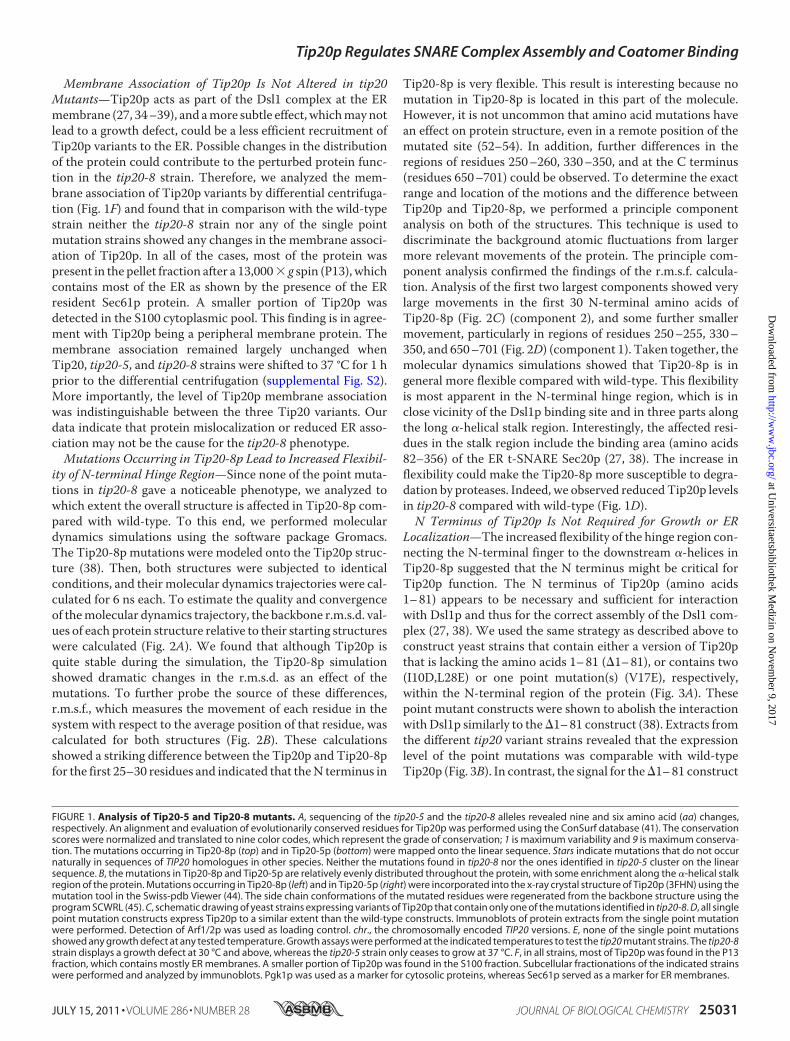
ity of N-terminal Hinge Region—Since none of the point muta-tions in tip20-8 gave a noticeable phenotype, we analyzed towhich extent the overall structure is affected in Tip20-8p com-pared with wild-type. To this end, we performed moleculardynamics simulations using the software package Gromacs.The Tip20-8p mutations were modeled onto the Tip20p struc-ture (38). Then, both structures were subjected to identicalconditions, and their molecular dynamics trajectories were cal-culated for 6 ns each. To estimate the quality and convergenceof themolecular dynamics trajectory, the backbone r.m.s.d. val-ues of each protein structure relative to their starting structureswere calculated (Fig. 2A). We found that although Tip20p isquite stable during the simulation, the Tip20-8p simulationshowed dramatic changes in the r.m.s.d. as an effect of themutations. To further probe the source of these differences,r.m.s.f., which measures the movement of each residue in thesystem with respect to the average position of that residue, wascalculated for both structures (Fig. 2B). These calculationsshowed a striking difference between the Tip20p and Tip20-8pfor the first 25–30 residues and indicated that theN terminus in
Tip20-8p is very flexible. This result is interesting because nomutation in Tip20-8p is located in this part of the molecule.However, it is not uncommon that amino acid mutations havean effect on protein structure, even in a remote position of themutated site (52–54). In addition, further differences in theregions of residues 250–260, 330–350, and at the C terminus(residues 650–701) could be observed. To determine the exactrange and location of the motions and the difference betweenTip20p and Tip20-8p, we performed a principle componentanalysis on both of the structures. This technique is used todiscriminate the background atomic fluctuations from largermore relevant movements of the protein. The principle com-ponent analysis confirmed the findings of the r.m.s.f. calcula-tion. Analysis of the first two largest components showed verylarge movements in the first 30 N-terminal amino acids ofTip20-8p (Fig. 2C) (component 2), and some further smallermovement, particularly in regions of residues 250–255, 330–350, and 650–701 (Fig. 2D) (component 1). Taken together, themolecular dynamics simulations showed that Tip20-8p is ingeneral more flexible compared with wild-type. This flexibilityis most apparent in the N-terminal hinge region, which is inclose vicinity of the Dsl1p binding site and in three parts alongthe long �-helical stalk region. Interestingly, the affected resi-dues in the stalk region include the binding area (amino acids82–356) of the ER t-SNARE Sec20p (27, 38). The increase inflexibility could make the Tip20-8p more susceptible to degra-dation by proteases. Indeed, we observed reducedTip20p levelsin tip20-8 compared with wild-type (Fig. 1D).N Terminus of Tip20p Is Not Required for Growth or ER
Localization—The increased flexibility of the hinge region con-necting the N-terminal finger to the downstream �-helices inTip20-8p suggested that the N terminus might be critical forTip20p function. The N terminus of Tip20p (amino acids1–81) appears to be necessary and sufficient for interactionwith Dsl1p and thus for the correct assembly of the Dsl1 com-plex (27, 38). We used the same strategy as described above toconstruct yeast strains that contain either a version of Tip20pthat is lacking the amino acids 1–81 (�1–81), or contains two(I10D,L28E) or one point mutation(s) (V17E), respectively,within the N-terminal region of the protein (Fig. 3A). Thesepoint mutant constructs were shown to abolish the interactionwithDsl1p similarly to the�1–81 construct (38). Extracts fromthe different tip20 variant strains revealed that the expressionlevel of the point mutations was comparable with wild-typeTip20p (Fig. 3B). In contrast, the signal for the�1–81 construct
FIGURE 1. Analysis of Tip20-5 and Tip20-8 mutants. A, sequencing of the tip20-5 and the tip20-8 alleles revealed nine and six amino acid (aa) changes,respectively. An alignment and evaluation of evolutionarily conserved residues for Tip20p was performed using the ConSurf database (41). The conservationscores were normalized and translated to nine color codes, which represent the grade of conservation; 1 is maximum variability and 9 is maximum conserva-tion. The mutations occurring in Tip20-8p (top) and in Tip20-5p (bottom) were mapped onto the linear sequence. Stars indicate mutations that do not occurnaturally in sequences of TIP20 homologues in other species. Neither the mutations found in tip20-8 nor the ones identified in tip20-5 cluster on the linearsequence. B, the mutations in Tip20-8p and Tip20-5p are relatively evenly distributed throughout the protein, with some enrichment along the �-helical stalkregion of the protein. Mutations occurring in Tip20-8p (left) and in Tip20-5p (right) were incorporated into the x-ray crystal structure of Tip20p (3FHN) using themutation tool in the Swiss-pdb Viewer (44). The side chain conformations of the mutated residues were regenerated from the backbone structure using theprogram SCWRL (45). C, schematic drawing of yeast strains expressing variants of Tip20p that contain only one of the mutations identified in tip20-8. D, all singlepoint mutation constructs express Tip20p to a similar extent than the wild-type constructs. Immunoblots of protein extracts from the single point mutationwere performed. Detection of Arf1/2p was used as loading control. chr., the chromosomally encoded TIP20 versions. E, none of the single point mutationsshowed any growth defect at any tested temperature. Growth assays were performed at the indicated temperatures to test the tip20 mutant strains. The tip20-8strain displays a growth defect at 30 °C and above, whereas the tip20-5 strain only ceases to grow at 37 °C. F, in all strains, most of Tip20p was found in the P13fraction, which contains mostly ER membranes. A smaller portion of Tip20p was found in the S100 fraction. Subcellular fractionations of the indicated strainswere performed and analyzed by immunoblots. Pgk1p was used as a marker for cytosolic proteins, whereas Sec61p served as a marker for ER membranes.
Tip20p Regulates SNARE Complex Assembly and Coatomer Binding
JULY 15, 2011 • VOLUME 286 • NUMBER 28 JOURNAL OF BIOLOGICAL CHEMISTRY 25031
at Universitaetsbibliothek M
edizin on Novem
ber 9, 2017http://w
ww
.jbc.org/D
ownloaded from
FIGURE 2. Tip20-8p is more flexible than wild-type Tip20p. A, although Tip20p (blue) behaves rather stably during the molecular dynamics simulation,Tip20-8p (red) as an effect of the mutations shows dramatic changes in the r.m.s.d.. The backbone r.m.s.d. values of each protein structure relative to theirstarting structures were calculated to estimate the quality and convergence of the molecular dynamics trajectory. B, a striking difference between the Tip20p(blue) and Tip20-8p (red) for the first 25–30 residues (indicated by an arrow) and further differences in the regions of residues 250 –260, 330 –350, and the Cterminus (residues 650 –701) (indicated by dashed arrows) could be detected. The sources of the observed differences in r.m.s.d. were determined by compu-tation of the r.m.s.f. Thereby, the movement of each residue in the system with respect to the average position of that residue was calculated for bothstructures. C, component 2 of the principal component analysis reflects the very large movements in the first 30 N-terminal amino acids. The maximal range aswell as intermediate states of the movements for the wild-type Tip20p (left, blue) and the Tip20-8p (middle, red) is shown. On the right, a superposition (Tip20pin blue, Tip20-8p in red) is displayed. D, component 1 of the principle component analysis mirrors the observed fluctuations in the regions of the long �-helicalstalk. A superposition of the maximal range as well as intermediate states of the movements for wild-type Tip20p (blue) and Tip20-8p (red) is shown. The boxesrepresent an enlargement of the regions (amino acids 250 –255, 330 –350, and 650 –701) that displayed the biggest amplitude in movement.
Tip20p Regulates SNARE Complex Assembly and Coatomer Binding
25032 JOURNAL OF BIOLOGICAL CHEMISTRY VOLUME 286 • NUMBER 28 • JULY 15, 2011
at Universitaetsbibliothek M
edizin on Novem
ber 9, 2017http://w
ww
.jbc.org/D
ownloaded from
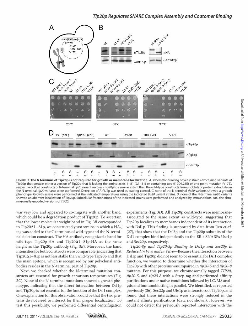
was very low and appeared to co-migrate with another band,which could be a degradation product of Tip20p. To ascertainthat the lower molecular weight band in Fig. 3B correspondedto Tip20�1–81p, we constructed yeast strains in which a HA3tag was added to the C terminus of wild-type and the N-termi-nal deletion construct. The HA antibody recognized a band forwild-type Tip20p-HA and Tip20�1–81p-HA at the sameheight as the Tip20p antibody (Fig. 3B). Moreover, the bandintensities for both constructswere comparable, indicating thatTip20�1–81p is not less stable than wild-type Tip20p and thatthe main epitope, which is recognized by our polyclonal anti-bodies resides in the N-terminal part of Tip20p.Next, we checked whether the N-terminal mutation con-
structs are essential for growth at various temperatures (Fig.3C). None of the N-terminal mutations showed a growth phe-notype, indicating that the direct interaction between Dsl1pandTip20p is not essential for the function of theDsl1 complex.One explanation for this observation could be that the two pro-teins do not need to interact for their proper localization. Totest this possibility, we performed differential centrifugation
experiments (Fig. 3D). All Tip20p constructs were membrane-associated to the same extent as wild-type, suggesting thatTip20p localizes to membranes independent of its interactionwith Dsl1p. This finding is supported by data from Ren et al.(27), that show that the Dsl3p and the Tip20p subunits of theDsl1 complex bind independently to the ER t-SNAREs Use1pand Sec20p, respectively.Tip20-8p and Tip20-5p Binding to Dsl1p and Sec20p Is
Reduced inVivo and inVitro—Because the interaction betweenDsl1p andTip20p did not seem to be essential for Dsl1 complexfunction, we wanted to determine whether the interaction ofTip20pwith other proteins was impaired in tip20-5 and tip20-8mutants. For this purpose, we chromosomally tagged TIP20,tip20-5, and tip20-8 with a Strep-tag and performed affinitypurifications under native conditions followed by LC/MS anal-ysis and immunoblotting in parallel. We identified, as reportedpreviously (36), Sec22p andUfe1p as interactors of Tip20p, andfound that these interactions were strongly reduced in themutant affinity purifications (data not shown). However, wecould not detect the previously reported interaction with the
FIGURE 3. The N terminus of Tip20p is not required for growth or membrane localization. A, schematic drawing of yeast strains expressing variants ofTip20p that contain either a version of Tip20p that is lacking the amino acids 1– 81 (�1– 81) or containing two (I10D,L28E) or one point mutation (V17E),respectively. B, all constructs of N-terminal tip20 variants express Tip20p to a similar extent than the wild-type constructs. Immunoblots of protein extracts fromthe N-terminal tip20 variants were performed. Detection of Arf1/2p was used as loading control. C, none of the N-terminal tip20 variants showed a growthphenotype. Growth assays were performed at the indicated temperatures using the indicated tip20 variant strains. D, none of the N-terminal tip20 variantsshowed an aberrant localization of Tip20p. Subcellular fractionations of the indicated strains were performed and analyzed by immunoblots. chr., the chro-mosomally encoded versions of TIP20.
Tip20p Regulates SNARE Complex Assembly and Coatomer Binding
JULY 15, 2011 • VOLUME 286 • NUMBER 28 JOURNAL OF BIOLOGICAL CHEMISTRY 25033
at Universitaetsbibliothek M
edizin on Novem
ber 9, 2017http://w
ww
.jbc.org/D
ownloaded from
ER t-SNARE Sec20p (36, 38, 55). To overcome this shortcom-ing, we performed purifications under denaturing conditionsand included a cross-linking step using chromosomally taggedSEC20 with a His6-biotinylation sequence-His6 (HBH) tag inTip20p wild-type and mutant strains. We found that lessTip20-5p and Tip20-8p were associated with Sec20p, whencompared with wild-type (data not shown). These results areconsistent with the wide distribution of the mutations over theentire sequence of TIP20 in the mutants and an increase offlexibility in various parts of at least Tip20-8p. We conclude
that the interaction with all known Tip20p binding proteinswas strongly reduced in both Tip20-5p and Tip20-8p in vivo.To confirm our results, we performed in vitro pulldown
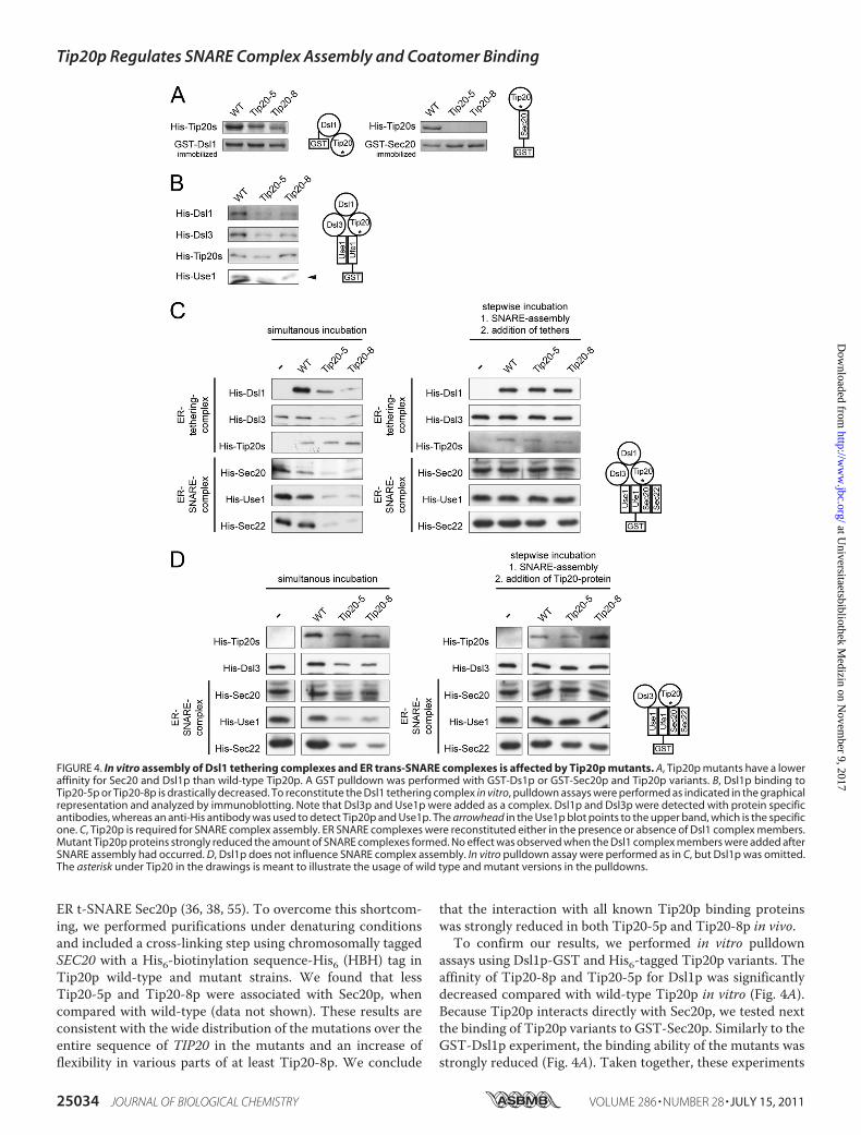
assays using Dsl1p-GST and His6-tagged Tip20p variants. Theaffinity of Tip20-8p and Tip20-5p for Dsl1p was significantlydecreased compared with wild-type Tip20p in vitro (Fig. 4A).Because Tip20p interacts directly with Sec20p, we tested nextthe binding of Tip20p variants to GST-Sec20p. Similarly to theGST-Dsl1p experiment, the binding ability of the mutants wasstrongly reduced (Fig. 4A). Taken together, these experiments
FIGURE 4. In vitro assembly of Dsl1 tethering complexes and ER trans-SNARE complexes is affected by Tip20p mutants. A, Tip20p mutants have a loweraffinity for Sec20 and Dsl1p than wild-type Tip20p. A GST pulldown was performed with GST-Ds1p or GST-Sec20p and Tip20p variants. B, Dsl1p binding toTip20-5p or Tip20-8p is drastically decreased. To reconstitute the Dsl1 tethering complex in vitro, pulldown assays were performed as indicated in the graphicalrepresentation and analyzed by immunoblotting. Note that Dsl3p and Use1p were added as a complex. Dsl1p and Dsl3p were detected with protein specificantibodies, whereas an anti-His antibody was used to detect Tip20p and Use1p. The arrowhead in the Use1p blot points to the upper band, which is the specificone. C, Tip20p is required for SNARE complex assembly. ER SNARE complexes were reconstituted either in the presence or absence of Dsl1 complex members.Mutant Tip20p proteins strongly reduced the amount of SNARE complexes formed. No effect was observed when the Dsl1 complex members were added afterSNARE assembly had occurred. D, Dsl1p does not influence SNARE complex assembly. In vitro pulldown assay were performed as in C, but Dsl1p was omitted.The asterisk under Tip20 in the drawings is meant to illustrate the usage of wild type and mutant versions in the pulldowns.
Tip20p Regulates SNARE Complex Assembly and Coatomer Binding
25034 JOURNAL OF BIOLOGICAL CHEMISTRY VOLUME 286 • NUMBER 28 • JULY 15, 2011
at Universitaetsbibliothek M
edizin on Novem
ber 9, 2017http://w
ww
.jbc.org/D
ownloaded from
demonstrate a reduced binding capacity of Tip20pmutant pro-teins to Sec20p andDsl1p in vivo and in vitro and are consistentwith previously published data (36).Tip20-8p and Tip20-5p Block Dsl1 Complex Assembly in
Vitro—Because the Tip20p-Dsl1p interaction was reduced inthe presence of Tip20pmutant proteins, we asked nextwhetherthe mutants would generally affect Dsl1 complex assembly.Although it had been shown previously that Dsl1p interactsdirectly with Dsl3p (27), our attempts to pull down His6-Dsl3pwith immobilized GST-Dsl1p failed, probably due to nonfunc-tional Dsl3p. Dsl3p directly interacts with the ER t-SNAREUse1p (27, 36), and Use1p could be only efficiently purifiedwhen coexpressed with Dsl3p (27). Thus, we used as a source ofDsl3p the complex ofHis6-Use1p/Dsl3p. Given this slight com-plication, we decided to build up the Dsl1 complex from theSNARE site and used a GST fusion to the ER t-SNARE, Ufe1p,(32), which has been shown to interact with bothUse1p and theDsl1 complex (30, 31, 36) and performed GST pulldowns (Fig.4B). Interestingly, Tip20-5p and Tip20-8p bound toUfe1pwithsimilar efficiencies than wild-type, indicating that Tip20p canbind directly to Ufe1p (supplemental Fig. S3), independent ofSec20p, and that the mutations do not destabilize this interac-tion, whereas Dsl1p bindingwas strongly impaired. The affinityof Tip20p to Ufe1p appears to be relatively low (supplementalFig. S3) and may be stabilized by the interaction with otherproteins in vivo. Because Dsl3p is expressed in a complex withUse1p, andUse1p can bind directly toUfe1p, we refrained fromdrawing any conclusions about Dsl3p directly. However, Dsl3pwas unable to efficiently immobilize Dsl1p, indicating thateither this interaction is too weak to stably recruit Dsl1p or thatthe Dsl3p-Use1p complex was also recruited inefficiently toUfe1p-GST. The interaction of the Tip20p mutant proteinswith Ufe1p explains why Tip20p mutants are still ER localized,despite the loss of interaction with Sec20p. Moreover, thesedata provide strong evidence that the Dsl1 complex is destabi-lized in tip20-5 and tip20-8mutants, probably also in vivo.Tip20pMutants Inhibit Trans-SNARE Complex Assembly in
Vitro—Formation of a SNARE complex consisting of the ER-localized SNAREs Sec20p, Ufe1p, and Use1p with thev-SNAREs Sec22p and/or Bet1p is necessary for the fusion ofCOPI vesicles with the ER (24, 30–33). Moreover, a recentstudy suggests that the Dsl1 complex accelerates SNARE com-plex assembly at the ER (27). Because Tip20-5p and Tip20-8pfailed to bind to Sec20p, but not to Ufe1p, we asked whetherTip20-8p and Tip20-5p would influence ER SNARE complexassembly. To this end, GST-Ufe1p was immobilized and incu-bated with the remaining ER t-SNAREs, the v-SNARE Sec22pand Dsl1 complex members (Fig. 4C). As expected, SNAREcomplex assembly occurred in the presence of wild-typeTip20p. In contrast, in the presence of the Tip20p mutant pro-teins, ER SNARE complex assembly was severely perturbed,and none of the other SNARE proteins was efficiently incorpo-rated into the SNARE complex. Tip20p must have a regulatoryfunction during SNARE complex assembly at the ER becauseSNARE complexes containing Sec20p, Use1p, and Sec22pwereformed properly, when first SNARE complexes were formed,and then Dsl1 complex members were added in a second incu-bation step (Fig. 4C). Dsl1p was recruited efficiently to these
SNARE complexes. However, this binding was most likelydependent on the Dsl3p-Use1p complex, which was presentduring the SNARE complex assembly step. These data are con-sistent with the observation that ER SNARE complex assemblyis accelerated in vitro by the presence of the Dsl1 complex (27).Taken together, our data indicate that Tip20p is required forproper SNARE complex assembly.Tip20p and Not Dsl1p Is Required for Proper SNARE Com-
plex Assembly—So far, the defect in SNARE complex assemblycould be due to the reduced interaction of Tip20-5p andTip20-8p with Dsl1p. Alternatively, the phenotype could becompletely independent of Dsl1p. To distinguish betweenthose possibilities, we repeated the above experiment in theabsence of Dsl1p (Fig. 4D). Again, SNARE complexes assembledin the absence of Dsl1p when wild-type Tip20p was present, andthis assembly was reducedwhenmutant Tip20pwas added to theincubation mixture. Interestingly, the reduction in SNARE com-plex assembly was independent of the presence of Dsl1p in theassay.Therefore,weconclude thatTip20pmayhaveamoreprom-inent role in SNARE complex assembly than Dsl1p.ER SNARE Complex Assembly Is Not Rescued by Alternative
v-SNAREs in tip20 Mutants—Sec22p is not the only v-SNAREthat could potentially engage in a trans-SNARE complex at theER. Bet1p and Ykt6p have been shown to be substitutes forSec22p (18, 33). In addition, it has been shown that SNARE-SNARE interactions under some circumstances are promiscu-ous and that the formations of nonphysiological SNARE com-plexes can take place (20, 21, 56, 57). Therefore, we testedwhether in the presence of Tip20-5p and Tip20-8p, ER SNAREcomplexeswould becomemore promiscuous. First, we decided tolook at Ykt6p, which seems to be able to substitute for v-SNAREsin more than one type of SNARE complexes (18, 24, 58). Ykt6pinteractedwithUfe1p equallywell, independent of the presence ofwild-type or amutant form of Tip20p in the assay (Fig. 5A). How-ever, this interaction did not improve the recruitment of Use1p orSec20p into the complex. Therefore, binding of Ykt6p to Ufe1p isindependentof theotherER t-SNAREs.AddingYkt6pandSec22psimultaneously did not improve the SNARE complex assembly atthe ER. Because neither Sec20p nor Use1p were efficientlyrecruited to Ufe1p in the presence of Ykt6p, Ykt6p does not driveSNARE assembly and cannot overcome the defects introduced bythe Tip20pmutant proteins.Next, we tested whether Bet1p was efficiently recruited into
SNARE complexes. Bet1p behaved similarly to Sec22p and wasonly incorporated into SNARE complexes in the presence ofwild-type Tip20p (Fig. 5B). Tip20p mutants caused a strongreduction of Bet1p binding, and less SNARE complexes wereformed. The effects for the ER v-SNAREs were specific becausenoncognate SNAREs as the v-SNAREat theGolgi, Bos1p, or theplasmamembrane v-SNARE Snc1p could not be recruited at allto engage into SNARE complex formation (Fig. 5B). Our dataindicate that in the presence of Tip20p mutant proteins,SNARE complex assembly at ER is severely altered but properrecognition of cognate v-SNAREs is not affected, as noncognatev-SNAREswere not recruited into ER SNARE complexes underany conditions tested in these assays.Tip20p Regulates Binding of Dsl1p to Coatomer—So far, we
have shown that Tip20pmutant proteins disturb the formation
Tip20p Regulates SNARE Complex Assembly and Coatomer Binding
JULY 15, 2011 • VOLUME 286 • NUMBER 28 JOURNAL OF BIOLOGICAL CHEMISTRY 25035
at Universitaetsbibliothek M
edizin on Novem
ber 9, 2017http://w
ww
.jbc.org/D
ownloaded from
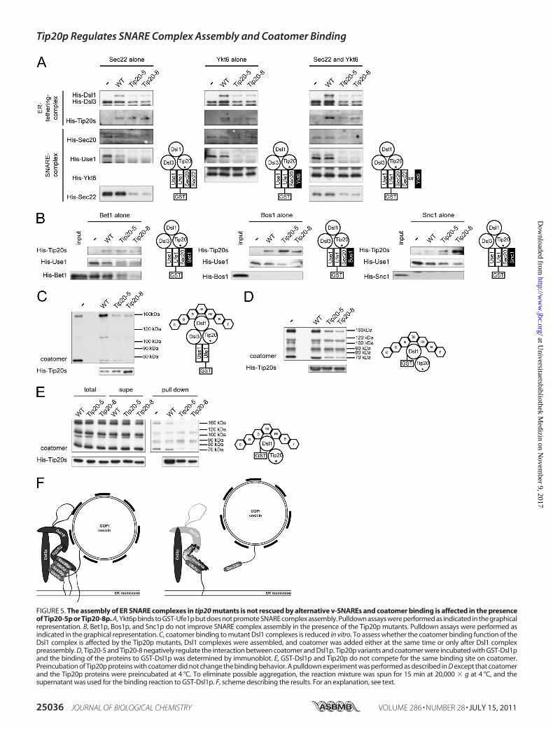
FIGURE 5. The assembly of ER SNARE complexes in tip20 mutants is not rescued by alternative v-SNAREs and coatomer binding is affected in the presenceof Tip20-5p or Tip20-8p. A, Ykt6p binds to GST-Ufe1p but does not promote SNARE complex assembly. Pulldown assays were performed as indicated in the graphicalrepresentation. B, Bet1p, Bos1p, and Snc1p do not improve SNARE complex assembly in the presence of the Tip20p mutants. Pulldown assays were performed asindicated in the graphical representation. C, coatomer binding to mutant Dsl1 complexes is reduced in vitro. To assess whether the coatomer binding function of theDsl1 complex is affected by the Tip20p mutants, Dsl1 complexes were assembled, and coatomer was added either at the same time or only after Dsl1 complexpreassembly. D, Tip20-5 and Tip20-8 negatively regulate the interaction between coatomer and Dsl1p. Tip20p variants and coatomer were incubated with GST-Dsl1pand the binding of the proteins to GST-Dsl1p was determined by immunoblot. E, GST-Dsl1p and Tip20p do not compete for the same binding site on coatomer.Preincubation of Tip20p proteins with coatomer did not change the binding behavior. A pulldown experiment was performed as described in D except that coatomerand the Tip20p proteins were preincubated at 4 °C. To eliminate possible aggregation, the reaction mixture was spun for 15 min at 20,000 � g at 4 °C, and thesupernatant was used for the binding reaction to GST-Dsl1p. F, scheme describing the results. For an explanation, see text.
Tip20p Regulates SNARE Complex Assembly and Coatomer Binding
25036 JOURNAL OF BIOLOGICAL CHEMISTRY VOLUME 286 • NUMBER 28 • JULY 15, 2011
at Universitaetsbibliothek M
edizin on Novem
ber 9, 2017http://w
ww
.jbc.org/D
ownloaded from
of ER SNARE complexes. We next wanted to test whether theupstream function the Dsl1 complex, namely the tethering ofCOPI vesicles through direct interaction with Dsl1p is also per-turbed by Tip20p mutant proteins (27, 34–38). First, we per-formed a pulldown with GST-Ufe1p and the members of theDsl1 complex and coatomer. Indeed, less coatomer was precip-itated in the Tip20p mutant incubations compared with wild-type (Fig. 5C). This result could be explained by either lessDsl1p binding to mutant Tip20p and hence less coatomerrecruitment or a more direct role of Tip20p in coatomer bind-ing. To distinguish between these two possibilities, we per-formed a pulldown with GST-Dsl1p, coatomer, and Tip20pvariants. Interestingly, the presence of Tip20pmutants reducedthe amount of immobilized coaotmer, suggesting that Tip20pmayplay amore active role inDsl1pbinding to coatomer by eitherstabilizing the interactionbetweenDsl1p and coatomer or provid-ing an additional coatomer binding site (Fig. 5D). Moreover, pre-incubation of Tip20p with coatomer did not change the bindingefficiency of Tip20p or coatomer to Dsl1p, indicating that Tip20pand Dsl1p are not competing for the same binding site (Fig. 5E).Taken together, our results suggest a prominent role forTip20p inthe recognition of incoming COPI vesicles and in the assembly oftrans-SNARE complexes at the ER.
DISCUSSION
In this paper, we investigated the function of the Dsl1 complexmemberTip20pbyanalyzing thephenotypesof tip20mutantsandfound that they interfered with the proper assembly of trans-SNARE complexes at the ER (Fig. 5F). Trans-SNARE complexesconsisting of the t-SNAREs Sec20p, Ufe1p, and Use1p and thev-SNARE Sec22p and/or Bet1p promote fusion of Golgi-derivedCOPI-coated vesicles with the ER (24, 30–32). Ufe1p, Sec20p,Use1p, and Sec22p/Bet1p could not be efficiently assembled intoER SNARE complexes in the presence of mutant Tip20p in vitro.Thisdefectwasnotcompensated forby the thirdvesicleSNAREofthe ER-Golgi shuttle, Bos1p, which was not incorporated into theERSNAREcomplexunderanyconditions tested.WhereasSec22pand Bet1p can participate in SNARE complex formation at boththe ER and the Golgi (18, 33, 59), Bos1p appears only to act in thefusion process of COPII vesicles at the Golgi (18, 24, 33, 59, 60).The noncognate v-SNARE Snc1p, which acts at the plasmamem-brane, could also not be engaged in SNARE complex formation.Interestingly, Ykt6p, another v-SNARE, which can functionallyreplace Sec22p in the fusionof ER-derivedCOPII vesicleswith theGolgi (18) binds efficiently toUfe1p, irrespectiveof thepresenceofwild-type or Tip20 mutants, and even when added together withSec22p. Yet, the presence of Ykt6p did not improve the incorpo-ration of Sec20p, Use1p, or Sec22p in vitro, and hence, no func-tional SNARE complexes were formed. Our in vitro data suggestthatTip20-5pandTip20-8pcould act asdominantnegative inhib-itors of SNARE complex assembly. However, we cannot excludethat in vivo, when e.g. other factors like the Sec1/Munc18 proteinSly1p are present, Ykt6p could potentially assist in the incorpora-tion of cognate SNAREs in the trans-SNARE complex at the ER.Our data are consistent with findings byMeiringer et al. (61), thatYkt6p can bind to the ER t-SNARE-Dsl1 complex but that it doesnot act as v-SNARE in this scenario. We propose that Tip20pmutant proteins do not change the specificity for cognate SNARE
complex assembly but rather prevent trans-SNARE complexassembly. Consistentwith this notion,mutantTip20p already dis-turbed the interaction of the ER t-SNAREswith each other. How-ever,whenSNAREcomplexeswere assembled in vitroprior to theaddition of Tip20p variants, no effect on SNARE complex assem-bly was observed, indicating that Tip20p mutant proteins mayhave adecreasedoff-rate fromtheSNAREs.Aplausible scenario isthat theDsl1 complexmay associate with ER t-SNAREs and handover the COPI vesicle to the ER SNAREs, which may be in a par-tially preassembled state. Sec22p interacts with the ER t-SNAREsand forms a trans-SNARE complex and allows fusion. In the pres-ence of Tip20p mutant proteins, less COPI vesicles would bebrought into close proximity to the ER because, although themutantTip20pare still ER-associated, thebinding toDsl1p,whichinteracts with coatomer, is reduced (27, 34–39). Tip20p seems toplay also a regulatory role in the binding ofDsl1p to coatomer, thelevels of which were drastically reduced in the presence of Tip20pmutant proteins. Alternatively, Tip20p could possess a crypticcoatomer-binding site, which would become more important inthe mutant situation, and the mutant proteins would displacecoatomer fromDsl1p. Such a cryptic interaction site is thought tobe located in the N terminus of Tip20p of Schizosaccharomycesspecies (62). Thus, in principle, it is conceivable that the N-termi-nal region, which contains the Dsl1p binding site (27, 38) andwhichbecomes very flexible inTip20-8p, could adopt a conforma-tion that is unable to recognize Dsl1p and becomes instead acoatomer-binding site. Yet, SNARE complex formationmight notonly be reduced because of the lower amount of COPI vesicles attheERmembranebut alsobecauseTip20pmutantproteinswouldblockefficientERSNAREassemblyby interactingwithUfe1p.Thefinding that defective Dsl1 complexes interfere with a properassembly of cognateER trans-SNAREcomplexes and theobserva-tion that theDsl1 complex accelerates SNAREcomplex formation(27) provide evidence for a novel function of the Dsl1 complex,namely a role in actively contributing to SNARE complex assem-bly. Such a function has been suggested before for other tetheringcomplexes. Uso1p, an essential tethering factor at the Golgi inyeast, is required for the assemblyof the v-SNARE-t-SNAREcom-plexes (63). In mammalian cells, defects in the function of theintra-Golgi-tethering complex, the conserved oligomeric Golgi(COG) complex, led to a significant decrease in Golgi SNAREmobility and in the steady-state level of intra-Golgi SNARE com-plexes, accompanied by an accumulation of uncomplexed syn-taxin 5 (64). Furthermore, the trans-Golgi located mammaliantethering complex GARP interacted directly with SNAREs thatparticipate in the endosome-to-trans-Golgi network (TGN) retro-grade route. Further functional analyses placed the Golgi-associ-ated retrograde protein (GARP) complex upstream of theSNAREs, regulating their localization and assembly into SNAREcomplexes (26).Finally, theHOPScomplex, the tetheringcomplexpresent at the yeast vacuole, proofreads SNARE domain andN-terminal domain structures of vacuolar SNAREs and regulatesthe fusion capacity of trans-SNARE complexes, only allowing fullfunction for wild-type SNARE configurations (28).How mutant Tip20-5p and Tip20-8p prevent assembly of the
SNARE complexes remains unclear. At least Tip20-8p is moreflexible than wild-type Tip20p, and this flexibility may renderTip20p more susceptible to degradation. Indeed, Tip20-8 levels
Tip20p Regulates SNARE Complex Assembly and Coatomer Binding
JULY 15, 2011 • VOLUME 286 • NUMBER 28 JOURNAL OF BIOLOGICAL CHEMISTRY 25037
at Universitaetsbibliothek M
edizin on Novem
ber 9, 2017http://w
ww
.jbc.org/D
ownloaded from
are slightly reduced in yeast, and recombinant Tip20-8p hasmoredegradation products than wild-type. More importantly, parts ofthe helical stalk region become more flexible, and these regionscomprise the binding site of Sec20p. The binding to Sec20p seemstobeof regulatory importancebecauseTip20-8pandTip20-5parestill efficiently recruited tomembranes, albeit the interactionwithSec20p is severely reduced. Although we find direct binding ofTip20p to Ufe1p in vitro, the interaction of Tip20p with Sec20pmight be themore important one in vivo and could potentially bethe driving force for SNARE complex assembly. A plausible sce-nario is that the arrival of a vesicle is signaled via the Tip20p-Sec20p interaction, leading to an efficient recruitment of cognateSNAREs. The role of other players, like Sec1/Munc18 proteins,known to have a role in orchestrating and stabilizing fusion eventsat the ER, needs to be further examined in this context.
Acknowledgments—We are grateful to M. Spiess, F. Hughson, H. D.Schmitt, C. Ungermann, D. Banfield, J. Gerst, R.-W. Peng, P. Cosson,and R. Schekman for reagents. We thank C. Ungermann for sharingresults prior to publication. Members of the Spang laboratory areacknowledged for discussions and critical comments.
REFERENCES1. Bonifacino, J. S., and Glick, B. S. (2004) Cell 116, 153–1662. Lee, M. C., Miller, E. A., Goldberg, J., Orci, L., and Schekman, R. (2004)
Annu. Rev. Cell Dev. Biol. 20, 87–1233. Springer, S., Spang, A., and Schekman, R. (1999) Cell 97, 145–1484. Barlowe,C.,Orci, L., Yeung, T.,Hosobuchi,M.,Hamamoto, S., Salama,N.,
Rexach, M. F., Ravazzola, M., Amherdt, M., and Schekman, R. (1994) Cell77, 895–907
5. Letourneur, F., Gaynor, E. C., Hennecke, S., Demolliere, C., Duden, R.,Emr, S. D., Riezman, H., and Cosson, P. (1994) Cell 79, 1199–1207
6. Waters, M. G., Serafini, T., and Rothman, J. E. (1991) Nature 349,248–251
7. Serafini, T., Orci, L., Amherdt, M., Brunner, M., Kahn, R. A., and Roth-man, J. E. (1991) Cell 67, 239–253
8. Yang, J. S., Lee, S. Y., Gao, M., Bourgoin, S., Randazzo, P. A., Premont,R. T., and Hsu, V. W. (2002) Journal of Cell Biology 159, 69–78
9. Antonny, B., Madden, D., Hamamoto, S., Orci, L., and Schekman, R.(2001) Nat. Cell Biol. 3, 531–537
10. Spang, A. (2009) Curr. Opin. Cell Biol. 21, 531–53611. Tanigawa, G., Orci, L., Amherdt, M., Ravazzola, M., Helms, J. B., and
Rothman, J. E. (1993) J. Cell Biol. 123, 1365–137112. Fasshauer, D., Bruns, D., Shen, B., Jahn, R., and Brunger, A. T. (1997)
J. Biol. Chem. 272, 4582–459013. Fiebig, K.M., Rice, L.M., Pollock, E., and Brunger, A. T. (1999)Nat. Struct.
Biol. 6, 117–12314. Poirier, M. A., Xiao,W.,Macosko, J. C., Chan, C., Shin, Y. K., and Bennett,
M. K. (1998) Nat. Struct. Biol. 5, 765–76915. Sutton, R. B., Fasshauer,D., Jahn, R., andBrunger, A. T. (1998)Nature395,
347–35316. Borisovska,M., Zhao, Y., Tsytsyura, Y., Glyvuk,N., Takamori, S.,Matti, U.,
Rettig, J., Sudhof, T., and Bruns, D. (2005) EMBO J. 24, 2114–212617. Gotte, M., and Gallwitz, D. (1997) FEBS Lett. 411, 48–5218. Liu, Y., and Barlowe, C. (2002)Mol. Biol. Cell 13, 3314–332419. Pelham, H. R. (2001) Trends Cell Biol. 11, 99–10120. Tsui, M. M., and Banfield, D. K. (2000) J. Cell Sci. 113, 145–15221. Fasshauer, D., Antonin, W., Margittai, M., Pabst, S., and Jahn, R. (1999)
J. Biol. Chem. 274, 15440–1544622. Cai, H., Reinisch, K., and Ferro-Novick, S. (2007) Dev. Cell 12, 671–68223. Carr, C. M., and Rizo, J. (2010) Curr. Opin. Cell Biol. 22, 488–49524. Jahn, R., and Scheller, R. H. (2006) Nat. Rev. Mol. Cell Biol. 7, 631–64325. Markgraf, D. F., Peplowska, K., and Ungermann, C. (2007) FEBS Lett. 581,
2125–213026. Perez-Victoria, F. J., and Bonifacino, J. S. (2009) Mol. Cell Biol. 29,
5251–526327. Ren, Y., Yip, C. K., Tripathi, A., Huie, D., Jeffrey, P. D., Walz, T., and
Hughson, F. M. (2009) Cell 139, 1119–112928. Starai, V. J., Hickey, C. M., and Wickner, W. (2008) Mol. Biol. Cell 19,
2500–250829. Ungermann, C., and Langosch, D. (2005) J. Cell Sci. 118, 3819–382830. Burri, L., Varlamov, O., Doege, C. A., Hofmann, K., Beilharz, T., Rothman,
J. E., Sollner, T. H., and Lithgow, T. (2003) Proc. Natl. Acad. Sci. U.S.A.100, 9873–9877
31. Dilcher,M., Veith, B., Chidambaram, S., Hartmann, E., Schmitt, H. D., andFischer von Mollard, G. (2003) EMBO J. 22, 3664–3674
32. Lewis, M. J., Rayner, J. C., and Pelham, H. R. (1997) EMBO J. 16,3017–3024
33. Spang, A., and Schekman, R. (1998) J. Cell Biol. 143, 589–59934. Andag, U., Neumann, T., and Schmitt, H. D. (2001) J. Biol. Chem. 276,
39150–3916035. Andag, U., and Schmitt, H. D. (2003) J. Biol. Chem. 278, 51722–5173436. Kraynack, B. A., Chan, A., Rosenthal, E., Essid, M., Umansky, B., Waters,
M. G., and Schmitt, H. D. (2005)Mol. Biol. Cell 16, 3963–397737. Reilly, B. A., Kraynack, B. A., VanRheenen, S.M., andWaters,M.G. (2001)
Mol. Biol. Cell 12, 3783–379638. Tripathi, A., Ren, Y., Jeffrey, P. D., and Hughson, F. M. (2009)Nat. Struct.
Mol. Biol. 16, 114–12339. Zink, S., Wenzel, D., Wurm, C. A., and Schmitt, H. D. (2009)Dev. Cell 17,
403–41640. Kamena, F., and Spang, A. (2004) Science 304, 286–28941. Goldenberg,O., Erez, E., Nimrod,G., andBen-Tal, N. (2009)Nucleic Acids
Res. 37, D323–32742. Pupko, T., Bell, R. E., Mayrose, I., Glaser, F., and Ben-Tal, N. (2002) Bioin-
formatics 18, S71–7743. Fiser, A., and Sali, A. (2003) Bioinformatics 19, 2500–250144. Guex, N., and Peitsch, M. C. (1997) Electrophoresis 18, 2714–272345. Canutescu, A. A., Shelenkov, A. A., and Dunbrack, R. L., Jr. (2003) Protein
Sci. 12, 2001–201446. Sambrook, J., Fritsch, E., and Maniatis, T. (1989) Molecular Cloning: A
Laboratory Manual, Cold Spring Harbor Laboratory Press, New York47. Sherman, F. (1991)Methods Enzymol. 194, 3–2148. Rexach, M. F., Latterich, M., and Schekman, R.W. (1994) J. Cell Biol. 126,
1133–114849. Van Der Spoel, D., Lindahl, E., Hess, B., Groenhof, G., Mark, A. E., and
Berendsen, H. J. (2005) J. Comput. Chem. 26, 1701–171850. Hosobuchi, M., Kreis, T., and Schekman, R. (1992) Nature 360, 603–60551. Cosson, P., Schroder-Kohne, S., Sweet, D. S., Demolliere, C., Hennecke, S.,
Frigerio, G., and Letourneur, F. (1997) Eur J. Cell Biol. 73, 93–9752. Brzovic, P. S., Sawa, Y., Hyde, C. C., Miles, E. W., and Dunn, M. F. (1992)
J. Biol. Chem. 267, 13028–1303853. Galiano, L., Ding, F., Veloro, A. M., Blackburn, M. E., Simmerling, C., and
Fanucci, G. E. (2009) J. Am. Chem. Soc. 131, 430–43154. Ingram, V. M. (1956) Nature 178, 792–79455. Sweet, D. J., and Pelham, H. R. (1993) EMBO J. 12, 2831–284056. Wendler, F., and Tooze, S. (2001) Traffic 2, 606–61157. Yang, B., Gonzalez, L., Jr., Prekeris, R., Steegmaier, M., Advani, R. J., and
Scheller, R. H. (1999) J. Biol. Chem. 274, 5649–565358. Fischer von Mollard, G., and Stevens, T. H. (1999) Mol. Biol. Cell 10,
1719–173259. Cao, X., and Barlowe, C. (2000) J. Cell Biol. 149, 55–6660. Parlati, F., McNew, J. A., Fukuda, R., Miller, R., Sollner, T. H., and Roth-
man, J. E. (2000) Nature 407, 194–19861. Meiringer, C., Rethmeier, R., Auffarth, K.,Wilson, J., Perz, A., Barlowe, C.,
Schmitt, H. D., and Ungermann, C. (2011) J. Biol. Chem. 286,25039–25046
62. Schmitt, H. D. (2010) Trends Cell Biol. 20, 257–26863. Sapperstein, S. K., Lupashin, V. V., Schmitt, H. D., and Waters, M. G.
(1996) J. Cell Biol. 132, 755–76764. Shestakova, A., Suvorova, E., Pavliv, O., Khaidakova, G., and Lupashin, V.
(2007) J. Cell Biol. 179, 1179–1192
Tip20p Regulates SNARE Complex Assembly and Coatomer Binding
25038 JOURNAL OF BIOLOGICAL CHEMISTRY VOLUME 286 • NUMBER 28 • JULY 15, 2011
at Universitaetsbibliothek M
edizin on Novem
ber 9, 2017http://w
ww
.jbc.org/D
ownloaded from
Melanie Diefenbacher, Holmfridur Thorsteinsdottir and Anne SpangSaccharomyces cerevisiaeComplex Assembly at the Endoplasmic Reticulum in
-Ethylmaleimide-sensitive Factor) Attachment Protein Receptor (SNARE)NThe Dsl1 Tethering Complex Actively Participates in Soluble NSF (
doi: 10.1074/jbc.M110.215657 originally published online April 11, 20112011, 286:25027-25038.J. Biol. Chem.
10.1074/jbc.M110.215657Access the most updated version of this article at doi:
Alerts:
When a correction for this article is posted•
When this article is cited•
to choose from all of JBC's e-mail alertsClick here
Supplemental material:
http://www.jbc.org/content/suppl/2011/04/11/M110.215657.DC1
http://www.jbc.org/content/286/28/25027.full.html#ref-list-1
This article cites 63 references, 27 of which can be accessed free at at Universitaetsbibliothek M
edizin on Novem
ber 9, 2017http://w
ww
.jbc.org/D
ownloaded from