Embed Size (px)
Citation preview
Therapeutic and diagnostic radiopharmaceuticals
Csaba TömbölyInstitute of BiochemistrySeptember 21, 2016
„Practice-oriented, student-friendly modernization of the biomedical education for strengthening the international competitiveness of the rural Hungarian universities”TÁMOP-4.1.1.C-13/1/KONV-2014-0001
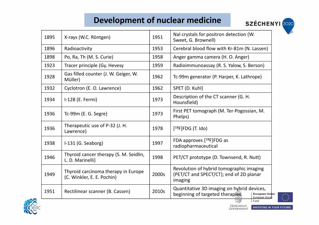
1895 X‐rays (W.C. Röntgen) 1951 NaI crystals for positron detection (W. Sweet, G. Brownell)
1896 Radioactivity 1953 Cerebral blood flow with Kr‐81m (N. Lassen)
1898 Po, Ra, Th (M. S. Curie) 1958 Anger gamma camera (H. O. Anger)
1923 Tracer principle (Gy. Hevesy 1959 Radioimmunoassay (R. S. Yalow, S. Berson)
1928 Gas filled counter (J. W. Geiger, W. Müller) 1962 Tc‐99m generator (P. Harper, K. Lathrope)
1932 Cyclotron (E. O. Lawrence) 1962 SPET (D. Kuhl)
1934 I‐128 (E. Fermi) 1973 Description of the CT scanner (G. H. Hounsfield)
1936 Tc‐99m (E. G. Segre) 1973 First PET tomograph (M. Ter‐Pogossian, M. Phelps)
1936 Therapeutic use of P‐32 (J. H. Lawrence) 1978 [18F]FDG (T. Ido)
1938 I‐131 (G. Seaborg) 1997 FDA approves [18F]FDG as radiopharmaceutical
1946 Thyroid cancer therapy (S. M. Seidlin, L. D. Marinelli) 1998 PET/CT prototype (D. Townsend, R. Nutt)
1949 Thyroid carcinoma therapy in Europe (C. Winkler, E. E. Pochin) 2000s
Revolution of hybrid tomographic imaging (PET/CT and SPECT/CT); end of 2D planar imaging
1951 Rectilinear scanner (B. Cassen) 2010s Quantitative 3D imaging on hybrid devices, beginning of targeted therapies
Development of nuclear medicine
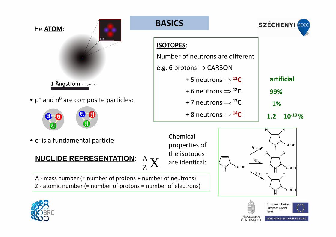
• p+ and n0 are composite particles:
• e‐ is a fundamental particle
ISOTOPES:Number of neutrons are differente.g. 6 protons CARBON
+ 5 neutrons 11C
+ 6 neutrons 12C+ 7 neutrons 13C
+ 8 neutrons 14C
BASICSHe ATOM:
artificial
A ‐mass number (= number of protons + number of neutrons)Z ‐ atomic number (= number of protons = number of electrons)
XZANUCLIDE REPRESENTATION:
NH
COOH
NH
COOH
NH
COOH
NH
COOH
DD
TT3H2
2H2
1H2
HH
Chemical properties of the isotopes are identical:
99%
1%
1.2 10‐10 %
0 5 10 15 20 25
2
4
6
8
30 60 90 120 150 180 210 240
4He
12C16O
56Fe
238U
Mass number (A)
E B/A
(MeV
)
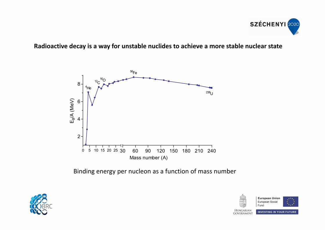
Binding energy per nucleon as a function of mass number
Radioactive decay is a way for unstable nuclides to achieve a more stable nuclear state
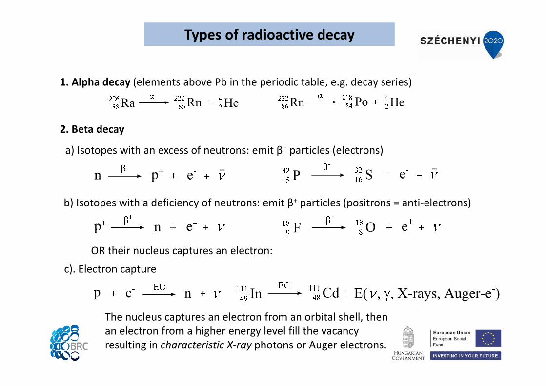
2. Beta decay
a) Isotopes with an excess of neutrons: emit β− particles (electrons)
1. Alpha decay (elements above Pb in the periodic table, e.g. decay series)
b) Isotopes with a deficiency of neutrons: emit β+ particles (positrons = anti‐electrons)
c). Electron capture
The nucleus captures an electron from an orbital shell, then an electron from a higher energy level fill the vacancy resulting in characteristic X‐ray photons or Auger electrons.
OR their nucleus captures an electron:
Types of radioactive decay
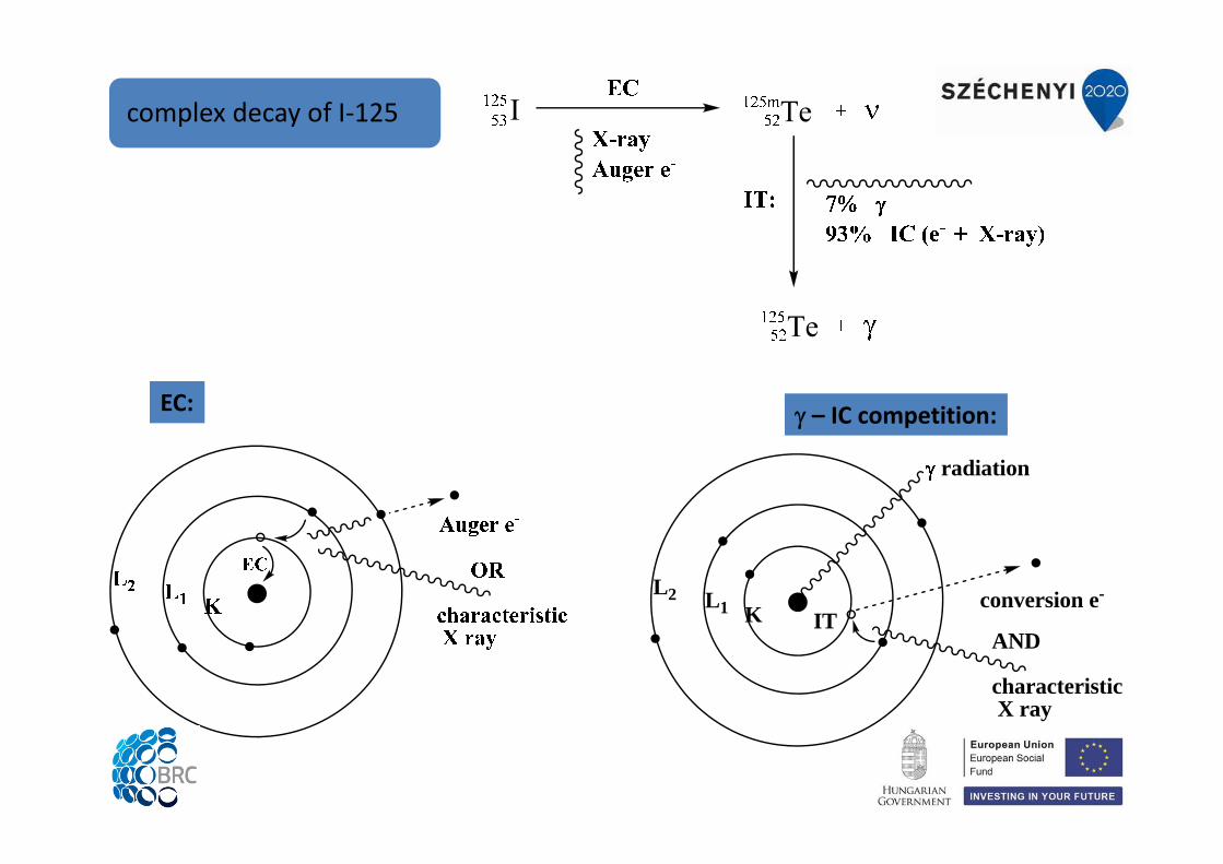
complex decay of I‐125
conversion e-KL1
L2
AND
characteristicX ray
radiation
IT
– IC competition:EC:
P P PP
P P PP
P P PP
P P PP
P S SP
P S SP
P S SP
P S SP
S S SP
S S SP
S S SS
P S SP
S S SP
P P PS
S S PS
P P PS
P S SS
P P SS
P P SS
S P PP
S P PP
S P PS
P S SP
P S SS
14 days 14 days
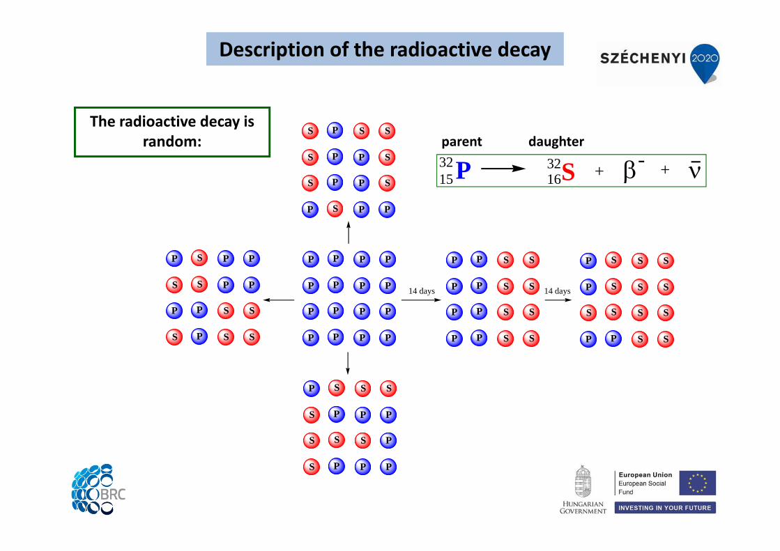
P S3215
3216 - + +
parent daughterThe radioactive decay is
random:
Description of the radioactive decay

Description of the radioactive decay
Radioactivity is the decay rate of a radioactive substance:
dNdt = N
The decay rate is proportional to the number of radionuclides, N. If N and the decay rate are great enough:
If the original number of nuclei at t= 0 s is N0, then integration yields:
Substitution with A/λ for N and A0/λ for N0 results in the practical expression:
0
200
400
600
800
1000
0 10 20 30 40 50 60 70
time (days)
no. o
f rad
ioac
tive
atom
s
1000 unstable atoms decay10 times faster than
100 unstable atoms of the same isotope
0 12 24 36 480
20
40
60
80
100
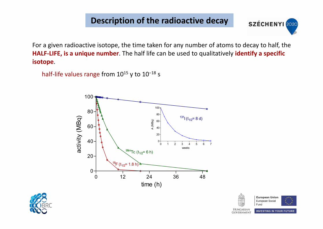
99mTc (t1/2= 6 h)
18F (t1/2= 1.8 h)
131I (t1/2= 8 d)
time (h)
activ
ity (M
Bq)
0 1 2 3 4 5 6 70
20
40
60
80
100
weeks
A (M
Bq)
For a given radioactive isotope, the time taken for any number of atoms to decay to half, the HALF‐LIFE, is a unique number. The half life can be used to qualitatively identify a specific isotope.
half‐life values range from 1015 y to 10−18 s
Description of the radioactive decay
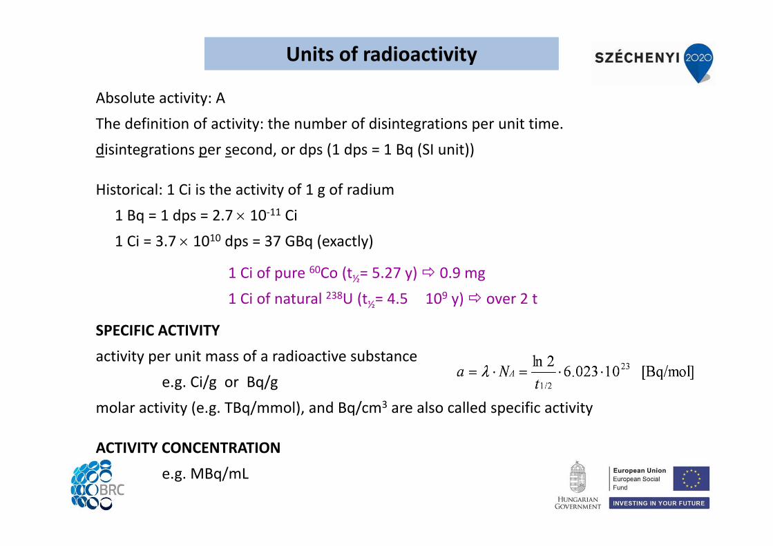
Absolute activity: AThe definition of activity: the number of disintegrations per unit time.disintegrations per second, or dps (1 dps = 1 Bq (SI unit))
Historical: 1 Ci is the activity of 1 g of radium1 Bq = 1 dps = 2.7 10‐11 Ci1 Ci = 3.7 1010 dps = 37 GBq (exactly)
1 Ci of pure 60Co (t½= 5.27 y) 0.9 mg1 Ci of natural 238U (t½= 4.5 109 y) over 2 t
SPECIFIC ACTIVITYactivity per unit mass of a radioactive substance
e.g. Ci/g or Bq/gmolar activity (e.g. TBq/mmol), and Bq/cm3 are also called specific activity
ACTIVITY CONCENTRATIONe.g. MBq/mL
Units of radioactivity
+__+
+__+
+__
_++
+
_
_
++
_
_
+
+
___
+
+
photoelectron
e-h
Compton-electron
e-
h
h
e-
h
e-
e+
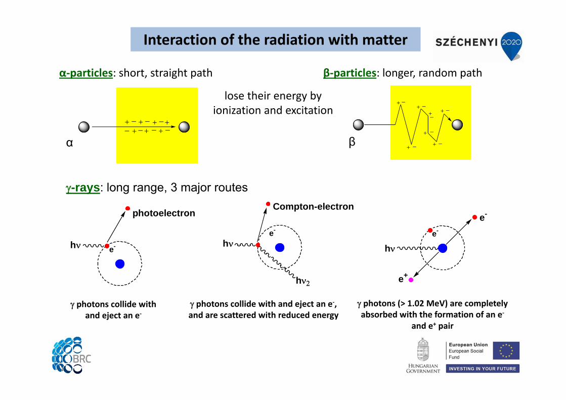
α‐particles: short, straight path β‐particles: longer, random path
α β
lose their energy by ionization and excitation
-rays: long range, 3 major routes
photons collide withand eject an e‐
photons collide with and eject an e‐, and are scattered with reduced energy
photons (> 1.02 MeV) are completely absorbed with the formation of an e‐
and e+ pair
Interaction of the radiation with matter
Measurement of radioactivity
Based on the radiation – matter interactions(mainly ionization and excitation)
GAS IONIZATION DETECTORSe.g. Geiger‐Müller ionization chamber
proportional counter
SCINTILLATION COUNTERSLiquid scintillation analysisČerenkov counting
SEMICONDUCTOR DETECTORS
Measurement of radioactivity
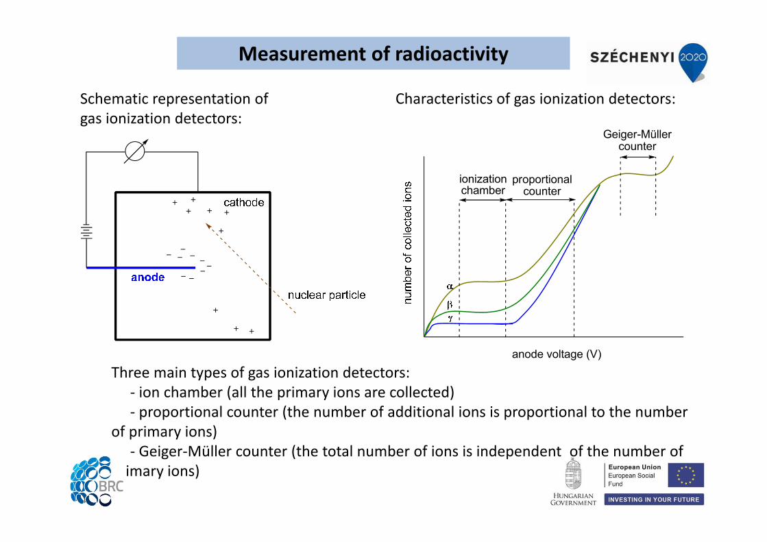
anode voltage (V)
ionizationchamber
proportionalcounter
Geiger-Müllercounter
Characteristics of gas ionization detectors:Schematic representation of gas ionization detectors:
Three main types of gas ionization detectors:‐ ion chamber (all the primary ions are collected)‐ proportional counter (the number of additional ions is proportional to the number
of primary ions) ‐ Geiger‐Müller counter (the total number of ions is independent of the number of
primary ions)
Measurement of radioactivity
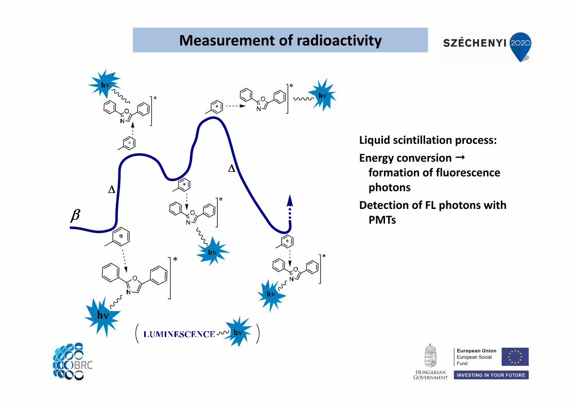
Liquid scintillation process:Energy conversionformation of fluorescence photons
Detection of FL photons with PMTs
N
O
*
NO
h
*
*
NO
h
*
*
NO
h
*
*N
O*
*
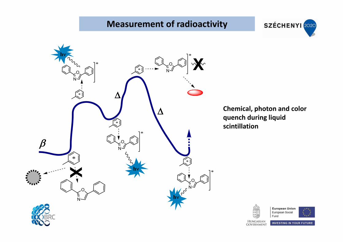
Measurement of radioactivity
Chemical, photon and color quench during liquid scintillation
Measurement of radioactivity
photon
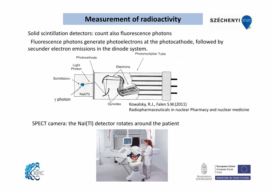
SPECT camera: the NaI(Tl) detector rotates around the patient
Solid scintillation detectors: count also fluorescence photonsFluorescence photons generate photoelectrons at the photocathode, followed by secunder electron emissions in the dinode system.
Kowalsky, R.J., Falen S.W.(2011)Radiopharmaceuticals in nuclear Pharmacy and nuclear medicine
Measurement of radioactivity
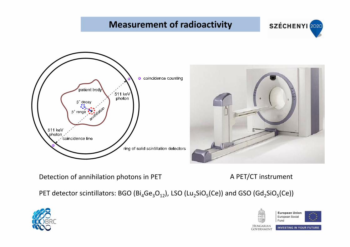
A PET/CT instrumentDetection of annihilation photons in PET
PET detector scintillators: BGO (Bi4Ge3O12), LSO (Lu2SiO5(Ce)) and GSO (Gd2SiO5(Ce))
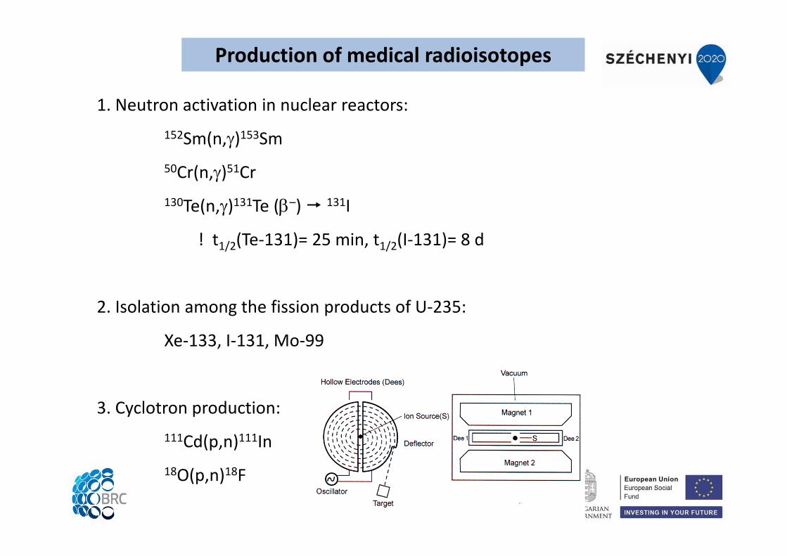
Production of medical radioisotopes
1. Neutron activation in nuclear reactors:152Sm(n,)153Sm50Cr(n,)51Cr130Te(n,)131Te (–) 131I
! t1/2(Te‐131)= 25 min, t1/2(I‐131)= 8 d
2. Isolation among the fission products of U‐235:
Xe‐133, I‐131, Mo‐99
3. Cyclotron production:111Cd(p,n)111In18O(p,n)18F
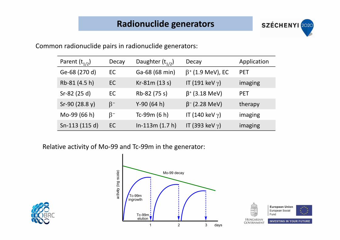
Parent (t1/2) Decay Daughter (t1/2) Decay Application
Ge‐68 (270 d) EC Ga‐68 (68 min) + (1.9 MeV), EC PET
Rb‐81 (4.5 h) EC Kr‐81m (13 s) IT (191 keV ) imaging
Sr‐82 (25 d) EC Rb‐82 (75 s) β+ (3.18 MeV) PET
Sr‐90 (28.8 y) – Y‐90 (64 h) β– (2.28 MeV) therapy
Mo‐99 (66 h) – Tc‐99m (6 h) IT (140 keV ) imaging
Sn‐113 (115 d) EC In‐113m (1.7 h) IT (393 keV ) imaging
Radionuclide generators
Common radionuclide pairs in radionuclide generators:
Mo-99 decay
Tc-99melution
Tc-99mingrowth
days1 2 3
Relative activity of Mo‐99 and Tc‐99m in the generator:

Linear energy transfer (LET): transferred energy on the unit length of the particel path
mkeV
lELET
high LETradiation low LET
radiation
LET (keV/m)
Co‐60 0.2
250 kV X‐ray 2.0
‐ 10
50 ‐ 70
Biological effects of ionizing radiations
This energy transfer responsible for the biological effects:
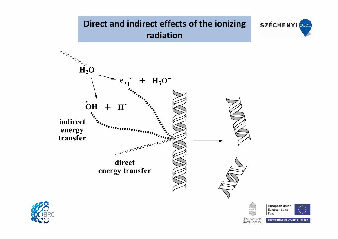
Direct and indirect effects of the ionizing radiation
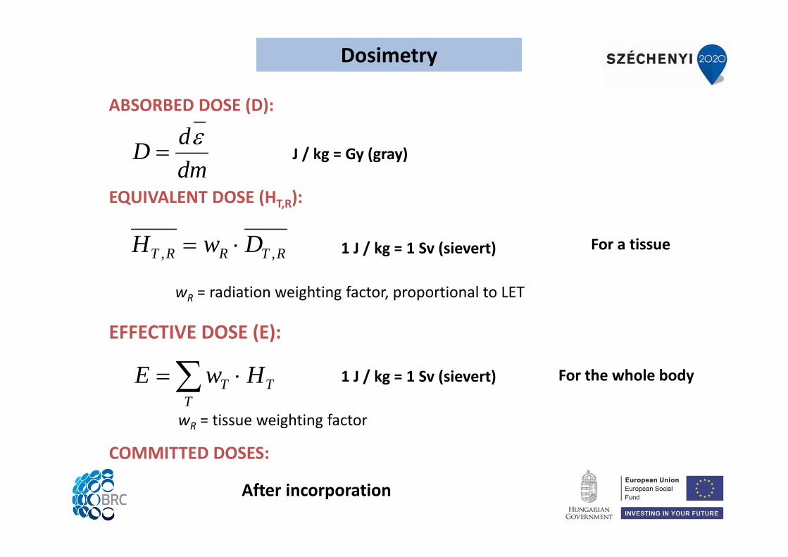
ABSORBED DOSE (D):
dmdD
J / kg = Gy (gray)
EQUIVALENT DOSE (HT,R):
RTRRT DwH ,, 1 J / kg = 1 Sv (sievert)
wR = radiation weighting factor, proportional to LET
EFFECTIVE DOSE (E):
T
TT HwE
wR = tissue weighting factor
For a tissue
For the whole body1 J / kg = 1 Sv (sievert)
COMMITTED DOSES:
After incorporation
Dosimetry
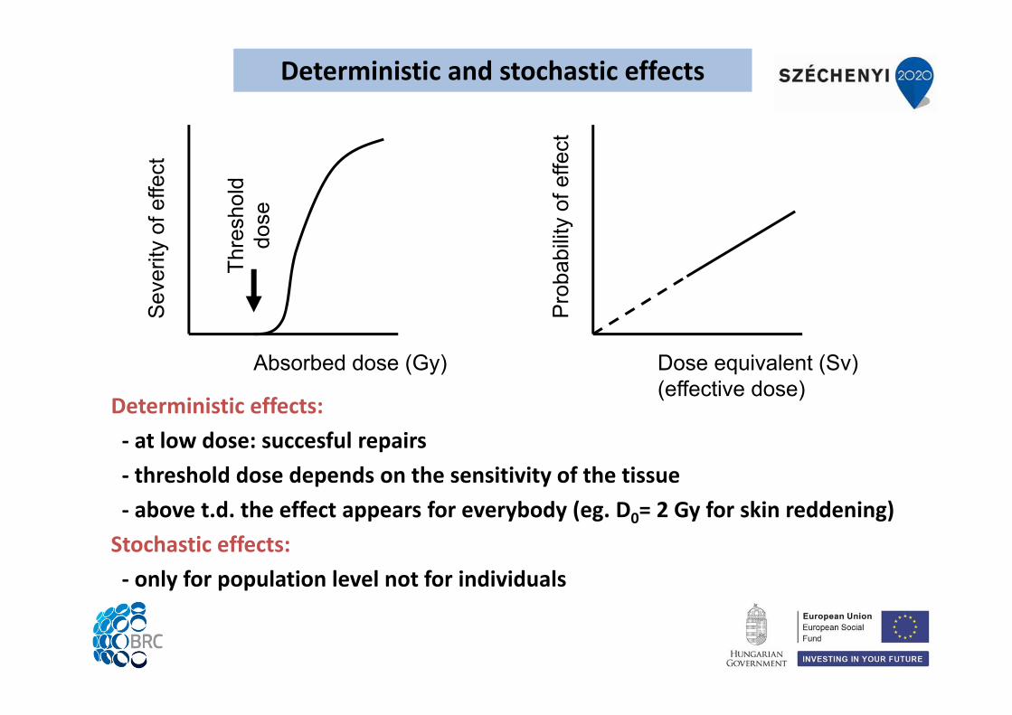
Absorbed dose (Gy)
Sev
erity
of e
ffect
Thre
shol
ddo
se
Dose equivalent (Sv)(effective dose)
Pro
babi
lity
of e
ffect
Deterministic effects:‐ at low dose: succesful repairs‐ threshold dose depends on the sensitivity of the tissue‐ above t.d. the effect appears for everybody (eg. D0= 2 Gy for skin reddening)Stochastic effects:‐ only for population level not for individuals
Deterministic and stochastic effects

Source of radiation exposure Effective dose
Radiation sickness > 1 Sv immediately
Coronary angiography 16 mSv
[67Ga]Ga‐citrate (4 mCi) body scan 15 mSv
[18F]FDG (20 mCi) brain PET scan 14 mSv
Mammography (X‐ray) 4 mSv
Natural background radiation (worldwide average) 2.4 mSv/y
Head CT 2 mSv
[123I]NaI (0.25 mCi) thyroid scan 1.9 mSv
[111In]pentetreotide (6 mCi) body scan 1.2 mSv
Dose limit for the general public 1 mSv/y
[133Xe]Xe (20 mCi) lung ventillation 0.5 mSv
Chest X‐ray 0.1 mSv
Exemption levels of radioactive substances < 10 mSv/y
Effective dose values of different procedures
Currently, the major problem in radionuclide therapy is that there is no acceptedstandard method for calculating the absorbed dose from internal radionuclides.
The European Association of Nuclear Medicine (EANM) has recently issued aguidance document on “Good Practice of Dosimetry Reporting”.
Definitions issued by the committee:‐Medical internal radiation dose (MIRD): Calculation of the average tumor‐absorbeddose at macroscopic level. This calculated dose assumes a homogenous distributionof radionuclide in organs. It does not calculate with intra‐tumoral heterogenicdistribution of the radionuclides.
‐Equivalent uniform biologically effective dose (EUBED): A uniform value of thebiologically effective dose that gives the same surviving fraction as the non‐uniformdistribution.
(M. Lassmann, Eur. J. Nucl. Med. Mol. Imaging 2011)
Dosimetry of radionuclide therapy
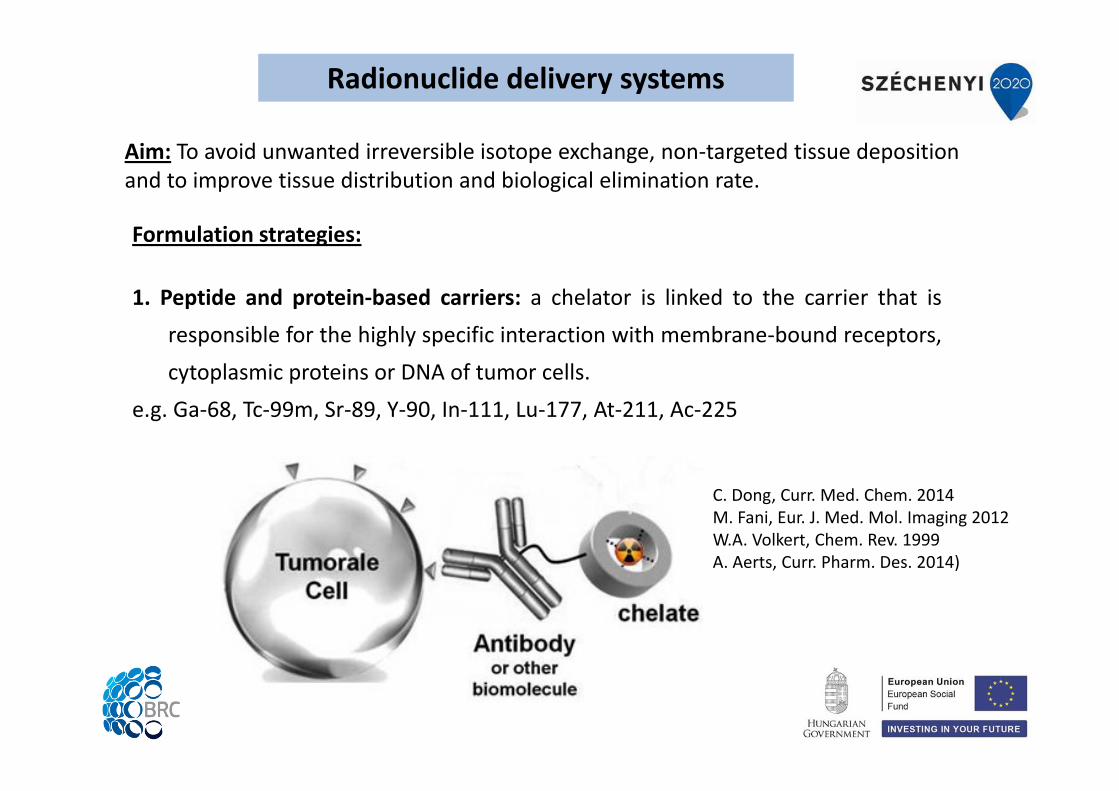
Aim: To avoid unwanted irreversible isotope exchange, non‐targeted tissue depositionand to improve tissue distribution and biological elimination rate.
C. Dong, Curr. Med. Chem. 2014M. Fani, Eur. J. Med. Mol. Imaging 2012W.A. Volkert, Chem. Rev. 1999A. Aerts, Curr. Pharm. Des. 2014)
Formulation strategies:
1. Peptide and protein‐based carriers: a chelator is linked to the carrier that isresponsible for the highly specific interaction with membrane‐bound receptors,cytoplasmic proteins or DNA of tumor cells.
e.g. Ga‐68, Tc‐99m, Sr‐89, Y‐90, In‐111, Lu‐177, At‐211, Ac‐225
Radionuclide delivery systems
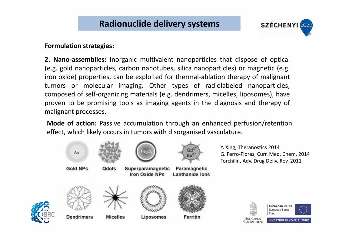
Formulation strategies:
2. Nano‐assemblies: Inorganic multivalent nanoparticles that dispose of optical(e.g. gold nanoparticles, carbon nanotubes, silica nanoparticles) or magnetic (e.g.iron oxide) properties, can be exploited for thermal‐ablation therapy of malignanttumors or molecular imaging. Other types of radiolabeled nanoparticles,composed of self‐organizing materials (e.g. dendrimers, micelles, liposomes), haveproven to be promising tools as imaging agents in the diagnosis and therapy ofmalignant processes.
Mode of action: Passive accumulation through an enhanced perfusion/retentioneffect, which likely occurs in tumors with disorganised vasculature.
Y. Xing, Theranostics 2014G. Ferro‐Flores, Curr. Med. Chem. 2014Torchilin, Adv. Drug Deliv. Rev. 2011
Radionuclide delivery systems
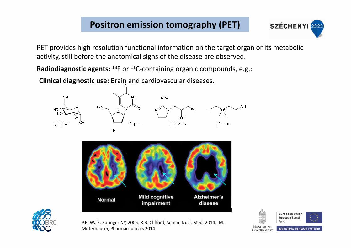
PET provides high resolution functional information on the target organ or its metabolic activity, still before the anatomical signs of the disease are observed.
P.E. Walk, Springer NY, 2005, R.B. Clifford, Semin. Nucl. Med. 2014, M. Mitterhauser, Pharmaceuticals 2014
Clinical diagnostic use: Brain and cardiovascular diseases.
Radiodiagnostic agents: 18F or 11C‐containing organic compounds, e.g.:
Positron emission tomography (PET)

Gamma‐emitting radiopharmaceuticals are exploited to visualize tumors and to investigate
energy imbalances and cardiovascular defficiencies by SPECT system.
Radiodiagnostic agents: Tc‐99m or I‐123 containing organic compounds
[99mTc]sestamibi (breast imaging) [99mTc]albumin (lung imaging)
[99mTc]tetrofosmin (heart imaging) [99mTc]oxidronate (skeletal imaging)
[99mTc]medronate (bone imaging) [99mTc]mertiatide (kidney imaging)
[99mTc]bicisate (localiztaion of stroke) [99mTc]apcitide (det. of venous thrombosis)
[99mTc]pentetate and gluceptate (brain and kidney imaging)
[99mTc]mebrofenin and disofenin (hepatobiliary imaging)
[99mTc]exametazime (detection of altered cerebral perfusion in stroke)
[123I]MIBG (or iobenguane sulfate) (localization of neuroblastomas, pheochromocytomas)
Advantage: no on site cyclotron is required
Disadvantage: lower resolution than that of PET
Single‐photon emission computed tomography (SPECT)
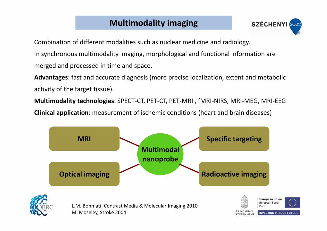
Combination of different modalities such as nuclear medicine and radiology.
In synchronous multimodality imaging, morphological and functional information are
merged and processed in time and space.
Advantages: fast and accurate diagnosis (more precise localization, extent and metabolic
activity of the target tissue).
Multimodality technologies: SPECT‐CT, PET‐CT, PET‐MRI , fMRI‐NIRS, MRI‐MEG, MRI‐EEG
Clinical application: measurement of ischemic conditions (heart and brain diseases)
L.M. Bonmati, Contrast Media & Molecular Imaging 2010M. Moseley, Stroke 2004
Multimodality imaging
Multimodalnanoprobe
Specific targeting
Radioactive imaging
MRI
Optical imaging
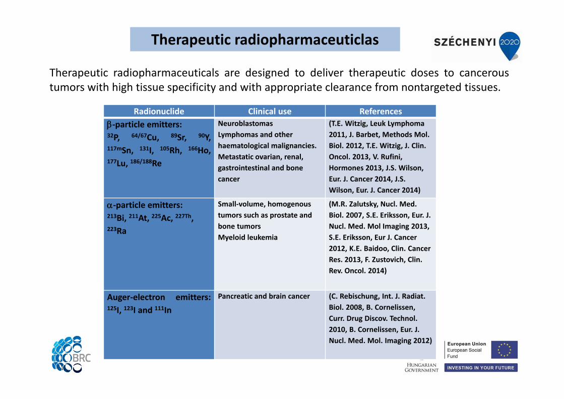
Radionuclide Clinical use References‐particle emitters:32P, 64/67Cu, 89Sr, 90Y,117mSn, 131I, 105Rh, 166Ho,177Lu, 186/188Re
NeuroblastomasLymphomas and other haematological malignancies.Metastatic ovarian, renal, gastrointestinal and bone cancer
(T.E. Witzig, Leuk Lymphoma 2011, J. Barbet, Methods Mol. Biol. 2012, T.E. Witzig, J. Clin. Oncol. 2013, V. Rufini, Hormones 2013, J.S. Wilson, Eur. J. Cancer 2014, J.S. Wilson, Eur. J. Cancer 2014)
‐particle emitters:213Bi, 211At, 225Ac, 227Th, 223Ra
Small‐volume, homogenous tumors such as prostate and bone tumorsMyeloid leukemia
(M.R. Zalutsky, Nucl. Med. Biol. 2007, S.E. Eriksson, Eur. J. Nucl. Med. Mol Imaging 2013, S.E. Eriksson, Eur J. Cancer 2012, K.E. Baidoo, Clin. Cancer Res. 2013, F. Zustovich, Clin. Rev. Oncol. 2014)
Auger‐electron emitters:125I, 123I and 111In
Pancreatic and brain cancer (C. Rebischung, Int. J. Radiat. Biol. 2008, B. Cornelissen, Curr. Drug Discov. Technol. 2010, B. Cornelissen, Eur. J. Nucl. Med. Mol. Imaging 2012)
Therapeutic radiopharmaceuticlas
Therapeutic radiopharmaceuticals are designed to deliver therapeutic doses to canceroustumors with high tissue specificity and with appropriate clearance from nontargeted tissues.
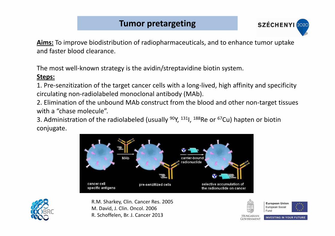
Aims: To improve biodistribution of radiopharmaceuticals, and to enhance tumor uptake and faster blood clearance.
The most well‐known strategy is the avidin/streptavidine biotin system.Steps:1. Pre‐senzitization of the target cancer cells with a long‐lived, high affinity and specificity circulating non‐radiolabeled monoclonal antibody (MAb).2. Elimination of the unbound MAb construct from the blood and other non‐target tissues with a “chase molecule”. 3. Administration of the radiolabeled (usually 90Y, 131I, 188Re or 67Cu) hapten or biotin conjugate.
R.M. Sharkey, Clin. Cancer Res. 2005M. David, J. Clin. Oncol. 2006R. Schoffelen, Br. J. Cancer 2013
Tumor pretargeting

Thank you for your attention!
This work is supported by the European Union, co-financed by the European Social Fund, within the framework of " Practice-
oriented, student-friendly modernization of the biomedical education for strengthening the international
competitiveness of the rural Hungarian universities " TÁMOP-4.1.1.C-13/1/KONV-2014-0001 project.