Embed Size (px)
Citation preview
Therapeutic Effects of Human STRO-3-Selected Mesenchymal Precursor Cells and their Soluble
Factors in Experimental Myocardial Ischemia
Fiona See PhD1, 2*, Tetsunori Seki PhD2, Peter J Psaltis MBBS3, Hugo P Sondermeijer MD MS2, Stan
Gronthos PhD3, Andrew CW Zannettino PhD3, Klaas M Govaert2, Michael D Schuster MBA2, Paul A
Kurlansky MD4, Darren J Kelly PhD1*, Henry Krum MBBS, PhD5, Silviu Itescu MBBS1,2
1The University of Melbourne, St Vincent’s Hospital, Department of Medicine, Victoria, Australia
2Columbia University, Department of Surgery, New York, USA
3Centre for Stem Cell Research, Department of Medicine, University of Adelaide, South Australia,
Australia
4Miami Heart Research Institute, Florida, USA
5NHMRC CCRE in Therapeutics, Monash University, Alfred Hospital, Victoria, Australia
Received date: 15-Jun-2010, Accepted date: 02-Nov-2010
*Addresses for Correspondence:
Fiona See, PhD
The Leon H. Charney Division of Cardiology, Cardiovascular Research Program
New York University Langone School of Medicine
522 First Avenue, Smilow 8, New York, NY 10016, USA
Tel: +1.212.263.4120 | Fax: +1.212.263.4129 | Email: [email protected]
Darren J Kelly, PhD
Department of Medicine, The University of Melbourne, St Vincent’s Hospital
29 Regent Street, Fitzroy, Victoria 3065, Australia
Tel: +61.3.9288.2546 | Fax: +61.3.9288.2581 | Email: [email protected] This is an Accepted Work that has been peer-reviewed and approved for publication in the Journal of Cellular and Molecular Medicine, but has yet to undergo copy-editing and proof correction. See http://www.blackwell-synergy.com/loi/jcmm for details. Please cite this article as a “Postprint”; 10.1111/j.1582-4934.2010.01241.x
2
ABSTRACT
Background: The STRO-3 antigen has previously been shown to identify a subset of adult human bone
marrow (BM)-derived mesenchymal lineage precursors, which may have cardioprotective potential. We
sought to characterize STRO-3+-immunoselected and culture-expanded mesenchymal precursor cells
(MPCs) with respect to their biology and therapeutic potential in myocardial ischemia.
Methods and Results: Immunoselection of STRO-3+ MPCs enriched for fibroblastic colony forming
units from unfractionated BM MNCs. Compared to mesenchymal stem cells conventionally isolated by
plastic adherence, MPCs demonstrated increased proliferative capacity during culture-expansion,
expressed higher levels of early “stem cell” markers and various proangiogenic and cardioprotective
cytokines, and exhibited greater trilineage developmental efficiency. Intramyocardial injection of MPCs
into a rat model of myocardial infarction (MI) promoted LV recovery and inhibited LV dilatation. These
beneficial effects were associated with cardioprotective and proangiogenic effects at the tissue level,
despite poor engraftment of cells. Treatment of MI rats with MPC-conditioned medium (CM) preserved
LV function and dimensions, reduced myocyte apoptosis and fibrosis, and augmented neovascularization,
involving both resident vascular cells and circulating endothelial progenitor cells (EPCs). Profiling of CM
revealed various cardioprotective and proangiogenic factors, which had biological activity in cultures of
myocytes, tissue-resident vascular cells and EPCs.
Conclusions: Prospective immunoselection of STRO-3+ MPCs from BM MNCs confers advantage in
maintaining a population of immature mesenchymal precursor cells during ex vivo expansion.
Transplantation of culture-expanded MPCs into the post-MI heart results in therapeutic benefit,
attributable at least in part to paracrine mechanisms of action. Thus, MPCs represent a promising therapy
for myocardial ischemia.
KEYWORDS
cell therapy; adult stem/progenitor cells; paracrine
3
INTRODUCTION
Mesenchymal stem/progenitor cells (MSCs), derived from adult human bone marrow (BM),
represent a leading strategy in the development of cell-based therapies for ischemic heart disease. The
salutary effects of MSC administration in animal models of myocardial infarction (MI) include recovery
of LV function and attenuation of LV remodeling, associated with augmentation of endogenous
cardioprotective processes, such as neovascularization, myocyte survival, and activation of resident
cardiac progenitor cells[1-3]. MSC differentiation into cardiomyocyte and endothelial lineage cell types
are unlikely to contribute significantly to these therapeutic benefits, given the infrequency of this
phenomenon and the typically low engraftment rates of transplanted cells[4-6]. Growing evidence
indicates that the cardioprotective and proangiogenic effects of MSC therapy may be attributed, at least in
part, to paracrine-based mechanisms of action[7,8]. Clinical application of MSC therapy is favored by the
ready availability of source tissue, the ease of isolation and culture-expansion of cells to therapeutically-
relevant numbers, and their ability to evade all immune recognition and response[9].
Conventional methods to isolate MSCs from bone marrow (BM) mononuclear cells (MNCs)
entail initial selection by plastic-adherence. This approach yields a starting population of cells
heterogeneous in morphology, immunophenotype and biological activity, including fibroblastic colony
forming unit (CFU-f) and developmental and proliferative efficiencies[10]. Culture-expansion of MSCs is
associated with downregulation of stem cell markers, decline of proliferative and differentiation
capacities and ultimately cell senescence[11,12]. An alternative approach to MSC isolation involves
prospective immunoselection of MSCs on the basis of a common cell surface antigen, with the goal of
isolating a more homogenous cell set with respect to biology and function. This strategy may be
advantageous in maintaining the population of precursor cells of interest during culture-expansion, and
may in turn enhance therapeutic outcome[13]. Antigenic definition of MSCs typically relies upon a panel
of surface molecules, including co-expression of CD105, CD73, CD90, and negativity for CD45, CD34,
CD14/CD11b, CD79α/CD19 and HLA-DR, despite these markers being non-specific for MSCs[14,15].

4
A number of novel candidate markers have been investigated as targets for direct selection of
MSCs, including STRO-1, STRO-3, CD49a and VCAM-1[13,16-18]. Growing evidence suggests that
STRO-1 marks an immature precursor cell type, which resides in the perivascular niche of human bone
marrow[16,19]. STRO-1 expression correlates with various stem cell characteristics including fibroblastic
clonogenicity, multipotentiality, telomerase expression and proliferative capacity[11,16]. In the context of
myocardial ischemia, STRO-1bright cells may represent a candidate cell type for therapy since
intramyocardial delivery of these cells to nude rats post-MI dose-dependently augmented LV functional
recovery and arteriolar density, compared to treatment with STRO-1-depleted MNCs[2]. These beneficial
effects post-MI may be attributable, at least in part, to soluble factors secreted by STRO-1bright cells since
a recent report demonstrated STRO-1 expression to be a determinant for cardioprotective and
proangiogenic paracrine activity[13]. Therefore, strategies to select for STRO-1bright cells may represent
an important step forward in the development of STRO-1-based therapy.
The recently-described STRO-3 monoclonal antibody, which reacts with a novel epitope of
human tissue non-specific alkaline phosphatase, has been shown to identify a subset of STRO-1bright BM
MNCs, designated mesenchymal precursor cells (MPCs)[17]. Freshly isolated STRO-1bright/STRO-3+
MPCs comprise virtually all CFU-f in BM MNC and demonstrate trilineage developmental potential in in
vitro and in vivo assays[17]. Given the low incidence of MPCs, development of these cells for potential
therapy post-MI necessitates culture-expansion to clinically-relevant numbers.
Based on this previous body of work, we hypothesized that culture-expanded MPCs would
demonstrate a cardioprotective phenotype. Accordingly, we examined the biological characteristics of
culture-expanded MPCs in vitro, and we evaluated the therapeutic potential and mechanisms of action of
MPCs in a nude rat model of MI.

5
METHODS
Full details are provided in the Supplementary Methods section.
In vitro biological characterization of MSCs and MPCs
The MNC fraction from human bone marrow aspirates was used to prepare (1) MSC by
conventional, plastic-adherence isolation (MSC) [10] and (2) MPC by STRO-3-based prospective
immunoselection by magnetic activated cell sorting (MACS) [17]. Following the establishment of CFU-f,
passage (P) 0 MSC and MPC were plated as single cell suspensions for ex vivo expansion. P4 MSCs and
MPCs were compared for: (1) CFU-f efficiency; (2) in vitro expansion potential; (3) immunophenotypic
profiles and (4) genetic expression of stem cell-related transcripts; and (5) developmental capacities under
osteogenic, chondrogenic and adipogenic inductive conditions.
Animal Studies
Supplementary Figure 1 illustrates the 3 animal experiments performed in this study. An athymic
nude rat model of MI induced by permanent coronary artery ligation was used in each experiment. In the
14 day MPC study, animals received either MPCs (1x10⁶) suspended in saline (N=10) or saline as vehicle
control (N=12) by intramyocardial (IM) injection at 48 h post-MI. In the 7 day conditioned medium (CM)
studies, animals were treated with IM injections of either concentrated CM derived from 1x10⁶ MPCs
(P5) cultured in serum-free αMEM (N=12) or control SF-αMEM (N=10) at 48 h post-MI. In the EPC
homing study, 1x10⁶ human endothelial progenitor cells (EPCs) suspended in saline were delivered by
intracardiac injection into the LV of rats at 48 h post-MI. EPCs were allowed to circulate for 15 min prior
to intramyocardial injections of CM or control medium (N=12/group).

6
Cytokine profiling and in vitro biological activity of MPC-CM
Soluble factors present in CM were profiled using a membrane-based antibody array.
Concentrations of IL-6, VEGF and MCP-1 were determined using a spectral bead-based immunoassay.
The direct effects of CM on neonatal rat cardiac myocytes, human umbilical vein endothelial cells
(HUVECs), A7r5 rat vascular smooth muscle cells (rVSMCs) and EPCs were examined in cell culture
experiments, in the presence or absence of neutralizing antibodies raised against IL-6, MCP-1 or VEGF.

7
RESULTS
Biological characterization of STRO-3-immunoselected and culture-expanded MPCs
STRO-3+ cells were immunoselected from the MNC fraction of adult human bone marrow
aspirate. While CFU-f were detected in unfractionated MNCs, STRO-3-immunoselection resulted in an 8-
fold enrichment of CFU-f (Unfractionated vs. STRO-3+, P<0.05). The STRO-3-depeleted fraction of
MNCs was negative for CFU-f (STRO-3+ vs. STRO-3-, P<0.05) (Figure 1A). Cultures of immunoselected
STRO-3+ MPCs and MSC isolated from MNCs by plastic adherence were expanded up to 9 passages. In
comparison to MSCs, population doublings in cultures of STRO-3+ MPCs tended to be higher over
passages 1-6, and were significantly increased from passages 7-9 (P<0.05) (Figure 1B). At passage 4, cell
surface expression of STRO-1 and STRO-3 each tended to be higher in MPCs compared with MSCs
(Figure 1C). Culture-expanded MPCs demonstrated increased gene expression of a range of stem cell
markers, TWIST-1, DERMO-1, Msx2, CBFA-1 and TERT, relative to MSCs (Figure 1D). Levels of
transcripts for SDF-1, HGF, IGF-1, VEGF and IL-6, were also elevated in passaged MPCs above MSCs
(Figure 1E). Culture-expanded MPCs exhibited a greater capacity to undergo osteogenic (P<0.05) (Figure
1F), adipogenic (P<0.05) (Figure 1G) and chondrogenic (P<0.05) (Figure 1H) differentiation.
Intramyocardial injection of MPCs post-MI in nude rats attenuates LV dysfunction and remodeling
The therapeutic potential of culture-expanded MPCs was examined in a nude rat model of MI.
Echocardiographic measurements of fractional shortening (FS) and LV diastolic area (LVAD) obtained at
24 h post-MI indicated that rats randomized to the 2 study groups were well-matched prior to treatment
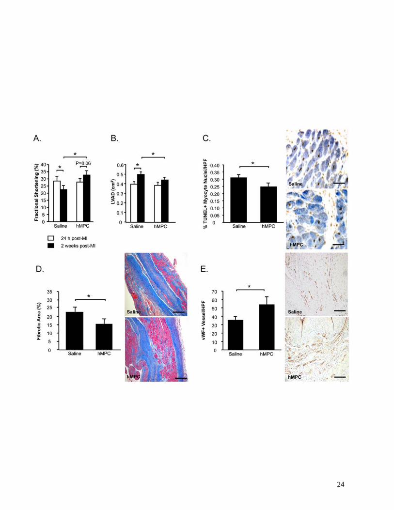
(Figures 2A, 2B). LV function declined following saline injections (-19%, 24 h vs. 2 weeks, P<0.05)
(Figure 2A). In contrast, FS was preserved in MPC-treated rats (+20%, 24 h vs. 2 weeks, P=0.06) and was
higher compared to saline-treated controls at the end of the 2 week study (MPC vs. Saline, P<0.05). MPC-
treatment post-MI also attenuated LV dilatation (+17%, 24 h vs. 2 weeks, P=NS) compared to saline-
treatment (+27%, 24 h vs. 2 weeks, P<0.05), and resulted in significantly reduced LVAD at the end of the
study period (MPC vs. Saline, P<0.05) (Figure 2B).
8
At the tissue level, MPC-treated hearts exhibited fewer apoptotic myocyte nuclei at the infarct
border compared with saline-treatment (P<0.05) (Figure 2C), as well as less extensive areas of fibrosis
(P<0.05) (Figure 2D) and augmentation of peri-infarct capillary density (P<0.05) (Figure 2E).
MPC engraftment in myocardial tissue at 2 weeks following transplantation was examined by
immunostaining for human mitochondrial protein. However analysis of stained tissues was confounded
by the presence of false positive signals in vehicle-treated MI hearts. These signals were likely
attributable to inflammatory cells, given their localization in the infarct region and granular staining
pattern (data not shown). As an alternative approach to assessing MPC engraftment, we employed PCR to
detect human-specific RPL32 (hRPL32) in DNA extracts from rat MI hearts at 24 h (N=2) and 7 d (N=2)
following intramyocardial injection of MPCs. MPCs were positive for hRPL32, with band intensities
corresponding with cell number (Figure 3). DNA from saline-treated rat heart was negative for RPL32.
Only weak signals for hRPL32 were detected in MPC-treated hearts at 24 h and 7 d following injection,
despite equal loading of samples indicated by comparable levels of rat-specific RPL32. These data
indicate rapid loss of MPCs occurs following direct intramyocardial injection at 48 h post-MI.
Intramyocardial injection of MPC-derived CM post-MI preserves LV function and structure, and
promotes cardioprotection and neovascularization
We went on to hypothesize that MPC-derived paracrine factors may augment endogenous repair
mechanisms in the ischemic heart. MI rats received intramyocardial injections of CM or control medium
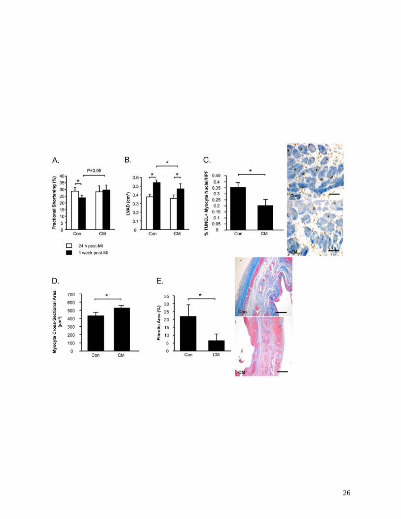
at 48 h post-MI. FS was similar in both groups prior to treatment, but declined following control medium
treatment (-16%, 24 h vs. 1 week, P<0.05) (Figure 4A). In contrast LV function was preserved in MI rats
treated with CM, such that FS was higher in this group compared to controls at 1 week (CM vs. Control,
P=0.05). LVAD was also similar in both treatment groups at baseline (Figure 4B). While LVAD
increased by 43% in control medium-treated rats (24 h vs. 1 week, P<0.05), LV dilatation was attenuated
in CM-treated animals (+26%, 24 h vs. 1 week, P<0.05), which demonstrated significantly smaller LV
cavities at the end of the study (P<0.05 vs. Saline).

9
The benefits of CM on LV function and remodeling over control were associated with reduced
myocyte apoptosis in the peri-infarct region (P<0.05) (Figure 4C), increased cross-sectional areas of
surviving myocytes (P<0.05) (Figure 4D) and reduced myocardial fibrosis (P<0.05) (Figure 4E).
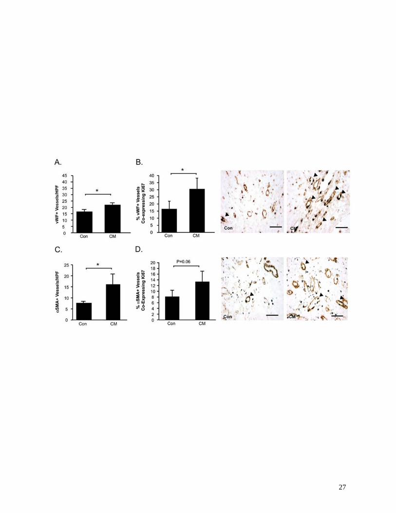
CM-treatment post-MI also resulted in enhanced myocardial neovascularization, associated with
increased proliferation of resident vascular cells compared with control medium. vWF+ vessel counts
were higher in the peri-infarct region of CM-treated hearts compared with control medium-treated
counterparts (P<0.05) (Figure 5A). More specifically, CM-treated hearts demonstrated a higher
proportion of vWF+ vessels co-expressing rat-specific Ki67, a nuclear antigen expressed by proliferating
cells, compared with controls (P<0.05) (Figure 5B). Intramyocardial injection of CM post-MI also
resulted in increases in both the density of αSMA+ arterioles in the peri-infarct region (P<0.05) (Figure
5C), as well as the proportion of αSMA+ vessels co-expressing Ki67 (P=0.06) (Figure 5D).
MPC-CM augments EPC mobilization and homing post-MI
To address the question of whether CM promotes EPC mobilization, peripheral blood was
collected from MI rats in both treatment groups at the end of the 1 week study period. The concentration
of circulating MNCs was increased following intramyocardial CM injection compared to control-medium
treatment (P<0.05) (Figure 6A). Counts of acLDL+lectin+ EPCs derived from MNCs of CM-treated MI
rats were higher compared with those derived from control-treated counterparts (P<0.05) (Figure 6B).
An additional study was undertaken to investigate the effects of CM on EPC homing to the
ischemic heart. MI rats received intracardiac injections of DiI+ human EPCs, which were allowed to
circulate prior to intramyocardial delivery of CM or control medium. At 5 days following injections, DiI+
cells were found in the peri-infarct region of myocardial sections taken from both treatment groups.
However CM-treated hearts demonstrated significantly higher numbers of DiI+ cells compared to control
medium-treated hearts (Figure 6C). The increased presence of DiI+ cells in CM-treated hearts was
associated with increased capillary density (P<0.05, vs. Saline) (Figure 6D).
10
MPC-CM contains cardioprotective and proangiogenic factors
The cytokine profile of CM samples generated under serum-free culture conditions was
determined using a membrane-based antibody array system. Detectable signals were semi-quantified by
densitometric analysis and expressed relative to control medium (Supplementary Table 1). IL-6, VEGF
and MCP-1 were among the factors expressed by MPCs at 2-fold or greater levels above control medium.
The concentrations of these factors were quantified: IL-6: 118.04±0.27 pg/ml; MCP-1: 521.89±1.48
pg/ml; VEGF: 33.95±2.98 pg/ml.
The direct effects of CM on cardiac myocytes were examined in vitro. Hypoxic conditions
induced apoptosis in myocytes cultured in ischemia-mimetic medium, as indicated by TUNEL (Figure
7A). Myocyte apoptosis was reduced in cells pre-treated with CM+mouse IgG isotype control
(CM+mIgG vs. Control, P<0.05). This protective effect of CM was partially abrogated by blockade of IL-
6 activity (CM+αIL-6 MAb vs. CM+mIgG, P<0.05), though the number of TUNEL+ cells in co-treated
cultures remained lower than control-treated cells (CM+αIL-6 MAb vs. Control, P<0.05).
CM stimulated hypertrophic growth of myocytes, as evidenced by a marked increase in total
cellular protein content, without significant change in DNA content (CM+mIgG vs. Control, P<0.05)
(Figure 7B). Neutralization of IL-6 attenuated the growth-promoting effect of CM (CM+αIL-6 MAb vs.
CM+mIgG, P<0.05). However protein content in myocyte cultures treated with CM+αIL-6 MAb
remained greater than that in control-treated cells (CM+αIL-6 MAb vs. Control, P<0.05). Consistent
with these findings, phalloidin-staining of myocytes treated with CM+mIgG for 72 h demonstrated
increased size and obvious striation and organization of the contractile protein, actin, into sarcomeric
units (Figure 7C). These morphological features were less pronounced in myocytes treated with CM+αIL-
6 MAb.
Cellular proliferation was increased in HUVECs treated with CM+mIgG compared with control
medium (P<0.05) (Figure 8A). Blockade of VEGF activity partially inhibited this effect (CM+αVEGF
MAb vs. CM+mIgG, P<0.05; CM+αVEGF MAb vs. Control, P<0.05). In comparison to control-

11
treatment, CM stimulated HUVEC migration (CM+mIgG vs. Control, P<0.05), which was attenuated by
an anti-VEGF antibody (CM+αVEGF MAb vs. CM+mIgG, P<0.05; CM+αVEGF MAb vs. Control,
P<0.05) (Figure 8B). CM also promoted HUVEC cord formation on growth factor-reduced Matrigel
substrate (Figure 8C and D). Neutralization of VEGF activity in CM disrupted the formation of these
cord-like structures (CM+αVEGF MAb vs. CM+mIgG, P<0.05).
CM increased rVSMC proliferation above controls (CM+mIgG vs. Control, P<0.05) (Figure 8E).
Anti-MCP-1 antibody partially reduced the proliferative effects of CM (CM+αMCP-1 MAb vs.
CM+mIgG, P<0.05; CM+αMCP-1 MAb vs. Control, P<0.05). In comparison to control medium, CM
stimulated increased rVSMC chemotaxis (CM+mIgG vs. Control, P<0.05) (Figure 8F). Inhibition of
MCP-1 activity attenuated this chemotactic effect (CM+αMCP-1 MAb vs. CM+mIgG, P<0.05).
EPC proliferation was increased in cultures treated with CM, above levels observed in
unstimulated controls (CM+mIgG vs. Control, P<0.05) (Figure 9A). Neutralization of VEGF tended to
inhibit this effect (CM+mIgG vs. CM+αVEGF MAb, P<0.05). CM also exhibited chemotactic effects on
EPCs (CM+mIgG vs. Control, P<0.05) (Figure 9B), which were partly attenuated in CM+αVEGF MAb
(CM+αVEGF MAb vs. CM+mIgG, P<0.05; CM+αVEGF MAb vs. Control, P<0.05). CM increased
survival of EPCs cultured under hypoxic conditions (CM+mIgG vs. Control, P<0.05) (Figure 9C). This
protective effect was reduced by blockade of VEGF (CM+αVEGF MAb vs. CM+mIgG, P<0.05). CM
stimulated formation of cord-like structures in EPCs grown on Matrigel substrate (Figure 9D). Blockade
of VEGF inhibited formation of these structures (CM+αVEGF MAb vs. CM+mIgG, P<0.05).
12
DISCUSSION
In the present study, we examined the biological characteristics of the culture-expanded progeny
of STRO-3-immunoselected MPCs and their therapeutic potential post-MI. The key findings of this study
are: (1) ex vivo-grown MPCs exhibited increased proliferative capacity, gene expression for various early
stem cell markers and cytokines, and differentiation efficiency compared with conventionally-prepared
MSCs; (2) intramyocardial injection of MPCs into rats post-MI attenuated LV dysfunction and
remodeling, reduced myocyte apoptosis and augmented myocardial neovascularization, despite poor
engraftment of transplanted cells; (3) administration of MPC-CM to the MI heart also mitigated LV
dysfunction and dilatation, promoted myocyte survival and hypertrophy and increased vascular density;
(4) MPC-CM contained soluble factors with biological activity in cultures of cardiac myocytes, vascular
cells, and peripheral blood-derived EPCs.
The recently described STRO-3 monoclonal antibody has previously been shown to enrich for a
subset of STRO-1bright MNCs, which is clonogenic and multipotential [17]. Co-expression of high levels
of STRO-1 suggest that STRO-3+ MPCs may be a relatively immature precursor cell-type with
cardioprotective potential[2,16]. The present findings extend on existing data by demonstrating that the
culture-expanded progeny of STRO-3-selected MPCs remain proliferative and multipotential following
culture expansion over several passages. These functional characteristics were associated with cell-
surface expression of STRO-1, and gene expression of a panel of early “stem cell” markers, which are
implicated in the regulation of progenitor cell cycle progression and differentiation[12,20-22]. Culture-
expanded MPCs also expressed transcripts of various cytokines, reflective of the native function of
mesenchymal lineage cells in supporting the population of naïve hematopoietic progenitors in bone
marrow. Notably, the proliferative and developmental potentials and stem cell marker and cytokine
expression levels were each augmented in cultures initiated by STRO-3+ MPCs above that observed in
cultures of MSCs established by conventional methods. In sum our data suggest that prospective
immunoselection of STRO-3+ MPCs enriches for CFU-f, and may be advantageous in maintaining a
population of immature mesenchymal precursor cells during culture-expansion, compared with

13
preliminary isolation of MSCs by plastic-adherence alone. Further, these data support the possibility of
generating therapeutically-relevant numbers of precursor cells by ex vivo expansion for clinical
application.
Our in vivo studies are the first to demonstrate the cardioprotective and proangiogenic properties
of STRO-3-selected and culture-expanded human MPCs in a rat model of MI. Intramyocardial
transplantation of 1x10⁶ MPCs to rats at 48 h post-MI resulted in improved LV function, reduced
dilatation, increased myocyte survival and capillary density associated with reduced fibrosis, compared
with saline control at 2 weeks. The dose, route and timing of administration of MPCs were selected based
on our previous work, which demonstrated the efficacy of 1x10⁶ STRO-1+ culture-expanded MPCs in a
rat model of MI[2]. The present findings echo our earlier observations of functional recovery and
arteriogenesis in the ischemic heart following treatment with STRO-1+ MPCs[2]. Similarly, recent reports
have demonstrated that in a sheep model of myocardial ischemia, allogeneic-transplantation of STRO-3-
selected, ex vivo-cultured ovine MPCs attenuated infarct expansion, accompanied by increased vascular
density, in a dose-dependent manner, associated with modulation of matrix remodeling[23,24]. Together,
these data suggest that the STRO-3 antibody may be employed to select a subset of BM mesenchymal
lineage cells, which exhibit a cardioprotective phenotype following culture-expansion.
Interestingly, we found that the beneficial effects of MPC therapy post-MI in rats occurred
despite poor engraftment of cells. Specifically, the numbers of MPCs present in the post-MI heart at 24 h
and 7 d following injection were dramatically diminished compared to the number injected, and were
negligible by 2 weeks. Our observations are similar to reports in the literature of rapid and progressive
attrition of MSCs following intramyocardial transplantation under both syngeneic and xenogeneic
conditions[5,25]. In the acute phase post-MI, transplanted cells are likely to be beset by dynamic
processes including inflammation, cellular necrosis, fibrosis and metabolic and mechanical disturbances,
which may impair engraftment. Our data suggest that under the present study conditions, mechanisms
such as MPC differentiation or fusion are unlikely to account significantly for the observed effects of
MPC administration post-MI.

14
Instead, our observations fall in line with a current working hypothesis that the therapeutic
benefits observed following MSC administration to the ischemic heart occur, at least in part, via
paracrine-mediated mechanisms of action. Various studies have demonstrated beneficial effects of MSC
therapy without long-term engraftment, and a link between donor cell-derived factors and LV recovery
following MSC transplantation[26-28]. Moreover, injection of MSC-derived factors to the post-MI heart
has been shown to rapidly promote angiogenesis and cardioprotection[7,29-31]. Consistent with this body
of work, intramyocardial injection of MPC-derived soluble factors post-MI resulted in therapeutic
benefits, which mirrored the outcomes observed following treatment of MI rats with MPCs. Profiling of
CM revealed the presence of a range of multifunctional soluble factors. Our in vitro studies evidence the
contribution of IL-6, VEGF and MCP-1 present in CM to its direct actions on myocytes, HUVECs,
rVSMCs and EPCs, and suggest possible mechanistic underpinnings for the effects of CM in the post-MI
heart. It remains to be determined whether resident cardiac progenitors cells are activated in response to
CM treatment, and whether other mechanisms of tissue repair may be involved in the attenuation of LV
dysfunction. However, in sum, the present data from our CM studies support the notion that MPCs may
exert paracrine-mediated actions when transplanted into the ischemic myocardium.
Interestingly we found that MPC- or CM-treatment post-MI resulted in myocyte hypertrophy both
in vivo and in vitro. Although better function is often coupled with attenuation of myocyte enlargement,
hypertrophy of the residual myocardium occurs in the acute phase post-MI as a compensatory response to
the loss of tissue at the site of infarct to maintain LV contractile function. Previous studies support the
hypothesis that insufficient compensatory hypertrophy post-MI may contribute to LV dilatation and heart
failure disease progression[32]. Given the improvements in LV function observed in our study, we
speculate that administration of MPCs or CM at 48 h post-MI may augment an early compensatory
response, which may contribute to their salutary effects in this context.
Treatment of MI rats with either MPCs or CM also attenuated myocardial fibrosis at the site of
infarct. Alongside the concomitant reduction in myocyte apoptosis in the same hearts, our data may

15
suggest that reduced tissue fibrosis in the infarcted region is secondary to augmentation of myocyte
survival. In a previous report, the anti-fibrotic and infarct-limiting effects of allogeneic transplantation of
STRO-3-derived MPCs into an ovine model of myocardial ischemia were associated with increased
indices of collagen degradation[24]. However, the direct actions of MPCs on myocardial collagen
turnover remain to be determined. Alternatively, reductions in infarct size and tissue fibrosis following
cell therapy in animal models of MI may be attributable to partial regeneration of the necrotic
myocardium[33]. In light of poor engraftment of MPCs in the post-MI rat heart, our data support a
paracrine mechanism of action for these cells in this setting. However, our data do not preclude the
possibility that MPCs may also have the capacity for cardiogenesis, and the potential of MPCs for cardiac
development and to activate resident cardiac progenitors or myocytes remains a subject for future
investigation.
Our CM studies cast light on a possible paracrine-based mechanism of action of MPC therapy
post-MI, though we acknowledge that the data does not provide direct documentation of a paracrine
action of MPCs transplanted into the ischemic myocardium. However, our data may provide some insight
into the biology of ex vivo-grown MPCs. From this perspective, it is important to emphasize that MPC-
CM was generated under serum-free and normoxic culture conditions. Thus, our data demonstrate that the
constitutive secretome of culture-expanded MPCs includes proangiogenic and cardioprotective factors
with biological activity both in vitro and in vivo. The profile of cytokines identified in this study overlaps
qualitatively with that previously detected in MSC-CM, and similarly produced proangiogenic effects in
cultures of HUVECs[8]. Interestingly, MPC-CM also promoted myocyte survival under ischemia-
mimetic culture conditions and attenuated LV remodeling and dysfunction post-MI, without need for
potentiation of MPCs via transfection of survival genes and/or hypoxic culture, which was previously
found to be necessary to generate MSC-CM with similar effects[7]. The reasons for these differences in
cardioprotective potential between MPC-CM and MSC-CM require further investigation, though we note
that in the present study, transcripts for various proangiogenic and cardioprotective cytokines were more
abundant in MPCs compared with MSCs prepared by conventional methods. Similarly, Psaltis and
colleagues recently reported that CM generated by STRO-1-immunoselected cells demonstrated enhanced

16
cardioprotective and proangiogenic activity compared to MSC-CM[13]. Clearly, the functional
consequences of these differences remain to be examined. However, the present data suggest that
prospective immunoselection and culture-expansion of STRO-3+ MPCs may enrich for a population of
cells, which intrinsically expresses a profile of factors that is both qualitatively and quantitatively
sufficient to yield therapeutic benefit in myocardial ischemia.
Speculatively, these observations point to the intriguing possibility of MPC-derived soluble
factors as therapy per se. To this end, the sufficiency of CM generated under serum-free conditions to
produce beneficial effects in the ischemic myocardium, and the persistence of these effects at 7 days post-
MI, are encouraging. Further investigations might include examination of MPC-CM therapy alone, with
animals followed to later time points to determine whether the effects of CM post-MI are sustained.
MPC-CM might also be administered in combination with cells types, including EPCs, given the potential
synergy between MPC-CM and circulating EPCs observed in this study. In looking ahead toward the
possible clinical application of MPCs or CM for post-MI therapy, further efforts are required to optimize
these approaches to ensure full exploitation of their respective therapeutic potentials. For example, our
data indicate a need for strategies that enhance persistence of MPCs in the ischemic myocardium, since
previous studies have shown cell engraftment to correlate positively with therapeutic outcome [34].
Additional experiments, such as a head-to-head comparison of the efficacy of MPC versus CM injections,
would also inform evaluation of the most promising way to translate MPC-based therapy for myocardial
ischemia into the clinical setting.
The present data support the potential utility of STRO-3 as a marker for a subset of mesenchymal
lineage precursors present in adult BM MNCs. Prospective immunoselection of STRO-3+ MPCs may be
advantageous in maintaining an immature mesenchymal precursor population during ex vivo expansion.
The culture-expanded progeny of STRO-3+ MPCs demonstrated therapeutic potential in the post-MI
heart, attributable at least in part, to the secretion of proangiogenic and cardioprotective factors. These
characteristics endow MPCs with clinical relevance and promise as therapy for ischemic heart disease.

17
ACKNOWLEDGEMENTS
We acknowledge the technical assistance of Geping Zhang.
FUNDING SOURCES
Grant support for this study was provided by the NIH/NHLBI (P50HL077096) and by the Miami
Heart Research Institute.
DISCLOSURES
Silviu Itescu is Director, Scientific Founder, Chief Scientist, Chairman of Scientific Advisory
Board and equity holder of Angioblast Systems, Inc., NY, NY; Director and Chief Scientific Adviser of
Mesoblast Ltd, Melbourne, Australia. Michael Schuster is a co-founder and Vice President of Operations
of Angioblast Systems, Inc., NY, NY. Henry Krum is a member of the Scientific Advisory Board of
Mesoblast Ltd. Melbourne, Australia.

18
FIGURE LEGENDS
Figure 1. Biological comparisons between MSC and MPC.
(A) The clonogenic efficiency of each MNC fraction. Results shown are the mean±SEM number of CFU-
f per 105 cells plated for (*P<0.001, average of five donor experiments). (B) Mean number of population
doublings (±SEM) of MSC and MPC during ex vivo culture from P0 to senescence. (*P<0.05, average of
3 donor experiments). (C) Examples of flow cytometric histograms comparing MPC and MSC from the
same donor, for surface STRO-1 and STRO-3 expression at P4. Dotted line – isotype control. Red line -
MSC. Blue line – MPC. P4 MPC gene expression of various immature “stem cell” markers (D) and
cytokines (E). Results are presented as mean ratio±SEM of gene expression in MPC relative to MSC
(average of three donor experiments). P4 MSC and MPC differentiation capacity for mineralization (F),
adipogenesis (G) and glycosaminoglycan synthesis (H), under the respective in vitro inductive conditions.
Data are presented as mean±SEM. (*P<0.05, average of 3 donor experiments.)
Figure 2. Intramyocardial injection of MPCs post-MI attenuates LV dysfunction and remodeling.
Echocardiography was performed to assess fractional shortening (A) and LV diastolic areas (LVAD) (B).
Myocardial sections were stained to count TUNEL+ apoptotic myocytes (C). Masson’s Trichrome-stained
sections were examined for myocardial fibrosis (D). vWF+ vessels per high power field (HPF) were
quantified (E). For figures A-E, data is represented as mean±sem. N=10-12/group. *P<0.05.
Figure 3. MPCs demonstrate poor engraftment in the ischemic myocardium.
Human-specific RPL32 expression (384 bp) in DNA extracts from known numbers of MPCs, untreated
control rat heart and human MPC (hMPC)-treated MI hearts harvested at 24 h (N=2) and 7 d (N=2)
following intramyocardial injection, was determined by PCR. Rat-specific RPL32 was used as loading
control.
Figure 4. Intramyocardial injection of CM post-MI attenuates LV dysfunction and remodeling.

19
Echocardiography was performed to assess fractional shortening (A) and LV diastolic areas (LVAD) (B).
Myocardial sections were stained to count TUNEL+ apoptotic myocytes (C). Masson’s Trichrome-stained
sections were assessed for myocardial fibrosis (D) and myocyte cross-sectional areas (E).
Figure 5. Intramyocardial injection of CM post-MI promotes neovascularization of the ischemic
myocardium.
Myocardial sections were immunostained to identify capillaries and arterioles. vWF+ vessels were
counted (A). Proliferating vWF+ vessels were identified by co-expression of the proliferating cell nuclear
antigen Ki67 (arrow heads) (B). Arterioles were immunostained with αSMA (C). Serial sections were
double-stained for αSMA and Ki67 (arrow heads) (D). Data is presented at mean±sem. N=10-12/group.
*P<0.05.
Figure 6. CM augments mobilization and homing of EPCs to the post-MI heart.
To examine the effect of CM treatment on EPC mobilization, peripheral blood was sampled from rats at
the end of the 1 week study and total MNC were counted (A). acLDL+lectin+ EPCs cultured from the
MNC fraction were counted (B). In a separate experiment, the effects of control medium or CM on DiI+
EPCs homing to the ischemic rat heart were examined. At 1 week post-MI myocardial sections were
examined for the presence of DiI+ cells (C). Myocardial sections were stained to identify vWF+ vessels co
localized with DiI+ EPCs (D). Data is presented at mean±sem. N=10-12/group. *P<0.05.
Figure 7. CM protects cardiac myocytes from apoptotic death and promotes myocyte hypertrophy.
The direct effects of control medium, CM+mouse IgG isotype control (mIgG) or CM+αIL-6 MAb on
neonatal rat cardiac myocytes were examined. (A) Hypoxia-induced apoptosis was determined by
TUNEL (brown). Blue: viable nuclei. Myocyte hypertrophy was determined by measuring total protein
content, relative to DNA content (B) and phalloidin-staining of fixed cells (C). Data is represented as
mean±SEM. Each condition was represented in triplicate. N=3 experiments. *P<0.05.
Figure 8. CM promotes vascular cell proliferation, migration, survival and cord formation.

20
The direct effects of control medium, CM+mouse IgG isotype control (mIgG) or CM+αMCP-1 MAb on
cultures of HUVECs were examined in assays for proliferation (A), migration (B) and cord formation (C).
Representative images of cord formation in HUVEC cultures (D). The effects of control medium,
CM+mouse IgG isotype control (mIgG) or CM+αMCP-1 MAb on rat A7r5 smooth muscle cells were
examined with respect to proliferation (E) and migration (F). Data is represented as mean±SEM. Each
condition was represented in triplicate. N=3 experiments. *P<0.05.
Figure 9. CM promotes EPC proliferation, migration, survival and cord formation.
The direct effects of control medium, CM+mouse IgG isotype control (mIgG) or CM+αVEGF MAb on
isolated EPCs were examined in assays for proliferation (A), migration (B), apoptosis (C) and cord
formation (D). Data is represented as mean±SEM. Each condition was represented in triplicate. N=3
experiments. *P<0.05.
21
References
1. Amado LC, Saliaris AP, Schuleri KH, et al. Cardiac repair with intramyocardial injection of allogeneic mesenchymal stem cells after myocardial infarction. Proc Natl Acad Sci USA. 2005; 102: 11474-9.
2. Martens TP, See F, Schuster MD, et al. Mesenchymal lineage precursor cells induce vascular network formation in ischemic myocardium. Nat Clin Pract Cardiovasc Med. 2006; 3: S18-22.
3. Nakanishi C, Yamagishi M, Yamahara K, et al. Activation of cardiac progenitor cells through paracrine effects of mesenchymal stem cells. Biochem Biophys Res Commun. 2008; 374: 11-6.
4. Silva GV, Litovsky S, Assad JA, et al. Mesenchymal stem cells differentiate into an endothelial phenotype, enhance vascular density, and improve heart function in a canine chronic ischemia model. Circulation. 2005; 111:150-6.
5. Terrovitis J, Stuber M, Youssef A, et al. Magnetic resonance imaging overestimates ferumoxide-labeled stem cell survival after transplantation in the heart. Circulation. 2008; 117: 1555-62.
6. Toma C, Pittenger MF, Cahill KS, et al. Human mesenchymal stem cells differentiate to a cardiomyocyte phenotype in the adult murine heart. Circulation. 2002; 105: 93-8.
7. Gnecchi M, He H, Noiseux N, et al. Evidence supporting paracrine hypothesis for Akt-modified mesenchymal stem cell-mediated cardiac protection and functional improvement. FASEB J. 2006; 20: 661-9.
8. Hung SC, Pochampally RR, Chen SC, et al. Angiogenic effects of human multipotent stromal cell conditioned medium activate the PI3K-Akt pathway in hypoxic endothelial cells to inhibit apoptosis, increase survival, and stimulate angiogenesis. Stem Cells. 2007; 25: 2363-70.
9. Aggarwal S, Pittenger MF. Human mesenchymal stem cells modulate allogeneic immune cell responses. Blood. 2005; 105: 1815-22.
10. Pittenger MF, Mackay AM, Beck SC, et al. Multilineage potential of adult human mesenchymal stem cells. Science. 1999; 284: 143-7.
11. Shi S, Gronthos S, Chen S, et al. Bone formation by human postnatal bone marrow stromal stem cells is enhanced by telomerase expression. Nat Biotechnol. 2002; 20: 587-91.
12. Simonsen JL, Rosada C, Serakinci N, et al. Telomerase expression extends the proliferative life-span and maintains the osteogenic potential of human bone marrow stromal cells. Nat Biotechnol. 2002; 20: 592-6.
13. Psaltis PJ, Paton S, See F, et al. Enrichment for STRO-1 expression enhances the cardiovascular paracrine activity of human bone marrow-derived mesenchymal cell populations. J Cell Physiol. 2010;223: 530-40.
14. Dominici M, Le Blanc K, Mueller I, et al. Minimal criteria for defining multipotent mesenchymal stromal cells. The International Society for Cellular Therapy position statement. Cytotherapy. 2006; 8: 315-7.
15. Sabatini F, Petecchia L, Tavian M, et al. Human bronchial fibroblasts exhibit a mesenchymal stem cell phenotype and multilineage differentiating potentialities. Lab Invest. 2005; 85: 962-71.
16. Gronthos S, Zannettino AC, Hay SJ, et al. Molecular and cellular characterisation of highly purified stromal stem cells derived from human bone marrow. J Cell Sci. 2003; 116: 1827-35.
22
17. Gronthos S, Fitter S, Diamond P, et al. A novel monoclonal antibody (STRO-3) identifies an isoform of tissue nonspecific alkaline phosphatase expressed by multipotent bone marrow stromal stem cells. Stem Cells Dev. 2007; 16: 953-63.
18. Gindraux F, Selmani Z, Obert L, et al. Human and rodent bone marrow mesenchymal stem cells that express primitive stem cell markers can be directly enriched by using the CD49a molecule. Cell Tissue Res. 2007; 327: 471-83.
19. Shi S, Gronthos S. Perivascular niche of postnatal mesenchymal stem cells in human bone marrow and dental pulp. J Bone Miner Res. 2003; 18: 696-704.
20. Hu G, Lee H, Price SM, et al. Msx homeobox genes inhibit differentiation through upregulation of cyclin D1. Development. 2001; 128: 2373-84.
21. Isenmann S, Arthur A, Zannettino AC, et al. TWIST family of basic Helix-Loop-Helix Transcription Factors Mediate Human Mesenchymal Stromal/Stem Cell Growth and Commitment. Stem Cells. 2009; 27: 2457-68.
22. Shui C, Spelsberg TC, Riggs BL, Khosla S. Changes in Runx2/Cbfa1 expression and activity during osteoblastic differentiation of human bone marrow stromal cells. J Bone Miner Res. 2003; 18: 213-21.
23. Hamamoto H, Gorman JH, Ryan LP, et al. Allogeneic mesenchymal precursor cell therapy to limit remodeling after myocardial infarction: the effect of cell dosage. Ann Thorac Surg. 2009; 87: 794-801.
24. Dixon JA, Gorman RC, Stroud RE, et al. Mesenchymal cell transplantation and myocardial remodeling after myocardial infarction. Circulation. 2009; 120:S220-9.
25. Muller-Ehmsen J, Krausgrill B, Burst V, et al. Effective engraftment but poor mid-term persistence of mononuclear and mesenchymal bone marrow cells in acute and chronic rat myocardial infarction. J Mol Cell Cardiol. 2006; 41: 876-84.
26. Zhang M, Mal N, Kiedrowski M, et al. SDF-1 expression by mesenchymal stem cells results in trophic support of cardiac myocytes after myocardial infarction. FASEB J. 2007; 21: 3197-207.
27. Iso Y, Spees JL, Serrano C, et al. Multipotent human stromal cells improve cardiac function after myocardial infarction in mice without long-term engraftment. Biochem Biophys Res Commun. 2007; 354: 700-6.
28. Mirotsou M, Zhang Z, Deb A, et al. Secreted frizzled related protein 2 (Sfrp2) is the key Akt-mesenchymal stem cell-released paracrine factor mediating myocardial survival and repair. Proc Natl Acad Sci USA. 2007; 104: 1643-8.
29. Gnecchi M, He H, Liang OD, et al. Paracrine action accounts for marked protection of ischemic heart by Akt-modified mesenchymal stem cells. Nat Med. 2005; 11:367-8.
30. Dai W, Hale SL, Kloner RA. Role of a paracrine action of mesenchymal stem cells in the improvement of left ventricular function after coronary artery occlusion in rats. Regen Med. 2007; 2: 63-8.
31. Timmers L, Lim SK, Arslan F, et al. Reduction of myocardial infarct size by human mesenchymal stem cell conditioned medium. Stem Cell Res. 2007; 1: 129-37.
32. Litwin SE, Raya TE, Anderson PG, et al. Induction of myocardial hypertrophy after coronary ligation in rats decreases ventricular dilatation and improves systolic function. Circulation. 1991; 84: 1819-27.
33. Orlic D, Kajstura J, Chimenti S, et al. Bone marrow cells regenerate infarcted myocardium. Nature. 2001; 410: 701-5.
34. Huang J, Zhang Z, Guo J, et al. Genetic modification of mesenchymal stem cells overexpressing CCR1 increases cell viability, migration, engraftment, and capillary density in the injured myocardium. Circ. Res. 2010; 106: 1753-62.

23
24

25
26
27

28

29

30

32


SUPPLEMENTARY MATERIALS
MATERIALS AND METHODS
Preparation of human bone marrow mesenchymal cells for biological characterization in
vitro
Normal human bone marrow aspirates were obtained from donors with the approval
of the Human Ethics Committee of the Royal Adelaide Hospital, South Australia. The BM
MNC fraction from each donor was used to prepare (1) MSC by conventional, plastic-
adherence isolation (MSC) [1] and (2) MPC by STRO-3-based prospective immunoselection
by magnetic activated cell sorting (MACS) (Miltenyi Biotec, Auburn, CA, USA) [2].
Ex vivo culture of MSCs and MPCs for in vitro biological characterization
Following the establishment of colony forming units-fibroblastic (CFU-f), passage 0
(P0) MSC and STRO-3-MPC were plated as single cell suspensions at 8x10³ per cm2, in
αMEM (Invitrogen, Carlsbad, CA, USA) supplemented with 10% FCS, L-ascorbate-2-
phosphate (100 µM), β-mercaptoethanol (50 µM) and Primocin (100 µg/ml, Invivogen, San
Diego, CA, USA) (αMEM-10). For the purpose of ex vivo expansion, cultures were continued
until fourth passage (P4), at which point cells were compared for: (1) CFU-f efficiency; (2)
STRO-1 and STRO-3 expression; (3) in vitro expansion potential; (4) genetic expression of
stem cell-related transcripts (Twist-1, Dermo-1; Msx2, CBFA1, TERT, SDF-1α, VEGF, IGF-
1, IL-6, HGF, β-actin); and (5) developmental capacities under osteogenic, chondrogenic and
adipogenic inductive conditions as described previously[3].
Animal Studies
Surgery and Echocardiography
Animal studies were performed in accordance with the “Position of the American
Heart Association on Research Animal Use”, adopted by the AHA on November 11, 1984.

Figure 1 illustrates the time course of each study. MI was induced in male athymic nude rats
(rnu/rnu, 6-8 weeks of age) by permanent ligation of the left anterior descending coronary
artery [4]. At 48 h post-MI, animals underwent a second thoracotomy for intramyocardial
injections of cells, saline, CM or control medium. Total injection volume was 250 µl, which
was administered using a 27 GA needle over 5 sites along the peri-infarct border. In the 14
day MPC study, animals received either MPCs (1x10⁶) suspended in saline (N=10) or saline
as vehicle control (N=12). In the 7 day CM studies, animals were treated with either
concentrated CM (N=12) or control medium (N=10). In the EPC homing study, 1x10⁶ EPCs
suspended in saline were delivered by intracardiac injection into the LV and allowed to
circulate for 15 min prior to intramyocardial injections of CM or control medium
(N=12/group). Echocardiography studies were performed on anaesthetized rats at 24 h post-
MI to obtain baseline measurements of LV structure and function, and again at the end of
each study period. A Sonos 5500 ultrasound machine equipped with a 12 MHz probe (Philips,
Andover, MA, USA) was used to capture transthoracic images of the LV. Two-dimensional
(2D) and m-mode images were acquired through the short axis of the LV at the mid-level of
the papillary muscles. LV internal dimensions and posterior and anterior wall thicknesses in
diastole and systole were measured offline from m-mode images over 3 consecutive cardiac
cycles and averaged for each animal. After final echocardiography studies, hearts were
harvested and myocardial sections were examined by various techniques to determine fibrotic
area, myocyte cross-sectional areas, myocyte apoptosis, vascular density, MPC engraftment,
EPC homing. In the CM study, peripheral blood MNC samples were collected and assayed
for rat EPC mobilization.
MPC Preparation
Cryopreserved GMP-grade MPCs (P5) (Angioblast Systems, New York, NY, USA)
were used in all animal studies. Immediately prior to injection, cells were thawed, washed and
resuspended in saline at a concentration of 1x10⁶ cells in 250 µl per rat.

Preparation of CM
CM was generated by culturing 1x10⁶ P5 MPCs in 10 ml of serum-free αMEM (0.5%
BSA/L-ascorbate-2-phosphate (100 µM), β-mercaptoethanol (50 µM) and Primocin (100
µg/ml) at 37°C with 5% CO2 for 24 h. Control medium was generated by exposing serum-
free medium without cells to the same conditions. Control medium and CM were collected
and stored at -80°C for in vivo and in vitro experiments. CM and control medium samples
were thawed and concentrated 40x using Centricon-20 ultrafiltration tubes (Millipore,
Bedford, MA, USA) immediately prior to use.
Preparation of Human CD34+ EPCs
CD34+ EPCs were immunoselected from cryopreserved human peripheral blood
MNC by MACs (Miltenyi Biotec). Cells were used immediately in in vivo or in vitro studies.
EPCs were labeled with DiI (Invitrogen) for in vivo studies.
Histochemical Analyses of Myocardial Sections
At the end of each study, hearts were harvested and myocardial sections were
formalin-fixed and paraffin-embedded. Masson’s trichrome staining was performed for
assessment of fibrosis and myocyte cross-sectional areas. Immunohistochemistry using the
avidin-biotin detection method was used to identify von Willebrand Factor (vWF)-positive
vessels, α-smooth muscle actin (αSMA)-positive vessels, vessels co-expressing the
proliferating cell nuclear antigen Ki67, and human mitochondria in MPCs [5]. Additional
antibodies used in the present study were: rabbit anti-human vWF (Dako, Carpinteria, CA,
USA) and anti-human mitochondria (Clone: 113-1, dilution: Millipore).
Myocyte apoptosis was assessed by Terminal deoxynucleotide transferase dUTP
Nick End Labeling (TUNEL) using the peroxidase-based FragEL DNA Fragmentation
Detection Kit (EMD Chemicals, San Diego, CA, USA), followed by immunostaining for

myocytes using a mouse-anti-troponin I antibody (Clone 2D5, Accurate Scientific, Westbury,
NY, USA).
Digital images of stained sections in 8 to 10 random, non-overlapping fields were
captured under high power magnification for computer-based image analysis (ImagePro Plus
3.0, Media Cybernetics, Bethesda, MD, USA).
Detection of Human DNA in Rat Hearts Injected with MPCs
DNA was extracted from MPCs or snap-frozen whole heart samples using DNA
Isolation Kit for Cells and Tissue (Roche, Indianapolis, IN). DNA extract from 4x10⁶ MPCs
was serially diluted to represent 10,000, 1,000, 100, 10 and 1 cell samples. Primers for human
ribosomal protein (RP) L32 and rat RPL32 were designed based on its published sequences
(Human: NM 000994.3, rat: NM_013226). Human RPL32 forward primer:
TGGCCGCCCTCAGACCCCTT; reverse primer: GCCTGGCATTGGGGTTGGTG. Rat
RPL32 forward primer: TGGCTGCCCTTCGGCCTCTG; reverse primer:
GCCTGGCGTTGGGATTGGTG. DNA in each sample was amplified using Platinum
Blue PCR Supermix (Invitrogen) using the following reaction sequence: Denature: 94°C for 5
min. 40 cycles of amplification: 94°C for 30 s, 63°C for 30 s, 72°C for 1.5 min, 72°C for 5
min. 10 µl of each sample was electrophoresed through a 1% agarose gel containing ethidium
bromide (10 µg/ml) for UV-visualization of bands. A low DNA mass ladder was run in
parallel with the samples (Invitrogen).
Assessment of Rat EPC Mobilization
Peripheral blood samples from rats in the CM study were collected in EDTA-
containing tubes. MNCs were isolated from whole blood by centrifugation (3000 rpm, 30
min, RT) through Lymphoprep (Axis-Shield, Oslo, Norway). Cells were seeded onto
vitronectin-coated plates (8x10⁶ cells/ml) in EBM-2 supplemented with EGM-2 SingleQuot
Kit Supplements and Growth Factors (comprising FBS, gentamicin/amphotericin,
hydrocortisone, heparin, VEGF, rhEGF, rhFGF-B, R3-IGF-1 (Lonza, Walkersville, MD,
USA). After 3 days of culture at 37°C with 5% CO2, non-adherent cells were discarded and
adherent cells were cultured for a further 4 days in fresh EBM-2. After a total of 7 days of
culture, EPCs were identified by uptake of DiI-conjugated acetylated low density lipoproteins
(LDL) (Invitrogen, 5 µg/ml final concentration in the presence of Ca2+ over 1 h at 37°C),
followed by brief fixation in 2% PFA for labeling with Griffonia simplicifolia-derived
isolectin IB4 conjugated to Alexa Fluor 488 (Invitrogen, 10 µg/ml final concentration, 1 h at
37°C).
Assessment of EPC Homing
In the EPC homing study, the presence of DiI-labeled cells in the myocardium were
detected in OCT-embedded, frozen sections by fluorescence microscopy. To examine the co-
localization of DiI-positive cells with capillaries, sections were stained using antibodies
against vWF and αSMA detailed above, followed by FITC-conjugated goat anti-rabbit or
FITC-conjugated horse anti-mouse secondary antibodies respectively (Vector Laboratories).
Nuclei were counterstained with DAPI. Digital images of 8-10 random fields of each section
were captured and the number of DiI+ cells and vascular density in each view were counted.
Cytokine Profiling
Soluble factors contained in CM were profiled using a membrane-based Human
Cytokine Antibody Array (RayBiotech, Inc., Norcross, GA, USA). Expression levels of
individual factors relative to control medium were semi-quantified by densitometric analysis.
Determination of IL-6, MCP-1 and VEGF Concentrations in CM
Concentration of IL-6, VEGF and MCP-1 were determined using a spectral bead-
based immunoassay (Invitrogen). The plate was read using a Luminex 100 instrument
(Luminex, Austin, TX, USA). A standard curve for each cytokine was determined to
calculate sample concentrations.
Cell Culture Experiments
For blocking studies, CM was incubated with neutralizing monoclononal antibodies
or isotype control for 30 min to block target cytokine activity, prior to addition to cells.
Monoclonal antibodies used were anti-human IL-6, anti-human MCP-1 and anti-human
VEGF (Invitrogen). Final concentration of antibody in CM was 10 µg/ml.
Neonatal Rat Cardiac Myocytes: Apoptosis and hypertrophy
Cardiac myocytes were isolated from 1-3 day old neonatal Sprague-Dawley rats and
cultured as described previously [6]. Following isolation, myocytes suspended in DMEM/F12
supplemented with 5% horse serum were allowed to adhere onto gelatin-coated culture
vessels. After 24 h, cells were washed and medium was replaced with serum-free DMEM/F12
and cultured for a further 24 h before use in experiments.
For apoptosis, myocytes were cultured on 4-well glass chamber slides (1x10⁶
cells/well) in ischemia-mimetic solution (NaCl (125 mM), KCl (8 mM), KH2PO4 (1.2 mM),
MgSO4 (1.25 mM), CaCl2 (1.2 mM), NaHCO3 (6.25 mM), sodium lactate (5 mM), HEPES
(20 mM), pH 6.6) containing concentrated control medium, CM or CM+αIL-6 MAb. Cells
were placed into a hypoxia chamber and incubated at 37°C for 24 h. At the end of the culture
period, cells were fixed for TUNEL, using the FragEL DNA Fragmentation Detection Kit.
The numbers of apoptotic nuclei in 10 non-overlapping random fields under 200x
magnification were counted in each well.
For hypertrophy, cells were cultured for 72 h in the absence or presence of
concentrated control medium, CM or CM+IL-6 MAb. Following the stimulation period
myocyte protein content in each sample was determined (DC Protein Assay, BioRad,

Hercules, CA, USA) [6]. DNA in parallel samples was extracted and quantified (DNeasy
Blood and Tissue Kit, Qiagen, Valencia, CA, USA). Myocyte protein content was expressed
as a ratio to DNA content, to confirm hypertrophic growth in the absence of mitosis [6].
Morphological signs of hypertrophic growth were also examined by staining
paraformaldehyde-fixed myocyte cultures with Alexa Fluor -633-conjugated phalloidin
diluted in 1% BSA (Invitrogen) [6]. Cells were coverslipped in permanent mounting medium
containing DAPI to counterstain cell nuclei for fluorescence microscopic examination.
Experiments in Vascular Cells and EPCs: Proliferation, migration and cord formation
Human umbilical vein endothelial cells (HUVECs, American Type Culture
Collection, Manassas, VA, USA) were grown in F12K supplemented with 10% FBS,
endothelial growth supplement (0.3 mg/ml, BD Biosciences, San Jose, CA, USA) and heparin
(10 µg/ml). A7r5 rat vascular smooth muscle cells (American Type Culture Collection) were
grown in DMEM supplemented with 10% FBS. Human peripheral blood CD34+ EPCs were
used immediately following isolation.
For proliferation studies, cells were seeded at 1,500 cells/well in 96 well plates. After
24 h, serum was withdrawn for 48 h. Cells were then stimulated for 48 h with concentrated
control medium, CM, CM+αVEGF MAb or CM+αMCP-1 MAb. Cellular proliferation was
assessed by WST-1 assay (Roche, Indianapolis, IN).
In vitro migration assays were performed using a 48-well microchemotaxis chamber
(Neuro Probe, Gaithersburg, MD, USA). Each well of the bottom chamber was filled with
RPMI/0.5% BSA containing control medium, CM, CM+αVEGF MAb or CM+αMCP-1
MAb. Cells suspended in RPMI/0.5% BSA were loaded into the wells of the top chamber
(0.5–1x10⁶ cells/ml). Cells were allowed to migrate across a collagen-coated, porous
polyvinylpropylene-free polycarbonate filter over 4 h at 37°C. Cells which had migrated
through the membrane were fixed in ice-cold methanol then stained using the Diff-Quick

stain set (Dade Behring, Newark, DE, USA). Cells were visualized under 200x magnification
and the number of cells in 5 fields per well was counted. A migration index was calculated by
dividing the number of cells migrated in response to the agonist by the number of cells that
migrated under unstimulated conditions.
For cord formation studies, HUVECs or EPCs were suspended in serum-free F12K
medium supplemented with concentrated control medium, CM or CM+αVEGF MAb. Cells
were seeded in triplicate onto growth factor-reduced Matrigel-plated plates (BD Biosciences)
(4x10⁴ cells/cm2). After incubation at 37°C for 16-18 h, cells were examined microscopically
for the formation of tube-like structures, which were counted in 8-10 random, non-
overlapping fields.
Apoptosis
EPCs were cultured in serum-free EBM-2 under hypoxic conditions for 24 h and
stained with Annexin V-FITC and propidium iodide staining (Invitrogen) for flow cytometric
analysis of apoptosis.
Statistics
For in vitro characterization studies of MPCs, data are presented as mean±sem for
multiple experiments (N≥3 donors), unless otherwise specified. Statistical comparisons
between donor-matched STRO-1+ MPC and MSC were performed using paired Student’s t-
test or one-way ANOVA with post-hoc Tukey analysis, where appropriate. Statistical
significance was established for two-tailed P values <0.05. For in vivo studies and in vitro
studies using CM, data are expressed at mean±sem. Paired Student’s t-test was used to
compare before and after treatment data sets. Comparisons between 2 groups were analyzed
by unpaired Student’s t-test, and comparisons between 3 groups were analyzed by one-way
ANOVA followed by Bonferonni’s post-hoc comparisons. P<0.05 was considered
statistically significant.


Supplemental Table 1. Cytokines detected in MPC-CM cultured under serum-free
conditions.
Cytokine Expression in CM relative to Control Medium
Acrp30 2.02
AgRP 5.12
Angiogenin 2.66
Angiopoietin-2 0.36
Amphiregulin 1.66
Axl 4.14
BDNF 1.32
bFGF 1.37
BLC 1.25
BMP-4 2.42
BMP-6 2.97
b-NGF 1.98
BTC 2.72
CCL-28 2.73
CK β 8-1 1.64
CNTF 0.87
CTACK 1.58
Dtk 1.75
EGF 1.01
EGF-R 4.75
ENA-78 3.09
Eotaxin 0.88
Eotaxin-2 0.53
Eotaxin-3 0.56
Fas 1.35
FGF-4 1.56
FGF-6 1.23
FGF-7 1.58
FGF-9 2.90
Flt-3 Ligand 0.92
Fractalkine 0.71
GCP-2 1.31

GCSF 0.41
GDNF 7.88
GITR-Ligand 1.25
GITR 1.44
GM-CSF 1.80
GRO 2.77
GRO-α 2.08
HCC-4 1.60
HGF 2.29
I-309 1.02
ICAM-1 1.81
ICAM-3 1.66
IFN-γ 1.06
IGF-1 1.17
IGFBP-1 4.45
IGFBP-2 2.02
IGFBP-3 1.55
IGFBP-4 1.08
IGFBP-6 3.06
IGF-I SR 1.41
IL-1α 3.41
IL-Iβ 0.86
IL-1rα 1.30
IL-1 R4/ST2 1.24
IL-1RI 0.85
IL-2 0.60
IL-2 Rα 1.12
IL-3 0.94
IL-4 0.76
IL-5 1.94
IL-6 34.80
IL-6R 2.54
IL-7 5.15
IL-8 1.95
IL-10 8.30
IL-11 0.69
IL-12 p40 1.33
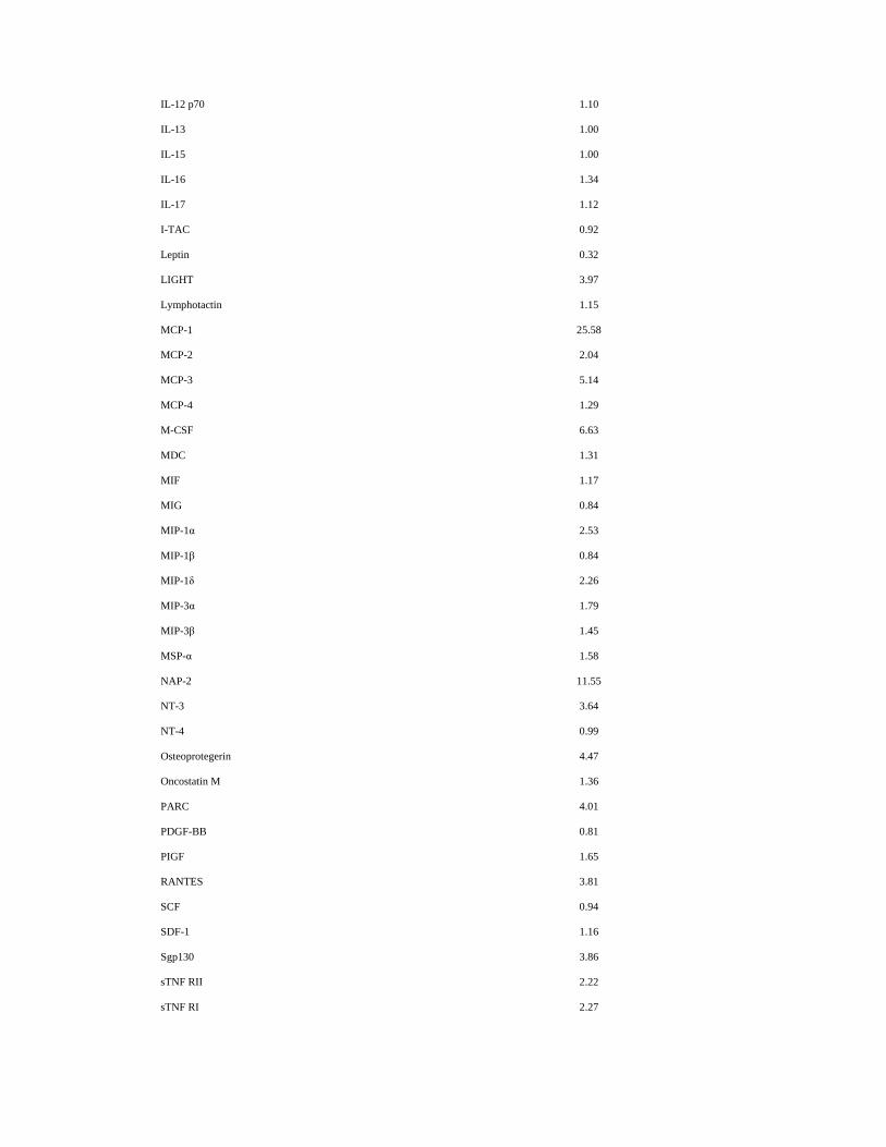
IL-12 p70 1.10
IL-13 1.00
IL-15 1.00
IL-16 1.34
IL-17 1.12
I-TAC 0.92
Leptin 0.32
LIGHT 3.97
Lymphotactin 1.15
MCP-1 25.58
MCP-2 2.04
MCP-3 5.14
MCP-4 1.29
M-CSF 6.63
MDC 1.31
MIF 1.17
MIG 0.84
MIP-1α 2.53
MIP-1β 0.84
MIP-1δ 2.26
MIP-3α 1.79
MIP-3β 1.45
MSP-α 1.58
NAP-2 11.55
NT-3 3.64
NT-4 0.99
Osteoprotegerin 4.47
Oncostatin M 1.36
PARC 4.01
PDGF-BB 0.81
PIGF 1.65
RANTES 3.81
SCF 0.94
SDF-1 1.16
Sgp130 3.86
sTNF RII 2.22
sTNF RI 2.27
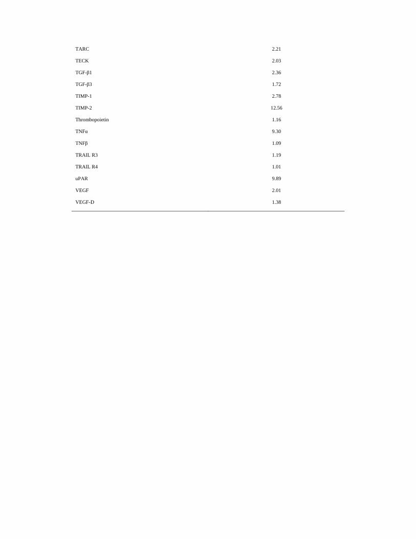
TARC 2.21
TECK 2.03
TGF-β1 2.36
TGF-β3 1.72
TIMP-1 2.78
TIMP-2 12.56
Thrombopoietin 1.16
TNFα 9.30
TNFβ 1.09
TRAIL R3 1.19
TRAIL R4 1.01
uPAR 9.89
VEGF 2.01
VEGF-D 1.38

SUPPLEMENTARY FIGURE LEGEND
Supplementary Figure 1. Study design of animal experiments.
Animal studies were performed to examine the effects of intramyocardial (IM) delivery of
MPCs (A) and CM (B) post-MI and of CM on EPC homing to the infarcted heart (C). (IC:
intracardiac).

References
1. Pittenger MF, Mackay AM, Beck SC, et al. Multilineage potential of adult
human mesenchymal stem cells. Science. 1999; 284: 143-7.
2. Gronthos S, Fitter S, Diamond P, et al. A novel monoclonal antibody
(STRO-3) identifies an isoform of tissue nonspecific alkaline phosphatase
expressed by multipotent bone marrow stromal stem cells. Stem Cells Dev.
2007; 16: 953-63.
3. Psaltis PJ, Paton S, See F, et al. Enrichment for STRO-1 expression
enhances the cardiovascular paracrine activity of human bone marrow-derived
mesenchymal cell populations. J Cell Physiol. 2010; 223: 530-40.
4. Kocher AA, Schuster MD, Szabolcs MJ, et al. Neovascularization of
ischemic myocardium by human bone-marrow-derived angioblasts prevents
cardiomyocyte apoptosis, reduces remodeling and improves cardiac function.
Nat Med. 2001; 7: 430-6.
5. Witkowski P, Seki T, Xiang G, et al. A DNA enzyme against plasminogen
activator inhibitor- type 1 (PAI-1) limits neointima formation after angioplasty
in an obese diabetic rodent model. J Cardiovasc Pharmacol. 2007; 50: 633-
40.
6. Thomas WG, Brandenburger Y, Autelitano DJ, et al. Adenoviral-directed
expression of the type 1A angiotensin receptor promotes cardiomyocyte
hypertrophy via transactivation of the epidermal growth factor receptor. Circ
Res. 2002; 90: 135-42.


![A review of therapeutic effects of mesenchymal stem cell ... · Wharton’s jelly [5], dental pulp [6] peripheral blood [7], cord blood [8], and more recently menstrual blood [9-11]](https://img.pdfslide.net/doc/110x75/600b187c3bcba55c3807aaaa/a-review-of-therapeutic-effects-of-mesenchymal-stem-cell-whartonas-jelly-5.jpg)