Embed Size (px)
Citation preview
Therapeutic Strategy Against Tumour Acidity Induced Immune-Suppression
STREAM Summer School
Medical University of Warsaw
June 2016
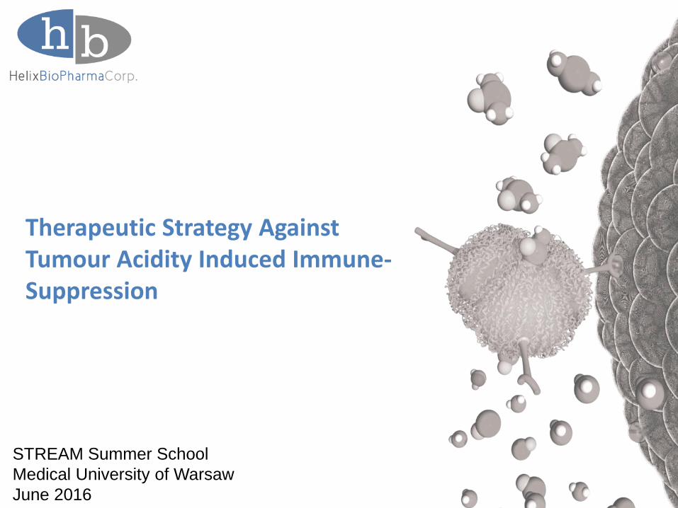
The Hallmarks of Cancer
Cell 2011 144, 646-674 2
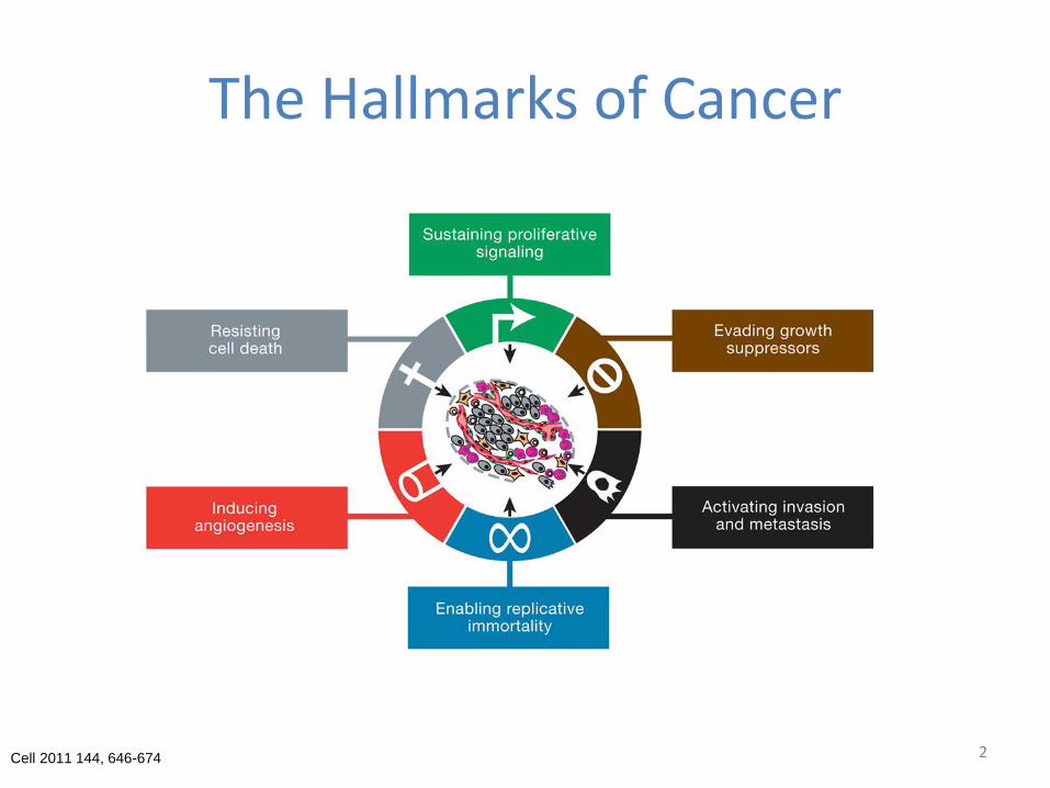
The Hallmarks of Cancer: Next Generation
Cell 2011 144, 646-674 3
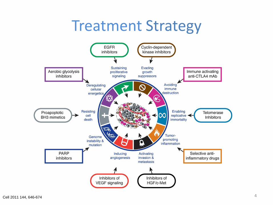
Treatment Strategy
Cell 2011 144, 646-674 4
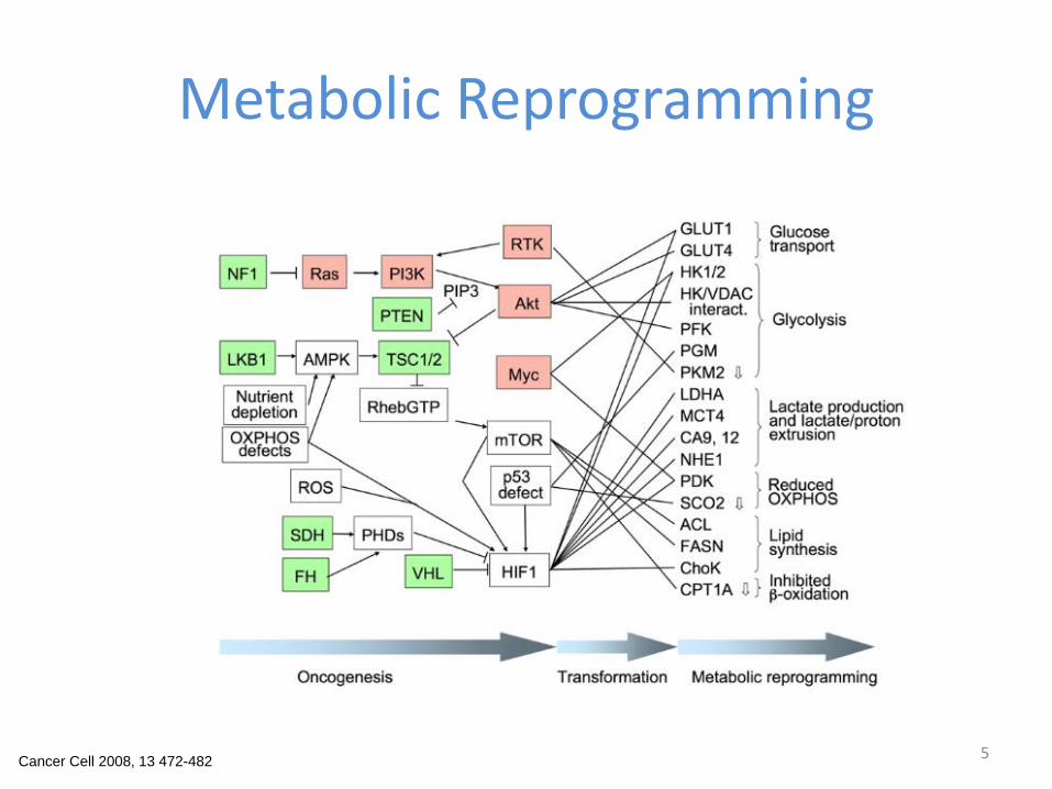
Metabolic Reprogramming
Cancer Cell 2008, 13 472-482 5

Cancer Hall Marks Link to Metabolism
Cancer Cell 2008, 13 472-482 6
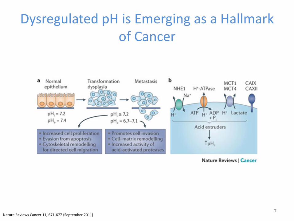
Dysregulated pH is Emerging as a Hallmark of Cancer
7Nature Reviews Cancer 11, 671-677 (September 2011)
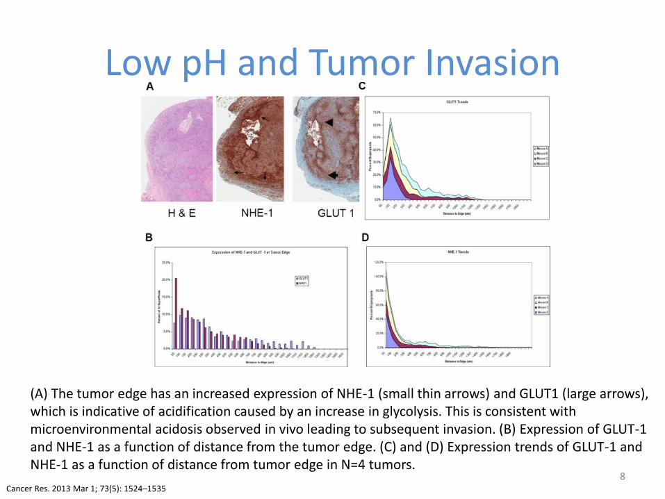
Low pH and Tumor Invasion
8
(A) The tumor edge has an increased expression of NHE-1 (small thin arrows) and GLUT1 (large arrows), which is indicative of acidification caused by an increase in glycolysis. This is consistent with microenvironmental acidosis observed in vivo leading to subsequent invasion. (B) Expression of GLUT-1 and NHE-1 as a function of distance from the tumor edge. (C) and (D) Expression trends of GLUT-1 and NHE-1 as a function of distance from tumor edge in N=4 tumors.
Cancer Res. 2013 Mar 1; 73(5): 1524–1535
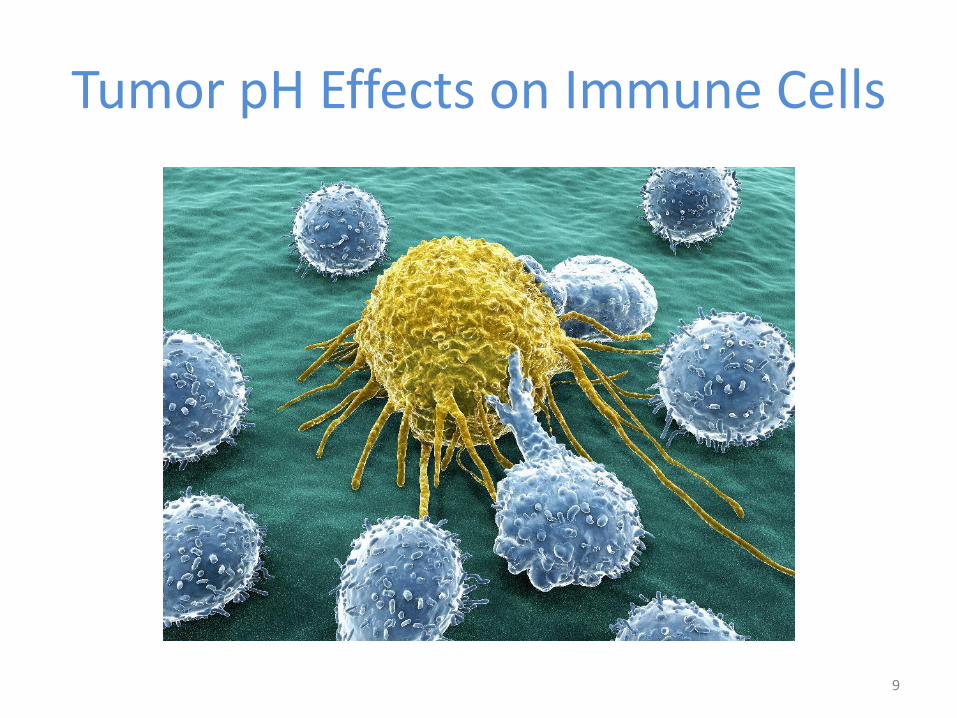
Tumor pH Effects on Immune Cells
9

Lactate Lower Tumour pH and Polarize Macrophages
Macrophages integrate metabolic andenvironmental signals to promotetumor growth. Tumor lactate whichlower pH polarizes macrophage andup-regulate Arg1. Area within dottedrectangle indicates proposedmechanisms of action. ARG, arginase;HIF, hypoxia-inducible factor; MCT,monocarboxylate transporter; NADH,nicotine adenine dinucleotide,reduced; PKM2, M2 isoform ofpyruvate kinase; VEGF, vascularendothelial growth factor.
10
Colegio et al. Nature 513:559-563 (2014)
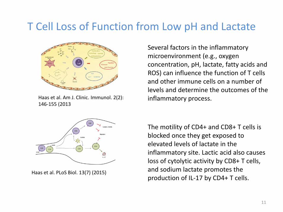
T Cell Loss of Function from Low pH and Lactate
Several factors in the inflammatory microenvironment (e.g., oxygen concentration, pH, lactate, fatty acids and ROS) can influence the function of T cells and other immune cells on a number of levels and determine the outcomes of the inflammatory process.
The motility of CD4+ and CD8+ T cells is blocked once they get exposed to elevated levels of lactate in the inflammatory site. Lactic acid also causes loss of cytolytic activity by CD8+ T cells, and sodium lactate promotes the production of IL-17 by CD4+ T cells.
11
Haas et al. Am J. Clinic. Immunol. 2(2): 146-155 (2013
Haas et al. PLoS Biol. 13(7) (2015)
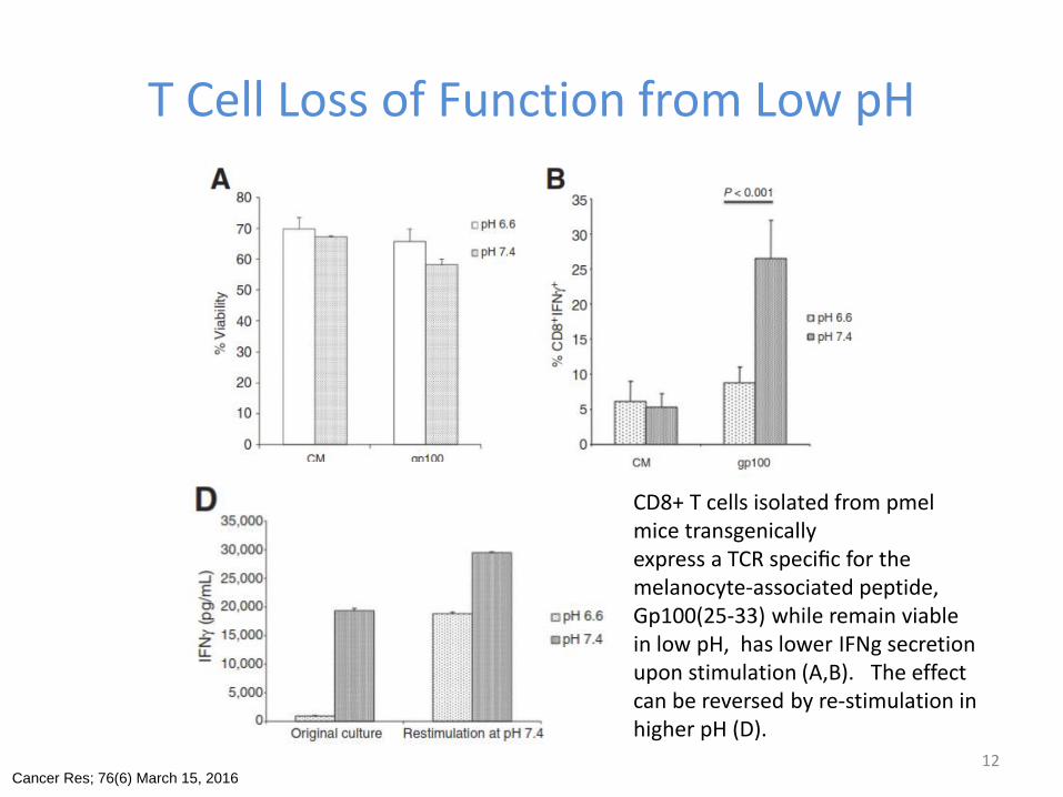
T Cell Loss of Function from Low pH
CD8+ T cells isolated from pmelmice transgenicallyexpress a TCR specific for the melanocyte-associated peptide, Gp100(25-33) while remain viable in low pH, has lower IFNg secretion upon stimulation (A,B). The effect can be reversed by re-stimulation in higher pH (D).
Cancer Res; 76(6) March 15, 2016 12
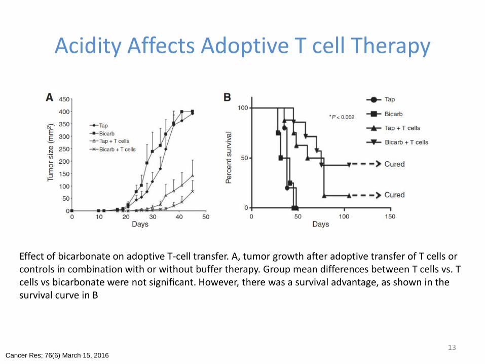
Acidity Affects Adoptive T cell Therapy
Cancer Res; 76(6) March 15, 2016
Effect of bicarbonate on adoptive T-cell transfer. A, tumor growth after adoptive transfer of T cells or controls in combination with or without buffer therapy. Group mean differences between T cells vs. T cells vs bicarbonate were not significant. However, there was a survival advantage, as shown in thesurvival curve in B
13

Tumor pH and Check-Point Inhibitors
14
Buffer therapy enhances efficacy of anti-immunotherapy in B16 melanoma. C57BL/6
Cancer Res; 76(6) March 15, 2016
DOS47Targeting the tumor microenvironment
15
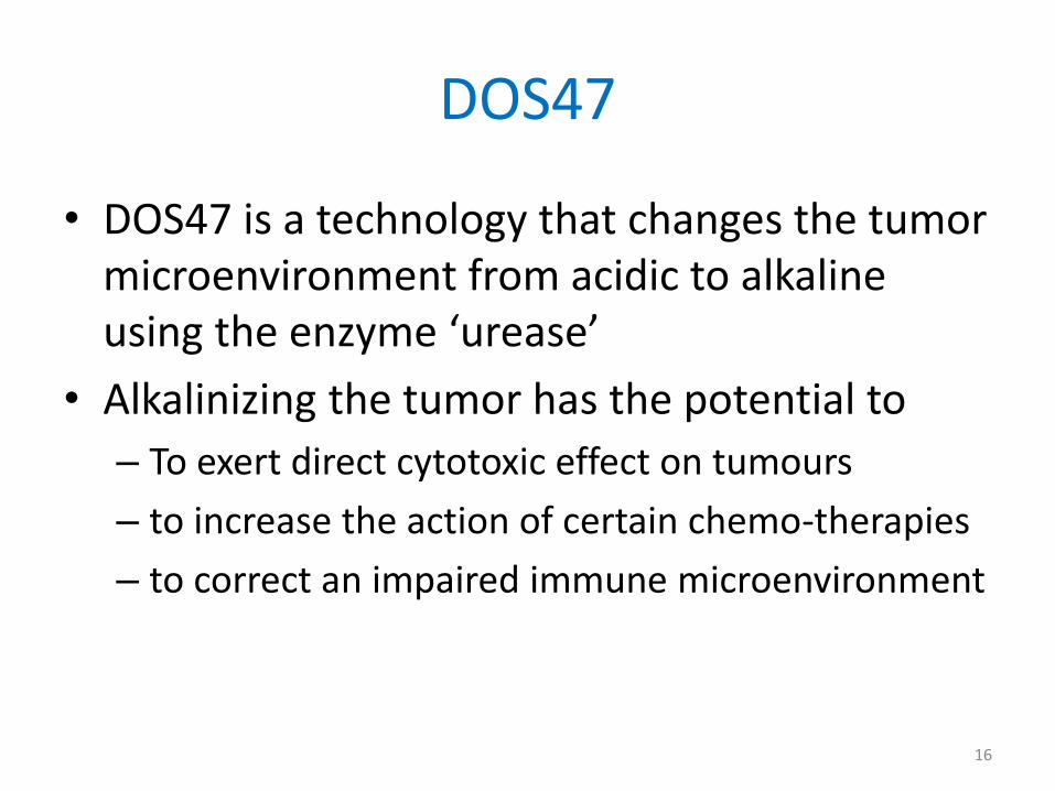
DOS47
• DOS47 is a technology that changes the tumor microenvironment from acidic to alkaline using the enzyme ‘urease’
• Alkalinizing the tumor has the potential to
– To exert direct cytotoxic effect on tumours
– to increase the action of certain chemo-therapies
– to correct an impaired immune microenvironment
16

L-DOS47Helix First Clinical Drug Candidate
17
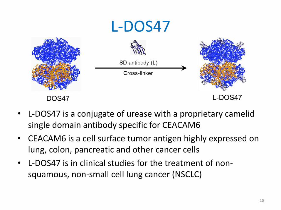
L-DOS47
• L-DOS47 is a conjugate of urease with a proprietary camelid single domain antibody specific for CEACAM6
• CEACAM6 is a cell surface tumor antigen highly expressed on lung, colon, pancreatic and other cancer cells
• L-DOS47 is in clinical studies for the treatment of non-squamous, non-small cell lung cancer (NSCLC)
18
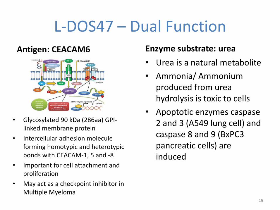
L-DOS47 – Dual FunctionAntigen: CEACAM6
• Glycosylated 90 kDa (286aa) GPI-linked membrane protein
• Intercellular adhesion molecule forming homotypic and heterotypic bonds with CEACAM-1, 5 and -8
• Important for cell attachment and proliferation
• May act as a checkpoint inhibitor in Multiple Myeloma
Enzyme substrate: urea
• Urea is a natural metabolite
• Ammonia/ Ammonium produced from urea hydrolysis is toxic to cells
• Apoptotic enzymes caspase 2 and 3 (A549 lung cell) and caspase 8 and 9 (BxPC3 pancreatic cells) are induced
19
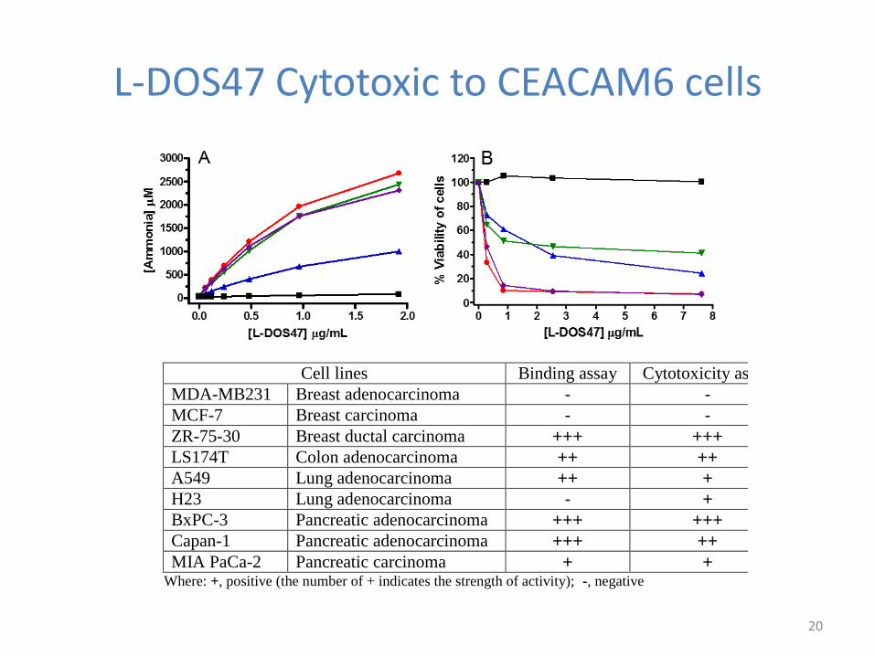
L-DOS47 Cytotoxic to CEACAM6 cells
Cell lines Binding assay Cytotoxicity assay
MDA-MB231 Breast adenocarcinoma - - MCF-7 Breast carcinoma - -
ZR-75-30 Breast ductal carcinoma +++ +++ LS174T Colon adenocarcinoma ++ ++ A549 Lung adenocarcinoma ++ + H23 Lung adenocarcinoma - + BxPC-3 Pancreatic adenocarcinoma +++ +++ Capan-1 Pancreatic adenocarcinoma +++ ++
MIA PaCa-2 Pancreatic carcinoma + + Where: +, positive (the number of + indicates the strength of activity); -, negative
20
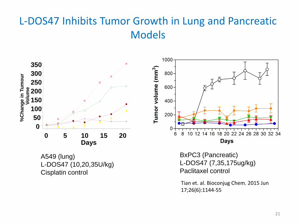
L-DOS47 Inhibits Tumor Growth in Lung and Pancreatic Models
0 5 10 15 20
0
50
100
150
200
250
300
350
Days
%C
ha
ng
e i
n T
um
ou
rV
olu
me
A549 (lung)
L-DOS47 (10,20,35U/kg)
Cisplatin control
BxPC3 (Pancreatic)
L-DOS47 (7,35,175ug/kg)
Paclitaxel control
Tian et. al. Bioconjug Chem. 2015 Jun 17;26(6):1144-55
21
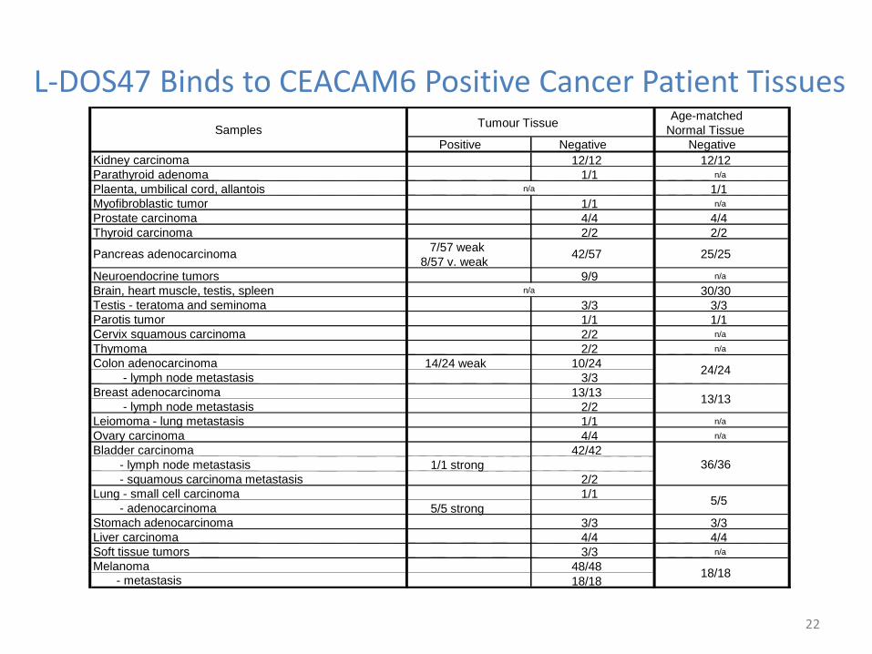
L-DOS47 Binds to CEACAM6 Positive Cancer Patient Tissues
Positive Negative Negative
Kidney carcinoma 12/12 12/12
Parathyroid adenoma 1/1 n/a
Plaenta, umbilical cord, allantois 1/1
Myofibroblastic tumor 1/1 n/a
Prostate carcinoma 4/4 4/4
Thyroid carcinoma 2/2 2/2
7/57 weak
8/57 v. weak
Neuroendocrine tumors 9/9 n/a
Brain, heart muscle, testis, spleen 30/30
Testis - teratoma and seminoma 3/3 3/3
Parotis tumor 1/1 1/1
Cervix squamous carcinoma 2/2 n/a
Thymoma 2/2 n/a
Colon adenocarcinoma 14/24 weak 10/24
- lymph node metastasis 3/3
Breast adenocarcinoma 13/13
- lymph node metastasis 2/2
Leiomoma - lung metastasis 1/1 n/a
Ovary carcinoma 4/4 n/a
Bladder carcinoma 42/42
- lymph node metastasis 1/1 strong
- squamous carcinoma metastasis 2/2
Lung - small cell carcinoma 1/1
- adenocarcinoma 5/5 strong
Stomach adenocarcinoma 3/3 3/3
Liver carcinoma 4/4 4/4
Soft tissue tumors 3/3 n/a
Melanoma 48/48- metastasis 18/18
18/18
36/36
24/24
13/13
n/a
5/5
Samples
Pancreas adenocarcinoma 42/57
Age-matched
Normal TissueTumour Tissue
n/a
25/25
22
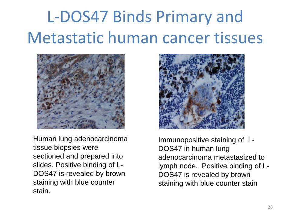
L-DOS47 Binds Primary and Metastatic human cancer tissues
Human lung adenocarcinoma
tissue biopsies were
sectioned and prepared into
slides. Positive binding of L-
DOS47 is revealed by brown
staining with blue counter
stain.
Immunopositive staining of L-
DOS47 in human lung
adenocarcinoma metastasized to
lymph node. Positive binding of L-
DOS47 is revealed by brown
staining with blue counter stain
23
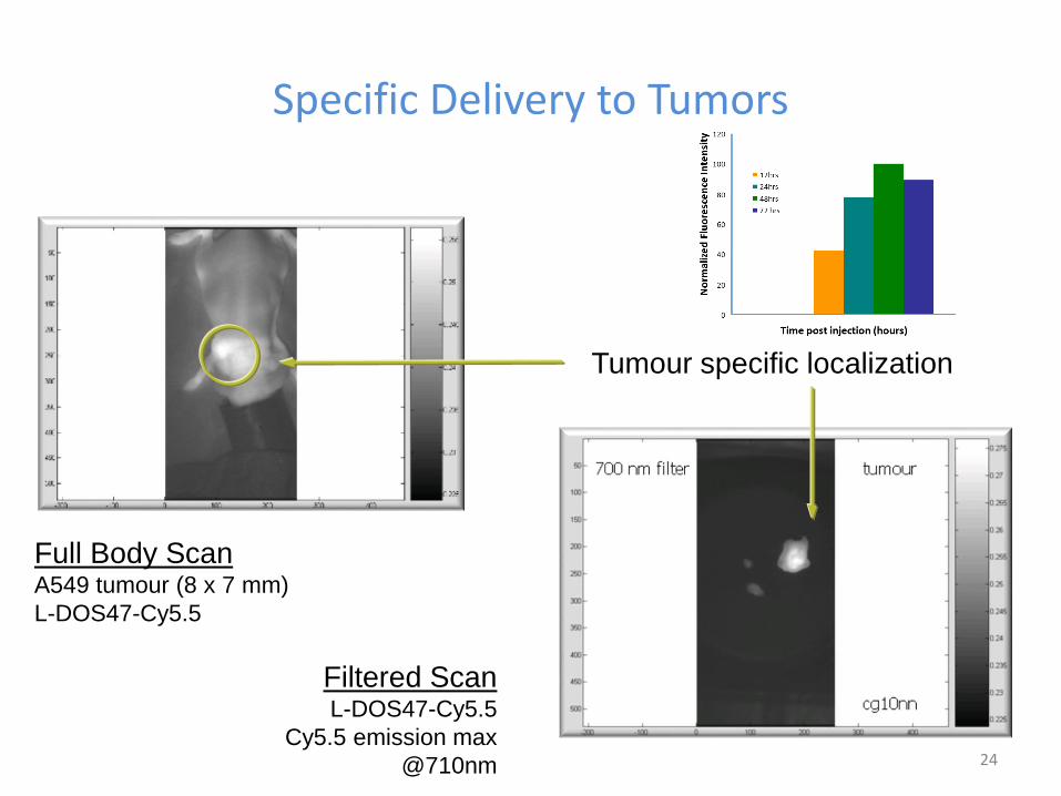
Specific Delivery to Tumors
24
Full Body ScanA549 tumour (8 x 7 mm)
L-DOS47-Cy5.5
Filtered ScanL-DOS47-Cy5.5
Cy5.5 emission max
@710nm
Tumour specific localization
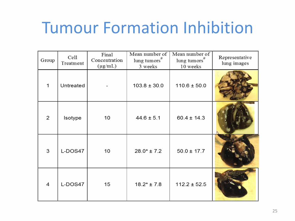
Tumour Formation Inhibition
25

L-DOS47 Action Monitored by NMR1H-NMR anatomical imaging
control
Treatment
32P-NMR microenvironment
NMR imaging on A549 xenograft mice showing a change in energy metabolism
(Pi/Pcr) as a result of L-DOS47 treatment26
L-DOS47 Clinical Update
• L-DOS47 Phase I / II Trial (LDOS002) – Monotherapy in advanced NSCLC patients
– Currently enrolling Phase II patients
• L-DOS47 Phase I with Expansion Trial (LDOS001)– Combination with pemetrexed and carboplatin
– Currently enrolling in cohort 2
• L-DOS47 Phase II (LDOS003)– Combination with vinorelbine and cisplatin
– In the planning phase
27
L-DOS47 Phase I / II Trial (LDOS002)• Monotherapy treatment protocol in NSCLC patients that have not
responded to other treatments;
• Stage IIIb / IV, metastatic, and progression after several lines of chemo, rad, surgery or chemo-naïve patients that have refused other lines of therapy;
• Dosed once a week for 2 weeks, 1 week rest (3-week cycle);
• Conducted in 5 Centers in Poland to assess safety (phase I) and then preliminary efficacy (phase II);
• Centers include The Maria Sklodowska-Curie Institute of Oncology, Military Institute of Health Institute, Mazovian Centre of Pulmonary Diseases and Tuberculosis in Otwock, Department of Oncology, Poznan University of Medical Sciences, National Tuberculosis and Lung Diseases Research Institute
• Phase II dosing regimen changed to twice a week dosing for 2 weeks, 1 week rest (3-week cycle);
28
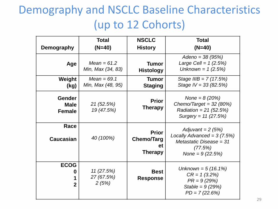
Demography
Total
(N=40)
NSCLC
History
Total
(N=40)
Age Mean = 61.2
Min, Max (34, 83)Tumor
Histology
Adeno = 38 (95%)
Large Cell = 1 (2.5%)
Unknown = 1 (2.5%)
Weight
(kg)
Mean = 69.1
Min, Max (48, 95)Tumor
Staging
Stage IIIB = 7 (17.5%)
Stage IV = 33 (82.5%)
Gender
Male
Female
21 (52.5%)
19 (47.5%)
Prior
Therapy
None = 8 (20%)
Chemo/Target = 32 (80%)
Radiation = 21 (52.5%)
Surgery = 11 (27.5%)
Race
Caucasian 40 (100%)Prior
Chemo/Targ
et
Therapy
Adjuvant = 2 (5%)
Locally Advanced = 3 (7.5%)
Metastatic Disease = 31
(77.5%)
None = 9 (22.5%)
ECOG
0
1
2
11 (27.5%)
27 (67.5%)
2 (5%)
Best
Response
Unknown = 5 (16.1%)
CR = 1 (3.2%)
PR = 9 (29%)
Stable = 9 (29%)
PD = 7 (22.6%)
Demography and NSCLC Baseline Characteristics (up to 12 Cohorts)
29
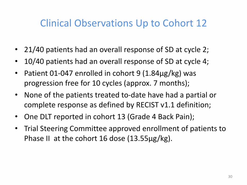
Clinical Observations Up to Cohort 12
• 21/40 patients had an overall response of SD at cycle 2;
• 10/40 patients had an overall response of SD at cycle 4;
• Patient 01-047 enrolled in cohort 9 (1.84µg/kg) was progression free for 10 cycles (approx. 7 months);
• None of the patients treated to-date have had a partial or complete response as defined by RECIST v1.1 definition;
• One DLT reported in cohort 13 (Grade 4 Back Pain);
• Trial Steering Committee approved enrollment of patients to Phase II at the cohort 16 dose (13.55µg/kg).
30

L-DOS002 Summary of Results
31
• One Dose Limiting Toxicity (DLT);• No safety issues beyond those observed in pre-clinical
toxicology studies or expected in the population of patients being studied;
• Immunogenicity consistent with what was observed pre-clinically;
• Clinical trends observed to-date are encouraging;• Phase II currently enrolling.
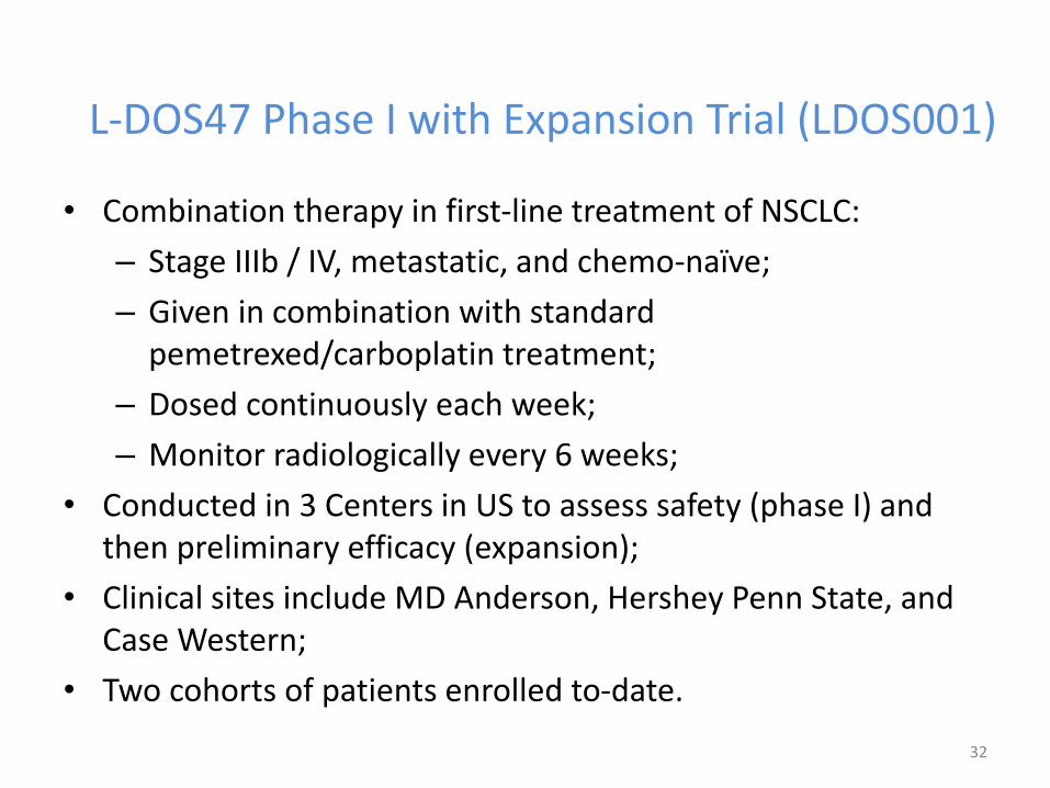
L-DOS47 Phase I with Expansion Trial (LDOS001)
• Combination therapy in first-line treatment of NSCLC:
– Stage IIIb / IV, metastatic, and chemo-naïve;
– Given in combination with standard pemetrexed/carboplatin treatment;
– Dosed continuously each week;
– Monitor radiologically every 6 weeks;
• Conducted in 3 Centers in US to assess safety (phase I) and then preliminary efficacy (expansion);
• Clinical sites include MD Anderson, Hershey Penn State, and Case Western;
• Two cohorts of patients enrolled to-date.
32

Tumor pH Treatment Strategy: DOS47*
Cancer Cell 2008, 13 472-482 33
*
*
*

THANK YOUHelix BioPharma Corp
34