Embed Size (px)
Citation preview

Pediatric Cancer
Roswell Park Cancer Institute
Department of Pediatrics
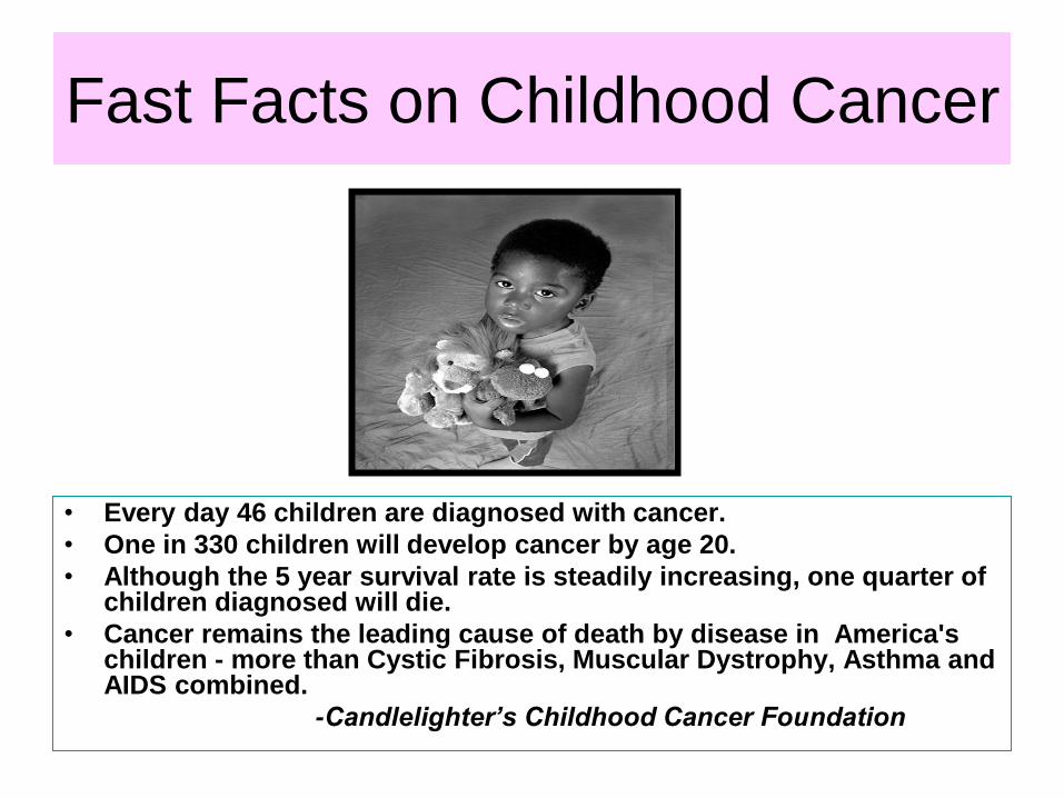
Fast Facts on Childhood Cancer
• Every day 46 children are diagnosed with cancer.
• One in 330 children will develop cancer by age 20.
• Although the 5 year survival rate is steadily increasing, one quarter of children diagnosed will die.
• Cancer remains the leading cause of death by disease in America's children - more than Cystic Fibrosis, Muscular Dystrophy, Asthma and AIDS combined.
-Candlelighter’s Childhood Cancer Foundation

Leading Causes of Death in
Children

Types of Cancer Distribution

Etiology
• Unknown
• Genetics – some chromosomal
abnormalities
– identical twins - 20% concordance rate for leukemia if < 6 yrs
• Environmental - prenatal vs post natal – radiation
– chemical carcinogens
– diet - no evidence in children
• Viral Oncogenesis – EBV in Burkitt’s,
Hodgkin’s
• Immune deficiency – increase in lymphoid
malignancies with congenital immunodeficiency
– cancer in AIDS patients

Acute Lymphoblastic Leukemia
• Most common
pediatric
malignancy
• 80% of pediatric
leukemias
• 1/25,000
• peaks at 3-5 years
• boys>girls

Normal Bone Marrow
Lymphoblasts Normal Bone Marrow
=

ALL
• Presentation
– Related to bone marrow not functioning properly: High or low WBC (bone pain), anemia, thrombocytopenia (petechiae, epistaxis), elevated LDH, elevated uric acid
– CNS disease Headaches, blurred vision
– Testicular disease, swelling, mass
• Differential diagnosis
– Mono, CMV, ITP, JRA, aplastic anemia, neuroblastoma,

ALL
• Diagnosis - bone marrow aspirate
– morphology, histochemistry, flow cytometry,
cytogenetics
• LP for CNS disease - prognosis poorer,
prophylaxis for low/standard risk
• Prognosis
– WBC > 50K, age <1 or >10 high risk
– DI > 1.16 favorable
– Chromosones favorable vs. unfavorable

Philadelphia Chromosone

ALL
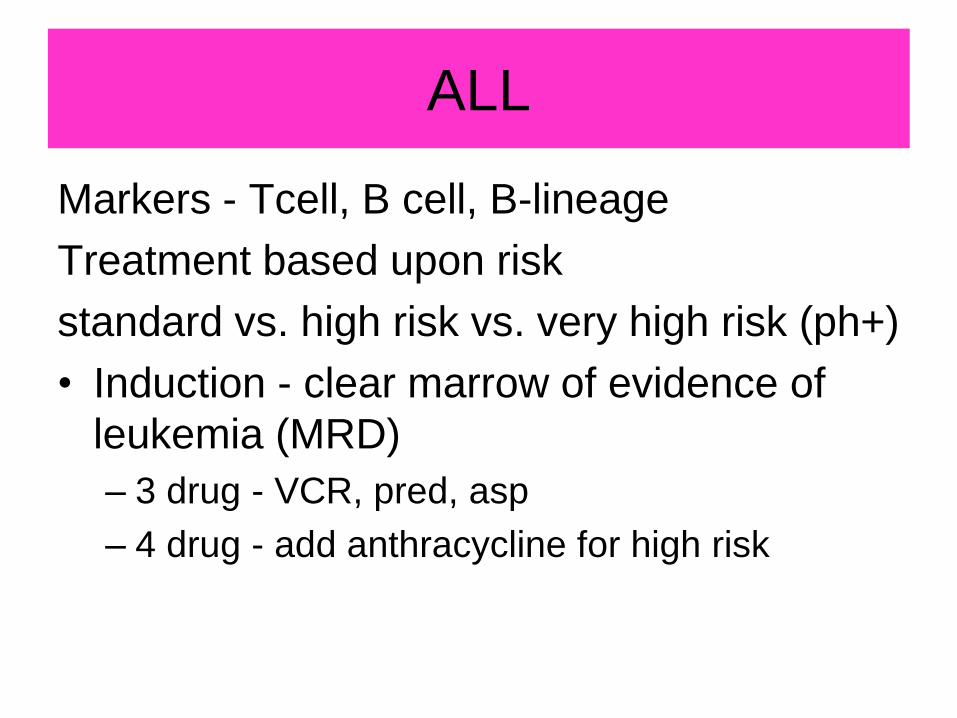
Markers - Tcell, B cell, B-lineage
Treatment based upon risk
standard vs. high risk vs. very high risk (ph+)
• Induction - clear marrow of evidence of
leukemia (MRD)
– 3 drug - VCR, pred, asp
– 4 drug - add anthracycline for high risk

ALL
• Intensification/Consoli
dation
• Maintenance
outpatient
• CNS prophylaxis - IT
meds vs. Cranial RT
• Treatment 2 ½ yrs
girls 3 yrs boys
• 80% cure overall

What to do when you refractory
or relapse? • Additional chemotherapy
• Transplant – depends on timing of relapse
• Where you come in – novel therapy
– CarT cells
– Blinatumomab – antibody with two binding
sites: a CD3 site for T cells and a CD19 site
for the target B cells
– Targeted therapy based on mutations –
Gleevac, dasatinib

Car T Cells

Acute Myelogenous Leukemia
• 500 new cases per year
• No age, sex preference
• presentation –anemia, thrombocytopenia (bruising, epistaxis), elevated/low WBC, DIC, gingivitis, hepatospleenomegaly
• 20% with WBC > 100,000
• Bone marrow aspirate/biopsy
• LP CNS disease less common than ALL

Myeloblasts

AML
• Treatment
– induction -Dauno,
AraC, 6TG
– intensification - HD
AraC
– Allogeneic BMT vs
chemo
• Cure rates - 60-70%
sib allo BMT vs. 30-
40% chemo only

So where do you come?
• Tyrosine Kinase inhibitors
– Sorafanib and FLt3-ITD
• Histone deacteylase Inhibitors
– Valproic acid, new agents, combination
• DNA-hypomethylating agent
– 5-azacitadine, decitabine
• Targeted therapy
• Immune Modulation
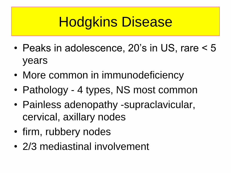
Hodgkins Disease
• Peaks in adolescence, 20’s in US, rare < 5
years
• More common in immunodeficiency
• Pathology - 4 types, NS most common
• Painless adenopathy -supraclavicular,
cervical, axillary nodes
• firm, rubbery nodes
• 2/3 mediastinal involvement

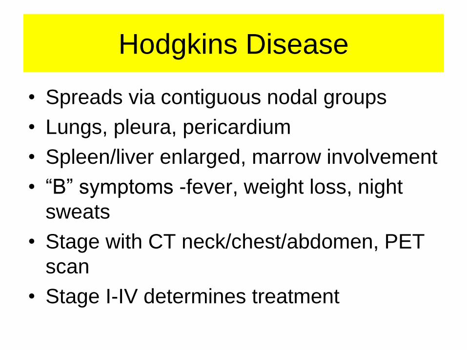
Hodgkins Disease
• Spreads via contiguous nodal groups
• Lungs, pleura, pericardium
• Spleen/liver enlarged, marrow involvement
• “B” symptoms -fever, weight loss, night
sweats
• Stage with CT neck/chest/abdomen, PET
scan
• Stage I-IV determines treatment

Hodgkins Disease
• Bone marrow aspirate/biopsy if advanced
disease
• Therapy
– Trend toward low dose chemo + low dose
XRT in local disease
– Advanced stage more aggressive chemo and
involved field radiation
– Recurrence Autologous BMT

Where do you come in?
• Brentuximab Vedotin
– Anti CD 30 linked to antitubulin agent
monomethyl auristatin E
• Lintuzumab anti-CD33
– a humanized IgG1 anti-CD33 antibody
– inhibit tumor assocaiated macrophage
function (thought to promote tumor growth)

Non-Hodgkins Lymphoma
• Very different from
adults - nearly all high
grade
• Small noncleaved
(undifferentiated) -
Burkitt’s
• Lymphoblastic
• Large cell

NHL
Small non-cleaved - “Burkitt’s”
– B cell, express surface immunoglobulin,
– abdominal mass +/- ascites, pain
– May have inguinal, iliac adenopathy
– Fast growing triplicates in 24 hours
Differential diagnosis: intussception, right iliac
fossa mass, confused with appendicitis
Large cell
– usually B cell phenotype
– presentation similar to small noncleaved

NHL
• Lymphoblastic
– T cell phenotype
– mediastinal mass, pleural effusion
– SVC syndrome, dyspnea, often ICU admit
– cervical adenopathy
– abdominal involvement uncommon
• Staging - CT chest/abdomen/pelvis, bone
marrow, LP
• Prognosis - tumor burden

Normal
Chest X-Ray

Mediastinal Mass

NHL
• Therapy –
Chemotherapy
– Radiation no benefit
except in
emergencies
– chemo differs, based
on cell type
– intensive, multiagent
chemotherapy
– CNS prophylaxis:
intrathecal
chemotherapy and
cranial radiation.

Brain Tumors
• Most common solid
tumor (1200 per year)
• Presentation dependent
upon site of origin, not
histology
• Obstructing, increased
ICP - classic triad:
- morning headaches, -
- nausea/vomiting,
- diplopia

Brain Tumors
• Subacute ICP - poor school performance, fatigue, personality change, Headaches
• Infants, toddlers - irritable, anorexia, developmental delay, loss of milestones, optic pallor, macrocephaly
• Infratentorial -balance, truncal instability, difficulty with coordination, gait disturbance (ataxia)
• Supratentorial - seizure, hemiparesis, hemisensory loss, visual field defect

Brain Tumors
• Staging - MRI brain, spine for mets, LP for
cytology, bone marrow aspirate/biopsy
• Treatment - SURGERY -prognosis better
• Craniospinal radiation
• Chemo - less of role for many types
– advanced, metastatic
– attempt to decrease XRT dose due to
long term effects


Wilm’s Tumor
• Most common
malignant renal tumor
in children
• 460 cases per year
• Mean age 3-4 yrs
• WAGR Syndrome -
del 11p13
• Beckwith-Wiedemann

Wilm’s Tumor
• Abdominal swelling
mass
• Abdominal pain,
hematuria, fever
• Imaging
– US, abdominal CT
– MRI for caval
patency
– CXR for pulmonary
mets

Wilm’s Tumor
• Surgery upfront -
nephrectomy
• Chemotherapy - VCR,
actino +/- doxo
• XRT for advanced
stages
• 65-90% RFS, overall
80%
• 5-7% bilateral

Neuroblastoma
• Most common
extracranial solid
tumor (525 cases per
year)
• Histology
– Small round blue cell
tumor
– derived from post
ganglionic sympathetic
neuroblasts
• Arise in any site along
sympathetic chain

Neuroblastoma
• Most primaries - abdomen (adrenal)
• Infants - thoracic, cervical
• Most cases < 5yrs, rare > 10 yrs
• Metastasis - lymphatic, hematogenous
• Infants more localized vs older children
more metastatic
• Cytogenetic chromosonal abnormalities

Neuroblastoma
• Surgery - pivotal role
• Chemotherapy -
aggressive,
multiagent
• radiation for
advanced stages
• High-dose chemo
with auto BMT? -
delay recurrence?

What science as done and what
we still need to do • Immune modulation and antibody therapy
• Teasing the disease apart to decrease
toxicity and increase cure.

Horner’s Syndrome

Mediastinal mass in neuroblastoma

MIBG
Bone
metastasis

Bone Tumors Osteogenic Sarcoma
• 7th largest, 3rd largest group in adolescents
• Osteosarcoma – distal femur, proximal tibia, proximal humerus
– Metaphysis of the bone
– pain, soft tissue mass
– 20% metastatic at diagnosis- lung, bone
– “Codman’s triangle”
– stage - MRI primary, CT chest, bone scan
– Neo-adjuvant chemo - limb-sparing surgery
– 80-90% RFS
– Lung mets at diagnosis decrease survival rate


Bone Tumors Ewings Sarcoma
• Ewing’s Sarcoma – any bone - pelvis,
femur, tibia, fibula, scapula, spine, ribs (axial)
– pain, swelling, fever
– metastasis - lungs, bone, marrow
– Plain film “onion skin” appearance
– Chemo, radiation