Embed Size (px)
Citation preview
The Structure of the Staphylococcus aureus Sortase-SubstrateComplex Reveals How the Universally Conserved LPXTGSorting Signal Is Recognized*□S
Received for publication, May 18, 2009, and in revised form, June 16, 2009 Published, JBC Papers in Press, July 10, 2009, DOI 10.1074/jbc.M109.022624
Nuttee Suree‡§1, Chu Kong Liew‡2, Valerie A. Villareal‡§3, William Thieu‡§, Evgeny A. Fadeev‡§4,Jeremy J. Clemens‡5, Michael E. Jung‡, and Robert T. Clubb‡§6
From the ‡Department of Chemistry and Biochemistry and the §UCLA-Department of Energy Institute for Genomics andProteomics, UCLA, Los Angeles, California 90095-1570
In Gram-positive bacteria, sortase enzymes assemble surfaceproteins and pili in the cell wall envelope. Sortases catalyze atranspeptidation reaction that joins a highly conserved LPXTGsorting signal within their polypeptide substrate to the cell wall orto other pilin subunits. The molecular basis of transpeptidationandsorting signal recognitionarenotwell understood,because theintermediates of catalysis are short lived. We have overcome thisproblemby synthesizing an analog of the LPXTGsignalwhose sta-ble covalent complex with the enzymemimics a key thioacyl cata-lytic intermediate. Here we report the solution structure anddynamics of its covalent complex with the Staphylococcus aureusSrtA sortase. In marked contrast to a previously reported crystalstructure, we show that SrtA adaptively recognizes the LPXTGsorting signal by closing and immobilizing an active site loop. Wehave also used chemical shift mapping experiments to localize thebinding site for the triglycine portion of lipid II, the second sub-strate towhich surfaceproteins are attached.Wepropose aunifiedmodel of the transpeptidation reaction that explains the functionsof key active site residues. Since the sortase-catalyzed anchoringreaction is required for the virulence of a number of bacterialpathogens, the results presented here may facilitate the develop-ment of new anti-infective agents.
Bacterial surface proteins function as virulence factors thatenable pathogens to adhere to sites of infection, evade theimmune response, acquire essential nutrients, and enter hostcells (1). Gram-positive bacteria use a common mechanism tocovalently attach proteins to the cell wall. This process is cata-lyzed by sortase transpeptidase enzymes, which join proteinsbearing a highly conserved Leu-Pro-X-Thr-Gly (LPXTG,where
X is any amino acid) sorting signal to the cross-bridge peptide ofthe peptidylglycan (2–4). Sortases also polymerize proteinscontaining sorting signals into pili, filamentous surface exposedstructures that promote bacterial adhesion (5, 6). The searchfor small molecule sortase inhibitors is an active area ofresearch, since these enzymes contribute to the virulence of anumber of important pathogens, including among othersStaphylococcus aureus, Listeria monocytogenes, Streptococcuspyogenes, and Streptococcus pneumoniae (reviewed in Refs. 7and 8). Sortase enzymes are also promising molecular biologyreagents that can be used to site-specifically attach proteins to avariety of biomolecules (9–14, 72).The sortaseA (SrtA)7 enzyme from S. aureus is the prototypical
member of the sortase enzyme family (15, 16). It anchors proteinsto the murein sacculus that possess a COOH-terminal cell wallsorting signal that consists of aLPXTGmotif, followedby ahydro-phobic segment of amino acids and a tail composed of mostlypositively charged residues (17). SrtA is located on the extracellu-lar side of themembrane.After partial secretion of its protein sub-strate across the cell membrane, SrtA cleaves the LPXTG motifbetween the threonine and glycine residues, forming a thioacyl-linked protein-sortase intermediate (16). It then catalyzes the for-mation of an amide bond between the carboxyl group of thethreonine and the cell wall precursor molecule lipid II (un-decaprenyl-pyrophosphate-MurNAc(-L-Ala-D-iGln-L-Lys(NH2-Gly5)-D-Ala-D-Ala)-�1–4-GlcNAc)), creating a protein-lipidII-linked product that is incorporated into the peptidylglycanvia the transglycosylation and transpeptidation reactions ofbacterial cell wall synthesis (18–20). Over 900 sortase-attachedproteins in 72 different strains of bacteria have thus far beenidentified (21, 22). The vast majority of these proteins contain aCOOH-terminal sorting signal harboring an LPXTGmotif andare anchored to the cell wall by enzymes closely related to SrtA.In vitro studies of SrtA have begun to define the mechanism
of transpeptidation. SrtA consists of two parts: an unstructuredamino-terminal tail that contains a stretch of nonpolar residuesthat embed it in the membrane and an autonomously foldedcatalytic domain that competently performs the transpeptida-tion reaction in vitro (SrtA�N59, residues 60–206) (16, 23–25).
* This work was supported, in whole or in part, by National Institutes of HealthGrant AI52217 (to R. T. C. and M. E. J.).
□S The on-line version of this article (available at http://www.jbc.org) containssupplemental Fig. S1 and Table S1.
1 Recipient of DPST and JSTP scholarships from the Royal Thai Government.2 Present address: Dept. of Molecular Cardiology and Biophysics, The Victor
Chang Cardiac Research Institute, Level 6, 384 Victoria St., Darlinghurst,New South Wales 2010, Australia.
3 Supported by National Institutes of Health, NIGMS, Grant F31GM075564.4 Present address: University of California, Irvine, CA 92697.5 Present address: Wyeth Research, 401 N. Middletown Rd., Pearl River, NY
10965.6 To whom correspondence should be addressed: Dept. of Chemistry and
Biochemistry, 611 Charles E. Young Dr., Los Angeles, CA 90095-1570. Tel.:310-206-2334; Fax: 310-206-4749; E-mail: [email protected].
7 The abbreviations used are: SrtA, sortase A; Cbz, benzylozycarbonyl; T*,(2R,3S)-3-amino-4-mercapto-2-butanol analog of threonine that replacesthe carbonyl group with -CH2-SH; NOE, nuclear Overhauser effect; NOESY,NOE spectroscopy; MALDI-TOF, matrix-assisted laser desorption ionizationtime-of-flight; r.m.s., root mean square.
THE JOURNAL OF BIOLOGICAL CHEMISTRY VOL. 284, NO. 36, pp. 24465–24477, September 4, 2009© 2009 by The American Society for Biochemistry and Molecular Biology, Inc. Printed in the U.S.A.
SEPTEMBER 4, 2009 • VOLUME 284 • NUMBER 36 JOURNAL OF BIOLOGICAL CHEMISTRY 24465
by guest on July 29, 2019http://w
ww
.jbc.org/D
ownloaded from
Catalysis occurs through a ping-pong mechanism that is initi-ated when the thiol group of amino acid Cys184 nucleophilicallyattacks the carbonyl carbon of the threonine residue within theLPXTG sorting signal (16, 23–25). This forms a transient tetra-hedral intermediate that, upon breakage of the threonine-gly-cine peptide bond, rearranges into a more stable thioacylenzyme-substrate linkage. SrtA then joins the terminal aminegroup within the pentaglycine branch of lipid II to the carbonylcarbon of the threonine, creating a second tetrahedral interme-diate that is resolved into the lipid II-linked protein product(23).Sortase enzymes contain three conserved residues within
their active sites: His120, Cys184, and Arg197 (SrtA numbering).These residues play a critical role in catalysis, since their muta-tion in SrtA causes severe reductions in enzyme activity (16,26–30). Although it is well established that Cys184 forms acovalent linkage to the sorting signal, the functions of His120
and Arg197 are controversial. A variety of disparate functionshave been ascribed to Arg197. These include deprotonatingCys184 (28), deprotonating lipid II (31), or stabilizing the bind-ing of either the LPXTG sorting signal (28, 32) or oxyanionintermediates (31, 32). Different functions have also been pro-posed for His120. Originally, it was suggested that it activatedCys184 by forming an imidazolium-thiolate ion pair (26). How-ever, subsequent pKa measurements revealed that both His120
and Cys184 are predominantly uncharged at physiological pHvalues, leading to the suggestion that His120 functions as a gen-eral base during catalysis (33). Most recently, it has been pro-posed that the most active form of the enzyme contains His120
and Cys184 in their charged states but that only a small fractionof SrtA exists in this form (�0.06%) prior to binding to thesorting signal (25).NMR and crystal structures of SrtA�N59 have revealed that
it adopts an eight-stranded �-barrel fold (31, 34). Other sor-tase enzymes have also been shown to possess a similar over-all structure, including SrtB from S. aureus (27, 35), SrtBfrom Bacillus anthracis (27, 36), SrtA from S. pyogenes (37),and the SrtC-1 and SrtC-3 enzymes from S. pneumoniae(38). However, the molecular basis of substrate recognitionremains poorly understood, because all of the structuresreported to date have not contained a sorting signal bound tothe enzyme. The lone exception is the crystal structure ofSrtA�N59 bound to an LPETG peptide (31). However, in thisstructure the peptide substrate is bound nonspecifically (seebelow) (32, 39).In this paper, we report the structure and dynamics of SrtA
covalently bound to an analog of the LPXTG sorting signal. Thestructure of the complex resembles the thioacyl intermediate ofcatalysis, providing insights into the molecular basis of bindingof the LPXTG sorting signal and the functions of key active siteresidues. Notably, the mechanism of substrate binding visual-ized in the NMR structure differs substantially from a previ-ously reported crystal structure of SrtA�N59 non-covalentlybound to a LPETGpeptide (31).Wehave also usedNMRchem-ical shift mapping experiments to localize the binding site for atriglycine cell wall substrate analog. A mechanism of transpep-tidation compatible with these new data is proposed.
EXPERIMENTAL PROCEDURES
Preparation of the Covalent Complex for NMR Studies—Wild-type SrtA from S. aureus containing amino acid residues60–206 (SrtA�N59) was produced as described previously (29).Uniformly 15N- and 13C- or 15N-labeled SrtA�N59 protein wascovalently attached to an analog of the LPXTG sorting signal,Cbz-LPAT* (where T* is (2R,3S)-3-amino-4-mercapto-2-buta-nol, and Cbz is a carbobenzyloxy protecting group). The meth-ods used to synthesize the analog and to prepare its covalentcomplex with SrtA�N59 have been described previously (40).Three �1 mM samples of the complex were studied. Each wasdissolved in 50 mM Tris-HCl (pH 6.0), 100 mM NaCl, 20 mM
CaCl2, and 0.01% NaN3. The complexes contained either 1)15N-labeled SrtA�N59 bound to the unlabeled peptide dissolvedin H2O (7% 2H2O), 2) 13C,15N-labeled protein bound to theunlabeled peptide dissolved in H2O (7% 2H2O), or 3) 13C,15N-labeled protein bound to the unlabeled peptide dissolved in2H2O.NMR Spectroscopy and Structure Determination—NMR
spectra were acquired at 302 K on Bruker Avance 500-, 600-,and 800-MHz spectrometers equipped with triple resonancecryogenic probes. 1H, 13C, and 15N protein chemical shiftassignments were obtained using standard methods (41, 42).Chemical shift assignments for the sorting signal were obtainedby analyzing two-dimensional (F1,F2) 13C-filtered NOESY (43)and (F1) 13C-filtered TOCSY (44) spectra. Distance restraintsto define the structure of the protein were obtained from three-dimensional 15N- and 13C-edited NOESY spectra (mixing time100 ms), and intramolecular restraints for the sorting signalanalog were obtained by analyzing two-dimensional (F1,F2)13C-filteredNOESY spectra. Intermolecular distance restraintsbetween SrtA�N59 and the bound peptide were identified inthree-dimensional (F1) 13C,15N-filtered (F2) 13C-editedNOESY-HSQC and (F1) 13C,15N-filtered (F2) 15N-editedNOESY-HSQC spectra (45) and in a two-dimensional (F1) 13C-filtered NOESY spectrum (43). 3JHN-H� values were measuredfrom a water flip-back three-dimensional HNHA spectrum(46), and backbone� and� dihedral angleswere obtained usingthe programTALOS (47). TheNMRdata were processed usingNMRPipe (48) and analyzed using the PIPP (49) and CARA(version 1.4.1) (50) software packages.Structure calculations were performed using the ATNOS/
CANDID and NIH-XPLOR programs (51–53). Three three-dimensional NOESY data sets were used as input for ATNOS/CANDID: three-dimensional 13C-edited NOESY-HSQC and15N-edited NOESY-HSQC spectra of the complex dissolved inH2O and a 13C-editedNOESY-HSQC spectrum of the complexdissolved in 2H2O. These data were supplemented withrestraints for the backbone and side chain dihedral angles.Seven cycles of ATNOS/CANDID calculations yielded a con-verged ensemble of the protein in the complex. The structurewas then refined in an iterative manner by manually checkingall of the NOEs assigned by CANDID and by including 3JHN-H�
couplings and carbon chemical shifts in the calculations. Newmanually identified intra- and intermolecular distancerestraints were also added, and at the final stages of refinement,hydrogen bonds were identified and included as distance
NMR Structure of the Covalent Sortase-sorting Signal Complex
24466 JOURNAL OF BIOLOGICAL CHEMISTRY VOLUME 284 • NUMBER 36 • SEPTEMBER 4, 2009
by guest on July 29, 2019http://w
ww
.jbc.org/D
ownloaded from
restraints. The latter were obtained by inspecting the structuresof the complex and by identifying NOE patterns characteristic ofdistinct secondarystructures.Allof theNOEswithin theactive siteand binding pocket weremanually checked. Residual dipolar cou-plings were measured by taking the difference in J couplingsbetween partially aligned and unaligned protein samples. Theprotein was aligned using 15% (w/v) charged bicelles (30:10:1molar ratio of dimyristoylphosphatidylcholine/dihexanoyl-phosphatidylcholine/hexadecyl(cetyl)trimethylammonium bro-mide). The programs MOLMOL (54) and PyMOL (55) wereused to generate figures.NMR Relaxation Studies—NMR data were collected using
the 15N-labeled sample of the complex at 600 MHz. The strat-egy used to collect and analyze the relaxation data has beendescribed previously (56, 57). The well resolved 1H-15N HSQCspectrum enabled the reliable measurement of relaxationparameters for 86 of a total of 148 residues. The average R1, R2,and 15N{1H} NOE values for the backbone nitrogen atoms inthe complex are 1.50 � 0.02 s�1, 12.28 � 0.16 s�1, and 0.62 �0.10, respectively. The tensor parameters were calculated usingthe program Quadric_Diffusion, which follows the approachoutlined by Bruschweiler et al. (58, 59). This yielded a correla-tion time of 8.56 ns, and the axial symmetric model was pre-ferred over the isotropic model. A total of 76 of 86 quantifiableresidues could be fit satisfactorily. The data from Ala81, Val87,Tyr88, Arg99, Ser102, Asn132, Ala135, Lys137, Met155, and Asp165could not be fit to any model, possibly because they undergomore complicated motions. Residues were classified as follows:model 1 (S2-only) was selected for 52 residues, 2 residues fit tomodel 2 (S2 and �e), 15 residues fit to model 3 (S2 and Rex), 1residue fit tomodel 4 (S2, �e, andRex), and 6 residues fit tomodel5 (Sf2, Ss2, and �e). For residues located in regions of regularsecondary structure, the average order parameter is 0.93 �0.01. The relaxation data were analyzed using the suite of anal-ysis programs kindly provided by Prof. Arthur G. Palmer III(60–63).Localization of the Lipid II Binding Pocket—These studies
made use of a 1 mM sample of the complex containing 15N-labeled SrtA�N59 covalently attached to the sorting signal ana-log (NMR buffer: 50mMTris-HCl, 100mMNaCl, 20mMCaCl2,0.01% NaN3, and 7% 2H2O, pH 6.0). Triglycine (Gly3) wasobtained from Sigma. A 500 mM concentrated stock solution ofGly3 dissolved in NMR buffer was used. A series of two-dimen-sional 1H,15NHSQCspectrawere recordedat302Kafter theaddi-tion of small aliquots of Gly3. A total of 12 spectra were acquiredwith the following molar ratios of Gly3 to the SrtA�N59-LPAT*complex: 0:1, 0.5:1, 1:1, 2:1, 3:1, 4:1, 8:1, 16:1, 40:1, 50:1, 80:1, and100:1. No significant chemical shift changes were observed after a40:1molar ratiowas achieved.Anormalizedchemical shift change(��) was calculated as �� � ((��H)2 � (��N/6.49)2)
1⁄2, where ��Nand��Hare, respectively, theamidenitrogenandprotonchemicalshift difference for a given residue in the presence and absence ofGly3. The titration experiments using the apo-formof the enzymewere performed in an identical manner.Site-directed Mutagenesis and Enzyme Assays—Five single
amino acid mutants of SrtA�N59 containing a COOH-terminalsix-histidine tag were produced in Escherichia coli from apET15b expression vector. The presence of the histidine tag
does not affect the enzymatic activity of the protein (9, 56, 64).Mutations were made using the QuikChange� method (Strat-agene) and confirmed by DNA sequencing. Mutant and wild-type enzymes were purified as described previously (56), andtheir proper folding was confirmed by one-dimensional 1HNMR.The enzyme kinetic parameters of five SrtA�N59mutants
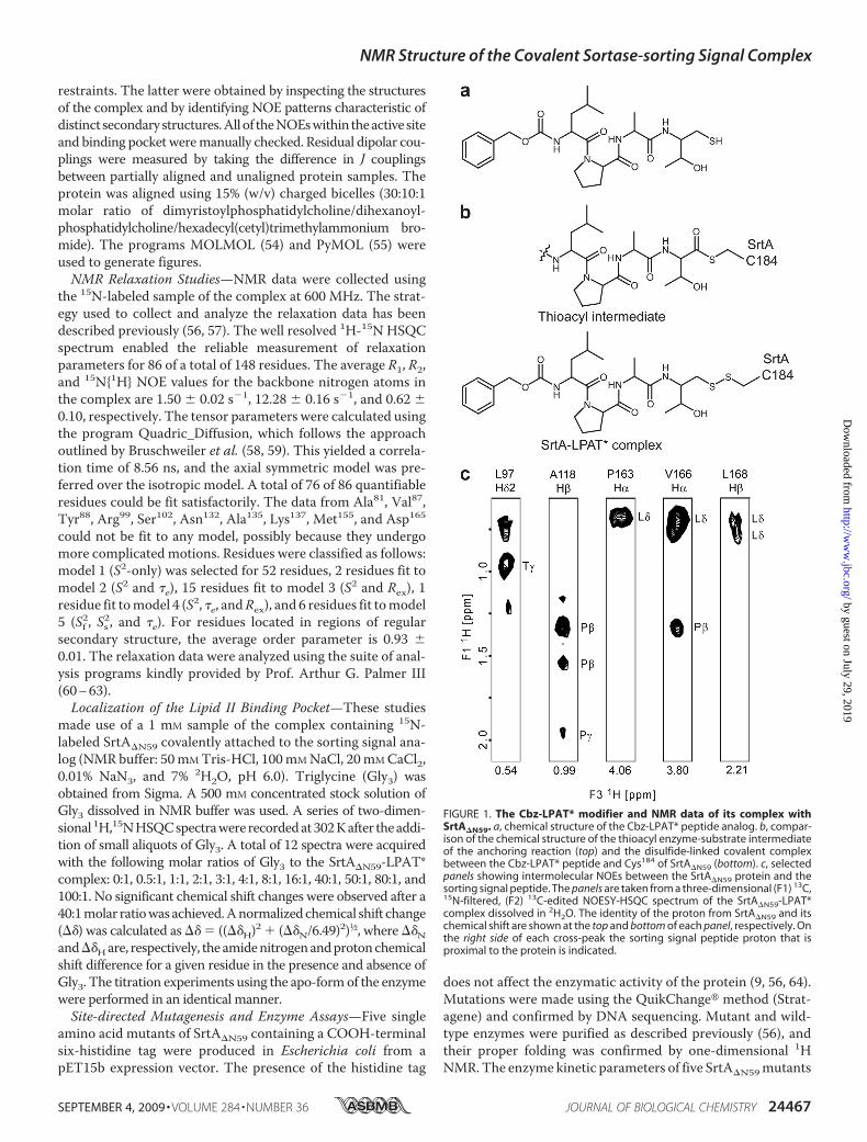
FIGURE 1. The Cbz-LPAT* modifier and NMR data of its complex withSrtA�N59. a, chemical structure of the Cbz-LPAT* peptide analog. b, compar-ison of the chemical structure of the thioacyl enzyme-substrate intermediateof the anchoring reaction (top) and the disulfide-linked covalent complexbetween the Cbz-LPAT* peptide and Cys184 of SrtA�N59 (bottom). c, selectedpanels showing intermolecular NOEs between the SrtA�N59 protein and thesorting signal peptide. The panels are taken from a three-dimensional (F1) 13C,15N-filtered, (F2) 13C-edited NOESY-HSQC spectrum of the SrtA�N59-LPAT*complex dissolved in 2H2O. The identity of the proton from SrtA�N59 and itschemical shift are shown at the top and bottom of each panel, respectively. Onthe right side of each cross-peak the sorting signal peptide proton that isproximal to the protein is indicated.
NMR Structure of the Covalent Sortase-sorting Signal Complex
SEPTEMBER 4, 2009 • VOLUME 284 • NUMBER 36 JOURNAL OF BIOLOGICAL CHEMISTRY 24467
by guest on July 29, 2019http://w
ww
.jbc.org/D
ownloaded from
(L97A, A104G, E105A, D112A, and A118G) were measured asdescribed previously (24, 56). Briefly, A self-quenched fluores-cent peptide, o-aminobenzoyl-LPETG-2,4-dinitrophenyl, wasused as a substrate in the cleavage reaction containing 1.5 �M
SrtA enzyme dissolved in assay buffer (20 mM HEPES, pH 7.5,with various concentrations of CaCl2). The o-aminobenzoyl-LPETG-2,4-dinitrophenyl substrate was dissolved in dimethylsulfoxide and added to the reaction to a final concentrationbetween 6.25 and 25 �M, for a total reaction volume of 200 �l.The increase in fluorescence intensity was monitored at roomtemperature using excitation at 335 nm and recording theemissionmaximum at 420 nm on a SpectramaxM5 spectroflu-orometer (Molecular Devices). The steady-state velocities (Vs)from the biphasic progress curves were calculated. Data setswere collected in triplicate and were corrected for inner filtereffects (65).Substrate Specificity Assay—An 18-member peptide library
containing alterations in the LPXTG sorting signal was pur-chased from Biopeptide Co., Inc. Each peptide in the librarycontains the amino acid sequence from the sorting signal ofprotein A (LPETG) but randomizes the leucine position
(SKRQAXPETGEESTE; where Xcan represent any amino acid exceptfor Ile or Cys). The ability of theSrtA�N59 enzyme to selectivelyprocess peptides within the librarywas ascertained by mass spectrom-etry. A 40-�l reaction containingSrtA�N59 and the library was incu-bated at 37 °C for 16 h (final reactionconcentrations: 0.1 mg/ml peptidelibrary, 15 �M SrtA�N59 dissolved in20 mM HEPES, 5 mM CaCl2, and 2mM Gly3, pH 7.5). A 4-�l aliquotfrom the reaction was thenquenched by the addition of 4 �l of0.2% trifluoroacetic acid. After mix-ing with an equal amount of�-cyano-4-hydroxycinnamic acid,the products and reactants wereanalyzed by MALDI-TOF using aVoyager-DE STR BiospectrometryWork station (Applied Biosystems)under the positive ion mode. Bothsubstrates and transpeptidationproducts can be simultaneouslyobserved in the mass spectrum.
RESULTS AND DISCUSSION
Structure of the Covalent Com-plex between Sortase and an Analogof the Sorting Signal—The molecu-lar basis of sorting signal recogni-tion and transpeptidation is notwell understood, because the reac-tion intermediates of catalysis areshort lived and thus difficult tovisualize by crystallography or
NMR spectroscopy. To overcome this problem, we synthe-sized a peptide analog of the sorting signal that covalentlymodifies the enzyme. The peptide contains the amino acidsequence Cbz-LPAT*, where Cbz is a carbobenzyloxy pro-tecting group and T* is a threonine derivative that replacesthe carbonyl group with -CH2-SH (Fig. 1a) (40). Via its T*moiety, the peptide forms a disulfide bond with the activesite Cys184 thiol generating a covalent SrtA�N59-LPAT*complex that structurally mimics the thioacyl intermediateof catalysis (compared in Fig. 1b).The structure of the SrtA�N59-LPAT* complex was deter-
mined using heteronuclear NMR methods (41). Previously, weassigned the backbone chemical shifts of the SrtA�N59 proteinin the complex (29). In this report, to solve the structure of thecomplex, we assigned nearly all of the 1H, 13C, and 15N chemicalshifts of the protein and the 1H chemical shifts of the boundpeptide. As shown in Fig. 1c, the covalent SrtA�N59-LPAT*complex exhibits good quality NMR spectra, enabling 36 inter-molecular NOE distance restraints between the protein andpeptide to be identified. The structure of the complex was cal-culated using 3,186 experimental restraints: 2,454 intraprotein
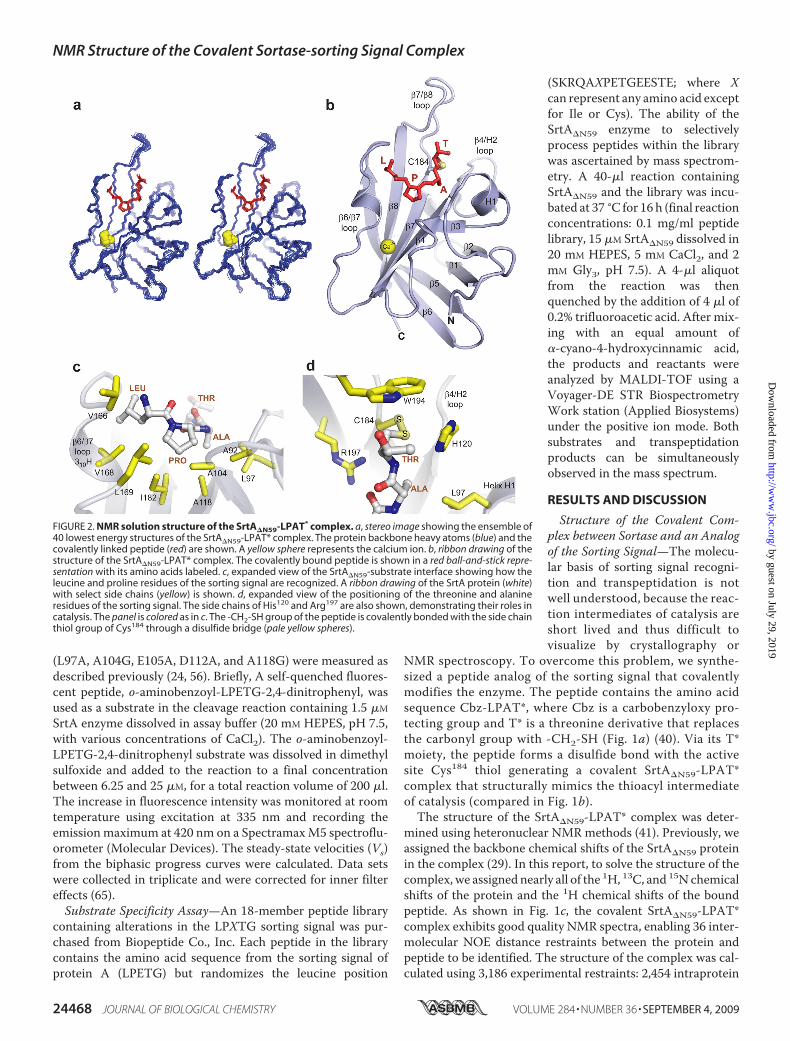
FIGURE 2. NMR solution structure of the SrtA�N59-LPAT* complex. a, stereo image showing the ensemble of40 lowest energy structures of the SrtA�N59-LPAT* complex. The protein backbone heavy atoms (blue) and thecovalently linked peptide (red) are shown. A yellow sphere represents the calcium ion. b, ribbon drawing of thestructure of the SrtA�N59-LPAT* complex. The covalently bound peptide is shown in a red ball-and-stick repre-sentation with its amino acids labeled. c, expanded view of the SrtA�N59-substrate interface showing how theleucine and proline residues of the sorting signal are recognized. A ribbon drawing of the SrtA protein (white)with select side chains (yellow) is shown. d, expanded view of the positioning of the threonine and alanineresidues of the sorting signal. The side chains of His120 and Arg197 are also shown, demonstrating their roles incatalysis. The panel is colored as in c. The -CH2-SH group of the peptide is covalently bonded with the side chainthiol group of Cys184 through a disulfide bridge (pale yellow spheres).
NMR Structure of the Covalent Sortase-sorting Signal Complex
24468 JOURNAL OF BIOLOGICAL CHEMISTRY VOLUME 284 • NUMBER 36 • SEPTEMBER 4, 2009
by guest on July 29, 2019http://w
ww
.jbc.org/D
ownloaded from
distances, 36 intermolecular distances, 94 hydrogen bonds, 883JHN� couplings, 254 carbon chemical shifts, and 260 dihedralangles. This produced an ensemble of 20 conformers that pos-sess good covalent geometry with no NOE, dihedral angle, orscalar coupling violations greater than 0.5Å, 5°, or 2Hz, respec-tively (Fig. 2a). The amino acids of the peptide analog and res-idues Pro63–Ile207 of SrtA�N59 are well defined by the NMRdata and have backbone and heavy atom coordinate root meansquare deviations to the mean structure of 0.28 � 0.06 and0.87 � 0.08 Å, respectively. Complete structure and restraintstatistics are presented in Table 1.Structural Basis of LPXTG Binding—SrtA recognizes the
LPXTG sorting signal through a large groove that leads into theactive site (Fig. 2b). Residues in strands�4 and�7 form the floorof the groove, whereas the walls are formed by surface loopsthat connect strand �6 to strand �7 (�6/�7 loop), strand �7 tostrand �8 (�7/�8 loop), strand �3 to strand �4 (�3/�4 loop),and strand �2 to helix H1 (�2/H1 loop). The leucine residue of
the signal rests against the �6/�7 loop, where residues Val166–Leu169 adopt a 310 helix that only forms when the substrate isbound (Fig. 2c). Helix formation enables the leucine methylgroups of the analog to be partially encircled by hydrophobiccontacts. From above, the leucine side chain is in close proxim-ity to the �-protons of Thr164 and Val166, whereas from below,it is contacted by the side chains of Val168 and Arg197. Theproline ring of the sorting signal is buried in the binding grooveby contacts from the side chains of Ile182 (�7) and Ala118 (�4)that project from the underlying sheet and by contacts fromresidues within both walls of the groove (Leu169 (310 helix),Ala92 (�2/H1 loop), and Ala104 (�3/�4 loop)). This latter inter-action is supported by the observation of strong intermolecularNOEs between the methyl groups of the alanine residues andprotons within the proline ring.The LPAT* peptide adopts an “L-shaped” structure as a
result of a�90° kink at the alanine-proline peptide bond, whichis in a trans conformation. The kink redirects the trajectory ofthe signal, enabling it to approach the active site in parallel withthe underlying �-strands of the binding groove. In the LPAT*peptide, an alanine residuemimics theX position of the LPXTGmotif. In the structure, the alanine is packed against the sidechain of Leu97 located in helix H1 as a result of several strongNOEs to the Leu97 H� protons (Fig. 2d). This explains the dem-onstrated promiscuity of SrtA for this site within the sortingsignal, since modeling studies suggest that larger side chainscould project away from the enzyme via a cleft located betweenhelix H1 and His120 (39). Recognition is completed by packingof the threonine �-methyl group beneath the indole ring ofTrp194, as substantiated by several NOEs between the methyland the H1 proton of the tryptophan. These contacts partiallyshield the active site from the solvent and help to project the-CH2-SH portion of the threonine analog toward Cys184 fordisulfide bond formation.Sorting Signal Binding Closes and Immobilizes the �6/�7
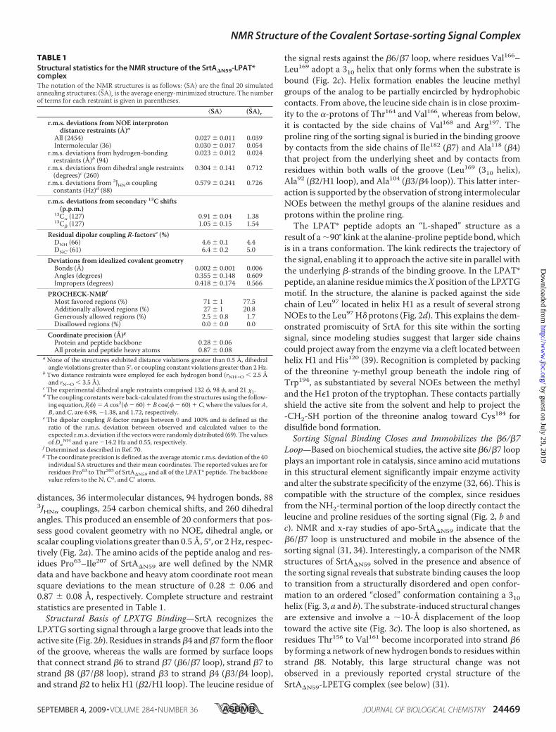
Loop—Based on biochemical studies, the active site �6/�7 loopplays an important role in catalysis, since amino acidmutationsin this structural element significantly impair enzyme activityand alter the substrate specificity of the enzyme (32, 66). This iscompatible with the structure of the complex, since residuesfrom the NH2-terminal portion of the loop directly contact theleucine and proline residues of the sorting signal (Fig. 2, b andc). NMR and x-ray studies of apo-SrtA�N59 indicate that the�6/�7 loop is unstructured and mobile in the absence of thesorting signal (31, 34). Interestingly, a comparison of the NMRstructures of SrtA�N59 solved in the presence and absence ofthe sorting signal reveals that substrate binding causes the loopto transition from a structurally disordered and open confor-mation to an ordered “closed” conformation containing a 310helix (Fig. 3, a and b). The substrate-induced structural changesare extensive and involve a �10-Å displacement of the looptoward the active site (Fig. 3c). The loop is also shortened, asresidues Thr156 to Val161 become incorporated into strand �6by forming a network of newhydrogen bonds to residueswithinstrand �8. Notably, this large structural change was notobserved in a previously reported crystal structure of theSrtA�N59-LPETG complex (see below) (31).
TABLE 1Structural statistics for the NMR structure of the SrtA�N59-LPAT*complexThe notation of the NMR structures is as follows: �SA� are the final 20 simulatedannealing structures; (S�A)r is the average energy-minimized structure. The numberof terms for each restraint is given in parentheses.
�SA� (S�A)rr.m.s. deviations from NOE interproton
distance restraints (Å)aAll (2454) 0.027 � 0.011 0.039Intermolecular (36) 0.030 � 0.017 0.054
r.m.s. deviations from hydrogen-bondingrestraints (Å)b (94)
0.023 � 0.012 0.024
r.m.s. deviations from dihedral angle restraints(degrees)c (260)
0.304 � 0.141 0.712
r.m.s. deviations from 3JHN� couplingconstants (Hz)d (88)
0.579 � 0.241 0.726
r.m.s. deviations from secondary 13C shifts(p.p.m.)
13C� (127) 0.91 � 0.04 1.3813C� (127) 1.05 � 0.15 1.54
Residual dipolar coupling R-factorse (%)DNH (66) 4.6 � 0.1 4.4DNC’ (61) 6.4 � 0.2 5.0
Deviations from idealized covalent geometryBonds (Å) 0.002 � 0.001 0.006Angles (degrees) 0.355 � 0.148 0.609Impropers (degrees) 0.418 � 0.174 0.566
PROCHECK-NMRf
Most favored regions (%) 71 � 1 77.5Additionally allowed regions (%) 27 � 1 20.8Generously allowed regions (%) 2.5 � 0.8 1.7Disallowed regions (%) 0.0 � 0.0 0.0
Coordinate precision (Å)gProtein and peptide backbone 0.28 � 0.06All protein and peptide heavy atoms 0.87 � 0.08
a None of the structures exhibited distance violations greater than 0.5 Å, dihedralangle violations greater than 5°, or coupling constant violations greater than 2 Hz.
b Two distance restraints were employed for each hydrogen bond (rNH���O 2.5 Åand rN���O 3.5 Å).
c The experimental dihedral angle restraints comprised 132 �, 98 �, and 21 1.d The coupling constants were back-calculated from the structures using the follow-ing equation, J(�) � A cos2(� � 60) � B cos(� � 60) � C, where the values for A,B, and C, are 6.98, �1.38, and 1.72, respectively.
e The dipolar coupling R-factor ranges between 0 and 100% and is defined as theratio of the r.m.s. deviation between observed and calculated values to theexpected r.m.s. deviation if the vectors were randomly distributed (69). The valuesof Da
NH and � are �14.2 Hz and 0.55, respectively.f Determined as described in Ref. 70.g The coordinate precision is defined as the average atomic r.m.s. deviation of the 40individual SA structures and their mean coordinates. The reported values are forresidues Pro63 to Thr203 of SrtA�N59 and all of the LPAT* peptide. The backbonevalue refers to the N, C�, and C atoms.
NMR Structure of the Covalent Sortase-sorting Signal Complex
SEPTEMBER 4, 2009 • VOLUME 284 • NUMBER 36 JOURNAL OF BIOLOGICAL CHEMISTRY 24469
by guest on July 29, 2019http://w
ww
.jbc.org/D
ownloaded from
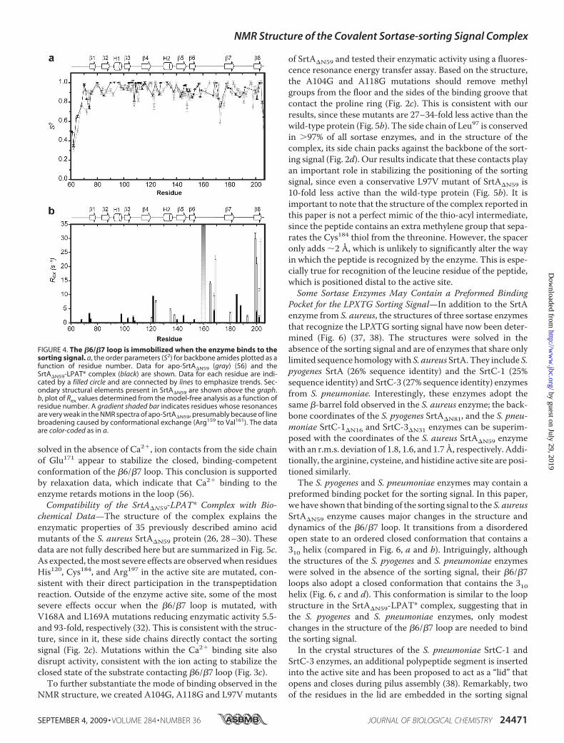
To determine whether loop closure over the sorting signalanalog quenches its mobility, we measured the R1, R2, and15N{1H} NOE relaxation parameters of the protein backbonenitrogen atoms in the complex and interpreted these data usingthe model-free formalism (60–63). This analysis yields theorder parameter (S2), which gives a concise account of the back-bone amide’s mobility on the picosecond time scale. It rangesfrom 0 to 1, with values of 1 indicating that the amide is com-pletely immobilized. The model-free analysis also yields an Rexterm that is diagnostic for the presence of slower micro- tomillisecond time scale motions. Fig. 4 compares the S2 and Rexvalues of SrtA�N59 in the SrtA�N59-LPAT* complex with simi-lar data reported for the apo-form of the enzyme (56). In theSrtA�N59-LPAT* complex, the �6/�7 loop is rigid on fast timescales, as evidenced by S2 values that are on average 0.90� 0.01(Fig. 4a, black). It is also immobile on slower time scales, sinceonly a few residues distributed throughout the protein exhibitsmall magnitude Rex terms (Fig. 4b, black). This is in markedcontrast to the apoenzyme, since many of the residues in the�6/�7 exhibit elevated Rex values and/or weak NMR reso-nances that indicate that they undergo slow micro- to millisec-ond time scalemotions (Fig. 4b,white and gradient bars). Inter-
estingly, in the apoenzyme, severalresidues in the loop have S2 values of�0.7, demonstrating that they donot participate in large amplitudepicosecond time scale motions (Fig.4a, gray). Combined, the structuraland relaxation data suggest that theloop in the apoenzyme adopts asemirigid state that undergoesmicro- to millisecond segmentalmotions that toggle it between openand closed conformations. Sub-strate binding quenches thesemotions, locking the loop in a closedstate for productive interactionswith the sorting signal.Closure and immobilization of
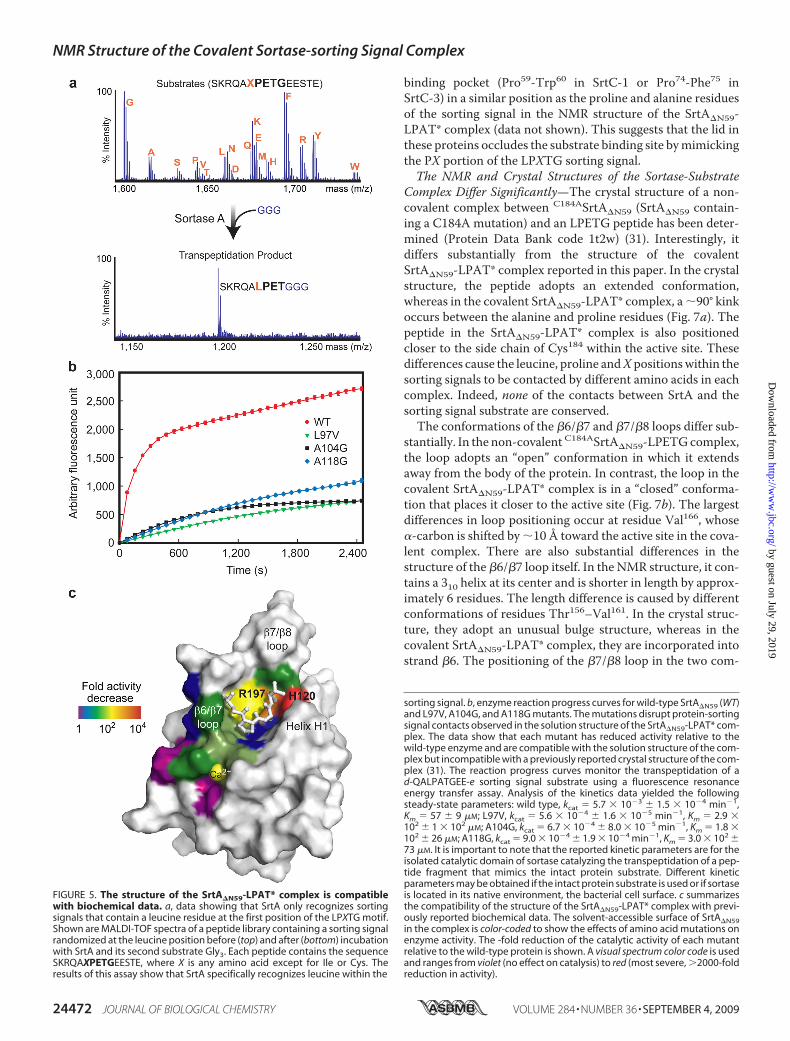
the �6/�7 loop enables extensiveenzyme contacts to the leucine res-idue within the sorting signal. Wetested whether these interactionsconfer specificity for this site bychallenging SrtA�N59 with a peptidelibrary containing the LPETGsequence randomized at the leucineposition (SKRQAXPETGEESTE,where X represents any amino acidexcept for Ile or Cys). Monitoringproduct formation usingmass spec-trometry reveals that only peptidesbearing a leucine amino acid atthis site are effectively processedby the enzyme (Fig. 5a). This sup-ports the notion that loop closureover the signal plays a key role insubstrate recognition.
Ca2� Stabilizes the Closed Conformation of the �6/�7 Loop—In vitro, Ca2� increases the enzymatic activity of SrtA�N59�8-fold by lowering theKm of the enzyme for the sorting signal(34, 56). This adaptation may enable S. aureus to increase thenumber of displayed proteins if elevated concentrations ofCa2� are encountered at sites of infection. The atomic basis ofdivalent ion binding is incompletely understood, because allpreviously reported crystal structures of SrtA�N59 were solvedin the metal-free state (31). Moreover, the binding mechanismof Ca2� cannot easily be defined by NMRmethods, since Ca2�
lacks proton atoms needed to identify NOE distance restraints.In structure calculations of the complex, we employed threeartificial distance restraints between a single Ca2� ion and theside chains of residues Glu105, Glu108, and Glu171. These wereincluded because mutant proteins that replace these residueswith alanine are insensitive to Ca2� and because the chemicalshifts of these residues are significantly perturbedwhenCa2� isadded (56). Importantly, structures calculatedwith the artificialrestraints are compatible with all of the experimental data. Inthe structure of the complex, Ca2� binds to a pocket formed bythe �3/�4 (Glu105 and Glu108) and �6/�7 loops (Glu171) (Fig.3c). As compared with the crystal structure of the apoenzyme
FIGURE 3. Sorting signal binding induces changes in the structure of SrtA. a, overlay of the ensemble ofNMR structures of apo-SrtA�N59 (34) (Protein Data Bank code 1ija; red) and the SrtA�N59-LPAT* complex (blue).The comparison shows that the structurally disordered �6/�7 loop becomes ordered upon binding the sortingsignal. b, superposition of the average NMR structures of apo-SrtA�N59 (34) (pink) and the SrtA�N59-LPAT*complex (blue). Each structure is presented as a schematic diagram with relevant loops labeled. The largestsubstrate-induced conformational changes occur in residues located within the �6/�7 and �7/�8 loops. Thistransition is accentuated in the figure by showing hypothetical structural intermediates calculated using theYale Morph Server (available on the World Wide Web) (68). c, as in b but expanded to show the shift of the �6/�7loop over the sorting signal and the role that calcium plays in stabilizing the closed conformation of the �6/�7loop.
NMR Structure of the Covalent Sortase-sorting Signal Complex
24470 JOURNAL OF BIOLOGICAL CHEMISTRY VOLUME 284 • NUMBER 36 • SEPTEMBER 4, 2009
by guest on July 29, 2019http://w
ww
.jbc.org/D
ownloaded from
solved in the absence of Ca2�, ion contacts from the side chainof Glu171 appear to stabilize the closed, binding-competentconformation of the �6/�7 loop. This conclusion is supportedby relaxation data, which indicate that Ca2� binding to theenzyme retards motions in the loop (56).Compatibility of the SrtA�N59-LPAT* Complex with Bio-
chemical Data—The structure of the complex explains theenzymatic properties of 35 previously described amino acidmutants of the S. aureus SrtA�N59 protein (26, 28–30). Thesedata are not fully described here but are summarized in Fig. 5c.As expected, themost severe effects are observedwhen residuesHis120, Cys184, and Arg197 in the active site are mutated, con-sistent with their direct participation in the transpeptidationreaction. Outside of the enzyme active site, some of the mostsevere effects occur when the �6/�7 loop is mutated, withV168A and L169A mutations reducing enzymatic activity 5.5-and 93-fold, respectively (32). This is consistent with the struc-ture, since in it, these side chains directly contact the sortingsignal (Fig. 2c). Mutations within the Ca2� binding site alsodisrupt activity, consistent with the ion acting to stabilize theclosed state of the substrate contacting �6/�7 loop (Fig. 3c).To further substantiate the mode of binding observed in the
NMR structure, we created A104G, A118G and L97V mutants
of SrtA�N59 and tested their enzymatic activity using a fluores-cence resonance energy transfer assay. Based on the structure,the A104G and A118G mutations should remove methylgroups from the floor and the sides of the binding groove thatcontact the proline ring (Fig. 2c). This is consistent with ourresults, since these mutants are 27–34-fold less active than thewild-type protein (Fig. 5b). The side chain of Leu97 is conservedin �97% of all sortase enzymes, and in the structure of thecomplex, its side chain packs against the backbone of the sort-ing signal (Fig. 2d). Our results indicate that these contacts playan important role in stabilizing the positioning of the sortingsignal, since even a conservative L97V mutant of SrtA�N59 is10-fold less active than the wild-type protein (Fig. 5b). It isimportant to note that the structure of the complex reported inthis paper is not a perfect mimic of the thio-acyl intermediate,since the peptide contains an extra methylene group that sepa-rates the Cys184 thiol from the threonine. However, the spaceronly adds �2 Å, which is unlikely to significantly alter the wayin which the peptide is recognized by the enzyme. This is espe-cially true for recognition of the leucine residue of the peptide,which is positioned distal to the active site.Some Sortase Enzymes May Contain a Preformed Binding
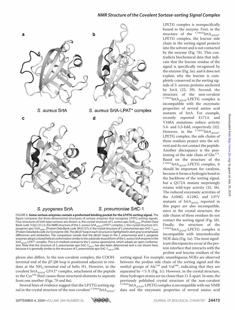
Pocket for the LPXTG Sorting Signal—In addition to the SrtAenzyme from S. aureus, the structures of three sortase enzymesthat recognize the LPXTG sorting signal have now been deter-mined (Fig. 6) (37, 38). The structures were solved in theabsence of the sorting signal and are of enzymes that share onlylimited sequence homologywith S. aureus SrtA.They include S.pyogenes SrtA (26% sequence identity) and the SrtC-1 (25%sequence identity) and SrtC-3 (27% sequence identity) enzymesfrom S. pneumoniae. Interestingly, these enzymes adopt thesame �-barrel fold observed in the S. aureus enzyme; the back-bone coordinates of the S. pyogenes SrtA�N81, and the S. pneu-moniae SrtC-1�N16 and SrtC-3�N31 enzymes can be superim-posed with the coordinates of the S. aureus SrtA�N59 enzymewith an r.m.s. deviation of 1.8, 1.6, and 1.7Å, respectively. Addi-tionally, the arginine, cysteine, and histidine active site are posi-tioned similarly.The S. pyogenes and S. pneumoniae enzymes may contain a
preformed binding pocket for the sorting signal. In this paper,we have shown that binding of the sorting signal to the S. aureusSrtA�N59 enzyme causes major changes in the structure anddynamics of the �6/�7 loop. It transitions from a disorderedopen state to an ordered closed conformation that contains a310 helix (compared in Fig. 6, a and b). Intriguingly, althoughthe structures of the S. pyogenes and S. pneumoniae enzymeswere solved in the absence of the sorting signal, their �6/�7loops also adopt a closed conformation that contains the 310helix (Fig. 6, c and d). This conformation is similar to the loopstructure in the SrtA�N59-LPAT* complex, suggesting that inthe S. pyogenes and S. pneumoniae enzymes, only modestchanges in the structure of the �6/�7 loop are needed to bindthe sorting signal.In the crystal structures of the S. pneumoniae SrtC-1 and
SrtC-3 enzymes, an additional polypeptide segment is insertedinto the active site and has been proposed to act as a “lid” thatopens and closes during pilus assembly (38). Remarkably, twoof the residues in the lid are embedded in the sorting signal
FIGURE 4. The �6/�7 loop is immobilized when the enzyme binds to thesorting signal. a, the order parameters (S2) for backbone amides plotted as afunction of residue number. Data for apo-SrtA�N59 (gray) (56) and theSrtA�N59-LPAT* complex (black) are shown. Data for each residue are indi-cated by a filled circle and are connected by lines to emphasize trends. Sec-ondary structural elements present in SrtA�N59 are shown above the graph.b, plot of Rex values determined from the model-free analysis as a function ofresidue number. A gradient shaded bar indicates residues whose resonancesare very weak in the NMR spectra of apo-SrtA�N59, presumably because of linebroadening caused by conformational exchange (Arg159 to Val161). The dataare color-coded as in a.
NMR Structure of the Covalent Sortase-sorting Signal Complex
SEPTEMBER 4, 2009 • VOLUME 284 • NUMBER 36 JOURNAL OF BIOLOGICAL CHEMISTRY 24471
by guest on July 29, 2019http://w
ww
.jbc.org/D
ownloaded from
binding pocket (Pro59-Trp60 in SrtC-1 or Pro74-Phe75 inSrtC-3) in a similar position as the proline and alanine residuesof the sorting signal in the NMR structure of the SrtA�N59-LPAT* complex (data not shown). This suggests that the lid inthese proteins occludes the substrate binding site bymimickingthe PX portion of the LPXTG sorting signal.The NMR and Crystal Structures of the Sortase-Substrate
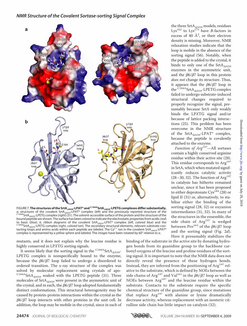
Complex Differ Significantly—The crystal structure of a non-covalent complex between C184ASrtA�N59 (SrtA�N59 contain-ing a C184A mutation) and an LPETG peptide has been deter-mined (Protein Data Bank code 1t2w) (31). Interestingly, itdiffers substantially from the structure of the covalentSrtA�N59-LPAT* complex reported in this paper. In the crystalstructure, the peptide adopts an extended conformation,whereas in the covalent SrtA�N59-LPAT* complex, a�90° kinkoccurs between the alanine and proline residues (Fig. 7a). Thepeptide in the SrtA�N59-LPAT* complex is also positionedcloser to the side chain of Cys184 within the active site. Thesedifferences cause the leucine, proline andXpositionswithin thesorting signals to be contacted by different amino acids in eachcomplex. Indeed, none of the contacts between SrtA and thesorting signal substrate are conserved.The conformations of the �6/�7 and �7/�8 loops differ sub-
stantially. In the non-covalent C184ASrtA�N59-LPETGcomplex,the loop adopts an “open” conformation in which it extendsaway from the body of the protein. In contrast, the loop in thecovalent SrtA�N59-LPAT* complex is in a “closed” conforma-tion that places it closer to the active site (Fig. 7b). The largestdifferences in loop positioning occur at residue Val166, whose�-carbon is shifted by �10 Å toward the active site in the cova-lent complex. There are also substantial differences in thestructure of the �6/�7 loop itself. In the NMR structure, it con-tains a 310 helix at its center and is shorter in length by approx-imately 6 residues. The length difference is caused by differentconformations of residues Thr156–Val161. In the crystal struc-ture, they adopt an unusual bulge structure, whereas in thecovalent SrtA�N59-LPAT* complex, they are incorporated intostrand �6. The positioning of the �7/�8 loop in the two com-
FIGURE 5. The structure of the SrtA�N59-LPAT* complex is compatiblewith biochemical data. a, data showing that SrtA only recognizes sortingsignals that contain a leucine residue at the first position of the LPXTG motif.Shown are MALDI-TOF spectra of a peptide library containing a sorting signalrandomized at the leucine position before (top) and after (bottom) incubationwith SrtA and its second substrate Gly3. Each peptide contains the sequenceSKRQAXPETGEESTE, where X is any amino acid except for Ile or Cys. Theresults of this assay show that SrtA specifically recognizes leucine within the
sorting signal. b, enzyme reaction progress curves for wild-type SrtA�N59 (WT)and L97V, A104G, and A118G mutants. The mutations disrupt protein-sortingsignal contacts observed in the solution structure of the SrtA�N59-LPAT* com-plex. The data show that each mutant has reduced activity relative to thewild-type enzyme and are compatible with the solution structure of the com-plex but incompatible with a previously reported crystal structure of the com-plex (31). The reaction progress curves monitor the transpeptidation of ad-QALPATGEE-e sorting signal substrate using a fluorescence resonanceenergy transfer assay. Analysis of the kinetics data yielded the followingsteady-state parameters: wild type, kcat � 5.7 � 10�3 � 1.5 � 10�4 min�1,Km � 57 � 9 �M; L97V, kcat � 5.6 � 10�4 � 1.6 � 10�5 min�1, Km � 2.9 �102 � 1 � 102 �M; A104G, kcat � 6.7 � 10�4 � 8.0 � 10�5 min�1, Km � 1.8 �102 � 26 �M; A118G, kcat � 9.0 � 10�4 � 1.9 � 10�4 min�1, Km � 3.0 � 102 �73 �M. It is important to note that the reported kinetic parameters are for theisolated catalytic domain of sortase catalyzing the transpeptidation of a pep-tide fragment that mimics the intact protein substrate. Different kineticparameters may be obtained if the intact protein substrate is used or if sortaseis located in its native environment, the bacterial cell surface. c summarizesthe compatibility of the structure of the SrtA�N59-LPAT* complex with previ-ously reported biochemical data. The solvent-accessible surface of SrtA�N59in the complex is color-coded to show the effects of amino acid mutations onenzyme activity. The -fold reduction of the catalytic activity of each mutantrelative to the wild-type protein is shown. A visual spectrum color code is usedand ranges from violet (no effect on catalysis) to red (most severe, �2000-foldreduction in activity).
NMR Structure of the Covalent Sortase-sorting Signal Complex
24472 JOURNAL OF BIOLOGICAL CHEMISTRY VOLUME 284 • NUMBER 36 • SEPTEMBER 4, 2009
by guest on July 29, 2019http://w
ww
.jbc.org/D
ownloaded from
plexes also differs. In the non-covalent complex, the COOH-terminal end of the �7/�8 loop is positioned adjacent to resi-dues at the NH2-terminal end of helix H1. However, in thecovalent SrtA�N59-LPAT* complex, attachment of the peptideto the Cys184 thiol causes these structural elements to separatefrom one another (Figs. 7b and 8a).Several lines of evidence suggest that the LPXTG sorting sig-
nal in the crystal structure of the non-covalent C184ASrtA�N59-
LPETG complex is nonspecificallybound to the enzyme. First, in thestructure of the C184ASrtA�N59-LPETG complex, the leucine sidechain in the sorting signal projectsinto the solvent and is not contactedby the enzyme (Fig. 7b). This con-tradicts biochemical data that indi-cate that the leucine residue of thesignal is specifically recognized bythe enzyme (Fig. 5a), and it does notexplain why the leucine is com-pletely conserved in the sorting sig-nals of S. aureus proteins anchoredby SrtA (22, 39). Second, thestructure of the non-covalentC184ASrtA�N59-LPETG complex isincompatible with the enzymaticproperties of several amino acidmutants of SrtA. For example,recently reported E171A andV168A mutations reduce activity5.4- and 5.5-fold, respectively (32).However, in the C184ASrtA�N59-LPETG complex, the side chains ofthese residues project into the sol-vent and do not contact the peptide.Another discrepancy is the posi-tioning of the side chain of Gln172.Based on the structure of theC184ASrtA�N59-LPETG complex, itshould be important for catalysis,because it forms a hydrogen bond tothe backbone of the sorting signal,but a Q172A mutant surprisinglyretains wild-type activity (32, 56).The reduced enzymatic activities ofthe A104G, A118G, and L97Vmutants of SrtA�N59 reported inthis paper are also incompatible,since in the crystal structure, theside chains of these residues do notcontact the sorting signal (Fig. 5b).Finally, the structure of theC184ASrtA�N59-LPETG complex isincompatible with intermolecularNOE data (Fig. 1a). Themost signif-icant discrepancies occur at the pro-tein interface that interacts with theproline and leucine residues of the
sorting signal. For example, unambiguous NOEs are observedbetween the proline side chain of the sorting signal and themethyl groups of Ala118 and Val166, indicating that they areseparated by 5 Å (Fig. 1c). However, in the crystal structure,these hydrogen atoms are no closer than 11Å apart. In sum, thepreviously published crystal structure of the non-covalentC184ASrtA�N59-LPETGcomplex is incompatible with ourNMRdata and the enzymatic properties of several amino acid
FIGURE 6. Some sortase enzymes contain a preformed binding pocket for the LPXTG sorting signal. Thefigure compares the three-dimensional structures of sortase enzymes that recognize LPXTG sorting signals.Four structures of SrtA-type sortases are shown. a, the crystal structure of S. aureus apo-SrtA�N59 (Protein DataBank code 1t2p) (31); b, the NMR structure of the S. aureus SrtA�N59-LPAT* complex; c, the crystal structure of S.pyogenes apo-SrtA�N81 (Protein Data Bank code 3fn5) (37); d, the crystal structure of S. pneumoniae apo-SrtC-1�N16(Protein Data Bank code 2w1j) enzyme (38). The�6/�7 loop in each structure is highlighted in dark gray to emphasizedifferences and similarities. The comparison reveals that the �6/�7 loops in the S. pneumoniae and S. pyogenesenzymes adopt a closed helical conformation similar to the substrate-bound form of the S. aureus SrtA enzyme in theSrtA�N59-LPAT* complex. This is in marked contrast to the S. aureus apoenzyme, which adopts an open conforma-tion. Note that the structure of S. pneumoniae apo-SrtC-3�N31 has also been determined and is not shown here,because it is generally similar to the structure of S. pneumoniae apo-SrtC-1�N16 (38).
NMR Structure of the Covalent Sortase-sorting Signal Complex
SEPTEMBER 4, 2009 • VOLUME 284 • NUMBER 36 JOURNAL OF BIOLOGICAL CHEMISTRY 24473
by guest on July 29, 2019http://w
ww
.jbc.org/D
ownloaded from
mutants, and it does not explain why the leucine residue ishighly conserved in LPXTG sorting signals.It seems likely that the sorting signal in the C184ASrtA�N59-
LPETG complex is nonspecifically bound to the enzyme,because the �6/�7 loop failed to undergo a disordered toordered transition. The x-ray structure of the complex wassolved by molecular replacement using crystals of apo-C184ASrtA�N59 soaked with the LPETG peptide (31). Threemolecules of SrtA�N59 were present in the asymmetric unit ofthe crystal, and in each, the �6/�7 loop adopted fundamentallydistinct conformations. This structural heterogeneity may becaused by protein-protein interactions within the crystal as the�6/�7 loop interacts with other proteins in the unit cell. Inaddition, the loop may be mobile in the crystal, since in each of
the three SrtA�N59 models, residuesLys162 to Lys175 have B-factors inexcess of 40 Å2, or their electrondensity is missing. Moreover, NMRrelaxation studies indicate that theloop is mobile in the absence of thesorting signal (56). Notably, whenthe peptide is added to the crystal, itbinds to only one of the SrtA�N59enzymes in the asymmetric unit,and the �6/�7 loop in this proteindoes not change its structure. Thus,it appears that the �6/�7 loop inthe C184ASrtA�N59-LPETG complexfailed to undergo substrate-inducedstructural changes required toproperly recognize the signal, pre-sumably because SrtA only weaklybinds the LPXTG signal and/orbecause of lattice packing interac-tions (25). This problem has beenovercome in the NMR structureof the SrtA�N59-LPAT* complex,because the peptide is covalentlyattached to the enzyme.Function of Arg197—All sortases
contain a highly conserved arginineresidue within their active site (28).This residue corresponds to Arg197in SrtA, whichwhenmutated signif-icantly reduces catalytic activity(28–30, 32). The function of Arg197in catalysis has hitherto remainedunclear, since it has been proposedto either deprotonate Cys184 (28) orlipid II (31) or, alternatively, to sta-bilize either the binding of thesorting signal (28, 32) or oxyanionintermediates (31, 32). In many ofthe structures in the ensemble, theside chain of Arg197 is wedgedbetween Pro163 of the �6/�7 loopand the sorting signal (Fig. 2d).Here it presumably stabilizes the
binding of the substrate in the active site by donating hydro-gen bonds from its guanidine group to the backbone car-bonyl oxygens of the leucine and proline residues of the sort-ing signal. It is important to note that the NMR data does notdirectly reveal the presence of these hydrogen bonds.Instead, they are inferred from the positioning of Arg197 rel-ative to the substrate, which is defined by NOEs between theside chains of Arg197 and Val161 in the �6/�7 loop as well asNOEs between Arg197 and the leucine residue within thesubstrate. Contacts to the substrate require the specificchemical structure of the guanidine group, since mutationsthat replace Arg197 with alanine or lysine dramaticallydecrease activity, whereas replacement with an isosteric cit-rulline side chain has little impact on catalysis (32).
FIGURE 7. The structures of the SrtA�N59-LPAT* and C184ASrtA�N59-LPETG complexes differ substantially.a, structures of the covalent SrtA�N59-LPAT* complex (left) and the previously reported structure of theC184ASrtA�N59-LPETG complex (right) (31). The solvent-accessible surface of the protein and the structure of thebound peptide are shown. The surface has been colored to indicate the electrostatic properties from acidic (red)to basic (blue). b, ribbon diagrams of the covalent SrtA�N59-LPAT* complex (left, colored blue) and theC184ASrtA�N59-LPETG complex (right, colored tan). The secondary structural elements, relevant substrate con-tacting loops and amino acids within each peptide are labeled. The Ca2� ion in the covalent SrtA�N59-LPAT*complex is represented by a yellow sphere and labeled. The images have been rotated by 60° relative to a.
NMR Structure of the Covalent Sortase-sorting Signal Complex
24474 JOURNAL OF BIOLOGICAL CHEMISTRY VOLUME 284 • NUMBER 36 • SEPTEMBER 4, 2009
by guest on July 29, 2019http://w
ww
.jbc.org/D
ownloaded from
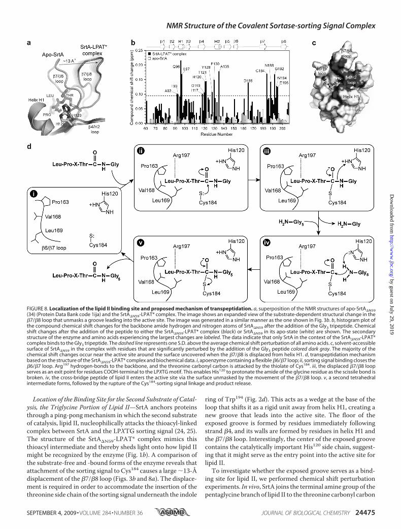
Location of the Binding Site for the Second Substrate of Catal-ysis, the Triglycine Portion of Lipid II—SrtA anchors proteinsthrough a ping-pongmechanism in which the second substrateof catalysis, lipid II, nucleophilically attacks the thioacyl-linkedcomplex between SrtA and the LPXTG sorting signal (24, 25).The structure of the SrtA�N59-LPAT* complex mimics thisthioacyl intermediate and thereby sheds light onto how lipid IImight be recognized by the enzyme (Fig. 1b). A comparison ofthe substrate-free and -bound forms of the enzyme reveals thatattachment of the sorting signal to Cys184 causes a large �13-Ådisplacement of the �7/�8 loop (Figs. 3b and 8a). The displace-ment is required in order to accommodate the insertion of thethreonine side chain of the sorting signal underneath the indole
ring of Trp194 (Fig. 2d). This acts as a wedge at the base of theloop that shifts it as a rigid unit away from helix H1, creating anew groove that leads into the active site. The floor of theexposed groove is formed by residues immediately followingstrand �4, and its walls are formed by residues in helix H1 andthe �7/�8 loop. Interestingly, the center of the exposed groovecontains the catalytically important His120 side chain, suggest-ing that it might serve as the entry point into the active site forlipid II.To investigate whether the exposed groove serves as a bind-
ing site for lipid II, we performed chemical shift perturbationexperiments. In vivo, SrtA joins the terminal amine group of thepentaglycine branch of lipid II to the threonine carbonyl carbon
FIGURE 8. Localization of the lipid II binding site and proposed mechanism of transpeptidation. a, superposition of the NMR structures of apo-SrtA�N59(34) (Protein Data Bank code 1ija) and the SrtA�N59-LPAT* complex. The image shows an expanded view of the substrate-dependent structural change in the�7/�8 loop that unmasks a groove leading into the active site. The image was generated in a similar manner as the one shown in Fig. 3b. b, histogram plot ofthe compound chemical shift changes for the backbone amide hydrogen and nitrogen atoms of SrtA�N59 after the addition of the Gly3 tripeptide. Chemicalshift changes after the addition of the peptide to either the SrtA�N59-LPAT* complex (black) or SrtA�N59 in its apo-state (white) are shown. The secondarystructure of the enzyme and amino acids experiencing the largest changes are labeled. The data indicate that only SrtA in the context of the SrtA�N59-LPAT*complex binds to the Gly3 tripeptide. The dashed line represents one S.D. above the average chemical shift perturbation of all amino acids. c, solvent-accessiblesurface of SrtA�N59 in the complex with residues that are significantly perturbed by the addition of the Gly3 peptide colored dark gray. The majority of thechemical shift changes occur near the active site around the surface uncovered when the �7/�8 is displaced from helix H1. d, transpeptidation mechanismbased on the structure of the SrtA�N59-LPAT* complex and biochemical data. i, apoenzyme containing a flexible �6/�7 loop; ii, sorting signal binding closes the�6/�7 loop. Arg197 hydrogen-bonds to the backbone, and the threonine carbonyl carbon is attacked by the thiolate of Cys184. iii, the displaced �7/�8 loopserves as an exit point for residues COOH-terminal to the LPXTG motif. This enables His120 to protonate the amide of the glycine residue as the scissile bond isbroken. iv, the cross-bridge peptide of lipid II enters the active site via the surface unmasked by the movement of the �7/�8 loop. v, a second tetrahedralintermediate forms, followed by the rupture of the Cys184-sorting signal linkage and product release.
NMR Structure of the Covalent Sortase-sorting Signal Complex
SEPTEMBER 4, 2009 • VOLUME 284 • NUMBER 36 JOURNAL OF BIOLOGICAL CHEMISTRY 24475
by guest on July 29, 2019http://w
ww
.jbc.org/D
ownloaded from
of the LPXTG sorting signal (18–20). A Gly3 peptide mimicsthis portion of lipid II and can be effectively used by the enzymeas a substrate in vitro (23). To locate the surface on the enzymethat interacts with Gly3, we titrated a sample of the SrtA�N59-LPAT* complexwith the peptide and usedNMR tomonitor thechemical shifts of the backbone amide atoms of the protein. Ahistogram plot of the chemical shift differences in the presenceand absence of Gly3 reveals that the peptide selectively perturbstheNMR spectrum of the protein (Fig. 8b, black bars). Interest-ingly, when the most significantly perturbed residues aremapped onto the structure of the protein in the SrtA�N59-LPAT*, they define a continuous surface that encompasses thegroove that is unmasked when the sorting signal binds (Fig. 8c).When the apo-form of the enzyme is titrated with the Gly3peptide, the backbone amide chemical shifts of residues withinthis surface are not perturbed (Fig. 8b, white bars). Combined,these data suggest that sorting signal-induced displacement ofthe �7/�8 loop unmasks the binding site for the Gly3 portion oflipid II and may thereby direct catalysis toward productformation.Mechanism of Catalysis—The structure of the complex
enables a more detailed mechanism of the SrtA-catalyzedtranspeptidation reaction to be proposed (Fig. 8d). The apoen-zyme is dynamic, with residues in the �6/�7 loop undergoingmotions that periodically displace them from the active site (56)(step i in Fig. 8d). Substrate binding nucleates the folding andclosure of the loop over the sorting signal, enabling recognition(step ii). Based on NMR and enzyme kinetic studies of the apo-form of sortase, the pKa values of His120 and Cys184 are �6.3–7and �9.4, respectively (25, 33). Therefore, at physiological pHvalues, the predominant form of the enzyme contains His120and Cys184 in their uncharged states. Interestingly, solvent iso-tope effect measurements have led to the suggestion that thisform of the enzyme is inactive. Instead, the active form of sor-tase has been proposed to be sparsely populated (0.06% of thetotal protein) and to contain His120 and Cys184 both in theirionized states (25). Therefore, the incoming threonine carbonylcarbon of the sorting signal is probably attacked by the thiolateof Cys184, resulting in the concerted displacement of the �7/�8loop (step iii). This opens a large groove leading out of the activesite that accommodates residues positioned on the COOH-ter-minal side of the threonine residue of the sorting signal in thesurface protein precursor. The groove contains the imidazo-lium side chain of His120 at its center, which is poised to pro-tonate the amide leaving group as the scissile bond is broken(67). NMR chemical shift experiments indicate that the groovealso functions as the entry point for the Gly5 portion of lipid II,suggesting that the newly deprotonated His120 side chain acti-vates the incoming terminal amine of lipid II for nucleophilicattack on the enzyme-linked thioacyl intermediate (step iv).The dual function of His120 as a general acid and base is con-sistent with its measured pKa value of �6.3–7 and is function-ally similar to histidine residues in a number of other enzymes,including serine proteases (33, 71). The reaction is then com-pleted by formation of the second tetrahedral intermediate andsubsequent breakage of the enzyme-substrate bond to liberatethe protein-lipid II-linked product (step v). In all steps, Arg197plays a key role in catalysis by stabilizing the positioning of the
substrate through direct hydrogen bonding to its polypeptidebackbone. In addition, the side chain of Arg197 is properly posi-tioned in several conformers in the NMR ensemble to stabilizeboth tetrahedral intermediates of catalysis by interacting withthe oxyanion.SrtA is required for the virulence of multidrug-resistant
methicillin resistant S. aureus, which in the United States killsan estimated 18,000 people annually (67). In addition, a numberof other important human pathogens contain SrtA homologsthatwhen genetically eliminated cause defects in virulence. Thedata presented here should therefore aid in the design of smallmolecule sortase inhibitors that are useful in treating a range ofbacterial infections.
Acknowledgments—We thank Dr. Robert Peterson for assistance withthe NMR experiments, Dr. Joseph A. Loo formass spectrometry exper-iments, Drs. Mandar T. Naik and Rosemarie L. Pilpa for technicalassistance with the NMR relaxation studies, and Drs. James U. Bowieand Scott A. Robson for useful discussions.
REFERENCES1. Navarre, W.W., and Schneewind, O. (1999)Microbiol. Mol. Biol. Rev. 63,
174–2292. Marraffini, L. A., Dedent, A. C., and Schneewind, O. (2006) Microbiol.
Mol. Biol. Rev. 70, 192–2213. Paterson, G. K., and Mitchell, T. J. (2004) Trends Microbiol. 12, 89–954. Ton-That, H., Marraffini, L. A., and Schneewind, O. (2004) Biochim. Bio-
phys. Acta 1694, 269–2785. Mandlik, A., Swierczynski, A., Das, A., and Ton-That, H. (2008) Trends
Microbiol. 16, 33–406. Scott, J. R., and Zahner, D. (2006)Mol. Microbiol. 62, 320–3307. Maresso, A.W., and Schneewind, O. (2008) Pharmacol. Rev. 60, 128–1418. Suree, N., Jung, M. E., and Clubb, R. T. (2007) Mini Rev. Med. Chem. 7,
991–10009. Mao, H., Hart, S. A., Schink, A., and Pollok, B. A. (2004) J. Am. Chem. Soc.
126, 2670–267110. Chan, L., Cross, H. F., She, J. K., Cavalli, G., Martins, H. F., and Neylon, C.
(2007) PLoS ONE 2, e116411. Popp, M. W., Antos, J. M., Grotenbreg, G. M., Spooner, E., and Ploegh,
H. L. (2007) Nat. Chem. Biol. 3, 707–70812. Clow, F., Fraser, J. D., and Proft, T. (2008) Biotechnol. Lett. 30, 1603–160713. Tanaka, T., Yamamoto, T., Tsukiji, S., and Nagamune, T. (2008) Chem-
BioChem 9, 802–80714. Samantaray, S.,Marathe, U., Dasgupta, S., Nandicoori, V. K., andRoy, R. P.
(2008) J. Am. Chem. Soc. 130, 2132–213315. Mazmanian, S. K., Liu, G., Ton-That, H., and Schneewind, O. (1999) Sci-
ence 285, 760–76316. Ton-That, H., Liu, G., Mazmanian, S. K., Faull, K. F., and Schneewind, O.
(1999) Proc. Natl. Acad. Sci. U.S.A. 96, 12424–1242917. Schneewind, O., Model, P., and Fischetti, V. A. (1992) Cell 70, 267–28118. Perry, A. M., Ton-That, H., Mazmanian, S. K., and Schneewind, O. (2002)
J. Biol. Chem. 277, 16241–1624819. Ruzin, A., Severin, A., Ritacco, F., Tabei, K., Singh, G., Bradford, P. A.,
Siegel, M. M., Projan, S. J., and Shlaes, D. M. (2002) J. Bacteriol. 184,2141–2147
20. Schneewind, O., Fowler, A., and Faull, K. F. (1995) Science 268, 103–10621. Comfort, D., and Clubb, R. T. (2004) Infect. Immun. 72, 2710–272222. Pallen, M. J., Lam, A. C., Antonio, M., and Dunbar, K. (2001) Trends
Microbiol. 9, 97–10223. Ton-That, H., Mazmanian, S. K., Faull, K. F., and Schneewind, O. (2000)
J. Biol. Chem. 275, 9876–988124. Huang, X., Aulabaugh, A., Ding, W., Kapoor, B., Alksne, L., Tabei, K., and
Ellestad, G. (2003) Biochemistry 42, 11307–1131525. Frankel, B. A., Kruger, R. G., Robinson, D. E., Kelleher, N. L., and
NMR Structure of the Covalent Sortase-sorting Signal Complex
24476 JOURNAL OF BIOLOGICAL CHEMISTRY VOLUME 284 • NUMBER 36 • SEPTEMBER 4, 2009
by guest on July 29, 2019http://w
ww
.jbc.org/D
ownloaded from
McCafferty, D. G. (2005) Biochemistry 44, 11188–1120026. Ton-That, H., Mazmanian, S. K., Alksne, L., and Schneewind, O. (2002)
J. Biol. Chem. 277, 7447–745227. Zhang, R., Wu, R., Joachimiak, G., Mazmanian, S. K., Missiakas, D. M.,
Gornicki, P., Schneewind, O., and Joachimiak, A. (2004) Structure 12,1147–1156
28. Marraffini, L. A., Ton-That, H., Zong, Y., Narayana, S. V., and Schnee-wind, O. (2004) J. Biol. Chem. 279, 37763–37770
29. Liew,C. K., Smith, B. T., Pilpa, R., Suree,N., Ilangovan,U., Connolly, K.M.,Jung, M. E., and Clubb, R. T. (2004) FEBS Lett. 571, 221–226
30. Frankel, B. A., Tong, Y., Bentley, M. L., Fitzgerald, M. C., andMcCafferty,D. G. (2007) Biochemistry 46, 7269–7278
31. Zong, Y., Bice, T. W., Ton-That, H., Schneewind, O., and Narayana, S. V.(2004) J. Biol. Chem. 279, 31383–31389
32. Bentley, M. L., Lamb, E. C., and McCafferty, D. G. (2008) J. Biol. Chem.283, 14762–14771
33. Connolly, K. M., Smith, B. T., Pilpa, R., Ilangovan, U., Jung, M. E., andClubb, R. T. (2003) J. Biol. Chem. 278, 34061–34065
34. Ilangovan, U., Ton-That, H., Iwahara, J., Schneewind, O., and Clubb, R. T.(2001) Proc. Natl. Acad. Sci. U.S.A. 98, 6056–6061
35. Zong, Y., Mazmanian, S. K., Schneewind, O., and Narayana, S. V. (2004)Structure 12, 105–112
36. Maresso, A.W.,Wu, R., Kern, J. W., Zhang, R., Janik, D., Missiakas, D.M.,Duban, M. E., Joachimiak, A., and Schneewind, O. (2007) J. Biol. Chem.282, 23129–23139
37. Race, P. R., Bentley, M. L., Melvin, J. A., Crow, A., Hughes, R. K., Smith,W. D., Sessions, R. B., Kehoe, M. A., McCafferty, D. G., and Banfield, M. J.(2009) J. Biol. Chem. 284, 6924–6933
38. Manzano, C., Contreras-Martel, C., El Mortaji, L., Izore, T., Fenel, D.,Vernet, T., Schoehn, G., Di Guilmi, A.M., andDessen, A. (2008) Structure16, 1838–1848
39. Kruger, R. G., Otvos, B., Frankel, B. A., Bentley, M., Dostal, P., andMcCafferty, D. G. (2004) Biochemistry 43, 1541–1551
40. Jung,M. E., Clemens, J. J., Suree, N., Liew, C. K., Pilpa, R., Campbell, D. O.,and Clubb, R. T. (2005) Bioorg. Med. Chem. Lett. 15, 5076–5079
41. Cavanagh, J., Fairbrother, W. J., Palmer, A. G., and Skelton, N. J. (2006)Protein NMR spectroscopy, 2nd Ed., Elsevier Science and Technology, SanDiego, CA
42. Iwahara, J., Iwahara,M., Daughdrill, G.W., Ford, J., andClubb, R. T. (2002)EMBO J. 21, 1197–1209
43. Iwahara, J., Wojciak, J. M., and Clubb, R. T. (2001) J. Biomol. NMR 19,231–241
44. Ogura, K., Terasawa, H., and Inagaki, F. (1996) J. Biomol. NMR 8, 492–49845. Zwahlen, C., Legault, P., Vincent, S. J., Greenblatt, J., Konrat, R., and Kay,
L. E. (1997) J. Am. Chem. Soc. 119, 6711–672146. Vuister, G. W., and Bax, A. (1993) J. Am. Chem. Soc. 115, 7772–7777
47. Cornilescu, G., Delaglio, F., and Bax, A. (1999) J. Biomol. NMR 13,289–302
48. Delaglio, F., Grzesiek, S., Vuister, G. W., Zhu, G., Pfeifer, J., and Bax, A.(1995) J. Biomol. NMR 6, 277–293
49. Garrett, D. S., Powers, R., Gronenborn, A. M., and Clore, G. M. (1991) J.Magn. Reson. 95, 214–220
50. Keller, R. (2004) The Computer Aided Resonance Assignment Tutorial,Cantina Verlag, Goldau, Switzerland
51. Herrmann, T., Guntert, P., and Wuthrich, K. (2002) J. Mol. Biol. 319,209–227
52. Herrmann, T., Guntert, P., and Wuthrich, K. (2002) J. Biomol. NMR 24,171–189
53. Schwieters, C. D., Kuszewski, J. J., Tjandra, N., and Clore, G. M. (2003) J.Magn. Reson. 160, 65–73
54. Koradi, R., Billeter, M., andWuthrich, K. (1996) J. Mol. Graph. 14, 51–5555. DeLano, W. L. (2006) The PyMOL Molecular Graphics System, DeLano
Scientific, LLC, Palo Alto, CA56. Naik, M. T., Suree, N., Ilangovan, U., Liew, C. K., Thieu, W., Campbell,
D. O., Clemens, J. J., Jung,M. E., and Clubb, R. T. (2006) J. Biol. Chem. 281,1817–1826
57. Iwahara, J., Peterson, R. D., and Clubb, R. T. (2005) Protein Sci. 14,1140–1150
58. Bruschweiler, R. (2003) Curr. Opin. Struct. Biol. 13, 175–18359. Bruschweiler, R., Liao, X., and Wright, P. E. (1995) Science 268, 886–88960. Lipari, G., and Szabo, A. (1982) J. Am. Chem. Soc. 104, 4559–457061. Lipari, G., and Szabo, A. (1982) J. Am. Chem. Soc. 104, 4546–455962. Mandel, A. M., Akke, M., and Palmer, A. G., 3rd (1995) J. Mol. Biol. 246,
144–16363. Mandel, A. M., Akke, M., and Palmer, A. G., 3rd (1996) Biochemistry 35,
16009–1602364. Mao, H. (2004) Protein Expr. Purif. 37, 253–26365. Kruger, R. G., Dostal, P., andMcCafferty, D. G. (2004)Anal. Biochem. 326,
42–4866. Bentley, M. L., Gaweska, H., Kielec, J. M., and McCafferty, D. G. (2007)
J. Biol. Chem. 282, 6571–658167. Klevens, R. M., Morrison, M. A., Nadle, J., Petit, S., Gershman, K., Ray, S.,
Harrison, L. H., Lynfield, R., Dumyati, G., Townes, J. M., Craig, A. S., Zell,E. R., Fosheim,G. E.,McDougal, L. K., Carey, R. B., and Fridkin, S. K. (2007)JAMA 298, 1763–1771
68. Krebs, W. G., and Gerstein, M. (2000) Nucleic Acids Res. 28, 1665–167569. Clore, G. M., and Garrett, D. S. (1999) J. Am. Chem. Soc. 121, 9008–901270. Laskowski, R. A., Rullmannn, J. A., MacArthur, M. W., Kaptein, R., and
Thornton, J. M. (1996) J. Biomol. NMR 8, 477–48671. Hedstrom, L. (2002) Chem. Rev. 102, 4501–452472. Parthasarathy, R., Subramanian, S., and Boder, E. T. (2007) Bioconjugate
Chem. 18, 469–476
NMR Structure of the Covalent Sortase-sorting Signal Complex
SEPTEMBER 4, 2009 • VOLUME 284 • NUMBER 36 JOURNAL OF BIOLOGICAL CHEMISTRY 24477
by guest on July 29, 2019http://w
ww
.jbc.org/D
ownloaded from
Jeremy J. Clemens, Michael E. Jung and Robert T. ClubbNuttee Suree, Chu Kong Liew, Valerie A. Villareal, William Thieu, Evgeny A. Fadeev,
TG Sorting Signal Is RecognizedXHow the Universally Conserved LP Sortase-Substrate Complex RevealsStaphylococcus aureusThe Structure of the
doi: 10.1074/jbc.M109.022624 originally published online July 10, 20092009, 284:24465-24477.J. Biol. Chem.
10.1074/jbc.M109.022624Access the most updated version of this article at doi:
Alerts:
When a correction for this article is posted•
When this article is cited•
to choose from all of JBC's e-mail alertsClick here
Supplemental material:
http://www.jbc.org/content/suppl/2009/07/10/M109.022624.DC1
http://www.jbc.org/content/284/36/24465.full.html#ref-list-1
This article cites 69 references, 21 of which can be accessed free at
by guest on July 29, 2019http://w
ww
.jbc.org/D
ownloaded from