Embed Size (px)
Citation preview

Journal: Head & Neck
Title: Surgical molecular navigation with a Ratiometric Activatable Cell Penetrating Peptide improves
intraoperative identification and resection of small salivary gland cancers
Authors: Timon Hussain1, MD; Elamprakash N. Savariar2, PhD; Julio A. Diaz‐Perez1, MD; Karen Messer3,
PhD; Minya Pu3, MA; Roger Y. Tsien2, 4, PhD; Quyen T. Nguyen1, MD/PhD
1Division of Head and Neck Surgery, University of California San Diego
2Department of Pharmacology, University of California San Diego
3Division of Biostatistics, Moores Cancer Center, University of California San Diego
3Howard Hughes Medical Institute, University of California San Diego
Running Title: RACPP‐assisted molecular navigation improves tumor identification
Keywords:
Fluorescence Guided Surgery, Long Term Survival, Molecular Imaging, Molecular Navigation, RACPP
Preliminary results of this study were presented at the annual World Molecular Imaging Congress 2013,
Savannah, GA, September 20, 2013.
This article has been accepted for publication and undergone full peer review but has not beenthrough the copyediting, typesetting, pagination and proofreading process which may lead todifferences between this version and the Version of Record. Please cite this article as an‘Accepted Article’, doi: 10.1002/hed.23946
This article is protected by copyright. All rights reserved.
2
Conflict of interest statement:
R.Y. Tsien, and Q.T. Nguyen are scientific advisors to Avelas Biosciences, which has licensed the ACPP
technology from University of California Regents. No potential conflicts of interest were disclosed by the
other authors.
Financial support:
1. NIH (NIBIB) R01 EB014929‐01
08/08/2012 – 06/31/2016
PI: Quyen Nguyen
Title: Testing Fluorescently Labeled Probes for Nerve Imaging during Surgery
2. Burroughs‐Wellcome Fund – (CAMS)
09/01/2009‐08/31/2014
PI: Quyen Nguyen
Title: Testing surgery guided by molecular fluorescence imaging
3. NIH (NCI/NIBIB) 1R01CA158448‐01A1
09/16/2011‐08/31/2016
PI: Roger Tsien
Title: Injectable reporters to image tumors and guide resection
Corresponding Author: Timon Hussain, University of California San Diego, 9500 Gilman Drive La Jolla, CA
92093, Phone: 858‐366‐8472, Fax: 858‐534‐5270, E‐mail: [email protected]
This article is protected by copyright. All rights reserved.
3
Abstract
Background
We evaluated the use of intraoperative fluorescence guidance by enzymatically cleavable ratiometric
activatable cell‐penetrating peptide (RACPPPLGC(Me)AG) containing Cy5 as a fluorescent donor and Cy7 as a
fluorescent acceptor for salivary gland cancer surgery in a mouse model.
Methods
Surgical resection of small parotid gland cancers in mice was performed with fluorescence guidance or
white light (WL) imaging alone. Tumor identification accuracy, operating time and tumor free survival
were compared.
Results
RACPP guidance aided tumor detection (positive histology in 90% (27/30) vs. 48% (15/31) for WL,
p<0.001). A ~25% ratiometric signal increase as the threshold to distinguish between tumor and
adjacent tissue, yielded >90% detection sensitivity and specificity. Operating time was reduced by 54%
(p<0.001), tumor free survival was increased with RACPP guidance (p=0.025).
Conclusions
RACPP provides real‐time intraoperative guidance leading to improved survival. Ratiometric signal
thresholds can be set according to desired detection accuracy levels for future RACPP applications.
This article is protected by copyright. All rights reserved.
4
Introduction
Currently, surgery is the primary treatment approach for most types of head and neck cancer.(1) Despite
technical advancements such as improvements in ultrasound technology (US) and magnetic resonance
imaging (MRI) which provide an improved pre‐operative understanding of tumor location and
characteristics,(2), the reported rate of positive or close surgical margins after head and neck cancer
surgery remains considerable, ranging up to 43% depending on the location of the tumor(3, 4) even with
intraoperative histological analysis of frozen sections. Surgeons have to rely on white light visualization
and palpation to identify malignant tissue in an anatomical field where the immediate vicinity of vital
structures such as the cranial nerves, the external and internal carotid arteries and the skull base
prohibits an overly generous resection to ensure the removal of all malignant tissue. Surgical margin
status correlates strongly with local recurrence and overall prognosis for head and neck cancer
patients,(5, 6) emphasizing that there is a great need for tools to improve the intraoperative tumor
identification and completeness of resection while preserving surrounding healthy tissue.(7)
Molecularly targeted fluorescently labeled imaging probes could potentially fill this void. They provide
real time guidance for the surgeon by increasing the dynamic range of visual cues to differentiate
between tumor and non‐tumor tissue. Recently developed Ratiometric Activatable Cell Penetrating
Peptides (RACPPPLGC(Me)AG) contain Cy5 as far red fluorescent donor and Cy7 as near‐infrared fluorescent
acceptor.13 Until enzymatic cleavage of the peptide sequence PLGC(Me)AG by matrix metalloproteinases
(MMPs) 2 and 9 occurs, Cy5 is quenched in favor of Cy7 emission. Increased MMP 2 and 9 activity has
been shown to be associated with many different types of cancer and we have previously shown tumor
labeling in animal models of melanoma and breast cancer.(8) MMP 2 and 9 are also overexpressed in the
microenvironment of human head and neck cancer variants,(9) and MMP2/9 mRNA has recently been
reported to be increased in Head and Neck Squamous Cell Carcinoma (HNSCC) specimens from The
This article is protected by copyright. All rights reserved.
5
Cancer Genomic Atlas (TCGA) cohort compared to paired control normal tissue,(10) emphasizing the
relevance of this technology in HNSCC.
_ENREF_12Upon cleavage by MMP 2 and 9, tissue retention of the Cy5 containing fragment occurs,
while uncleaved peptides continue to emit fluorescence signal at Cy7 wavelength. Cancer to background
contrast is generated based on the ratio of the two fluorescence emissions (Cy5/Cy7). Compared to
single fluorophore probes, the ratiometric approach is relatively independent of interindividual
differences in pharmacokinetics such as total probe uptake and washout, as well as thresholding. The
rapidity of ratiometric change is particularly useful for surgical application of RACPPPLGC(Me)AG. Malignant
tissue (including primary tumors, distant metastases or lymph nodes) to background contrast is
generated within 1‐2 hours after systemic application, allowing for a real‐time assessment of tumor
margins and lymph node status.
In this study, we evaluated whether RACPPPLGC(Me)AG could improve the intraoperative identification and
removal of head and neck tumors in a mouse model of parotid gland cancer. The parotid gland is a
particularly delicate surgical field due to the immediate proximity of the facial nerve, making additional
intraoperative guidance highly desirable to avoid over‐resection while at the same ensuring tumor free
surgical margins. We compared RACPP guided parotid gland cancer surgeries to procedures performed
under WL alone and quantified operating time, sensitivity and specificity of the probe as well as long‐
term post‐operative tumor‐free survival. We also determined the ratiometric threshold for RACPP to
allow for optimal tumor removal without over‐resection of normal tissue.
(11)(12, 13)(14)(8, 15)(8, 15)Materials and Methods
Animals
This article is protected by copyright. All rights reserved.

6
70 female Swiss‐Webster mice (Jackson Laboratories, Bar Harbor, ME) were used in this study. All animal
studies were approved by the UCSD Institutional Animal Care and Use Committee (protocol number
S05536). 61 mice were included in the tumor removal study, 9 mice were used to establish the tumor
model and for additional confocal microscopic imaging.
Establishment of the Tumor Model
To assess the kinetics of tumor growth after orthotopic injection of murine salivary gland cancer cells
(SCA‐9 clone 15, ATCC CRL‐1734, American Type Culture Collection) and to determine the optimal cell
injection dose to create small (1‐2 mm) parotid gland cancers after 14d, 2.5x10⁵, 5x10⁵, 1x10⁶ or 2x10⁶
cells were injected into the right parotid glands of 8 syngeneic immune competent mice, n=2 per cell
amount. Animals were anaesthetized with ketamine (50 mg/kg) and midazolam (1 mg/kg) for the
procedure, and hair on the right side of the face was removed using depilatory cream (Nair, Church and
Dwight Co., Ewing, NJ) Tumor growth patterns were observed by daily observation and palpation as well
by autopsy and histological analysis after sacrifice of the mice 14 days after cell injection.
Molecular Imaging Probe
The ratiometric activatable cell penetrating peptide (RACPP) was synthesized as previously described.(11)
In brief, H2N‐e9‐c(SS‐tBu)o‐PLGC(Me)AG‐r9‐c‐CONH2 was reacted with Cy5‐maleimide, subsequently
treated with triethylphosphine to deprotect the tert‐butylthio group, and then purified using high‐
performance liquid chromatography (HPLC). The purified compound was then reacted with m‐dPEG12‐
MAL (Quanta Biodesign). After completion of the reaction, Cy7 mono NHS ester (Cy7‐NHS, GE Health
Sciences) was added to get RACPP (Cy7‐NH‐e9‐c(peg12)‐oPLGC(Me)AG‐r9‐c(Cy5)). Then, the final
compound was purified using C‐18 reverse‐phase HPLC.
This article is protected by copyright. All rights reserved.
7
Intraoperative Fluorescent Optical Imaging
Fluorescent intraoperative optical imaging was performed with a modified Olympus OV100 small animal
imaging system (Cy5: 640 nm excitation/685 nm emission; Cy7 762 nm excitation/785 nm emission).
Survival Surgery
61 Swiss Webster mice with orthotopic salivary gland tumors generated by percutaneous injection of
murine salivary gland cancer cells into the right parotid gland were included in the study. Prior to the
surgical procedure, animals were either placed into the RACPP guidance arm (n=30) or the control group
on which surgeries were performed with WL visualization alone (n=31).
Animals that were in the RACPP arm were briefly anaesthetized with inhalational isoflurane and
intravenously injected with RACPP 2 hours before the beginning of the tumor removal surgery to allow
for a sufficient MMP‐cleavage induced accumulation in tumor and washout of nonspecific binding from
non‐tumor tissue.
For the surgeries, animals were injected with ketamine (150 mg/kg) and xylazine (10 mg/kg). Following
facial hair removal with depilatory cream, a 2 cm infra‐auricular skin incision was made and the skin
retracted. Depending on their respective group allocation, salivary gland tumor identification was then
attempted under WL or with RACPP guidance by surgically exploring the parotid gland and the
surrounding infra‐auricular region. The time from skin incision to tumor exposure was quantified for
both groups. In the WL group, tissue which seemed most likely to be malignant in the parotid gland or
the immediate vicinity of the gland was removed. In the RACPP guided group, fluorescently labeled foci
in the same anatomical region were removed. All tissue was carefully excised in an attempt to preserve
crucial anatomical structures, such as the facial nerve or the external jugular vein and its branches.
However, if tumor had invaded those tissues, they were removed to ensure a maximal completeness of
resection. Heat‐cautery was used as needed to achieve hemostasis. After completion of tumor
This article is protected by copyright. All rights reserved.
8
resection, the skin was closed in a single layer. All animals were monitored daily for 5 days
postoperatively to ensure adequate recovery from the surgical procedure.
Tumor Free Survival
After the initial 5‐day postoperative observation period, all mice were monitored by research personnel
blinded to the experimental condition once every four weeks for potential tumor recurrence by clinical
inspection and manual palpation under a brief inhalational isoflurane anesthesia. If a deterioration in
animal health was indicated by substantial weight loss, obvious discomfort or abnormal behavior,
animals were sacrificed and removed from the study. For every clinical inspection, facial hair was
removed with depilatory cream. If tumors were palpable or visible and the tumor diameter exceeded 5
mm, animals were sacrificed and the respective post‐operative observation time point was considered
the endpoint of tumor free survival. Animals, in which tumors smaller than 5 mm in diameter were
clinically observed were further monitored. If the clinical tumor detection could be confirmed at the
following inspection time points, the time point of first detection was considered the end‐point of tumor
free survival.
The final post‐operative monitoring time point was 24 weeks after tumor removal surgery. At this point,
all remaining mice were sacrificed. To definitely determine the presence or absence of tumor as
previously assessed by the clinical inspection, tissue samples were collected from the parotid gland
region for histological analysis. The majority of mice underwent a limited tissue collection procedure
during which approximately 1x1x1 mm3 biopsies were collected from the surgical field. This limited
collection yielded inconclusive results, largely due to insufficient tissue. Consequently, the remaining
mice (n = 18 (10 RACPP, 8 controls)) underwent an extensive tissue collection procedure including the
tissue surrounding the parotid gland such as the infraauricular fat pad, connective tissue as well as
lymph nodes. On average, 4 samples (one from each quadrant of the surgical field) of approximately
This article is protected by copyright. All rights reserved.

9
3x3x3 mm3 were excised from the parotid gland region. Thus, the entire former surgical field could be
histologically analyzed for potential tumor recurrence.
Histological Analysis
Tissue which was excised during the initial tumor removal surgery and during the postoperative follow
up analysis, was immediately embedded in optimum tissue cutting (OCT) formulation (Tissue Tek,
Thermo Fisher Scientific, Waltham, MA) and frozen. Multiple cryosections (8 µm) were obtained from
every sample and histological analysis was conducted using hematoxylin and eosin (H&E) staining. A
board certified pathologist blinded to the experimental conditions performed all histological analysis.
Microscopic Tumor Imaging
To further characterize fluorescent staining of malignant cells and their environment, additional in vivo
confocal imaging was performed with a Nikon A1 upright confocal microscope (Fig. 4).
Determination of ratiometric threshold
A receiver operator characteristic (ROC) analysis was performed to determine the accuracy of the
ratiometric imaging probe and to identify an optimal threshold for the discrimination between tumor
and healthy tissue. Cy5/Cy7 ratios were measured on images acquired during surgery from tumor tissue
(n = 25), as well as tumor‐free adjacent tissue (n = 34). Measurements were performed on ratiometric
Cy5/Cy7 images acquired intraoperatively from mice which had later undergone an extensive tissue
collection procedure followed by histological analysis to confirm the presence or absence of tumor.
Cy5/Cy7 intensity ratios were measured in Image J by hand selecting regions of interest (ROIs) which had
been histologically confirmed to be tumor as well as from immediately adjacent tissue in quadrants
around the tumor confirmed to be tumor free. The mean pixel intensity values were measured. Cy5/Cy7
This article is protected by copyright. All rights reserved.
10
ratios were in all cases normalized to the Cy5/Cy7 background signal which was measured from skin
tissue located outside the surgical bed.
Data Analysis
Statistical data analysis was performed using SigmaPlot software (Systat, San Jose, CA). An unpaired t‐
test was used to compare time to tumor exposure, Fisher’s exact test was utilized to compare binary
operative and postoperative histology results. A ROC analysis was conducted to analyze sensitivity and
specificity of RACPP. The differences in tumor free survival assessed by clinical observation were
compared with a log rank test; Kaplan‐Meier curves were plotted for post‐operative tumor free survival
times. To accommodate possible errors in clinical assessment of tumor occurrence status in some mice,
a simulation study was performed based on the observed error rates.
Results
Establishment of tumor model
Injection of 1x10⁶ salivary gland cancer cells into the right parotid gland region led to the development
of tumors with a diameter of 1‐2 mm after 14 days as confirmed by autopsy and histological analysis.
This tumor size was determined to be suitable for the purpose of this study in which we attempted to
evaluate the value of fluorescence guidance for the surgery of small salivary gland tumors. In order to
simulate spontaneous tumor behavior, the malignant cells were injected through the skin without direct
visualization of the gland in order to produce variability in the location of the cancer. This technique
resulted in some tumors being located directly in the parotid gland, while others grew deeper within the
masseter muscle or the infraauricular fat pad. A single surgeon blinded to the exact location of tumor
injection performed all surgical resections.
This article is protected by copyright. All rights reserved.
11
Time to intraoperative tumor exposure
The operative time after skin incision to tumor exposure was quantified for both experimental groups.
Tumor identification was significantly faster with ratiometric fluorescence imaging compared to white
light alone (p<0.001, Table 1). With RACPP guidance, foci with high ratiometric values detected and
surgically exposed in 5.1 minutes ± 1.8 (n=30). Under WL, the infrauricular region had to be extensively
explored surgically and potentially malignant foci were exposed on average after 11.2 minutes ± 2.5
(n=31).
Intraoperative detection of malignant tissue
Intraoperative guidance with RACPPs significantly aided tumor identification compared to WL alone.
27/30 (90%) fluorescent foci excised were histologically positive for cancer, compared to 15/31 (48%)
suspicious foci excised from the white light group (p<0.001, Table 1, Figures 1 and 2). Thus, the positive
predictive value of the imaging probe was 90.0%, compared to 48.4% for WL. Sensitivity and specificity
of the RACPP compared to WL in this experiment were calculated based on histology of tissue samples
excised during the surgical procedure, which were assumed to be positive for tumor (test positive
condition), as well as samples excised from the surgical bed postoperatively, which intraoperatively had
been considered tumor‐free (test negative condition). With RACPP guidance, 27/29 samples taken
during the surgical procedure were correctly identified as tumor positive compared to 15/19 under WL.
With RACPP guidance, 3/41 (7.31%) samples from the surgical bed were false positives, compared to
16/44 (36.4%) with WL. Sensitivity for RACPP was 93.1% (n = 27/29) vs. 79.0% for WL (n = 15/19) and
specificity was 92.7% for RACPP (n = 38/41) vs. 63.6% for WL (n = 28/44) (Figure 3A).
Based on the ROC analysis (Figure 3B), RACPP showed very high accuracy for discriminating between
tumor and adjacent non‐tumor tissue (normal parotid gland, adipose tissue, connective tissue, muscle)
This article is protected by copyright. All rights reserved.
12
(AUC = 0.98 ± 0.02, p<0.0001). A ratiometric threshold in the range of 1.2 and 1.3 to discriminate
between tumor and healthy tissue yielded the best sensitivity and specificity values. For example, a
1.265 fold increase in Cy5/Cy7 ratio between tumor and background resulted in 92.0% sensitivity and
91.2% specificity for tumor detection. This range of ratiometric change in fluorescence between tumor
(Cy5/Cy7)/background (Cy5/Cy7) is consistent with previous report of RACPP performance.(11)
Tumor‐Free Survival
Of the 61 mice on which tumor removal surgery was performed, 6 mice (2 in the RACPP group, 4 in WL
group) died during the initial post‐operative observation period or over the course of the follow‐up
period, most likely due to surgical complications, and these were excluded from further analysis.
Follow‐up by clinical inspection and palpation
55 mice (28 RACPP group, 27 WL group) mice were assessed by clinical inspection and palpation with
monthly follow up. The post‐operative assessment indicated that tumor free survival was significantly
increased (p = 0.025) when mice had received tumor removal surgery aided by fluorescence guidance
rather than WL surgery (Figure 5 A). Over the course of the 6 months follow‐up survival period, 4 mice
(3 RACPP and 1 WL) developed tumors exceeding 5 mm in diameter and were sacrificed; time to tumor
recurrence was noted for the survival analysis with time of death the day of sacrifice. The remaining
mice were followed to 24 weeks after surgery, at which time they were sacrificed and tissue obtained
from the surgical bed for and histological analyses.
Histological analysis
The final histological analysis after 6 months included tissue samples from 51 mice. The results from the
initial limited tissue collection procedure (n=33) were inconclusive, largely due to insufficient tissue
samples, showing detection of tumor in only 2/33 (6%) mice. A more extensive tissue collection
procedure (3x3x3mm sample from each surgical quadrant) was performed on the remaining 18 mice (10
This article is protected by copyright. All rights reserved.

13
RACPP group, 8 controls). Histological results from these mice showed that 20% (2/10 mice) of mice
which had undergone RACPP guided surgery were positive for tumor, compared to 50% (4/8 mice) of
mice from the white light group. In 83% (15/18 mice) of cases, the histological findings confirmed the
presence or absence of tumor as assessed by the clinical follow‐up. Two mice (one RACPP and one WL)
which had clinically shown signs of a parotid mass did not have histologically confirmed cancer, one
mouse (RACPP group) showed positive histological evidence of cancer despite the absence of any
clinically detectable mass. An analysis of tumor free survival for the subgroup of 18 mice (Figure 5 B),
taking into account the clinical follow‐up findings, and corrected according to the final histology results
showed that RACPP guided surgery tended to increase tumor free survival and the two curves were well
separated, but did not attain statistical significance (p=0.19), perhaps because of the limited sample size.
After correcting tumor recurrence times and statuses for these three mice, using the whole data set of
55 mice, the RACPP group still had a significantly longer tumor free survival time than the control group
(p=0.04).
We conducted a sensitivity analysis to address the concern that 33 mice did not go through the intensive
histological examination, which had to be considered the gold standard to determine potential presence
of tumor, leaving some uncertainty regarding tumor recurrence status. Among these mice there were 16
recurrences and 17 non‐recurrences based on clinical assessment. We thus performed a simulation
study to assess the robustness of the treatment effect after accommodating possible response
assessment errors. From the 18 mice with certain tumor statuses, we estimated that the probability of
mistreating tumor non‐recurrence as recurrence was 28% (2/7); whereas the probability of mistreating
tumor recurrence as non‐recurrence was 9.1% (1/11). Using these rates, we randomly imputed 4 out of
16 observed recurrences to be true non‐recurrences and set their survival times to be 24 weeks; we also
randomly imputed 2 out of 17 the observed non‐recurrences to be true recurrences and randomly chose
their event times from the set of 4, 8, 12, 16, 20 and 24 weeks. We then combined these 33 mice with
This article is protected by copyright. All rights reserved.
14
imputed recurrence status together with the rest of the 23 mice with certain event statuses. A p‐value
was generated from this combined full data set. We repeated this process 1000 times and summarized
p‐values. The median of these p‐values was 0.096 with interquartiles ranging from 0.03 and 0.13. Thus,
the simulation analysis showed that the RACPP group was at least marginally significantly associated
with longer tumor free survival, even accounting for possible estimated ascertainment error.
Discussion
The utilization of fluorescently labeled intraoperative molecular markers is still in the early stages of
development but could substantially improve oncologic surgery.(12, 13) Fluorescent dyes and fluorescently
labeled targeted probes have been shown to improve surgical results in human brain, liver and ovarian
cancer surgery.(14‐17) For head and neck cancer surgery, no intraoperative fluorescence guidance tools
are in routine clinical use, but recent studies in animal models have suggested that fluorescence
guidance can potentially be a valuable addition to existing treatment modalities.(18‐20) A variety of
potential tumor‐specific targets for intraoperative optical imaging are evolving,(21) and for the
intraoperative identification of head and neck cancers prior investigative approaches have focused
primarily on antibody‐based probes.(22‐25)(31‐33) (34)While antibody‐targeted approaches depend on
adequate expression levels of highly specific markers, RACPPPLGC(Me)AG offers a more comprehensive
approach because it is enzymatically activated by matrix metalloproteinases‐2 and ‐9, which are
associated with cancer invasion and metastasis across many different types of cancer.(26‐29)
_ENREF_28_ENREF_28Prior promising results in models of melanoma and breast cancer prompted us to
assess the value intraoperative RACPPPLGC(Me)AG guidance for head and neck cancer surgery. Elevated
MMP levels in the tumor microenvironment of head and neck cancers are a result of overexpression by
multiple tumor‐associated cell types, including immune cells,(30) reinforcing the necessity to perform the
This article is protected by copyright. All rights reserved.

15
experiments in immune competent mice; our particular orthotopic model also allowed for a long‐term
postoperative follow‐up in this study.
Our results show that RACPPPLGC(Me)AG fluorescently labels small salivary gland cancers in mice with high
specificity and sensitivity, hereby significantly aiding their intraoperative detection and surgical removal.
The high specificity for labeling tumors two hours after probe injection emphasizes the value of the
ratiometric imaging approach to minimize nonspecific signal generation, one of the key limiting factors
of optical imaging approaches.(31, 32) Tumors generated in this study were only 1‐2 mm in diameter at the
time of surgery and not detectable clinically. Without fluorescence guidance, only 48% of the small
lesions could be located compared to 90% in the RACPP guided group where the surgeon was guided to
their location after skin incision. Our results suggest that intraoperative fluorescence guidance could be
particularly valuable for the detection of clinically occult tumors of the head and neck. Especially when
searching for an unknown primary in patients with metastatic head and neck cancer, diagnostic
surgeries are required and large amounts of tissue have to be resected to yield positive results.(33, 34) In
this study, an accurate detection and resection of small tumors was facilitated by intraoperative
fluorescence guidance and time to tumor exposure was reduced by over 50%. Reducing operating time
not only lowers associated cost but potentially favorably impacts postoperative patient morbidity, which
has been shown to correlate with the extent of parotid gland surgery.(35) It should be noted no pre‐
operative US or MR imaging was performed in this study, though they are part of today’s clinical staging
process for salivary gland cancers. Both imaging modalities could have potentially provided additional
pre‐operative information in this particular model, offering high sensitivity and spatial resolution.(36,
37)_ENREF_11 Pre‐operative imaging however cannot provide the same level of guidance for the
surgeon as real time imaging during the surgical resection as the orientation provided preoperatively is
often lost after initial tissue dissection, particularly complicating the identification of small tumors.
This article is protected by copyright. All rights reserved.
16
Although the field of fluorescence guided surgery is gaining momentum with development of novel
probes and instrumentation, one fundamental question remaining is how to determine the threshold for
optimal tumor removal without over‐resection of normal tissue. In this study, we leveraged the
ratiometric nature of RACPP to address this issue by ROC, a commonly used tool to assess detection
accuracy levels for imaging tools in cancer surgery.(38‐40) The use of RACPP improved the intraoperative
detection of very small tumors compared to WL with >90% sensitivity and specificity, and these levels
corresponded to the detection accuracy for a ~25% ratiometric signal increase as the discrimination
threshold between malignant and healthy tissue, as determined by postoperative receiver operator
curve (ROC) analysis. By selecting a certain ratiometric signal threshold, sensitivity or specificity can be
emphasized more strongly, depending on the preferred surgical outcome. We believe that this finding
presents a significant advance in the field of molecular surgical navigation as it demonstrates proof of
concept using ratiometric fluorescence to determine a threshold for the accuracy of tumor detection. In
clinical practice, selection of a ratiometric threshold yielding high sensitivity could help to ensure tumor‐
free surgical margins while still allowing for an accurate resection without sacrificing healthy tissue.
(11)(44)(45, 46)(50‐52)In this study the long‐term (6 months) follow‐up suggested increased tumor free survival
for mice which had been operated with RACPP guidance compared to the mice which were operated on
using white light alone, indicating that RACPP guidance enabled a complete tumor resection, i.e. clear
surgical margins in the majority of mice.
It should be noted that the final histological analysis performed in this study to confirm our clinical
findings demonstrated some ascertainment errors. The extensive histological analysis performed on a
subgroup of mice confirmed the presence or absence of tumor as assessed by the clinical follow‐up in
83% of cases and revealed that recurring or progressing tumors were mostly less than 3 mm in diameter
and sometimes located deep in the tissue surrounding the parotid gland as well as adjacent lymph
nodes. The small size of tumors detected by histological analysis after the 6 month follow‐up period also
This article is protected by copyright. All rights reserved.
17
suggests that the findings of the clinical postoperative assessment can most likely not be attributed to
the manual or visual detection of tumors in all cases. However, the extensive histological analysis
showed that mice which had tumors regularly also had grossly enlarged cervical lymph nodes. We
therefore hypothesize that these may have been manually detected during the clinical follow up period,
indirectly but correctly indicating tumor prevalence. We note, however, that clinical assessment was
blinded as to group status, supporting our findings of a significant difference. In addition, extensive and
conservative simulation results incorporating the uncertainty of assessment generally supported our
findings.
In conclusion, we show that RACPP is a promising optical molecular imaging probe with very sensitive
and specific intraoperative tumor visualization. The broad applicability for many types of cancer should
benefit future clinical translation of this imaging probe. In head and neck cancer surgery in particular,
the high accuracy in labeling small tumors can be very valuable to aid identification and resection while
reducing operating time and limiting damage to surrounding important structures. Post‐operative
follow‐up data suggests that RACPP may increase tumor free survival after surgery by facilitating
complete tumor resection.
Acknowledgements:
The authors thank the members of our laboratory for discussions and comments on the manuscript; P.
Steinbach, N. Nashi and P. Arcaria for technical assistance.
This article is protected by copyright. All rights reserved.
18
References
1. Argiris A, Karamouzis MV, Raben D, Ferris RL. Head and neck cancer. Lancet 2008;371(9625):1695‐709. 2. Lee YY, Wong KT, King AD, Ahuja AT. Imaging of salivary gland tumours. European journal of radiology 2008;66(3):419‐36. 3. Woolgar JA, Triantafyllou A. A histopathological appraisal of surgical margins in oral and oropharyngeal cancer resection specimens. Oral oncology 2005;41(10):1034‐43. 4. McMahon J, O'Brien CJ, Pathak I, et al. Influence of condition of surgical margins on local recurrence and disease‐specific survival in oral and oropharyngeal cancer. The British journal of oral & maxillofacial surgery 2003;41(4):224‐31. 5. Cook JA, Jones AS, Phillips DE, Soler Lluch E. Implications of tumour in resection margins following surgical treatment of squamous cell carcinoma of the head and neck. Clinical otolaryngology and allied sciences 1993;18(1):37‐41. 6. Haque R, Contreras R, McNicoll MP, Eckberg EC, Petitti DB. Surgical margins and survival after head and neck cancer surgery. BMC ear, nose, and throat disorders 2006;6:2. 7. Keereweer S, Sterenborg HJ, Kerrebijn JD, Van Driel PB, Baatenburg de Jong RJ, Lowik CW. Image‐guided surgery in head and neck cancer: current practice and future directions of optical imaging. Head & neck 2012;34(1):120‐6. 8. Nguyen QT, Olson ES, Aguilera TA, et al. Surgery with molecular fluorescence imaging using activatable cell‐penetrating peptides decreases residual cancer and improves survival. Proceedings of the National Academy of Sciences of the United States of America 2010;107(9):4317‐22. 9. Rosenthal EL, Matrisian LM. Matrix metalloproteases in head and neck cancer. Head & neck 2006;28(7):639‐48. 10. Hauff SJ, Raju SC, Orosco RK, et al. Matrix‐Metalloproteinases in Head and Neck Carcinoma‐Cancer Genome Atlas Analysis and Fluorescence Imaging in Mice. Otolaryngology‐‐head and neck surgery : official journal of American Academy of Otolaryngology‐Head and Neck Surgery 2014. 11. Savariar EN, Felsen CN, Nashi N, et al. Real‐time in vivo molecular detection of primary tumors and metastases with ratiometric activatable cell‐penetrating peptides. Cancer research 2013;73(2):855‐64. 12. Nguyen QT, Tsien RY. Fluorescence‐guided surgery with live molecular navigation ‐ a new cutting edge. Nature reviews Cancer 2013. 13. Frangioni JV. New technologies for human cancer imaging. Journal of clinical oncology : official journal of the American Society of Clinical Oncology 2008;26(24):4012‐21.
This article is protected by copyright. All rights reserved.
19
14. Stummer W, Pichlmeier U, Meinel T, et al. Fluorescence‐guided surgery with 5‐aminolevulinic acid for resection of malignant glioma: a randomised controlled multicentre phase III trial. The lancet oncology 2006;7(5):392‐401. 15. Widhalm G, Wolfsberger S, Minchev G, et al. 5‐Aminolevulinic acid is a promising marker for detection of anaplastic foci in diffusely infiltrating gliomas with nonsignificant contrast enhancement. Cancer 2010;116(6):1545‐52. 16. Gotoh K, Yamada T, Ishikawa O, et al. A novel image‐guided surgery of hepatocellular carcinoma by indocyanine green fluorescence imaging navigation. Journal of surgical oncology 2009;100(1):75‐9. 17. van Dam GM, Themelis G, Crane LM, et al. Intraoperative tumor‐specific fluorescence imaging in ovarian cancer by folate receptor‐alpha targeting: first in‐human results. Nature medicine 2011;17(10):1315‐9. 18. Reddy NP, Miyamoto S, Araki K, et al. A novel orthotopic mouse model of head and neck cancer with molecular imaging. The Laryngoscope 2011;121(6):1202‐7. 19. Miyamoto S, Sperry S, Yamashita T, Reddy NP, O'Malley BW, Jr., Li D. Molecular imaging assisted surgery improves survival in a murine head and neck cancer model. International journal of cancer Journal international du cancer 2012;131(5):1235‐42. 20. Loja MN, Luo Z, Greg Farwell D, et al. Optical molecular imaging detects changes in extracellular pH with the development of head and neck cancer. International journal of cancer Journal international du cancer 2013;132(7):1613‐23. 21. Keereweer S, Kerrebijn JD, van Driel PB, et al. Optical image‐guided surgery‐‐where do we stand? Molecular imaging and biology : MIB : the official publication of the Academy of Molecular Imaging 2011;13(2):199‐207. 22. Kulbersh BD, Duncan RD, Magnuson JS, Skipper JB, Zinn K, Rosenthal EL. Sensitivity and specificity of fluorescent immunoguided neoplasm detection in head and neck cancer xenografts. Archives of otolaryngology‐‐head & neck surgery 2007;133(5):511‐5. 23. Rosenthal EL, Kulbersh BD, Duncan RD, et al. In vivo detection of head and neck cancer orthotopic xenografts by immunofluorescence. The Laryngoscope 2006;116(9):1636‐41. 24. Gleysteen JP, Newman JR, Chhieng D, Frost A, Zinn KR, Rosenthal EL. Fluorescent labeled anti‐EGFR antibody for identification of regional and distant metastasis in a preclinical xenograft model. Head & neck 2008;30(6):782‐9. 25. Heath CH, Deep NL, Sweeny L, Zinn KR, Rosenthal EL. Use of panitumumab‐IRDye800 to image microscopic head and neck cancer in an orthotopic surgical model. Annals of surgical oncology 2012;19(12):3879‐87. 26. Jinga DC, Blidaru A, Condrea I, et al. MMP‐9 and MMP‐2 gelatinases and TIMP‐1 and TIMP‐2 inhibitors in breast cancer: correlations with prognostic factors. Journal of cellular and molecular medicine 2006;10(2):499‐510. 27. Maatta M, Soini Y, Liakka A, Autio‐Harmainen H. Differential expression of matrix metalloproteinase (MMP)‐2, MMP‐9, and membrane type 1‐MMP in hepatocellular and pancreatic adenocarcinoma: implications for tumor progression and clinical prognosis. Clinical cancer research : an official journal of the American Association for Cancer Research 2000;6(7):2726‐34. 28. Bauvois B. New facets of matrix metalloproteinases MMP‐2 and MMP‐9 as cell surface transducers: outside‐in signaling and relationship to tumor progression. Biochimica et biophysica acta 2012;1825(1):29‐36. 29. Egeblad M, Werb Z. New functions for the matrix metalloproteinases in cancer progression. Nature reviews Cancer 2002;2(3):161‐74. 30. Curry JM, Sprandio J, Cognetti D, et al. Tumor microenvironment in head and neck squamous cell carcinoma. Seminars in oncology 2014;41(2):217‐34.
This article is protected by copyright. All rights reserved.

20
31. Keereweer S, Mol IM, Vahrmeijer AL, et al. Dual wavelength tumor targeting for detection of hypopharyngeal cancer using near‐infrared optical imaging in an animal model. International journal of cancer Journal international du cancer 2012;131(7):1633‐40. 32. Baeten J, Haller J, Shih H, Ntziachristos V. In vivo investigation of breast cancer progression by use of an internal control. Neoplasia 2009;11(3):220‐7. 33. Waltonen JD, Ozer E, Schuller DE, Agrawal A. Tonsillectomy vs. deep tonsil biopsies in detecting occult tonsil tumors. The Laryngoscope 2009;119(1):102‐6. 34. Nagel TH, Hinni ML, Hayden RE, Lott DG. Transoral laser microsurgery for the unknown primary: role for lingual tonsillectomy. Head & neck 2014;36(7):942‐6. 35. Koch M, Zenk J, Iro H. Long‐term results of morbidity after parotid gland surgery in benign disease. The Laryngoscope 2010;120(4):724‐30. 36. Yabuuchi H, Fukuya T, Tajima T, Hachitanda Y, Tomita K, Koga M. Salivary gland tumors: diagnostic value of gadolinium‐enhanced dynamic MR imaging with histopathologic correlation. Radiology 2003;226(2):345‐54. 37. Massoud TF, Gambhir SS. Molecular imaging in living subjects: seeing fundamental biological processes in a new light. Genes & development 2003;17(5):545‐80. 38. Hricak H, Wang L, Wei DC, et al. The role of preoperative endorectal magnetic resonance imaging in the decision regarding whether to preserve or resect neurovascular bundles during radical retropubic prostatectomy. Cancer 2004;100(12):2655‐63. 39. Junaid M, Choudhary MM, Sobani ZA, et al. A comparative analysis of toluidine blue with frozen section in oral squamous cell carcinoma. World journal of surgical oncology 2012;10:57. 40. Roder C, Bender B, Ritz R, et al. Intraoperative visualization of residual tumor: the role of perfusion‐weighted imaging in a high‐field intraoperative magnetic resonance scanner. Neurosurgery 2013;72(2 Suppl Operative):ons151‐8; discussion ons158.
This article is protected by copyright. All rights reserved.

21
Table
Summary of operative results
Group No. per group
Time to tumor exposure after skin incision ± SD
Presence of cancer in resected tissue
RACPP 30 5.1 min ± 1.8 27/30 (90%) WL 31 11.2 min ± 2.5 15/31 (48%)
p < 0.0001 p = 0.0007
Table 1: Summary of operative results. Time to exposure of potentially malignant tissue foci after skin
incision was significantly reduced with RACPP guidance compared to procedures performed under white
light (WL) alone. Fluorescence guidance also significantly aided the surgeon in identifying the small
malignant lesions, indicated by the results of the histological analysis of excised tissue samples.
Figure Legends
Figure 1 A. Intraoperative view of the surgical bed after skin incision. B. Cy5 fluorescence signal was
visible through overlying healthy tissue and guided the surgeon to the location of the small tumor. C.
The ratiometric image depicting Cy5/Cy7 ratios further aids the visual identification of the tumor. Here
This article is protected by copyright. All rights reserved.

22
the tumor clearly shows the highest Cy5 intensity and low Cy7 intensity (i.e. high Cy5/Cy7 ratio, depicted
in red) providing high detection specificity.
D. Intraoperative view of the surgical bed after dissection of overlying connective tissue and parts of the
parotid gland. E. Fluorescence signal helps the surgeon to identify the extent of the tumor, enabling a
more complete resection. Compared to the white light image the assessment of the extent of the tumor
is easier with the fluorescence signal. F. Ratiometric imaging further aids the visual identification by
indicating the location of highest Cy5/Cy7 ratio. The ratio scale bars indicate the level of Cy5/Cy7 ratio.
Scale bars: 5 mm.
Figure 2 All excised tissue samples were analyzed by a pathologist blinded to experimental conditions
after hematoxylin and eosin staining. A, B. Histological section of normal parotid gland tissue from a
negative tissue sample C, D. Tumor positive tissue sample. Scale bars: 100 µm.
Figure 3 A. Based on histological analysis of tissue samples excised during the surgical procedure and
postoperatively, RACPP increases the accuracy of intraoperative tumor detection compared to WL. B.
Receiver Operating Characteristic (ROC) Analysis illustrates the high accuracy of the RACPP for tumor
detection (AUC = 0.98, p<0.0001). Selecting a ratio threshold in the range of 1.2 and 1.3 to discriminate
between tumor and healthy tissue yields the best trade‐off between sensitivity and specificity.
Figure 4 A. Macroscopic ratiometric image of a large parotid gland tumor (white stippled outline)
imaged after RACPP injection. Red pseudocolor indicates high Cy5/Cy7 ratio, while blue/green color
indicates low Cy5/Cy7 ratio. The overlying skin has been retracted from the tumor and shows high ratio
due to cancer invasion (white arrows). Scale bar: 5 mm. B. Area of the anterior tumor border (white
asterisk in A) imaged at higher magnification with confocal microscopy. Individual cells are labeled with
This article is protected by copyright. All rights reserved.
23
higher Cy5/Cy7 ratio than others and can be distinguished from fat vacuoles (white asterisks in B) and
healthy surrounding tissue (white stippled outline) around the tumor. Scale bar: 100 µm.
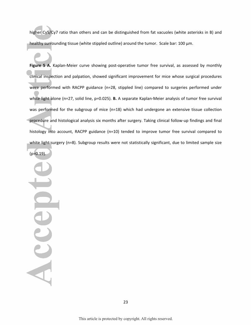
Figure 5 A. Kaplan‐Meier curve showing post‐operative tumor free survival, as assessed by monthly
clinical inspection and palpation, showed significant improvement for mice whose surgical procedures
were performed with RACPP guidance (n=28, stippled line) compared to surgeries performed under
white light alone (n=27, solid line, p=0.025). B. A separate Kaplan‐Meier analysis of tumor free survival
was performed for the subgroup of mice (n=18) which had undergone an extensive tissue collection
procedure and histological analysis six months after surgery. Taking clinical follow‐up findings and final
histology into account, RACPP guidance (n=10) tended to improve tumor free survival compared to
white light surgery (n=8). Subgroup results were not statistically significant, due to limited sample size
(p=0.19).
This article is protected by copyright. All rights reserved.
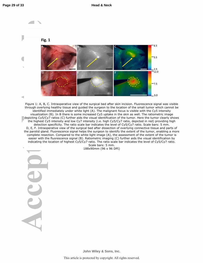
Figure 1: A, B, C. Intraoperative view of the surgical bed after skin incision. Fluorescence signal was visible through overlying healthy tissue and guided the surgeon to the location of the small tumor which cannot be
identified immediately under white light (A). The malignant focus is visible with the Cy5 intensity
visualization (B). In B there is some increased Cy5 uptake in the skin as well. The ratiometric image depicting Cy5/Cy7 ratios (C) further aids the visual identification of the tumor. Here the tumor clearly shows the highest Cy5 intensity and low Cy7 intensity (i.e. high Cy5/Cy7 ratio, depicted in red) providing high
detection specificity. The ratio scale bar indicates the level of Cy5/Cy7 ratio. Scale bars: 5 mm. D, E, F. Intraoperative view of the surgical bed after dissection of overlying connective tissue and parts of the parotid gland. Fluorescence signal helps the surgeon to identify the extent of the tumor, enabling a more complete resection. Compared to the white light image (A), the assessment of the extent of the tumor is easier with the fluorescence signal (B). Ratiometric imaging (C) further aids the visual identification by indicating the location of highest Cy5/Cy7 ratio. The ratio scale bar indicates the level of Cy5/Cy7 ratio.
Scale bars: 5 mm. 188x90mm (96 x 96 DPI)
Page 29 of 33
John Wiley & Sons, Inc.
Head & Neck
This article is protected by copyright. All rights reserved.

Figure 2: All excised tissue samples were analyzed by a pathologist blinded to experimental conditions after hematoxylin and eosin staining. Histological section of normal parotid gland tissue from a negative tissue
sample (A and B) compared to a tumor positive tissue sample (C and D). Scale bars: 100 µm. 179x129mm (96 x 96 DPI)
Page 30 of 33
John Wiley & Sons, Inc.
Head & Neck
This article is protected by copyright. All rights reserved.
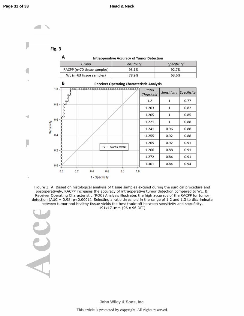
Figure 3: A. Based on histological analysis of tissue samples excised during the surgical procedure and postoperatively, RACPP increases the accuracy of intraoperative tumor detection compared to WL. B. Receiver Operating Characteristic (ROC) Analysis illustrates the high accuracy of the RACPP for tumor
detection (AUC = 0.98, p<0.0001). Selecting a ratio threshold in the range of 1.2 and 1.3 to discriminate between tumor and healthy tissue yields the best trade-off between sensitivity and specificity.
191x171mm (96 x 96 DPI)
Page 31 of 33
John Wiley & Sons, Inc.
Head & Neck
This article is protected by copyright. All rights reserved.
Figure 4: A. Macroscopic ratiometric image of a large parotid gland tumor (white stippled outline) imaged after RACPP injection. Red pseudocolor indicates high Cy5/Cy7 ratio, while blue/green color indicates low Cy5/Cy7 ratio. The overlying skin has been retracted from the tumor and shows high ratio due to cancer
invasion (white arrows). Scale bar: 5 mm. B shows an area of the anterior tumor border (white asterisk in A) imaged at higher magnification with confocal microscopy. Individual cells are labeled with higher Cy5/Cy7 ratio than others and can be distinguished from fat vacuoles (white asterisks in B) and healthy surrounding
tissue (white stippled outline) around the tumor. Scale bar: 100 µm. 208x112mm (96 x 96 DPI)
Page 32 of 33
John Wiley & Sons, Inc.
Head & Neck
This article is protected by copyright. All rights reserved.
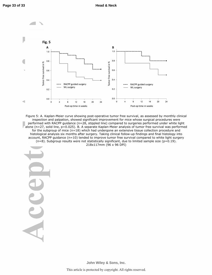
Figure 5: A. Kaplan-Meier curve showing post-operative tumor free survival, as assessed by monthly clinical inspection and palpation, showed significant improvement for mice whose surgical procedures were
performed with RACPP guidance (n=28, stippled line) compared to surgeries performed under white light
alone (n=27, solid line, p=0.025). B. A separate Kaplan-Meier analysis of tumor free survival was performed for the subgroup of mice (n=18) which had undergone an extensive tissue collection procedure and histological analysis six months after surgery. Taking clinical follow-up findings and final histology into account, RACPP guidance (n=10) tended to improve tumor free survival compared to white light surgery
(n=8). Subgroup results were not statistically significant, due to limited sample size (p=0.19). 218x117mm (96 x 96 DPI)
Page 33 of 33
John Wiley & Sons, Inc.
Head & Neck
This article is protected by copyright. All rights reserved.