Embed Size (px)
Citation preview

This chapter is divided into the following sections:Introduction, •••Anatomy and Physiology, •••Vocabulary, •••Terminology, •••Pathology of the Digestive System, •••Exercises, •••Answers to Exercises, •••Pronunciation of Terms, •••
Chapter goals• Name the organs of the digestive system and describe their
locations and functions.
• Defi ne combining forms for organs and know the meaning of related terminology.
• Describe signs, symptoms, and disease conditions affecting the digestive system.
Digestive System
5

5
144 DIGESTIVE SYSTEM
Students and teachers ask why I begin study of body system terminology with the digestive system, rather than with the more traditional musculoskeletal system. After many years of teaching, I recognized that both my students and I found it easier to start with the digestive system because it was more familiar. Also, the anatomy and physiology of the system were easier to explain and understand: The gastrointestinal tract resembles a long conveyor belt, with the mouth at the entrance and the anus at the exit. Keep in mind, however, that the text is organized so that you may begin study of the body systems with any chapter, to create the order that best refl ects your interests.
INTRODUCTION
The digestive or gastrointestinal tract begins with the mouth, where food enters, and ends with the anus, where solid waste material leaves the body. The four functions of the system are ingestion, digestion, absorption, and elimination.
First, complex food material taken into the mouth is ingested. Second, it is digested,or broken down, mechanically and chemically, as it travels through the gastrointestinal tract. Digestive enzymes speed up chemical reactions and aid the breakdown (digestion) of complex nutrients. Complex proteins are digested to simpler amino acids; complicated sugars are reduced to simple sugars, such as glucose; and large fat or lipid molecules are broken down to simpler substances such as fatty acids and triglycerides (three parts fatty acids and one part glycerol).
Third, via absorption, digested food passes through the lining cells or epithelium of the small intestine and into the bloodstream. Nutrients thus travel to all cells of the body. Cells then breakdown nutrients in the presence of oxygen to release energy. Cells also use amino acid nutrients to build up large protein molecules needed for growth and development. In addition, fat molecules are absorbed into lymphatic vessels from the intestine.
The fourth function of the digestive system is elimination of the solid waste materials that cannot be absorbed into the bloodstream. The large intestine concentrates these solid wastes, called feces, and the wastes fi nally pass out of the body through the anus.
ANATOMY AND PHYSIOLOGY
ORAL CAVITY
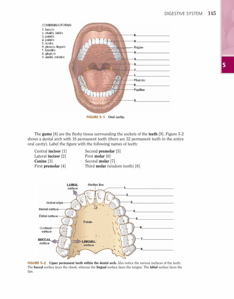
The gastrointestinal tract begins with the oral cavity. Oral means pertaining to the mouth (or/o). Label Figure 5-1 as you learn the major parts of the oral cavity.
The cheeks [1] form the walls of the oval-shaped oral cavity, and the lips [2] surround the opening to the cavity.
The hard palate [3] forms the anterior portion of the roof of the mouth, and the mus-cular soft palate [4] lies posterior to it. Rugae are irregular ridges in the mucous mem-brane covering the anterior portion of the hard palate. The uvula [5], a small soft tissue projection, hangs from the soft palate. It aids production of sounds and speech.
The tongue [6] extends across the fl oor of the oral cavity, and muscles attach it to the lower jaw bone. It moves food around during mastication (chewing) and deglutition (swal-lowing). Papillae, small raised areas on the tongue, contain taste buds that are sensitive to the chemical nature of foods and allow discrimination of different tastes as food moves across the tongue.
The tonsils [7], masses of lymphatic tissue located in depressions of the mucous mem-branes, lie on both sides of the oropharynx (part of the throat near the mouth). They are fi lters to protect the body from the invasion of microorganisms and they produce lympho-cytes, disease-fi ghting white blood cells.
5
DIGESTIVE SYSTEM 145
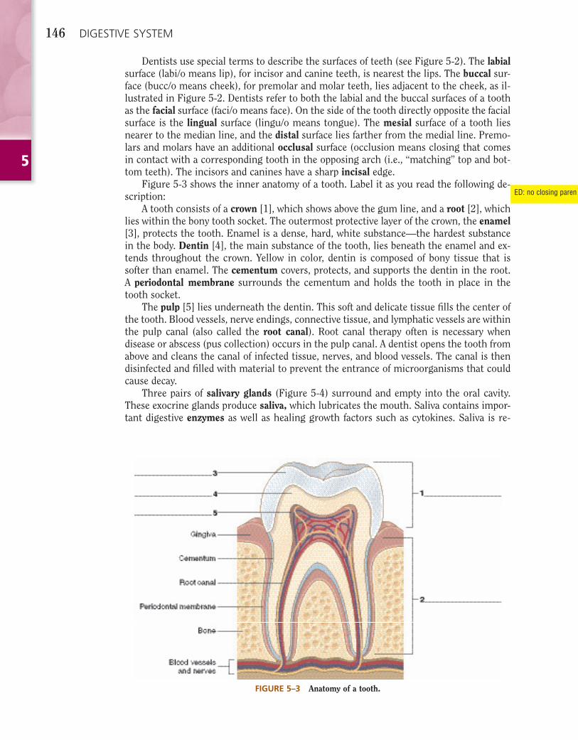
The gums [8] are the fl eshy tissue surrounding the sockets of the teeth [9]. Figure 5-2 shows a dental arch with 16 permanent teeth (there are 32 permanent teeth in the entire oral cavity). Label the fi gure with the following names of teeth:
Central incisor [1] Second premolar [5]Lateral incisor [2] First molar [6]Canine [3] Second molar [7]First premolar [4] Third molar (wisdom tooth) [8]
FIGURE 5–1 Oral cavity.
FIGURE 5–2 Upper permanent teeth within the dental arch. Also notice the various surfaces of the teeth. The buccal surface faces the cheek, whereas the lingual surface faces the tongue. The labial surface faces the lips.
5
146 DIGESTIVE SYSTEM
Dentists use special terms to describe the surfaces of teeth (see Figure 5-2). The labialsurface (labi/o means lip), for incisor and canine teeth, is nearest the lips. The buccal sur-face (bucc/o means cheek), for premolar and molar teeth, lies adjacent to the cheek, as il-lustrated in Figure 5-2. Dentists refer to both the labial and the buccal surfaces of a tooth as the facial surface (faci/o means face). On the side of the tooth directly opposite the facial surface is the lingual surface (lingu/o means tongue). The mesial surface of a tooth lies nearer to the median line, and the distal surface lies farther from the medial line. Premo-lars and molars have an additional occlusal surface (occlusion means closing that comes in contact with a corresponding tooth in the opposing arch (i.e., “matching” top and bot-tom teeth). The incisors and canines have a sharp incisal edge.
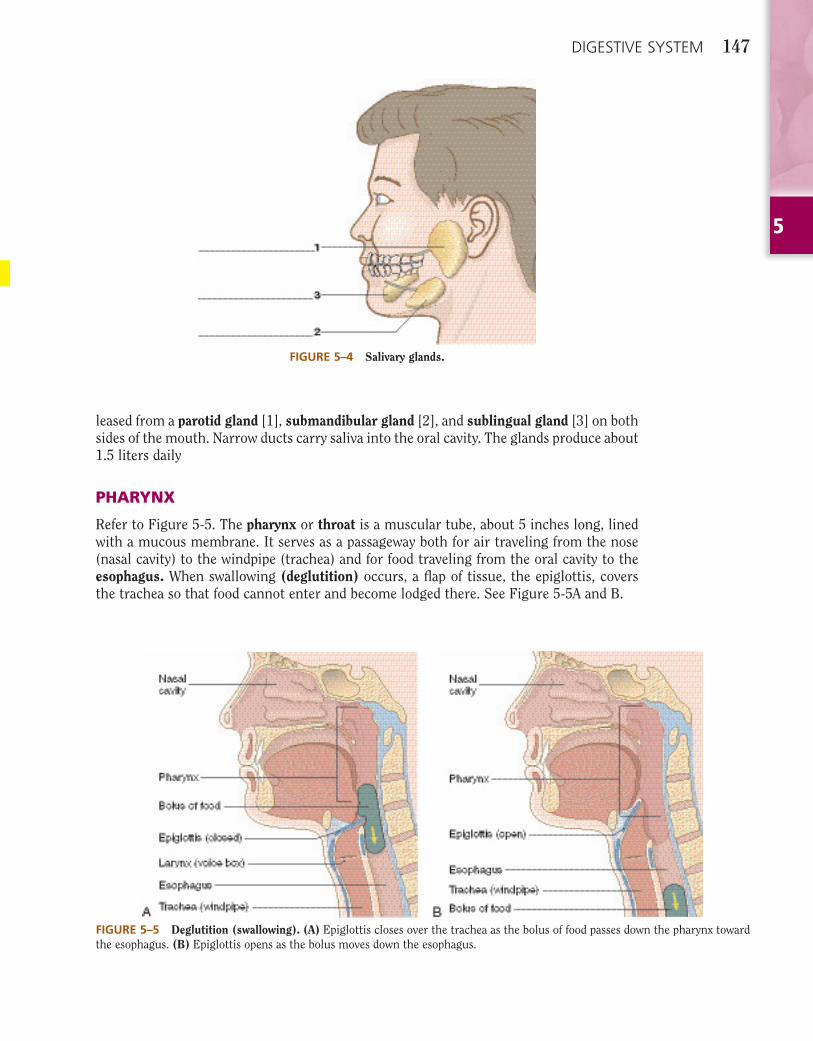
Figure 5-3 shows the inner anatomy of a tooth. Label it as you read the following de-scription:
A tooth consists of a crown [1], which shows above the gum line, and a root [2], which lies within the bony tooth socket. The outermost protective layer of the crown, the enamel[3], protects the tooth. Enamel is a dense, hard, white substance—the hardest substance in the body. Dentin [4], the main substance of the tooth, lies beneath the enamel and ex-tends throughout the crown. Yellow in color, dentin is composed of bony tissue that is softer than enamel. The cementum covers, protects, and supports the dentin in the root. A periodontal membrane surrounds the cementum and holds the tooth in place in the tooth socket.
The pulp [5] lies underneath the dentin. This soft and delicate tissue fi lls the center of the tooth. Blood vessels, nerve endings, connective tissue, and lymphatic vessels are within the pulp canal (also called the root canal). Root canal therapy often is necessary when disease or abscess (pus collection) occurs in the pulp canal. A dentist opens the tooth from above and cleans the canal of infected tissue, nerves, and blood vessels. The canal is then disinfected and fi lled with material to prevent the entrance of microorganisms that could cause decay.
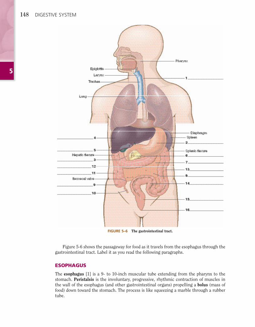
Three pairs of salivary glands (Figure 5-4) surround and empty into the oral cavity. These exocrine glands produce saliva, which lubricates the mouth. Saliva contains impor-tant digestive enzymes as well as healing growth factors such as cytokines. Saliva is re-
FIGURE 5–3 Anatomy of a tooth.
ED: no closing paren
5
DIGESTIVE SYSTEM 147
leased from a parotid gland [1], submandibular gland [2], and sublingual gland [3] on both sides of the mouth. Narrow ducts carry saliva into the oral cavity. The glands produce about 1.5 liters daily
PHARYNX
Refer to Figure 5-5. The pharynx or throat is a muscular tube, about 5 inches long, lined with a mucous membrane. It serves as a passageway both for air traveling from the nose (nasal cavity) to the windpipe (trachea) and for food traveling from the oral cavity to the esophagus. When swallowing (deglutition) occurs, a fl ap of tissue, the epiglottis, covers the trachea so that food cannot enter and become lodged there. See Figure 5-5A and B.
FIGURE 5–4 Salivary glands.
FIGURE 5–5 Deglutition (swallowing). (A) Epiglottis closes over the trachea as the bolus of food passes down the pharynx toward the esophagus. (B) Epiglottis opens as the bolus moves down the esophagus.
5
148 DIGESTIVE SYSTEM
Figure 5-6 shows the passageway for food as it travels from the esophagus through the gastrointestinal tract. Label it as you read the following paragraphs.
ESOPHAGUS
The esophagus [1] is a 9- to 10-inch muscular tube extending from the pharynx to the stomach. Peristalsis is the involuntary, progressive, rhythmic contraction of muscles in the wall of the esophagus (and other gastrointestinal organs) propelling a bolus (mass of food) down toward the stomach. The process is like squeezing a marble through a rubber tube.
FIGURE 5–6 The gastrointestinal tract.
5
DIGESTIVE SYSTEM 149
STOMACH
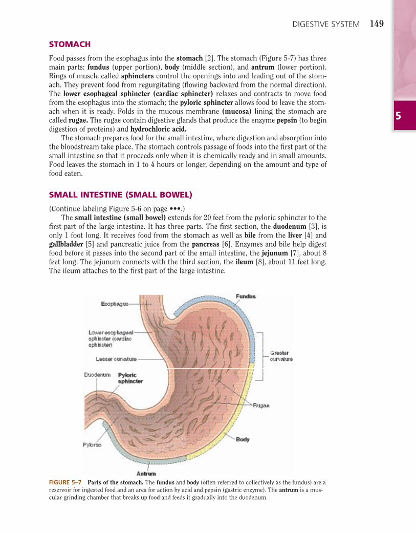
Food passes from the esophagus into the stomach [2]. The stomach (Figure 5-7) has three main parts: fundus (upper portion), body (middle section), and antrum (lower portion). Rings of muscle called sphincters control the openings into and leading out of the stom-ach. They prevent food from regurgitating (fl owing backward from the normal direction). The lower esophageal sphincter (cardiac sphincter) relaxes and contracts to move food from the esophagus into the stomach; the pyloric sphincter allows food to leave the stom-ach when it is ready. Folds in the mucous membrane (mucosa) lining the stomach are called rugae. The rugae contain digestive glands that produce the enzyme pepsin (to begin digestion of proteins) and hydrochloric acid.
The stomach prepares food for the small intestine, where digestion and absorption into the bloodstream take place. The stomach controls passage of foods into the fi rst part of the small intestine so that it proceeds only when it is chemically ready and in small amounts. Food leaves the stomach in 1 to 4 hours or longer, depending on the amount and type of food eaten.
SMALL INTESTINE (SMALL BOWEL)
(Continue labeling Figure 5-6 on page •••.)The small intestine (small bowel) extends for 20 feet from the pyloric sphincter to the
fi rst part of the large intestine. It has three parts. The fi rst section, the duodenum [3], is only 1 foot long. It receives food from the stomach as well as bile from the liver [4] and gallbladder [5] and pancreatic juice from the pancreas [6]. Enzymes and bile help digest food before it passes into the second part of the small intestine, the jejunum [7], about 8 feet long. The jejunum connects with the third section, the ileum [8], about 11 feet long. The ileum attaches to the fi rst part of the large intestine.
FIGURE 5–7 Parts of the stomach. The fundus and body (often referred to collectively as the fundus) are a reservoir for ingested food and an area for action by acid and pepsin (gastric enzyme). The antrum is a mus-cular grinding chamber that breaks up food and feeds it gradually into the duodenum.
5
150 DIGESTIVE SYSTEM
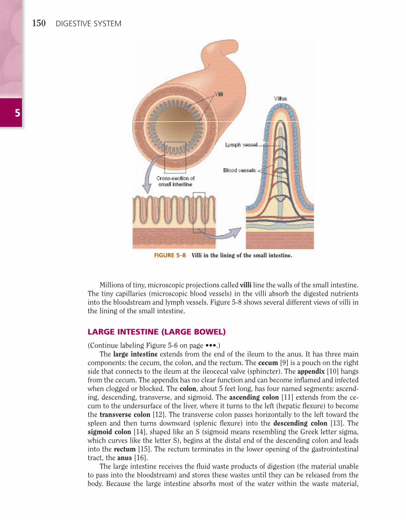
FIGURE 5–8 Villi in the lining of the small intestine.
Millions of tiny, microscopic projections called villi line the walls of the small intestine. The tiny capillaries (microscopic blood vessels) in the villi absorb the digested nutrients into the bloodstream and lymph vessels. Figure 5-8 shows several different views of villi in the lining of the small intestine.
LARGE INTESTINE (LARGE BOWEL)
(Continue labeling Figure 5-6 on page •••.)The large intestine extends from the end of the ileum to the anus. It has three main
components: the cecum, the colon, and the rectum. The cecum [9] is a pouch on the right side that connects to the ileum at the ileocecal valve (sphincter). The appendix [10] hangs from the cecum. The appendix has no clear function and can become infl amed and infected when clogged or blocked. The colon, about 5 feet long, has four named segments: ascend-ing, descending, transverse, and sigmoid. The ascending colon [11] extends from the ce-cum to the undersurface of the liver, where it turns to the left (hepatic fl exure) to become the transverse colon [12]. The transverse colon passes horizontally to the left toward the spleen and then turns downward (splenic fl exure) into the descending colon [13]. The sigmoid colon [14], shaped like an S (sigmoid means resembling the Greek letter sigma, which curves like the letter S), begins at the distal end of the descending colon and leads into the rectum [15]. The rectum terminates in the lower opening of the gastrointestinal tract, the anus [16].
The large intestine receives the fl uid waste products of digestion (the material unable to pass into the bloodstream) and stores these wastes until they can be released from the body. Because the large intestine absorbs most of the water within the waste material,
5
DIGESTIVE SYSTEM 151
the body can expel solid feces (stools). Defecation is the expulsion or passage of feces from the body through the anus. Diarrhea, or passage of watery stools, results from reduced water absorption into the bloodstream through the walls of the large intestine.
LIVER, GALLBLADDER, AND PANCREAS
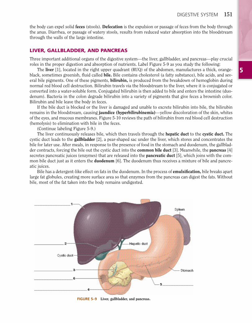
Three important additional organs of the digestive system—the liver, gallbladder, and pancreas—play crucial roles in the proper digestion and absorption of nutrients. Label Figure 5-9 as you study the following:
The liver [1], located in the right upper quadrant (RUQ) of the abdomen, manufactures a thick, orange-black, sometimes greenish, fl uid called bile. Bile contains cholesterol (a fatty substance), bile acids, and sev-eral bile pigments. One of these pigments, bilirubin, is produced from the breakdown of hemoglobin during normal red blood cell destruction. Bilirubin travels via the bloodstream to the liver, where it is conjugated or converted into a water-soluble form. Conjugated bilirubin is then added to bile and enters the intestine (duo-denum). Bacteria in the colon degrade bilirubin into a variety of pigments that give feces a brownish color. Bilirubin and bile leave the body in feces.
If the bile duct is blocked or the liver is damaged and unable to excrete bilirubin into bile, the bilirubin remains in the bloodstream, causing jaundice (hyperbilirubinemia)—yellow discoloration of the skin, whites of the eyes, and mucous membranes. Figure 5-10 reviews the path of bilirubin from red blood cell destruction (hemolysis) to elimination with bile in the feces.
(Continue labeling Figure 5-9.)The liver continuously releases bile, which then travels through the hepatic duct to the cystic duct. The
cystic duct leads to the gallbladder [2], a pear-shaped sac under the liver, which stores and concentrates the bile for later use. After meals, in response to the presence of food in the stomach and duodenum, the gallblad-der contracts, forcing the bile out the cystic duct into the common bile duct [3]. Meanwhile, the pancreas [4] secretes pancreatic juices (enzymes) that are released into the pancreatic duct [5], which joins with the com-mon bile duct just as it enters the duodenum [6]. The duodenum thus receives a mixture of bile and pancre-atic juices.
Bile has a detergent-like effect on fats in the duodenum. In the process of emulsifi cation, bile breaks apart large fat globules, creating more surface area so that enzymes from the pancreas can digest the fats. Without bile, most of the fat taken into the body remains undigested.
FIGURE 5–9 Liver, gallbladder, and pancreas.
5
152 DIGESTIVE SYSTEM
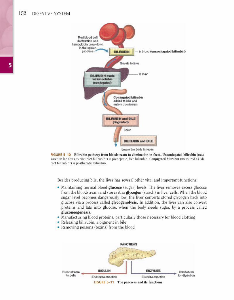
FIGURE 5–10 Bilirubin pathway from bloodstream to elimination in feces. Unconjugated bilirubin (mea-sured in lab tests as “indirect bilirubin”) is prehepatic, free bilirubin. Conjugated bilirubin (measured as “di-rect bilirubin”) is posthepatic bilirubin.
Besides producing bile, the liver has several other vital and important functions:
• Maintaining normal blood glucose (sugar) levels. The liver removes excess glucose from the bloodstream and stores it as glycogen (starch) in liver cells. When the blood sugar level becomes dangerously low, the liver converts stored glycogen back into glucose via a process called glycogenolysis. In addition, the liver can also convert proteins and fats into glucose, when the body needs sugar, by a process called gluconeogenesis.
• Manufacturing blood proteins, particularly those necessary for blood clotting• Releasing bilirubin, a pigment in bile• Removing poisons (toxins) from the blood
FIGURE 5–11 The pancreas and its functions.
5
DIGESTIVE SYSTEM 153
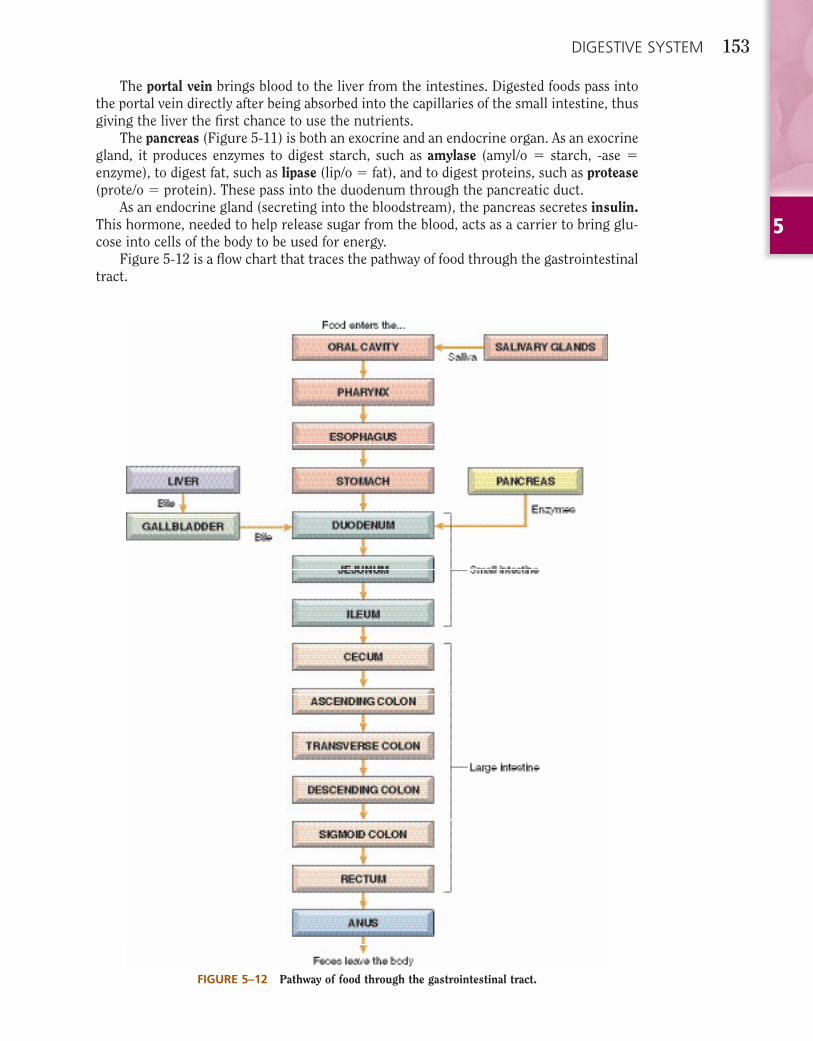
FIGURE 5–12 Pathway of food through the gastrointestinal tract.
The portal vein brings blood to the liver from the intestines. Digested foods pass into the portal vein directly after being absorbed into the capillaries of the small intestine, thus giving the liver the fi rst chance to use the nutrients.
The pancreas (Figure 5-11) is both an exocrine and an endocrine organ. As an exocrine gland, it produces enzymes to digest starch, such as amylase (amyl/o � starch, -ase �enzyme), to digest fat, such as lipase (lip/o � fat), and to digest proteins, such as protease(prote/o � protein). These pass into the duodenum through the pancreatic duct.
As an endocrine gland (secreting into the bloodstream), the pancreas secretes insulin.This hormone, needed to help release sugar from the blood, acts as a carrier to bring glu-cose into cells of the body to be used for energy.
Figure 5-12 is a fl ow chart that traces the pathway of food through the gastrointestinal tract.
5
154 DIGESTIVE SYSTEM
VOCABULARYThe following list reviews many of the terms introduced in this chapter. Short defi nitions
and additional information reinforce your understanding of the terms. All of the terms are included in the Pronunciation of Terms section later in the chapter.
absorption Passage of materials through the walls of the small intestine into the bloodstream.
amino acids Small building blocks of proteins (like links in a chain), released when proteins are digested.
amylase Enzyme secreted by the pancreas to digest starch.
anus Terminal end or opening of the digestive tract to the outside of the body.
appendix Blind pouch hanging from the cecum (in the right lower quadrant [RLQ]). It literally means hanging (pend/o) onto (ap-, which is a form of ad-).
bile Digestive juice made in the liver and stored in the gallbladder. It breaks up (emulsifi es) large fat globules. Bile originally was called gall (Latin bilis, meaning gall or anger), probably because it has a bitter taste. It is composed of bile pigments (colored materials), cholesterol, and bile salts.
bilirubin Pigment released by the liver in bile.
bowel Intestine.
canine teeth Pointed, dog-like teeth (canine means pertaining to dog) next to the incisors. Also called cuspids or eyeteeth.
cecum First part of the large intestine.
colon Large intestine, consisting of the cecum; the ascending, transverse, and descending segments of the colon; and the rectum.
common bile duct Carries bile from the liver and gallbladder to the duodenum. Also called the choledochus.
defecation Elimination of feces from the digestive tract through the anus.
deglutition Swallowing.
dentin The primary material found in teeth. It is covered by the enamel in the crown and a protective layer of cementum in the root.
digestion Breakdown of complex foods to simpler forms.
duodenum First part of the small intestine. Duo � 2, den � 10; the duodenum measures 12 inches long.
elimination Act of removal of materials from the body; in the digestive system, the removal of indigestible materials as feces.
emulsifi cation Physical process of breaking up large fat globules into smaller globules, thereby increasing the surface area that enzymes can use to digest the fat.
5
DIGESTIVE SYSTEM 155
enamel Hard, outermost layer of a tooth.
enzyme A chemical that speeds up a reaction between substances. Digestive enzymes break down complex foods to simpler substances. Enzymes are given names that end in -ase.
esophagus Tube connecting the throat to the stomach. Eso- means inward; phag/o means swallowing.
fatty acids Substances produced when fats are digested.
feces Solid wastes; stool.
gallbladder Small sac under the liver; stores bile. Remember: gallbladder is one word!
glucose Simple sugar.
glycogen Starch; glucose is stored in the form of glycogen in liver cells.
hydrochloric acid Substance produced by the stomach; necessary for digestion of food.
ileum Third part of the small intestine; from the Greek eilos, meaning twisted. When the abdomen was viewed at autopsy, the intestine appeared twisted, and the ileum often was an area of obstruction.
incisor One of four front teeth in the dental arch.
insulin Hormone produced by the endocrine cells of the pancreas. It transports sugar from the blood into cells and stimulates glycogen formation by the liver.
jejunum Second part of the small intestine. The Latin jejunus means empty; this part of the intestine was always empty when a body was examined after death.
lipase Pancreatic enzyme necessary to digest fats.
liver A large organ located in the RUQ of the abdomen. The liver secretes bile; stores sugar, iron, and vitamins; produces blood proteins; and destroys worn-out red blood cells. The normal adult liver weighs about 21⁄2 to 3 pounds.
lower esophageal sphincter (LES)
Ring of muscles between the esophagus and the stomach. Also called cardiac sphincter.
mastication Chewing.
molar teeth The sixth, seventh, and eighth teeth from the middle on either side of the dental arch. Premolar teeth are the fourth and fi fth teeth, before the molars.
palate Roof of the mouth. The hard palate lies anterior to the soft palate and is supported by the upper jaw bone (maxilla). The soft palate is the posterior fl eshy part between the mouth and the throat.
pancreas Organ under the stomach; produces insulin (for transport of sugar into cells) and enzymes (for digestion of foods).
papillae (singular: papilla) Small elevations on the tongue. A papilla is a nipple-like elevation.
parotid gland Salivary gland within the cheek, just anterior to the ear.
5
156 DIGESTIVE SYSTEM
peristalsis Rhythmic contractions of the tubular organs. In the gastrointestinal tract, peristalsis moves the contents through at different rates: stomach, 0.5 to 2 hours; small intestine, 2 to 6 hours; and colon, 6 to 72 hours. Peri- means surrounding; -stalsis is constriction.
pharynx Throat, the common passageway for food from the mouth and for air from the nose.
portal vein Large vein bringing blood to the liver from the intestines.
protease Enzyme that digests protein.
pulp Soft tissue within a tooth, containing nerves and blood vessels.
pyloric sphincter Ring of muscle at the end of the stomach, near the duodenum. From the Greek pyloros, meaning gatekeeper. It is normally closed, but opens when a wave of peristalsis passes over it.
pylorus Distal region of the stomach, opening to the duodenum.
rectum Last section of the large intestine, connecting the end of the colon and the anus.
rugae Ridges on the hard palate and the wall of the stomach.
saliva Digestive juice produced by salivary glands.
salivary glands Parotid, sublingual, and submandibular glands.
sigmoid colon Fourth and last, S-shaped segment of the colon, just before the rectum; empties into the rectum.
sphincter Circular ring of muscle that constricts a passage or closes a natural opening.
stomach Muscular organ that receives food from the esophagus. The stomach’s parts are the fundus (proximal section), body (middle section), and antrum (distal section).
triglycerides Fat molecules composed of three parts fatty acids and one part glycerol.
uvula Soft tissue hanging from the middle of the soft palate. The Latin uva means bunch of grapes.
villi (singular: villus) Microscopic projections in the wall of the small intestine that absorb nutrients into the bloodstream.

5
DIGESTIVE SYSTEM 157
TERMINOLOGYWrite the meaning of the medical term in the space provided. Check the Pronunciation
of Terms section later in the chapter for any unfamiliar words.
PARTS OF THE BODY
COMBINING FORM MEANING TERMINOLOGY MEANING
an/o anus perianal ________________________________________
append/o appendix appendectomy ___________________________________
appendic/o appendicitis _____________________________________See Figure 5–13.
bucc/o cheek buccal mucosa ___________________________________A mucosa is a mucous membrane lining cavities or canals that open to the outside of the body.
cec/o cecum cecal ___________________________________________
celi/o belly, abdomen celiac __________________________________________Abdomin/o and lapar/o also mean abdomen. With combining forms that have the same basic meaning, no rule exists for the proper usage of one or the other. You will learn to recognize each in its proper context.
cheil/o lip cheilosis ________________________________________Labi/o also means lip.
cholecyst/o gallbladder cholecystectomyDon’t confuse cholecyst/o with cyst/o, which means urinary bladder!
FIGURE 5–13 Stages of appendicitis. (A) Obstruction and bacterial infection cause red, swollen, and in-fl amed appendix. (B) Pus and bacteria invade the wall of the appendix. (C) Pus perforates (ruptures through) the wall of the appendix into the abdomen, leading to peritonitis (infl ammation of the peritoneum). (Modi-fi ed from Damjanov I: Pathology for the Health-Related Professions, 3rd ed., Philadelphia, Saunders, 2006, p. 260.)
5
158 DIGESTIVE SYSTEM
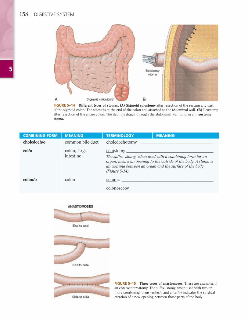
FIGURE 5–14 Different types of stomas. (A) Sigmoid colostomy after resection of the rectum and part of the sigmoid colon. The stoma is at the end of the colon and attached to the abdominal wall. (B) Ileostomy after resection of the entire colon. The ileum is drawn through the abdominal wall to form an ileostomy stoma.
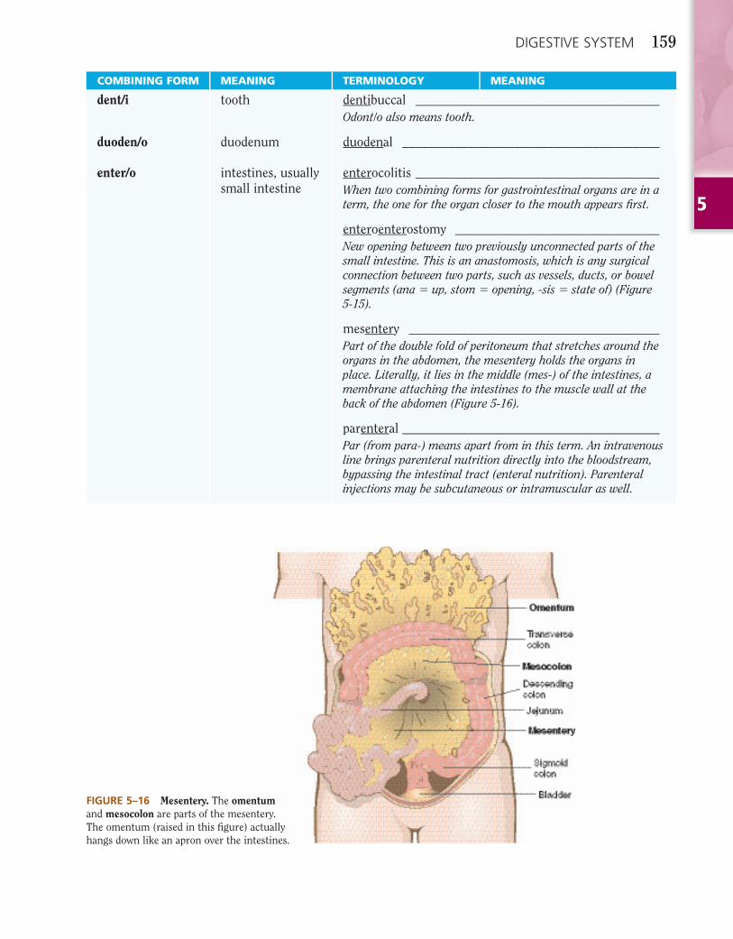
FIGURE 5–15 Three types of anastomoses. These are examples of an enteroenterostomy. The suffi x -stomy, when used with two or more combining forms (enter/o and enter/o) indicates the surgical creation of a new opening between those parts of the body.
COMBINING FORM MEANING TERMINOLOGY MEANING
choledoch/o common bile duct choledochotomy _________________________________
col/o colon, large intestine
colostomy _______________________________________The suffi x -stomy, when used with a combining form for an organ, means an opening to the outside of the body. A stoma is an opening between an organ and the surface of the body (Figure 5-14).
colon/o colon colonic _________________________________________
colonoscopy _____________________________________
5
DIGESTIVE SYSTEM 159
COMBINING FORM MEANING TERMINOLOGY MEANING
dent/i tooth dentibuccal _____________________________________Odont/o also means tooth.
duoden/o duodenum duodenal _______________________________________
enter/o intestines, usually small intestine
enterocolitis _____________________________________When two combining forms for gastrointestinal organs are in a term, the one for the organ closer to the mouth appears fi rst.
enteroenterostomy _______________________________New opening between two previously unconnected parts of the small intestine. This is an anastomosis, which is any surgical connection between two parts, such as vessels, ducts, or bowel segments (ana � up, stom � opening, -sis � state of) (Figure 5-15).
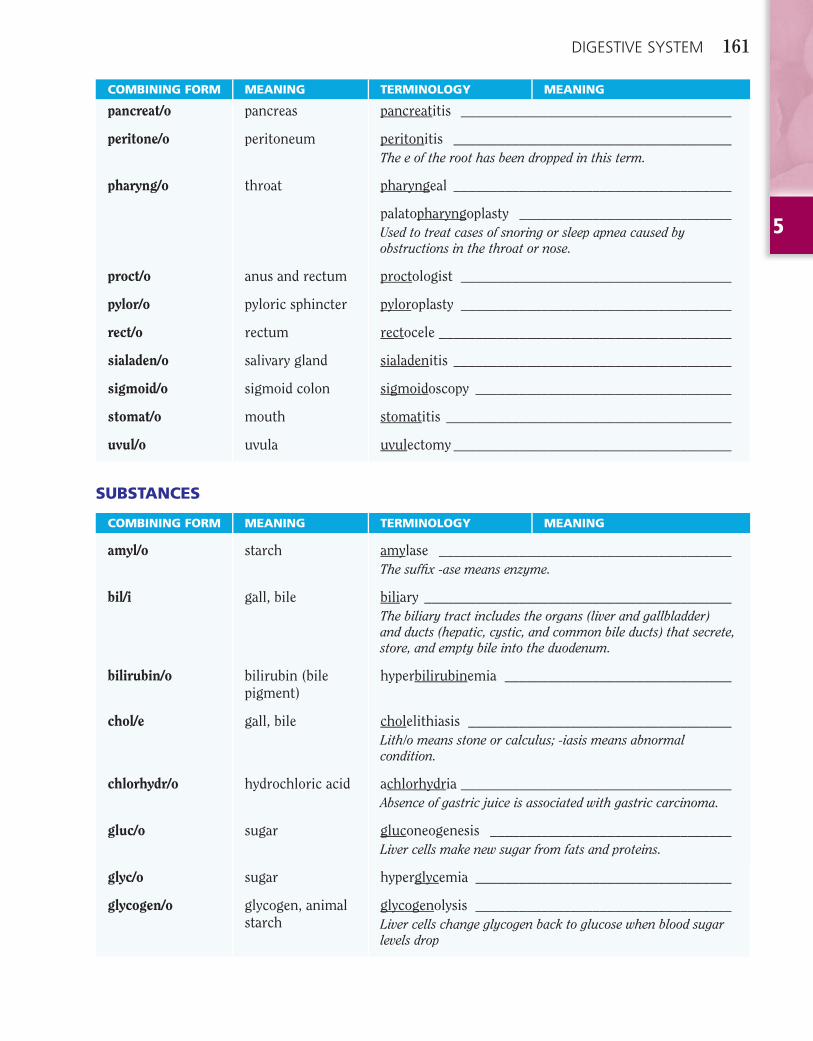
mesentery ______________________________________Part of the double fold of peritoneum that stretches around the organs in the abdomen, the mesentery holds the organs in place. Literally, it lies in the middle (mes-) of the intestines, a membrane attaching the intestines to the muscle wall at the back of the abdomen (Figure 5-16).
parenteral _______________________________________Par (from para-) means apart from in this term. An intravenous line brings parenteral nutrition directly into the bloodstream, bypassing the intestinal tract (enteral nutrition). Parenteral injections may be subcutaneous or intramuscular as well.
FIGURE 5–16 Mesentery. The omentum and mesocolon are parts of the mesentery. The omentum (raised in this fi gure) actually hangs down like an apron over the intestines.
5
160 DIGESTIVE SYSTEM
COMBINING FORM MEANING TERMINOLOGY MEANING
esophag/o esophagus esophageal ______________________________________Note: Changing the suffi x from -al to -eal softens the fi nal g (e-sof-a-JE-al).
faci/o face facial ___________________________________________
gastr/o stomach gastrostomy _____________________________________
gingiv/o gums gingivitis _______________________________________
gloss/o tongue hypoglossal _____________________________________Lingu/o also means tongue.
hepat/o liver hepatoma _______________________________________Also called hepatocellular carcinoma.
hepatomegaly ____________________________________
ile/o ileum ileocecal sphincterAlso called the ileocecal valve.
ileitis __________________________________________
ileostomy _______________________________________See Figure 5-14B.
jejun/o jejunum choledochojejunostomy ___________________________An anastomosis.
gastrojejunostomy ________________________________This is part of a gastric bypass procedure.
labi/o lip labial ___________________________________________
lapar/o abdomen laparoscopy _____________________________________A form of minimally invasive surgery (MIS). Examples are laparoscopic cholecystectomy and laparoscopic appendectomy.
lingu/o tongue sublingual ______________________________________
mandibul/o lower jaw, mandible
submandibular ___________________________________
odont/o tooth orthodontist _____________________________________Orth/o means straight.
periodontist _____________________________________
endodontist _____________________________________Performs root canal therapy.
or/o mouth oral ____________________________________________Stomat/o also means mouth.
palat/o palate palatoplasty _____________________________________Procedure to repair cleft palate and cleft lip; repair of a cleft palate.
5
DIGESTIVE SYSTEM 161
COMBINING FORM MEANING TERMINOLOGY MEANING
pancreat/o pancreas pancreatitis _____________________________________
peritone/o peritoneum peritonitis ______________________________________The e of the root has been dropped in this term.
pharyng/o throat pharyngeal ______________________________________
palatopharyngoplasty _____________________________Used to treat cases of snoring or sleep apnea caused by obstructions in the throat or nose.
proct/o anus and rectum proctologist _____________________________________
pylor/o pyloric sphincter pyloroplasty _____________________________________
rect/o rectum rectocele ________________________________________
sialaden/o salivary gland sialadenitis ______________________________________
sigmoid/o sigmoid colon sigmoidoscopy ___________________________________
stomat/o mouth stomatitis _______________________________________
uvul/o uvula uvulectomy ______________________________________
SUBSTANCES
COMBINING FORM MEANING TERMINOLOGY MEANING
amyl/o starch amylase ________________________________________The suffi x -ase means enzyme.
bil/i gall, bile biliary __________________________________________The biliary tract includes the organs (liver and gallbladder) and ducts (hepatic, cystic, and common bile ducts) that secrete, store, and empty bile into the duodenum.
bilirubin/o bilirubin (bile pigment)
hyperbilirubinemia _______________________________
chol/e gall, bile cholelithiasis ____________________________________Lith/o means stone or calculus; -iasis means abnormal condition.
chlorhydr/o hydrochloric acid achlorhydria _____________________________________Absence of gastric juice is associated with gastric carcinoma.
gluc/o sugar gluconeogenesis _________________________________Liver cells make new sugar from fats and proteins.
glyc/o sugar hyperglycemia ___________________________________
glycogen/o glycogen, animal starch
glycogenolysis ___________________________________Liver cells change glycogen back to glucose when blood sugar levels drop
5
162 DIGESTIVE SYSTEM
PATHOLOGY OF THE DIGESTIVE SYSTEM
This section presents medical terms that describe signs and symptoms (clinical indications of illness) and pathologic conditions of the gastrointestinal tract. Sentences following each defi nition describe the etiology(eti/o � cause) of the illness and treatment. When the etiology (cause) is not understood, the condition is idiopathic (idi/o � unknown). You can fi nd a list of drugs prescribed to treat gastrointestinal signs and symp-toms and conditions on page ••• in Chapter 21, Pharmacology.
COMBINING FORM MEANING TERMINOLOGY MEANING
lip/o fat, lipid lipoma _________________________________________
lith/o stone lithogenesis _____________________________________
prote/o protein protease ________________________________________
sial/o saliva, salivary sialolith ________________________________________
steat/o fat steatorrhea ______________________________________Improperly digested (malabsorbed) fats will appear in the feces.
SUFFIXES
SUFFIX MEANING TERMINOLOGY MEANING
-ase enzyme lipase __________________________________________Enzymes speed up chemical reactions. Lipase aids in the digestion of fats. In all types, liver enzyme levels may be elevated, indicating damage to liver cells. Signs and symptoms include malaise, anorexia, hepatomegaly, jaundice, and abdominal pain.
-chezia defecation, elimination of wastes
hematochezia ____________________________________(he-ma-to-KE-ze-a) Bright red blood is found in the feces.
-iasis abnormal condition
choledocholithiasis _______________________________
-prandial meal postprandial _____________________________________Post cibum (p.c.), seen on written prescriptions, also means after meals.
Signs and Symptoms
A sign is an objective fi nding—such as an increase in body temperature, a rash, or a sound heard on listening to the chest—indicating the presence of disease as perceived by an examiner. However, a symptom is a subjective sensation or change in health—such as itching, pain, fatigue, or nausea—as experienced by the patient. Clearly, the same feature may be noticed by both doctor and patient, which makes it at once both a sign and a symptom!
5
DIGESTIVE SYSTEM 163
SIGNS AND SYMPTOMS
anorexia Lack of appetiteAnorexia (-orexia � appetite) often is a sign of malignancy or liver disease. An-orexia nervosa is loss of appetite associated with emotional problems such as an-ger, anxiety, and irrational fear of weight gain. It is an eating disorder and is dis-cussed, along with a similar eating disorder, bulimia nervosa, in Chapter 22.
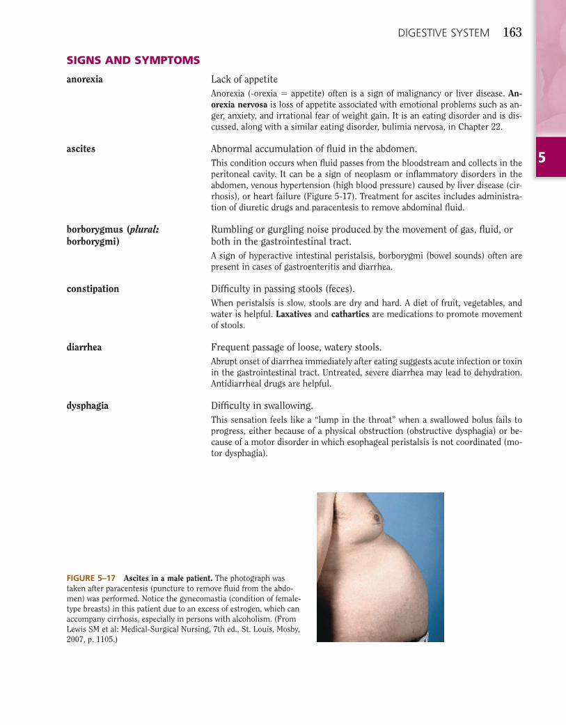
ascites Abnormal accumulation of fl uid in the abdomen.This condition occurs when fl uid passes from the bloodstream and collects in the peritoneal cavity. It can be a sign of neoplasm or infl ammatory disorders in the abdomen, venous hypertension (high blood pressure) caused by liver disease (cir-rhosis), or heart failure (Figure 5-17). Treatment for ascites includes administra-tion of diuretic drugs and paracentesis to remove abdominal fl uid.
borborygmus (plural: borborygmi)
Rumbling or gurgling noise produced by the movement of gas, fl uid, or both in the gastrointestinal tract.A sign of hyperactive intestinal peristalsis, borborygmi (bowel sounds) often are present in cases of gastroenteritis and diarrhea.
constipation Diffi culty in passing stools (feces).When peristalsis is slow, stools are dry and hard. A diet of fruit, vegetables, and water is helpful. Laxatives and cathartics are medications to promote movement of stools.
diarrhea Frequent passage of loose, watery stools.Abrupt onset of diarrhea immediately after eating suggests acute infection or toxin in the gastrointestinal tract. Untreated, severe diarrhea may lead to dehydration. Antidiarrheal drugs are helpful.
dysphagia Diffi culty in swallowing.This sensation feels like a “lump in the throat” when a swallowed bolus fails to progress, either because of a physical obstruction (obstructive dysphagia) or be-cause of a motor disorder in which esophageal peristalsis is not coordinated (mo-tor dysphagia).
FIGURE 5–17 Ascites in a male patient. The photograph was taken after paracentesis (puncture to remove fl uid from the abdo-men) was performed. Notice the gynecomastia (condition of female-type breasts) in this patient due to an excess of estrogen, which can accompany cirrhosis, especially in persons with alcoholism. (From Lewis SM et al: Medical-Surgical Nursing, 7th ed., St. Louis, Mosby, 2007, p. 1105.)
5
164 DIGESTIVE SYSTEM
eructation Gas expelled from the stomach through the mouth.Eructation produces a characteristic sound and also is called belching.
fl atus Gas expelled through the anus.Flatulence is the presence of excessive gas in both the stomach and the intes-tines.
hematochezia Passage of fresh, bright red blood from the rectum.The cause of hematochezia usually is bleeding due to colitis or from ulcers or polyps in the colon or rectum.
jaundice (icterus) Yellow-orange coloration of the skin and whites of the eyes caused by high levels of bilirubin in the blood (hyperbilirubinemia).Jaundice can occur when (1) excessive destruction of erythrocytes, as in hemoly-sis, causes excess bilirubin in the blood; (2) malfunction of liver cells (hepato-cytes) due to liver disease prevents the liver from excreting bilirubin with bile; or (3) obstruction of bile fl ow, such as from choledocholithiasis or tumor, prevents bilirubin in bile from being excreted into the duodenum.
melena Black, tarry stools; feces containing digested blood.This clinical sign usually refl ects a condition in which blood has had time to be digested (acted on by intestinal juices) and results from bleeding in the upper gastrointestinal tract (duodenal ulcer). A positive result on stool guaiac testing (see page •••) indicates blood in the stool.
nausea Unpleasant sensation in the stomach associated with a tendency to vomit.Common causes are sea and motion sickness and early pregnancy. Nausea and vomiting may be symptomatic of a perforation (hole in the wall) of an abdominal organ; obstruction of a bile duct, stomach, or intestine; or exposure to toxins (poisons).
steatorrhea Fat in the feces; frothy, foul-smelling fecal matter.Improper digestion or absorption of fat can cause fat to remain in the intestine. This may occur with disease of the pancreas (pancreatitis) when pancreatic en-zymes are not excreted. It also is a sign of intestinal disease that involves malab-sorption of fat.
PATHOLOGIC CONDITIONS
ORAL CAVITY AND TEETH
aphthous stomatitis Infl ammation of the mouth with small, painful ulcers.The ulcers associated with this condition are commonly called canker (KANK-er) sores; the cause is unknown. See Figure 5-18B.
dental caries Tooth decayDental plaque results from the accumulation of foods, proteins from saliva, and necrotic debris on the tooth enamel. Bacteria grow in the plaque and cause pro-duction of acid that dissolves the tooth enamel, resulting in a cavity (area of decay) (see Figure 15-18C). If the bacterial infection reaches the pulp of the tooth, root canal therapy may be necessary.

5
DIGESTIVE SYSTEM 165
FIGURE 5–18 Normal teeth and gums and pathologic conditions. (A) Normal teeth and gums. (B) Aphthous stomati-tis. (C) Dental caries. (D) Herpetic sto-matitis. (E) Oral leukoplakia. (F) Gingi-vitis. (A, from Christensen GJ: A Consumer’s Guide to Dentistry, St. Louis, Mosby, 2002; B, from Feldman M et al: Sleisenger and Fordtran’s Gastro-intestinal and Liver Disease, 8th ed., Philadelphia, Saunders, 2006; C, cour-tesy Dr. Frank Hodges, from Bird D, Robinson D: Torres and Ehrlich Modern Dental Assisting, 8th ed., Philadelphia, Saunders, 2005; D, from Swartz MH: Textbook of Physical Diagnosis, History and Examination, 5th ed., Philadelphia, Saunders, 2006; E, from Callen JP et al: Color Atlas of Dermatology, 2nd ed., Philadelphia, WB Saunders, 2002; F, from Bird D, Robinson D: Torres and Ehrlich Modern Dental Assisting, 8th ed., Philadelphia, Saunders, 2005.)
herpetic stomatitis Infl ammation of the mouth caused by infection with the herpesvirus.Painful fl uid-fi lled blisters on the lips, palate, gums, and tongue, commonly called fever blisters or cold sores (see Figure 15-18D). It is caused by herpes simplex virus type 1 (HSV1). Treatment is with medication to relieve symptoms. Herpes genitalis (due to HSV2) occurs on the reproductive organs. Both conditions are highly contagious.
oral leukoplakia White plaques or patches on the mucosa of the mouth.This precancerous lesion (see Figure 15-18E) can result from chronic tobacco use (pipe smoking or chewing tobacco). Malignant potential is assessed by micro-scopic study of biopsied tissue.
periodontal disease Infl ammation and degeneration of gums, teeth, and surrounding bone.Gingivitis (see Figure 15-18F) occurs as a result of accumulation of dental plaqueand dental calculus or tartar (a yellow-brown calcifi ed deposit on teeth). In gingi-vectomy, a periodontist uses a metal instrument to scrape away plaque and tartar from teeth; any pockets of pus are then drained and removed to allow new tissue to form. Localized infections are treated with systemic antibiotics.
5
166 DIGESTIVE SYSTEM
UPPER GASTROINTESTINAL TRACT
achalasia Failure of the lower esophagus sphincter (LES) muscle to relax.Achalasia (-chalasia � relaxation) results from the loss of peristalsis so that food cannot pass easily through the esophagus. Both failure of the LES to relax and the loss of peristalsis cause dilatation (widening) of the esophagus above the constric-tion. Physicians recommend a bland diet low in bulk and mechanical stretching of the LES to relieve symptoms.
esophageal cancer Malignant tumor of the esophagusThe most common symptom of esophageal cancer is diffi culty swallowing (dys-phagia). Smoking and chronic alcohol use are major risk factors. Long-term irri-tation of the esophagus caused by gastric refl ux is a premalignant condition called Barrett esophagus. Surgery, radiation therapy, and chemotherapy are treatment options.
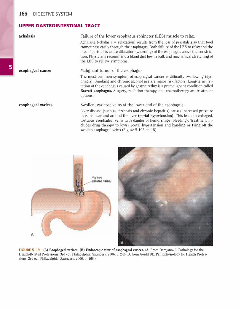
esophageal varices Swollen, varicose veins at the lower end of the esophagus.Liver disease (such as cirrhosis and chronic hepatitis) causes increased pressure in veins near and around the liver (portal hypertension). This leads to enlarged, tortuous esophageal veins with danger of hemorrhage (bleeding). Treatment in-cludes drug therapy to lower portal hypertension and banding or tying off the swollen esophageal veins (Figure 5-19A and B).
FIGURE 5–19 (A) Esophageal varices. (B) Endoscopic view of esophageal varices. (A, From Damjanov I: Pathology for the Health-Related Professions, 3rd ed., Philadelphia, Saunders, 2006, p. 246; B, from Gould BE: Pathophysiology for Health Profes-sions, 3rd ed., Philadelphia, Saunders, 2006, p. 466.)
5
DIGESTIVE SYSTEM 167
gastric cancer Malignant tumor of the stomach.Chronic gastritis associated with bacterial infection is a major risk factor for gas-tric carcinoma. Gastric endoscopy and biopsy diagnose the condition. Cure de-pends on early detection and surgical removal of the cancerous tissue.
gastroesophageal refl ux disease (GERD)
Solids and fl uids return to the mouth from the stomach.Heartburn is the burning sensation caused by regurgitation of hydrochloric acid from the stomach to the esophagus. Chronic exposure of esophageal mucosa to gastric acid and pepsin (an enzyme that digests protein) leads to refl ux esophagi-tis. Drug treatment for GERD includes antacid (acid-suppressive) agents and medication to increase the tone of the LES.
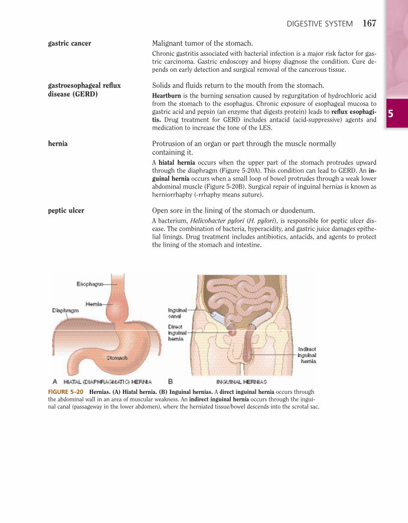
hernia Protrusion of an organ or part through the muscle normally containing it.A hiatal hernia occurs when the upper part of the stomach protrudes upward through the diaphragm (Figure 5-20A). This condition can lead to GERD. An in-guinal hernia occurs when a small loop of bowel protrudes through a weak lower abdominal muscle (Figure 5-20B). Surgical repair of inguinal hernias is known as herniorrhaphy (-rrhaphy means suture).
peptic ulcer Open sore in the lining of the stomach or duodenum.A bacterium, Helicobacter pylori (H. pylori), is responsible for peptic ulcer dis-ease. The combination of bacteria, hyperacidity, and gastric juice damages epithe-lial linings. Drug treatment includes antibiotics, antacids, and agents to protect the lining of the stomach and intestine.
FIGURE 5–20 Hernias. (A) Hiatal hernia. (B) Inguinal hernias. A direct inguinal hernia occurs through the abdominal wall in an area of muscular weakness. An indirect inguinal hernia occurs through the ingui-nal canal (passageway in the lower abdomen), where the herniated tissue/bowel descends into the scrotal sac.
5
168 DIGESTIVE SYSTEM
LOWER GASTROINTESTINAL TRACT (SMALL AND LARGE INTESTINE)
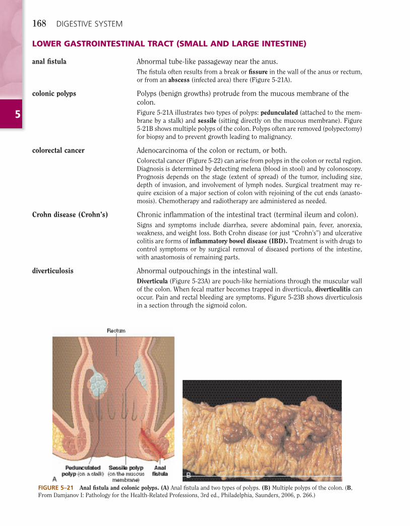
anal fi stula Abnormal tube-like passageway near the anus.The fi stula often results from a break or fi ssure in the wall of the anus or rectum, or from an abscess (infected area) there (Figure 5-21A).
colonic polyps Polyps (benign growths) protrude from the mucous membrane of the colon.Figure 5-21A illustrates two types of polyps: pedunculated (attached to the mem-brane by a stalk) and sessile (sitting directly on the mucous membrane). Figure 5-21B shows multiple polyps of the colon. Polyps often are removed (polypectomy) for biopsy and to prevent growth leading to malignancy.
colorectal cancer Adenocarcinoma of the colon or rectum, or both.Colorectal cancer (Figure 5-22) can arise from polyps in the colon or rectal region. Diagnosis is determined by detecting melena (blood in stool) and by colonoscopy. Prognosis depends on the stage (extent of spread) of the tumor, including size, depth of invasion, and involvement of lymph nodes. Surgical treatment may re-quire excision of a major section of colon with rejoining of the cut ends (anasto-mosis). Chemotherapy and radiotherapy are administered as needed.
Crohn disease (Crohn’s) Chronic infl ammation of the intestinal tract (terminal ileum and colon).Signs and symptoms include diarrhea, severe abdominal pain, fever, anorexia, weakness, and weight loss. Both Crohn disease (or just “Crohn’s”) and ulcerative colitis are forms of infl ammatory bowel disease (IBD). Treatment is with drugs to control symptoms or by surgical removal of diseased portions of the intestine, with anastomosis of remaining parts.
diverticulosis Abnormal outpouchings in the intestinal wall.Diverticula (Figure 5-23A) are pouch-like herniations through the muscular wall of the colon. When fecal matter becomes trapped in diverticula, diverticulitis can occur. Pain and rectal bleeding are symptoms. Figure 5-23B shows diverticulosis in a section through the sigmoid colon.
FIGURE 5–21 Anal fi stula and colonic polyps. (A) Anal fi stula and two types of polyps. (B) Multiple polyps of the colon. (B, From Damjanov I: Pathology for the Health-Related Professions, 3rd ed., Philadelphia, Saunders, 2006, p. 266.)

5
DIGESTIVE SYSTEM 169
dysentery Painful, infl amed intestines commonly caused by bacterial infection.Often occurring in the colon, dysentery results from ingestion of food or water containing bacteria (salmonellae or shigellae), amebae (one-celled organisms), or viruses. Symptoms are bloody stools and abdominal pain.
hemorrhoids Swollen, twisted, varicose veins in the rectal region.Varicose veins can be internal (within the rectum) or external (outside the anal sphincter). Pregnancy and chronic constipation, which put pressure on anal veins, often cause hemorrhoids.
ileus Loss of peristalsis with resulting obstruction of the intestines.Surgery, trauma, or bacterial injury to the peritoneum can lead to a paralytic ileus(acute, transient loss of peristalsis).
FIGURE 5–22 Adenocarcinoma of the colon. This tumor has “heaped-up” edges and an ulcerated central portion. (From Damjanov I: Pathol-ogy for the Health-Related Profes-sions, 3rd ed., Philadelphia, Saun-ders, 2006, p. 268.)
FIGURE 5–23 Diverticula and diverticulosis. (A) Diverticula form when the mucous membrane lining of the colon bulges through the muscular wall. (B) Diverticulosis can result when fecal material lodges in diverticula. Avoidance of foods with seeds and nuts decreases the risk of this condition. (B, From Kumar V et al: Robbins Basic Pathology, 8th ed., Philadelphia, Saunders, 2007, p. 604.)
5
170 DIGESTIVE SYSTEM
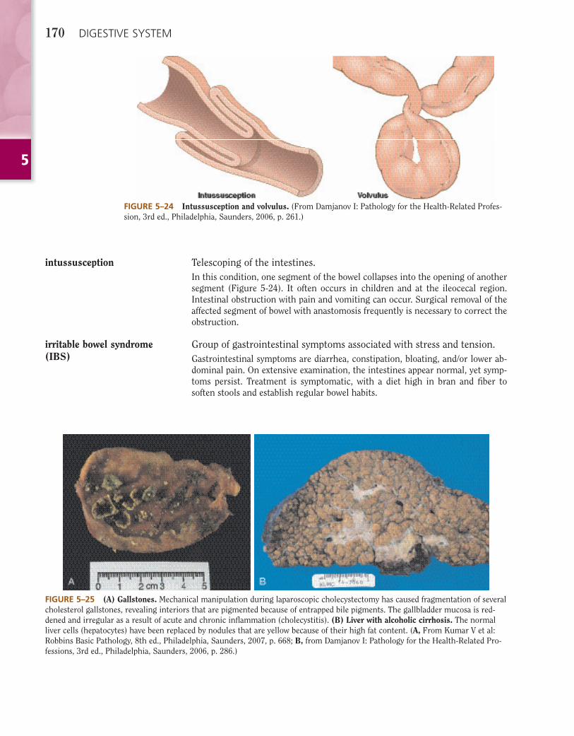
intussusception Telescoping of the intestines.In this condition, one segment of the bowel collapses into the opening of another segment (Figure 5-24). It often occurs in children and at the ileocecal region. Intestinal obstruction with pain and vomiting can occur. Surgical removal of the affected segment of bowel with anastomosis frequently is necessary to correct the obstruction.
irritable bowel syndrome (IBS)
Group of gastrointestinal symptoms associated with stress and tension.Gastrointestinal symptoms are diarrhea, constipation, bloating, and/or lower ab-dominal pain. On extensive examination, the intestines appear normal, yet symp-toms persist. Treatment is symptomatic, with a diet high in bran and fi ber to soften stools and establish regular bowel habits.
FIGURE 5–24 Intussusception and volvulus. (From Damjanov I: Pathology for the Health-Related Profes-sion, 3rd ed., Philadelphia, Saunders, 2006, p. 261.)
FIGURE 5–25 (A) Gallstones. Mechanical manipulation during laparoscopic cholecystectomy has caused fragmentation of several cholesterol gallstones, revealing interiors that are pigmented because of entrapped bile pigments. The gallbladder mucosa is red-dened and irregular as a result of acute and chronic infl ammation (cholecystitis). (B) Liver with alcoholic cirrhosis. The normal liver cells (hepatocytes) have been replaced by nodules that are yellow because of their high fat content. (A, From Kumar V et al: Robbins Basic Pathology, 8th ed., Philadelphia, Saunders, 2007, p. 668; B, from Damjanov I: Pathology for the Health- Related Pro-fessions, 3rd ed., Philadelphia, Saunders, 2006, p. 286.)

5
DIGESTIVE SYSTEM 171
FIGURE 5–26 Gallstone positions. (A) Stone in the gallbladder causing mild or no symptoms. (B) Stone obstructing the cystic duct, causing pain. (C) Stone obstructing the common bile duct, causing pain and jaundice. (D) Stone at the lower end of the common bile duct and pancreatic duct, causing pain, jaundice, and pancreatitis.
ulcerative colitis Chronic infl ammation of the colon with presence of ulcers.This idiopathic, chronic, recurrent diarrheal disease (an infl ammatory bowel dis-ease) presents with rectal bleeding and pain. Often beginning in the colon, the infl ammation spreads proximally, involving the entire colon. Drug treatment and careful attention to diet are recommended. Resection of diseased bowel with ileos-tomy may be necessary. Patients with ulcerative colitis have a higher risk of colon cancer.
volvulus Twisting of the intestine on itself.Volvulus causes intestinal obstruction. Severe pain, nausea and vomiting, and absence of bowel sounds are clinical features. Surgical correction is necessary to prevent necrosis of the affected segment of the bowel (see Figure 5-24).
LIVER, GALLBLADDER, AND PANCREAS
cholelithiasis Gallstones in the gallbladder (Figure 5-25A).Calculi (stones) prevent bile from leaving the gallbladder and bile ducts (Figure 5-26). Many patients remain asymptomatic and do not require treatment; how-ever, if a patient experiences episodes of biliary colic (pain from blocked cystic or common bile duct), treatment may be required. Currently, laparoscopic or mini-mally invasive surgery (laparoscopic cholecystectomy) is performed to remove the gallbladder and stones (Figure 5-27).
5
172 DIGESTIVE SYSTEM
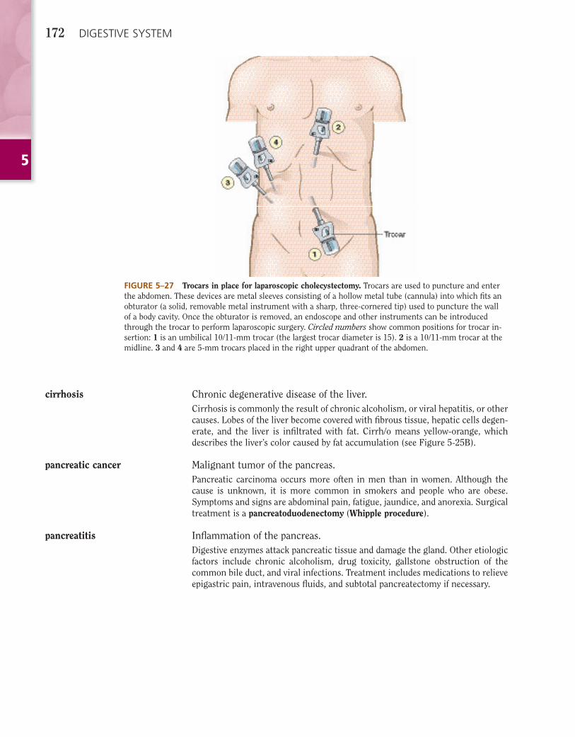
FIGURE 5–27 Trocars in place for laparoscopic cholecystectomy. Trocars are used to puncture and enter the abdomen. These devices are metal sleeves consisting of a hollow metal tube (cannula) into which fi ts an obturator (a solid, removable metal instrument with a sharp, three-cornered tip) used to puncture the wall of a body cavity. Once the obturator is removed, an endoscope and other instruments can be introduced through the trocar to perform laparoscopic surgery. Circled numbers show common positions for trocar in-sertion: 1 is an umbilical 10/11-mm trocar (the largest trocar diameter is 15). 2 is a 10/11-mm trocar at the midline. 3 and 4 are 5-mm trocars placed in the right upper quadrant of the abdomen.
cirrhosis Chronic degenerative disease of the liver.Cirrhosis is commonly the result of chronic alcoholism, or viral hepatitis, or other causes. Lobes of the liver become covered with fi brous tissue, hepatic cells degen-erate, and the liver is infi ltrated with fat. Cirrh/o means yellow-orange, which describes the liver’s color caused by fat accumulation (see Figure 5-25B).
pancreatic cancer Malignant tumor of the pancreas.Pancreatic carcinoma occurs more often in men than in women. Although the cause is unknown, it is more common in smokers and people who are obese. Symptoms and signs are abdominal pain, fatigue, jaundice, and anorexia. Surgical treatment is a pancreatoduodenectomy (Whipple procedure).
pancreatitis Infl ammation of the pancreas.Digestive enzymes attack pancreatic tissue and damage the gland. Other etiologic factors include chronic alcoholism, drug toxicity, gallstone obstruction of the common bile duct, and viral infections. Treatment includes medications to relieve epigastric pain, intravenous fl uids, and subtotal pancreatectomy if necessary.
5
DIGESTIVE SYSTEM 173
EXERCISESRemember to check your answers carefully with the Answers to Exercises, page •••.
A Match the following digestive system structures with their meanings below.
anuscecumcolonduodenum
esophagusgallbladderileumjejunum
liverpancreaspharynxsigmoid colon
1. large intestine ___________________________________________________________________________________________________________________________
2. small sac under the liver; stores bile _________________________________________________
3. fi rst part of the large intestine ______________________________________________________
4. end of the digestive tract opening to the outside of the body ______________________________
5. second part of the small intestine ___________________________________________________
6. tube connecting the throat to the stomach ____________________________________________
7. third part of the small intestine _____________________________________________________
8. large organ located in the RUQ; secretes bile, stores sugar, produces blood proteins
________________________________________________________________________________
9. throat __________________________________________________________________________
10. lower part of the colon ____________________________________________________________
11. fi rst part of the small intestine ______________________________________________________
12. organ under the stomach; produces insulin and digestive enzymes ________________________
B Circle the term that fi ts the given defi nition. You should be able to defi ne the other terms as well!
1. microscopic projections in the walls of the small intestinepapillae villi rugae
2. salivary gland near the ear:submandibular sublingual parotid
3. ring of muscle at the end of the stomachpyloric sphincter uvula lower esophageal sphincter
4. soft, inner section of a toothdentin enamel pulp
5. chemical that speeds up reactions and helps digest foodstriglyceride amino acid enzyme
5
174 DIGESTIVE SYSTEM
6. pigment released with bileglycogen bilirubin melena
7. hormone produced by endocrine cells of the pancreasinsulin amylase lipase
8. rhythm-like contraction of the muscles in the walls of the gastrointestinal tractdeglutition mastication peristalsis
9. breakdown of large fat globulesabsorption emulsifi cation anabolism
10. pointed, dog-like tooth medial to premolarsincisor canine molar
C Complete the following.
1. Labi/o and cheil/o both mean ____________________________________________________________________________________________________ .
2. Gloss/o and lingu/o both mean __________________________________________________________________________________________________ .
3. Or/o and stomat/o both mean ___________________________________________________________________________________________________ .
4. Dent/i and odont/o both mean __________________________________________________________________________________________________ .
5. Lapar/o and celi/o both mean ____________________________________________________________________________________________________ .
6. Gluc/o and glyc/o both mean ____________________________________________________________________________________________________ .
7. Lip/o, steat/o, and adip/o all mean _____________________________________________________________________________________________ .
8. The suffi xes -iasis and -osis both mean ______________________________________________________________________________________ .
9. Chol/e and bil/i both mean ________________________________________________________________________________________________________ .
10. Resection and -ectomy both mean ____________________________________________________________________________________________ .
D Build medical terms based on the given defi nitions.
1. removal of a salivary gland _________________________________________________________
2. pertaining to the throat ___________________________________________________________
3. hernia of the rectum ______________________________________________________________
4. enlargement of the liver ___________________________________________________________
5. surgical repair of the roof of the mouth ______________________________________________
6. after meals ______________________________________________________________________
7. visual examination of the anal and rectal region ________________________________________
8. study of the cause (of disease) ______________________________________________________
9. incision of the common bile duct ___________________________________________________
10. pertaining to teeth and cheek _______________________________________________________
5
DIGESTIVE SYSTEM 175
11. disease condition of the small intestine _______________________________________________
12. new opening between the common bile duct and the jejunum ____________________________
13. pertaining to surrounding the anus __________________________________________________
14. new opening from the colon to the outside of the body __________________________________
15. under the lower jaw ______________________________________________________________
16. pertaining to the face _____________________________________________________________
E Match the following doctors or dentists with their specialties.
colorectal surgeonendodontistgastroenterologist
nephrologistoral surgeonorthodontist
periodontistproctologisturologist
1. treats disorders of the anus and rectum ______________________________________________
2. operates on the organs of the urinary tract ____________________________________________
3. straightens teeth _________________________________________________________________
4. performs root canal therapy ________________________________________________________
5. operates on the mouth and teeth ____________________________________________________
6. treats kidney disorders ____________________________________________________________
7. diagnoses and treats gastrointestinal disorders _________________________________________
8. treats gum disease ________________________________________________________________
9. operates on the intestinal tract _____________________________________________________
F Build medical terms to describe the following infl ammations.
1. infl ammation of the appendix _______________________________________________________
2. infl ammation of the large intestine __________________________________________________
3. infl ammation of the passageway from the throat to the stomach __________________________
4. infl ammation of the membrane surrounding the abdomen _______________________________
5. infl ammation of the gallbladder _____________________________________________________
6. infl ammation of the third part of the small intestine ____________________________________
7. infl ammation of the pancreas _______________________________________________________
8. infl ammation of the gums _________________________________________________________
9. infl ammation of the liver __________________________________________________________
10. infl ammation of the mouth ________________________________________________________
11. infl ammation of the salivary gland ___________________________________________________
12. infl ammation of the small and large intestines _________________________________________

5
176 DIGESTIVE SYSTEM
G Match the following terms with their meanings below.
anastomosisbiliarydefecationcheilitis
gluconeogenesisglycogenolysishyperbilirubinemiahyperglycemia
mesenterymucosaparenteralportal vein
1. high level of blood sugar
2. infl ammation of the lip
3. pertaining to administration of medicines and fl uid by mouth
4. mucous membrane
5. expulsion of feces from the body through the anus
6. breakdown (conversion) of starch to sugar
7. fan-like membrane that connects the small intestine to the abdominal wall _________________
8. large vessel that takes blood to the liver from the intestines
9. new surgical connection between structures or organs
10. pertaining to bile ducts
11. process of forming new sugar from proteins and fats
12. high levels of a bile pigment in the bloodstream
H Give the names of the following gastrointestinal signs or symptoms based on their descriptions.
1. passage of bright red blood from the rectum __________________________________________
2. lack of appetite __________________________________________________________________
3. fat in the feces ___________________________________________________________________
4. black, tarry stools; feces containing digested blood _____________________________________
5. abnormal accumulation of fl uid in the abdomen _______________________________________
6. rumbling noise produced by gas in the GI tract ________________________________________
7. gas expelled through the anus ______________________________________________________
8. an unpleasant sensation in the stomach and a tendency to vomit __________________________
9. loose, watery stools _______________________________________________________________
10. diffi culty in passing stools (feces) ____________________________________________________
11. diffi culty in swallowing ____________________________________________________________
I Write short answers for the following questions.
1. What is jaundice? ________________________________________________________________
5
DIGESTIVE SYSTEM 177
2. List three ways in which a patient can become jaundiced:
a. _____________________________________________________________________________
b. _____________________________________________________________________________
c. _____________________________________________________________________________
3. What does it mean when a disease is described as idiopathic? _____________________________
________________________________________________________________________________
J Select from the list of pathologic conditions to make a diagnosis.
achalasiaanal fi stulaaphthous stomatitiscolonic polyps
colorectal cancerCrohn diseasedental cariesesophageal cancer
herpetic stomatitisoral leukoplakiaperiodontal disease
1. Mr. Jones, a smoker and heavy drinker, complained of dysphagia in recent months. A longstanding condition of Barrett esophagus resulted in his malignant condition. Diagnosis: .
2. An abnormal tube-like passageway near his anus caused Mr. Rosen’s proctalgia. His doctor performed surgery to close off the abnormality. Diagnosis: .
3. Carol’s dentist informed her that the enamel of three teeth was damaged by bacteria-producing acid. Diagnosis: .
4. Paola’s symptoms of chronic diarrhea, abdominal cramps, and fever led her doctor to suspect that she suffered from an infl ammatory bowel disease affecting the distal portion of her ileum. The doctor prescribed steroid drugs to heal her condition. Diagnosis: .
5. Mr. Hart learned that his colonoscopy showed the presence of small benign growths protruding from the mucous membrane of his large intestine. Diagnosis: .
6. During a routine dental checkup, Dr. Friedman discovered white plaques on Mr. Longo’s buccal mucosa. He advised Mr. Longo, who was a chronic smoker and heavy drinker, to have these precancerous lesions removed. Diagnosis: .
7. Every time Carl had a stressful time at work, he developed a fever blister (cold sore) on his lip, resulting from reactivation of a previous viral infection. His doctor told him that there was no treatment 100% effective in preventing the reappearance of these lesions. Diagnosis: .
8. Mr. Green had a biopsy of a neoplastic lesion in his descending colon. The pathology report indicated a malignancy. A partial colectomy was necessary. Diagnosis: .
9. Small ulcers (canker sores) appeared on Diane’s gums. They were painful and annoying. Diagnosis: .
10. Sharon’s failure to fl oss her teeth and remove dental plaque regularly led to development of gingivitis. Her dentist advised consulting a specialist who could treat her condition. Diagnosis: .
11. Imaging tests revealed a tumor in a section of Mr. Smith’s pancreas. His physician told him that since it had not spread, he could hope for a cure with surgery. He had a pancreatoduodenectomy (Whipple procedure), which was successful. Diagnosis: .
12. Mr. Clark complained of pain during swallowing. His physician explained that the pain was caused by a failure of muscles in his lower esophagus to relax during swallowing. Diagnosis: .
5
178 DIGESTIVE SYSTEM
cholecystolithiasiscirrhosisdiverticulosisdysenteryesophageal varices
hemorrhoidshiatal herniaileusintussusceptionirritable bowel syndrome
pancreatitispeptic ulcerulcerative colitisviral hepatitisvolvulus
1. protrusion of the upper part of the stomach through the diaphragm _______________________
2. painful, infl amed intestines caused by bacterial infection ________________________________
3. swollen, twisted veins in the rectal region _____________________________________________
4. open sore or lesion of the mucous membrane of the stomach or duodenum _________________
5. loss of peristalsis _________________________________________________________________
6. twisting of the intestine on itself ____________________________________________________
7. swollen, varicose veins on the surface of the distal portion of the esophagus _________________
8. a condition of abnormal outpouchings in the intestinal wall ______________________________
9. chronic infl ammation of the colon with destruction of its inner surface ____________________
10. telescoping of the intestines ________________________________________________________
11. infl ammation of the liver caused by type A, type B, or type C virus _________________________
12. infl ammation of the pancreas _______________________________________________________
13. calculi in the sac that stores bile ____________________________________________________
14. chronic degenerative liver disease with scarring resulting from alcoholism or infectious
hepatitis ________________________________________________________________________
15. symptoms (diarrhea or constipation, abdominal pain, bloating) associated with stress and
tension, but without infl ammation of the intestine _____________________________________
K Match the following pathologic diagnoses with their defi nitions.
5
DIGESTIVE SYSTEM 179
L Complete the following terms from their meanings given below.
1. membrane (peritoneal fold) that holds the intestines together: mes
2. removal of the gallbladder: ectomy
3. black or dark brown, tarry stools containing blood: mel
4. high levels of pigment in the blood (jaundice): hyper
5. pertaining to under the tongue: sub
6. twisting of the intestine on itself: vol
7. organ under the stomach that produces insulin and digestive enzymes: pan
8. lack of appetite: an
9. swollen, twisted veins in the rectal region: oids
10. new connection between two previously unconnected tubes: ana
11. absence of acid in the stomach: a
12. return of solids and fl uids to the mouth from the stomach: gastro
re disease
13. removal of soft tissue hanging from the roof of the mouth: ectomy
14. formation of stones: genesis.
5
180 DIGESTIVE SYSTEM
ANSWERS TO EXERCISES A
1. colon 2. gallbladder 3. cecum 4. anus
5. jejunum 6. esophagus 7. ileum 8. liver
B 1. Villi. Papillae are nipple-like
projections in the tongue where taste buds are located, and rugae are folds in the mucous membrane of the stomach and hard palate.
2. Parotid. The submandibular gland is under the lower jaw, and the sublingual gland is under the tongue.
3. Pyloric sphincter. The uvula is soft tissue hanging from the soft palate, and the lower esophageal sphincter is a ring of muscle between the esophagus and stomach.
4. Pulp. Dentin is the hard part of the tooth directly under the enamel and in the root, and enamel is the hard, outermost part of the tooth composing the crown.
5. Enzyme. A triglyceride is a large fat molecule, and an amino acid is a substance produced when proteins are digested.
6. Bilirubin. Glycogen is animal starch that is produced in liver cells from sugar, and melena is dark, tarry stools.
7. Insulin. Amylase and lipase are digestive enzymes produced by the exocrine cells of the pancreas.
8. Peristalsis. Deglutition is swallowing, and mastication is chewing.
9. Emulsifi cation. Absorption is the passage of materials through the walls of the small intestine into the bloodstream, and anabolism is the process of building up proteins in a cell (protein synthesis).
10. Canine. An incisor is one of the four front teeth in the dental arch (not pointed or like a dog’s tooth), and a molar is one of three large teeth just behind (distal to) the two premolar teeth.
C 1. lip 2. tongue 3. mouth 4. tooth
5. abdomen 6. sugar 7. fat
8. abnormal condition 9. gall, bile 10. removal, excision
D 1. sialadenectomy 2. pharyngeal 3. rectocele 4. hepatomegaly 5. palatoplasty
E 1. proctologist 2. urologist 3. orthodontist
4. endodontist 5. oral surgeon 6. nephrologist
7. gastroenterologist 8. periodontist 9. colorectal surgeon
F 1. appendicitis 2. colitis 3. esophagitis 4. peritonitis (note that the e is
dropped) 5. cholecystitis
6. ileitis 7. pancreatitis 8. gingivitis 9. hepatitis 10. stomatitis
G 1. hyperglycemia 2. cheilitis 3. parenteral 4. mucosa
5. defecation 6. glycogenolysis 7. mesentery 8. portal vein
9. anastomosis 10. biliary 11. gluconeogenesis 12. hyperbilirubinemia
11. sialadenitis 12. enterocolitis (when two combining
forms for gastrointestinal organs are in a term, use the one that is closest to the mouth fi rst)
6. postprandial (post cibum—cib/o refers to meals or feeding)
7. proctoscopy 8. etiology 9. choledochotomy 10. dentibuccal
11. enteropathy 12. choledochojejunostomy 13. perianal 14. colostomy 15. submandibular 16. facial
9. pharynx 10. sigmoid colon 11. duodenum 12. pancreas
5
DIGESTIVE SYSTEM 181
H 1. hematochezia 2. anorexia 3. steatorrhea 4. melena
5. ascites 6. borborygmus 7. fl atus 8. nausea
9. diarrhea 10. constipation 11. dysphagia 12. odynophagia
I 1. yellow-orange coloration of the
skin and other tissues (hyperbilirubinemia)
2. a. any liver disease (hepatopathy—such as cirrhosis, hepatoma, or hepatitis), so that bilirubin is
not processed into bile and cannot be excreted in feces
b. obstruction of bile fl ow, so that bile and bilirubin are not excreted and accumulate in the bloodstream
c. excessive hemolysis leading to overproduction of bilirubin and high levels in the bloodstream
3. cause is not known
J 1. esophageal cancer 2. anal fi stula 3. dental caries 4. Crohn disease (Crohn’s)
5. colonic polyps 6. oral leukoplakia 7. herpetic stomatitis 8. colorectal cancer
9. aphthous stomatitis 10. periodontal disease 11. pancreatic cancer 12. achalasia
K 1. hiatal hernia 2. dysentery 3. hemorrhoids 4. peptic ulcer 5. ileus
6. volvulus 7. esophageal varices 8. diverticulosis 9. ulcerative colitis 10. intussusception
11. viral hepatitis 12. pancreatitis 13. cholecystolithiasis (gallstones) 14. cirrhosis 15. irritable bowel syndrome
L 1. mesentery 2. cholecystectomy 3. melena 4. hyperbilirubinemia 5. sublingual
6. volvulus 7. pancreas 8. anorexia 9. hemorrhoids 10. anastomosis
11. achlorhydria 12. gastroesophageal refl ux disease 13. uvulectomy 14. lithogenesis

5
182 DIGESTIVE SYSTEM
PRONUNCIATION OF TERMSTo test your understanding of the terminology in
this chapter, write the meaning of each term in the space provided. In addition, you may wish to cover the terms and write them by look-ing at your defi nitions. Make sure your spelling is correct. The page number after each term indicates where it is defi ned or used in the book, so you can easily check your responses. You will fi nd complete defi nitions for all of these terms and their audio pronunciations on the CD.
PRONUNCIATION GUIDEa as in ape a as in applee as in even e as in everyı as in ıce ı as in ınteresto as in open o as in potu as in unit u as in under
VOCABULARY AND TERMINOLOGY TERM PRONUNCIATION MEANING
absorption (•••) ab-SORP-shun ____________________________________________________________
achlorhydria (•••) a-chlor-HID-re-a ____________________________________________________________
amino acids (•••) a-ME-no AS-ıdz ____________________________________________________________
amylase (•••) AM-ı-las ____________________________________________________________
anastomosis (•••) a-nas-to-MO-sıs ____________________________________________________________
anus (•••) A-nus ____________________________________________________________
appendectomy (•••) ap-en-DEK-to-me ____________________________________________________________
appendicitis (•••) a-pen-dı-SI-tıs ____________________________________________________________
appendix (•••) a-PEN-dıks ____________________________________________________________
bile (•••) bıl ____________________________________________________________
biliary (•••) BIL-e-ar-e ____________________________________________________________
bilirubin (•••) bıl-ı-ROO-bın ____________________________________________________________
bowel (•••) BOW-el ____________________________________________________________
buccal mucosa (•••) BUK-al mu-KO-sa ____________________________________________________________
canine teeth (•••) KA-nın teth ____________________________________________________________
cecal (•••) SE-kal ____________________________________________________________
cecum (•••) SE-kum ____________________________________________________________
celiac (•••) SE-le-ak ____________________________________________________________
cheilitis (•••) kı-LI-tıs ____________________________________________________________
cholecystectomy (•••) ko-le-sıs-TEK-to-me ____________________________________________________________
choledocholithiasis (•••) ko-le-do-ko-lı-THI-a-sıs ____________________________________________________________
choledochojejunostomy (•••) ko-le-do-ko-jı-ju-NOS-to-me ____________________________________________________________

5
DIGESTIVE SYSTEM 183
TERM PRONUNCIATION MEANING
choledochotomy (•••) ko-le-do-KOT-o-me ____________________________________________________________
cholelithiasis (•••) ko-le-lı-THI-a-sıs ____________________________________________________________
colon (•••) KO-lon ____________________________________________________________
colonic (•••) ko-LON-ık ____________________________________________________________
colonoscopy (•••) ko-lon-OS-ko-pe ____________________________________________________________
colostomy (•••) ko-LOS-to-me ____________________________________________________________
common bile duct (•••) KOM-on bıl dukt ____________________________________________________________
defecation (•••) def-e-KA-shun ____________________________________________________________
deglutition (•••) de-gloo-TISH-un ____________________________________________________________
dentibuccal (•••) den-tı-BUK-al ____________________________________________________________
dentin (•••) DEN-tın ____________________________________________________________
digestion (•••) dı-JEST-yun ____________________________________________________________
duodenal (•••) du-o-DE-nal or du-OD-e-nal ____________________________________________________________
duodenum (•••) du-o-DE-num or du-OD-e-num ____________________________________________________________
elimination (•••) e-lım-ı-NA-shun ____________________________________________________________
emulsifi cation (•••) e-mul-sı-fı-KA-shun ____________________________________________________________
enamel (•••) e-NAM-el ____________________________________________________________
endodontist (•••) en-do-DON-tıst ____________________________________________________________
enterocolitis (•••) en-ter-o-ko-LI-tıs ____________________________________________________________
enteroenterostomy (•••) en-ter-o-en-ter-OS-to-me ____________________________________________________________
enzyme (•••) EN-zım ____________________________________________________________
esophageal (•••) e-sof-a-JE-al ____________________________________________________________
esophagus (•••) e-SOF-a-gus ____________________________________________________________
fatty acid (•••) FAT-te A-sıd ____________________________________________________________
facial (•••) FA-shul ____________________________________________________________
feces (•••) FE-sez ____________________________________________________________
gallbladder (•••) GAWL-bla-der ____________________________________________________________
gastrointestinal tract (•••) gas-tro-ın-TES-tın-al tract ____________________________________________________________
gastrojejunostomy (•••) gas-tro-je-ju-NOS-to-me ____________________________________________________________
gastrostomy (•••) gas-TROS-to-me ____________________________________________________________
gingivitis (•••) jın-jı-VI-tıs ____________________________________________________________
gluconeogenesis (•••) gloo-ko-ne-o-JEN-e-sıs ____________________________________________________________
5
184 DIGESTIVE SYSTEM
TERM PRONUNCIATION MEANING
glucose (•••) GLOO-kos ____________________________________________________________
glycogen (•••) GLI-ko-jen ____________________________________________________________
glycogenolysis (•••) glı-ko-je-NOL-ı-sıs ____________________________________________________________
hepatoma (•••) he-pa-TO-ma ____________________________________________________________
hepatomegaly (•••) he-pa-to-MEG-a-le ____________________________________________________________
hydrochloric acid (•••) hı-dro-KLOR-ık A-sıd ____________________________________________________________
hyperbilirubinemia (•••) hı-per-bıl-ı-roo-bı-NE-me-a ____________________________________________________________
hyperglycemia (•••) hı-per-glı-SE-me-a ____________________________________________________________
hypoglossal (•••) hı-po-GLO-sal ____________________________________________________________
ileitis (•••) ıl-e-I-tıs ____________________________________________________________
ileocecal sphincter (•••) ıl-e-o-SE-kal SFINK-ter ____________________________________________________________
ileostomy (•••) ıl-e-OS-to-me ____________________________________________________________
ileum (•••) IL-e-um ____________________________________________________________
incisor (•••) ın-SI-zor ____________________________________________________________
insulin (•••) IN-su-lın ____________________________________________________________
jejunum (•••) je-JOO-num ____________________________________________________________
labial (•••) LA-be-al ____________________________________________________________
laparoscopy (•••) la-pa-ROS-ko-pe ____________________________________________________________
lipase (•••) LI-pas ____________________________________________________________
lithogenesis (•••) lıth-o-JEN-e-sıs ____________________________________________________________
liver (•••) LI-ver ____________________________________________________________
lower esophageal sphincter (•••) LOW-er e-sof-a-JE-al SFINGK-ter ____________________________________________________________
mastication (•••) mas-tı-KA-shun ____________________________________________________________
mesentery (•••) MES-en-ter-e ____________________________________________________________
molar teeth (•••) MO-lar teth ____________________________________________________________
oral (•••) OR-al ____________________________________________________________
orthodontist (•••) or-tho-DON-tıst ____________________________________________________________
palate (•••) PAL-at ____________________________________________________________
palatopharyngoplasty (•••) pal-a-to-fa-RING-go-plas-te ____________________________________________________________
palatoplasty (•••) PAL-a-to-plas-te ____________________________________________________________
pancreas (•••) PAN-kre-as ____________________________________________________________
pancreatitis (•••) pan-kre-a-TI-tıs ____________________________________________________________
5
DIGESTIVE SYSTEM 185
TERM PRONUNCIATION MEANING
papillae (•••) pa-PIL-e ____________________________________________________________
parenteral (•••) pa-REN-ter-al ____________________________________________________________
parotid gland (•••) pa-ROT-ıd gland ____________________________________________________________
perianal (•••) pe-re-A-nal ____________________________________________________________
periodontist (•••) per-e-o-DON-tıst ____________________________________________________________
peritonitis (•••) per-ı-to-NI-tıs ____________________________________________________________
peristalsis (•••) per-ı-STAL-sıs ____________________________________________________________
pharyngeal (•••) far-an-JE-al or fa-RIN-je-al ____________________________________________________________
pharynx (•••) FAR-ınks ____________________________________________________________
portal vein (•••) POR-tal van ____________________________________________________________
postprandial (•••) post-PRAN-de-al ____________________________________________________________
premolar teeth (•••) pre-MO-lar teth ____________________________________________________________
proctologist (•••) prok-TOL-o-jıst ____________________________________________________________
protease (•••) PRO-te-ase ____________________________________________________________
pulp (•••) pulp ____________________________________________________________
pyloric sphincter (•••) pı-LOR-ık SFINK-ter ____________________________________________________________
pyloroplasty (•••) pı-LOR-o-plas-te ____________________________________________________________
pylorus (•••) pı-LOR-us ____________________________________________________________
rectocele (•••) REK-to-sel ____________________________________________________________
rectum (•••) REK-tum ____________________________________________________________
rugae (•••) ROO-ge ____________________________________________________________
saliva (•••) sa-LI-va ____________________________________________________________
salivary glands (•••) SAL-ı-var-e glandz ____________________________________________________________
sialadenitis (•••) sı-al-a-de-NI-tıs ____________________________________________________________
sialolith (•••) sı-AL-o-lıth ____________________________________________________________
sigmoid colon (•••) SIG-moyd KO-lon ____________________________________________________________
sigmoidoscopy (•••) sıg-moyd-OS-ko-pe ____________________________________________________________
sphincter (•••) SFINGK-ter ____________________________________________________________
steatorrhea (•••) ste-a-to-RE-a ____________________________________________________________
stomatitis (•••) sto-ma-TI-tıs ____________________________________________________________
sublingual (•••) sub-LING-wal ____________________________________________________________
submandibular (•••) sub-man-DIB-u-lar ____________________________________________________________
5
186 DIGESTIVE SYSTEM
TERM PRONUNCIATION MEANING
triglycerides (•••) trı-GLI-se-rıdz ____________________________________________________________
uvula (•••) U-vu-la ____________________________________________________________
uvulectomy (•••) u-vu-LEK-to-me ____________________________________________________________
villi (•••) VIL-ı ____________________________________________________________
PATHOLOGIC TERMINOLOGY TERM PRONUNCIATION MEANING
achalasia (•••) ak-ah-LA-ze-a ____________________________________________________________
anal fi stula (•••) A-nal FIS-tu-la ____________________________________________________________
anorexia (•••) an-o-REK-se-a ____________________________________________________________
aphthous stomatitis (•••) AF-thus sto-ma-TI-tıs ____________________________________________________________
ascites (•••) a-SI-tez ____________________________________________________________
borborygmus (•••) bor-bo-RIG-mus ____________________________________________________________
cholelithiasis (•••) ko-le-lı-THI-a-sıs ____________________________________________________________
cirrhosis (•••) sır-RO-sıs ____________________________________________________________
colonic polyps (•••) ko-LON-ık polıps ____________________________________________________________
colorectal cancer (•••) ko-lo-REK-tal KAN-ser ____________________________________________________________
constipation (•••) con-stı-PA-shun ____________________________________________________________
Crohn disease (•••) kron dı-ZEZ ____________________________________________________________
dental caries (158) DEN-tal KAR-ez ____________________________________________________________
diarrhea (•••) dı-ah-RE-a ____________________________________________________________
diverticula (•••) dı-ver-TIK-u-la ____________________________________________________________
diverticulitis (•••) dı-ver-tık-u-LI-tıs ____________________________________________________________
diverticulosis (•••) dı-ver-tık-u-LO-sıs ____________________________________________________________
dysentery (•••) DIS-en-ter-e ____________________________________________________________
dysphagia (•••) dıs-PHA-je-a ____________________________________________________________
eructation (•••) e-ruk-TA-shun ____________________________________________________________
esophageal cancer (•••) e-sof-a-JE-al KaN-ser ____________________________________________________________
esophageal varices (•••) e-sof-a-JE-al VAR-ı-sez ____________________________________________________________
etiology (•••) e-te-OL-o-je ____________________________________________________________
fl atus (•••) FLA-tus ____________________________________________________________
gastric carcinoma (•••) GAS-trık kar-sı-NO-ma ____________________________________________________________

5
DIGESTIVE SYSTEM 187
TERM PRONUNCIATION MEANING
gastroesophageal refl ux disease (•••)
gas-tro-e-sof-a-JE-al RE-fl ux dı-ZEZ
____________________________________________________________
hematochezia (•••) he-ma-to-KE-ze-a ____________________________________________________________
hemorrhoids (•••) HEM-o-roydz ____________________________________________________________
herpetic stomatitis (•••) her-PET-ık sto-ma-TI-tıs ____________________________________________________________
hiatal hernia (•••) hı-A-tal HER-ne-a ____________________________________________________________
icterus (•••) IK-ter-us ____________________________________________________________
idiopathic (•••) ıd-e-o-PATH-ık ____________________________________________________________
ileus (•••) IL-e-us ____________________________________________________________
infl ammatory bowel disease (•••) ın-FLA-ma-to-re BOW-el dı-ZEZ ____________________________________________________________
inguinal hernia (•••) ING-wı-nal HER-ne-a ____________________________________________________________
intussusception (•••) ın-tus-sus-SEP-shun ____________________________________________________________
irritable bowel syndrome (•••) IR-ı-ta-b’l BOW-el SIN-drom ____________________________________________________________
jaundice (•••) JAWN-dıs ____________________________________________________________
melena (•••) MEL-e-na or me-LE-na ____________________________________________________________
nausea (•••) NAW-ze-a ____________________________________________________________
oral leukoplakia (•••) OR-al lu-ko-PLA-ke-a ____________________________________________________________
pancreatic cancer (•••) pan-kre-A-tıc KAN-ser ____________________________________________________________
pancreatitis (•••) pan-kre-a-TI-tıs ____________________________________________________________
peptic ulcer (•••) PEP-tıc UL-ser ____________________________________________________________
periodontal disease (•••) per-e-o-DON-tal dı-ZEZ ____________________________________________________________
ulcerative colitis (•••) UL-ser-a-tıv ko-LI-tıs ____________________________________________________________
viral hepatitis (•••) VI-ral hep-a-TI-tıs ____________________________________________________________
Note: The Review Sheet for this chapter is combined with the Review Sheet for Chapter 6 on page •••.
Please refer to the enclosed CD for additional exercises and images related to this chapter.


![Simulating and Testing the TRIUMF Bragg Ionisation Chamber · UMF [6]. The facility is divided into two sections, in ISAC-I beams are used The facility is divided into two sections,](https://img.pdfslide.net/doc/110x75/5dd090eed6be591ccb619c65/simulating-and-testing-the-triumf-bragg-ionisation-chamber-umf-6-the-facility.jpg)