Embed Size (px)
Citation preview

The impact of hepatitis E in the liver transplant setting.
Item Type Article
Authors Behrendt, Patrick; Steinmann, Eike; Manns, Michael P;Wedemeyer, Heiner
Citation The impact of hepatitis E in the liver transplant setting. 2014, 61(6):1418-29 J. Hepatol.
DOI 10.1016/j.jhep.2014.08.047
Journal Journal of hepatology
Download date 13/07/2018 08:11:21
Link to Item http://hdl.handle.net/10033/346563

This is an Open Access article's PDF published inBehrendt, P., Steinmann, E., Manns, M.P., Wedemeyer, H.The impact of hepatitis e in the liver transplant
setting(2014) Journal of Hepatology, 61 (6), pp. 1418-1429.
The impact of hepatitis E in the liver transplant setting
Patrick Behrendt1,2,3, Eike Steinmann3, Michael P. Manns1,2, Heiner Wedemeyer1,2,⇑
1Department for Gastroenterology, Hepatology and Endocrinology, Hannover Medical School, Hannover, Germany; 2German Centerfor Infection Research, Hannover, Germany; 3Twincore, Centre for Experimental and Clinical Infection Research, A Joint Venture
Between Medical School Hannover and Helmholtz Centre for Infection Research, Hannover, Germany
Summary
Hepatitis E virus (HEV) infection has been identified as a cause ofgraft hepatitis in liver transplant recipients. The true frequencyand clinical importance of HEV infections after liver transplanta-tions is a matter of debate. It is proposed that consumption ofHEV-contaminated undercooked meat is a main source for HEVinfections in developed countries – which might also accountfor some hepatitis E cases after organ transplantation. However,HEV is also transmitted by transfusion of blood products, likelyrepresenting a previously underestimated risk particularly forpatients in the transplant setting. HEV infection can take chroniccourses in immunocompromised individuals, associated in somecases with rapid progression to cirrhosis within 1–2 years ofinfection. Diagnosis in transplanted patients is based on HEVRNA testing as antibody assays are not sensitive enough. Selec-tion of immunosuppressive drugs is important as different com-pounds may influence viral replication and the course of liverdisease. Ribavirin has antiviral activity against HEV and shouldbe administered for at least three months in chronically infectedindividuals; however, treatment failure may occur. HEV infec-tions have also been linked to a variety of extrahepatic manifes-tations both during and after resolution of infection.
In this review we summarize the emerging data on hepatitis Ewith a particular focus on the importance of HEV infections forliver transplant recipients.� 2014 European Association for the Study of the Liver. Publishedby Elsevier B.V. All rights reserved.
Introduction
Hepatitis E is caused by infection with the hepatitis E virus (HEV).An infectious agent, leading to acute hepatitis that differed fromHAV and HBV, was already suspected in the 1970s. In 1983,Balayan and colleagues showed that oral administration ofpooled stool extracts from patients with non-A/non-B hepatitisled to acute hepatitis in a human volunteer in whom virus-likeparticles were identified in stool samples [1]. Hepatitis E was ini-tially recognized only as an acute self-limited liver disease, whichvery rarely progressed to acute liver failure. Severe courses weremost often observed in pregnant women [2,3] and individualswith chronic liver diseases [4–7]. HEV infections were reportedmainly from endemic countries such as the Indian subcontinent,South-East Asia and Sub-Saharan Africa. During the last 3 decadeslarge scale outbreaks of hepatitis E were reported [8–11], evencontinuing until recently when many hepatitis E cases werereported in refugee camps in South Sudan [12].
For more than 25 years HEV infection was not considered amajor clinical problem in developed countries including Europeand the United States. The description of chronic courses ofHEV infections in solid organ transplant recipients in France in2008 [13] increased the awareness for a potentially largelyunderestimated disease. Since then, persistent HEV infectionswere described in different cohorts of immunocompromisedpatients [9,14] but also beyond organ transplantation [15–19].Subsequently, mechanisms leading to persistent infection wereexplored in more detail and antiviral therapies were tested inlarge case series. Importantly, HEV infections seem to take veryrapid and aggressive courses in many patients receiving immuno-suppressive medications and HEV has been linked with end-stageliver disease and even liver-associated mortality. HEV infectionsseem to be much more common in Europe than previouslythought. Between 10% and 50% of the populations were anti-HEV-positive in various epidemiological studies [20–22]. Thus,millions of – usually clinically silent – infections occur each yearin Europe but only a small proportion of infected individualsdevelop clinical symptoms. HEV infection should also be consid-ered in the differential diagnosis of drug-induced liver disease[23,24], which has also a major importance in the managementof liver transplant recipients, receiving various medication beforeand after transplantation. Of note, HEV has also been linked toextrahepatic manifestations and thus, the clinical implications
Journal of Hepatology 2014 vol. 61 j 1418–1429
Keywords: Hepatitis E; HEV; Liver transplantation; Treatment.Received 8 July 2014; received in revised form 25 August 2014; accepted 29 August2014⇑ Corresponding author. Address: Abteilung für Gastroenterologie, Hepatologieund Endokrinologie, Medizinische Hochschule Hannover, Carl-Neuberg-Str.1,30625 Hannover, Germany. Tel.: +49 511 532 6814; fax: +49 511 532 6820.E-mail address: [email protected] (H. Wedemeyer).Abbreviations: HEV, hepatitis E virus; HVR, hypervariable region; ORF,open-reading frame; GBS, Guillain–Barrè syndrome.
Frontiers in Liver Transplantation

of hepatitis E virus infections are likely to be much broader thanpreviously estimated [25–28].
Key Points
• Hepatitis E virus infection can take chronic courses in immunocompromised individuals. There is limited data suggesting that chronic courses may be more frequent after liver transplantation
• Chronic hepatitis E is often associated with rapid progression to liver cirrhosis
• HEV genotype 3 infection can be a zoonosis and HEV is believed to be transmitted mainly by ingestion of raw uncooked meat. Other sources of transmission are food hygiene and transfusion of HEV positive blood products
• Different immunosuppressive drugs may have distinct effects on HEV replication (either inhibitory or stimulatory) and should therefore be selected accordingly
• Ribavirin can be used to treat HEV infection and should be given for at least 3 months. Treatment failures may occur and require further investigation
HEV virology
HEV is a small (27–34 mm) non-enveloped virus which belongsto the family of Hepeviridae. It is encoded by a single-strandedRNA (7.2 kb) which consists of three open-reading frames(ORF1–3) [29]. The non-structural proteins are encoded byORF1 and their translation leads to the synthesis of proteinswhich are primarily essential for viral replication. In particular,it consists of a methyltransferase, a protease, a macrodomain, ahelicase and an RNA-dependent RNA-polymerase [30]. Of note,a hypervariable region (HVR) is localized between the proteaseand the macrodomain. ORF 2 and ORF 3 on the other hand encodefor the structural proteins. The capsid, which is essential for viralattachment and entry into liver cells, is the translational productof ORF2 [31,32]. ORF 3 partly overlaps with ORF2 and leads to theproduction of a small phosphoprotein, which is involved in theassembly and release of the virus and has been shown to interactwith cellular host factors [33].
Similar to other RNA-viruses different genotypes of HEV existdue to a lack of proof-reading activity of the RNA-dependentRNA-polymerase. So far, 5 different HEV genotypes have beenidentified, which differ in their nucleotide sequences by 19%.Intra-genotypic differentiation into sub-genotypes is made upona variation of approximately 12% [34]. These distinct genotypesdiffer markedly in their distribution, clinical presentation andspecies-specificity.
It is believed, that only genotypes 1, 2, 3 and 4 are able tocause apparent disease in humans, while genotype 5 has beenidentified only in birds thus far [14]. While genotype 1 and 2solely infect humans, genotype 3 and 4 are zoonotic pathogenswith likely major reservoirs in pigs [35–37], wild boars and deer[38–40]. Some data suggest that genotype 1 may also be able toinfect pigs [41], which however needs to be confirmed by
additional studies. The worldwide distribution of the differentgenotypes varies markedly. Genotype 1 and 2 infections havebeen reported mainly in Asia, India and North-Africa. Genotype2 HEV has been also identified in Mexico [14,42]. Genotype 3 ispresent in Western countries as well as in Asia and North Amer-ica while genotype 4 has been detected in Asian- as well as inEuropean countries [14]. However, genotype 4 might also playa minor role in Europe as supported by recent data, demonstrat-ing evidence for a genotype 4 infection in France [43].
Additionally, several related viral strains have been identifiedin a variety of species like bats [44], chicken [45], ferrets [46],rabbits [47], rats [48] and trout [49]. However, it is believed thatthese viruses are not able to infect humans.
Routes of HEV transmission
Distinct HEV genotypes differ in their route of transmission. Con-taminated water is the main source for genotypes 1 and 2 infec-tions. Large hepatitis E outbreaks have been described in variousAfrican and Asian countries including India, China, Somalia, SouthSudan or Uganda [8–10,14]. In contrast, HEV genotype 3 and 4usually cause sporadic infections, most likely due to consumptionof contaminated food. HEV-RNA has been detected in a variety offood products in particular porcine livers and pig sausages inFrance, the US, the Netherlands and Germany [35,36,50,51].Consumption of such food increases the likelihood for the devel-opment of HEV-infection [43,51–54]. In line with this, peoplewho have close contact to swine and deer – farmers, slaughter-house-workers, veterinarians – display a significant higher prev-alence of HEV-infections compared to the general population[55,56]. Recent publications have delineated that HEV-RNA iseven more widely distributed than previously thought, and hasbeen found in various food-products like shellfish [57], mussels[58,59], green vegetables [60] as well as field-grown strawberries[61] (Fig. 1).
HEV transmission by blood products and organtransplantation
Organ transplant recipients frequently receive blood products,either prior to transplantation, during, or after transplantation.In most Western countries, blood products are tested forHCV-RNA and HIV-RNA by mini-pool testing and often even forHBV-DNA. However, blood products are not tested for HEV-RNA, even if transfused to individuals at risk. Several previouscase reports suggested that HEV transmission by blood productsis possible [14,62], which can obviously also occur after livertransplantation as demonstrated recently by Coilly et al. [63].Clinically healthy European blood donors may carry HEV RNAas reported during the last 2 years for Scotland [64], Sweden[65], England [66], Germany [67,68] and the Netherlands[69] – even though the absolute HEV RNA prevalence is low(0.01–0.04%). Still, the risk for HEV transmission may becomesubstantial if blood products are pooled. In this case, the riskfor HEV transmission seems to be substantial as up to 10% ofpooled plasma products tested HEV RNA positive in Europe[65]. Indeed, plasma products seem to be a specific cause forHEV infections [62,70]. However, screening of blood productsfor HEV has not been standardized in any country until now.Currently, there is an ongoing debate concerning this issue.
JOURNAL OF HEPATOLOGY
Journal of Hepatology 2014 vol. 61 j 1418–1429 1419

Recently, balancing miscellaneous aspects, Féray et al. delineatedthe potential significance of testing blood products for HEV [71].This has been further supported by the work of Hewitt et al.,which was published in Lancet very recently. Here, the authorsretrospectively screened blood donations that were collected insoutheast England between October 2012 and September 2013for HEV-RNA. Out of 225,000 blood donations, 79 patients wereviraemic with genotype 3 HEV. The authors could follow-up 43recipients of HEV-positive blood components and 42% of theseindividuals displayed evidence for infection with HEV thereafter.Short-term morbidity was rather low as only one of these indi-viduals developed clinical significant symptoms. However, 10patients, including organ transplant recipients who were receiv-ing immunosuppressive medications, displayed prolonged viralinfection [72].
HEV is not only transmitted by blood products but also by thetransplanted organ itself (Fig. 1). Of interest, in one case report aHEV-RNA positive liver has been transplanted to a HEV-negativerecipient. The development of a chronic HEV-infection rapidly ledto re-cirrhosis of the graft [73]. It is important to note that organtransplants are not tested for HEV RNA. Stem cells may representanother risk for HEV infection. Recently, a stem cell donor has
been identified who was clinically healthy at the time of evalua-tion but who suffered from acute hepatitis E at the time of leu-copheresis [74].
Seroprevalence
The evaluation of the prevalence of HEV, as determined by thedetection of anti-HEV IgG, is hampered by marked differencesin the sensitivities and specificities of the antibody assays[75,76]. For example, a comparison of different commercialassays revealed a variation of anti-HEV IgG-positive individualsbetween 3.6% and 16.2% in blood donors from the UK [21]. Thesensitivity thereby ranged from 56 to 98%. In the US an anti-HEV prevalence of around 25% has been proposed [77]. Similarvariations can be seen in endemic areas where reported HEVprevalence may range between 30% and 80% [78]. It is well estab-lished that the HEV seroprevalence increases with age [20,22,79]but an overall decline of anti-HEV has been observed during thelast two decades as e.g. recently shown for Germany. Here theauthors screened 1092 sera and found a prevalence of 51% in1996, which dropped to 34% in 2011 [22].
HEV
DeathNeed for liver transplantation
Pigs
Blood products Shellfish
FruitsVegetables
Transplantationof infected organs
Water
40-50% 50-60%
10%
● Possible increased likelihood for liver transplanted individuals● Only genotype 3
Approximately 30% by reduction of immunosuppression
Rapid progression
Solid organ transplanted individual
Treatment options:
Ribavirin
Possible positive influence by Favouring mycophenolate-mofetilAvoiding calcineurin- and mTOR inhibitors
Clearance
Cirrhosis
Chronic infection
[Interferon-alpha] - second line
••
Fig. 1. Transmission and disease-progression in transplanted individuals. The possible mode of HEV-transmissions is illustrated. Additionally, known factors whichinfluence the clinical course are depicted. Treatment options are highlighted in blue.
Frontiers in Liver Transplantation
1420 Journal of Hepatology 2014 vol. 61 j 1418–1429
Clinical course of HEV infection
Clinical symptoms occur in only 2–5% of patients with acute HEVinfection as suggested by data from a large, double-blinded pla-cebo controlled vaccine trial from China where more than110,000 individuals have been followed [80]. From apparentcases, mortality rates were estimated to be between 0.5–3%[81], leading up to 70,000 deaths worldwide each year [82].Importantly, risk factors have been identified, which are associ-ated with a more severe outcome of infection. In particular, preg-nant women seem to be at risk where infection with HEVgenotype 1 has been linked with foetal and maternal mortalityin up to 25% of cases [3]. Distinct polymorphisms in the proges-terone receptor may explain in part the particular severe out-come in pregnant women [79], but the detailed underlyingpathomechanism has not been fully understood thus far. Fulmin-ant disease progression seems also to occur more often inpatients who already suffer from an underlying chronic liver dis-ease [4–7]. Additionally, prolonged alcohol abuse serves as a riskfactor for fulminant acute hepatitis E [83]. The course of acuteHEV infection seems to differ between HEV genotypes as geno-type 1 infection is associated with more severe cases than infec-tion with genotype 3 HEV [84], however specific viral sequencevariations could not be linked to fulminant hepatitis E [85].
Acute hepatitis E in immunocompetent individuals
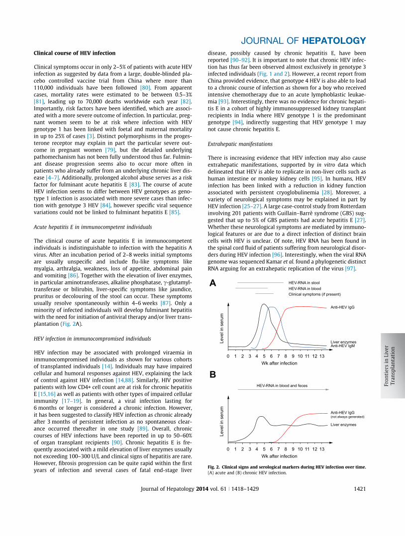
The clinical course of acute hepatitis E in immunocompetentindividuals is indistinguishable to infection with the hepatitis Avirus. After an incubation period of 2–8 weeks initial symptomsare usually unspecific and include flu-like symptoms likemyalgia, arthralgia, weakness, loss of appetite, abdominal painand vomiting [86]. Together with the elevation of liver enzymes,in particular aminotransferases, alkaline phosphatase, c-glutamyl-transferase or bilirubin, liver-specific symptoms like jaundice,pruritus or decolouring of the stool can occur. These symptomsusually resolve spontaneously within 4–6 weeks [87]. Only aminority of infected individuals will develop fulminant hepatitiswith the need for initiation of antiviral therapy and/or liver trans-plantation (Fig. 2A).
HEV infection in immunocompromised individuals
HEV infection may be associated with prolonged viraemia inimmunocompromised individuals as shown for various cohortsof transplanted individuals [14]. Individuals may have impairedcellular and humoral responses against HEV, explaining the lackof control against HEV infection [14,88]. Similarly, HIV positivepatients with low CD4+ cell count are at risk for chronic hepatitisE [15,16] as well as patients with other types of impaired cellularimmunity [17–19]. In general, a viral infection lasting for6 months or longer is considered a chronic infection. However,it has been suggested to classify HEV infection as chronic alreadyafter 3 months of persistent infection as no spontaneous clear-ance occurred thereafter in one study [89]. Overall, chroniccourses of HEV infections have been reported in up to 50–60%of organ transplant recipients [90]. Chronic hepatitis E is fre-quently associated with a mild elevation of liver enzymes usuallynot exceeding 100–300 U/L and clinical signs of hepatitis are rare.However, fibrosis progression can be quite rapid within the firstyears of infection and several cases of fatal end-stage liver
disease, possibly caused by chronic hepatitis E, have beenreported [90–92]. It is important to note that chronic HEV infec-tion has thus far been observed almost exclusively in genotype 3infected individuals (Fig. 1 and 2). However, a recent report fromChina provided evidence, that genotype 4 HEV is also able to leadto a chronic course of infection as shown for a boy who receivedintensive chemotherapy due to an acute lymphoblastic leukae-mia [93]. Interestingly, there was no evidence for chronic hepati-tis E in a cohort of highly immunosuppressed kidney transplantrecipients in India where HEV genotype 1 is the predominantgenotype [94], indirectly suggesting that HEV genotype 1 maynot cause chronic hepatitis E.
Extrahepatic manifestations
There is increasing evidence that HEV infection may also causeextrahepatic manifestations, supported by in vitro data whichdelineated that HEV is able to replicate in non-liver cells such ashuman intestine or monkey kidney cells [95]. In humans, HEVinfection has been linked with a reduction in kidney functionassociated with persistent cryoglobulinemia [28]. Moreover, avariety of neurological symptoms may be explained in part byHEV infection [25–27]. A large case-control study from Rotterdaminvolving 201 patients with Guillain–Barrè syndrome (GBS) sug-gested that up to 5% of GBS patients had acute hepatitis E [27].Whether these neurological symptoms are mediated by immuno-logical features or are due to a direct infection of distinct braincells with HEV is unclear. Of note, HEV RNA has been found inthe spinal cord fluid of patients suffering from neurological disor-ders during HEV infection [96]. Interestingly, when the viral RNAgenome was sequenced Kamar et al. found a phylogenetic distinctRNA arguing for an extrahepatic replication of the virus [97].
HEV-RNA in stoolHEV-RNA in bloodClinical symptoms (if present)
Leve
l in
seru
m
Wk after infection0 1 2 3 4 5 6 7 8 9 10 11 12 13
Liver enzymesAnti-HEV IgM
Anti-HEV IgG
A
B
Leve
l in
seru
m
Wk after infection0 1 2 3 4 5 6 7 8 9 10 11 12 13
Liver enzymes
Anti-HEV IgG(not always generated)
HEV-RNA in blood and feces
Fig. 2. Clinical signs and serological markers during HEV infection over time.(A) acute and (B) chronic HEV infection.
JOURNAL OF HEPATOLOGY
Journal of Hepatology 2014 vol. 61 j 1418–1429 1421

Further manifestations, associated with HEV include caseswith acute pancreatitis, haematological abnormalities as well asmyopathies [98]. Of note, extrahepatic symptoms may alsodevelop even after HEV clearance as recently suggested for a livertransplant recipient who developed vasculitis and enteritis afterHEV RNA became negative [99].
Diagnosis of HEV infection in immunosuppressed patients
HEV infection can be diagnosed in immunocompetent individualsby detection of anti-HEV IgM antibodies. Anti-HEV IgM titersincrease with the onset of clinical symptoms. However, the ana-lytical sensitivity of the anti-HEV IgM assays can differ [100],which potentially represents a major problem in patients receiv-ing immunosuppressive medications. Indeed, delayed diagnosisof HEV infection has been reported in a liver transplant recipientdue to variable performance of serologic assays [101]. Detectionof anti-HEV IgG indicates successful clearance of a previous infec-tion and may be associated with some level of protection againstHEV re-infection even though HEV antibodies are not able to pro-vide complete sterilizing immunity against HEV [102] (Fig. 2A).Anti-HEV-IgG can also diminish over time [103] and thus, sero-negativity may not exclude previous exposure to HEV. Overall,testing for immunoglobulins has limitations in specificity andsensitivity [75,76,104]. Reliability seems more accurate ingenotype 3 infection than in other genotypes. In addition,cross-reactivity to other viruses has been observed [105,106].Anti-HEV testing may frequently be false negative in immuno-compromised individuals. Time to seroconversion – meaningdevelopment of anti-HEV IgG – is delayed in patients after livertransplantation and some patients may never develop detectableanti-HEV IgG antibodies during chronic infection [92].
Considering the limitations of the antibody assays, diagnosisof HEV infection should be based on detection of HEV-RNA inorgan transplant recipients. Still, even HEV PCR assays are notperfect as sensitivities may show substantial variability and mostprevious studies have been performed with in-house assays[107]. Moreover, not all published assays have been optimizedfor different HEV genotypes and a recent comparative studyshowed that up to one out of six HEV RNA positive samples wereincorrectly classified as HEV RNA negative [108]. Importantly, theWHO established an internal control for RNA amplification tech-nology in 2011, which should help to solve some of the technicalissues [107].
An alternative to HEV RNA assays could be HEV antigen test-ing. HEV antigens may show different kinetics than HEV RNA andhave been shown to be longer positive than HEV RNA in patientswith acute hepatitis E [109]. A commercial sandwich ELISA forthe detection of the HEV-capsid is available. However, this testseems to be less sensitive than RNA-techniques and the role fordetection of infection needs to be evaluated in more detail infuture studies [110] (Fig. 2B).
Prevalence and clinical course of hepatitis E in liver-transplant recipients (summarized in Table 1)
The first case series demonstrating chronic HEV infection intransplanted individuals also included three liver transplantrecipients [13]. Overall, 14 solid-organ transplanted patients with
acute HEV infections were identified. Out of these, 8 patientsdeveloped persistent infection, which interestingly included allthree liver transplanted individuals. The study demonstrated arather rapid histological disease progression within a mediantime of follow-up of only 18 months. Subsequently, variousfurther studies have been published, addressing the rolechronic infection in organ transplant recipients including liver-transplanted individuals. Overall, several cases of chronic hepati-tis E after liver transplantation were identified even though theabsolute frequency was low with 1–3% in most studies. Consider-able differences between countries were evident.
In 2009, a study from the Netherlands was published [111],screening 285 patients, which had undergone liver transplanta-tion, for HEV-RNA, anti-HEV IgM and IgG levels. 96% of these indi-viduals were HEV RNA and anti-HEV IgG/IgM negative and thus,had no evidence for post-transplant HEV infection. One femalepatient was identified with ongoing chronic hepatitis caused byHEV – without displaying any HEV-specific antibody-titres. Shesubsequently had to be re-transplanted due to liver cirrhosis.However, the new graft became re-infected and persistent HEVinfection sustained in the absence of anti-HEV antibodies. Over-all, 9 patients were IgG positive after transplantation (3.1%). Ret-rospective analysis of stored samples revealed the presence ofanti-HEV already before transplantation in 6 of these 9 individu-als. Two patients seroconverted to anti–HEV IgG positive aftertransplantation. One of the patients developed anti-HEV IgG dur-ing a chronic phase of infection, which started 18 month aftertransplantation and lasted until 60 month post-transplantation.This patient then cleared the infection spontaneously. Whethera change in immunosuppressive medication was associated withHEV clearance was not reported in the publication.
The findings were later confirmed by another study from TheNetherlands [112]. The authors retrospectively screened a largecohort of solid-organ recipients (total N = 1200, liver: n = 300)for HEV-RNA. In total, 12 patients were HEV-RNA positive ofwhich 4 had undergone liver transplantation. One of thesecleared the virus within a few days, all others – and in particularall of the liver transplanted – became chronically infected. Impor-tantly, the authors also delineated that the time of initial HEV-infection and the first detection of anti-HEV-IgG spanned onaverage 32–124 days, supporting the need for evaluation ofHEV-RNA when HEV infection is suspected in immunocompro-mised individuals.
The experience from the Netherlands was largely in line withobservations from northern Germany, which were published in2010 [92]. A retrospective screening of liver transplanted individ-uals tested 226 patients who all had elevated liver enzymes. HEVinfection was identified as a probable cause of hepatitis in threeindividuals with two patients becoming chronically infected. Oneof the persistently infected patients developed rapid progressionof liver fibrosis within 2 years. The prevalence of anti-HEV washigher in the liver transplanted group (4.4%) compared to 3% innon-transplanted individuals with chronic liver diseases and 1%in healthy controls.
Several studies from France investigated the prevalence ofhepatitis E in immunosuppressed patients. Markers of HEV infec-tion were studied in a cohort of liver transplanted individualswho received transplantation between 2005 and 2012 in theRhône-Alpes-region [113]. HEV seroconversion was observed in7.7% of patients after a median follow-up of 33 months. Theauthors estimated the HEV incidence rate to be 2.83 cases per
Frontiers in Liver Transplantation
1422 Journal of Hepatology 2014 vol. 61 j 1418–1429
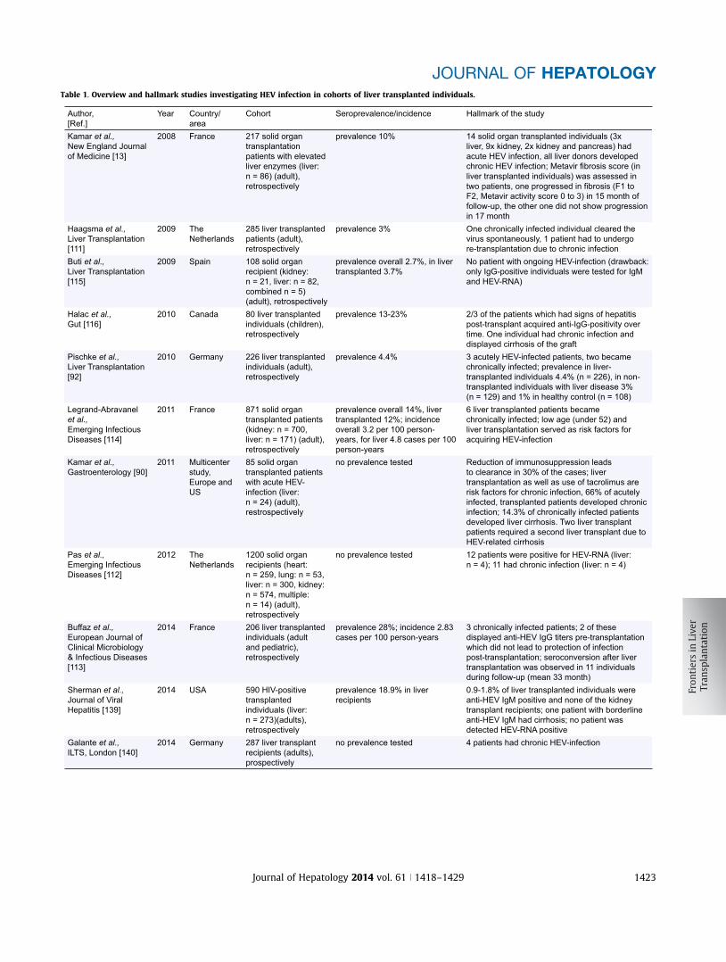
Table 1. Overview and hallmark studies investigating HEV infection in cohorts of liver transplanted individuals.
Author,[Ref.]
Year Country/area
Cohort Seroprevalence/incidence Hallmark of the study
Kamar et al.,New England Journal of Medicine [13]
2008 France 217 solid organ transplantation patients with elevated liver enzymes (liver: n = 86) (adult), retrospectively
prevalence 10% 14 solid organ transplanted individuals (3x liver, 9x kidney, 2x kidney and pancreas) had acute HEV infection, all liver donors developed chronic HEV infection; Metavir fibrosis score (in liver transplanted individuals) was assessed in two patients, one progressed in fibrosis (F1 toF2, Metavir activity score 0 to 3) in 15 month of follow-up, the other one did not show progression in 17 month
Haagsma et al.,Liver Transplantation [111]
2009 The Netherlands
285 liver transplanted patients (adult), retrospectively
prevalence 3% One chronically infected individual cleared the virus spontaneously, 1 patient had to undergo re-transplantation due to chronic infection
Buti et al.,Liver Transplantation [115]
2009 Spain 108 solid organ recipient (kidney: n = 21, liver: n = 82, combined n = 5)(adult), retrospectively
prevalence overall 2.7%, in liver transplanted 3.7%
No patient with ongoing HEV-infection (drawback: only IgG-positive individuals were tested for IgM and HEV-RNA)
Halac et al.,Gut [116]
2010 Canada 80 liver transplanted individuals (children), retrospectively
prevalence 13-23% 2/3 of the patients which had signs of hepatitis post-transplant acquired anti-IgG-positivity over time. One individual had chronic infection and displayed cirrhosis of the graft
Pischke et al.,Liver Transplantation [92]
2010 Germany 226 liver transplanted individuals (adult), retrospectively
prevalence 4.4% 3 acutely HEV-infected patients, two became chronically infected; prevalence in liver-transplanted individuals 4.4% (n = 226), in non-transplanted individuals with liver disease 3% (n = 129) and 1% in healthy control (n = 108)
Legrand-Abravanel et al.,Emerging Infectious Diseases [114]
2011 France 871 solid organ transplanted patients (kidney: n = 700, liver: n = 171) (adult), retrospectively
prevalence overall 14%, liver transplanted 12%; incidence overall 3.2 per 100 person-years, for liver 4.8 cases per 100 person-years
6 liver transplanted patients became chronically infected; low age (under 52) and liver transplantation served as risk factors for acquiring HEV-infection
Kamar et al.,Gastroenterology [90]
2011 Multicenter study, Europe and US
85 solid organ transplanted patients with acute HEV-infection (liver: n = 24) (adult), restrospectively
no prevalence tested Reduction of immunosuppression leads to clearance in 30% of the cases; liver transplantation as well as use of tacrolimus are risk factors for chronic infection, 66% of acutely infected, transplanted patients developed chronic infection; 14.3% of chronically infected patients developed liver cirrhosis. Two liver transplant patients required a second liver transplant due to HEV-related cirrhosis
Pas et al.,Emerging Infectious Diseases [112]
2012 The Netherlands
1200 solid organ recipients (heart:n = 259, lung: n = 53, liver: n = 300, kidney: n = 574, multiple: n = 14) (adult), retrospectively
no prevalence tested 12 patients were positive for HEV-RNA (liver: n = 4); 11 had chronic infection (liver: n = 4)
Buffaz et al.,European Journal of Clinical Microbiology & Infectious Diseases [113]
2014 France 206 liver transplanted individuals (adult and pediatric), retrospectively
prevalence 28%; incidence 2.83 cases per 100 person-years
3 chronically infected patients; 2 of these displayed anti-HEV IgG titers pre-transplantation which did not lead to protection of infection post-transplantation; seroconversion after liver transplantation was observed in 11 individuals during follow-up (mean 33 month)
Sherman et al.,Journal of Viral Hepatitis [139]
2014 USA 590 HIV-positive transplanted individuals (liver: n = 273)(adults), retrospectively
prevalence 18.9% in liver recipients
0.9-1.8% of liver transplanted individuals were anti-HEV IgM positive and none of the kidney transplant recipients; one patient with borderline anti-HEV IgM had cirrhosis; no patient was detected HEV-RNA positive
Galante et al.,ILTS, London [140]
2014 Germany 287 liver transplant recipients (adults), prospectively
no prevalence tested 4 patients had chronic HEV-infection
JOURNAL OF HEPATOLOGY
Journal of Hepatology 2014 vol. 61 j 1418–1429 1423

100 person-years. Only one of the seroconverted patients wasalso HEV RNA positive. In addition, the authors identified 2patients with chronic hepatitis E who had already been anti-HEV IgG seropositive prior to transplantation. Based on thisobservation the authors concluded that HEV clearance beforetransplantation and presence of HEV antibodies may not protectfrom HEV infection in the context of immunosuppression afterliver transplantation. However, one drawback of the study wasthat HEV-RNA-testing was performed only in individuals whowere anti-HEV-IgM and IgG positive. Therefore, anti-HEV anti-body-negative/HEV-RNA-positive patients would not have beendetected with this screening approach.
In another study from Toulouse (France) a large number ofkidney- (n = 529) and liver-transplanted (n = 171) individuals –transplanted between 2004 and 2008 – were retrospectivelyassessed for signs of HEV infection [114]. HEV infection aftertransplantation occurred in 34 patients (kidney recipientsn = 22, liver n = 12). 14 patients seroconverted post-transplanta-tion, while HEV-RNA remained negative. However, 20 patientswho did not develop antibodies against HEV had detectable HEVRNA. The HEV incidence rates were calculated with 2.7 per 100person-years in kidney recipients and 4.8 per 100 person-yearsin liver recipients. A chronic course of HEV infection was observedin 16 patients (47%). Strikingly, age under 52 at time of transplan-tation as well as receiving a liver transplantation were risk factorsfor acquiring HEV infection in the multivariate analysis.
There are also data from Spain. In 2010, Buti et al. investigatedthe HEV-prevalence in solid-organ-transplanted individuals bytesting anti-HEV IgG when an elevation of liver enzymes wasobserved (kidney: n = 21, liver: n = 82, combined n = 5) [115].Patients positive for IgG were also tested for anti-HEV-IgM aswell as for HEV-RNA. Overall, only three liver transplantedpatients were anti-HEV IgG positive. There was no evidence forpersistent infections. Also in this study it has to be consideredthat HEV-RNA testing was performed only in anti-HEV IgG posi-tive samples and thus the true prevalence of HEV infection couldhave been underestimated.
Factors potentially associated with chronic HEV infection insolid organ transplant recipients were investigated in a largemulti-centre study involving 17 transplant centres throughoutEurope and the US [90]. Overall 83 patients were enrolled includ-ing 26 liver transplant recipients. Interestingly, in this cohort themajority of liver-transplanted individuals developed chronicinfection (22 of 26 infected individuals) which was significantlymore frequent than in non-liver transplanted patients (34 of59) supporting the hypothesis that the risk for persistent infec-tions is higher after liver transplantation.
Sherman et al. studied HIV-infected kidney and liver trans-planted individuals in the US for markers of HEV-infection. In590 individuals (including 273 patients after liver transplanta-tion) the authors found an anti-HEV IgG prevalence of 20% inboth, liver and kidney transplanted patients. However, testingfor HEV-RNA revealed no ongoing infection in any case.
The prevalence of HEV infection in children after livertransplantation was investigated in a Canadian study [116]. Theauthors screened 80 children transplanted between 1992 and2010. Liver enzymes were normal in 66 children while 14 hadpersistently elevated ALT levels. Individuals with normal liverenzymes tested anti-HEV IgG positive in 15% of cases whiletwo-thirds of the children with elevated liver enzymesseroconverted after liver transplantation. Additionally, seven of
the children with elevated liver enzymes had detectableanti-HEV IgM more than once during follow-up and thesepatients also had histological signs of inflammation and fibrosisprogression. One girl tested repeatedly HEV-RNA positive andunfortunately developed liver cirrhosis further highlighting theprogressive nature of persistent HEV infection in immunosup-pressed patients.
Role of immunosuppression in solid-organ transplantedindividuals and HEV
Immunosuppressive drugs may interfere with replication of dif-ferent viruses. It has, for example, been shown for HCV that ste-roids may increase HCV infectivity by upregulation of distinctentry receptors [117] and that cyclosporine can inhibit viral rep-lication by interfering with cyclophilins [118]. There is also someevidence that the immunosuppressive regime may be importantfor HEV infection. Initial data suggested that chronic cases of hep-atitis E are more likely in liver- and kidney transplanted patients ifpatients were treated with the calcineurin-inhibitor tacrolimus ascompared to patients receiving cyclosporine [90]. In contrast,heart transplant recipients receiving mycophenolate did notdevelop persistent infection [91]. Of note, these clinical data havemeanwhile been supported by studies investigating the effects ofdifferent drugs on HEV infection in vitro. The studies were possibledue to the development of a robust in vitro model for HEV geno-type 3 infection [119]. Interestingly, addition of tacrolimusincreased viral replication supporting the clinical observations[120]. However, the other calcineurin inhibitor cyclosporine Aalso increased replication, which could not be confirmed yet inpatient cohorts. Different mTOR-inhibitors, rapamycin and ever-olimus also led to an enhanced replication of HEV transfected cellsin vitro, involving phosphorylated 4E-BP1 in infected hepatocytes[121]. However, the clinical impact of this finding is unclear at thisstage. The use of corticosteroids does not seem to impact replica-tion of HEV, both in vivo as well as in vitro. Strikingly, the use ofmycophenolate-mofetil leads to a significant decrease of viral rep-lication in vitro, which would on the first view support the abovementioned clinical observation in heart transplant recipients[91,120]. The mechanism is likely to be linked to IMPDH inhibi-tion by mycophenolate [122]. However, mycophenolate-mofetilmight also negatively influence leucocyte cell count and subse-quently promote chronicity of infection [14,88]. Thus, it is unclearto what extent the beneficial in vitro effects of mycophenolatereally translate into clinical benefits. Indeed, many patients withchronic hepatitis E in the literature received mycophenolate aspart of their immunosuppressive regimen.
Overall, the choice of the immunosuppressive drugs seems tobe of important for liver transplant recipients with hepatitis E.Still, both experimental and clinical data are very limited. Atpresent one could suggest that tacrolimus and mTOR inhibitorsshould be avoided once chronic infection is evident, while ste-roids, cyclosporine and mycophenolate might be preferred com-pounds. However, there is not sufficient evidence yet topropose distinct ‘‘pre-emptive’’ immunosuppressive regimens toavoid chronic infections after exposure or to propose specificdrug changes once persistent HEV infection has developed.Rather than discussing one drug vs. another it seems to be moreimportant to reduce the overall level of immunosuppression asmuch as possible in patients with chronic hepatitis E.
Frontiers in Liver Transplantation
1424 Journal of Hepatology 2014 vol. 61 j 1418–1429
HEV reactivation
In 2009 le Coutre et al. reported the case of a patient with acutelymphatic leukaemia who displayed an acute, self-limiting HEVinfection prior to allogenic stem cell transplantation [123].14 weeks after transplantation reappearance of HEV viraemiawas observed. Sequencing analysis confirmed the presence ofidentical viral strains prior to transplantation and at the time orreactivation suggesting prolonged viral persistence in theabsence of detectable viraemia. For kidney-transplanted individ-uals no HEV reactivation has yet been described [124] while HEVcan be transmitted by liver transplantation as for example delin-eated by Haagsma et al. [125] and Schlosser et al. [73].
Prevention of HEV infection
HEV, being a non-enveloped virus, is relatively robust and stableupon environmental harms. The risk of food-borne HEV can besignificantly reduced by cooking of meat. In vitro assays suggest,that cooking meat for 1 min at 70 �C is efficient in reducing viralinfectivity [126,127]. In theory, transmission by contact withraw meet seems possible, although not described in the litera-ture so far. Therefore, this risk should be reduced by washinghands after exposure to potentially infected meat. Blood prod-ucts are not routinely tested until now and it is unclear whethercommon inactivation procedures cause elimination of infectiousHEV-particles. Although, direct human-to-human transmissionof HEV is usually uncommon, patients should be advised to per-form regular cleaning of shared sanitary facilities. If one pre-sumes similar stabilities of HEV and HAV, HAV is partiallyresistant to 80% ethanol-based disinfectants, which should notbe recommended alone for the decontamination of HEV either[128]. Overall, intra-family or direct person-to person transmis-sion seems to be a seldom event but have been described andsuggested in a few cases [129]. Therefore, testing of partners/family-members should be considered if risk factors for thedevelopment of severe infection are present including organtransplantation.
A vaccine against HEV was licensed in China which showed anefficacy of 94%-100% in a large-scale vaccine trial involving morethan 100,000 individuals [80]. Unfortunately, this vaccine has notbeen approved yet in other countries and the efficacy to protectfrom HEV genotype 3 infections has also not formally been pro-ven – even though the genotype 1- based vaccine was effectiveagainst HEV genotype 4 infections in China. A vaccine againstHEV would be highly desirable, especially for patients at risk todevelop fulminant acute HEV infection or to become chronicallyinfected. However, even if a vaccine would be available it isunclear if the approach would be effective in immunosuppressedpatients as antibody-titres might not be sufficient to protectagainst infection. This is supported by the study from Buffazet al. who observed chronic HEV infections even in patientswho displayed anti-HEV IgG before transplantation [113]. In linewith this, Abravanel et al. recently also reported infection ofsolid-organ transplanted individuals, which were anti-HEV IgGpositive prior to liver transplantation. They propose that anHEV antibody-titre below 7 WHO units/ml is not sufficient toprotect from HEV-infection in this cohort. Additionally, theydelineated that these infections can lead to both, acute andchronic HEV infection [130].
Treatment
HEV-infection is usually subclinical in immunocompetent indi-viduals and does therefore not require specific therapies in thevast majority of cases. However, antiviral treatment of severeacute infections should be considered in patients with risk factorsfor fulminant liver failure. Ribavirin seems to be the treatment ofchoice as it can lead to a rapid decrease of viral load in patientswith acute severe hepatitis E [131–133]. Of note, ribavirin wasalso beneficial not only in genotype 3 infection but also in onepatient with very severe acute HEV genotype 1 infection [84].Based on this – still rather limited – data, we would recommendto treat acute hepatitis E patients at risk of developing liver fail-ure with ribavirin not only to prevent organ failure but also toavoid HEV transmission if liver transplantation is still needed.However, the specific role of ribavirin as pre-emptive therapyto avoid infection of the transplanted graft requires furtherinvestigation.
Chronic hepatitis E should be avoided in liver transplant recip-ients to prevent fibrosis progression. The first steps in the man-agement of a transplanted patient with persistent HEV infectionwould be the re-evaluation of immunosuppressive drugs basedon effects on HEV replication as discussed above and – if possible– to reduce the intensity of immunosuppression, which may leadto HEV RNA clearance in approximately 30% of cases [90]. If HEVis not cleared, antiviral therapies should be considered.
Two case reports describing in total 5 liver transplanted indi-viduals showed that chronic hepatitis E may be treated with PEG-interferon alpha [134,135]. HEV RNA became negative over atreatment course of 3–12 months. However, interferon alphatreatment is associated with side effects and may also inducegraft rejection. Thus, only well selected patients with chronichepatitis E may be candidates of PEG-IFNa therapy.
First case series, reporting an antiviral efficacy of ribavirinmonotherapy have been reported by two French centres in 2and 6 individuals with persistent HEV infection, respectively[136,137]. Patients were treated for 3 months with a ribavirindose of 600–800 mg per day and six of the eight patients experi-enced a virological response with sustained clearance of HEV,while two patients relapsed after the end of therapy. These find-ings were then reproduced by different centres. In Hannover(Germany), patients were treated for a longer time (5 months)and 9/11 patients achieved HEV RNA clearance. However andimportantly, one patient showed a virological breakthroughwhen the ribavirin dose was reduced due to anaemia. Clinicalresistance was evident as no further viral decline was observedeven when the ribavirin dosing was increased again [84]. Theso far largest cohort of ribavirin-treated patients was reportedrecently from France describing a retrospective analysis of 59solid-organ transplanted patients with chronic hepatitis E[138]. Patients received ribavirin at a dose of 8 mg per kg bodyweight for a median of 3 months (range 1–18 months). A sus-tained virological response could be reached in 46 of the 59patients. Interestingly, all patients who failed to clear HEV duringthe initial course of ribavirin therapy showed a sustained virolog-ical response when re-treated with ribavirin for a longer period.Overall, clearance of HEV was observed in 95% of the cases (2patient still received retreatment, 2 patients died of HEV- andtreatment-unrelated reasons, 2 patients declined to undergo re-therapy and 2 were lost during follow-up). In the multivariateanalysis a low lymphocyte count was predictive for non-response
JOURNAL OF HEPATOLOGY
Journal of Hepatology 2014 vol. 61 j 1418–1429 1425

to treatment. Of note, anaemia was a dominant side effect requir-ing reduction of ribavirin in 29% of the patients. This underlinesthat patients with poor performance status and comorbiditiesmay not be applicable for ribavirin treatment and that alternativeantiviral treatment are still needed.
Studies, investigating antiviral regimes specifically in livertransplanted individuals are not yet available. However, availabledata prompt for ribavirin being the treatment of choice (Table 2).
Concluding remarks
It is now well established that HEV infections can take chroniccourses in immunocompromised individuals and that the riskfor chronicity – even though based on limited data – may be evenhigher for patients after liver transplantation. However, the over-all frequency of HEV infection in European and North Americantransplant cohorts is low. HEV RNA testing of patients with ele-vated liver enzymes only seems to be a reasonable approach.HEV genotype 3 infection is a zoonosis and liver transplant recip-ients have to be advised to avoid the consumption of raw anduncooked meat. Transfusion of HEV by blood has to be consideredand HEV RNA testing of blood products is currently under debatein several countries. Chronic hepatitis E is often associated with aparticular rapid progression to liver cirrhosis and should there-fore not be missed. The choice of immunosuppressive drugs isimportant in case of HEV infections as distinct effects on HEV rep-lication (either inhibitory or stimulatory) have been described.
An antiviral treatment option is available with ribavirin, whichshould be given for at least 3 months, however, treatment failuresmay occur supporting ongoing research to identify additionaltreatment options for a frequent and largely underestimatedinfectious complication after liver transplantation.
Conflict of interest
Prof. Manns has received payments for consulting services fromAbbott, Falk Foundation, Merck, and Roche.
Prof. Wedemeyer has received payments for consulting ser-vices from Abbott, Falk Foundation, Merck, Roche, Roche Diag-nostics, and Siemens, as well as payments for his expert advicefrom Abbott, Roche, Roche Diagnostics, and Siemens.
Steinmann, PD PhD and Behrendt, MD have nothing to declare.
References
[1] Balayan MS, Andjaparidze AG, Savinskaya SS, Ketiladze ES, Braginsky DM,Savinov AP, et al. Evidence for a virus in non-A, non-B hepatitis transmittedvia the fecal-oral route. Intervirology 1983;20:23–31.
[2] Bhatia V, Singhal A, Panda SK, Acharya SK. A 20-year single-centerexperience with acute liver failure during pregnancy: is the prognosisreally worse? Hepatology 2008;48:1577–1585.
[3] Patra S, Kumar A, Trivedi SS, Puri M, Sarin SK. Maternal and fetal outcomesin pregnant women with acute hepatitis E virus infection. Ann Intern Med2007;147:28–33.
[4] Dalton HR, Hazeldine S, Banks M, Ijaz S, Bendall R. Locally acquiredhepatitis E in chronic liver disease. Lancet 2007;369:1260.
Table 2. Overview and hallmark reports investigating different treatment options of chronic HEV infection in liver transplanted individuals.
Author, [Ref.] Treated individuals Antiviral drug/intervention Outcome/HallmarksKamar et al., 2010;Clinical Infectious Disease [92]
3 liver transplanted PegIFNα-2a 2 patients cleared infection, 1 individual relapsed
Haagsma et al., 2010;Liver Transplantation [135]
2 liver transplanted PegIFNα-2a One patient cleared under treatment; the other patient was treated for 16 weeks, but still displayed detectable HEV-RNA. This person resolved 4 weeks after discontinuation, during which period levels of tacrolimus were rather low
Pischke et al., 2013;Liver International [84]
5 kidney-, 4 heart-, 4 lung-, 2 liver transplanted
Ribavirin or reduction of immunosuppression
Overall, reduction of immunosuppression led to HEV-clearance in three patients. 11 patients recieved ribavirin, 9 of these cleared the virus, one patient died due to treatment- and HEV-unrelated disease, one patient remained HEV-RNA positive and died later due to liver related disorders. Both liver transplanted individuals resolved by reduction of immunosuppression
Junge et al., 2013;Pediatric Transplantation [141]
1 liver transplanted Ribavirin The patient cleared infection under treatment
Kamar et al., 2014;New England Journal of Medicine [138]
37 kidney-, 5 heart-, 5 kidney & pancreas-, 2 lung-, 10 liver transplanted
Ribavirin Overall, HEV clearance under therapy was achieved in 95% of the patients, recurrence of HEV-RNA was detected in 10 patients after the end of treatment, 7 of these patients underwent re-treatment which led to sustained virological response in 4 individuals, 2 were HEV-RNA negative and still recieved therapy and 1 patient was HEV-RNA negative 3 month after the end of re-treatment; in liver transplanted individuals 8 had sustained virological response, while 2 had no sustained virological response after initial treatment;
associated with a sustained virologic response; a positive test for HEV RNA at month 1 of treatment increased the likelihood for failure to achieve a sustained virological response
Klein et al., 2014;Experimental and Clinical Transplantation [142]
1 liver transplanted Ribavirin The patient cleared infection under treatment
Galante et al., 2014;oral presentation ILTS London[140]
4 liver transplanted Ribavirin 3 patients resolved, 1 patient relapsed after therapy and is still receiving retreatment
a higher lymphocyte count at the initiation of ribavirin was significantly
Frontiers in Liver Transplantation
1426 Journal of Hepatology 2014 vol. 61 j 1418–1429
[5] Kumar M, Sharma BC, Sarin SK. Hepatitis E virus as an etiology of acuteexacerbation of previously unrecognized asymptomatic patients withhepatitis B virus-related chronic liver disease. J Gastroenterol Hepatol2008;23:883–887.
[6] Hamid SS, Atiq M, Shehzad F, Yasmeen A, Nissa T, Salam A, et al. Hepatitis Evirus superinfection in patients with chronic liver disease. Hepatology2002;36:474–478.
[7] Ramachandran J, Eapen CE, Kang G, Abraham P, Hubert DD, Kurian G, et al.Hepatitis E superinfection produces severe decompensation in patientswith chronic liver disease. J Gastroenterol Hepatol 2004;19:134–138.
[8] Aye TT, Uchida T, Ma XZ, Iida F, Shikata T, Zhuang H, et al. Completenucleotide sequence of a hepatitis E virus isolated from the Xinjiangepidemic (1986–1988) of China. Nucleic Acids Res 1992;20:3512.
[9] Kamar N, Bendall R, Legrand-Abravanel F, Xia NS, Ijaz S, Izopet J, et al.Hepatitis E. Lancet 2012;379:2477–2488.
[10] Aggarwal R, Naik S. Epidemiology of hepatitis E: current status. JGastroenterol Hepatol 2009;24:1484–1493.
[11] Kim JH, Nelson KE, Panzner U, Kasture Y, Labrique AB, Wierzba TF. Asystematic review of the epidemiology of hepatitis E virus in Africa. BMCInfect Dis 2014;14:308.
[12] Centers for Disease C, Prevention. Investigation of hepatitis E outbreakamong refugees – Upper Nile, South Sudan, 2012-2013. MMWR Morbidityand Mortality Weekly Report 2013;62:581–586.
[13] Kamar N, Selves J, Mansuy JM, Ouezzani L, Peron JM, Guitard J, et al.Hepatitis E virus and chronic hepatitis in organ-transplant recipients. NewEngl J Med 2008;358:811–817.
[14] Wedemeyer H, Pischke S, Manns MP. Pathogenesis and treatment ofhepatitis E virus infection. Gastroenterology 2012;142:e1381.
[15] Dalton HR, Bendall RP, Keane FE, Tedder RS, Ijaz S. Persistent carriage ofhepatitis E virus in patients with HIV infection. New Engl J Med2009;361:1025–1027.
[16] Kaba M, Richet H, Ravaux I, Moreau J, Poizot-Martin I, Motte A, et al.Hepatitis E virus infection in patients infected with the human immuno-deficiency virus. J Med Virol 2011;83:1704–1716.
[17] Peron JM, Mansuy JM, Recher C, Bureau C, Poirson H, Alric L, et al. Prolongedhepatitis E in an immunocompromised patient. J Gastroenterol Hepatol2006;21:1223–1224.
[18] Tavitian S, Peron JM, Huynh A, Mansuy JM, Ysebaert L, Huguet F, et al.Hepatitis E virus excretion can be prolonged in patients with hematologicalmalignancies. J Clin Virol 2010;49:141–144.
[19] Honer Zu Siederdissen C, Pischke S, Schlue J, Deterding K, Hellms T, Schuler-Luttmann S, et al. Chronic HEV infection beyond transplantation or HIVinfection. Hepatology 2013.
[20] Mansuy JM, Bendall R, Legrand-Abravanel F, Saune K, Miedouge M, Ellis V,et al. Hepatitis E virus antibodies in blood donors, France. Emerg Infect Dis2011;17:2309–2312.
[21] Bendall R, Ellis V, Ijaz S, Ali R, Dalton H. A comparison of two commerciallyavailable anti-HEV IgG kits and a re-evaluation of anti-HEV IgG seroprev-alence data in developed countries. J Med Virol 2010;82:799–805.
[22] Wenzel JJ, Sichler M, Schemmerer M, Behrens G, Leitzmann MF, Jilg W.Decline in hepatitis E virus antibody prevalence in Southeastern Germany,1996–2011. Hepatology 2014.
[23] Davern TJ, Chalasani N, Fontana RJ, Hayashi PH, Protiva P, Kleiner DE, et al.Acute hepatitis E infection accounts for some cases of suspected drug-induced liver injury. Gastroenterology 2011;141:e1661–1669.
[24] Chen EY, Baum K, Collins W, Love A, Merz M, Olafsson S, et al. Hepatitis Emasquerading as drug-induced liver injury. Hepatology2012;56:2420–2423.
[25] Kamar N, Bendall RP, Peron JM, Cintas P, Prudhomme L, Mansuy JM, et al.Hepatitis E virus and neurologic disorders. Emerg Infect Dis2011;17:173–179.
[26] van Eijk JJ, Madden RG, van der Eijk AA, Hunter JG, Reimerink JH, BendallRP, et al. Neuralgic amyotrophy and hepatitis E virus infection. Neurology2014;82:498–503.
[27] van den Berg B, van der Eijk AA, Pas SD, Hunter JG, Madden RG, Tio-GillenAP, et al. Guillain–Barre syndrome associated with preceding hepatitis Evirus infection. Neurology 2014;82:491–497.
[28] Kamar N, Weclawiak H, Guilbeau-Frugier C, Legrand-Abravanel F, CointaultO, Ribes D, et al. Hepatitis E virus and the kidney in solid-organ transplantpatients. Transplantation 2012;93:617–623.
[29] Koonin EV, Gorbalenya AE, Purdy MA, Rozanov MN, Reyes GR, Bradley DW.Computer-assisted assignment of functional domains in the nonstructuralpolyprotein of hepatitis E virus: delineation of an additional group ofpositive-strand RNA plant and animal viruses. Proc Natl Acad Sci U S A1992;89:8259–8263.
[30] Smith DB, Vanek J, Ramalingam S, Johannessen I, Templeton K, SimmondsP. Evolution of the hepatitis E virus hypervariable region. J Gen Virol2012;93:2408–2418.
[31] Guu TS, Liu Z, Ye Q, Mata DA, Li K, Yin C, et al. Structure of the hepatitis Evirus-like particle suggests mechanisms for virus assembly and receptorbinding. Proc Natl Acad Sci U S A 2009;106:12992–12997.
[32] Yamashita T, Mori Y, Miyazaki N, Cheng RH, Yoshimura M, Unno H, et al.Biological and immunological characteristics of hepatitis E virus-likeparticles based on the crystal structure. Proc Natl Acad Sci U S A2009;106:12986–12991.
[33] Korkaya H, Jameel S, Gupta D, Tyagi S, Kumar R, Zafrullah M, et al. The ORF3protein of hepatitis E virus binds to Src homology 3 domains and activatesMAPK. J Biol Chem 2001;276:42389–42400.
[34] Okamoto H. Genetic variability and evolution of hepatitis E virus. Virus Res2007;127:216–228.
[35] Feagins AR, Opriessnig T, Guenette DK, Halbur PG, Meng XJ. Detection andcharacterization of infectious Hepatitis E virus from commercial pig liverssold in local grocery stores in the USA. J Gen Virol 2007;88:912–917.
[36] Bouwknegt M, Lodder-Verschoor F, van der Poel WH, Rutjes SA, de RodaHusman AM. Hepatitis E virus RNA in commercial porcine livers in TheNetherlands. J Food Protect 2007;70:2889–2895.
[37] Berto A, Backer JA, Mesquita JR, Nascimento MS, Banks M, Martelli F, et al.Prevalence and transmission of hepatitis E virus in domestic swinepopulations in different European countries. BMC Res Notes 2012;5:190.
[38] Smith DB, Purdy MA, Simmonds P. Genetic variability and the classificationof hepatitis E virus. J Virol 2013;87:4161–4169.
[39] Burri C, Vial F, Ryser-Degiorgis MP, Schwermer H, Darling K, Reist M, et al.Seroprevalence of hepatitis E virus in domestic pigs and wild boars inSwitzerland. Zoonoses Public Health 2014.
[40] Hara Y, Terada Y, Yonemitsu K, Shimoda H, Noguchi K, Suzuki K, et al. Highprevalence of hepatitis e virus in wild boar (Sus scrofa) in YamaguchiPrefecture, Japan. J Wildlife Dis 2014.
[41] Caron M, Enouf V, Than SC, Dellamonica L, Buisson Y, Nicand E. Identifi-cation of genotype 1 hepatitis E virus in samples from swine in Cambodia. JClin Microbiol 2006;44:3440–3442.
[42] Hakze-van der Honing RW, van Coillie E, Antonis AF, van der Poel WH. Firstisolation of hepatitis E virus genotype 4 in Europe through swinesurveillance in the Netherlands and Belgium. PLoS One 2011;6:e22673.
[43] Colson P, Romanet P, Moal V, Borentain P, Purgus R, Benezech A, et al.Autochthonous infections with hepatitis E virus genotype 4, France. EmergInfect Dis 2012;18:1361–1364.
[44] Drexler JF, Seelen A, Corman VM, Fumie Tateno A, Cottontail V, MelimZerbinati R, et al. Bats worldwide carry hepatitis E virus-related viruses thatform a putative novel genus within the family Hepeviridae. J Virol2012;86:9134–9147.
[45] Haqshenas G, Shivaprasad HL, Woolcock PR, Read DH, Meng XJ. Geneticidentification and characterization of a novel virus related to humanhepatitis E virus from chickens with hepatitis-splenomegaly syndrome inthe United States. J Gen Virol 2001;82:2449–2462.
[46] Raj VS, Smits SL, Pas SD, Provacia LB, Moorman-Roest H, Osterhaus AD, et al.Novel hepatitis E virus in ferrets, the Netherlands. Emerg Infect Dis2012;18:1369–1370.
[47] Zhao C, Ma Z, Harrison TJ, Feng R, Zhang C, Qiao Z, et al. A novel genotype ofhepatitis E virus prevalent among farmed rabbits in China. J Med Virol2009;81:1371–1379.
[48] Johne R, Heckel G, Plenge-Bonig A, Kindler E, Maresch C, Reetz J, et al. Novelhepatitis E virus genotype in Norway rats, Germany. Emerg Infect Dis2010;16:1452–1455.
[49] Batts W, Yun S, Hedrick R, Winton J. A novel member of the familyHepeviridae from cutthroat trout (Oncorhynchus clarkii). Virus Res2011;158:116–123.
[50] Wenzel JJ, Preiss J, Schemmerer M, Huber B, Plentz A, Jilg W. Detection ofhepatitis E virus (HEV) from porcine livers in Southeastern Germany andhigh sequence homology to human HEV isolates. J Clin Virol2011;52:50–54.
[51] Wichmann O, Schimanski S, Koch J, Kohler M, Rothe C, Plentz A, et al.Phylogenetic and case-control study on hepatitis E virus infection inGermany. J Infect Dis 2008;198:1732–1741.
[52] Tei S, Kitajima N, Takahashi K, Mishiro S. Zoonotic transmission of hepatitisE virus from deer to human beings. Lancet 2003;362:371–373.
[53] Tomiyama D, Inoue E, Osawa Y, Okazaki K. Serological evidence of infectionwith hepatitis E virus among wild Yezo-deer, Cervus nippon yesoensis, inHokkaido, Japan. J Viral Hepatitis 2009;16:524–528.
[54] Meng XJ. Zoonotic and foodborne transmission of hepatitis E virus. SeminLiver Dis 2013;33:41–49.
JOURNAL OF HEPATOLOGY
Journal of Hepatology 2014 vol. 61 j 1418–1429 1427
[55] Bouwknegt M, Engel B, Herremans MM, Widdowson MA, Worm HC,Koopmans MP, et al. Bayesian estimation of hepatitis E virus seropreva-lence for populations with different exposure levels to swine in TheNetherlands. Epidemiol Infect 2008;136:567–576.
[56] Krumbholz A, Mohn U, Lange J, Motz M, Wenzel JJ, Jilg W, et al. Prevalenceof hepatitis E virus-specific antibodies in humans with occupationalexposure to pigs. Med Microbiol Immunol 2012;201:239–244.
[57] Crossan C, Baker PJ, Craft J, Takeuchi Y, Dalton HR, Scobie L. Hepatitis Evirus genotype 3 in shellfish, United Kingdom. Emerg Infect Dis2012;18:2085–2087.
[58] Donia D, Dell’Amico MC, Petrinca AR, Martinucci I, Mazzei M, Tolari F, et al.Presence of hepatitis E RNA in mussels used as bio-monitors of viral marinepollution. J Virol Methods 2012;186:198–202.
[59] Diez-Valcarce M, Kokkinos P, Soderberg K, Bouwknegt M, Willems K, deRoda-Husman AM, et al. Occurrence of human enteric viruses in commer-cial mussels at retail level in three European countries. Food Environ Virol2012;4:73–80.
[60] Kokkinos P, Kozyra I, Lazic S, Bouwknegt M, Rutjes S, Willems K, et al.Harmonised investigation of the occurrence of human enteric viruses in theleafy green vegetable supply chain in three European countries. FoodEnviron Virol 2012;4:179–191.
[61] Brassard J, Gagne MJ, Genereux M, Cote C. Detection of human food-borneand zoonotic viruses on irrigated, field-grown strawberries. Appl EnvironMicrobiol 2012;78:3763–3766.
[62] Hauser L, Roque-Afonso AM, Beyloune A, Simonet M, Deau Fischer B, Burindes Roziers N. Hepatitis E transmission by transfusion of Intercept bloodsystem-treated plasma. Blood 2014;123:796–797.
[63] Coilly A, Haim-Boukobza S, Roche B, Antonini TM, Pause A, Mokhtari C,et al. Posttransplantation hepatitis E: transfusion-transmitted hepatitisrising from the ashes. Transplantation 2013;96:e4–6.
[64] Cleland A, Smith L, Crossan C, Blatchford O, Dalton HR, Scobie L, et al.Hepatitis E virus in Scottish blood donors. Vox Sang 2013;105:283–289.
[65] Baylis SA, Koc O, Nick S, Blumel J. Widespread distribution of hepatitis Evirus in plasma fractionation pools. Vox Sang 2012;102:182–183.
[66] Ijaz S, Said B, Boxall E, Smit E, Morgan D, Tedder RS. Indigenous hepatitis Ein England and wales from 2003 to 2012: evidence of an emerging novelphylotype of viruses. J Infect Dis 2014;209:1212–1218.
[67] Corman VM, Drexler JF, Eckerle I, Roth WK, Drosten C, Eis-Hubinger AM.Zoonotic hepatitis E virus strains in German blood donors. Vox Sang2013;104:179–180.
[68] Juhl D, Baylis SA, Blumel J, Gorg S, Hennig H. Seroprevalence and incidenceof hepatitis E virus infection in German blood donors. Transfusion2014;54:49–56.
[69] Slot E, Hogema BM, Riezebos-Brilman A, Kok TM, Molier M, Zaaijer HL.Silent hepatitis E virus infection in Dutch blood donors, 2011 to 2012. EuroSurveill 2013;18.
[70] Haim-Boukobza S, Ferey MP, Vetillard AL, Jeblaoui A, Pelissier E, Pelletier G,et al. Transfusion-transmitted hepatitis E in a misleading context ofautoimmunity and drug-induced toxicity. J Hepatol 2012;57:1374–1378.
[71] Feray C, Pawlotsky JM, Roque-Afonso AM, Samuel D, Dhumeaux D. Shouldwe screen blood products for hepatitis E virus RNA? Lancet 2014;383:218.
[72] Hewitt PE, Ijaz S, Brailsford SR, Brett R, Dicks S, Haywood B, et al. Hepatitis Evirus in blood components: a prevalence and transmission study insoutheast England. Lancet 2014.
[73] Schlosser B, Stein A, Neuhaus R, Pahl S, Ramez B, Kruger DH, et al. Livertransplant from a donor with occult HEV infection induced chronichepatitis and cirrhosis in the recipient. J Hepatol 2012;56:500–502.
[74] Koenecke C, Pischke S, Beutel G, Ritter U, Ganser A, Wedemeyer H, et al.Hepatitis E virus infection in a hematopoietic stem cell donor. BoneMarrow Transplant 2014;49:159–160.
[75] Drobeniuc J, Meng J, Reuter G, Greene-Montfort T, Khudyakova N,Dimitrova Z, et al. Serologic assays specific to immunoglobulin M antibod-ies against hepatitis E virus: pangenotypic evaluation of performances. ClinInfect Dis 2010;51:e24–e27.
[76] Herremans M, Bakker J, Duizer E, Vennema H, Koopmans MP. Use ofserological assays for diagnosis of hepatitis E virus genotype 1 and 3infections in a setting of low endemicity. Clin Vaccine Immunol2007;14:562–568.
[77] Kuniholm MH, Purcell RH, McQuillan GM, Engle RE, Wasley A, Nelson KE.Epidemiology of hepatitis E virus in the United States: results from theThird National Health and Nutrition Examination Survey, 1988–1994. JInfect Dis 2009;200:48–56.
[78] Hoofnagle JH, Nelson KE, Purcell RH. Hepatitis E. New Engl J Med2012;367:1237–1244.
[79] Krumbholz A, Neubert A, Joel S, Girschick H, Huppertz HI, Kaiser P, et al.Prevalence of Hepatitis E virus antibodies in children in Germany. PediatrInfect Dis J 2013.
[80] Zhu FC, Zhang J, Zhang XF, Zhou C, Wang ZZ, Huang SJ, et al. Efficacy andsafety of a recombinant hepatitis E vaccine in healthy adults: a large-scale,randomised, double-blind placebo-controlled, phase 3 trial. Lancet2010;376:895–902.
[81] Teshale EH, Hu DJ, Holmberg SD. The two faces of hepatitis E virus. ClinInfect Dis 2010;51:328–334.
[82] Rein DB, Stevens GA, Theaker J, Wittenborn JS, Wiersma ST. The globalburden of hepatitis E virus genotypes 1 and 2 in 2005. Hepatology2012;55:988–997.
[83] Peron JM, Bureau C, Poirson H, Mansuy JM, Alric L, Selves J, et al. Fulminantliver failure from acute autochthonous hepatitis E in France: description ofseven patients with acute hepatitis E and encephalopathy. J Viral Hepatitis2007;14:298–303.
[84] Pischke S, Hardtke S, Bode U, Birkner S, Chatzikyrkou C, Kauffmann W, et al.Ribavirin treatment of acute and chronic hepatitis E: a single-centreexperience. Liver Int 2013;33:722–726.
[85] Smith DB, Simmonds P. Hepatitis E virus and fulminant hepatitis – a virusor host-specific pathology? Liver Int 2014:10–15.
[86] Purcell RH, Emerson SU. Hepatitis E: an emerging awareness of an olddisease. J Hepatol 2008;48:494–503.
[87] Scobie L, Dalton HR. Hepatitis E: source and route of infection, clinicalmanifestations and new developments. J Viral Hepatitis 2013;20:1–11.
[88] Suneetha PV, Pischke S, Schlaphoff V, Grabowski J, Fytili P, Gronert A, et al.Hepatitis E virus (HEV)-specific T-cell responses are associated with controlof HEV infection. Hepatology 2012;55:695–708.
[89] Kamar N, Rostaing L, Legrand-Abravanel F, Izopet J. How should hepatitis evirus infection be defined in organ-transplant recipients? Am J Transplant2013;13:1935–1936.
[90] Kamar N, Garrouste C, Haagsma EB, Garrigue V, Pischke S, Chauvet C, et al.Factors associated with chronic hepatitis in patients with hepatitis E virusinfection who have received solid organ transplants. Gastroenterology2011;140:1481–1489.
[91] Pischke S, Stiefel P, Franz B, Bremer B, Suneetha PV, Heim A, et al. Chronichepatitis E in heart transplant recipients. Am J Transplant2012;12:3128–3133.
[92] Pischke S, Suneetha PV, Baechlein C, Barg-Hock H, Heim A, Kamar N, et al.Hepatitis E virus infection as a cause of graft hepatitis in liver transplantrecipients. Liver Transplant 2010;16:74–82.
[93] Geng Y, Zhang H, Huang W, J Harrison T, Geng K, Li Z, et al. Persistenthepatitis e virus genotype 4 infection in a child with acute lymphoblasticleukemia. Hepatitis Monthly 2014;14:e15618.
[94] Naik A, Gupta N, Goel D, Ippagunta SK, Sharma RK, Aggarwal R. Lack ofevidence of hepatitis E virus infection among renal transplant recipients ina disease-endemic area. J Viral Hepatitis 2013;20:e138–e140.
[95] Emerson SU, Nguyen H, Graff J, Stephany DA, Brockington A, Purcell RH. Invitro replication of hepatitis E virus (HEV) genomes and of an HEV repliconexpressing green fluorescent protein. J Virol 2004;78:4838–4846.
[96] Despierres LA, Kaphan E, Attarian S, Cohen-Bacrie S, Pelletier J, Pouget J,et al. Neurologic disorders and hepatitis E, France, 2010. Emerg Infect Dis2011;17:1510–1512.
[97] Kamar N, Izopet J, Cintas P, Garrouste C, Uro-Coste E, Cointault O, et al.Hepatitis E virus-induced neurological symptoms in a kidney-transplantpatient with chronic hepatitis. Am J Transplant 2010;10:1321–1324.
[98] Aggarwal R. Clinical presentation of hepatitis E. Virus Res 2011;161:15–22.[99] Pischke S, Behrendt P, Manns MP, Wedemeyer H. HEV-associated cryo-
globulinaemia and extrahepatic manifestations of hepatitis E. Lancet InfectDis 2014;14:678–679.
[100] Aggarwal R. Hepatitis E: clinical presentation in disease-endemic areas anddiagnosis. Semin Liver Dis 2013;33:30–40.
[101] Yoo N, Bernstein J, Caldwell C, Dong C, Drobeniuc J, Kamili S, et al. HepatitisE virus infection in a liver transplant recipient: delayed diagnosis due tovariable performance of serologic assays. Transplant Infect Dis2013;15:E166–E168.
[102] Huang SJ, Liu XH, Zhang J, Ng MH. Protective immunity against HEV. CurrOpin Virol 2014;5:1–6.
[103] Zhang J, Zhang XF, Zhou C, Wang ZZ, Huang SJ, Yao X, et al. Protectionagainst hepatitis E virus infection by naturally acquired and vaccine-induced immunity. Clin Microbiol Infect 2013.
[104] Rossi-Tamisier M, Moal V, Gerolami R, Colson P. Discrepancy between anti-hepatitis E virus immunoglobulin G prevalence assessed by two assays inkidney and liver transplant recipients. J Clin Virol 2013;56:62–64.
Frontiers in Liver Transplantation
1428 Journal of Hepatology 2014 vol. 61 j 1418–1429

[105] Pas SD, Streefkerk RH, Pronk M, de Man RA, Beersma MF, Osterhaus AD,et al. Diagnostic performance of selected commercial HEV IgM and IgGELISAs for immunocompromised and immunocompetent patients. J ClinVirol 2013;58:629–634.
[106] Hyams C, Mabayoje DA, Copping R, Maranao D, Patel M, Labbett W, et al.Serological cross reactivity to CMV and EBV causes problems in thediagnosis of acute hepatitis E virus infection. J Med Virol 2014;86:478–483.
[107] Baylis SA, Hanschmann KM, Blumel J, Nubling CM, Group HEVCS.Standardization of hepatitis E virus (HEV) nucleic acid amplificationtechnique-based assays: an initial study to evaluate a panel of HEV strainsand investigate laboratory performance. J Clin Microbiol2011;49:1234–1239.
[108] Mokhtari C, Marchadier E, Haim-Boukobza S, Jeblaoui A, Tesse S, Savary J,et al. Comparison of real-time RT-PCR assays for hepatitis E virus RNAdetection. J Clin Virol 2013;58:36–40.
[109] Majumdar M, Singh MP, Pujhari SK, Bhatia D, Chawla Y, Ratho RK. HepatitisE virus antigen detection as an early diagnostic marker: report from India. JMed Virol 2013;85:823–827.
[110] Vollmer T, Knabbe C, Dreier J. Comparison of real-time PCR and antigenassays for detection of hepatitis e virus in blood donors. J Clin Microbiol2014;52:2150–2156.
[111] Haagsma EB, Niesters HG, van den Berg AP, Riezebos-Brilman A, Porte RJ,Vennema H, et al. Prevalence of hepatitis E virus infection in livertransplant recipients. Liver Transplant 2009;15:1225–1228.
[112] Pas SD, de Man RA, Mulders C, Balk AH, van Hal PT, Weimar W, et al.Hepatitis E virus infection among solid organ transplant recipients, theNetherlands. Emerg Infect Dis 2012;18:869–872.
[113] Buffaz C, Scholtes C, Dron AG, Chevallier-Queyron P, Ritter J, Andre P, et al.Hepatitis E in liver transplant recipients in the Rhone-Alpes region inFrance. Eur J Clin Microbiol Infect Dis 2014;33:1037–1043.
[114] Legrand-Abravanel F, Kamar N, Sandres-Saune K, Lhomme S, Mansuy JM,Muscari F, et al. Hepatitis E virus infection without reactivation in solid-organ transplant recipients, France. Emerg Infect Dis 2011;17:30–37.
[115] Buti M, Cabrera C, Jardi R, Castells L, Esteban R. Are recipients of solid organtransplantation a high-risk population for hepatitis E virus infection? LiverTransplant 2010;16:106–107, author reply 108.
[116] Halac U, Beland K, Lapierre P, Patey N, Ward P, Brassard J, et al. Chronichepatitis E infection in children with liver transplantation. Gut2012;61:597–603.
[117] Ciesek S, Steinmann E, Iken M, Ott M, Helfritz FA, Wappler I, et al.Glucocorticosteroids increase cell entry by hepatitis C virus. Gastroenter-ology 2010;138:1875–1884.
[118] Ciesek S, Steinmann E, Wedemeyer H, Manns MP, Neyts J, Tautz N, et al.Cyclosporine A inhibits hepatitis C virus nonstructural protein 2 throughcyclophilin A. Hepatology 2009;50:1638–1645.
[119] Shukla P, Nguyen HT, Torian U, Engle RE, Faulk K, Dalton HR, et al. Cross-species infections of cultured cells by hepatitis E virus and discovery of aninfectious virus-host recombinant. Proc Natl Acad Sci U S A2011;108:2438–2443.
[120] Wang Y, Zhou X, Debing Y, Chen K, VanderLaan LJ, Neyts J, et al. Calcineurininhibitors stimulate and mycophenolic acid inhibits replication of hepatitisE virus. Gastroenterology 2014.
[121] Zhou X, Wang Y, Metselaar HJ, Janssen HL, Peppelenbosch MP, Pan Q.Rapamycin and everolimus facilitate hepatitis E virus replication: revealinga basal defense mechanism of PI3K-PKB-mTOR pathway. J Hepatol 2014.
[122] Debing Y, Neyts J. Antiviral strategies for hepatitis E virus. Antiviral Res2014;102:106–118.
[123] le Coutre P, Meisel H, Hofmann J, Rocken C, Vuong GL, Neuburger S, et al.Reactivation of hepatitis E infection in a patient with acute lymphoblastic
leukaemia after allogeneic stem cell transplantation. Gut2009;58:699–702.
[124] Kamar N, Izopet J, Rostaing L. No reactivation of hepatitis E virus afterkidney retransplantation. Am J Transplant 2012;12:507–508.
[125] Haagsma EB, van den Berg AP, Porte RJ, Benne CA, Vennema H, ReimerinkJH, et al. Chronic hepatitis E virus infection in liver transplant recipients.Liver Transplant 2008;14:547–553.
[126] Emerson SU, Arankalle VA, Purcell RH. Thermal stability of hepatitis E virus.J Infect Dis 2005;192:930–933.
[127] Schielke A, Filter M, Appel B, Johne R. Thermal stability of hepatitis E virusassessed by a molecular biological approach. Virol J 2011;8:487.
[128] Terpstra FG, van den Blink AE, Bos LM, Boots AG, Brinkhuis FH, Gijsen E,et al. Resistance of surface-dried virus to common disinfection procedures.J Hospital Infect 2007;66:332–338.
[129] Mansuy JM, Huynh A, Abravanel F, Recher C, Peron JM, Izopet J. Molecularevidence of patient-to-patient transmission of hepatitis E virus in ahematology ward. Clin Infect Dis 2009;48:373–374.
[130] Abravanel F, Lhomme S, Chapuy-Regaud S, Mansuy JM, Muscari F, SallustoF, et al. Hepatitis E virus reinfections in solid-organ-transplant recipientscan evolve into chronic infections. J Infect Dis 2014;209:1900–1906.
[131] Gerolami R, Borentain P, Raissouni F, Motte A, Solas C, Colson P. Treatmentof severe acute hepatitis E by ribavirin. J Clin Virol 2011;52:60–62.
[132] Goyal R, Kumar A, Panda SK, Paul SB, Acharya SK. Ribavirin therapy forhepatitis E virus-induced acute on chronic liver failure: a preliminaryreport. Antiviral Ther 2012;17:1091–1096.
[133] Peron JM, Dalton H, Izopet J, Kamar N. Acute autochthonous hepatitis E inwestern patients with underlying chronic liver disease: a role for ribavirin?J Hepatol 2011;54:1323–1324, author reply 1324–1325.
[134] Kamar N, Rostaing L, Abravanel F, Garrouste C, Esposito L, Cardeau-Desangles I, et al. Pegylated interferon-alpha for treating chronic hepatitisE virus infection after liver transplantation. Clin Infect Dis2010;50:e30–e33.
[135] Haagsma EB, Riezebos-Brilman A, van den Berg AP, Porte RJ, Niesters HG.Treatment of chronic hepatitis E in liver transplant recipients withpegylated interferon alpha-2b. Liver Transplant 2010;16:474–477.
[136] Mallet V, Nicand E, Sultanik P, Chakvetadze C, Tesse S, Thervet E, et al. Briefcommunication: case reports of ribavirin treatment for chronic hepatitis E.Ann Intern Med 2010;153:85–89.
[137] Kamar N, Rostaing L, Abravanel F, Garrouste C, Lhomme S, Esposito L, et al.Ribavirin therapy inhibits viral replication on patients with chronichepatitis e virus infection. Gastroenterology 2010;139:1612–1618.
[138] Kamar N, Izopet J, Tripon S, Bismuth M, Hillaire S, Dumortier J, et al.Ribavirin for chronic hepatitis E virus infection in transplant recipients.New Engl J Med 2014;370:1111–1120.
[139] Sherman KE, Terrault N, Barin B, Rouster SD, Shata MT. Hepatitis E infectionin HIV-infected liver and kidney transplant candidates. J Viral hepatitis2014.
[140] Galante A, Pischke S, Lang M, Lütgehetmann M, Polywka S, Nashan B, et al.Relevance of chronic hepatitis E in liver transplant recipients: a real-world-setting. The 2014 Joint International Congress of ILTS, ELITA & LICAGE2014; Abstract O-68.
[141] Junge N, Pischke S, Baumann U, Goldschmidt I, Manns M, Wedemeyer H,et al. Results of single-center screening for chronic hepatitis E in childrenafter liver transplantation and report on successful treatment withribavirin. Pediatr Transplant 2013;17:343–347.
[142] Klein F, Neuhaus R, Hofmann J, Rudolph B, Neuhaus P, Bahra M. Successfultreatment of chronic hepatitis E after an orthotopic liver transplant withribavirin monotherapy. Exp Clin Transplant 2014.
JOURNAL OF HEPATOLOGY
Journal of Hepatology 2014 vol. 61 j 1418–1429 1429