Embed Size (px)
Citation preview


This presentation contains information in the slides and
notes pages – please review in Normal View

Module Outline: Imaging Basics
• Highlight key events in the historical development of imaging
• Identify techniques used in modern imaging departments
• Review which techniques do or do not use ionizing radiation
• Explain radiological orientations, directions & conventions
This module will …
Module Author: Imaging Basics
Andrew Farrall
Dr Farrall came to the Division of Clinical Neurosciences in 2002 from Canada, where he trained as a Radiologist in Halifax, Nova Scotia. He obtained an MD in 1997 from the University of Calgary in Alberta, an MSc in 1995 from the University of Western Ontario in London, Ontario, and a BSc (Hons) from the University of British Columbia in Vancouver in 1990.
He has part time appointments as a Fellow at the University of Edinburgh and as a Consultant Neuroradiologist in the NHS, with sessions at the Western General Hospital & at St. John's Hospital. His main interests are in Stroke Neuroradiology & the role of Magnetic Resonance in its investigation. Lacunar stroke, dementias & small vessel disease form the core of his research. He is also interested in teaching issues in Neuroradiology.

The slides that follow are those used for the final
presentation


Module: Techniques & Physics
Lecture: Imaging Basics
Description: History, terminology & orientation
Author: Dr. Andrew Farrall

• Highlight key events in the historical development of imaging
• Identify techniques used in modern imaging departments
• Review which techniques do or do not use ionizing radiation
• Explain radiological orientations, directions & conventions
Objectives:

• There are no pre-requisites for this session
Prerequisites:

Brief Historical Outline
X-rays were discovered by Wilhelm Conrad Röntgen in 1895 which is generally accepted as being the birth of radiology.
Since that time, X-ray radiology technology has taken advantage of technological advancements in image capture & representation, and also has evolved with computing advances.
Also, other techniques for imaging have been developed, some using radiation and others not.
What follows is a timeline summary of some key events in the history of imaging.

Imaging Orientation & Direction
“Superior” refers to anything above your point of reference, where above means in a direction towards the top of the head e.g. the nose is superior to the lips
“Inferior” refers to anything below your point of reference, where below means in a direction towards the feet e.g. the lips are inferior to the nose.
Superiorly
Inferiorly

Image Conventions
By convention, when we view images, we look at them as though we are actually looking at the patient “face-to-face”.
Therefore, the RIGHT side of a radiological image as you look at it is the patient’s LEFT side; the LEFT side of a radiological image is the patient’s RIGHT side.
Often a marker “R” or “L” embedded in the film indicates which is the patient’s right or left.
Pat
ient
’s r
ight
sid
e
Patient’s left side
Skull X-ray
Superior
Inferior

Modern Imaging Departments
Imaging techniques used in modern imaging departments can be divided into those which use ionizing radiation and those which do not.
Radiation No Radiation

Modern Imaging Departments
Two commonly used techniques which do not use ionizing radiation to create images are Magnetic Resonance Imaging (MRI) and Ultrasound (US).
No Radiation
Ultrasound
Magnetic Resonance
Imaging
Radiation

Imaging Orientation & Direction
“Lateral” refers to anything lying towards the sides, left or right, relative to your point of reference
“Medial” refers to anything lying towards a plane running through the middle of the body, dividing it into left and right halves e.g. the nose lies medially to the eyes.
Laterally right Laterally left Medial

Summary
• Outline the historical development of imaging
• Identify techniques used in modern imaging departments
• Identify which techniques do or do not use ionizing radiation
• Distinguish between techniques which use ionizing radiation
• Understand radiological orientations, directions & convention
You should now be able to:


End of presentation

Module Resources: Imaging Basics
• Bookso Huda W, Stone R, Review of radiologic physics.
Williams & Wilkins, Media, PA, 1995.

Learning Activities: Imaging Basics
Question (Matching):
(1) Match the correct date with the corresponding event:
First magnetic resonance image published
1946
Functional magnetic resonance imaging becomes feasible
1973
First successful magnetic resonance experiment
1977
First human magnetic resonance image obtained
1991

Learning Activities: Imaging Basics
Answer:
(1) Match the correct date with the corresponding event:
First magnetic resonance image published
1946
Functional magnetic resonance imaging becomes feasible
1973
First successful magnetic resonance experiment
1977
First human magnetic resonance image obtained
1991

Learning Activities: Imaging Basics
Question (Ranking):
(2) Put the following events into the correct order:
First successful (NMR) experiment
Radioactivity discovered
X-rays discovered
Scintillation camera developed
Ultrasound in clinical practice starts

Learning Activities: Imaging Basics
Answer:
(2) Put the following events into the correct order:
First successful (NMR) experiment 3
Radioactivity discovered 2
X-rays discovered 1
Scintillation camera developed 5
Ultrasound in clinical practice starts 4

Learning Activities: Imaging Basics
Question (Matrix):
(3) Match the person with the underlying imaging principle:
Edward Purcell X-rays Radioactivity MR Ultrasound
Antoine-Henri Becquerel X-rays Radioactivity MR Ultrasound
Ernest Lawrence X-rays Radioactivity MR Ultrasound
Godfrey Hounsfield X-rays Radioactivity MR Ultrasound
Paul Lauterbur X-rays Radioactivity MR Ultrasound
Felix Bloch X-rays Radioactivity MR Ultrasound
Wilhelm Conrad Röntgen X-rays Radioactivity MR Ultrasound

Learning Activities: Imaging Basics
Answers:
(3) Match the person with the underlying imaging principle:
Edward Purcell X-rays Radioactivity MR Ultrasound
Antoine-Henri Becquerel X-rays Radioactivity MR Ultrasound
Ernest Lawrence X-rays Radioactivity MR Ultrasound
Godfrey Hounsfield X-rays Radioactivity MR Ultrasound
Paul Lauterbur X-rays Radioactivity MR Ultrasound
Felix Bloch X-rays Radioactivity MR Ultrasound
Wilhelm Conrad Röntgen X-rays Radioactivity MR Ultrasound
Learning Activities: Imaging Basics
Question (Multiple response):
(4) Mark all imaging techniques which rely on ionizing radiationto create images:
PET
Ultrasound
CT
MRI
SPECT

Learning Activities: Imaging Basics
Answer:
(4) Mark all imaging techniques which rely on ionizing radiationto create images:
PET
Ultrasound
CT
MRI
SPECT

Learning Activities: Imaging Basics
Question (Multiple response):
(5) Mark all imaging techniques which rely on injection ofradiating isotopes to create images:
Ultrasound
CT
SPECT
PET
MRI

Learning Activities: Imaging Basics
Answer:
(5) Mark all imaging techniques which rely on injection ofradiating isotopes to create images:
Ultrasound
CT
SPECT
PET
MRI

Learning Activities: Imaging Basics
Question (True/False):
(6) With reference to the image of the head:
a b c
d
e
“a” lies laterally to the nearest eye
“b” lies medially to both eyes
“b” lies superiorly to the mouth
“d” lies superiorly to the mouth
“e” lies inferiorly to the nose

Learning Activities: Imaging Basics
Answer:
(6) With reference to the image of the head:
a b c
d
e
“a” lies laterally to the nearest eye True
“b” lies medially to both eyes True
“b” lies superiorly to the mouth True
“d” lies superiorly to the mouth True
“e” lies inferiorly to the nose True
Learning Activities: Imaging Basics
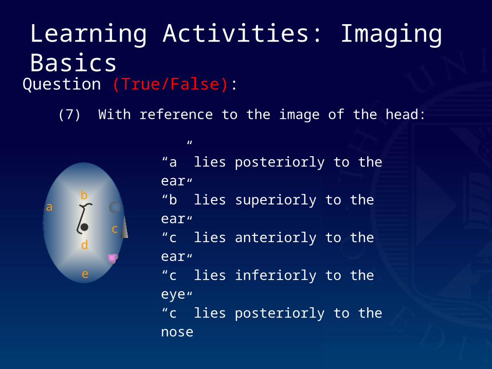
Question (True/False):
(7) With reference to the image of the head:
ab
cd
e
“a” lies posteriorly to the ear
“b” lies superiorly to the ear
“c” lies anteriorly to the ear
“c” lies inferiorly to the eye
“c” lies posteriorly to the nose

Learning Activities: Imaging Basics
Answer:
(7) With reference to the image of the head:
ab
cd
e
“a” lies posteriorly to the ear True
“b” lies superiorly to the ear True
“c” lies anteriorly to the ear True
“c” lies inferiorly to the eye True
“c” lies posteriorly to the nose True

Learning Activities: Imaging Basics
Question (Drag & drop):
(8) Place the labels adjacent to the appropriate images:
Upright or erect
Prone
Supine
Ground or Floor

Learning Activities: Imaging Basics
Answer:
(8) Place the labels adjacent to the appropriate images:
Ground or Floor
Prone Upright or erect Supine

Learning Activities: Imaging Basics
Question (Drag & drop):
(9) Place any applicable labels for this conventionally displayed frontal skull X-ray in the appropriate locations:
Inferior
Left
Right
Superior
Posterior
Anterior

Learning Activities: Imaging Basics
Answer:
(9) Place any applicable labels for this conventionally displayed frontal skull X-ray in the appropriate locations:
Inferior
LeftRight
Superior
Posterior
Anterior

Learning Activities: Imaging Basics
Question (Drag & drop):
(10) Place any applicable labels for this conventionally displayed axial head CT image in the appropriate locations:
Inferior
Left
Right
Superior
Posterior
Anterior

Learning Activities: Imaging Basics
Answer:
(10) Place any applicable labels for this conventionally displayed axial head CT image in the appropriate locations:
InferiorLeftRight
Superior
Posterior
Anterior

Learning Activities: Imaging Basics
Question (Fill in the blank):
(11) Using conventional imaging terminology, the projection of the skull X-ray illustrated below is: ________________

Learning Activities: Imaging Basics
Answer:
(11) Using conventional imaging terminology, the projection of the skull X-ray illustrated below is: lateral

Learning Activities: Imaging Basics
Question (Matrix):
(12) Select the orientation of the section defined by the orange plane:
Axial Coronal Sagittal
Axial Coronal Sagittal
Axial Coronal Sagittal
Axial Coronal Sagittal

Learning Activities: Imaging Basics
Answer:
(12) Select the orientation of the section defined by the orange plane:
Axial Coronal Sagittal
Axial Coronal Sagittal
Axial Coronal Sagittal
Axial Coronal Sagittal


![[This question paper contains 03 printed pages] …...Page 1 of 3 [This question paper contains 03 printed pages] Roll Number: _____ HPAS (Main) Examination-2018 POLITICAL SCIENCE](https://img.pdfslide.net/doc/110x75/5fb72868c4792a31e164ffc7/this-question-paper-contains-03-printed-pages-page-1-of-3-this-question-paper.jpg)


![[This question paper contains pages.] OLLF](https://img.pdfslide.net/doc/110x75/62a1686b83cb7f61ea249393/this-question-paper-contains-pages-ollf.jpg)


![[This question paper contains 04 printed pages] MATHEMATICS-I](https://img.pdfslide.net/doc/110x75/6215f18ff69c7e12c215aad7/this-question-paper-contains-04-printed-pages-mathematics-i.jpg)


![[This question.paper contains 04 printed pages] Roll Number/~~](https://img.pdfslide.net/doc/110x75/617758727f35d805aa6fabb3/this-contains-04-printed-pages-roll-number.jpg)
