Embed Size (px)
Citation preview

Three-Dimensional Structure ofN5-Carboxyaminoimidazole RibonucleotideSynthetase: A Member of the ATP Grasp Protein Superfamily†,‡
James B. Thoden,§ T. Joseph Kappock,| JoAnne Stubbe,*,| and Hazel M. Holden*,§
Department of Biochemistry, UniVersity of WisconsinsMadison, 1710 UniVersity AVenue, Madison, Wisconsin 53705, andDepartments of Chemistry and Biology, Massachusetts Institute of Technology, Cambridge, Massachusetts 02139
ReceiVed July 13, 1999; ReVised Manuscript ReceiVed August 27, 1999
ABSTRACT: Escherichia coli PurK, a dimeric N5-carboxyaminoimidazole ribonucleotide (N5-CAIR)synthetase, catalyzes the conversion of 5-aminoimidazole ribonucleotide (AIR), ATP, and bicarbonate toN5-CAIR, ADP, and Pi. Crystallization of both a sulfate-liganded and the MgADP-ligandedE. coli PurKhas resulted in structures at 2.1 and 2.5 Å resolution, respectively. PurK belongs to the ATP graspsuperfamily of C-N ligase enzymes. Each subunit of PurK is composed of three domains (A, B, and C).The B domain contains a flexible, glycine-rich loop (B loop, T123-G130) that is disordered in the sulfate-PurK structure and becomes ordered in the MgADP-PurK structure. MgADP is wedged between the Band C domains, as with all members of the ATP grasp superfamily. Other enzymes in this superfamilycontain a conservedΩ loop proposed to interact with the B loop, define the specificity of their nonnucleotidesubstrate, and protect the acyl phosphate intermediate formed from this substrate. PurK contains a minimalΩ loop without conserved residues. In the reaction catalyzed by PurK, carboxyphosphate is the putativeacyl phosphate intermediate. The sulfate of the sulfate ion-liganded PurK interacts electrostatically withArg 242 and the backbone amide group of Asn 245, components of the J loop of the C domain. Thissulfate may reveal the location of the carboxyphosphate binding site. Conserved residues within theC-terminus of the C domain define a pocket that is proposed to bind AIR in collaboration with an N-terminalstrand loop helix motif in the A domain (P loop, G8-L12). The P loop is proposed to bind the phosphateof AIR on the basis of similar binding sites observed in PurN and PurE and proposed in PurD and PurT,four other enzymes in the purine pathway.
In most textbooks the de novo biosynthetic pathway forthe production of purines involves 10 enzymatic activitiesand results in the conversion of phosphoribosyl pyrophos-phate to inosine monophosphate (1). The sixth step in thispathway is usually represented as a bicarbonate or CO2-dependent conversion of 5-aminoimidazole ribonucleotide(AIR)1 to 4-carboxy-5-aminoimidazole ribonucleotide (CAIR),catalyzed by AIR carboxylase. This representation of the
pathway has been validated in higher eukaryotes with CO2
supplying the additional carbon (2).In most prokaryotes, plants, and yeast, however, the
conversion of AIR to CAIR requires two gene products:PurK and PurE. PurK has been shown to catalyze theconversion of AIR, ATP, and bicarbonate toN5-car-boxyaminoimidazole ribonucleotide (N5-CAIR), ADP, andPi (3, 4). PurE catalyzes the interconversion ofN5-CAIR andCAIR (Scheme 1) (4, 5). The instability ofN5-CAIR, witha t1/2 of 15 s at pH 7.5 and 25°C, delayed its identificationand consequently the identification of PurK as anN5-CAIRsynthetase and PurE as an unusual mutase (4, 5). Thus inE.coli, yeast, and plants the purine pathway is composed of11 catalytic activities.
The structures of PurK and PurE are of interest for manyreasons. First, channeling, direct transfer by transient protein-
† This research was supported in part by grants from the NIH(DK47814 to H.M.H. and GM32191 to J.S.) and the NSF (BIR-9317398). T.J.K. was supported by NIH training grant CA09112.
‡ X-ray coordinates have been deposited in the Brookhaven ProteinData Bank (1b6r, sulfate complex; 1b6s, MgADP complex) and willbe released upon publication.
* To whom correspondence should be addressed: phone (617) 253-1841; fax (617) 258-7247; e-mail [email protected].
| University of WisconsinsMadison.§ Massachusetts Institute of Technology.1 Abbreviations: N5-CAIR, N5-carboxyaminoimidazole ribonucle-
otide; PurK,N5-carboxyaminoimidazole ribonucleotide synthetase; AIR,5-aminoimidazole ribonucleotide; CAIR, 4-carboxy-5-aminoimidazoleribonucleotide; PurE,N5-carboxyaminoimidazole ribonucleotide mutase;BC, biotin carboxylase; CPS, carbamoyl phosphate synthetase; PurT,glycinamide ribonucleotide formyltransferase (folate-independent);DDL, D-alanine:D-alanine ligase; GTS, glutathione synthetase; SynC,C-terminal domain of synapsin Ia; GAR, glycinamide ribonucleotide;PurD, glycinamide ribonucleotide synthetase; PurN, glycinamide ri-bonucleotide formyltransferase (folate-dependent); AMPPNP, 5′-ade-nylyl imidodiphosphate; PurF, phosphoribosylpyrophosphate amido-transferase.
Scheme 1
15480 Biochemistry1999,38, 15480-15492
10.1021/bi991618s CCC: $18.00 © 1999 American Chemical SocietyPublished on Web 10/30/1999
protein interactions of the chemically unstable intermediateN5-CAIR, may be a possibility between PurK and PurE.Second, the mechanism of the biotin-independent carboxy-lation catalyzed by PurK is of chemical interest as is theunprecedented migration of a CO2 catalyzed by PurE (5).Third, the evolutionary link between microbial (class I) andanimal (class II) PurEs, which utilize different substrates, isof interest (2). Recent structures of PurE (6) and PurK insulfate- and MgADP-bound forms presented here provide astarting point to obtain answers to these intriguing questions.
PurK (Scheme 1) in the presence of AIR, ATP, and [18O]-bicarbonate catalyzes quantitative18O transfer into [18O]Pi.This observation led to the proposal that bicarbonate isactivated by ATP for nucleophilic attack by the amino groupof AIR through the generation of a carboxyphosphateintermediate (or its decomposition products CO2 and Pi) (4).Carboxyphosphate generated from bicarbonate and ATP hasalso been implicated in the mechanisms of biotin carboxy-lases (BC) and the carbamate-forming step of carbamoylphosphate synthetase (CPS) (7-12). Recent crystallographicstudies have shown that BC and two domains of CPS arestructurally homologous and part of a new superfamily ofproteins containing an ATP grasp fold (13-16).
On the basis of sequence alignments, Galperin and Koonin(17) suggested that PurK belongs to this superfamily ofproteins as well. This superfamily also includesD-alanine:D-alanine ligase (DDL), glutathione synthetase (GTS), suc-cinyl-CoA synthetase, pyruvate phosphate dikinase, a syn-apsin domain (SynC), and most recently glycinamideribonucleotide (GAR) synthetase (PurD), the second enzymein the purine biosynthetic pathway (18-22). All of the C-Nligases in this superfamily utilize MgATP and are likely toinvolve phosphoanhydride intermediates.
Here we describe the three-dimensional structures ofsulfate-liganded and MgADP-ligandedE. coli PurK refinedby X-ray crystallographic analysis to 2.1 and 2.5 Å resolu-tion, respectively. As anticipated, the molecular fold of PurKis structurally homologous to the ATP grasp superfamily. Amononucleotide (AIR) binding site in PurK is proposed onthe basis of conservation of PurK residues and structuralhomology with PurD. In turn, PurD’s mononucleotidebinding site was identified (22) by structural homology withmononucleotide-liganded PurN, the folate-dependent GARtransformylase (23). A comparison of PurK with other ATPgrasp family members is presented.
MATERIALS AND METHODS
Protein Purification and Crystallization. PurK was purifiedaccording to a previously published procedure (3) with theexception that the enzyme was dialyzed against 50 mM Tris(pH 8) for 8 h at 4°C prior to the final chromatographicstep with DEAE-Sepharose. Salt and buffer levels werereduced by dilution and reconcentration with a YM30membrane. The enzyme employed for the crystallizationtrials had a specific activity of 55 units/mg at 37°C and pH8.0 in the AIR-dependent ATPase assay where one unit isdefined as 1µmol of ADP formed/min.
For the initial sparse matrix crystallization screens of nativePurK, a stock solution of enzyme (at 36 mg/mL in 10 mMTris, pH 7.8, and 30 mM KCl) was diluted to 20 mg/mLwith 10 mM PIPES (pH 7.0). These screens, which employed
the hanging drop method of vapor diffusion, were conductedat both 4°C and room temperature. Small crystals appearedat both temperatures with ammonium sulfate as the precipi-tant. The crystal habits were better defined at room temper-ature and hence the precipitant conditions were optimizedat ∼22 °C. All of the crystals employed for the structuralanalysis of the native enzyme were grown by macroseedinginto small batch setups containing 0.525 M ammoniumsulfate and 100 mM HEPPS (pH 7.8). The protein concen-tration was typically 10 mg/mL.
Crystals of the native enzyme belonged to the space groupC2221 with unit cell dimensions ofa ) 93.4 Å,b ) 95.2 Å,and c ) 120.6 Å and one monomer per asymmetric unit.The Matthews’ coefficient orVm for these crystals was 3.4Å3/Da, which corresponded to a solvent content of ap-proximately 64%.
Crystals of the enzyme/MgADP complex were grown bybatch methods at room temperature as well. For theseexperiments, the precipitant was poly(ethylene glycol) 8000(PEG) and the protein sample, at a concentration of 10 mg/mL, contained 100 mM HEPES (pH 7.5), 125 mM KCl, 5.0mM MgCl2, and 5 mM 5′-adenylyl imidodiphosphate (AMP-PNP) (24). Rod-shaped crystals typically appeared from8-17% (w/v) PEG after 1-2 months. The crystals belongedto the space groupP1 with unit cell dimensions ofa ) 60.6Å, b ) 92.1 Å, c ) 102.6 Å,R ) 66.1°, â ) 82.7°, andγ) 81.8°. The asymmetric unit contained two complete PurKdimers. Once the structure of the complex was solved, itbecame clear that AMPPNP had been hydrolyzed during thecrystallization experiments as only MgADP remained in theactive site.
Preparation of HeaVy-Atom DeriVatiVes for the NatiVeEnzyme and X-ray Data Collection and Processing.For thepreparation of heavy-atom derivatives, the native crystalswere first transferred to solutions containing 0.75 M am-monium sulfate and 100 mM HEPPS (pH 7.8). Four heavyatom derivatives were readily prepared by soaking thecrystals in solutions containing 1 mM methylmercury acetatefor 3 h, 1 mM 2,5-bis(chloromercuri)furan for 6 h, 1 mMmeso-1,4-bis(acetoxymercuri)-2,3-diethoxybutane for 3 h, or10 mM K2PtCl4 for 60 h. X-ray data from the native andheavy-atom derivative crystals were collected at-2.5 °Cwith a Bruker AXS HiStar area detector system. The X-raysource was Ni-filtered Cu KR radiation from a RigakuRU200 X-ray generator operated at 50 mV and 90 mA andequipped with double focusing mirrors. Only one crystal wasrequired for each X-ray data set to 2.7 Å resolution. Friedelpairs were measured for each of the heavy-atom derivativex-data sets. All data sets were processed with the softwarepackage XDS (25, 26) and internally scaled with the programXCALIBRE (G. Wesenberg and I. Rayment, unpublishedsoftware). Relevant X-ray data collection statistics are givenin Table 1. The heavy-atom derivative data sets were scaledto the native by a “local” scaling procedure developed byDrs. G. Wesenberg, W. Rypniewski, and I. Rayment. TheR-factors (based on amplitudes) between the native and themethylmercury acetate, 2,5-bis(chloromercuri)furan,meso-1,4-bis(acetoxymercuri)-2,3-diethoxybutane, and K2PtCl4data sets were 23.9%, 15.7%, 16.8%, and 18.8%, respec-tively. Once the structure had been determined to 2.7 Åresolution, a final native X-ray data set was collected to 2.1Å resolution by using three crystals. This particular data set
Structure ofN5-CAIR Synthetase (PurK) Biochemistry, Vol. 38, No. 47, 199915481

was processed with the data reduction software packageSAINT (Bruker AXS INC) and internally scaled withXCALIBRE. The X-ray data set was 95.6% complete to 2.1Å resolution. Relevant statistics for this data set are alsopresented in Table 1. X-ray data from a single crystal of thePurK/MgADP complex were collected in a manner similarto that described above and processed with SAINT andXCALIBRE. Relevant statistics are given in Table 1.
Structural Determination and Least-Squares Refinementof the NatiVe PurK. The positions of the heavy atom bindingsites were determined by inspection of difference Pattersonmaps calculated with X-ray data from 30 to 5.0 Å. Themethylmercury acetate, 2,5-bis(chloromercuri)furan,meso-1,4-bis(acetoxymercuri)-2,3-diethoxybutane, and K2PtCl4derivatives contained two, six, one, and five heavy-atombinding sites, respectively. The heavy-atom derivatives wereplaced on a common origin by difference Fourier maps andthe positions and occupancies for each heavy-atom bindingsite were refined by the origin-removed Patterson-functioncorrelation method to 2.7 Å resolution (27, 28). All themercury-based derivatives had one common binding site nearCys 5, but the exact locations of these heavy atoms werenot identical. Anomalous difference Fourier maps calculatedfrom 30 to 5.0 Å were employed for determining the correcthand of the heavy-atom constellation. Protein phases werecalculated with the program HEAVY (27) and relevant phasecalculation statistics can be found in Table 1.
An electron density map calculated to 2.7 Å resolutionclearly revealed the overall course of the polypeptide chain.The map was subsequently improved by the technique ofsolvent flattening (29) as implemented by Dr. W. Kabsch,Heidelberg, Germany. From the density-modified map it waspossible to trace the entire polypeptide chain contained withinthe asymmetric unit. The PurK subunit contains 355 aminoacid residues (30, 31). There was one break in the proteinbackbone delineated by Thr123-Gly130. Additionally, inthe final electron density map the side chain correspondingto Phe41 was disordered with only enough electron densityto accommodate an alanine residue. According to thepublished amino acid sequence, residue 205 is a glutamine(30, 31). The electron density map, however, clearly revealedthe presence of an arginine at position 205 that forms a saltbridge with Glu208. Resequencing of the DNA confirms thisassignment. The model was refined by least-squares analysisto 2.1 Å resolution with the software package TNT (32).Relevant refinement statistics are given in Table 2. The finalR-factor was 19.6% for all measured X-ray data from 30 to2.1 Å resolution. In addition toR-factor values and the overallgeometry of the model, perhaps one of the strongest
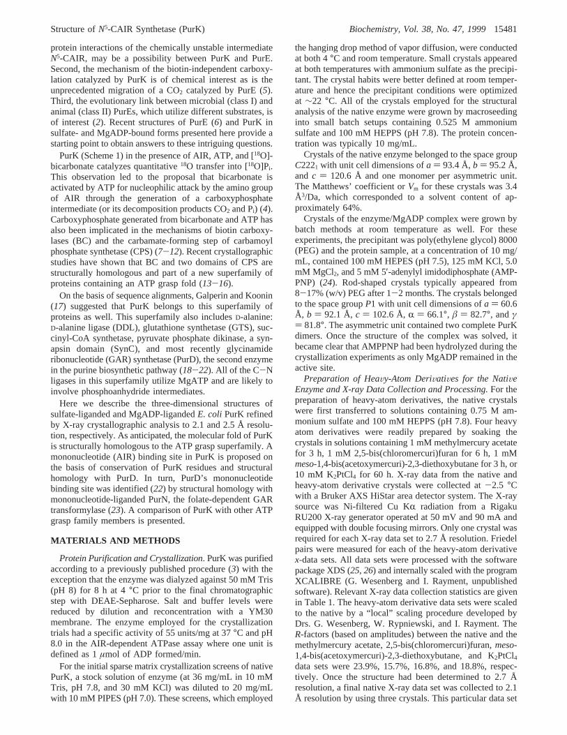
indications of model quality is a plot of the polypeptide chainbackbone dihedral angles. A Ramachandran diagram of suchtorsional angles is presented in Figure 1A, and as can beseen there are no significant outliers. A representative portionof the electron density map near the region surrounding theobserved sulfate anion is given in Figure 1B. In addition tothis sulfate ion, there were 92 water molecules included inthe refinement and these had an averageB-value of 49.0 Å2.The averageB-value for the polypeptide chain backboneatoms was 41.7 Å2.
Structural Determination and Least-Squares Refinementof the PurK/MgADP Complex. The structure of the PurK/MgADP complex was solved by molecular replacement withthe software package AMORE (33) with the native PurKdimer employed as a search model. A search sphere radiusof 25 Å was utilized. Both the cross-rotational functions andtranslational searches were conducted with X-ray data from20 to 4.0 Å. Two solutions were obtained corresponding tothe two dimers in the asymmetric unit. Following rigid-bodyrefinement with X-ray data from 20 to 4.0 Å, the correlationcoefficient was 65.3% and theR-factor was 38.9%. Toexpedite the model building process and to remove bias dueto the molecular replacement technique, the four subunitsin the asymmetric unit were averaged according to thealgorithm of Bricogne (34). From this averaged electrondensity map, one subunit of the PurK/MgADP complex wasconstructed. This averaged model was placed back into theunit cell and subjected to alternate cycles of least-squaresrefinement and model building. Relevant refinement statisticsare given in Table 2. In each subunit, there was anintramolecular disulfide bridge between Cys146 and Cys150.
Table 1: Data Statistics
data setresolution
(Å)independentreflections
completeness(%)
avgI/avgσ (I)
Rsyma
(%)Riso
b
(%)phasingpowerc
native I 30-2.7 14 589 96.4 31.5 4.3methylmercury acetate 30-2.7 14 719 97.3 13.6 5.6 23.9 1.062,5-bis(chloromercuri)furan 30-2.7 14 492 95.8 10.4 6.8 15.7 1.30meso-1,4-bis(acetoxymercuri)-2,3-diethoxybutane 30-2.7 13 726 90.7 13.2 4.6 16.8 1.10K2PtCl4 30-2.7 14 000 92.5 8.3 6.3 18.8 1.30
native II 30-2.1 30 332 95.6 24.3 4.7Mg2+ADP complex 30-2.5 66 345 95.7 12.6 7.2
a Rsym ) (∑|I - ⟨I⟩|/∑ I) × 100. b Riso ) (∑|Fh| - |Fn|)/∑n Fn × 100, whereFh andFn are the heavy atom and native structure factors, respectively.c Phasing power is the ratio of the root-mean-square heavy-atom scattering factor amplitude to the root-mean-square lack of closure error.
Table 2: Least-Squares Refinement Statistics
PurK/sulfatecomplex
PurK/MgADPcomplex
resolution limits (Å) 30.0-2.1 30.0-2.5R-factora (%) 19.6 18.5no. of reflections used 30 332 66 345no. of protein atoms 2737 11137no. of solvent atomsb 97 495
Weighted Root-Mean-Square Deviations from Idealitybond length (Å) 0.013 0.013bond angle (deg) 2.33 2.23planarity (trigonal) (Å) 0.005 0.006planarity (other planes) (Å) 0.011 0.013torsional anglec (deg) 17.6 19.3a R-factor ) ∑|Fo - Fc|/∑|Fo|, whereFo is the observed structure-
factor amplitude andFc is the calculated structure-factor amplitude.b These include 92 waters and one sulfate ion in the native model and491 waters and four magnesium ions in the complex model.c Thetorsional angles were not restrained during the refinement.
15482 Biochemistry, Vol. 38, No. 47, 1999 Thoden et al.
A Ramachandran plot of all nonglycinyl main-chain dihedralangles is displayed in Figure 2A, and electron densitycorresponding to the nucleotide in subunit II of the asym-metric unit is shown in Figure 2B.
RESULTS AND DISCUSSION
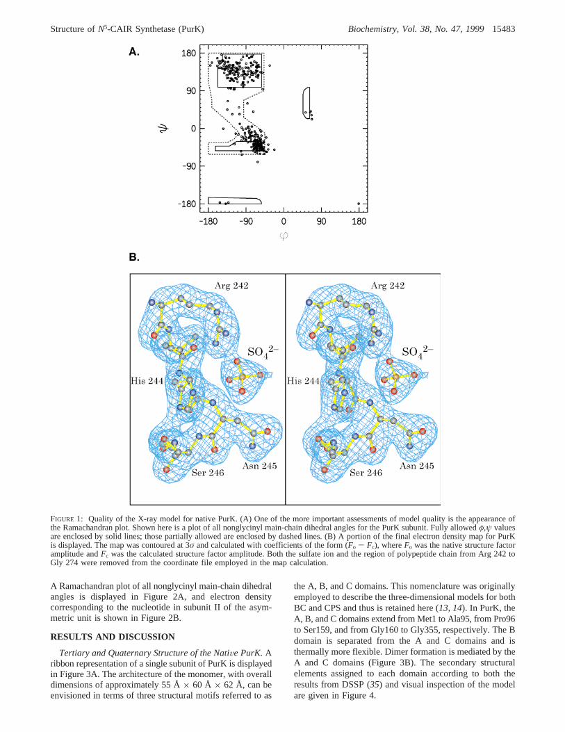
Tertiary and Quaternary Structure of the NatiVe PurK.Aribbon representation of a single subunit of PurK is displayedin Figure 3A. The architecture of the monomer, with overalldimensions of approximately 55 Å× 60 Å × 62 Å, can beenvisioned in terms of three structural motifs referred to as
the A, B, and C domains. This nomenclature was originallyemployed to describe the three-dimensional models for bothBC and CPS and thus is retained here (13, 14). In PurK, theA, B, and C domains extend from Met1 to Ala95, from Pro96to Ser159, and from Gly160 to Gly355, respectively. The Bdomain is separated from the A and C domains and isthermally more flexible. Dimer formation is mediated by theA and C domains (Figure 3B). The secondary structuralelements assigned to each domain according to both theresults from DSSP (35) and visual inspection of the modelare given in Figure 4.
FIGURE 1: Quality of the X-ray model for native PurK. (A) One of the more important assessments of model quality is the appearance ofthe Ramachandran plot. Shown here is a plot of all nonglycinyl main-chain dihedral angles for the PurK subunit. Fully allowedφ,ψ valuesare enclosed by solid lines; those partially allowed are enclosed by dashed lines. (B) A portion of the final electron density map for PurKis displayed. The map was contoured at 3σ and calculated with coefficients of the form (Fo - Fc), whereFo was the native structure factoramplitude andFc was the calculated structure factor amplitude. Both the sulfate ion and the region of polypeptide chain from Arg 242 toGly 274 were removed from the coordinate file employed in the map calculation.
Structure ofN5-CAIR Synthetase (PurK) Biochemistry, Vol. 38, No. 47, 199915483

The A-domain contains three strands of parallelâ-sheetthat range in length from four to seven residues and fourdistinct R-helical regions composed of 5-10 amino acids.Additionally, there are numerous classical reverse turnsconnecting these secondary structural elements. The molec-ular axes of the third (Phe74-Ala78) and fourth (Arg80-Phe87)R-helices in the A-domain are oriented at approxi-mately 90° with respect to one another and are connectedby one residue, Asp 79, which adopts dihedral angles ofφ
) -95.0° and ψ ) 119.8°. This type of tertiary pattern,namely, a helix-residue-helix motif, appears to be astructural hallmark for enzymes belonging to the ATP graspsuperfamily of proteins and serves as the bridge between
the A- and B-domains. Similar dihedral angles are observedfor the single connecting residues between the equivalenttwo R-helices in DDL (Asp96,φ ) -103°, ψ ) 104°), GTS(Asn 123,φ ) -70°, ψ ) 142°), BC (Asp 115,φ ) -113°,ψ ) 95°), both synthetase domains of the large subunit ofCPS (Asp 128,φ ) -81°, ψ ) 121°, and Asp 674,φ )-84°, ψ ) 119°), and PurD (Ser 104,φ ) -123°, ψ )111°).
A structural homology search of the Brookhaven ProteinData Bank using the A domain of PurK and the programDALI ( 36), revealed many Rossmann fold-containing pro-teins including the N-terminal domain of the third enzymein the purine biosynthetic pathway: GAR formyltransferase
FIGURE 2: Quality of the X-ray model for the PurK/MgADP complex. Shown in panel A is a plot of all nonglycinyl main-chain dihedralangles for the four subunits of PurK contained within the asymmetric unit. Electron density corresponding to the nucleotide in subunit IIis displayed in panel B. The map was contoured at 3σ and calculated with coefficients of the form (Fo - Fc), whereFo was the nativestructure factor amplitude andFc was the calculated structure factor amplitude. The nucleotide was not included in the coordinate fileemployed for the map calculation.
15484 Biochemistry, Vol. 38, No. 47, 1999 Thoden et al.
(PurN) and the A domain of PurD.2 The structure of PurNhas been solved in the presence of GAR (23). The structuralhomology of PurN with PurK and PurD allows an excellentguess about the mononucleotide (AIR andN5-CAIR) bindingsite on PurK. The phosphate is proposed to interact with theP loop (G8-L12) of the N-terminal strand loop helix (Figures3A and 4B). The P loop region consensus by alignment of22 full-length PurK sequences is G8[G/N/D]GQL12. This loopappears to provide the flexibility for phosphate binding andstabilization by the adjacent helixR1 dipole. A similarmononucleotide binding motif has been established structur-ally for two additional enzymes in the purine pathway: PurF
(37) and recently PurE (6). Thus five of the 11 enzymes inthis pathway have a similar binding site for a ribose5-phosphate moiety, common to all intermediate metabolitesin this pathway.
The B-domain, depicted in green in Figure 3A, containsfour strands of antiparallelâ-sheet ranging in length fromthree to nine amino acid residues (Figure 4B). Thisâ-sheetis flanked on one side by twoR-helices formed by Arg103-Leu113 and Ala136-Gln141. The second helix in thisdomain (R6) is decidedly distorted due to Glu138, whichadopts dihedral angles ofφ ) -84.6° and ψ ) 3.6°. Theregion of polypeptide chain from Thr123 to Gly130 isdisordered in the present structure and is referred to as theB loop. In 24 full-length or partial PurK sequences, the Bloop consensus sequence is X123X[G/A]YDG[R/K/Q/H]G 130.
2 The A domain of PurD was referred to as the N domain in theprevious paper (22).
FIGURE 3: Tertiary and quaternary structure of PurK. A single PurK subunit is shown in panel A. The A, B, and C domains of the subunitare color-coded in yellow, green, and blue, respectively. A sulfate molecule observed in the electron density map is displayed in a ball-and-stick representation. The ribbon representation of the PurK dimer shown in panel B is oriented perpendicular to the crystallographica-axis. Those amino acid side chains involved in electrostatic interactions within the subunit-subunit interface are depicted as ball-and-sticks.
Structure ofN5-CAIR Synthetase (PurK) Biochemistry, Vol. 38, No. 47, 199915485

All members of the ATP grasp superfamily whosestructures have been determined in the absence of a ligandhave disordered B loops. In the case of the availablenucleotide liganded structures these B loops are generallyordered. Structural alignments of these loops, which are ingeneral Gly- and Ser-rich, reveal that the PurK B loop hasone additional residue (Figure 5A). In the available ligandedstructures, the amide hydrogens of the two C-terminalresidues [MGG or GSS, 4 and2 in Figure 5A] participatein hydrogen-bonding interactions with theâ- andγ-phosphategroups of the nucleotide. In PurK the corresponding sequenceY126DG128 is unique in that one side chain is large andanother is charged. In the structure of sulfate-liganded PurK,Tyr126 makes an unusual salt bridge with Arg129. Asp127might function as a metal ligand, as discussed below. Most
members of the ATP grasp superfamily also possess a secondloop (called theΩ loop, Figure 6A) in their C domain thatinteracts with the B loop, providing a protective face overthe â- and γ-phosphates of ATP and the putative acylphosphate intermediate. As noted below, thisΩ loop is veryshort in the PurK structure. Hence the Tyr and Asp of PurK’sB loop may play an important role in sequestration of theactive site in conjunction with conserved residues includingthe J loop (Ala240-His244) of the C domain (Figure 6,discussed subsequently).
The C-domain, the most complicated of the three domains(Figures 3A and 4B), is dominated by a twisted eight-stranded antiparallelâ-sheet that forms a basket to cradlethe A- and B-motifs. Theseâ-strands range in length from4 to 11 amino acids with an average length of eight residues.
FIGURE 4: Secondary structure and strictly conserved amino acid residues in PurKs. (A) Sequence ofE. coli PurK. The white letters inblack boxes are conserved among 22 full-length PurK sequences. Black letters in white boxes are conserved among 21 PurK sequences.Triangles indicate where any insertion is required in theE. coli sequence to generate the indicated consensus sequence. (B) Topologydiagram of the secondary structure of the A, B, and C domains of PurK. For clarity, the linker betweenâ7 andâ8 is not shown, and theC domain has been moved to the right of the A and B domains.
15486 Biochemistry, Vol. 38, No. 47, 1999 Thoden et al.

The third, fourth, and fifthâ-strands (â10-â12) are inter-rupted by bulges resulting from the dihedral angles adoptedby Arg 190 (φ ) -115.4°, ψ ) -37.6°), Gly 221 (φ )125.9°, ψ ) -163.4°), and Asn 237 (φ ) -98.3°, ψ )-64.6°). There are also fourR-helices and numerous regionsof reverse turns distributed throughout the C-domain.
The C domain varies in size and complexity amongmembers of the ATP grasp superfamily (22). However, inall of the structurally well-characterized proteins, the C-domain J loop provides a structurally conserved strand-loopstructure (Figures 5B and 6C) that cradles the ATP. It iscalled a J loop because of the strand-loop structure’sshape: the loop hooks to the right as it extends fromâ12.The conservation of residues within this motif is high withinenzymes but not among superfamily members. The con-served residues in PurK are shown in boldface type (Figure5B), as are the conserved residues within other members ofthis superfamily. Several conserved features from PurK areimportant to note: Arg242 interacts electrostatically with twoof the oxygens of the sulfate from the sulfate-liganded PurK.The sulfate also interacts with the backbone amide ofconserved Asn245. Glu238 is conserved not only withinPurKs but among all of the members of this superfamily.From the Mg-nucleotide liganded structures available (20,38), this Glu is known to be a metal ligand. One feature thatis strikingly different between PurK and the other superfamilymembers is Ala240. This residue is an Asn and a knownmetal ligand in the other family members, except SynC.3 In
the case of PurK, portions of the C domain including this Jloop and the P loop from the A domain, unique to the purineenzymes, form the binding pocket for the mononucleotidesubstrate AIR and will be discussed subsequently.
The differences in complexity of the C domains of thesuperfamily members are most apparent in the regionC-terminal to the J loop. In DDL, GTS, SynC, and the twodomains of CPS, the J loop is near the end of the C domain.In PurK, PurD, and BC, an additional globular featurefollows the J loop (residues 247-355 in PurK). In the caseof PurK this feature appears to contribute a significant portionof the AIR binding site. A comparison of all of the structuresof this superfamily reveals that, aside from the proteintopology, the J loop is the only common feature in theC-domain.
Dimer Interface of PurK.PurK is known to function as adimer in solution (3). In the crystals employed in thisinvestigation, the dimeric protein is packed in the unit cellwith its molecular dyad coincident with a crystallographic2-fold, thereby leading to an asymmetric unit containing onlyone subunit. Shown in Figure 3B is a ribbon representationof the PurK dimer viewed perpendicular to the crystal-lographic a-axis. The dimer has overall dimensions ofapproximately 55 Å× 95 Å × 88 Å and a subunit-subunitburied surface area of 2850 Å2 as calculated according tothe method of Lee and Richards (40) with a probe sphere of1.4 Å. For comparison, the buried surface area of BC, alsoknown to be a dimer and similar in structure to PurK, is2600 Å2 (13). The specific manners by which the subunitsof PurK and BC associate to form functional dimers,however, are quite different even though in both cases theB-domains extend away from the main bodies of the proteins.PurD and PurT (a formate-dependent GAR transformylase),members of this superfamily and of the purine biosyntheticpathway, are monomers (41, 42).
There are four specific regions of polypeptide chain ineach PurK subunit that are involved in maintaining the properquaternary structure of the protein. One of these regions insubunit I of the dimer involves those residues lying in thefirst R-helical region of the A-domain as can be seen inFigure 3B. Here there are numerous hydrogen bonds betweenthe two subunits including those formed by O of Pro22(subunit I) and N of Trp304 (subunit II), O of Leu23 (subunitI) and Oη of Tyr292 (subunit II), and Nε2 of Gln18 (subunitI) and Oε1 of Glu21 (subunit II). Additionally, there is a saltbridge formed between the guanidinium group of Arg17(subunit I) and the carboxylate group of Glu21 (Subunit II).The second region of subunit-subunit contact occurs in thearea delineated by Ser255 and Asn277. Specific electrostaticinteractions are formed between Oγ of Ser255 (subunit I)and O of Pro289 (subunit II), Nη2 of Arg264 (subunit I) andO of Leu295 (subunit II), Nδ2 of Asn277 and Oδ2 of Asp322,and Oδ1 of Asn277 (subunit I) and Nη2 of Arg327 (subunitII). By the local symmetry of the dimer, the third and fourthareas of contact provided by subunit I are necessarily formedby Tyr292-Asp306 and Asp322-Arg327. In addition tothese specific electrostatic interactions, there are numerouswater molecules distributed throughout the subunit-subunitinterface.
Location of the ActiVe Site of PurK. As noted above, thestructures of MgADP and sulfate-liganded PurK define partof its active-site cleft. Additional insight into the active site,
3 A crystal structure of SynC contains Ca2+ and ATP (39). Thefunction of this synapsin and whether ATP is hydrolyzed remainunknown.
FIGURE 5: Comparison of B loop and J loop consensus sequencesfrom a structure-based alignment of four ATP grasp proteins. Ineach panel, secondary structure assignments from PurK areindicated. Boldface residues are positions conserved within eachenzyme. Sequence ranges are indicated (E. coli numbering, exceptSynC fromB. taurus). A sequence from the N-terminal carbamate-generating domain from the large subunit of CPS is shown (CPSN).(A) Comparison of B loop sequences in four ATP grasp proteinswith structures where a nucleotide is bound and the B loop is visible.Solid triangles indicate positions observed to contact ADP, and theopen triangle indicates a position expected to make a backboneNH contact with theâ- andγ-phosphates in ATP. (B) Comparisonof J loop sequences in six crystallographically characterized ATPgrasp enzymes. The solid triangle is a ligand to metal 1, and theshaded triangle is a ligand to metal 2 in some enzymes (boxedAsn residues).
Structure ofN5-CAIR Synthetase (PurK) Biochemistry, Vol. 38, No. 47, 199915487

including the AIR binding site, is provided by the locationof the 34 strictly conserved residues of∼355 amino acidsof PurK in 22 full-length sequences available (Figure 6). Asshown in Figure 5, the B loop region in the B domain andthe strand-J loop in the C domain contain five and eight
conserved residues, respectively. In addition, the P loop andthe adjacent helixR1 (five conserved residues) in the Adomain are proposed to bind the phosphate of AIR andprovide part of AIR’s binding pocket. The remainder ofAIR’s binding pocket is proposed to be provided by nine of
FIGURE 6: Active site of PurK. (A) Close-up showing the B loop (blue), theΩ loop (yellow), the P loop (red), and the strand-J loop (green)using the MgADP structure. Both sulfate and the MgADP are shown. The sulfate has been superimposed on the MgADP structure. (B)Molecular surface of PurK in the same orientation as in panel A. Surface-exposed conserved residues are colored green and the sulfatebinding site is shown (CPK model); in this view MgADP is mostly obscured by the B loop. This view provides a glimpse at the putativeAIR binding pocket. The figure was prepared with GRASP (50). (C) Closer view of the structure in panel A with the conserved residueslabeled. All of the conserved residues (Figure 4A) within the active site are depicted in ball-and-stick representations. The partially conservedGlu49 (Asp) and Ser247 (Thr) are shown with white C atoms. The view is tilted forward 45° relative to the view in panels A and B, andthe B domain has been removed for clarity. Panels A and C were prepared with Molscript (51) and Raster3D (52).
15488 Biochemistry, Vol. 38, No. 47, 1999 Thoden et al.

the 10 conserved residues in the C-terminus of the C domain(i.e., after the J loop in primary sequence, Figure 4A). Thisregion nuzzles up against the P-loop helixR1 in the Adomain and together they are proposed to form the AIRbinding pocket. A picture of the putative AIR binding pocketwith the B domain removed for clarity is shown in Figure6C.
Carboxyphosphate Binding Site.The sulfate binding siteof the sulfate-liganded PurK could be indicative of theγ-phosphate binding site of ATP, the binding site of theproduct phosphate, or the phosphate binding site of themononucleotide substrate, AIR. As discussed above, webelieve that the phosphate binding site of the mononucleotideresides in the A domain. Thus this last possibility seemsunlikely. A clue as to the function of the sulfate binding sitein PurK may be provided by the phosphate-liganded struc-tures of BC and the carbamate-forming domain of CPS. Inthese proteins, the phosphate ions are located in nearlyidentical positions to the sulfate in PurK and interact withan Arg side chain and a backbone amide group [Arg292 andVal295 in BC (13); Arg303 and Arg306 in CPS (43)]. Sincethe reaction mechanisms of PurK, BC, and the carbamate-forming domain of CPS all require bicarbonate and arethought to proceed through a carboxyphosphate intermediate,one can speculate that this site is involved in bindingbicarbonate and/or the carboxyphosphate intermediate.
MgADP and MgATP Binding Site.In attempt to moreclearly define the ATP binding site of PurK, crystals of theenzyme complexed with MgAMPPNP were prepared andthe structure solved. As described in the Materials andMethods section, the crystals belong to the space groupP1and contained four complete subunits in the asymmetric unit.For the sake of simplicity, only subunit II is discussed heresince the electron density corresponding to this polypeptidechain is the best-ordered.
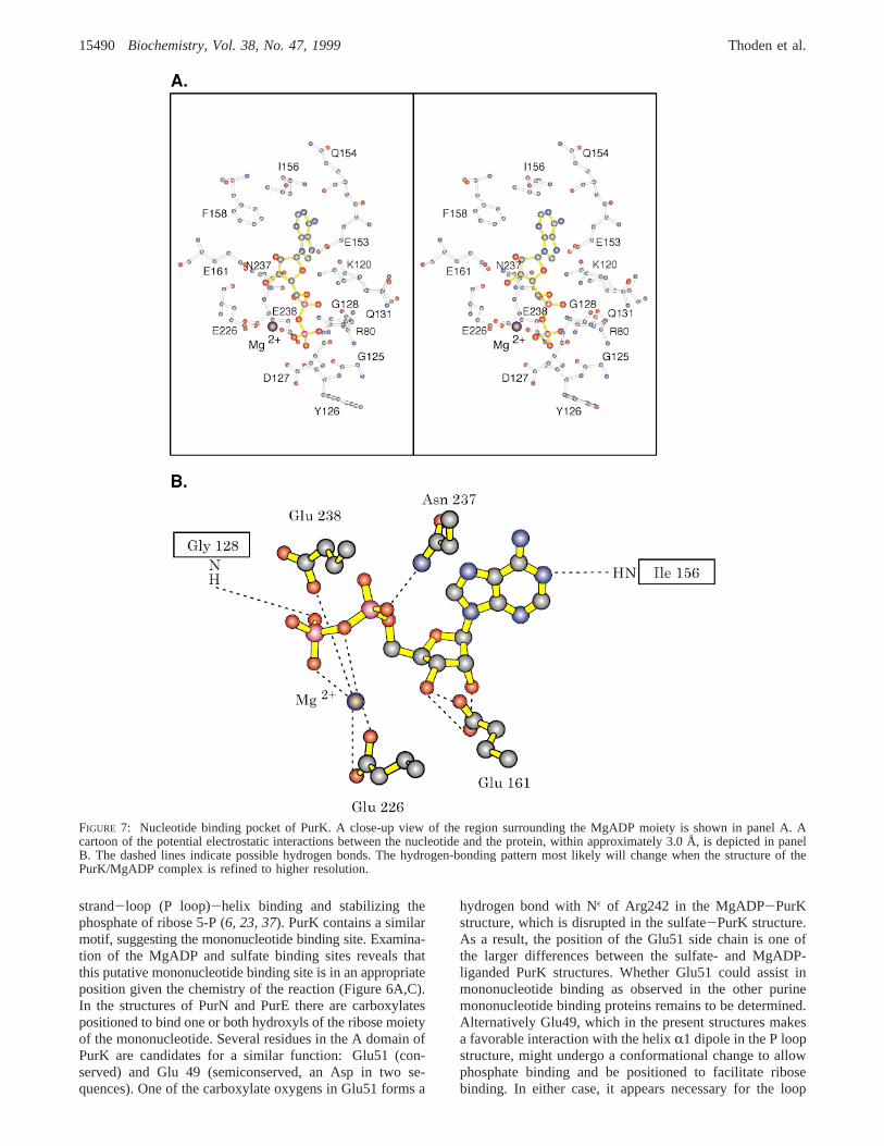
AMPPNP was chosen for cocrystallization due to itspresumed stability in the absence of the AIR. In fact, duringthe course of the crystallization experiments, the ligand washydrolyzed to MgADP as can be seen in Figure 7 (18). Froma detailed structural analysis of CPS, it is known that theB-domains in the large subunit of the enzyme close downtightly when either AMPPNP (and presumably ATP as well)or ADP/Pi is bound in the active sites (15, 16). Indeed, thetrigger for the B-domain closure in CPS appears to be theformation of hydrogen bonds between theγ-phosphates ofthe nucleotide moieties and the backbone amide nitrogensof glycine residues occupying the second and third positionsin type III′ reverse turns in the B loop. Strikingly, some ofthe atoms in the B-domains of CPS move by more than 7 Å(16). This transformation of the B loop from a disordered toan ordered state concomitant with a conformational changehas also been observed for GTS, for which both open andclosed structures of this protein are available (21, 38).
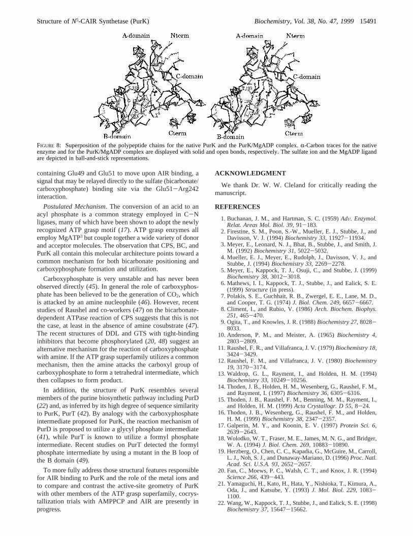
Since only MgADP is observed binding in the PurK activesite, rather than a combination of nucleoside diphosphate andinorganic phosphate, the B-domain closure is not as extensiveas that observed in CPS or GTS. Still, there is significantmovement with some atoms moving by more than 3.0 Å, ascan be seen in Figure 8. The major structural differencesbetween the two forms of PurK presented here are confinedto a region within the B domain, residues Leu100-Val152.Excluding this region, however, the polypeptide chains for
the two models are remarkably similar, such that theirbackbone atoms superimpose with a root-mean-square devia-tion of 0.43 Å. The actual conformation of the B loop maychange further upon binding of a Mg-nucleoside triphosphate.
Shown in Figure 7 is a close-up view of that portion ofthe PurK active site responsible for positioning the nucle-otide. The adenine ring is linked to the protein via a hydrogenbond to N-1 contributed by the backbone amide group ofIle156 and packed against a hydrophobic pocket formed byIle156 and Phe158. In addition, the 6-amino group appearsto be hydrogen-bonded to the carbonyl of Glu154 and oneof the carboxylate oxygens of Glu153. The ribose ring adoptsa C3′-endopucker with its 2′- and 3′-hydroxyl groups lyingwithin hydrogen-bonding distance of the carboxylate sidechain of Glu161. Arg80 interacts electrostatically with theR- andâ-phosphates of ADP, and Lys120 interacts with theR-phosphate and adenine N-7. At a resolution of 2.5 Å it isnot possible to unambiguously define the coordinationgeometry around the putative Mg ion observed in the electrondensity map for PurK. In PurK, Mg is ligated by a phosphoryloxygen and a bridging oxygen from the nucleotide, Oε1 andOε2 of Glu226 and Oε2 of Glu238. While the definition ofthe coordination geometry will change upon refinement ofthe model at higher resolution, it is clear that both Glu226and Glu238 act as ligands to the metal ion.
The availability of several high-resolution structures ofmembers of the ATP grasp superfamily with MgADP andMgATP bound allow similarities and differences in thenucleotide binding site to be discussed. In almost all caseseither the 2′ or 3′ hydroxyls of the ribose of ADP or ATPare bound to a conserved Glu (Glu161 in PurK). In moststructures either one or two Mg or Mn ions interact with theâ- andγ-phosphates of the nucleotides with two conservedmetal ion binding residues in the strand-J loop region: EXN(Figure 5B). For no ATP grasp enzyme is a detailedunderstanding of the role of the metal(s) in catalysisavailable. As we discussed above, the first metal bindingmotif in PurK is E238XA240, raising the possibility that theremay not be a second metal binding site in PurK, which wouldbe a unique situation for an ATP grasp enzyme known tohydrolyze ATP.3 Asp 127, a conserved residue at the tip ofthe B loop, is near the ADPâ-phosphate (Figure 7) and mightfunction as a ligand to a second metal.4
In most superfamily members, the charges on the phos-phates of the nucleotide are in part neutralized by threepositively charged Lys or Arg residues: one from the Adomain helix-residue-helix kink (Arg80 in PurK), one fromthe underside of the B domain (Lys120 in PurK), and onefrom the Ω loop.5 PurK differs from other ATP graspenzymes in that a third positive charge is not present. Giventhat there is a lower positive charge around the reactive endof ATP and a perturbed metal binding motif, there may besignificant differences in the activation of ATP by PurK.
AIR/N5-CAIR Binding Site. There is no definitive evidencefor the mononucleotide (AIR) binding site. Recent structuresof nucleotide-bound PurF, PurN, and PurE exhibit a common
4 A residue in the B loop of human GTS, a circularly permutedanalogue ofE. coli GTS, functions as a ligand to the second Mg in arecent structure with ADP, sulfate, two Mg ions, and glutathione boundat the active site (44).
5 In both ATP grasp domains in CPS, the third charge is an Arg atthe junction between the J-loop and the subsequent helix.
Structure ofN5-CAIR Synthetase (PurK) Biochemistry, Vol. 38, No. 47, 199915489
strand-loop (P loop)-helix binding and stabilizing thephosphate of ribose 5-P (6, 23, 37). PurK contains a similarmotif, suggesting the mononucleotide binding site. Examina-tion of the MgADP and sulfate binding sites reveals thatthis putative mononucleotide binding site is in an appropriateposition given the chemistry of the reaction (Figure 6A,C).In the structures of PurN and PurE there are carboxylatespositioned to bind one or both hydroxyls of the ribose moietyof the mononucleotide. Several residues in the A domain ofPurK are candidates for a similar function: Glu51 (con-served) and Glu 49 (semiconserved, an Asp in two se-quences). One of the carboxylate oxygens in Glu51 forms a
hydrogen bond with Nε of Arg242 in the MgADP-PurKstructure, which is disrupted in the sulfate-PurK structure.As a result, the position of the Glu51 side chain is one ofthe larger differences between the sulfate- and MgADP-liganded PurK structures. Whether Glu51 could assist inmononucleotide binding as observed in the other purinemononucleotide binding proteins remains to be determined.Alternatively Glu49, which in the present structures makesa favorable interaction with the helixR1 dipole in the P loopstructure, might undergo a conformational change to allowphosphate binding and be positioned to facilitate ribosebinding. In either case, it appears necessary for the loop
FIGURE 7: Nucleotide binding pocket of PurK. A close-up view of the region surrounding the MgADP moiety is shown in panel A. Acartoon of the potential electrostatic interactions between the nucleotide and the protein, within approximately 3.0 Å, is depicted in panelB. The dashed lines indicate possible hydrogen bonds. The hydrogen-bonding pattern most likely will change when the structure of thePurK/MgADP complex is refined to higher resolution.
15490 Biochemistry, Vol. 38, No. 47, 1999 Thoden et al.
containing Glu49 and Glu51 to move upon AIR binding, asignal that may be relayed directly to the sulfate (bicarbonate/carboxyphosphate) binding site via the Glu51-Arg242interaction.
Postulated Mechanism. The conversion of an acid to anacyl phosphate is a common strategy employed in C-Nligases, many of which have been shown to adopt the newlyrecognized ATP grasp motif (17). ATP grasp enzymes allemploy MgATP3 but couple together a wide variety of donorand acceptor molecules. The observation that CPS, BC, andPurK all contain this molecular architecture points toward acommon mechanism for both bicarbonate positioning andcarboxyphosphate formation and utilization.
Carboxyphosphate is very unstable and has never beenobserved directly (45). In general the role of carboxyphos-phate has been believed to be the generation of CO2, whichis attacked by an amine nucleophile (46). However, recentstudies of Raushel and co-workers (47) on the bicarbonate-dependent ATPase reaction of CPS suggests that this is notthe case, at least in the absence of amine cosubstrate (47).The recent structures of DDL and GTS with tight-bindinginhibitors that become phosphorylated (20, 48) suggest analternative mechanism for the reaction of carboxyphosphatewith amine. If the ATP grasp superfamily utilizes a commonmechanism, then the amine attacks the carboxyl group ofcarboxyphosphate to form a tetrahedral intermediate, whichthen collapses to form product.
In addition, the structure of PurK resembles severalmembers of the purine biosynthetic pathway including PurD(22) and, as inferred by its high degree of sequence similarityto PurK, PurT (42). By analogy with the carboxyphosphateintermediate proposed for PurK, the reaction mechanism ofPurD is proposed to utilize a glycyl phosphate intermediate(41), while PurT is known to utilize a formyl phosphateintermediate. Recent studies on PurT detected the formylphosphate intermediate by using a mutant in the B loop ofthe B domain (49).
To more fully address those structural features responsiblefor AIR binding to PurK and the role of the metal ions andto compare and contrast the active-site geometry of PurKwith other members of the ATP grasp superfamily, cocrys-tallization trials with AMPPCP and AIR are presently inprogress.
ACKNOWLEDGMENT
We thank Dr. W. W. Cleland for critically reading themanuscript.
REFERENCES
1. Buchanan, J. M., and Hartman, S. C. (1959)AdV. Enzymol.Relat. Areas Mol. Biol. 39, 91-183.
2. Firestine, S. M., Poon, S.-W., Mueller, E. J., Stubbe, J., andDavisson, V. J. (1994)Biochemistry 33, 11927-11934.
3. Meyer, E., Leonard, N. J., Bhat, B., Stubbe, J., and Smith, J.M. (1992)Biochemistry 31, 5022-5032.
4. Mueller, E. J., Meyer, E., Rudolph, J., Davisson, V. J., andStubbe, J. (1994)Biochemistry 33, 2269-2278.
5. Meyer, E., Kappock, T. J., Osuji, C., and Stubbe, J. (1999)Biochemistry 38, 3012-3018.
6. Mathews, I. I., Kappock, T. J., Stubbe, J., and Ealick, S. E.(1999)Structure(in press).
7. Polakis, S. E., Guchhait, R. B., Zwergel, E. E., Lane, M. D.,and Cooper, T. G. (1974)J. Biol. Chem. 249, 6657-6667.
8. Climent, I., and Rubio, V. (1986)Arch. Biochem. Biophys.251, 465-470.
9. Ogita, T., and Knowles, J. R. (1988)Biochemistry 27, 8028-8033.
10. Anderson, P. M., and Meister, A. (1965)Biochemistry 4,2803-2809.
11. Raushel, F. R., and Villafranca, J. V. (1979)Biochemistry 18,3424-3429.
12. Raushel, F. M., and Villafranca, J. V. (1980)Biochemistry19, 3170-3174.
13. Waldrop, G. L., Rayment, I., and Holden, H. M. (1994)Biochemistry 33, 10249-10256.
14. Thoden, J. B., Holden, H. M., Wesenberg, G., Raushel, F. M.,and Rayment, I. (1997)Biochemistry 36, 6305-6316.
15. Thoden, J. B., Raushel, F. M., Benning, M. M., Rayment, I.,and Holden, H. M. (1999)Acta Crystallogr. D 55, 8-24.
16. Thoden, J. B., Wesenberg, G., Raushel, F. M., and Holden,H. M. (1999)Biochemistry 38, 2347-2357.
17. Galperin, M. Y., and Koonin, E. V. (1997)Protein Sci. 6,2639-2643.
18. Wolodko, W. T., Fraser, M. E., James, M. N. G., and Bridger,W. A. (1994)J. Biol. Chem. 269, 10883-10890.
19. Herzberg, O., Chen, C. C., Kapadia, G., McGuire, M., Carroll,L. J., Noh, S. J., and Dunaway-Mariano, D. (1996)Proc. Natl.Acad. Sci. U.S.A. 93, 2652-2657.
20. Fan, C., Moews, P. C., Walsh, C. T., and Knox, J. R. (1994)Science 266, 439-443.
21. Yamaguchi, H., Kato, H., Hata, Y., Nishioka, T., Kimura, A.,Oda, J., and Katsube, Y. (1993)J. Mol. Biol. 229, 1083-1100.
22. Wang, W., Kappock, T. J., Stubbe, J., and Ealick, S. E. (1998)Biochemistry 37, 15647-15662.
FIGURE 8: Superposition of the polypeptide chains for the native PurK and the PurK/MgADP complex.R-Carbon traces for the nativeenzyme and for the PurK/MgADP complex are displayed with solid and open bonds, respectively. The sulfate ion and the MgADP ligandare depicted in ball-and-stick representations.
Structure ofN5-CAIR Synthetase (PurK) Biochemistry, Vol. 38, No. 47, 199915491

23. Almassy, R. J., Janson, C. A., Kan, C. C., and Hostomska, Z.(1992)Proc. Natl. Acad. Sci. U.S.A. 89, 6114-6118.
24. Yount, R. G., Babcock, D., Ballantyne, W., and Ojala, D.(1971)Biochemistry 10, 2484-2489.
25. Kabsch, W. (1988)J. Appl. Crystallogr. 21, 67-71.26. Kabsch, W. (1988)J. Appl. Crystallogr. 21, 916-924.27. Terwilliger, T. C., and Eisenberg, D. (1983)Acta Crystallogr.
A 39, 813-817.28. Rossmann, M. G. (1960)Acta Crystallogr. 13, 221-226.29. Wang, B. C. (1985)Methods Enzymol. 115, 90-112.30. Tiedeman, A. A., Keyhani, J., Kamholz, J., Daum, H. A., 3d,
Gots, J. S., and Smith, J. M. (1989)J. Bacteriol. 171, 205-212.
31. Watanabe, W., Sampei, G., Aiba, A., and Mizobuchi, K. (1989)J. Bacteriol. 171, 198-204.
32. Tronrud, D. E., Ten Eyck, L. F., and Matthews, B. W. (1987)Acta Crystallogr. A 43, 489-501.
33. Navaza, J. (1994)Acta Crystallogr. A 50, 157-163.34. Bricogne, G. (1976)Acta Crystallogr. A 32, 832-847.35. Kabsch, W., and Sander, C. (1983)Biopolymers 22, 832-
847.36. Holm, L., and Sander, C. (1996)Science 273, 595-602.37. Krahn, J. M., Kim, J. H., Burns, M. R., Parry, R. J., Zalkin,
H., and Smith, J. L. (1997)Biochemistry 36, 11061-11068.38. Hara, T., Kato, H., Katsube, Y., and Oda, J. I. (1996)
Biochemistry 35, 11967-11974.39. Esser, L., Wang, C. R., Hosaka, M., Smagula, C. S., Sudhof,
T. C., and Deisenhofer, J. (1998)EMBO J. 17, 977-984.
40. Lee, B., and Richards, F. M. (1971)J. Mol. Biol. 55, 379-400.
41. Cheng, Y.-S., Rudolph, J., Stern, M., Stubbe, J., Flannigan,K. A., and Smith, J. M. (1990)Biochemistry 29, 218-227.
42. Marolewski, A., Smith, J. M., and Benkovic, S. J. (1994)Biochemistry 33, 2531-2537.
43. Thoden, J. B., Miran, S. G., Phillips, J. C., Howard, A. J.,Raushel, F. M., and Holden, H. M. (1998)Biochemistry 37,8825-8831.
44. Polekhina, G., Board, P. G., Gali, R. R., Rossjohn, J., andParker, M. W. (1999)EMBO J. 18, 3204-3213.
45. Sauers, C. K., Jencks, W. P., and Groh, S. (1975)J. Am. Chem.Soc. 97, 5546-5553.
46. Knowles, J. R. (1989)Annu. ReV. Biochem. 58, 195-221.47. Gibson, G. E., Mullins, L. S., and Raushel, F. M. (1998)
Bioorg. Chem. 26, 255-268.48. Hiratake, J., Kato, H., and Oda, J. (1994)J. Am. Chem. Soc.
116, 12059-12060.49. Marolewski, A. E., Mattia, K. M., Warren, M. S., and
Benkovic, S. J. (1997)Biochemistry 36, 6709-6716.50. Nicholls, A., Sharp, K., and Honig, B. (1991)Proteins: Struct.,
Funct. Genet. 11, 281-296.51. Kraulis, P. J. (1991)J. Appl. Crystallogr. 24, 946-950.52. Merritt, E. A., and Bacon, D. J. (1997)Methods Enzymol. 277,
505-524.
BI991618S
15492 Biochemistry, Vol. 38, No. 47, 1999 Thoden et al.