Embed Size (px)
Citation preview
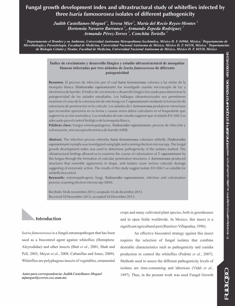
Fungal growth development index and ultrastructural study of whiteflies infected bythree Isaria fumosorosea isolates of different pathogenicity
1 1 2Judith Castellanos-Moguel , Teresa Mier , María del Rocío Reyes-Montes
2 3Hortensia Navarro Barranco , Armando Zepeda Rodríguez 3 2
Armando Pérez-Torres , Conchita Toriello
Autor para correspondencia: Judith [email protected]
crops and many cultivated plant species, both in greenhouses
and in open fields worldwide. In Mexico, this insect is a
significant agricultural pest (Ramírez-Villapudua, 1996).
An effective biocontrol strategy against this insect
requires the selection of fungal isolates that combine
desirable characteristics such as pathogenicity and conidia
production to control the whiteflies (Pedrini et al., 2007).
Methods used to assess the different pathogenicity levels of
isolates are time-consuming and laborious (Vidal et al.,
1997). Thus, in the present work was used Fungal Growth
Índice de crecimiento y desarrollo fúngico y estudio ultraestructural de mosquitas blancas infectadas por tres aislados de Isaria fumosorosea de diferente
patogenicidad
Resumen. El proceso de infección por el cual Isaria fumosorosea coloniza a las ninfas de la
mosquita blanca (Trialeurodes vaporariorum) fue investigado usando microscopía de luz y
electrónica de barrido. El índice de crecimiento y desarrollo fúngico fue usado para determinar la
patogenicidad de los aislados estudiados. Los hallazgos ultraestructurales nos permitieron
examinar el curso de la colonización de este hongo en T. vaporariorum mediante la formación de
estructuras de penetración en la cutícula. Los aislados de I. fumosorosea produjeron estructuras
que recuerdan apresorios en su forma y causan serios daños cuticulares en el hospedante que
sugieren la acción enzimática. Los resultados de este estudio sugieren que el aislado EH-506/3 es
adecuado para el control biológico de la mosquita blanca.
Palabras clave: hongos entomopatógenos, Trialeurodes vaporariorum, proceso de infección y
colonización, microscopía electrónica de barrido (MEB).
Abstract. The infection process whereby Isaria fumosorosea colonizes whitefly (Trialeurodes
vaporariorum) nymphs was investigated using light and scanning electron microscopy. The fungal
growth development index was used to determine pathogenicity of the isolates studied. The
ultrastructural findings allowed us to examine the course of colonization of T. vaporariorum by
this fungus through the formation of cuticular penetration structures. I. fumosorosea produced
structures that resemble appressoria in shape, and isolates cause serious cuticular damage
suggesting of enzymatic action. The results of this study suggest isolate EH-506/3 as suitable for
whitefly biocontrol.
Keywords: entomopathogenic fungi, Trialeurodes vaporariorum, infection and colonization
process, scanning electron microscopy (SEM).
Recibido 18 de noviembre 2013; aceptado 16 de diciembre 2013.
Received 18 November 2013; accepted 16 December 2013.
1 2 Departamento el Hombre y su Ambiente, Universidad Autónoma Metropolitana-Xochimilco, México D. F. 04960, México. Departamento de 3Microbiología y Parasitología, Facultad de Medicina, Universidad Nacional Autónoma de México, México D. F. 04510, México. Departamento
de Biología Celular y Tisular, Facultad de Medicina, Universidad Nacional Autónoma de México, México D. F. 04510, México
ORIG
INAL
© 2
013 R
evista M
exic
ana d
e M
icolo
gía
. Im
pre
sa e
n M
éxic
o
/
REVIS
TA M
EXIC
ANA D
E M
ICOLOGÍA
38: 23-3
3, 2013
Introduction
Isaria fumosorosea is a fungal entomopathogen that has been
used as a biocontrol agent against whiteflies (Hemiptera:
Aleyrodidae) and other insects (Butt et al., 2001; Shah and
Pell, 2003; Meyer et al., 2008; Cabanillas and Jones, 2009).
Whiteflies are polyphagous insects of vegetables, ornamental
Development Index (FGDI), described by Landa et al. (1994)
for determining pathogenicity of entomogenous fungi in
whiteflies. During the bioassay, the nymphs induced fungal
development, and the area of the inoculum drop closest to the
nymph was the most interactive zone.
Nonspecific attachment of the conidia to the host
surface is the first event of the infection process (Rangel et al.,
2008; Chouvenc et al., 2009), and it is followed by conidial
germination and the production of mycelia and different
structures, such as appressoria, that colonize the insect's
cuticle surface (Amóra et al., 2010). A successful fungal
invasion involves two main mechanisms: one is a mechanical
force employing enormous turgor pressures (Fang et al.,
2009), and the other is enzymatic digestion of the cuticle,
mainly by proteases (Zhang et al., 2008; Khan Pathan et al.,
2007) and chitinases (Staats et al., 2013). The first mechanism
is exerted by entomopathogenic fungal structures such as the
appressoria, which have been observed with electron
microscopy. Metarhizium anisopliae develop appressoria
when grown on the western flower thrips (Frankiniella
occidentalis) (Vestergaard et al., 1999), as do Lecanicillium
(=Verticillium) dimorphum or L. cf. psalliotae on red scale
insects of palms (Phoenicococcus marlatti) (Asensio et al.,
2005), and L. lecanii on brown scale (Coccus hesperidum)
(Liu et al., 2011). These structures have also been observed in
Paecilomyces lilacinus growing on nematode eggs
(Meloidogyne javanica) (Holland et al., 2002). However,
hyphae from other entomopathogenic fungi such as
Entomophaga maimaga and Beauveria bassiana, penetrate
the insect cuticle directly, without the need for appressoria
formation, or through natural orifices as mouthparts (Hajek
and Eastburn, 2003; Asensio et al., 2005; Mauchline et al.,
2011).
Due to the relevance of both pathogenicity and the
fungal structures in the infection processes of microbial
agents used in biological control, in this paper, the FGDI was
used to compare the pathogenicity of three I. fumosorosea
isolates with different lethal median concentrations (LC ). 50
An ultrastructural study was carried out to examine the
formation of infection structures and cuticular penetration
during the infection process of I. fumosorosea in T.
vaporariorum nymphs.
Materials and methods
Fungal isolates and growth conditions
Isaria fumosorosea PFCAM, MBP, and PSMB1 were isolated
from whiteflies and obtained from the National Center for
Biological Control, Mexico (Centro Nacional de Referencia
de Control Biológico-CNRCB). Single spore cultures
numbered EH-506/3, EH-503/3 and EH-520/3 from PFCAM,
MBP, and PSMB1, respectively, were prepared by the Goettel
and Inglis method (1997) as modified by Cavallazzi-Vargas et
al. (2001). The original and monospore cultures (isolates)
were preserved in sterile water, mineral oil, and in liquid
nitrogen cryopreservation at –196 °C and deposited in the
fungal collection of the Laboratorio de Micología Básica,
Departamento de Microbiología y Parasitología, Facultad de
Medicina, Universidad Nacional Autónoma de México,
UNAM, registered at the World Federal Culture Collection
(WFCC) as BMFM-UNAM 834, and in the Laboratorio de
Micología, Departamento El Hombre y su Ambiente,
Universidad Autónoma Metropolitana-Xochimilco (UAM-
X). Isolates were maintained on culture medium slants
containing 1 % sucrose, 0.5 % glucose, 0.05 % peptone, 0.5 %
yeast extract, and 2.3 % agar (SGPYE medium) until used.
The pathogenicity of the three isolates used in this
work had been tested previously by median lethal
concentration (Castellanos-Moguel et al., 2007). The high-
3pathogenicity isolate, EH-506/3, had an LC of 1.1 x 10 , the 50
medium pathogenicity isolate, EH-503/3, had an LC of 2.5 x 50
410 , and the low pathogenicity isolate, EH-520/3, had an LC50
4of 7.6 x 10 conidia/mL.
REVIS
TA M
EXIC
ANA D
E M
ICOLOGÍA
38, 2013
24

Insects
The whitefly nymphs (Trialeurodes vaporariorum) used for
the FGDI and scanning electronic microscopy (SEM)
protocols originated from colonies maintained at the
greenhouse and experimental field of the Centro de
Investigación en Biotecnología, Universidad Autónoma del
Estado de Morelos (CEIB-UAEM), México. Whiteflies were
reared on aretillo (Fuchsia sp.) leaves.
Bioassay: Fungal growth development index (FGDI)
procedure
We employed the method of Landa et al. (1994) with minor
modifications. Fungal conidia were produced in SGPYE
6medium cultures, and a dose of 1 x 10 conidia/mL was used.
Fuchsia leaves that were infested predominantly with early
fourth instar nymphs were used. The nymphs were disinfected
in a laminar flow hood by soaking them in the following
solutions: 70 % alcohol for 5 s, sterile distilled water for 40 s,
and 5 % sodium hypochlorite for 20 s, followed by rinsing in 3
changes of sterile distilled water for a total of 120 s. The
leaves were air-dried on sterile filter paper and the nymphs
were removed from the leaf surfaces with an entomological
needle and transferred to 3.5 cm Petri dishes containing
water-agar (2.3 % for light and 60 % for scanning
microscopy).
The nymphs were infected by dropping 4 L of the
conidial suspension described above on the nymph vicinity.
Control nymphs were treated with 0.05 % Tween 80. The Petri
dishes were placed in an incubator at 28 ºC with a photoperiod
of 16:8 hours light:dark, for a total of 96 h. In total, 75 nymphs
were used for each method (FDGI and SEM): 25 for each
replicate bioassay and another 25 nymphs as a control. For the
SEM, only isolates that had shown high (EH-506/3) and low
(EH-520/3) pathogenicity were used.
The nymphs were assessed by light microscopy and
valued individually at 0, 6, 12, 18, 24, 36, 48, 60, 72 and 96 h,
and the FGDI was calculated. The results were expressed as
the means of the FGDI values for each isolate. The FGDI
values were valued as in Landa et al. (1994). A value of 0
represented nymphs surrounded by nongerminated conidia. A
value of 0.5 was equated to the germination of conidia with
one or two germ tubes, especially in the area close to the
nymph. A value of 1.0 corresponded to germinated and non-
germinated conidia surrounding the nymph, while a value of
1.5 required initial fungal growth on the host. At this point,
hyphae were oriented toward the nymph and the first contact
between hyphae and nymph was noticed. At 2.0, mycelium
growth was observed on the host, hyphal growth was
observed on the surface of the nymph and in the area around
the nymph, and the presence of dense mycelium was noted. A
value of 2.5 denoted initial sporulation and the presence of
the first conidiophore on the surface of the nymph. At 3.0,
sporulation had completed, and the nymph was covered with
mycelia and conidia.
Any nymphs covered with the fungus were assumed
to be dead due to fungal infection. Photomicrographs were
taken with an Olympus BX-40 microscope (Olympus Optical
Co. Shibuya-Ku, Tokyo, Japan).
Statistical analysis
Analysis of variance (ANOVA, =0.05) was calculated for
the FGDI at the different assay times among the three
monospore cultures studied and followed by a Tukey multiple
means comparison test (Dowdy and Wearden, 1983). The
statistical analyses were performed using the SPSS Program,
version 10, 2003.
Scanning Electron Microscopy (SEM)
The germination and development of EH-506/3 and EH-
520/3 isolates were observed by SEM at the same times
mentioned above for the FGDI assay. Ten infected nymphs
were fixed at each time with 2.5 % glutaraldehyde (v/v) in 0.1
M phosphate buffer at pH 7.2 for 48 h at 4 ºC, and post-fixed
with 1 % osmium tetroxide (v/v) in 0.1 M phosphate buffer at
25
ORIG
INAL
Ca
ste
llan
os-
Mo
gu
el,
J., e
t a
l. Fu
ng
al g
row
th d
eve
lop
me
nt
ind
ex a
nd
ult
rast
ruct
ura
l stu
dy
of
wh
ite
flie
s in
fect
ed

pH 7.2 for 3 h at 4 ºC. The nymphs were then dehydrated with
ethanol and desiccated at their critical point in a CO chamber. 2
The insects were mounted on aluminum studs, sputter-coated
with silver and covered with coal and ionized gold. The
samples were examined with a Carl Zeiss DSM 950 electron
microscope operating at 15 Kv.
Chemicals
Unless otherwise stated, all chemicals used were from Sigma-
Aldrich Química (Toluca, México).
Results
Fungal growth development index
The FGDI provided an overview of the chronological events
of the whitefly infection by I. fumosorosea. Table 1 shows the
FGDI for the three isolates of different pathogenicity. At 6 h
into the bioassay, the FGDI values of EH-506/3 were
significantly different (P< 0.05) from those observed for EH-
503/3 and EH-520/3. The isolate with high pathogenicity,
EH-506/3, was the only one that displayed germination of
conidia with one or two germ tubes, particularly in the area
close to the T. vaporariorum nymph (FGDI = 0.5). Similarly,
after 12 h, the FGDI values of EH-506/3 were significantly
different (P< 0.05) from those observed for EH-503/3 and
EH-520/3. At the 18 and 24 h time points, the three isolates
showed significant differences (P< 0.05). At 36 and 48 h, the
FGDI values of EH-506/3 and EH-503/3 were the same, and
they were significantly different (P< 0.05) from that of EH-
520/3. At 60 h, the three isolates attained the same FGDI.
Isolate EH-506/3 germinated and colonized
whitefly nymphs faster than the other two isolates tested,
supporting our assessment of EH-506/3 as the most virulent
isolate. At 6 h, this isolate showed conidia germination in the
area near the nymph (FGDI = 0.5; data not shown), whereas
this value was not attained by the other two isolates until 12 h
(Table 1). Figure 1 shows the comparison of isolates EH-
506/3 (left column), EH-503/3 (middle column), and EH-
520/3 (right column) of Isaria fumosorosea by the Fungal
Growth Development Index (FGDI), at 12 (a, f, k), 18 (b, g, l),
24 (c, h, m), 60 (d, i, n) and 96 (e, j, o) hours of incubation. At
12 h, EH-506/3 mycelial growth was observed on the host
(FGDI = 2.0; Figure 1 a), suggesting hyphal emergence from
the host's body. At the same time, the other two isolates, EH-
503/3 and EH-520/3, had scarcely started germinating near
the nymph (Figures 1f and 1k), and EH-520/3 showed mostly
swollen conidia. At 18 h of incubation, EH-506/3-infected
nymphs showed few conidiophores with the characteristic
Isaria sporulation on the surface (FGDI = 2.5; Figure 1 b). At
this time point, EH-503/3 showed nymph-orientated hyphae
and the first contact between nymph and hyphae (FGDI = 1.5;
Figure 1 g). At 18 h, isolate EH-520/3 developed one or more
germ tubes near the nymph (FGDI = 0.5; Figure 1 l). At 24 h,
EH-506/3 retained a constant FGDI value (2.5) with Isaria
sporulation on the surface (Figure 1 c), while EH-503/3
developed dense mycelial growth on the nymph, suggesting
that hyphae were emerging from the host (FGDI = 2.0), and
nymph deformation and destruction were also evident (Figure
1 h). At this time, the FGDI value of EH-520/3 was 1.0;
germinating conidia and some germ tubes scarcely showed
the first contact with the nymph surface (Figure 1 m). At 36 h
(data not shown), EH-506/3 continued to show sporulation,
and EH-503/3 initiated sporulation on the nymph surfaces
(FGDI = 2.5). EH-503/3 continued to display dense mycelial
growth, and nymphs infected with this isolate showed
structural damage and altered morphology (data not shown).
Control nymphs and those infected with the other two isolates
conserved their shapes even during the manipulation
procedure at the equivalent time point. The EH-520/3-
infected nymphs showed an FGDI of 1.5, and hyphae started
to colonize the host. At 48 h of incubation (data not shown),
EH-506/3-infected nymphs were completely covered with
mycelia (FGDI = 3.0), but no further sporulation was
26
REVIS
TA M
EXIC
ANA D
E M
ICOLOGÍA
38, 2013
observed for the rest of the assay; in contrast, EH-503/3
(FGDI = 3.0) continued to display profuse sporulation (Table
1). At 60 h, EH-506/3-infected nymphs continued to have the
maximum FGDI value (3.0), and growth became denser on
the host (Figure 1 d). EH-503/3 exhibited profuse sporulation
(Figure 1 i), and conidiophores were observed on nearly every
surface of the nymph. The isolate EH-520/3 grew slower than
the other two isolates, and it did not reach the maximum FGDI
(3.0) value until 60 h, at which point it also showed profuse
sporulation (Figure 1n). All of the nymphs were incubated
until 96 h to follow the infection process, and, at this time, all
of the infected nymphs showed complete mycelial
colonization of their surfaces (Figures 1e, 1j and 1o). The EH-
503/3-infected nymphs showed more evidence of cuticle
damage, and some insects exhibited significant perforations.
This phenomenon was not observed with the other two
isolates.
Scanning Electron Microscopy (SEM)
The FGDI assay allowed us to select the appropriate time
points for SEM analysis of the two isolates tested: EH-506/3
(high pathogenicity and rapid nymph colonization) and EH-
520/3 (low pathogenicity and slow nymph colonization)
showing that at 6 h of incubation is a critical point for
whiteflies infection. The SEM results showed that the conidia
of both I. fumosorosea isolates were capable of adhering to
any site on the nymph surface, but they mainly did so at the
rachis and vasiform orifice areas. In general, EH-506/3
showed less mycelial growth but more severe cuticular
damage when compared with EH-520/3. The control nymph
maintained an intact shape, did not sustain cuticle damage
after manipulation and no fungal growth was observed on it
(Figure 2a). Moreover, almost all of the control nymphs
reached the 3rd instar stage of insect development.
All of the EH-506/3 and EH-520/3-infected nymphs
showed signs of infection; in most cases, a single germ tube
emerged from each conidium and extended up to a certain
distance before penetrating. At 0 h, conidia of EH-506/3 and
EH-520/3 were found on the nymph surface (Figure 2b) and
near the vasiform orifice of the insect. At this time, the nymph
surface showed, naturally occurring microbiota (Figure 2c).
At 0 h the vasiform orifice did not show any extracellular
matrix (Figure 2d). At 6 h, conidia of both isolates showed an
extracellular matrix. Figure 2e shows EH-506/3 conidia with
extracellular matrix at the rachis area. The EH-520/3-treated
nymphs showed large amounts of the matrix, even at a
27
ORIG
INAL
Ca
ste
llan
os-
Mo
gu
el,
J., e
t a
l. Fu
ng
al g
row
th d
eve
lop
me
nt
ind
ex a
nd
ult
rast
ruct
ura
l stu
dy
of
wh
ite
flie
s in
fect
ed
Bioassay
(h)
EH-506/3
X ± SD
EH-503/3
X ± SD
EH-520/3
X ± SD
0 0a 0a 0a
6 0 .5b ± 0.7 0a ± 0.14 0a ± 0.9
12 2.0b ± 0.3 0.5a ± 0.23 0.5a ± 0.18
18 2.5c ± 0.13 1.5b ± 0.21 0.5a ± 0.15
24 2.5c ± 0.1 2.0b ± 0.32 1.0a ±0.21
36 2.5b ± 0.06 2.5b ± 0.18 1.5a ± 0.25
48 3.0b
± 0.13 3.0b± 0.16 2.0
a ± 0.34
60 3.0a ± 0.08 3.0a ± 0.09 3.0a ± 0.19
Table 1. Fungal Growth Development Index (FGDI) of high (EH-506/3), medium (EH-503/3) and low (EH-520/3) pathogenicity Isaria fumosorosea isolates
X ± SD = median ± standard deviation.Values are representative of three independent experiments run in triplicate.Values in the same line marked with the same letter did not differ significantly according to Tukey´s test of multiple mean comparisons at a significance level of 5%.

28
REVIS
TA M
EXIC
ANA D
E M
ICOLOGÍA
38, 2013
Figure 1. Comparison of isolates EH-506/3 (left column), EH-503/3 (middle column), and EH-520/3 (right column) of Isaria fumosorosea by the Fungal Growth Development Index (FGDI), at 12 (a, f, k), 18 (b, g, l), 24 (c, h, m), 60 (d, i, n) and 96 (e, j, o) hours of incubation. a) EH-506/3 hyphae emerging from a whitefly nymph at 12 h of incubation (FGDI = 2.0). f) EH-503/3 germ tube close to the nymph area at 12 h of incubation (FGDI = 0.5). k) EH-520/3 swollen conidia starting germination close to the whitefly area at 12 h of incubation (FGDI = 0.5). b) EH-506/3 conidiophores at the nymph surface at 18 h of incubation (FGDI = 2.5). g) EH-503/3 hyphae having first contact with the nymph at 18 h of incubation (FGDI = 1.5). l) EH-520/3 conidia with germ tubes closer to the nymph area at 18 h of incubation (FDGI = 0.5). c) EH-506/3 conidiophores sporulating over the nymph at 24 h of incubation (FGDI = 2.5). h) EH-503/3 hyphae on the surface with nymph deformation and destruction at 24 h of incubation (FGDI = 2.0). m) EH-520/3 initial colonization of the nymph at 24 h of incubation (FGDI = 1.0). d) EH-506/3 growing abundantly on the nymph at 60 h of incubation; conidiophores are not very evident (FGDI = 3.0). i) EH-503/3 conidiophores emerging from all nymph surface at 60 h of incubation (FGDI = 3.0). n) EH-520/3 conidiophores over the nymph at 60 h of incubation (FGDI = 3). e) EH-506/3 growth on the nymph at 96 h of incubation (FGDI = 3.0). j) EH-503/3 growth over the nymph at 96 h of incubation (FGDI = 3.0). o) EH-520/3 conidiophore production at 96 h of incubation (FGDI = 3.0). All bars = 250 m; with the exception of f) and k) with 100 m, and i) = 400 m

distance from the conidia. The EH-506/3-treated nymphs
showed ample cuticular damage on the nymph surface after 6
h (Figure 2f). The vasiform orifice was occupied by this
matrix, which trapped the small pieces of cuticle debris that
were produced as a result of cuticle degradation (Figure 2g).
Conidia that were starting to germinate were also observed. At
this same time (6 h), the EH-520/3-treated nymphs also
showed cuticular damage, but it was less severe than that of
the EH-506/3-treated nymphs. After 12 h of incubation, the
EH-506/3-treated nymphs displayed sustained cuticular
damage and gaps on the nymph surface (Figure 2h). This
surface was covered with an extracellular matrix, which was
most likely produced by the isolate, as well as a large amount
of cuticle debris deposited on the nymph surface, even at a
distance from the fungal structures (Figure 2i). The
development of hyphae continued and was faster and denser
than that of EH-520/3. At this time (12 h), EH-520/3 formed
structures at the tip of the germ tubes whose shapes resembled
appressoria. These structures were observed mainly when
hyphae developed at the rachis, where a thin extracellular
matrix was also observed near the hyphae, appressoria and the
vasiform orifice. When appressoria-like structures were at the
29
ORIG
INAL
Ca
ste
llan
os-
Mo
gu
el,
J., e
t a
l. Fu
ng
al g
row
th d
eve
lop
me
nt
ind
ex a
nd
ult
rast
ruct
ura
l stu
dy
of
wh
ite
flie
s in
fect
ed
Figure 2. Scanning Electron Microscopy of Isaria fumosorosea EH-506/3 and EH-520/3 infected nymphs at 0, 6, 12, 18, 24 and 48 h of incubation. a) Intact whitefly control nymph on Fuchsia spp. b) EH-506/3 treated nymph showing a large amount of conidia at 0 h of incubation c) Naturally occurring microbiota at the nymph surface at 0 h of incubation. d) Vasiform orifice of EH-520/3 infected nymph at 0 h of incubation. e) EH-506/3 conidia adhering to the nymph surface with an extracellular matrix at 6 h of incubation. f) Ample cuticular damage on the EH-506/3 treated nymph surface after 6 h of incubation. g) EH-520/3 treated nymph with an extracellular matrix covering the vasiform orifice area. h) Cuticular damage on nymphs treated with EH-506/3 at 12 h of incubation, the photo shows gaps on the cuticle. i) EH-506/3 treated nymph covered with debris, most likely originating from cuticle degradation, at 12 h of incubation. j) EH-506/3 mycelial growth with mucilaginous matrix on the nymph at 18 h of incubation. k) Vasiform orifice of the nymph with EH-520/3 appressoria–like structures at 18 h of incubation. l) EH-520/3 hyphae at 18 h of incubation. The photo shows the extracellular matrix surrounding the hyphae. Direct penetration-like of the cuticle at the end of germ tubes is observed. m) EH-506/3 penetration-like hyphae with appressoria at the vasiform orifice, at 24 h of incubation. n) EH-520/3 mycelial growth on the nymph surface at 24 h of incubation. o) EH-520/3 hyphae penetration-like through the vasiform orifice at 24 h of incubation. p) EH-506/3 growing on the nymph surface at 48 h of incubation. The photo shows a gap on the cuticle and the mucilaginous matrix.

Discussion
The three isolates tested were shown to be virulent against T.
vaporariorum whitefly nymphs. In this study, use of the FGDI
permitted us to perform a detailed observation of the
induction of fungal growth on the nymphs, as reported by
Landa et al. (1994) regarding P. fumosoroseus (now Isaria
fumosorosea) infecting Bemisia argentifolii. Conidia of EH-
506/3, EH-503/3 and EH-520/3 were capable of adhering to
the host's cuticle, germinating and producing infections. The
EH-506/3 strain displayed the highest values of FGDI within
the shortest time period, highlighting the differing
pathogenicity of the isolates tested. The FGDI values and the
previous CL data (Castellanos-Moguel, 2002) show that 50
EH-506/3 is the most virulent isolate (FGDI = 2.5 at 18 h of
3incubation; CL = 1.1 x 10 conidia/mL), followed by EH-50
4503/3 (FGDI = 1.5 at 18 h of incubation; CL = 2.5 x 10 50
conidia/mL) and EH-520/3 (FGDI = 0.5 at 18 h of incubation,
4CL = 7.6 x 10 conidia/mL). Between 60 and 96 h, the three 50
tested isolates adhered, germinated, penetrated the nymphs
and later sporulated on their surface. This development was
faster than that reported by Landa et al. (1994) for P.
fumosoroseus growing in B. argentifolii nymphs. Previous
work regarding Verticillium lecanii (now Lecanicillium
lecanii) growing on the aphid Macrosiphum euphorbiae
reported the development of dense hyphal growth at 120 h of
incubation (Askary et al., 1999), and M. anisopliae var.
acridum growing on Lutzomya longipalpis formed mycelia,
anastomosis, appressoria and conidiophore primordia at 96 h
of incubation (Amóra et al., 2010).
EH-506/3-treated nymphs showed mycelial growth
that had apparently emerged from the nymphal body at 12 h of
incubation, suggesting that penetration was achieved between
6 and 12 h of incubation. The fungus emerged from the
whitefly to start the sporulation process at 18 h (Figure 1b).
rachis, gaps with germ tubes orienting towards them were also
observed. Apparent penetration of the cuticle and a slight
clear zone at the appressorium tip were also observed. At 18 h,
EH-506/3 showed dense mycelial growth and conidia with
elongated germ tubes embedded in a thin mucilaginous
matrix (Figure 2 j). Hyphal growth toward the vasiform
orifice was also observed (data not shown). At this time,
hyphae with appressoria-like structures were also observed.
Hyphae developed and appeared to penetrate the insect in the
area of the vasiform orifice in EH-520/3-treated nymphs
(Figure 2k). Direct penetration of the cuticle (without
appressoria-like structures) by the end of the germ tube was
also observed. A mucilaginous matrix was seen to cover
hyphae (in some instances) or other sites, suggesting the
existence of an adherence mechanism between hyphae and
the nearest cuticle (Figure 2 l).
At 24 h, the penetration of hyphae through the
vasiform orifice (Figure 2m) was observed in EH-506/3-
treated nymphs. EH-520/3-treated nymphs showed a dense
hyphal layer covered with the same extracellular
mucilaginous matrix observed on the nymph's surface (Figure
2n). At the vasiform orifice area, well-developed appressoria-
like structures (Figure 2 o) and penetration-like of the hyphae
through the orifice were observed. Structures that resembled
developing phialides were observed at the hyphal tips (Figure
2o).
At 48 h, EH-506/3-treated nymphs were densely
colonized with mycelia (Figure 2 p); under the mycelial mat, a
thin mucilaginous matrix, cuticular damage and gaps could be
observed. Conidia produced by mycelia were also observed.
This same type of growth was observed on EH-520/3-treated
nymphs (data not shown). At 60 h, all of the isolates had
completed their life cycles, and the fungus had emerged from
dead whitefly nymphs.
30
REVIS
TA M
EXIC
ANA D
E M
ICOLOGÍA
38, 2013

Although this isolate infected whiteflies with a low apparent
hyphal production, it grew abundantly on the insect's surface
after host death, suggesting saprobic growth.
The EH-503/3-treated nymphs showed more
abundant mycelial growth than EH-506/3. Moreover, the
cuticle of these whiteflies was severely deformed (36 h post-
incubation) or completely destroyed after 96 h of incubation.
An enzymatic hydrolysis (Charnley, 2003) mechanism could
lead to the cuticle damage induced by EH-503/3 because it
has high chitinase production (Castellanos-Moguel et al.,
2001). Nymphs treated with the other two isolates did not
show this particular cuticle damage. The infection patterns we
observed correspond to those reported by James et al. (2003)
for P. fumosoroseus growing on third-instar silverleaf
whiteflies.
Interestingly, more evident cuticle damage (and, in
some insects, large perforations) was not observed with EH-
520/3 and EH-506/3 isolates, suggesting that this damage was
caused only by the fungal action of the EH-503/3 isolate.
Cabanillas and Jones (2009) reported that I. fumosorosea
isolates caused fungal-produced patches and collapse of the
body wall of whitefly nymphs.
The SEM experiments were performed with EH-
506/3 (high pathogenicity) and EH-520/3 (low pathogenicity)
to compare the structure formation time and the mode of
action of the two isolates. EH-506/3 has a high protease
production, specifically Pr1 and Pr2 (Castellanos-Moguel et
al., 2007), which may explain the intense and wide cuticular
damage observed and most likely facilitated fungal
penetration. These enzymes could also be responsible for the
rapid emergence of the fungus after colonizing the insect
(Small and Bidochka, 2005), a phenomenon that was
observed for EH-506/3. These histolysis zones were covered
with EH-506/3 mucilage, even far from the vicinity of the
fungus. The EH-520/3-treated nymphs also displayed
cuticular damage, but it was less extensive and only in the
vicinity of the conidia. A similar phenomenon was observed
as egg shell degradation associated with hyphae in L.
dimorphum infection of the red scale insect of palms
(Phoenicoccus marlatii) (Asensio et al., 2005), as well as in
M. anisopliae infection of the western flower thrips
(Frankliniella occidentalis) (Vestergaard et al., 1999).
In the present study, both tested isolates adhered to
the cuticle in groups, consistent with the report of Yanagawa
et al. (2008) for P. fumosoroseus in Coptoptermes formosanus
cuticle; they then showed hyphal swelling, globose at the tip,
that resembled appressoria in shape, which occurred mainly
at the vasiform orifice and rachis areas of the nymph. In
addition to the fact that the humidity of these areas promotes
fungal germination, the presence of appressoria-like
structures could be relevant because these structures have
been implicated in cuticle penetration through exertion of
mechanical forces by the rice blast fungus Magnaporthe
grisea (Howard et al., 1991). However, these types of
structures have not been observed by other authors in assays
of P. fumosoroseus infection of Plutella xylostella (Altre and
Vandenberg, 2001). These researchers reported that the fungi
appeared to penetrate the cuticle directly with
undifferentiated germ tubes within 22 h of inoculation.
The EH-506/3 and EH-503/3 isolates produced a
mucilaginous extracellular matrix, which was more evident in
isolate EH-520/3. This isolate also displayed a slight
discoloration of the cuticle at the hyphal tips, which was likely
caused by enzyme production. The extracellular matrix
observed at the 6 h time point near conidia of both isolates
EH-506/3 and EH-520/3 apparently conferred adhesion
properties to the fungus. As mentioned by Askary et al.
(1999), the mucilage matrix could have adhesive properties
that facilitate penetration of V. lecanii during aphid invasion.
Invasive fungal growth was also observed in the two
isolates. EH-506/3 showed faster but less abundant growth
than EH-520/3, which is coincident with the development
observed in the FGDI experiments. Our SEM observations
showed that conidia formed a unipolar germ tube that
31
ORIG
INAL
Ca
ste
llan
os-
Mo
gu
el,
J., e
t a
l. Fu
ng
al g
row
th d
eve
lop
me
nt
ind
ex a
nd
ult
rast
ruct
ura
l stu
dy
of
wh
ite
flie
s in
fect
ed
extended over a distance before penetration, as has been
described by Askary et al. (1999) for V. lecanii invasion of the
potato aphid Macrosiphonella sanbornii. However, these last
authors mention that it was not possible to obtain clear
evidence of the fungal entrance with SEM. In contrast, our
observations showed that I. fumosorosea appears to penetrate
directly through the cuticle (Figure 2l) at the vasiform orifice
(Figure 2m) and rachis areas where appressoria-like
structures were observed, suggesting that the penetration may
be mediated by these structures. Liu et al. (2011) observed
that L. lecanii can invade through the anus (the vasiform
orifice in whiteflies) in nymphs of Coccus hesperidium. In
other cuticle sections, we observed direct penetration of the
fungus. This may be because the particular humidity
conditions of the rachis and vasiform orifice create a
microclimate that promotes appressoria development
(Charnley, 2003) as well as cuticular toughness, as has been
demonstrated for phytopathogenic fungi (Howard et al.,
1991). In the rachis area, structures that resembled
penetration pegs were observed; these structures have also
been observed when L. lecanii is penetrating C. hesperidum
nymphs (Liu et al., 2011). These authors also mentioned that
this fungus forms a thick mycelial layer that covers the insect
body, similar to that which we observed in both isolates of I.
fumosorosea infecting whiteflies. The presence of phialides
in both isolates after 24 h of incubation is intriguing, but
abundant sporulation was not observed. Our results suggest
that EH-506/3 is a suitable candidate for biocontrol due to the
fungus's ability to rapidly colonize and emerge from the
nymphs. However, this isolate has always shown a very low
conidial production on slant and rice cultures (unpublished
data). Therefore, we suggest the combined use of EH-506/3
with EH-503/3, an isolate with medium pathogenicity and
high conidia production on slant cultures, for biocontrol
purpose.
Acknowledgments
The authors thank to the Centro Nacional de Referencia en
Control Biológico for the original I. fumosorosea isolates and
the Centro de Investigaciones en Biotecnologia of the UAEM
for the whitefly nymphs.
References
Altre, J.A., J.D. Vandenberg, 2001. Comparison of blastospores of two Paecilomyces fumosoroseus isolates: In vitro traits and virulence when injected into fall armyworm Spodoptera frugiperda. Journal of Invertebrate Pathology 78: 170-175.
Amóra, S.S.A., C.M.L. Bevilaqua, F.M. Carneiro-Feijo, R.H. de Macedo Assunçao Pereira, N. Dutra Alves, F.A. de Morais Freire, M. T. Kamimura, D. M. de Oliveira, E.A. Luna-Alves Lima, M.F. Gadelha Rocha, 2010. The effects of the fungus Metarhizium anisopliae var. acridum on different stages of Lutzomya longipalpis (Diptera: Psychodidae). Acta Tropica 113: 214-220.
Asensio, l., L.V. Lopez-Llorca, J.A. Lopez-Jimenez, 2005. Use of light, scanning electron microscopy and bioassays to evaluate parasitism by entomopathogenic fungi of the red scale insect of palms (Phoenicococcus marlatti Ckll., 1899). Micron 36: 169-175.
Askary, H., N. Benhamou, J. Brodeur, 1999. Ultrastructural and cytochemical characterization of aphid invasion by the hyphomycete Verticillium lecanii. Journal of Invertebrate Pathology 74: 1-13.
Butt, T.M., C. Jackson, N. Magan, 2001. Fungi as Biocontrol Agents, Progress, Problems and Potential. CABI, New York.
Cabanillas, H.E., W.A. Jones, 2009. Pathogenicity of Isaria sp. (Hypocreales: Clavicipitaceae) against the sweet potato whitefly B biotype, Bemisia tabaci (Hemiptera: Aleyrodidae). Crop Protection 28: 333-337.
Castellanos-Moguel, J. 2002. Relación entre los niveles de proteasa y quitinasa en aislados de Paecilomyces fumosoroseus (Wize) Brown y Smith y su patogenicidad hacia la mosquita blanca. Tesis de Maestría, Escuela Nacional de Ciencias Biológicas, Instituto Politécnico Nacional, México, D.F.
Castellanos-Moguel, J., R. Cruz-Camarillo, E. Aranda, C. Toriello, 2001. Selección de aislados de Paecilomyces fumosoroseus (Wize) Brown y Smith, con base en sus niveles de proteasa y quitinasa. IX Congreso Nacional de Biotecnología y Bioingeniería; XIII Congreso Nacional de Ingeniería Bioquímica, II Congreso Internacional de Ingeniería Bioquímica. Veracruz, México, septiembre 10-14.
Castellanos-Moguel, J., M. González-Barajas, T. Mier, M.R. Reyes-Montes, E. Aranda, C. Toriello, 2007. Virulence testing and extracellular subtilisin-like (Pr1) and trypsin-like (Pr2) activity during propagule production of Paecilomyces fumosoroseus isolates from whiteflies (Homoptera:Aleyrodidae). Revista Iberoamericana de Micología 24: 62-68.
Cavallazzi-Vargas, G., A. Pérez-Mejía, A. Berlanga-Padilla, V. Hernández-Velázquez, Toriello, C., 2001. Selección de cultivos
32
REVIS
TA M
EXIC
ANA D
E M
ICOLOGÍA
38, 2013
33
ORIG
INAL
monospóricos de Paecilomyces fumosoroseus con base en sus características fenotípicas. In: Nevarez-Morillon, G.V., Sánchez-Martínez G., Muñoz-Castellanos L.N. (Eds.), Memorias del XXIV Congreso Nacional de Control Biológico, Chihuahua México. Sociedad Mexicana de Control Biológico, Chihuahua, México, pp. 112-115.
Charnley, A. K., 2003. Fungal pathogens of insects: cuticle degrading enzymes and toxins. Advances in Botanical Research 40: 242-321.
Chouvenc, T., N.-Y. Su, A. Robert, 2009. Cellular encapsulation in the Eastern subterranean termite, Reticulitermes flavipes (Isoptera), against infection by the entomopathogenic fungus Metarhizium anisopliae. Journal of Invertebrate Pathology 101: 234-241.
Dowdy, S., S. Wearden, 1983. Statistics for Research. John Wiley and Sons, New York.
Fang, W., M. Pava-ripoll, S. Wang, R. St. Leger. 2009. Protein kinase A regulates production of virulence determinants by the entomopathogenic fungus Metarhizium anisopliae. Fungal Genetics and Biology 46: 277-285.
Goettel, M.S., G.D. Inglis, 1997. Fungi: Hyphomycetes. In: Lacey, L.A., Manual of Techniques in Insect Pathology (Ed). Academic Press, London, UK. pp. 213-250.
Hajek, A.E., C.C. Eastburn, 2003. Attachment and germination of Entomophaga maimaiga conidia on host and non-host larval cuticle. Journal of Invertebrate Pathology 82: 12-22.
Holland, R.J., T.S. Gunasekera, K.L. Williams, K.M.H. Nevalainen, 2002. Ultrastructure and properties of Paecilomyces lilacinus spores. Canadian Journal of Microbiology 48: 879-885.
Howard, R.J., M.A. Ferrari, D. H. Roach, N.P. Money, 1991. Penetration of hard substrates by a fungus employing enormous turgor pressures. Proceedings of the National Academy of Sciences of the United States of America 88: 11281-11284.
James, J.J., J.S. Buckner, T.P. Freeman, 2003. Cuticular lipids and silverleaf whitefly stage affect conidial germination of Beauveria bassiana and Paecilomyces fumosoroseus. Journal of Invertebrate Pathology 84: 67-74.
Khan Pathan, A. A., K. Uma Devi, H. Vogel, A. Reineke, 2007. Analysis of differential gene expression in the generalist entomopathogenic fungus Beauveria bassiana (Bals.) Vuillemin grown in different insect cuticular extracts and synthetic medium through cDNa-AFLPs. Fungal Genetics and Biology 44: 1231-1241.
Landa, Z., L. Osborne, F. Lopez, J. Eyal, 1994. A bioassay for determining pathogenicity of entomogenous fungi on whiteflies. Biological Control 4: 341-350.
Liu, W., Y. Xue, J. Xue, Y. Zhang, X. Zhang, 2011. Ultrastructural and cytochemical characterization of brown soft scale Coccus
hesperidum (Hemiptera: Coccidae) infected by the Lecanicillium lecanii (Ascomycota: Hypocreales). Micron 42: 71-79.
Mauchline, N., I. Hallet, G. Hill, S. Casonato, 2011. Process of infection of armored scale insects (Diaspididae) by an entomopathogenic Cosmospora sp. Journal of Invertebrate Pathology 108: 46-51.
Meyer, J.M., M. A. Hoy, D.G. Boucias, 2008. Isolation and characterization of an Isaria fumosorosea isolate infecting the Asian citrus psillid in Florida. Journal of Invertebrate Pathology 99: 96-102.
Pedrini, N., R. Crespo, M.P. Juárez, 2007. Biochemistry of insect epicuticle degradation by entomopathogenic fungi. Comparative Biochemistry and Physiology, Part C 146: 124-137.
Ramírez-Villapudua, J., 1996. Manejo Integrado de la Mosquita blanca de la Hoja Plateada. Universidad Autónoma de Sinaloa, Facultad de Agronomía, Sinaloa.
Rangel, D.E.N., D.G. Alston, D. W. Roberts, 2008. Effects of physical and nutritional stress conditions during mycelial growth on conidial germination speed, adhesion to host cuticle, and virulence of Metarhizium anisopliae, an entomopathogenic fungus. Mycological Research 112: 1355-1361.
Shah, P.A., J.K. Pell, 2003. Entomopathogenic fungi as biological control agents. Applied and Microbiology and Biotechnology 61: 413-423.
Small, C.L., M.J. Bidochka, 2005. Up-regulation of Pr1, a subtilisin-like protease, during conidiation in the insect pathogen Metarhizium anisopliae. Mycological Research 109: 307-313.
Staats, C.C., L Kmetzch, I. Lubeck, A. Junges, M. H. Vainstein, A. Schrank, 2013. Metarhizium anisopliae chitinase CHIT30 is involved in heat-shock stress and contributes to virulence against Dysdercus peruvianus. Fungal Biology 117: 137-144.
Vestegard, S., T.M. Butt, J. Bresciani, A.T. Gillespie, J. Eilenberg, 1999. Light and electron microscopy studies of the infection of the western flower thrips Frankliniella occidentalis (Thysanoptera: Thripidae) by the entomopathogenic fungus Metarhizium anisopliae. Journal of Invertebrate Pathology 73: 25-33.
Vidal, C., L.A. Lacey, J. Fargues, 1997. Pathogenicity of Paecilomyces fumosoroseus (Deuteromycotina: Hyphomycetes) against Bemisia argentifolii (Homoptera: Aleyrodidae) with a description of a bioassay method. Journal of Economic Entomology 90: 765-772.
Yanagawa, A., F. Yokohari, S. Shimizu, 2008. Defense mechanism of the termite, Coptotermes formosanus Shiraki , to the entomopathogenic fungi. Journal of Invertebrate Pathology 97: 165-170.
Zhang, Y., X. Liu, M. Wang, 2008. Cloning, expression, and characterization of two novel cuticle-degrading serine proteases from the entomopathogenic fungus Cordyceps sinensis. Research in Microbiology 159: 462-469.
Ca
ste
llan
os-
Mo
gu
el,
J., e
t a
l. Fu
ng
al g
row
th d
eve
lop
me
nt
ind
ex a
nd
ult
rast
ruct
ura
l stu
dy
of
wh
ite
flie
s in
fect
ed