Embed Size (px)
Citation preview
THYMIC TUMORS
GENERAL THORACIC SURGERY
CHAPTER 167
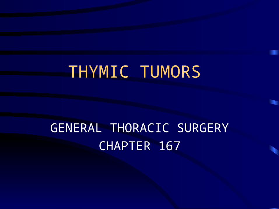
Thymic tumor
• Almost in the anterior mediastinum.
• Secondary to neurogenic tumor in mediastinal tumor.
• Rare in children younger than 16 y/o.
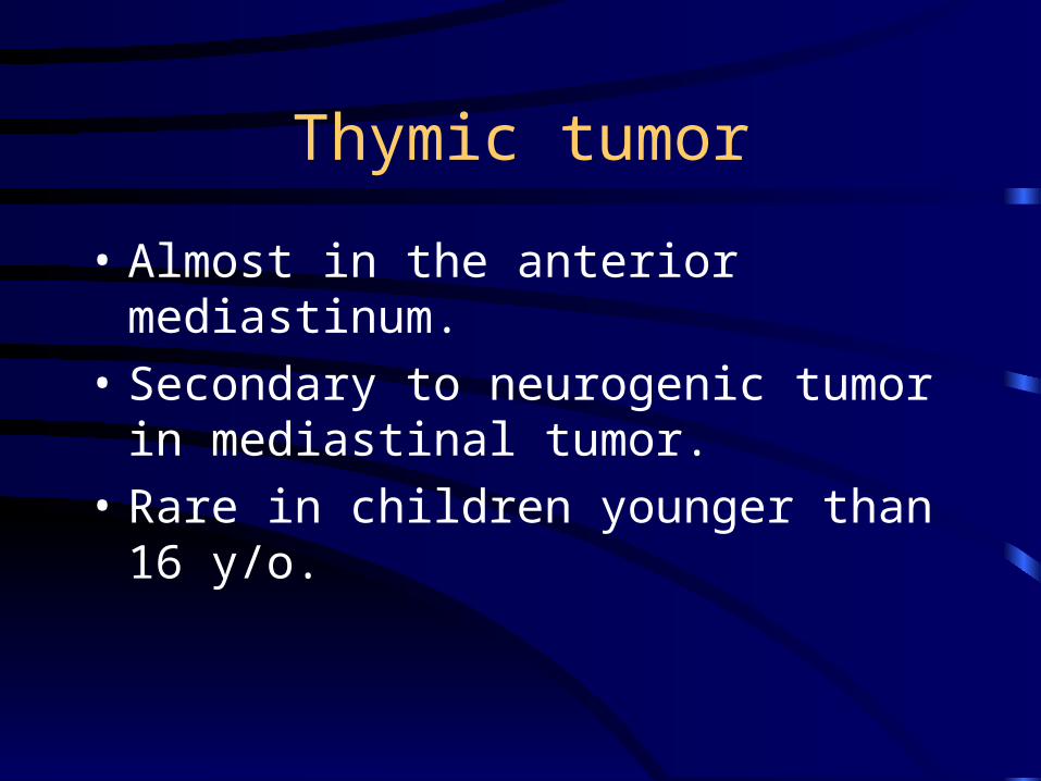
Thymic tumor
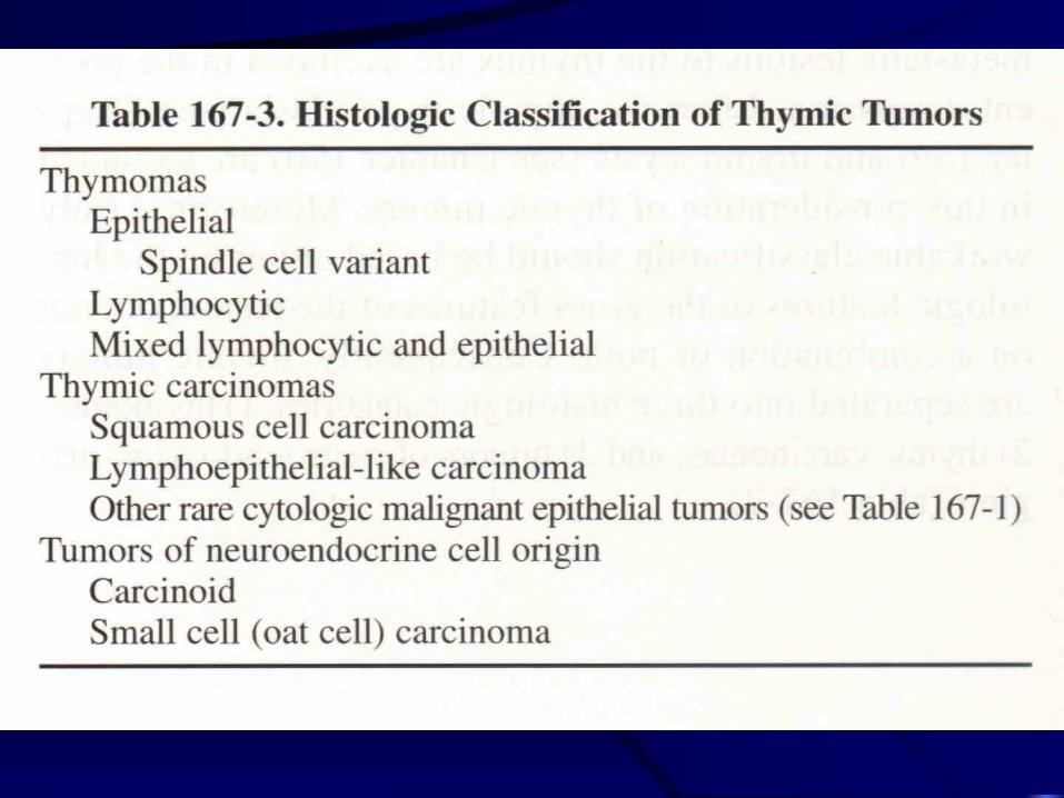
• Separated into three histologic categories—
Thymoma.
Thymic carcinoma.
Neuroendocrine tumor.
THYMOMA
Location
• 95% in anterior mediastinum.
• Neck.
• Left hilar region.
• Within lung parenchynma.
• Anterior cardiophrenic angle.
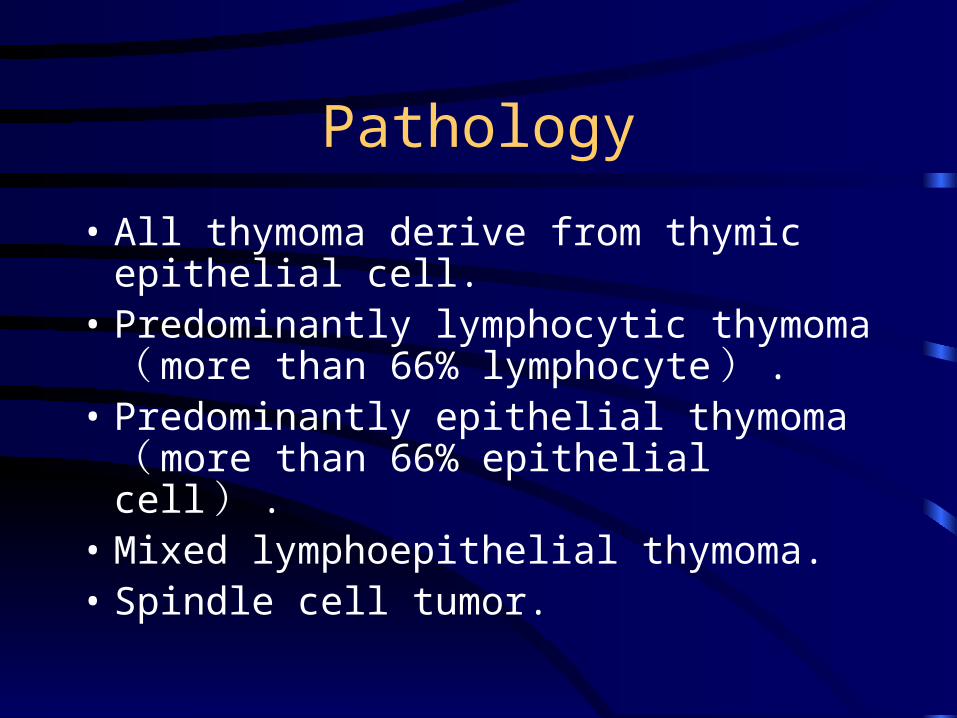
Pathology
• All thymoma derive from thymic epithelial cell.
• Predominantly lymphocytic thymoma ( more than 66% lymphocyte ) .
• Predominantly epithelial thymoma ( more than 66% epithelial cell ) .
• Mixed lymphoepithelial thymoma. • Spindle cell tumor.
Pathology
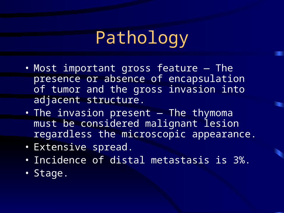
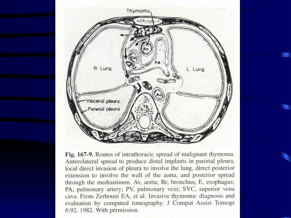
• Most important gross feature — The presence or absence of encapsulation of tumor and the gross invasion into adjacent structure.
• The invasion present — The thymoma must be considered malignant lesion regardless the microscopic appearance.
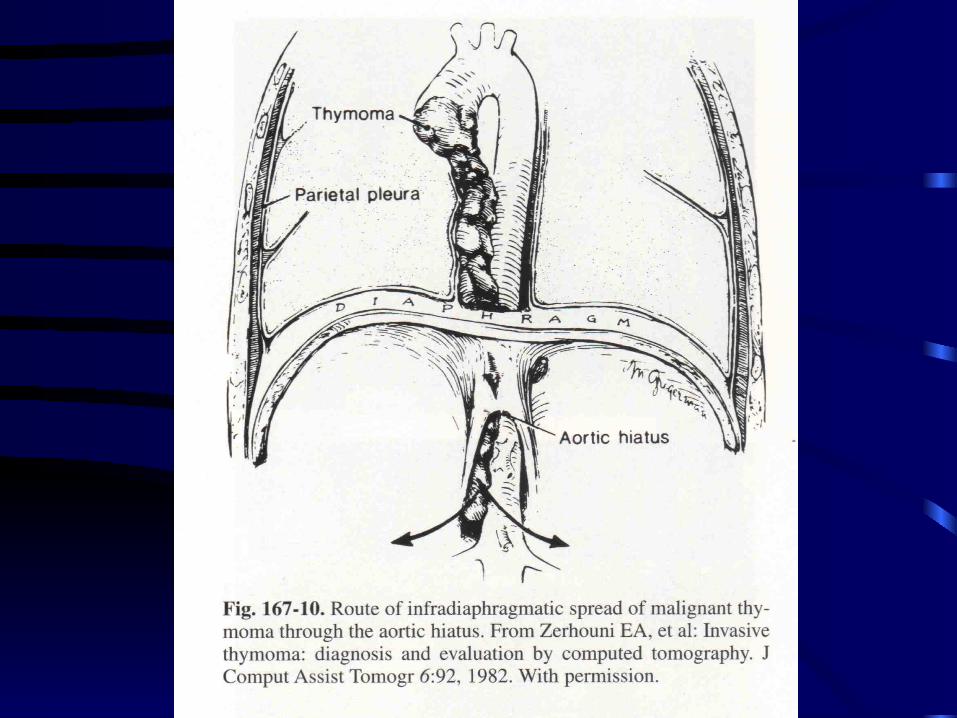
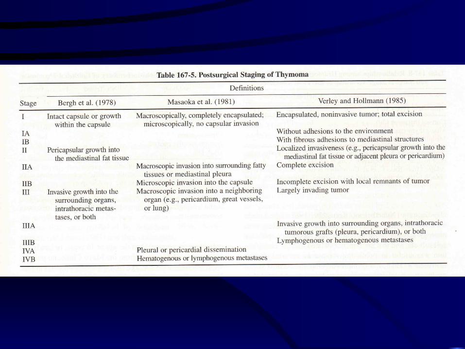
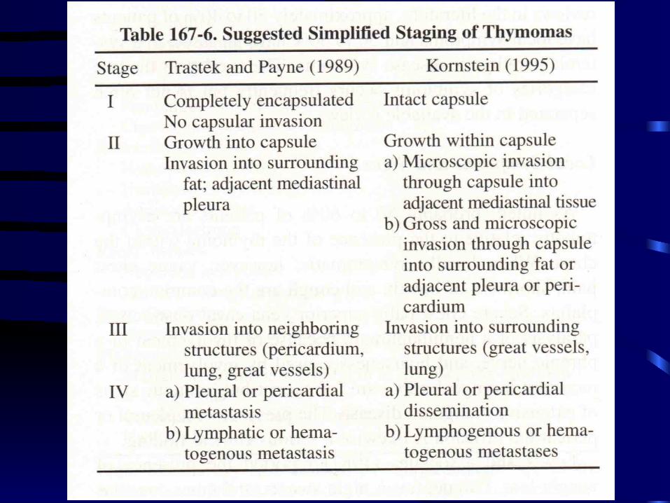
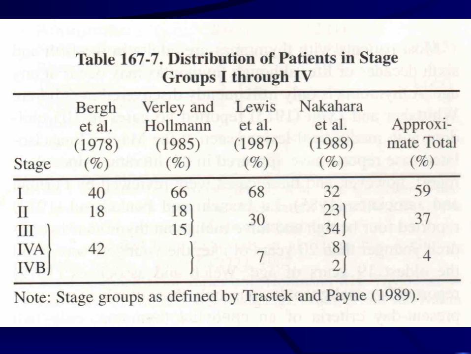
• Extensive spread. • Incidence of distal metastasis is 3%.• Stage.
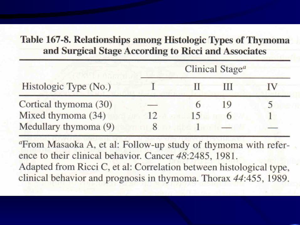
Another class
• Cortical.
• Medullary.
• Mixed thymoma.
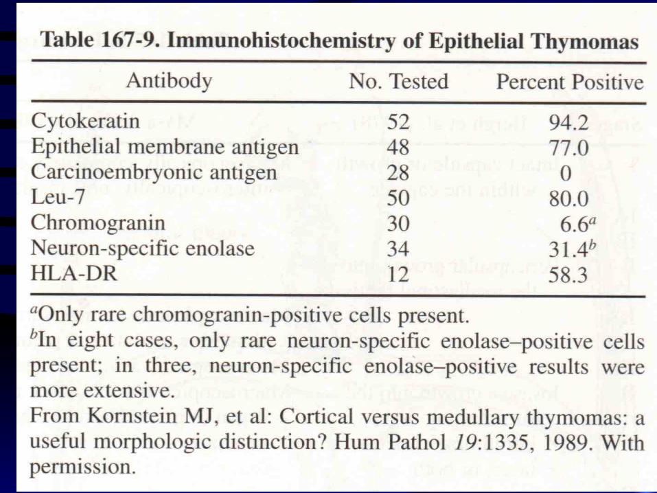
• Immunohistochemistry.
Clinical presentation
• 50-60 y/o.
• Sex distribution — Equal.
• s/s — chest pain, SOB, cough, SVC syndrome, paralysis of hemidiaphragm, hoarseness, weight loss, fatigue, fever, night sweats.
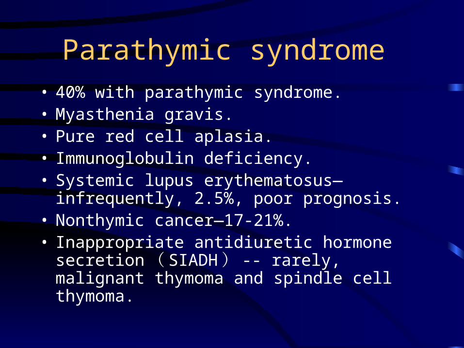
Parathymic syndrome • 40% with parathymic syndrome. • Myasthenia gravis. • Pure red cell aplasia. • Immunoglobulin deficiency. • Systemic lupus erythematosus—infrequently,
2.5%, poor prognosis. • Nonthymic cancer—17-21%. • Inappropriate antidiuretic hormone
secretion ( SIADH ) -- rarely, malignant thymoma and spindle cell thymoma.
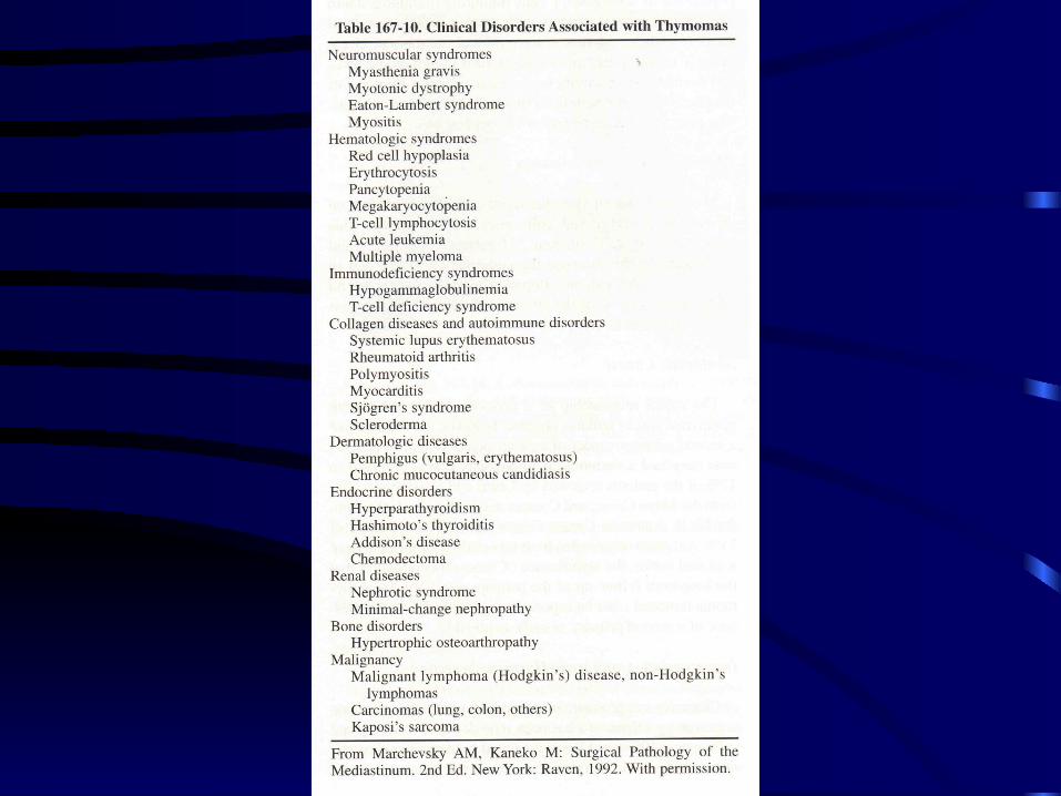
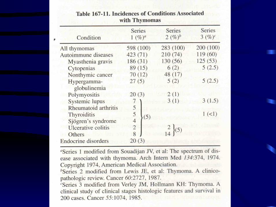
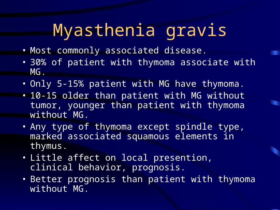
Myasthenia gravis• Most commonly associated disease. • 30% of patient with thymoma associate with MG. • Only 5-15% patient with MG have thymoma. • 10-15 older than patient with MG without tumor,
younger than patient with thymoma without MG. • Any type of thymoma except spindle type, marked
associated squamous elements in thymus. • Little affect on local presention, clinical behavior,
prognosis. • Better prognosis than patient with thymoma
without MG.
Pure red cell aplasia• Anemia. • Suppression erythrogenesis in bone marrow. • Mechanism--Not clear, IgG antibodies inhibit
erythropoietin or hemoglobin synthesis, cytotoxic to erythroblast, decrease B cell.
• 50 % patients with red cell aplasia have thymoma, 5% thymoma with red cell aplasia.
• Most ( 70% ) are non-invasive spindle cell. • 25-33% patient with red cell aplasia benefit from
excision of the thymoma.
Immunoglobulin deficiency
• Spindle cell type.
• Acquired hypogammaglobulinemia.
• Suppressor T-cell inhibiting immunoglobulin synthesis.
Diagnostic studies
• Standard posteroanterior and lateral chest radiographies.
• CXR—Smooth or lobulated mas, right side the silhouette sign present, left side the sign abscent.
• Calcification — 10%. • CT—Delineate the extent of mass, cannot not
differentiating benign and malignant, assessing intrathoracic spread of an invasive thymoma.
Surgical biopsy• Unnecessary for a suspected locally symptomatic
thymoma, because the capsule of tumor may be violated by invasive procedure.
• Only distinguish the tumor from the other malignant tumor, or locally symptomatic, clearly nonresectable, biopsy is to establish the diagnosis before making decision of therapy.
• Fine needle biopsy by CT or sono-guide. • Extend substernal mediastinoscopy.• Anterior mediastinotomy.• Lateral thoracotomy.• VATS.
Treatment
• Depend on clinical presentation. • Surgical resection — thymoma is encapsulated
and free from adjacent structure. • Radiation — in atage II, III.• chemotherapy — in locally nonresectable,
presence distal metastasis, neoadjuvant therapy for initially advanced local diasease or in locally recurrent disease.

Surgical excision
• All patient with thymoma should undergo as complete resection as possible.
• Pulmonary lesion should be excised at the same time.
• Tumor encapsulated — total thymectomy. • Simple enucleation is avoid except the unusual
condition ( excision through lateral thoracotomy with unknown preoperative diagnosis ) .
Surgical excision
• Preferred median sternotomy.
• Posterolateral thoracotomy — for large tumor in hemithorax or tumor from anterior cardiophrenic angle.
• Bilateral anterior fourth intercostals incisionwith transverse section of sternum ( clamshell — )for large midline tumors.
• The use of video-assisted thoracoscopic removal of thymoma is unacceptable even for stage I tumor.
Surgical excision
• Extend procedure the entire thymus and adjacent fat should be removed if possible.
• Tumor fixation to nonvital adjacent structure should be resect ( pleura, lung, pericardium, ) .
Surgical excision
• One phrenic nerve involve could be resected if patient could tolerate loss of hemidiaphragm function.
• If both phrenic are involved, only debulking is performed.
Surgical excision
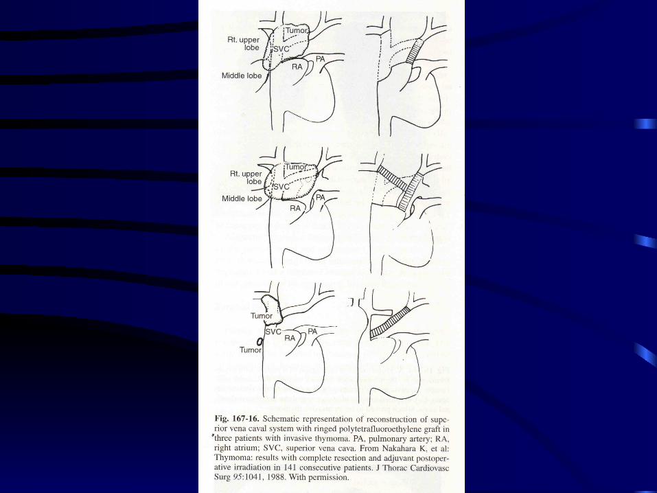
• The wall of SVC involve — if no SVC syndrome, lateral wall resection of SVC and replace graft.
• When the aorta, major pulmonary vessels, recurrent nerve trachea, are involve, only debulking.
• Operative mortality 3.1%-7.7%.
Radiation therapy
• For invasive thymoma. • In stage I is uncertain. • For resected stage II or completely or
incompletely resected stage III disease. • 4500-5000 cGy for suspected microscopic residual
disease. • 6000 cGy for known residual disease. • Brachytherapy with I-125 seed placed in gross
residual disease at time of operation.
Chemotherapy
• For stage III and IV.
• Cisplatin, doxorubucin, vincristine, cyclophosphamide, neoadjuvant.
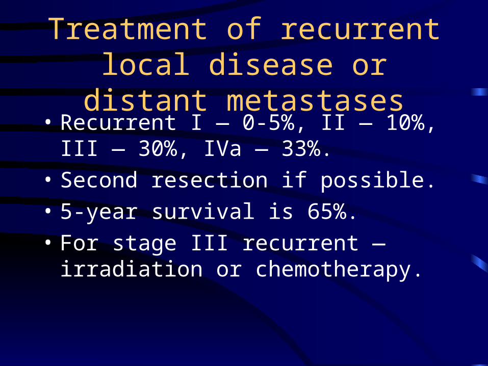
Treatment of recurrent local disease or distant metastases
• Recurrent I — 0-5%, II — 10%, III — 30%, IVa — 33%.
• Second resection if possible.
• 5-year survival is 65%.
• For stage III recurrent — irradiation or chemotherapy.
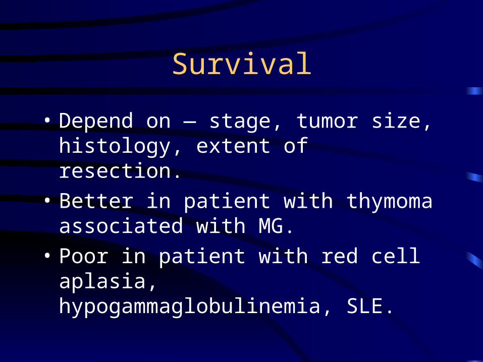
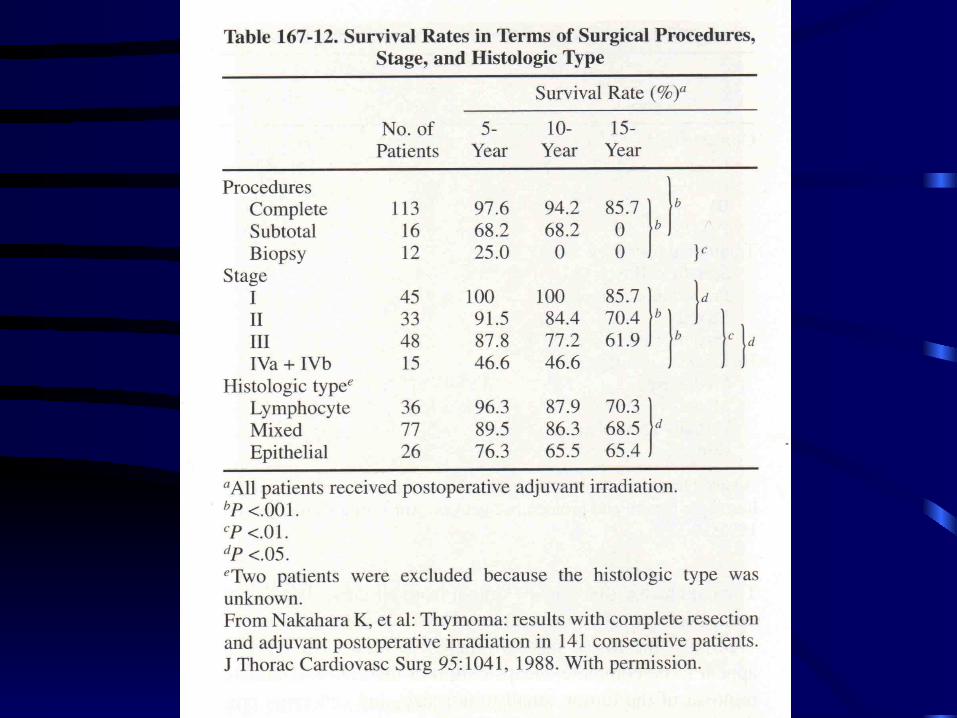
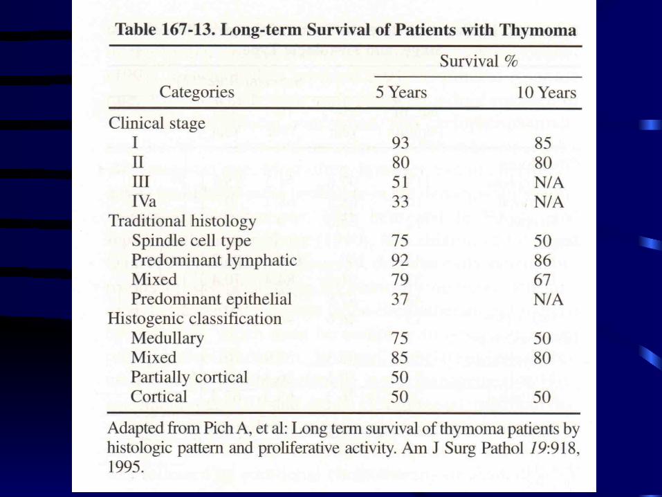
Survival
• Depend on — stage, tumor size, histology, extent of resection.
• Better in patient with thymoma associated with MG.
• Poor in patient with red cell aplasia, hypogammaglobulinemia, SLE.
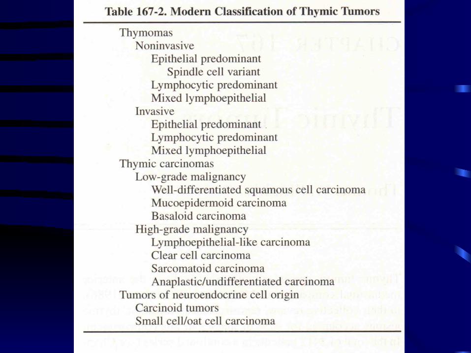
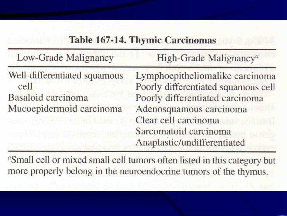
Thymic carcinoma
• Low and high grade.
• Malignant cytologic and architectural feature.
• Staging not standardized.
Squamous cell carcinoma
• Most common. • Men predominant. • 60 y/o. • Partially encapsulated. • s/s — weight loss, chest pain, cough, hemoptysis. • Treatment—Surgical resection, sensitive to
radiation, combination chemotherapy.• Prognosis excellent in well-differentiated
squamous cell carcnoma.
Lymphoepitheliomalike carcinoma
• Epstein-Barr virus.
• Treatment—irradiation therapy, chemotherapy.
Tumor of neuroendocrine cell origin
• Thymic carcinoid tumor.
• Small cell carcinoma.
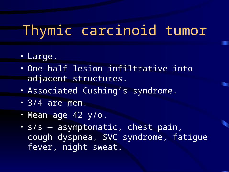
Thymic carcinoid tumor
• Large. • One-half lesion infiltrative into adjacent
structures.• Associated Cushing’s syndrome. • 3/4 are men. • Mean age 42 y/o. • s/s — asymptomatic, chest pain, cough dyspnea,
SVC syndrome, fatigue fever, night sweat.
Thymic carcinoid tumor
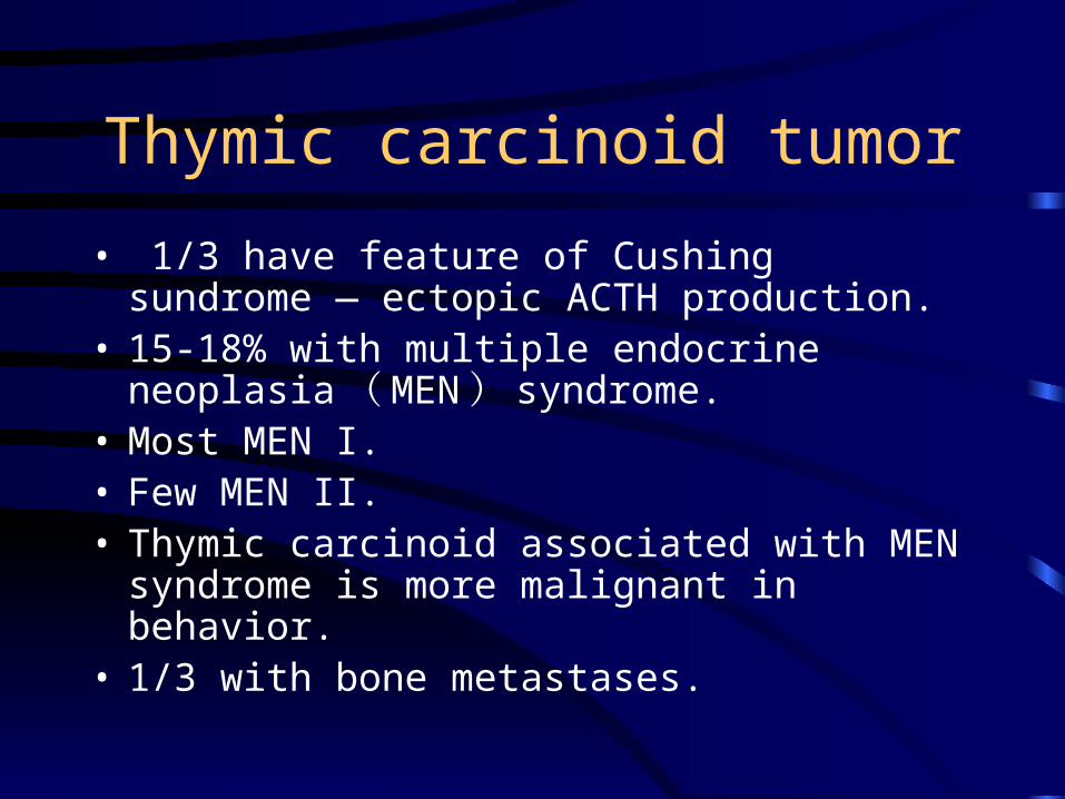
• 1/3 have feature of Cushing sundrome — ectopic ACTH production.
• 15-18% with multiple endocrine neoplasia ( MEN ) syndrome.
• Most MEN I. • Few MEN II. • Thymic carcinoid associated with MEN syndrome
is more malignant in behavior. • 1/3 with bone metastases.
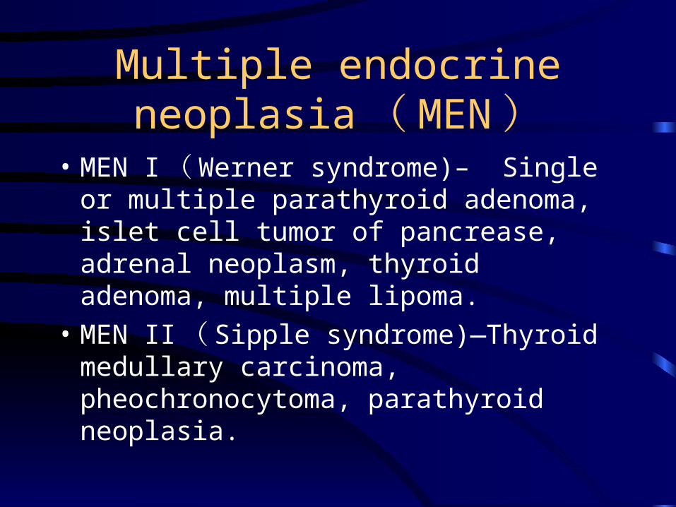
Multiple endocrine neoplasia ( MEN )
• MEN I ( Werner syndrome)– Single or multiple parathyroid adenoma, islet cell tumor of pancrease, adrenal neoplasm, thyroid adenoma, multiple lipoma.
• MEN II ( Sipple syndrome)—Thyroid medullary carcinoma, pheochronocytoma, parathyroid neoplasia.
Thymic carcinoid tumor
• Treatment—complete surgical resction or debulking tumor, radiation therapy.
• 73% local recurrence or metastases.
• Overall cure rate is low — 13%.
• Mean survival of metastases disease is 3 years.
Small ( Oat ) cell carcinoma
• Aggressive and metastases extensively,
• Associated with MEN I.
• Treatment—radiation therapy and chemotherapy.


![Solitary fi brous tumors of the pleura · tumor of the pleura from other lun g tumors, while the contribution of thoracic CT is rather moderate [4]. Although preoperative dia gnosis](https://img.pdfslide.net/doc/110x75/6081a9dfae78a40b630c556a/solitary-i-brous-tumors-of-the-pleura-tumor-of-the-pleura-from-other-lun-g-tumors.jpg)