Embed Size (px)
Citation preview
Thymocyte apoptosis drives the intrathymic generationof regulatory T cellsJoanne E. Konkela, Wenwen Jina, Brittany Abbatielloa, John R. Graingerb, and WanJun Chena,1
aMucosal Immunology Section, Oral and Pharyngeal Cancer Branch, National Institute of Dental and Craniofacial Research, National Institutes of Health,Bethesda, MD 20892; bProgram in Barrier Immunity and Repair, Mucosal Immunology Section, Laboratory of Parasitic Diseases, National Institute of Allergyand Infectious Diseases, National Institutes of Health, Bethesda, MD 20892
Edited by Philippa Marrack, Howard Hughes Medical Institute, National Jewish Health, Denver, CO, and approved December 18, 2013 (received for reviewOctober 28, 2013)
Maintenance of immune tolerance critically depends upon regula-tory T cells that express the transcription factor forkhead box P3(Foxp3). These CD4+ T cells can be generated in the thymus,termed thymus-derived regulatory T cells (tTregs), but their devel-opmental pathway remains incompletely understood. tTreg devel-opment has been shown to be delayed compared with that ofCD4+ single positive (SP) thymocytes, with tTregs being detectedonly in neonatal thymi by day 3 after birth. Here, we outline thereasons for this delayed emergence of Foxp3+ tTregs and demon-strate that thymocyte apoptosis is intrinsically tied to tTreg devel-opment. We show that thymic apoptosis leads to the productionof TGFβ intrathymically from thymic macrophages, dendritic cells,and epithelial cells. This TGFβ then induces foxp3 expression anddrives tTreg generation. Thymocyte apoptosis has previously beenshown to accelerate after birth, which drives increases in TGFβ inthe neonatal thymus. We highlight a paucity of TGFβ in the neo-natal thymus, accounting for the delayed development of tTregscompared with CD4+ SP thymocytes. Importantly, we show thatenhanced levels of apoptosis in the thymus result in an augmentedtTreg population and, moreover, that decreasing thymic apoptosisresults in reduced tTregs. In addition to this, we also show thatT-cell receptor (TCR) signals of different affinity were all capable ofdriving tTreg development; however, to achieve this TGFβ signalsmust also be received concomitant with the TCR signal. Collec-tively, our results indicate that thymic apoptosis is a key event intTreg generation and reveal a previously unrecognized apoptosis–TGFβ–Foxp3 axis that mediates the development of tTregs.
thymic Treg | phagocytes | TCR affinity
The thymus houses and controls the development of commit-ted T-cell precursors to thymocytes to T cells. Within the
thymus, checkpoints ensure that all T cells are capable of seeingantigen and can therefore contribute to an immune response(positive selection). In addition, carefully orchestrated mecha-nisms limit the pathogenicity that would be mediated by self-reactive T cells. First, those thymocytes expressing a TCR withhigh affinity for self are purged from the repertoire and undergoapoptosis (negative selection). Second, the thymus generatesregulatory T cells (thymus-derived Tregs; tTregs), a CD4+ T-cellpopulation characterized by their expression of the transcriptionfactor Foxp3 (1, 2), which play vital roles in suppressing auto-immunity and in maintaining immune homeostasis.Placing tTregs as a central player maintaining a balanced
immune system means that the factors governing the expressionof Foxp3 and tTreg development have received much attention.tTreg development is known to be delayed compared with CD4+
single positive (SP) thymocytes, with tTregs being detected onlyby day 3 after birth (3). One major hypothesis that has emergedis that differentiation of tTregs is initiated upon recognition ofhigh-affinity self-antigens in the thymus (4–6). These data sug-gest that tTreg generation is TCR-instructive, yet other datahave countered this, demonstrating that the higher frequenciesof tTregs associated with cognate antigen interactions are due toreduced populations of non-Tregs (7). Thus, whether tTreg
generation is a TCR-instructive process with a specific “quality”of TCR stimulus specifying the fate remains debatable. Notably,an exclusively TCR-instructive process cannot explain the lack oftTregs in the neonatal thymus until day 3 after birth. Indeed,delayed tTreg generation suggests the neonatal thymus lacks aFoxp3-inducing factor(s).In concert with TCR, other signals have been shown to be
important in thymic tTreg specification, including CD28 cos-timulation (8) and the transcription factors NFAT, AP-1, NF-κB,and Foxo1/3 (9–12). Cytokines also function in tTreg generation,with the common γ-chain cytokines, importantly IL-2 and TGFβ,both involved. A two-step model of tTreg differentiation wasproposed, which suggested that high-affinity TCR interactionspermit CD25 expression on CD4+SP thymocytes and that IL-2–signaling then promotes Foxp3 induction (13, 14). However, itnow seems likely that IL-2 instead functions to promote both thesurvival and the proliferation of thymocytes differentiating intotTregs (15, 16). TGFβ signals are vital for the induction of Foxp3in naive CD4+ T cells (17), but whether TGFβ plays a role intTreg specification remains debated with studies showing that itis both vital (15) and redundant (18).One process overlooked in tTreg generation is apoptosis; all
developmental processes in the thymus occur under a blanket ofthymocyte apoptosis as negative and positive selection occur.Despite these high levels of apoptosis occurring in the thymus,apoptotic thymocytes are not easily found due to the extremelyefficient uptake of apoptotic cells by thymic phagocytes. Moni-toring apoptosis in the neonatal thymus, Surh and Sprent showedthat thymocyte apoptosis suddenly accelerates after birth (19),with few apoptotic thymocytes present in fetal thymus yet sig-nificant populations of apoptotic cells being present by day 2after birth. One would envision that the presence of apoptoticcells, phagocytes, and the factors produced by phagocytes in
Significance
Thymus-derived regulatory T cells (tTregs) are vital to main-taining immune homeostasis, and as such the signals drivingtheir development have been extensively studied. Despite this,a cohesive model describing tTreg generation and aligning allconflicting data has been elusive. Here we outline a compre-hensive model controlling the generation of tTregs and showthat tTreg generation is tied to thymic apoptosis. We show thatthe presence of apoptotic cells in the thymus drives the pro-duction of TGFβ intrathymically and that this cytokine then actsto specify the tTreg fate. Thus, our results reveal an apoptosis–TGFβ–Foxp3 axis that mediates the development of tTregs.
Author contributions: J.E.K. and W.C. designed research; J.E.K., W.J., B.A., and J.R.G.performed research; J.E.K. and W.C. analyzed data; and J.E.K. and W.C. wrote the paper.
The authors declare no conflict of interest.
This article is a PNAS Direct Submission.1To whom correspondence should be addressed. E-mail: [email protected].
This article contains supporting information online at www.pnas.org/lookup/suppl/doi:10.1073/pnas.1320319111/-/DCSupplemental.
www.pnas.org/cgi/doi/10.1073/pnas.1320319111 PNAS | Published online January 13, 2014 | E465–E473
IMMUNOLO
GY
PNASPL
US
response to apoptotic cells may well influence thymocyte de-velopmental pathways. Indeed, here we show that thymocyteapoptosis is intrinsically tied to tTreg generation. It is wellestablished that sensing and uptake of apoptotic cells results inTGFβ secretion (20, 21), and we show the activity of this pathwayin the thymus. We demonstrate that TGFβ is vital for tTregdevelopment, and, importantly, we demonstrate that tTreggeneration occurs more readily in thymi with higher levels ofthymocyte apoptosis. Our data reveal that thymic apoptosisinduces TGFβ production intrathymically, driving tTreg gener-ation. Acceleration of thymocyte apoptosis after birth (19)occurs on a time course preceding the emergence of tTregs andcoincides with increasing levels of intrathymic TGFβ. Thus,thymocyte apoptosis leads to the production of TGFβ in thethymus, which, along with TCR engagement, induces foxp3 ex-pression and ultimately drives tTreg generation. We therefore
propose an apoptosis–TGFβ–Foxp3 axis that is responsible forthe development of tTregs.
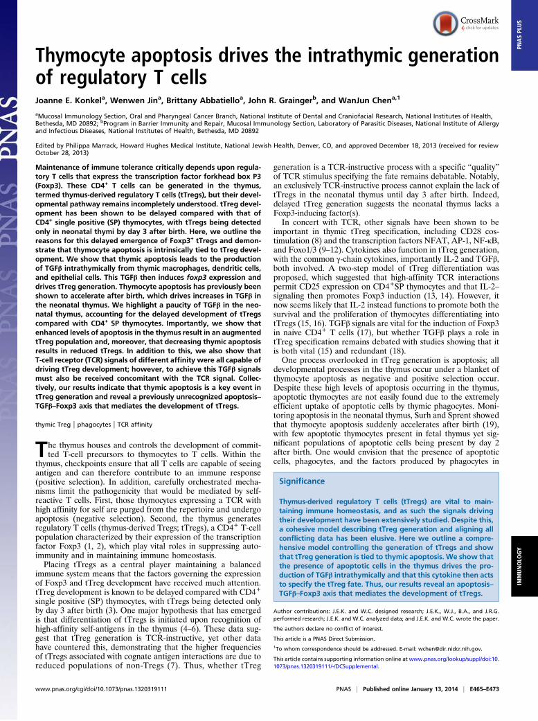
ResultsTGFβ Is Required for tTreg Generation Not to Selectively ProtecttTregs from Apoptosis. A role for TGFβ signaling in tTreg gen-eration remains contentious. Previous data have indicated thatTGFβ signaling is essential for tTreg generation (15); however,another study reported that TGFβ was instead required to spe-cifically keep tTregs alive (18). To better explore this issue, wegenerated Tgfbr1f/fFoxp3-cre+ mice to delete TGFβ Receptor(TβR) I expression following tTreg generation (Fig. S1 A and B).If the tTreg deficiencies that result from the absence of TGFβsignaling were exclusively due to increased tTreg death, thenTgfbr1f/fFoxp3-cre+ mice should exhibit the same tTreg defi-ciencies. We examined tTregs in neonate and adult thymi of
Fig. 1. TGF-β drives tTreg generation. (A and B) CD4+CD25− DO11.10xRag−/− thymocytes were i.t.-transferred to BALB/c mice with 1 μg pOVA plus 150 μganti-TGFβ or isotype control. (A) Thymi were harvested after 5 d, and Foxp3 expression in the transferred population was examined by FACS or (B) after 18 hwhen transferred cells were FACS-sorted and foxp3 expression examined by quantitative PCR (qPCR). Data represent three to five experiments. (C)CD4+CD25−GFP− SP thymocytes were FACS-sorted and cultured with anti-CD3+anti-CD28 for 18 h followed by 36 h rest in the presence or absence of anti-TGFβ and TGFβ inhibitor (SB431542) or isotype control and DMSO. Bar graph shows frequency of CD25+Foxp3+ cells in cultures. Data represent twoexperiments. (D and E) FTOC cultures were established with E16.5 fetal lobes and cultured for 1 wk with TGFβ inhibitor (SB431542) or DMSO control. (D)Representative FACS plots gated on CD4+SP thymocytes. (E) Bar graph showing frequency of CD25+Foxp3+ cells. Data represent three experiments. (F and G)FACS-sorted DN thymocytes from Tgfbr1+/fCD4-cre+ (control; f/+) or Tgfbr1f/fCD4-cre+ (KO; f/f) mice were transferred i.t. into congenic C57BL/6 mice. Thymi wereharvested after 12 d. (F) Representative FACS plots gated on transferred CD4+SP thymocytes. (G) Bar graph showing frequency of Foxp3+ cells in transferredCD4+SP thymocytes. Data represent five experiments. Error bars represent mean ± SEM. *P < 0.05, **P < 0.005 (unpaired two-tailed Student t test).
E466 | www.pnas.org/cgi/doi/10.1073/pnas.1320319111 Konkel et al.
Tgfbr1f/fFoxp3-cre+ mice and importantly found no decrease intTregs; instead, Tgfbr1f/fFoxp3-cre+ mice had similar fre-quencies of tTregs compared with controls (Fig. S1C). Thesedata exclude a simple anti-apoptotic role for TGFβ signalingin tTregs, indicating that TGFβ signaling does not function tospecifically keep Foxp3+ thymocytes alive and instead playsother roles in tTreg generation.Next we determined whether TGFβ signaling was required to
specify tTreg fate. As Foxp3+ thymocytes arise from CD4+SPcells (22), we examined whether TGFβ played a role in Foxp3induction in CD4+ SP thymocytes. We transferred CD4+CD25−
thymocytes from DO11.10xRag−/− mice (Foxp3− thymocytes)intrathymically into BALB/c mice, along with cognate antigen(ovalbumin peptide 323-339; pOVA). Consistent with pre-vious reports (23), Foxp3+DO11.10xRag−/− SP thymocyteswere detected after 5 d (Fig. 1A). Inhibition of TGFβ signalingby intrathymic injection of anti-TGFβ led to a significant de-crease in the frequency (Fig. 1A) and total number (control:5,821 ± 1,253; plus anti-TGFβ: 2,807 ± 461; P = 0.0332) ofFoxp3+DO11.10xRag−/− SP thymocytes. DO11.10xRag−/− thy-mocytes were also sorted from recipient thymi 18 h after intra-thymic transfer, and foxp3 mRNA expression was examined. TGFβinhibition led to decreased foxp3 mRNA (Fig. 1B), indicating thatTGFβ is key in switching on foxp3 gene expression.To further support a role for TGFβ in tTreg generation, we
performed similar experiments using CD4+SP thymocytes fromOT-IIxRag−/− mice crossed with Tgfbr1f/fCD4-cre+ mice (herecalled OT-II-TβR1KO and OT-II-TβR1WT). OT-II-TβR1KO orOT-II-TβR1WT CD4+SP thymocytes were intrathymically trans-ferred into congenic hosts along with pOVA. Examination ofthymocytes 5 d later showed few Foxp3+ OT-II-TβR1KO thy-mocytes, whereas Foxp3+ OT-II-TβR1WT developed as expected(Fig. S1 D–F), again indicating that TGFβ is vital for the gener-ation of Foxp3+ thymocytes.Early withdrawal of TCR stimulation has been shown to
promote Foxp3 expression (24). Culture of CD4+SP thymocyteswith TCR stimulation for 18 h followed by withdrawal of stim-ulation for 36 h promotes development of Foxp3+ thymocytes.Of note, generation of Foxp3+ cells in these cultures was sig-nificantly reduced when TGFβ signals were inhibited (Fig. 1C),suggesting that TGFβ plays a critical role in the generation ofFoxp3+ thymocytes in this setting.Examining tTreg generation in more physiological settings, we
established fetal thymic organ cultures (FTOC) with thymiobtained from wild-type embryonic day 16.5 (E16.5) embryos.When TGFβ signaling was impaired by addition of the TGFβinhibitor SB431542, the development of Foxp3+CD4+SP thy-mocytes was significantly reduced (Fig. 1 D and E).To examine this in vivo, we intrathymically injected sorted dou-
ble-negative (DN) thymocytes from Tgfbr1f/fCD4-cre+ (KO; f/f) orTgfbr1+/fCD4-cre+ (control; f/+) mice into congenic wild-typethymi and allowed them to develop within the thymus for 12 d (atime point by which SP populations had developed). As DN thy-mocytes were transferred into wild-type thymi, this experimentalapproach provided a normal, noninflammatory environment inwhich transferred polyclonal thymocytes could develop. Twelvedays following transfer of control DN thymocytes, Foxp3+CD4+SPthymocytes were present at frequencies normally found in wild-typethymi (3–5%) (Fig. 1 F and G). Transferred KO DN thymocytesgenerated a similar size population of CD4+SP thymocytes (Fig.S1G), yet these cells exhibited a dramatically reduced ability tobecome Foxp3+, as determined by both frequency (Fig. 1 F and G)and total number (control DN transfer: 307 ± 59.9; KO DNtransfer: 12.98 ± 3.16; P < 0.0001). Collectively, these data establishthat TGFβ signaling is vital for the generation of tTregs.
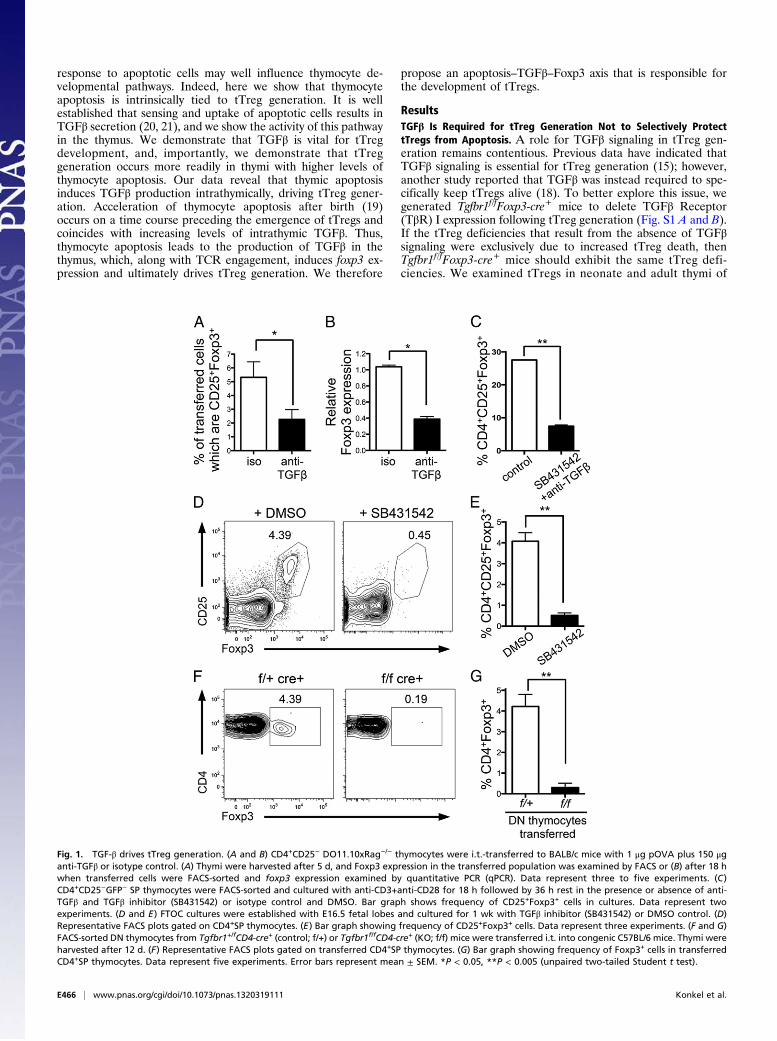
Thymic TGFβ Expression Increases After Birth. Thymic tTreg gener-ation is temporally restricted after birth (3, 15), with few tTregs
detected until day 3 after birth. As we have demonstrated thatTGFβ is vital for tTreg development, we next asked whether thisdelayed emergence of Foxp3+ thymocytes in the neonatal thy-mus was due to a paucity of TGFβ at early time points after birth.We examined levels of TGFβ activity within the neonatal thymus
by staining thymus sections with antibody against active TGFβ1[LC(1–30)] (25). There was an increase in active TGFβ1 in thethymus over the time period from birth (E18.5) through the neo-natal period (Fig. 2A). Although the thymic medulla regions areonly beginning to coalesce and form over the neonatal period, fromH&E staining (in particular by day 3) there was an indication thatthe areas rich in TGFβ1 activity were medullary regions. Indeed,this was confirmed by immunohistochemical (IHC) examination ofadult thymus sections (Fig. S2A). Enriched TGFβ1 activity in thethymic medulla is a vital observation considering that this is thethymic region in which the precursors of tTregs (CD4+ SP thy-mocytes) (22) are found, and which has also been shown to sup-port tTreg development (26, 27). Thus, our IHC staining indicatesthat the thymic medulla is an environment enriched for TGFβ1activity and, more importantly, that intrathymic TGFβ1 activitygradually increases after birth over the neonatal time frame.We also examined TGFβ protein levels by ELISA, and, in line
with our IHC data, intrathymic TGFβ1 increased significantly overthe neonatal period (Fig. 2B). Furthermore, we examined theprotein levels of TGFβ2 and TGFβ3 over the neonate period andfound these also increased over this time frame (Fig. S2 B and C).As CD4+ SP thymocytes are the immediate precursors of
tTregs, we next examined TGFβ-signaling events in these thy-mocytes after birth. We examined levels of phosphorylatedSmad2/3 (pSmad) in CD4+SP thymocytes from 1-, 3-, and 5-d-old pups. The frequency of pSmad2/3+ CD4+SP thymocytesincreased over this neonate period (Fig. 2C). To further confirmthat TGFβ signaling increased in CD4+SP thymocytes afterbirth, we FACS-sorted this population from the thymi of day 1, 2,3, and 4 neonates. We examined the level of Smad7 expression inthese sorted thymocytes, as Smad7 is induced in response toTGFβ signaling (28). After birth there was a gradual increase inSmad7 message, which was significantly enhanced by days 3 and4 post birth in CD4+SP thymocytes (Fig. 2D). Thus, we sawincreases in TGFβ in the thymus over the short period after birthand enhanced TGFβ signaling in tTreg precursors over this sametime frame. Collectively, our data show that there are reducedlevels of intrathymic TGFβ at early time points after birth, andhighlight a gradual increase in TGFβ levels in the neonatal thymusafter birth, which precedes the emergence of tTregs.
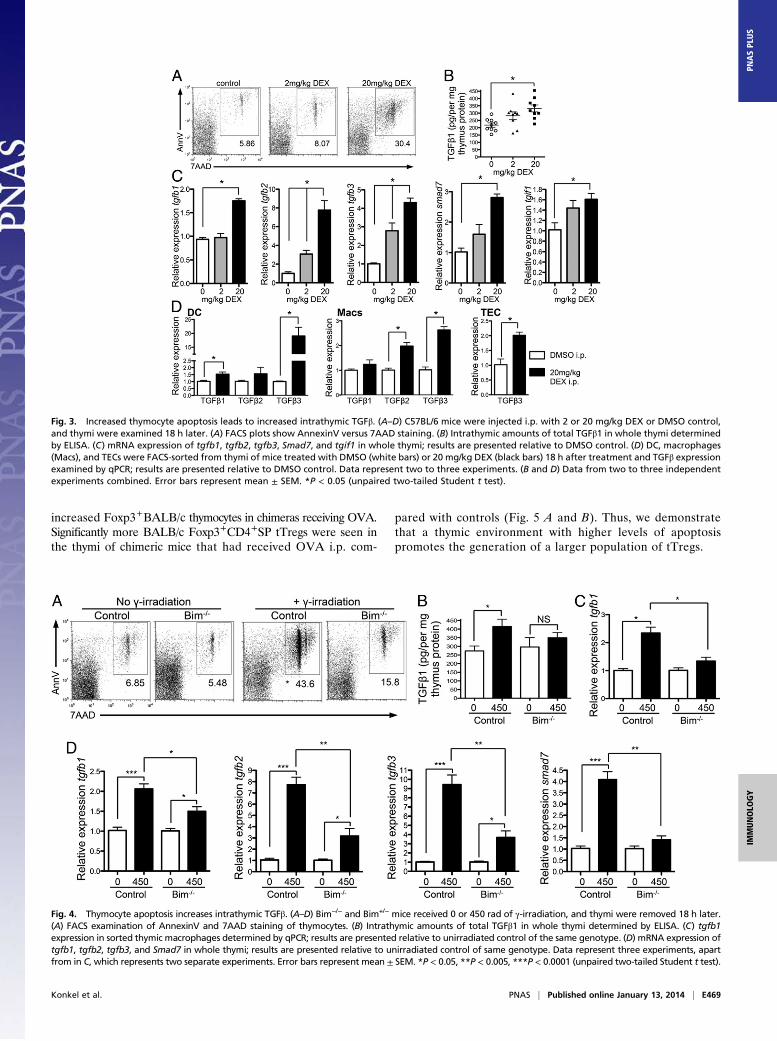
Thymocyte Apoptosis Increases Levels of Intrathymic TGFβ. Havingdemonstrated a vital role for TGFβ in tTreg generation anda paucity of TGFβ in neonatal thymus, we next wanted to identifythe stimulus for TGFβ production in the neonatal thymus. Weexamined the role of thymocyte apoptosis in this process. Thymo-cyte apoptosis has been shown to accelerate after birth; at fetal dayE18.5 few apoptotic cells were seen in the thymus, but by day 2after birth populations of apoptotic cells were identified (19). Thistiming in the induction of thymic apoptosis follows the same timecourse as the increase in TGFβ in the neonate thymus after birth(Fig. 2). As it is well established that uptake of apoptotic cells byphagocytes induces TGFβ secretion from phagocytes (20, 21), wehypothesized that thymocyte apoptosis, and consequent TGFβproduction, might be the initiation steps in tTreg generation.We tested this in adult thymus, asking whether induction of
apoptosis could cause elevated levels of intrathymic TGFβ. Weexamined thymic TGFβ levels after administration of dexa-methasone (DEX), which induces thymocyte apoptosis (Fig. 3A),to wild-type mice and examined intrathymic TGFβ 18 h later. Wesaw increased intrathymic TGFβ1 protein following induction ofthymocyte apoptosis (Fig. 3B), as well as increases in messagefor TGFβ1, -2, and -3 (Fig. 3C). Indeed, there was a positive
Konkel et al. PNAS | Published online January 13, 2014 | E467
IMMUNOLO
GY
PNASPL
US
correlation between mRNA expression of TGFβ1–3 and thefrequency of apoptotic cells in the thymus (Fig. S3). In addition,TGFβ signaling was also enhanced in thymi of mice with en-hanced levels of apoptosis, as we saw increased mRNA for Smad7and TGIF1 [genes induced in response to TGFβ signaling (29)](Fig. 3C). Importantly, we FACS-sorted dendritic cells (DC) andmacrophages from thymus following DEX treatment and saw in-creased message for TGFβ1, -2, and/or -3 in these phagocytes fol-lowing enhancement of apoptosis (Fig. 3D). We also FACS-sortedthymic epithelial cells (TECs) and saw increased levels of TGFβ3mRNA in TECs from DEX-treated compared with DMSO-treatedcontrol mice (Fig. 3D). These data suggest that apoptotic thymo-cytes trigger thymic phagocytes to produce TGFβ.As IL-2 is another cytokine shown to be important in supporting
tTreg generation, we also examined levels of IL-2 intrathymicallyfollowing increases in apoptosis. However, unlike TGFβ, intra-thymic levels of IL-2 did not correlate with levels of apoptosis norincrease over the neonate period (Fig. S4).Next we used other methods to induce thymic apoptosis to
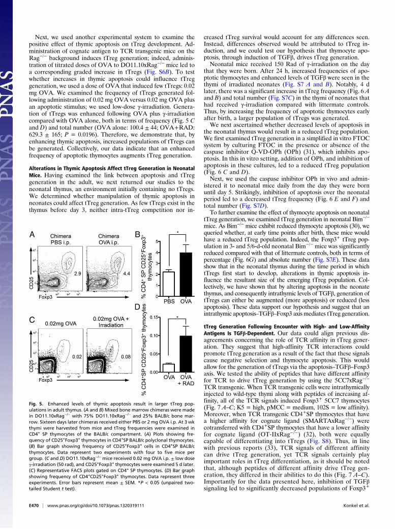
determine the generality of thymic apoptosis in driving TGFβproduction intrathymically. We exposed mice to γ-irradiation(Fig. 4A), or administered anti-CD3 (Fig. S5A), both of whichinduce apoptosis in the thymus. In both cases we saw increasedintrathymic TGFβ levels in mice that exhibited enhanced levelsof thymic apoptosis (γ-irradiation, Fig. 4 B–D; anti-CD3, Fig. S5B and C). Thus, these data suggest that enhanced thymic apo-ptosis leads to higher levels of intrathymic TGFβ.To directly assess whether thymic apoptosis was responsible
for this enhanced intrathymic TGFβ, we examined TGFβ levelsin Bim−/− thymi following induction of apoptosis. Thymocytes inBim−/− mice are more resistant to apoptosis as Bim is a proa-poptotic factor (30) (Fig. 4A). We found that the increased levelsof thymic TGFβ seen following γ-irradiation were indeed due to
increased apoptosis; TGFβ1 protein levels did not increase asmuch in Bim−/− thymi compared with control thymi followingγ-irradiation (Fig. 4B). This was also true for mRNA levels oftgfb1, -2, -3, and Smad7 in whole thymus and tgfb1 mRNA insorted thymic macrophages (Fig. 4 C and D). Therefore, in re-sponse to apoptotic stimuli, intrathymic TGFβ was reduced inBim−/− thymi compared with control thymi (γ-irradiation, Fig. 4;anti-CD3, Fig. S5). Collectively, our data indicate that thymicapoptosis stimulates production of TGFβ in the thymus.
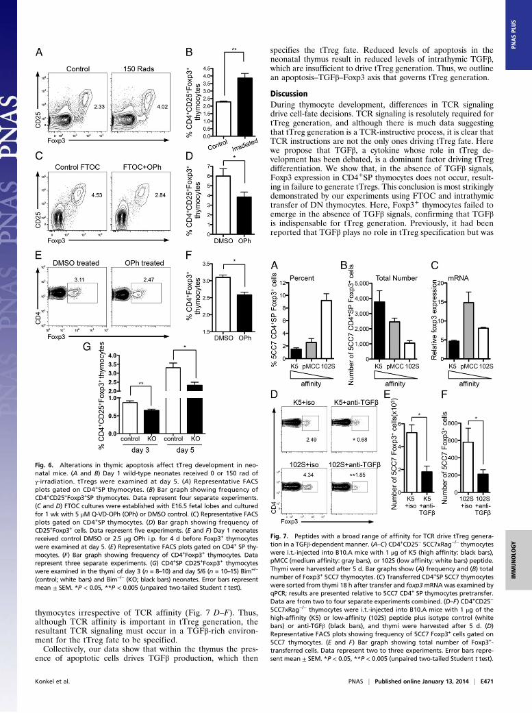
Thymic Apoptosis Promotes tTreg Generation.Having demonstratedthat increased thymic apoptosis caused increased levels of intra-thymic TGFβ, we next examined whether alterations in thymicapoptosis could affect tTreg generation. To do this, we experi-mentally altered levels of thymic apoptosis to determine whetherapoptosis, and subsequent TGFβ production, influenced tTreggeneration. First, we performed these experiments in the adultthymus, asking if increased levels of thymic apoptosis could promotetTreg generation (below). Second, we altered thymic apoptosis inday 1 neonates, which contain no tTregs (Alterations in ThymicApoptosis Affect tTreg Generation in Neonatal Mice).We generated mixed bone marrow chimeras in DO11.10xRag−/−
mice reconstituted with 75% DO11.10xRag−/− and 25% BALB/cbone marrow. Sixteen days after reconstitution chimeric mice re-ceived either 2 mg ovalbumin (OVA) or PBS intraperitoneally.Foxp3+CD4+SP thymocytes were examined 5 d later, a total of 3wk after reconstitution (Fig. S6A); tTregs were assessed 3 wk aftergeneration of chimeras to examine the Foxp3+ cells in a settingmost similar to that in neonates. OVA administration inducesapoptosis in only DO11.10xRag−/− thymocytes. We wanted todetermine whether increased apoptosis of DO11.10xRag−/−
thymocytes would influence generation of Foxp3+BALB/c poly-clonal thymocytes, our hypothesis being that there would be
Fig. 2. TGFβ levels increase in the thymus after birth. (A) Representative IHC sections of thymus from E18.5, day 1, and day 3 neonates stained (Upper) withantibody against active TGFβ1 (positive brown stain with counterstain in blue) and (Lower) H&E. Magnification is 10×. Sections are within five serial sectionsand are representative of sections from three to four separate mice. (B) Intrathymic amounts of active TGFβ1 were determined by ELISA in E18.5 fetal thymiand day 1, 2, 3, and 4 neonate thymi. Data are representative of two independent experiments with three to seven thymi per time point. (C) Flow cytometricanalysis of Smad2/3 phosphorylation in CD4+SP thymocytes from day 1, 3, and 5 neonatal thymus (n = 6–7). (D) CD4+SP thymocytes were FACS-sorted fromday 1–4 neonate thymi, and levels of Smad7 expression were examined by qPCR. Graph shows data from two separate experiments; results are presentedrelative to day 1. Error bars represent mean ± SEM. *P < 0.05 (unpaired two-tailed Student t test).
E468 | www.pnas.org/cgi/doi/10.1073/pnas.1320319111 Konkel et al.
increased Foxp3+BALB/c thymocytes in chimeras receiving OVA.Significantly more BALB/c Foxp3+CD4+SP tTregs were seen inthe thymi of chimeric mice that had received OVA i.p. com-
pared with controls (Fig. 5 A and B). Thus, we demonstratethat a thymic environment with higher levels of apoptosispromotes the generation of a larger population of tTregs.
Fig. 3. Increased thymocyte apoptosis leads to increased intrathymic TGFβ. (A–D) C57BL/6 mice were injected i.p. with 2 or 20 mg/kg DEX or DMSO control,and thymi were examined 18 h later. (A) FACS plots show AnnexinV versus 7AAD staining. (B) Intrathymic amounts of total TGFβ1 in whole thymi determinedby ELISA. (C) mRNA expression of tgfb1, tgfb2, tgfb3, Smad7, and tgif1 in whole thymi; results are presented relative to DMSO control. (D) DC, macrophages(Macs), and TECs were FACS-sorted from thymi of mice treated with DMSO (white bars) or 20 mg/kg DEX (black bars) 18 h after treatment and TGFβ expressionexamined by qPCR; results are presented relative to DMSO control. Data represent two to three experiments. (B and D) Data from two to three independentexperiments combined. Error bars represent mean ± SEM. *P < 0.05 (unpaired two-tailed Student t test).
Fig. 4. Thymocyte apoptosis increases intrathymic TGFβ. (A–D) Bim−/− and Bim+/− mice received 0 or 450 rad of γ-irradiation, and thymi were removed 18 h later.(A) FACS examination of AnnexinV and 7AAD staining of thymocytes. (B) Intrathymic amounts of total TGFβ1 in whole thymi determined by ELISA. (C) tgfb1expression in sorted thymic macrophages determined by qPCR; results are presented relative to unirradiated control of the same genotype. (D) mRNA expression oftgfb1, tgfb2, tgfb3, and Smad7 in whole thymi; results are presented relative to unirradiated control of same genotype. Data represent three experiments, apartfrom in C, which represents two separate experiments. Error bars represent mean ± SEM. *P < 0.05, **P < 0.005, ***P < 0.0001 (unpaired two-tailed Student t test).
Konkel et al. PNAS | Published online January 13, 2014 | E469
IMMUNOLO
GY
PNASPL
US
Next, we used another experimental system to examine thepositive effect of thymic apoptosis on tTreg development. Ad-ministration of cognate antigen to TCR transgenic mice on theRag−/− background induces tTreg generation; indeed, adminis-tration of titrated doses of OVA to DO11.10xRag−/− mice led toa corresponding graded increase in tTregs (Fig. S6B). To testwhether increases in thymic apoptosis could influence tTreggeneration, we used a dose of OVA that induced few tTregs: 0.02mg OVA. We examined the frequency of tTregs generated fol-lowing administration of 0.02 mg OVA versus 0.02 mg OVA plusan apoptotic stimulus; we used low-dose γ-irradiation. Genera-tion of tTregs was enhanced following OVA plus γ-irradiationcompared with OVA alone, both in terms of frequency (Fig. 5 Cand D) and total number (OVA alone: 100.4 ± 44; OVA+RAD:629.3 ± 165; P = 0.0196). Therefore, we demonstrate that, byenhancing thymic apoptosis, increased populations of tTregs canbe generated. Collectively, our data indicate that an enhancedfrequency of apoptotic thymocytes augments tTreg generation.
Alterations in Thymic Apoptosis Affect tTreg Generation in NeonatalMice. Having examined the link between apoptosis and tTreggeneration in the adult, we next returned our studies to theneonatal thymus, an environment initially containing no tTregs.We determined whether manipulation of thymic apoptosis inneonates could affect tTreg generation. As few tTregs exist in thethymus before day 3, neither intra-tTreg competition nor in-
creased tTreg survival would account for any differences seen.Instead, differences observed would be attributed to tTreg in-duction, and we could test our hypothesis that thymocyte apo-ptosis, through induction of TGFβ, drives tTreg generation.Neonatal mice received 150 Rad of γ-irradiation on the day
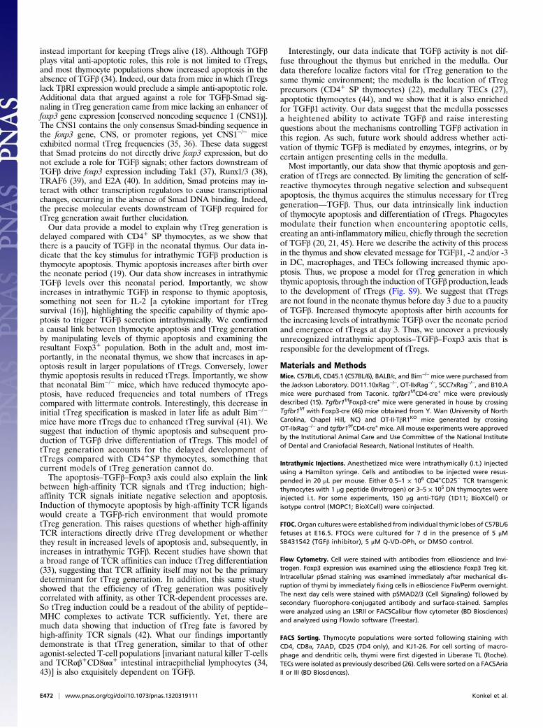
that they were born. After 24 h, increased frequencies of apo-ptotic thymocytes and enhanced levels of TGFβ were seen in thethymi of irradiated neonates (Fig. S7 A and B). Notably, 4 dlater, there was a significant increase in tTreg frequency (Fig. 6 Aand B) and total number (Fig. S7C) in the thymi of neonates thathad received γ-irradiation compared with littermate controls.Thus, by increasing the frequency of apoptotic thymocytes earlyafter birth, a larger population of tTregs was generated.We next ascertained whether decreased levels of apoptosis in
the neonatal thymus would result in a reduced tTreg population.We first examined tTreg generation in a simplified in vitro FTOCsystem by culturing FTOC in the presence or absence of thecaspase inhibitor Q-VD-OPh (OPh) (31), which inhibits apo-ptosis. In this in vitro setting, addition of OPh, and inhibition ofapoptosis in these cultures, led to a reduced tTreg population(Fig. 6 C and D).Next, we used the caspase inhibitor OPh in vivo and admin-
istered it to neonatal mice daily from the day they were bornuntil day 5. Strikingly, inhibition of apoptosis over the neonatalperiod led to a decreased tTreg frequency (Fig. 6 E and F) andtotal number (Fig. S7D).To further examine the effect of thymocyte apoptosis on neonatal
tTreg generation, we examined tTreg generation in neonatal Bim−/−
mice. As Bim−/− mice exhibit reduced thymocyte apoptosis (30), wequeried whether, at early time points after birth, these mice wouldhave a reduced tTreg population. Indeed, the Foxp3+ tTreg pop-ulation in 3- and 5/6-d-old neonatal Bim−/− mice was significantlyreduced compared with that of littermate controls, both in terms ofpercentage (Fig. 6G) and absolute number (Fig. S7E). These datashow that in the neonatal thymus during the time period in whichtTregs first start to develop, alterations in thymic apoptosis in-fluence the resultant size of the emerging tTreg population. Col-lectively, we have shown that by altering apoptosis in the neonatethymus, and consequently intrathymic levels of TGFβ, generation oftTregs can either be augmented (more apoptosis) or reduced (lessapoptosis). These data support our hypothesis and suggest that anintrathymic apoptosis–TGFβ–Foxp3 axis mediates tTreg generation.
tTreg Generation Following Encounter with High- and Low-AffinityAntigens Is TGFβ-Dependent. Our data could align previous dis-agreements concerning the role of TCR affinity in tTreg gener-ation. They suggest that high-affinity TCR interactions couldpromote tTreg generation as a result of the fact that these signalscause negative selection and thymocyte apoptosis. This wouldallow for the generation of tTregs via the apoptosis–TGFβ–Foxp3axis. We tested the ability of peptides that have different affinityfor TCR to drive tTreg generation by using the 5CC7xRag−/−
TCR transgenic. When TCR transgenic cells were intrathymicallyinjected to wild-type thymi along with peptides of increasing af-finity, all of the TCR signals induced Foxp3+ 5CC7 thymocytes(Fig. 7 A–C; K5 = high, pMCC = medium, 102S = low affinity).Moreover, when TCR transgenic CD4+SP thymocytes that havea higher affinity for cognate ligand (SMARTAxRag−/−) werecotransferred with CD4+SP thymocytes that have a lower affinityfor cognate ligand (OT-IIxRag−/−) (32), both were equallycapable of differentiating into tTregs (Fig. S8). Thus, in linewith previous reports (33), TCR signals of different affinitycan drive tTreg generation, yet TCR signals certainly playimportant roles in tTreg differentiation, as it should be notedthat, although peptides of different affinity drive tTreg gen-eration, they differed in their abilities to do this (Fig. 7 A–C).Importantly for the data presented here, inhibition of TGFβsignaling led to significantly decreased populations of Foxp3+
Fig. 5. Enhanced levels of thymic apoptosis result in larger tTreg pop-ulations in adult thymus. (A and B) Mixed bone marrow chimeras were madein DO11.10xRag−/− with 75% DO11.10xRag−/− and 25% BALB/c bone mar-row. Sixteen days later chimeras received either PBS or 2 mg OVA i.p. At 3 wkthymi were harvested from mice and tTreg frequencies were examined inCD4+ SP thymocytes of the BALB/c compartment. (A) Plots showing fre-quency of CD25+Foxp3+ thymocytes in CD4+SP BALB/c polyclonal thymocytes.(B) Bar graph showing frequency of CD25+Foxp3+ cells in CD4+SP BALB/cthymocytes. Data represent two experiments with four to five mice pergroup. (C and D) DO11.10xRag−/− mice received 0.02 mg OVA i.p. ± low doseγ-irradiation (50 rad), and CD25+Foxp3+ thymocytes were examined 5 d later.(C) Representative FACS plots gated on CD4+ SP thymocytes. (D) Bar graphshowing frequency of CD4+CD25+Foxp3+ thymocytes. Data represent threeexperiments. Error bars represent mean ± SEM. *P < 0.05 (unpaired two-tailed Student t test).
E470 | www.pnas.org/cgi/doi/10.1073/pnas.1320319111 Konkel et al.
thymocytes irrespective of TCR affinity (Fig. 7 D–F). Thus,although TCR affinity is important in tTreg generation, theresultant TCR signaling must occur in a TGFβ-rich environ-ment for the tTreg fate to be specified.Collectively, our data show that within the thymus the pres-
ence of apoptotic cells drives TGFβ production, which then
specifies the tTreg fate. Reduced levels of apoptosis in theneonatal thymus result in reduced levels of intrathymic TGFβ,which are insufficient to drive tTreg generation. Thus, we outlinean apoptosis–TGFβ–Foxp3 axis that governs tTreg generation.
DiscussionDuring thymocyte development, differences in TCR signalingdrive cell-fate decisions. TCR signaling is resolutely required fortTreg generation, and although there is much data suggestingthat tTreg generation is a TCR-instructive process, it is clear thatTCR instructions are not the only ones driving tTreg fate. Herewe propose that TGFβ, a cytokine whose role in tTreg de-velopment has been debated, is a dominant factor driving tTregdifferentiation. We show that, in the absence of TGFβ signals,Foxp3 expression in CD4+SP thymocytes does not occur, result-ing in failure to generate tTregs. This conclusion is most strikinglydemonstrated by our experiments using FTOC and intrathymictransfer of DN thymocytes. Here, Foxp3+ thymocytes failed toemerge in the absence of TGFβ signals, confirming that TGFβis indispensable for tTreg generation. Previously, it had beenreported that TGFβ plays no role in tTreg specification but was
Fig. 6. Alterations in thymic apoptosis affect tTreg development in neo-natal mice. (A and B) Day 1 wild-type neonates received 0 or 150 rad ofγ-irradiation. tTregs were examined at day 5. (A) Representative FACSplots gated on CD4+SP thymocytes. (B) Bar graph showing frequency ofCD4+CD25+Foxp3+SP thymocytes. Data represent four separate experiments.(C and D) FTOC cultures were established with E16.5 fetal lobes and culturedfor 1 wk with 5 μM Q-VD-OPh (OPh) or DMSO control. (C) Representative FACSplots gated on CD4+SP thymocytes. (D) Bar graph showing frequency ofCD25+Foxp3+ cells. Data represent five experiments. (E and F) Day 1 neonatesreceived control DMSO or 2.5 μg OPh i.p. for 4 d before Foxp3+ thymocyteswere examined at day 5. (E) Representative FACS plots gated on CD4+ SP thy-mocytes. (F) Bar graph showing frequency of CD4+Foxp3+ thymocytes. Datarepresent three separate experiments. (G) CD4+SP CD25+Foxp3+ thymocyteswere examined in the thymi of day 3 (n = 8–10) and day 5/6 (n = 10–15) Bim+/−
(control; white bars) and Bim−/− (KO; black bars) neonates. Error bars representmean ± SEM. *P < 0.05, **P < 0.005 (unpaired two-tailed Student t test).
Fig. 7. Peptides with a broad range of affinity for TCR drive tTreg genera-tion in a TGFβ-dependent manner. (A–C) CD4+CD25− 5CC7xRag−/− thymocyteswere i.t.-injected into B10.A mice with 1 μg of K5 (high affinity: black bars),pMCC (medium affinity: gray bars), or 102S (low affinity: white bars) peptide.Thymi were harvested after 5 d. Bar graphs show (A) frequency and (B) totalnumber of Foxp3+ 5CC7 thymocytes. (C) Transferred CD4+SP 5CC7 thymocyteswere sorted from thymi 18 h after transfer and foxp3mRNAwas examined byqPCR; results are presented relative to 5CC7 CD4+ SP thymocytes pretransfer.Data are from two to four separate experiments combined. (D–F) CD4+CD25−
5CC7xRag−/− thymocytes were i.t.-injected into B10.A mice with 1 μg of thehigh-affinity (K5) or low-affinity (102S) peptide plus isotype control (whitebars) or anti-TGFβ (black bars), and thymi were harvested after 5 d. (D)Representative FACS plots showing frequency of 5CC7 Foxp3+ cells gated on5CC7 thymocytes. (E and F) Bar graph showing total number of Foxp3+-transferred cells. Data represent two to three experiments. Error bars repre-sent mean ± SEM. *P < 0.05, **P < 0.005 (unpaired two-tailed Student t test).
Konkel et al. PNAS | Published online January 13, 2014 | E471
IMMUNOLO
GY
PNASPL
US
instead important for keeping tTregs alive (18). Although TGFβplays vital anti-apoptotic roles, this role is not limited to tTregs,and most thymocyte populations show increased apoptosis in theabsence of TGFβ (34). Indeed, our data from mice in which tTregslack TβRI expression would preclude a simple anti-apoptotic role.Additional data that argued against a role for TGFβ-Smad sig-naling in tTreg generation came from mice lacking an enhancer offoxp3 gene expression [conserved noncoding sequence 1 (CNS1)].The CNS1 contains the only consensus Smad-binding sequence inthe foxp3 gene, CNS, or promoter regions, yet CNS1−/− miceexhibited normal tTreg frequencies (35, 36). These data suggestthat Smad proteins do not directly drive foxp3 expression, but donot exclude a role for TGFβ signals; other factors downstream ofTGFβ drive foxp3 expression including Tak1 (37), Runx1/3 (38),TRAF6 (39), and E2A (40). In addition, Smad proteins may in-teract with other transcription regulators to cause transcriptionalchanges, occurring in the absence of Smad DNA binding. Indeed,the precise molecular events downstream of TGFβ required fortTreg generation await further elucidation.Our data provide a model to explain why tTreg generation is
delayed compared with CD4+ SP thymocytes, as we show thatthere is a paucity of TGFβ in the neonatal thymus. Our data in-dicate that the key stimulus for intrathymic TGFβ production isthymocyte apoptosis. Thymic apoptosis increases after birth overthe neonate period (19). Our data show increases in intrathymicTGFβ levels over this neonatal period. Importantly, we showincreases in intrathymic TGFβ in response to thymic apoptosis,something not seen for IL-2 [a cytokine important for tTregsurvival (16)], highlighting the specific capability of thymic apo-ptosis to trigger TGFβ secretion intrathymically. We confirmeda causal link between thymocyte apoptosis and tTreg generationby manipulating levels of thymic apoptosis and examining theresultant Foxp3+ population. Both in the adult and, most im-portantly, in the neonatal thymus, we show that increases in ap-optosis result in larger populations of tTregs. Conversely, lowerthymic apoptosis results in reduced tTregs. Importantly, we showthat neonatal Bim−/− mice, which have reduced thymocyte apo-ptosis, have reduced frequencies and total numbers of tTregscompared with littermate controls. Interestingly, this decrease ininitial tTreg specification is masked in later life as adult Bim−/−
mice have more tTregs due to enhanced tTreg survival (41). Wesuggest that induction of thymic apoptosis and subsequent pro-duction of TGFβ drive differentiation of tTregs. This model oftTreg generation accounts for the delayed development oftTregs compared with CD4+SP thymocytes, something thatcurrent models of tTreg generation cannot do.The apoptosis–TGFβ–Foxp3 axis could also explain the link
between high-affinity TCR signals and tTreg induction; high-affinity TCR signals initiate negative selection and apoptosis.Induction of thymocyte apoptosis by high-affinity TCR ligandswould create a TGFβ-rich environment that would promotetTreg generation. This raises questions of whether high-affinityTCR interactions directly drive tTreg development or whetherthey result in increased levels of apoptosis and, subsequently, inincreases in intrathymic TGFβ. Recent studies have shown thata broad range of TCR affinities can induce tTreg differentiation(33), suggesting that TCR affinity itself may not be the primarydeterminant for tTreg generation. In addition, this same studyshowed that the efficiency of tTreg generation was positivelycorrelated with affinity, as other TCR-dependent processes are.So tTreg induction could be a readout of the ability of peptide–MHC complexes to activate TCR sufficiently. Yet, there aremuch data showing that induction of tTreg fate is favored byhigh-affinity TCR signals (42). What our findings importantlydemonstrate is that tTreg generation, similar to that of otheragonist-selected T-cell populations [invariant natural killer T-cellsand TCRαβ+CD8αα+ intestinal intraepithelial lymphocytes (34,43)] is also exquisitely dependent on TGFβ.
Interestingly, our data indicate that TGFβ activity is not dif-fuse throughout the thymus but enriched in the medulla. Ourdata therefore localize factors vital for tTreg generation to thesame thymic environment; the medulla is the location of tTregprecursors (CD4+ SP thymocytes) (22), medullary TECs (27),apoptotic thymocytes (44), and we show that it is also enrichedfor TGFβ1 activity. Our data suggest that the medulla possessesa heightened ability to activate TGFβ and raise interestingquestions about the mechanisms controlling TGFβ activation inthis region. As such, future work should address whether acti-vation of thymic TGFβ is mediated by enzymes, integrins, or bycertain antigen presenting cells in the medulla.Most importantly, our data show that thymic apoptosis and gen-
eration of tTregs are connected. By limiting the generation of self-reactive thymocytes through negative selection and subsequentapoptosis, the thymus acquires the stimulus necessary for tTreggeneration—TGFβ. Thus, our data intrinsically link inductionof thymocyte apoptosis and differentiation of tTregs. Phagocytesmodulate their function when encountering apoptotic cells,creating an anti-inflammatory milieu, chiefly through the secretionof TGFβ (20, 21, 45). Here we describe the activity of this processin the thymus and show elevated message for TGFβ1, -2 and/or -3in DC, macrophages, and TECs following increased thymic apo-ptosis. Thus, we propose a model for tTreg generation in whichthymic apoptosis, through the induction of TGFβ production, leadsto the development of tTregs (Fig. S9). We suggest that tTregsare not found in the neonate thymus before day 3 due to a paucityof TGFβ. Increased thymocyte apoptosis after birth accounts forthe increasing levels of intrathymic TGFβ over the neonate periodand emergence of tTregs at day 3. Thus, we uncover a previouslyunrecognized intrathymic apoptosis–TGFβ–Foxp3 axis that isresponsible for the development of tTregs.
Materials and MethodsMice. C57BL/6, CD45.1 (C57BL/6), BALB/c, and Bim−/−mice were purchased fromthe Jackson Laboratory. DO11.10xRag−/−, OT-IIxRag−/−, 5CC7xRag−/−, and B10.Amice were purchased from Taconic. tgfbr1f/fCD4-cre+ mice were previouslydescribed (15). Tgfbr1f/fFoxp3-cre+ mice were generated in house by crossingTgfbr1f/f with Foxp3-cre (46) mice obtained from Y. Wan (University of NorthCarolina, Chapel Hill, NC) and OT-II-TβR1KO mice generated by crossingOT-IIxRag−/− and tgfbr1f/fCD4-cre+ mice. All mouse experiments were approvedby the Institutional Animal Care and Use Committee of the National Instituteof Dental and Craniofacial Research, National Institutes of Health.
Intrathymic Injections. Anesthetized mice were intrathymically (i.t.) injectedusing a Hamilton syringe. Cells and antibodies to be injected were resus-pended in 20 μL per mouse. Either 0.5–1 × 10⁶ CD4+CD25− TCR transgenicthymocytes with 1 μg peptide (Invitrogen) or 3–5 × 10⁵ DN thymocytes wereinjected i.t. For some experiments, 150 μg anti-TGFβ (1D11; BioXCell) orisotype control (MOPC1; BioXCell) were coinjected.
FTOC.Organ cultures were established from individual thymic lobes of C57BL/6fetuses at E16.5. FTOCs were cultured for 7 d in the presence of 5 μMSB431542 (TGFβ inhibitor), 5 μM Q-VD-OPh, or DMSO control.
Flow Cytometry. Cell were stained with antibodies from eBioscience and Invi-trogen. Foxp3 expression was examined using the eBioscience Foxp3 Treg kit.Intracellular pSmad staining was examined immediately after mechanical dis-ruption of thymi by immediately fixing cells in eBioscience Fix/Perm overnight.The next day cells were stained with pSMAD2/3 (Cell Signaling) followed bysecondary fluorophore-conjugated antibody and surface-stained. Sampleswere analyzed using an LSRII or FACSCalibur flow cytometer (BD Biosciences)and analyzed using FlowJo software (Treestar).
FACS Sorting. Thymocyte populations were sorted following staining withCD4, CD8α, 7AAD, CD25 (7D4 only), and KJ1-26. For cell sorting of macro-phage and dendritic cells, thymi were first digested in Liberase TL (Roche).TECs were isolated as previously described (26). Cells were sorted on a FACSAriaII or III (BD Biosciences).
E472 | www.pnas.org/cgi/doi/10.1073/pnas.1320319111 Konkel et al.
In Vitro Generation of tTregs. CD4+SP thymocytes (CD4+CD8−CD25−GFP−)were FACS-sorted from Foxp3GFP mice and cultured as previously described(24). TGFβ inhibitor SB431542 (5 μM; Selleck Chemicals) or DMSO and anti-TGFβ (50 μg/mL; 1D11) or isotype control (50 μg/mL; MOPC21) were addedfor the entire culture period.
Real-Time PCR. RNA was extracted from thymus or cells and reversed-tran-scribed. Quantitative PCR was done according to the protocol of TaqMan geneexpression assay kits (Applied Biosystems). For Tgfbr1 detection in cells sortedfrom Tgfbr1f/fFoxp3-cre+ mice, previously described primers were used (15).
ELISA. Thymus samples were lysed in defined volumes of buffer and proteinconcentration of the supernatant determined. Quantification of TGFβ1,-2, and -3 was determined using TGFβ ELISA kits (TGFβ1: Promega; TGFβ2and -3: R&D Systems) by loading equal amounts of protein.
Generation of Bone-Marrow Chimeras. Bone marrow was isolated from miceand depleted of T cells using anti-CD90 beads (Miltenyi Biotech). Cells (∼5 ×10⁶) were then injected i.v. at the indicated ratios into irradiated hosts.
Immunohistochemistry. Thymi were formalin-fixed and embedded in par-affin. Sections were cut and antigen retrieval performed. Staining for activeTGFβ1 was done using the antibody LC(1–30) (25), and detection wasperformed using the immPRESS anti-rabbit reagent kit and ImmPACTDAB kit (both Vector Labs). Slides were scanned by an Aperio ScanScope(Aperio Technologies).
Statistical Analysis. Unless otherwise noted, statistical analysis was performedusing the unpaired two-tailed Student t test in Prism (GraphPad).
ACKNOWLEDGMENTS. We thank Dr. N. Moutsopoulos and D. Zhang fortechnical expertise; Drs. J. J. O’Shea and E. Wohlfert for critical reading ofthe manuscript; Dr. K. Flanders for the LC(1-30) antibody and staining pro-tocols; the National Heart, Lung, and Blood Institute and National Instituteof Dental and Craniofacial Research (NIDCR) FACS cores for cell sorting;Dr. B. J. Fowlkes for FTOC advice; and Dr. K. Tarbell for use of equipment.This work was supported by the Intramural Research Programs of the NIDCRand of the National Institute of Allergy and Infectious Diseases, NationalInstitutes of Health.
1. Hori S, Nomura T, Sakaguchi S (2003) Control of regulatory T cell development by thetranscription factor Foxp3. Science 299(5609):1057–1061.
2. Fontenot JD, et al. (2005) Regulatory T cell lineage specification by the forkheadtranscription factor foxp3. Immunity 22(3):329–341.
3. Fontenot JD, Dooley JL, Farr AG, Rudensky AY (2005) Developmental regulation ofFoxp3 expression during ontogeny. J Exp Med 202(7):901–906.
4. Jordan MS, et al. (2001) Thymic selection of CD4+CD25+ regulatory T cells induced byan agonist self-peptide. Nat Immunol 2(4):301–306.
5. Apostolou I, Sarukhan A, Klein L, von Boehmer H (2002) Origin of regulatory T cellswith known specificity for antigen. Nat Immunol 3(8):756–763.
6. Moran AE, et al. (2011) T cell receptor signal strength in Treg and iNKT cell de-velopment demonstrated by a novel fluorescent reporter mouse. J Exp Med 208(6):1279–1289.
7. van Santen HM, Benoist C, Mathis D (2004) Number of T reg cells that differentiatedoes not increase upon encounter of agonist ligand on thymic epithelial cells. J ExpMed 200(10):1221–1230.
8. Tai X, Cowan M, Feigenbaum L, Singer A (2005) CD28 costimulation of developing thy-mocytes induces Foxp3 expression and regulatory T cell differentiation independently ofinterleukin 2. Nat Immunol 6(2):152–162.
9. Mantel PY, et al. (2006) Molecular mechanisms underlying FOXP3 induction in humanT cells. J Immunol 176(6):3593–3602.
10. Kerdiles YM, et al. (2010) Foxo transcription factors control regulatory T cell de-velopment and function. Immunity 33(6):890–904.
11. Long M, Park SG, Strickland I, Hayden MS, Ghosh S (2009) Nuclear factor-kappaBmodulates regulatory T cell development by directly regulating expression of Foxp3transcription factor. Immunity 31(6):921–931.
12. Ruan Q, et al. (2009) Development of Foxp3(+) regulatory t cells is driven by the c-Relenhanceosome. Immunity 31(6):932–940.
13. Burchill MA, et al. (2008) Linked T cell receptor and cytokine signaling govern thedevelopment of the regulatory T cell repertoire. Immunity 28(1):112–121.
14. Lio CW, Hsieh CS (2008) A two-step process for thymic regulatory T cell development.Immunity 28(1):100–111.
15. Liu Y, et al. (2008) A critical function for TGF-beta signaling in the development ofnatural CD4+CD25+Foxp3+ regulatory T cells. Nat Immunol 9(6):632–640.
16. Tai X, et al. (2013) Foxp3 transcription factor is proapoptotic and lethal to developingregulatory T cells unless counterbalanced by cytokine survival signals. Immunity 38(6):1116–1128.
17. ChenW, et al. (2003) Conversion of peripheral CD4+CD25- naive T cells to CD4+CD25+regulatory T cells by TGF-beta induction of transcription factor Foxp3. J Exp Med 198(12):1875–1886.
18. Ouyang W, Beckett O, Ma Q, Li MO (2010) Transforming growth factor-beta signalingcurbs thymic negative selection promoting regulatory T cell development. Immunity32(5):642–653.
19. Surh CD, Sprent J (1994) T-cell apoptosis detected in situ during positive and negativeselection in the thymus. Nature 372(6501):100–103.
20. Fadok VA, et al. (1998) Macrophages that have ingested apoptotic cells in vitro inhibitproinflammatory cytokine production through autocrine/paracrine mechanisms in-volving TGF-beta, PGE2, and PAF. J Clin Invest 101(4):890–898.
21. Xiao YQ, et al. (2008) Transcriptional and translational regulation of TGF-beta pro-duction in response to apoptotic cells. J Immunol 181(5):3575–3585.
22. Lee HM, Hsieh CS (2009) Rare development of Foxp3+ thymocytes in the CD4+CD8+subset. J Immunol 183(4):2261–2266.
23. Wirnsberger G, Mair F, Klein L (2009) Regulatory T cell differentiation of thymocytesdoes not require a dedicated antigen-presenting cell but is under T cell-intrinsic de-velopmental control. Proc Natl Acad Sci USA 106(25):10278–10283.
24. Sauer S, et al. (2008) T cell receptor signaling controls Foxp3 expression via PI3K, Akt,and mTOR. Proc Natl Acad Sci USA 105(22):7797–7802.
25. Flanders KC, et al. (1989) Transforming growth factor-beta 1: Histochemical locali-zation with antibodies to different epitopes. J Cell Biol 108(2):653–660.
26. Aschenbrenner K, et al. (2007) Selection of Foxp3+ regulatory T cells specific for selfantigen expressed and presented by Aire+ medullary thymic epithelial cells. Nat Im-munol 8(4):351–358.
27. Cowan JE, et al. (2013) The thymic medulla is required for Foxp3+ regulatory but notconventional CD4+ thymocyte development. J Exp Med 210(4):675–681.
28. Nakao A, et al. (1997) Identification of Smad7, a TGFbeta-inducible antagonist of TGF-beta signalling. Nature 389(6651):631–635.
29. Chen F, et al. (2003) Regulation of TG-interacting factor by transforming growthfactor-beta. Biochem J 371(Pt 2):257–263.
30. Bouillet P, et al. (2002) BH3-only Bcl-2 family member Bim is required for apoptosis ofautoreactive thymocytes. Nature 415(6874):922–926.
31. Caserta TM, Smith AN, Gultice AD, Reedy MA, Brown TL (2003) Q-VD-OPh, a broadspectrum caspase inhibitor with potent antiapoptotic properties. Apoptosis 8(4):345–352.
32. Mandl JN, Monteiro JP, Vrisekoop N, Germain RN (2013) T cell-positive selection usesself-ligand binding strength to optimize repertoire recognition of foreign antigens.Immunity 38(2):263–274.
33. Lee HM, Bautista JL, Scott-Browne J, Mohan JF, Hsieh CS (2012) A broad range of self-reactivity drives thymic regulatory T cell selection to limit responses to self. Immunity37(3):475–486.
34. Konkel JE, et al. (2011) Control of the development of CD8αα+ intestinal intra-epithelial lymphocytes by TGF-β. Nat Immunol 12(4):312–319.
35. Zheng Y, et al. (2010) Role of conserved non-coding DNA elements in the Foxp3 genein regulatory T-cell fate. Nature 463(7282):808–812.
36. Schlenner SM, Weigmann B, Ruan Q, Chen Y, von Boehmer H (2012) Smad3 bindingto the foxp3 enhancer is dispensable for the development of regulatory T cells withthe exception of the gut. J Exp Med 209(9):1529–1535.
37. Wan YY, Chi H, Xie M, Schneider MD, Flavell RA (2006) The kinase TAK1 integratesantigen and cytokine receptor signaling for T cell development, survival and function.Nat Immunol 7(8):851–858.
38. Bruno L, et al. (2009) Runx proteins regulate Foxp3 expression. J Exp Med 206(11):2329–2337.
39. Shimo Y, et al. (2011) TRAF6 directs commitment to regulatory T cells in thymocytes.Genes Cells 16(4):437–447.
40. Maruyama T, et al. (2011) Control of the differentiation of regulatory T cells and T(H)17 cells by the DNA-binding inhibitor Id3. Nat Immunol 12(1):86–95.
41. Zhan Y, et al. (2011) Defects in the Bcl-2-regulated apoptotic pathway lead to pref-erential increase of CD25 low Foxp3+ anergic CD4+ T cells. J Immunol 187(4):1566–1577.
42. Hsieh CS, Lee HM, Lio CW (2012) Selection of regulatory T cells in the thymus. Nat RevImmunol 12(3):157–167.
43. Doisne JM, et al. (2009) iNKT cell development is orchestrated by different branchesof TGF-beta signaling. J Exp Med 206(6):1365–1378.
44. Stritesky GL, et al. (2013) Murine thymic selection quantified using a unique methodto capture deleted T cells. Proc Natl Acad Sci USA 110(12):4679–4684.
45. Perruche S, et al. (2008) CD3-specific antibody-induced immune tolerance involvestransforming growth factor-beta from phagocytes digesting apoptotic T cells. NatMed 14(5):528–535.
46. Zhou X, et al. (2008) Selective miRNA disruption in T reg cells leads to uncontrolledautoimmunity. J Exp Med 205(9):1983–1991.
Konkel et al. PNAS | Published online January 13, 2014 | E473
IMMUNOLO
GY
PNASPL
US


![Thymoglobulin (anti-thymocyte globulin [rabbit]) · 2020. 12. 14. · DESCRIPTION . Thymoglobulin® (Anti-thymocyte globulin [rabbit]) is a purified, pasteurized, gamma immune globulin](https://img.pdfslide.net/doc/110x75/60c2dece3812e518472963b9/thymoglobulin-anti-thymocyte-globulin-rabbit-2020-12-14-description-thymoglobulin.jpg)