Embed Size (px)
Citation preview
Rheumatology, Thyroid Dysfunction and the Eye
February 24, 2018
Greg A Caldwell, OD, FAAO [email protected] 814-931-2030 cell 1
Greg Caldwell OD, FAAO2018 TOA Annual Convention
February 24, 2018
Rheumatology, Thyroid Dysfunction and the Eye
Disclosure Statement(next slide)
Disclosures- Greg Caldwell, OD, FAAO
$ Will mention many products, instruments and companies during our discussion
¬ I don�t have any financial interest in any of these products, instruments or companies
$ Pennsylvania Optometric Association –President 2010
2 POA Board of Directors 2006-2011 $ American Optometric Association, Trustee 2013-2016
¬ Thank you to the members and those who join $ I never used or will use my volunteer positions to further my lecturing
career$ Lectured for: Shire, BioTissue, Optovue
$ Advisory Board: Allergan
$ Envolve: PA Medical Director, Credential Committee $ Optometric Education Consultants- Scottsdale and Quebec City, Owner
Learning Objectives$Enhance clinical understanding of rheumatology and
thyroid dysfunction and their ocular associations
$Enhance clinical diagnosis of ocular manifestations of rheumatologic diseases and thyroid disease
$Enhance clinical management and treatment of ocular manifestations of rheumatologic diseases and thyroid eye disease
$Increase comfort level when ordering or interpreting laboratory tests in rheumatologic and thyroid diseases
$Gain confidence in working closer with rheumatology and endocrinology
Thyroid Disease and
Thyroid Eye Disease
Thyroid$Thyroid is an endocrine gland$Two types of glands
¬ Endocrine
¬ Exocrine
$Endocrine system is a control system of ductless endocrine glands that secrete hormones (chemical messenger) that circulate within the body via the bloodstream or lymph system to affect distant organs
¬ Hypothalamus
¬ Pituitary gland
¬ Thyroid
¬ Parathyroid glands
¬ Pancreas
¬ Adrenal glands¬ Gonads (testes and ovaries)
¬ Pineal gland
Thyroid
$Exocrine glands contain ducts. Ducts are tubes leading from a gland to its target organ¬ Digestive glands have ducts for releasing the digestive enzymes¬ Salivary glands, sweat glands and glands within the
gastrointestinal tract
$Pancreas is both endocrine and exocrine¬ Exocrine (ducted gland) secreting digestive enzymes into the small
intestine. ¬ Endocrine (ductless gland) in that the islets of Langerhans secrete
insulin and glucagon to regulate the blood sugar level.
Rheumatology, Thyroid Dysfunction and the Eye
February 24, 2018
Greg A Caldwell, OD, FAAO [email protected] 814-931-2030 cell 2
Thyroid
$Largest endocrine gland in the body$Butterfly shaped
$Two lobes located on either side of the trachea in the lower portion of the neck
$Lies just below skin and muscle layer surface
$The thyroid is controlled by the hypothalamus and pituitary
$The primary function of the thyroid is production of the hormones thyroxine (T4), triiodothyronine (T3), and calcitonin
Thyroid
$Thyroid regulates: heart rate, ventilation rate, metabolic rate, and development of cells
$Thyroid disorder- approx 1 in 13 or 7.35% or 20 million people in USA, estimated 2 million undiagnosed
$Diabetes- approx 1 in 13 or 7.8% or 17.9 million people in USA , 5.7 million undiagnosed
$Pathophysiology: >40 postulates (thyroid)
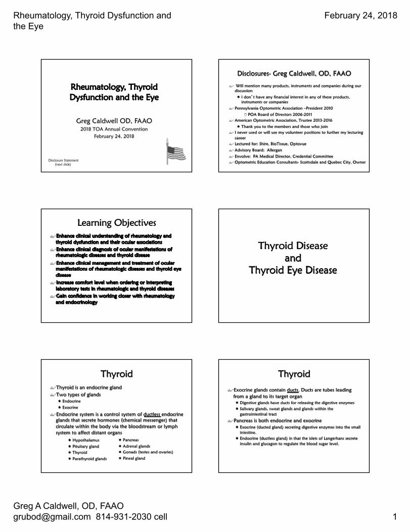
Normal Thyroid Function Discussion
Thyroid Dysfunction
$What is the most common cause of thyroid dysfunction?A. Cancer
B. Surgically inducedC. Medication toxicity or side effect
D. PregnancyE. Autoimmune disease
$ In autoimmune disease the body typically produces ______ that attacks itself, this can be systemic or organ specific¬ Antibodies, immunoglobulins
Thyroid Dysfunction
$Primary=Thyroid gland$Secondary= Pituitary failure
$Tertiary= Hypothalamic
Rheumatology, Thyroid Dysfunction and the Eye
February 24, 2018
Greg A Caldwell, OD, FAAO [email protected] 814-931-2030 cell 3
Antibodies of Thyroid Dysfunction
$TSH Receptor Antibodies¬ Stimulating TSH receptor antibody
2 Thyroid Stimulating Immunoglobulin (TSI)
¬ Thyroid blocking antibody (TBAb)
$Thyroid Peroxidase Antibodies (TPOAb)¬ TPO is found in thyroid follicle cells where it converts the thyroid
hormone T4 to T3
¬ TPOAb contributes to thyroid cellular destruction
$ Most autoimmune thyroid dysfunctions have a combination of thyroid antibodies, however depending on which AB is more abundant results in the outcome of the disease
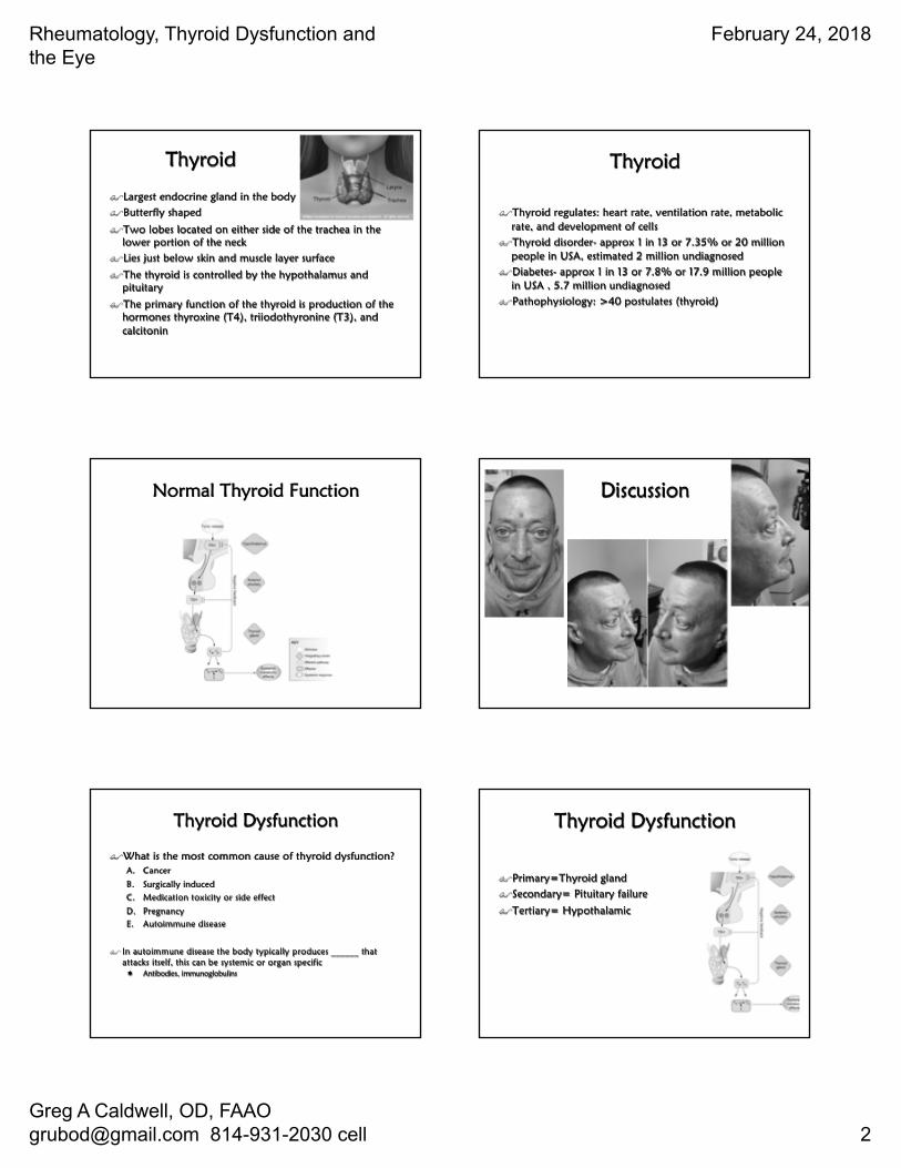
Hyperthyroid
$TSI attacks the thyroid
$T3 and T4 increase$TSH decreases
Hypothyroid
$TBAb attacks the thyroid
$T3 and T4 decrease$TSH increases
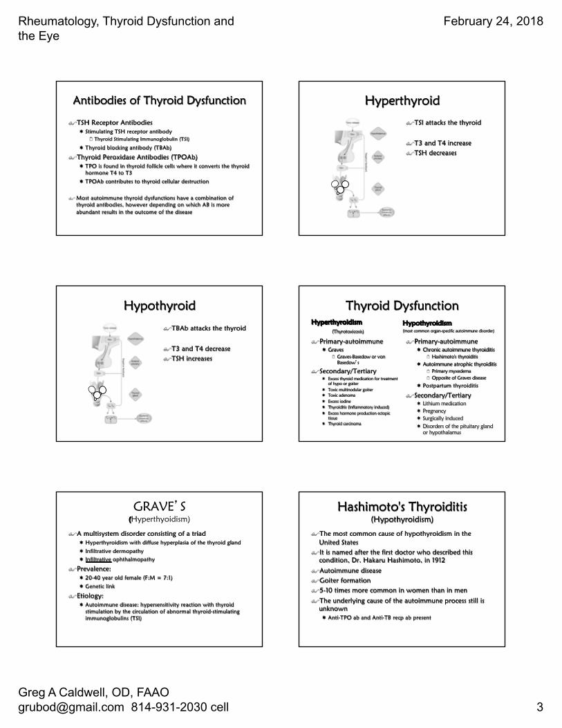
Thyroid DysfunctionHyperthyroidism
(Thyrotoxicosis)
$Primary-autoimmune¬ Graves
2 Graves-Basedow or von Basedow�s
$Secondary/Tertiary¬ Excess thyroid medication for treatment
of hypo or goiter¬ Toxic multinodular goiter¬ Toxic adenoma¬ Excess iodine¬ Thyroiditis (inflammatory induced)¬ Excess hormone production ectopic
tissue¬ Thyroid carcinoma
Hypothyroidism (most common organ-specific autoimmune disorder)
$Primary-autoimmune¬ Chronic autoimmune thyroiditis
2 Hashimoto's thyroiditis
¬ Autoimmune atrophic thyroiditis2 Primary myxedema2 Opposite of Graves disease
¬ Postpartum thyroiditis
$Secondary/Tertiary¬ Lithium medication¬ Pregnancy¬ Surgically induced¬ Disorders of the pituitary gland
or hypothalamus
GRAVE�S(Hyperthyoidism)
$A multisystem disorder consisting of a triad ¬ Hyperthyroidism with diffuse hyperplasia of the thyroid gland
¬ Infiltrative dermopathy¬ Infiltrative ophthalmopathy
$Prevalence: ¬ 20-40 year old female (F:M = 7:1)
¬ Genetic link
$Etiology: ¬ Autoimmune disease: hypersensitivity reaction with thyroid
stimulation by the circulation of abnormal thyroid-stimulating immunoglobulins (TSI)
Hashimoto's Thyroiditis(Hypothyroidism)
$The most common cause of hypothyroidism in the United States
$It is named after the first doctor who described this condition, Dr. Hakaru Hashimoto, in 1912
$Autoimmune disease$Goiter formation$5-10 times more common in women than in men
$The underlying cause of the autoimmune process still is unknown¬ Anti-TPO ab and Anti-TB recp ab present
Rheumatology, Thyroid Dysfunction and the Eye
February 24, 2018
Greg A Caldwell, OD, FAAO [email protected] 814-931-2030 cell 4
Autoimmune atrophic thyroiditis(Hypothyroidism)
$Atrophic thyroiditis is similar to Hashimoto's thyroiditis$A goiter is not present
Postpartum Thyroiditis (Hypothyroidism)
$These women develop antibodies to their own thyroid during pregnancy, causing an inflammation of the thyroid after delivery
Systemic Manifestations of Hyperthyroid(Primary or Secondary)
$Symptoms¬ Nervousness¬ Heat intolerance¬ Sweating¬ Fatigue¬ Palpitation¬ Insomnia¬ Early waking¬ Alopecia¬ Vitiligo¬ Brittle nails
$Signs¬ Sweating¬ Muscle Weakness¬ Emotionally labile ¬ Tremor¬ Tachycardia¬ Arrhythmia¬ Hypertension¬ Brisk tendon reflex¬ Diabetes¬ ↑Triglycerides & Ca, ↓CHO¬ Microcyticanemia¬ Possible goiter¬ Myxedema
Systemic Manifestations of Hypothyroid(Primary or Secondary)
$Symptoms¬ Cold intolerance
¬ Weakness¬ Reduced energy
¬ Lethargy¬ Muscle cramps
¬ Constipation
¬ Increased sleeping
¬ Weight gain
¬ Reduced appetite¬ Joint stiffness
$Signs¬ Cool, scaling skin
¬ Puffy hands and face¬ Deep voice
¬ Myotonia¬ Delirium
¬ Bradycardia
¬ Slow reflexes
¬ Obesity
¬ Hypothermia¬ Myxedema
Thyroid Eye Disease (TED)
$Other names used¬ Grave�s disease
¬ Grave's ophthalmopathy¬ Grave's orbitopathy
¬ Exophthalmos in Graves Disease ¬ Thyroid Associated Orbitopathy (TAO)
¬ Thyroid Orbitopathy
¬ Ophthalmic Graves Disease ¬ Inflammatory Eye Disease
¬ Endocrine Orbitopathy
Why is this so confusing?
$Thyroid Eye Disease¬ Is often seen in conjunction with Graves' Disease (hyperthyroid)
¬ Is seen in people with no other evidence of thyroid dysfunction¬ Is seen in patients who have Hashimoto's Disease (hypothyroid)
$Most thyroid patients, however, will not develop thyroid eye disease
Rheumatology, Thyroid Dysfunction and the Eye
February 24, 2018
Greg A Caldwell, OD, FAAO [email protected] 814-931-2030 cell 5
Why is this so confusing?$ The eye symptoms usually occur at the same time as the thyroid
disease¬ However they may precede or follow the obvious symptoms of the thyroid
abnormality
$ The incidence of thyroid eye disease associated with thyroid dysfunction is higher and more severe in smokers¬ There is no way to predict which thyroid patients will be affected
Why is this so confusing?$ While eye disease may be brought on by thyroid dysfunction
¬ Successful treatment of the thyroid gland does not guarantee that the eye disease will improve
¬ No particular thyroid treatment can guarantee that the eyes will not continue to deteriorate
¬ Once inflamed, the eye disease may remain active from several months to as long as three years
¬ There may be a gradual or, in some cases, a complete improvement
Thyroid Eye Disease $ Commonly known as Graves' ophthalmopathy$ About 80% of all patients with TED have the autoimmune hyperthyroid
disorder known as Graves' disease$ Another 10% of all cases are seen in patients with autoimmune
hypothyroidism, either Hashimoto's thyroiditis, atrophic thyroiditis or Hashitoxicosis
$ Another 10% of all cases are seen in people with normal thyroid function¬ When thyroid function is normal, the eye condition is referred to as euthyroid
Graves' disease¬ Euthyroid is a term meaning that thyroid function tests are normal. Most people
with euthyroid Graves' disease develop a thyroid disorder within eighteen months of the emergence of the eye disorder
¬ But some people with euthyroid Graves' disease never develop thyroid dysfunction
Thyroid Eye Disease
$What causes the Thyroid Eye Disease signs and symptoms?
$The high and low levels of T3 and T4
$The antibodies that are attacking the thyroid gland
Thyroid Eye Disease
$Thyroid Eye Disease has 2 phases¬ A phase secondary to abnormal thyroid hormone levels
2 Increased or decreased FT3 and FT4 levels2 Once these levels are normalized, ocular symptoms will resolve
¬ Congestive Autoimmune form of Thyroid Eye Disease2 Active phase-stimulating or blocking TRAb are causing ocular activity2 Plateau phase-reduced activity2 Resolution phase-symptoms regress and eyes return to normal
Phase secondary to abnormal thyroid hormone levels (T3/T4)(Thyroid Eye Disease)
$ Hyperthyroidism eye symptoms¬ Excess hormone acting on the nerves
that supply the eye ¬ Usually spastic and include staring¬ Dryness¬ Eyelid retraction
$ Hypothyroidism eye symptoms¬ Deficient hormone causing venous
congestion, impaired circulation and fluid stagnation
¬ Periorbital edema
$ This form of TED resolves within a few weeks after thyroid hormone levels (FT4 and FT3) are corrected and brought back into the normal range
$ The pituitary hormone TSH can stay low or suppressed for many months during the course of treatment for hyperthyroidism and doesn't mean that the patient is still hyperthyroid
$ TSH also lags at least 6 weeks behind thyroid hormone levels and often remains elevated longer in people who have been hypothyroid
$ Relying on the TSH level can be misleading and in treating TED
Rheumatology, Thyroid Dysfunction and the Eye
February 24, 2018
Greg A Caldwell, OD, FAAO [email protected] 814-931-2030 cell 6
Congestive Autoimmune form of Thyroid Eye Disease(Active phase, Plateau phase, Resolution phase)
$ Caused by both stimulating and blocking TSH receptor antibodies (TRAb) and also immune system chemicals known as cytokines
$ Secondary targets appear to be TSH receptor antigens (epitopes) located on orbital fibroblasts as well as dermal fibroblasts
$ Active �inflammatory� phase of TED varies¬ Symptoms resolve quickly although on average the active phase lasts
about 12-18 months¬ TRAb levels are high, patients are smokers, nutrient deficiencies are
present, or the patient continues to be exposed to environmental triggers such as excess dietary iodine, the active phase can last as long as 5 years
¬ Avoid any lid, muscle or orbital surgery
$ Plateau phase and Resolution �Passive� phase¬ An individual may be left with structural changes, such as eye protrusion, eyelid
retraction, and in some cases, double vision¬ There are corrective procedures that can be performed to address these problems
Euthyroid Graves' disease
$If thyroid function is normal. How does one develop thyroid eye disease?
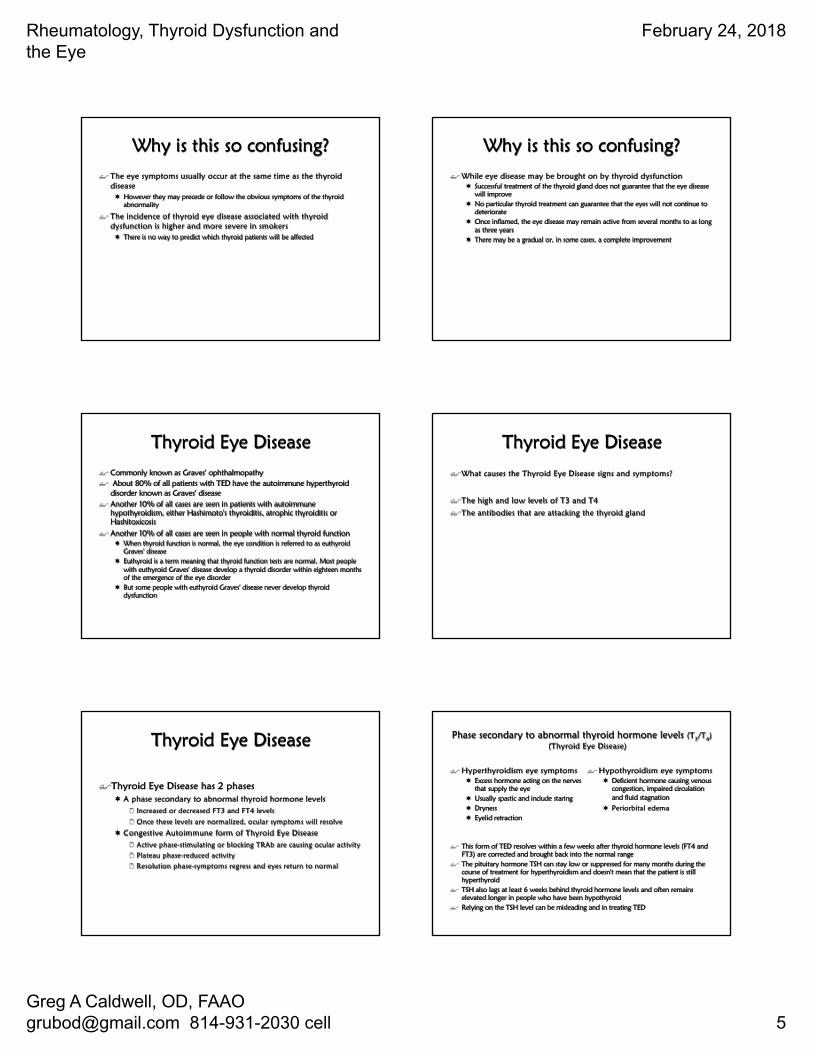
Similar receptors are found in the skin, fat and muscle of the orbit
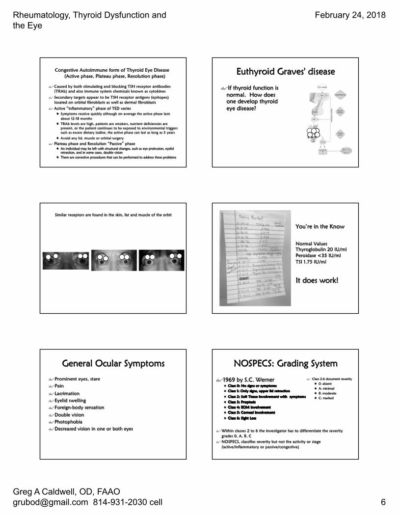
You’re in the Know
Normal Values Thyroglobulin 20 IU/ml Peroidase <35 IU/ml TSI 1.75 IU/ml
It does work!
General Ocular Symptoms
$Prominent eyes, stare$Pain
$Lacrimation$Eyelid swelling$Foreign-body sensation
$Double vision$Photophobia$Decreased vision in one or both eyes
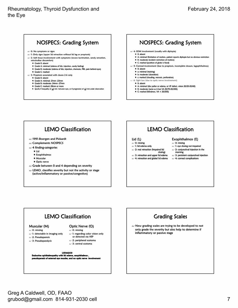
NOSPECS: Grading System
$1969 by S.C. Werner¬ Class 0: No signs or symptoms
¬ Class 1: Only signs, upper lid retraction
¬ Class 2: Soft Tissue involvement with symptoms
¬ Class 3: Proptosis
¬ Class 4: EOM involvement
¬ Class 5: Corneal Involvement
¬ Class 6: Sight Loss
$ Within classes 2 to 6 the investigator has to differentiate the severity grades 0, A, B, C
$ NOSPECS, classifies severity but not the activity or stage (active/inflammatory or passive/congestive)
$ Class 2-6 document severity¬ 0: absent¬ A: minimal¬ B: moderate¬ C: marked
Rheumatology, Thyroid Dysfunction and the Eye
February 24, 2018
Greg A Caldwell, OD, FAAO [email protected] 814-931-2030 cell 7
NOSPECS: Grading System$ 0: No symptoms or signs$ 1: Only signs (upper lid retraction without lid lag or proptosis)
$ 2: Soft tissue involvement with symptoms (excess lacrimation, sandy sensation, retrobulbar discomfort)¬ Grade 0: absent¬ Grade A: minimal (edema of lids, injection, sandy feeling)¬ Grade B: moderate (edema of lids, injection, chemosis, FBS, pain behind eyes)¬ Grade C: marked
$ 3: Proptosis associated with classes 2-6 only ¬ Grade 0: absent¬ Grade A: minimal: 21mm -23mm¬ Grade B: moderate: 24mm -27mm¬ Grade C: marked: 28mm or more¬ Specify if inequality of >3 mm between eyes, or if progression of >3 mm under observation
NOSPECS: Grading System$ 4: EOM involvement (usually with diplopia)
¬ 0: absent¬ A: minimal (limitation of motion, patient reports diplopia but no obvious restriction¬ B: moderate (evident restriction of motion)¬ C: marked (position of globe is fixed)
$ 5: Corneal involvement (due to proptosis, incomplete closure, lagophthalmos)¬ 0: absent¬ a: minimal (staining)¬ b: moderate (ulceration)¬ c: marked (clouding, necrosis, perforation)
$ 6: Sight loss (due to optic nerve involvement)¬ 0: absent¬ A: minimal (disc pallor or edema, or VF defect, vision 20/20-20/60)¬ B: moderate (same as A but VA 20/70-20/200)¬ C: marked (blindness, VA < 20/200)
LEMO Classification
$1991-Boergen and Pickardt$Complements NOSPECS
$4 finding-categories¬ Lid ¬ Exophthalmos
¬ Muscular ¬ Optic nerve
$Grade between 0 and 4 depending on severity
$LEMO, classifies severity but not the activity or stage (active/inflammatory or passive/congestive)
LEMO Classification
Lid (L)$ 0: missing$ 1: lid edema only$ 2: real retraction (impaired lid
closing)$ 3: retraction and upper lid edema$ 4: retraction and global lid edema
Exophthalmos (E)$ 0: missing$ 1: eye closing not impaired$ 2: conjunctival injection in the
morning$ 3: persistent conjunctival injection$ 4: corneal complications
LEMO Classification
Muscular (M)$ 0: missing
$ 1: detectable in imaging only$ 2: Pseudoparesis
$ 3: Pseudoparalysis
Optic Nerve (O)$ 0: missing
$ 1: regarding color vision only or detected via VEP
$ 2: peripheral scotoma$ 3: central scotoma
L1E1M2O0Endocrine ophthalmopathy with lid edema, exophthalmos , pseudoparesis of external eye muscles, and no optic nerve involvement
Grading Scales
$New grading scales are trying to be developed to not only grade the severity but also help to determine if inflammatory or passive stage
Rheumatology, Thyroid Dysfunction and the Eye
February 24, 2018
Greg A Caldwell, OD, FAAO [email protected] 814-931-2030 cell 8
Lid Involvement
$Lid Retraction$Lid Lag
$Lagophthalmus
Lid Retraction $ Scleral show in primary gaze
$ Occurs in ~90% of Grave�s patients¬ Excess stimulation of Muller�s muscle¬ Fibrotic inferior rectus¬ Mechanical restriction or infiltration
of levator¬ Increased orbital volume causes
exophthalmos
$ Normal Lid Position¬ Upper lid intersects cornea at the 2
and 10 o�clock positions2 ~2 mm below the limbus
¬ Lower lid coincident or 1-2mm below the limbus
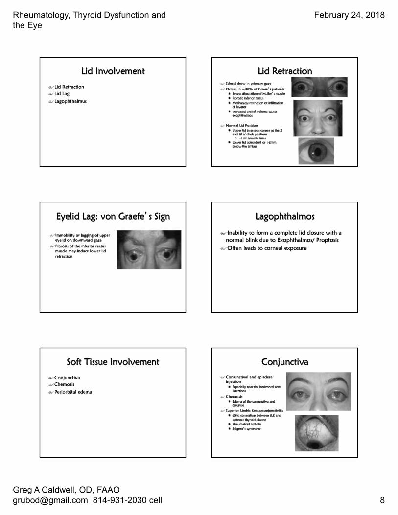
Eyelid Lag: von Graefe�s Sign
$Immobility or lagging of upper eyelid on downward gaze
$Fibrosis of the inferior rectus muscle may induce lower lid retraction
Lagophthalmos
$Inability to form a complete lid closure with a normal blink due to Exophthalmos/ Proptosis
$Often leads to corneal exposure
Soft Tissue Involvement
$Conjunctiva$Chemosis
$Periorbital edema
Conjunctiva$ Conjunctival and episcleral
injection ¬ Especially near the horizontal recti
insertions
$ Chemosis¬ Edema of the conjunctiva and
caruncle$ Superior Limbic Keratoconjunctivitis
¬ 65% correlation between SLK and systemic thyroid disease
¬ Rheumatoid arthritis ¬ Sjögren�s syndrome
Rheumatology, Thyroid Dysfunction and the Eye
February 24, 2018
Greg A Caldwell, OD, FAAO [email protected] 814-931-2030 cell 9
“If it is Red think TED”Dr. Andy Morgenstern 12-7-2013, OMS-Contemporary Resort
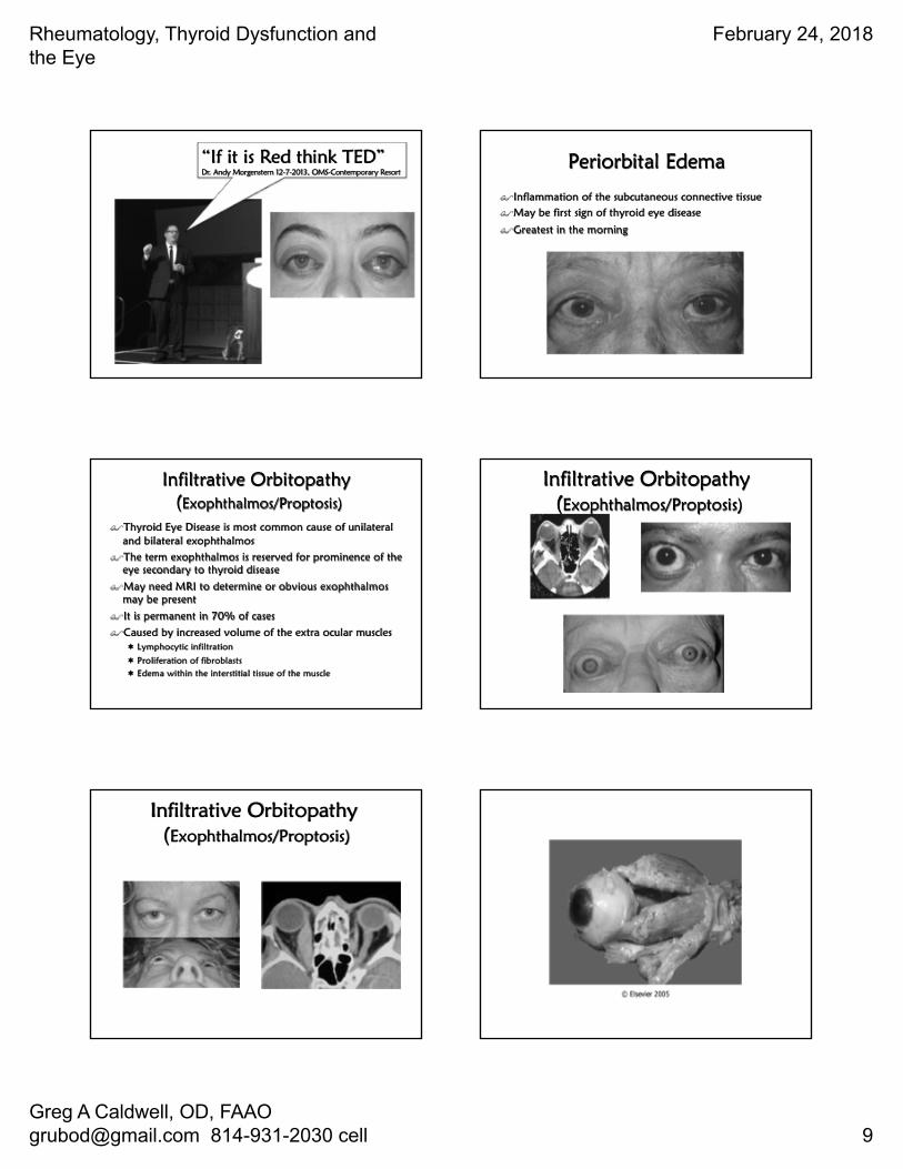
Periorbital Edema
$Inflammation of the subcutaneous connective tissue$May be first sign of thyroid eye disease
$Greatest in the morning
Infiltrative Orbitopathy(Exophthalmos/Proptosis)
$Thyroid Eye Disease is most common cause of unilateral and bilateral exophthalmos
$The term exophthalmos is reserved for prominence of the eye secondary to thyroid disease
$May need MRI to determine or obvious exophthalmosmay be present
$It is permanent in 70% of cases
$Caused by increased volume of the extra ocular muscles¬ Lymphocytic infiltration
¬ Proliferation of fibroblasts¬ Edema within the interstitial tissue of the muscle
Infiltrative Orbitopathy(Exophthalmos/Proptosis)
Infiltrative Orbitopathy(Exophthalmos/Proptosis)
Rheumatology, Thyroid Dysfunction and the Eye
February 24, 2018
Greg A Caldwell, OD, FAAO [email protected] 814-931-2030 cell 10
Exophthalmometry$ Is race dependent (Asians versus Black men is statistically significant)
$ Hertel or Luedde results
$ Adults¬ Average reading 17 mm¬ 95% of population have readings between 13-21mm
$ General concerns¬ A difference of 2 mm or more between the eyes¬ A measurement of more than 24 mm
Race Mean Normal Value Upper Limits
mm mm
White women 15.4 20.1
White men 16.5 21.7
Black women 17.8 23.1
Black men 18.5 24.7
Asians ---- 18.0
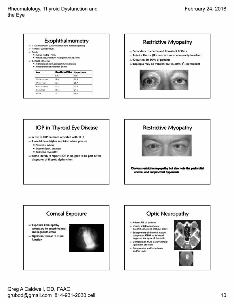
Restrictive Myopathy
$Secondary to edema and fibrosis of EOM�s $Inferior Rectus (IR) muscle is most commonly involved
$Occurs in 30-50% of patients$Diplopia may be transient but in 50% it�s permanent
IOP in Thyroid Eye Disease
$A rise in IOP has been reported with TED$I would have higher suspicion when you see
¬ Periorbital edema
¬ Exophthalmos, proptosis¬ Restrictive myopathy
$Some literature reports IOP in up gaze to be part of the diagnoses of thyroid dysfunction
Restrictive Myopathy
Obvious restrictive myopathy but also note the periorbitaledema, and conjunctival hyperemia
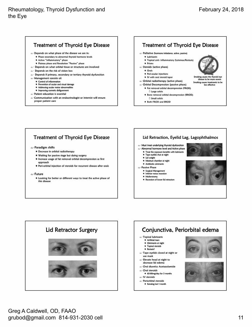
Corneal Exposure
$Exposure keratopathy secondary to exophthalmos and lagophthalmos
$Significant threat to visual function
Optic Neuropathy$ Affects 5% of patients
$ Usually mild to moderate exophthalmos and shallow orbits
$ Enlargement of the recti muscles compresses ONH or its blood supply at the apex of the orbit
$ Compression MAY occur without significant proptosis
$ Compressive and/or ischemic and/or toxic
Rheumatology, Thyroid Dysfunction and the Eye
February 24, 2018
Greg A Caldwell, OD, FAAO [email protected] 814-931-2030 cell 11
Treatment of Thyroid Eye Disease$ Depends on what phase of the disease we are in:
¬ Phase secondary to abnormal thyroid hormone levels¬ Active �inflammatory� phase¬ Plateau phase and Resolution �Passive� phase
$ Depends on what orbital tissue or structures are involved
$ Depends on the risk of vision loss
$ Depends if primary, secondary or tertiary thyroid dysfunction$ Management consists of:
¬ Control of inflammation¬ Prevention of ocular and visual damage¬ Addressing ocular motor abnormalities¬ Improving cosmetic disfigurement
$ Patient education is essential
$ Communication with an endocrinologist or internist will ensure proper patient care
Treatment of Thyroid Eye Disease$ Palliative (hormone imbalance, active, passive)
¬ Lubricants¬ Topical anti- inflammatory (Lotemax/Restasis)¬ Prisms
$ Steroids (active phase)¬ Orals¬ Peri-ocular injections¬ IV with oral steroid taper
$ Orbital radiotherapy (active phase)$ Orbital Decompression (passive phase)
¬ Fat removal orbital decompression (FROD)2 Large orbits
¬ Bone removal orbital decompression (BROD)
2 Small orbits
¬ Both FROD and BROD
Smoking causes the thyroid eye disease to be more severe
Smoking causes treatments to be less effective
Treatment of Thyroid Eye Disease
$Paradigm shifts¬ Decrease in orbital radiotherapy
¬ Waiting for passive stage but doing surgery¬ Increase usage of fat removal orbital decompression as first
approach¬ Peri-orbital injection of steroids for recurrent disease after orals
$Future¬ Looking for better or different ways to treat the active phase of
this disease
Lid Retraction, Eyelid Lag, Lagophthalmos
$ Must treat underlying thyroid dysfunction$ Abnormal hormone level and Active phase
¬ Treat the exposure keratitis with lubricants¬ Tape eyelids shut at night¬ Lid weight¬ Moisture chamber at night¬ Antibiotic ointments
$Passive Phase¬ Surgical Management¬ Inferior rectus recession¬ Mullerotomy¬ Recession of lower lid retractors
Lid Retractor Surgery Conjunctiva, Periorbital edema$ Topical lubricants
¬ Artificial tears¬ Ointments at night¬ Topical steroids¬ Restasis?
$ Tape eyelids closed at night or use mask
$ Elevate head at night to decrease lid edema
$ Oral diuretics Acetazolamide
$ Oral steroids¬ 60-80mg/day for 3 months
$ IV steroids
$ Periorbital steroids¬ Kenalog last 1 month
Rheumatology, Thyroid Dysfunction and the Eye
February 24, 2018
Greg A Caldwell, OD, FAAO [email protected] 814-931-2030 cell 12
Infiltrative Orbitopathy(Exophthalmos/Proptosis)
$ Orbital Disease Consult¬ Systemic steroids to reduce
inflammation¬ Low dose radiotherapy¬ Surgical orbital decompression
Restrictive Myopathy$ Non-surgical (while waiting for stability)
¬ Teach proper head position to alleviate diplopia
¬ Prism in spectacle correction (Fresnel or ground in)
¬ Oral steroids¬ Botulinum toxin injection
$ Surgical Consult¬ Recession of the rectus muscle/s involved¬ Diplopia in primary gaze, reading gaze or both¬ Stable angle of deviation for at least 6 months¬ No evidence of active disease¬ Binocular vision in at least primary and
reading positions
Corneal Exposure
$Manage the corneal defect as first line
¬ Lubricating and antibiotic¬ Lid taping
¬ Moisture barrier
$Orbital Disease Consult¬ High dose oral steroids
2 120-140mg /day x 7 days
¬ Orbital decompression
Optic Neuropathy
$Systemic Steroids¬ If rapidly progressive and painful
in the early stage of the disease¬ Only if no contraindications¬ Prednisolone 80-100mg, expect
results within 48hrs. Taper dose and d/c within 3 mo
$IV Methylprednisolone
$Radiotherapy: if contraindication to steroid
$Orbital decompression
Orbital Decompression
$ Not effective if no medical treatment ¬ Two-wall decompression
2 3-6 mm retro-placement of the globe
¬ Three-wall decompression2 6-10mm retro-placement
¬ Four-wall decompression2 10-16mm retro-placement
Orbital Decompression(Surgical/Cosmetic)
Rheumatology, Thyroid Dysfunction and the Eye
February 24, 2018
Greg A Caldwell, OD, FAAO [email protected] 814-931-2030 cell 13
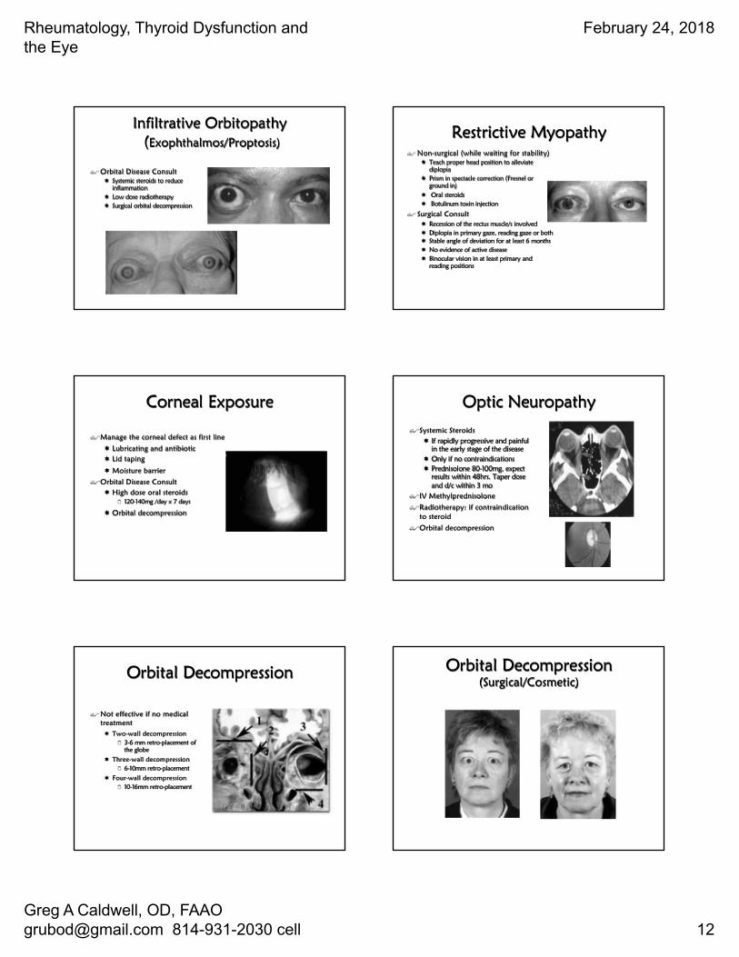
Thyroid Eye Disease and Depression
$ When facial disfigurement occurs, thyroid eye disease is equivalent to the diagnosis of cancer and AIDS
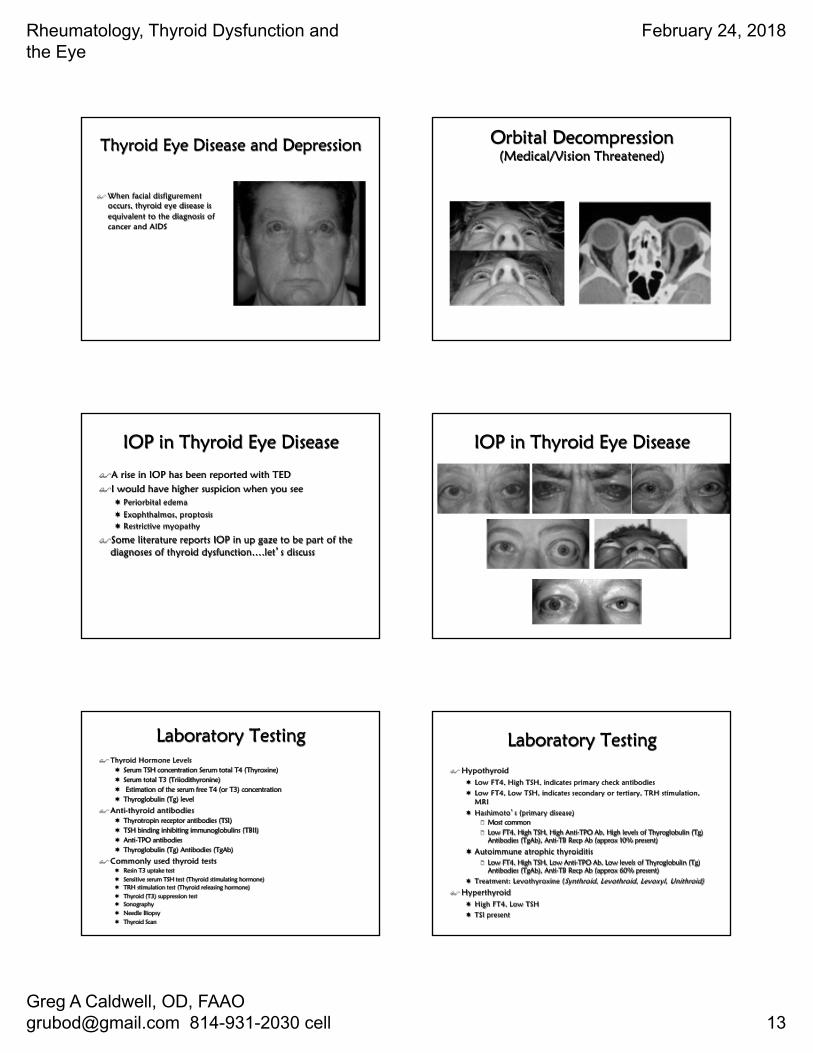
Orbital Decompression(Medical/Vision Threatened)
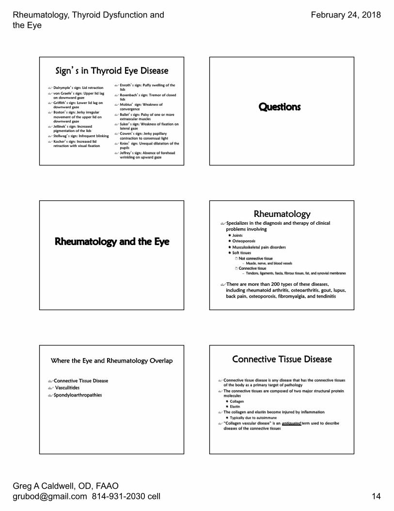
IOP in Thyroid Eye Disease
$A rise in IOP has been reported with TED$I would have higher suspicion when you see
¬ Periorbital edema
¬ Exophthalmos, proptosis¬ Restrictive myopathy
$Some literature reports IOP in up gaze to be part of the diagnoses of thyroid dysfunction….let�s discuss
IOP in Thyroid Eye Disease
Laboratory Testing$ Thyroid Hormone Levels
¬ Serum TSH concentration Serum total T4 (Thyroxine)¬ Serum total T3 (Triiodithyronine)¬ Estimation of the serum free T4 (or T3) concentration¬ Thyroglobulin (Tg) level
$ Anti-thyroid antibodies¬ Thyrotropin receptor antibodies (TSI)¬ TSH binding inhibiting immunoglobulins (TBII)¬ Anti-TPO antibodies¬ Thyroglobulin (Tg) Antibodies (TgAb)
$ Commonly used thyroid tests ¬ Resin T3 uptake test¬ Sensitive serum TSH test (Thyroid stimulating hormone)¬ TRH stimulation test (Thyroid releasing hormone)¬ Thyroid (T3) suppression test¬ Sonography¬ Needle Biopsy¬ Thyroid Scan
Laboratory Testing$ Hypothyroid
¬ Low FT4, High TSH, indicates primary check antibodies¬ Low FT4, Low TSH, indicates secondary or tertiary, TRH stimulation,
MRI¬ Hashimoto�s (primary disease)
2 Most common2 Low FT4, High TSH, High Anti-TPO Ab, High levels of Thyroglobulin (Tg)
Antibodies (TgAb), Anti-TB Recp Ab (approx 10% present)
¬ Autoimmune atrophic thyroiditis2 Low FT4, High TSH, Low Anti-TPO Ab, Low levels of Thyroglobulin (Tg)
Antibodies (TgAb), Anti-TB Recp Ab (approx 60% present)¬ Treatment: Levothyroxine (Synthroid, Levothroid, Levoxyl, Unithroid)
$ Hyperthyroid¬ High FT4, Low TSH¬ TSI present
Rheumatology, Thyroid Dysfunction and the Eye
February 24, 2018
Greg A Caldwell, OD, FAAO [email protected] 814-931-2030 cell 14
Sign�s in Thyroid Eye Disease
$ Dalrymple�s sign: Lid retraction
$ von Graefe�s sign: Upper lid lag on downward gaze
$ Griffith�s sign: Lower lid lag on downward gaze
$ Boston�s sign: Jerky irregular movement of the upper lid on downward gaze
$ Jellinek�s sign: Increased pigmentation of the lids
$ Stellwag�s sign: Infrequent blinking
$ Kocher�s sign: Increased lid retraction with visual fixation
$ Enroth�s sign: Puffy swelling of the lids
$ Rosenbach�s sign: Tremor of closed lids
$ Mobius� sign: Weakness of convergence
$ Ballet�s sign: Palsy of one or more extraocular muscles
$ Suker�s sign: Weakness of fixation on lateral gaze
$ Cowen�s sign: Jerky papillary contraction to consensual light
$ Knies� sign: Unequal dilatation of the pupils
$ Jeffrey�s sign: Absence of forehead wrinkling on upward gaze
Questions
Rheumatology and the Eye
Rheumatology$Specializes in the diagnosis and therapy of clinical
problems involving¬ Joints ¬ Osteoporosis
¬ Musculoskeletal pain disorders¬ Soft tissues
2 Not connective tissue– Muscle, nerve, and blood vessels
2 Connective tissue– Tendons, ligaments, fascia, fibrous tissues, fat, and synovial membranes
$There are more than 200 types of these diseases, including rheumatoid arthritis, osteoarthritis, gout, lupus, back pain, osteoporosis, fibromyalgia, and tendinitis
Where the Eye and Rheumatology Overlap
$Connective Tissue Disease$ Vasculitides
$Spondyloarthropathies
Connective Tissue Disease
$ Connective tissue disease is any disease that has the connective tissues of the body as a primary target of pathology
$ The connective tissues are composed of two major structural protein molecules¬ Collagen¬ Elastin
$ The collagen and elastin become injured by inflammation¬ Typically due to autoimmune
$ “Collagen vascular disease” is an antiquated term used to describe diseases of the connective tissues
Rheumatology, Thyroid Dysfunction and the Eye
February 24, 2018
Greg A Caldwell, OD, FAAO [email protected] 814-931-2030 cell 15
Connective tissue diseases secondary to gene abnormalities
$Connective tissue diseases that are strictly due to genetic inheritance include ¬ Marfan syndrome
2 Gene FBN1 on chromosome 152 Can have tissue abnormalities in the heart, aorta, lungs, eyes, and
skeleton
¬ Ehlers-Danlos syndrome2 Many types with numerous genes
2 Typically have loose, fragile skin and hyperextensible joints depending on type
Connective tissue diseases secondary to autoimmunity
$Cannot be regularly defined by gene abnormalities
$The spontaneous over activity of the immune system ¬ Results in the production of
extra antibodies into the circulation
$ Systemic Lupus Erythematosus
$ Rheumatoid Arthritis$ Sjogrens Syndrome
$ Systemic Sclerosis
$ Polymyositis /Dermatomyositis$ Mixed Connective Tissue
$ Wegner�s Granulomatous
Connective Tissue Diseases
Disease Auto-antibodySystemic Lupus Erythematosus Anti-dsDNA, Anti-SM
Rheumatoid Arthritis RF, Anti-RA33Sjogrens Syndrome Anti-Ro(SS-A),Anti-La(SS-B)
Systemic Sclerosis Anti-Scl-70, Anti-centromerePolymyositis/Dermatomyositis Anti-Jo-1
Mixed Connective Tissue Disease Anti-U1-RNP
Wegener�s Granulomatosus c-ANCA
Similar Structures
$The connective tissues are composed of two major structural protein molecules¬ Collagen¬ Elastin
$Synovial membrane: A layer of connective tissue that lines the cavities of joints, tendon sheaths, and bursae and makes synovial fluid , which has a lubricating function.
$Sclera- the opaque , white, fibrous, protective, outer layer of the eye containing collagenand elastin fibers
$Ténon� s Capsule –a layer of connective tissue which forms a thin membrane that envelops the eyeball from the optic nerve to the limbus , separating it from the orbital fat and forming a socket
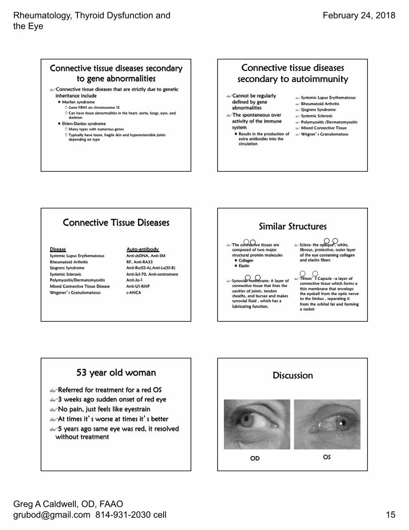
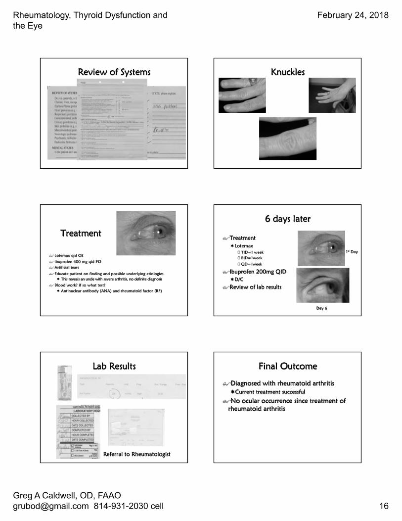
53 year old woman
$Referred for treatment for a red OS $3 weeks ago sudden onset of red eye$No pain, just feels like eyestrain$At times it�s worse at times it�s better$5 years ago same eye was red, it resolved
without treatment
Discussion
OD OS
Rheumatology, Thyroid Dysfunction and the Eye
February 24, 2018
Greg A Caldwell, OD, FAAO [email protected] 814-931-2030 cell 16
Review of Systems Knuckles
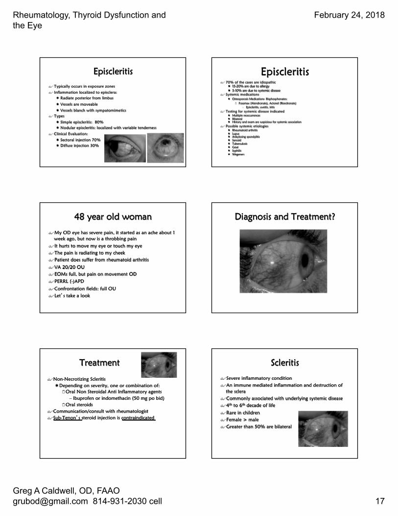
Treatment
$Lotemax qid OS
$Ibuprofen 400 mg qid PO$Artificial tears
$Educate patient on finding and possible underlying etiologies¬ This reveals an uncle with severe arthritis, no definite diagnosis
$Blood work? if so what test?¬ Antinuclear antibody (ANA) and rheumatoid factor (RF)
6 days later
$Treatment¬Lotemax
2 TID=1 week2 BID=1week
2 QD=1week
$Ibuprofen 200mg QID¬D/C
$Review of lab results
1st Day
Day 6
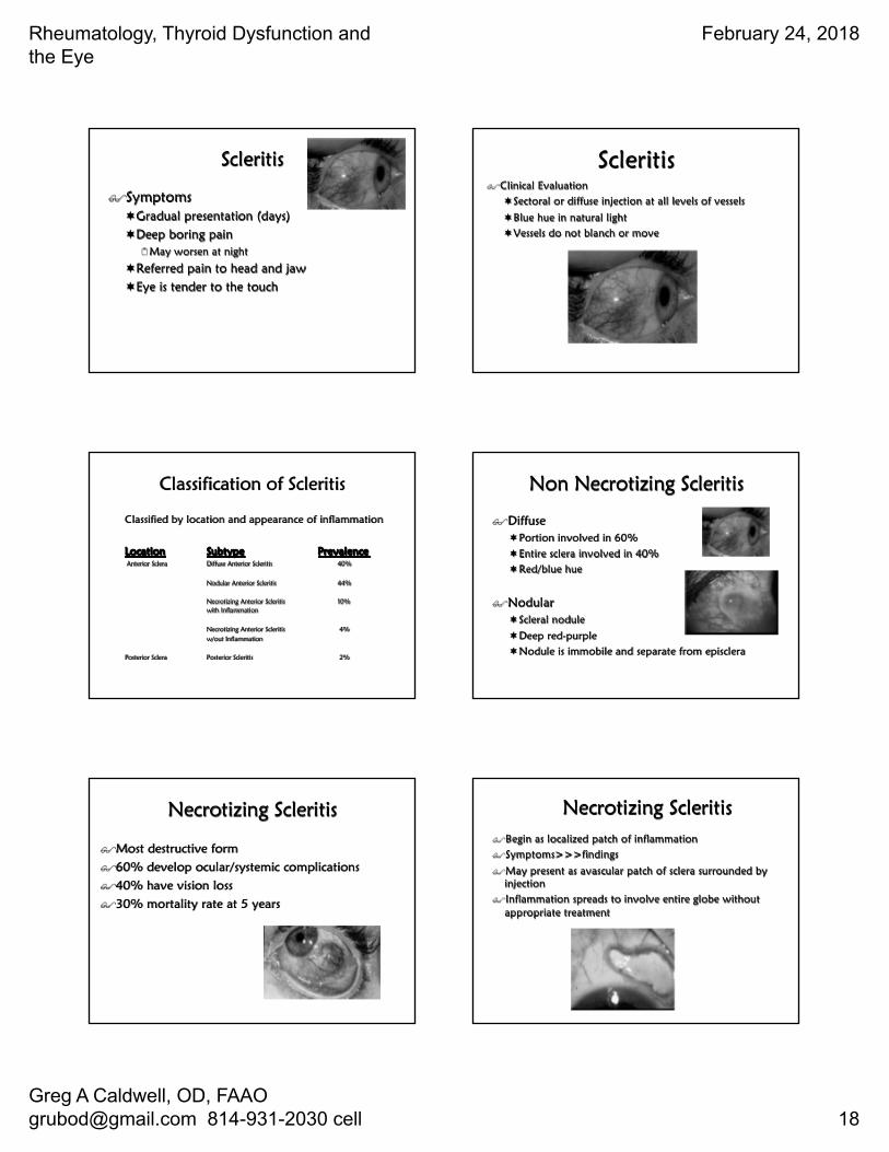
Lab Results
Referral to Rheumatologist
Final Outcome
$Diagnosed with rheumatoid arthritis¬Current treatment successful
$No ocular occurrence since treatment of rheumatoid arthritis
Rheumatology, Thyroid Dysfunction and the Eye
February 24, 2018
Greg A Caldwell, OD, FAAO [email protected] 814-931-2030 cell 17
Episcleritis$ Typically occurs in exposure zones
$ Inflammation localized to episclera:¬ Radiate posterior from limbus
¬ Vessels are moveable
¬ Vessels blanch with sympatomimetics$ Types
¬ Simple episcleritis: 80%¬ Nodular episcleritis: localized with variable tenderness
$ Clinical Evaluation:
¬ Sectoral injection 70%¬ Diffuse injection 30%
Episcleritis$ 70% of the cases are idiopathic
¬ 15-20% are due to allergy¬ 5-10% are due to systemic disease
$ Systemic medications¬ Osteoporosis Medications- Bisphosphonates:
2 Fosamax (Alendronate), Actonel (Risedronate)– Episcleritis, uveitis, iritis
$ Testing for systemic disease indicated¬ Multiple reoccurrences¬ Bilateral¬ History and exam are suspicious for systemic association
$ Possible systemic etiologies¬ Rheumatoid arthritis ¬ Lupus¬ Ankylosing spondylitis¬ Sarcoid¬ Tuberculosis¬ Gout¬ Syphilis ¬ Wegeners
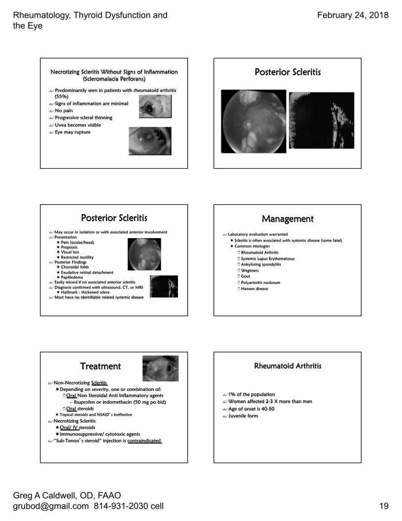
48 year old woman
$My OD eye has severe pain, it started as an ache about 1 week ago, but now is a throbbing pain
$It hurts to move my eye or touch my eye$The pain is radiating to my cheek$Patient does suffer from rheumatoid arthritis
$VA 20/20 OU$EOMs full, but pain on movement OD
$PERRL (-)APD
$Confrontation fields: full OU$Let�s take a look
Diagnosis and Treatment?
Treatment
$Non-Necrotizing Scleritis¬Depending on severity, one or combination of:
2Oral Non Steroidal Anti Inflammatory agents– Ibuprofen or indomethacin (50 mg po bid)
2Oral steroids $Communication/consult with rheumatologist$Sub-Tenon�s steroid injection is contraindicated
Scleritis
$Severe inflammatory condition$An immune mediated inflammation and destruction of
the sclera$Commonly associated with underlying systemic disease$4th to 6th decade of life
$Rare in children$Female > male$Greater than 50% are bilateral
Rheumatology, Thyroid Dysfunction and the Eye
February 24, 2018
Greg A Caldwell, OD, FAAO [email protected] 814-931-2030 cell 18
Scleritis
$Symptoms¬Gradual presentation (days)¬Deep boring pain
2May worsen at night
¬Referred pain to head and jaw¬Eye is tender to the touch
Scleritis$Clinical Evaluation
¬Sectoral or diffuse injection at all levels of vessels
¬Blue hue in natural light¬Vessels do not blanch or move
Classification of Scleritis
Classified by location and appearance of inflammation
Location Subtype PrevalenceAnterior Sclera Diffuse Anterior Scleritis 40%
Nodular Anterior Scleritis 44%
Necrotizing Anterior Scleritis 10%with Inflammation
Necrotizing Anterior Scleritis 4%w/out Inflammation
Posterior Sclera Posterior Scleritis 2%
Non Necrotizing Scleritis
$Diffuse ¬Portion involved in 60%¬Entire sclera involved in 40%¬Red/blue hue
$Nodular¬Scleral nodule
¬Deep red-purple¬Nodule is immobile and separate from episclera
Necrotizing Scleritis
$Most destructive form$60% develop ocular/systemic complications$40% have vision loss$30% mortality rate at 5 years
Necrotizing Scleritis$Begin as localized patch of inflammation$Symptoms>>>findings
$May present as avascular patch of sclera surrounded by injection
$Inflammation spreads to involve entire globe without appropriate treatment
Rheumatology, Thyroid Dysfunction and the Eye
February 24, 2018
Greg A Caldwell, OD, FAAO [email protected] 814-931-2030 cell 19
Necrotizing Scleritis Without Signs of Inflammation(Scleromalacia Perforans)
$Predominantly seen in patients with rheumatoid arthritis (55%)
$Signs of inflammation are minimal$No pain$Progressive scleral thinning
$Uvea becomes visible$Eye may rupture
Posterior Scleritis
Posterior Scleritis$May occur in isolation or with associated anterior involvement$Presentation
¬ Pain (ocular/head)¬ Proptosis¬ Visual loss¬ Restricted motility
$Posterior Findings¬ Choroidal folds¬ Exudative retinal detachment¬ Papilledema
$Easily missed if no associated anterior scleritis$Diagnosis confirmed with ultrasound, CT, or MRI
¬ Hallmark : thickened sclera$Most have no identifiable related systemic disease
Management
$Laboratory evaluation warranted
¬ Scleritis is often associated with systemic disease (some fatal)¬ Common etiologies
2 Rheumatoid Arthritis
2 Systemic Lupus Erythematosus2 Ankylosing spondylitis
2 Wegeners2 Gout
2 Polyarteritis nodosum
2 Hansen disease
Treatment
$Non-Necrotizing Scleritis¬Depending on severity, one or combination of:
2Oral Non Steroidal Anti Inflammatory agents– Ibuprofen or indomethacin (50 mg po bid)
2Oral steroids ¬ Topical steroids and NSAID�s ineffective
$Necrotizing Scleritis¬Oral/ IV steroids¬Immunosuppressive/ cytotoxic agents
$“Sub-Tenon�s steroid” injection is contraindicated
Rheumatoid Arthritis
$1% of the population$Women affected 2-3 X more than men
$Age of onset is 40-50$Juvenile form
Rheumatology, Thyroid Dysfunction and the Eye
February 24, 2018
Greg A Caldwell, OD, FAAO [email protected] 814-931-2030 cell 20
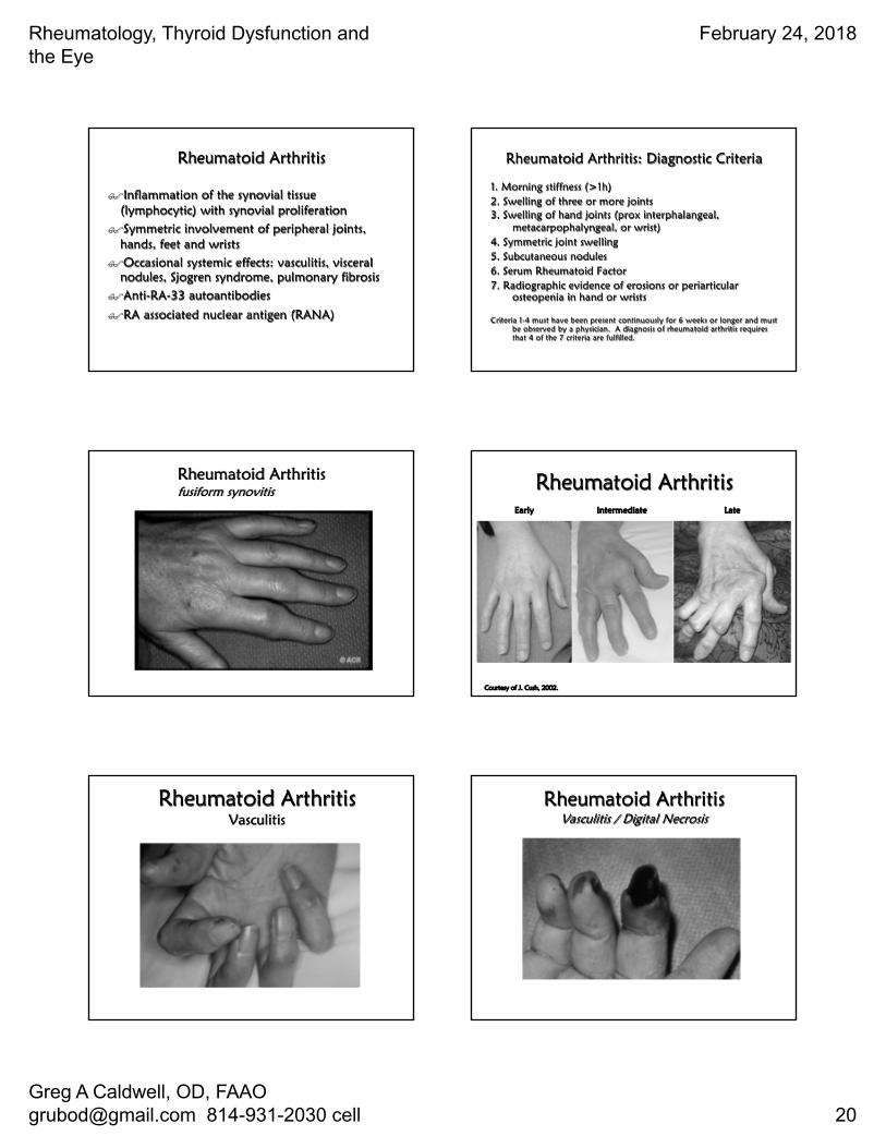
Rheumatoid Arthritis
$Inflammation of the synovial tissue (lymphocytic) with synovial proliferation
$Symmetric involvement of peripheral joints, hands, feet and wrists
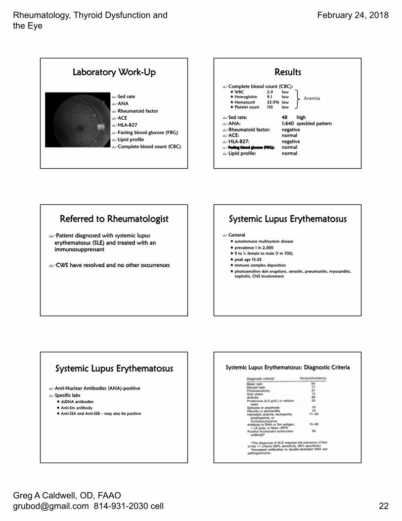
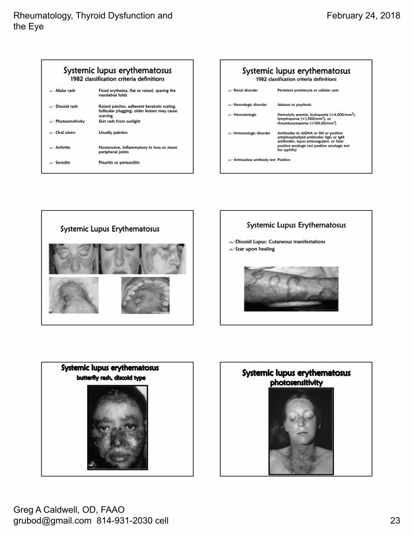
$Occasional systemic effects: vasculitis, visceral nodules, Sjogren syndrome, pulmonary fibrosis
$Anti-RA-33 autoantibodies
$RA associated nuclear antigen (RANA)
Rheumatoid Arthritis: Diagnostic Criteria
1. Morning stiffness (>1h)2. Swelling of three or more joints3. Swelling of hand joints (prox interphalangeal,
metacarpophalyngeal, or wrist)4. Symmetric joint swelling5. Subcutaneous nodules6. Serum Rheumatoid Factor7. Radiographic evidence of erosions or periarticular
osteopenia in hand or wrists
Criteria 1-4 must have been present continuously for 6 weeks or longer and must be observed by a physician. A diagnosis of rheumatoid arthritis requires that 4 of the 7 criteria are fulfilled.
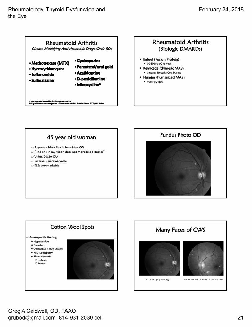
Rheumatoid Arthritisfusiform synovitis
Early Intermediate Late
Courtesy of J. Cush, 2002.
Rheumatoid Arthritis
Rheumatoid ArthritisVasculitis
Rheumatoid ArthritisVasculitis / Digital Necrosis
Rheumatology, Thyroid Dysfunction and the Eye
February 24, 2018
Greg A Caldwell, OD, FAAO [email protected] 814-931-2030 cell 21
§Methotrexate (MTX)§ Hydroxychloroquine
§Leflunomide
§Sulfasalazine
§Cyclosporine
§Parenteral/oral gold
§Azathioprine
§D-penicillamine §Minocycline*
* Not approved by the FDA for the treatment of RA.ACR guidelines for the management of rheumatoid arthritis. Arthritis Rheum. 2002;46:328-346.
Rheumatoid ArthritisDisease Modifying Anti-rheumatic Drugs /DMARDs
Rheumatoid Arthritis(Biologic DMARDs)
§ Enbrel (Fusion Protein)§ 50-100mg SQ q week
§ Remicade (chimeric MAB)§ 3mg/kg -10mg/kg Q 4-8weeks
§ Humira (humanized MAB)§ 40mg SQ qow
45 year old woman
$Reports a black line in her vision OD$�The line in my vision does not move like a floater�
$Vision 20/20 OU$Externals: unremarkable$SLE: unremarkable
Fundus Photo OD
Cotton Wool Spots
$Non-specific finding¬ Hypertension
¬ Diabetes¬ Connective Tissue Disease
¬ HIV Retinopathy¬ Blood dyscrasia
2 Leukemia2 Anemia
Many Faces of CWS
No under lying etiology History of uncontrolled HTN and DM
Rheumatology, Thyroid Dysfunction and the Eye
February 24, 2018
Greg A Caldwell, OD, FAAO [email protected] 814-931-2030 cell 22
Laboratory Work-Up
$Sed rate$ANA
$Rheumatoid factor$ACE$HLA-B27
$Fasting blood glucose (FBG)$Lipid profile$Complete blood count (CBC)
Results$Complete blood count (CBC):
¬ WBC 2.9 low¬ Hemoglobin 9.1 low¬ Hematocrit 33.9% low¬ Platelet count 110 low
$Sed rate: 48 high$ANA: 1:640 speckled pattern$Rheumatoid factor: negative$ACE: normal$HLA-B27: negative$ Fasting blood glucose (FBG): normal$Lipid profile: normal
Anemia
Referred to Rheumatologist
$Patient diagnosed with systemic lupus erythematosus (SLE) and treated with an immunosuppressant
$CWS have resolved and no other occurrences
Systemic Lupus Erythematosus
$General¬ autoimmune multisystem disease
¬ prevalence 1 in 2,000¬ 9 to 1; female to male (1 in 700)
¬ peak age 15-25¬ immune complex deposition
¬ photosensitive skin eruptions, serositis, pneumonitis, myocarditis, nephritis, CNS involvement
Systemic Lupus Erythematosus
$Anti-Nuclear Antibodies (ANA)-positive$Specific labs
¬ dsDNA antibodies
¬ Anti-Sm antibody¬ Anti-SSA and Anti-SSB – may also be positive
Systemic Lupus Erythematosus: Diagnostic Criteria
Rheumatology, Thyroid Dysfunction and the Eye
February 24, 2018
Greg A Caldwell, OD, FAAO [email protected] 814-931-2030 cell 23
Systemic lupus erythematosus 1982 classification criteria definitions
$ Malar rash Fixed erythema, flat or raised, sparing the nasolabial folds
$ Discoid rash Raised patches, adherent keratotic scaling, follicular plugging; older lesions may cause scarring
$ Photosensitivity Skin rash from sunlight
$ Oral ulcers Usually painless
$ Arthritis Nonerosive, inflammatory in two or more peripheral joints
$ Serositis Pleuritis or pericarditis
Systemic lupus erythematosus1982 classification criteria definitions
$ Renal disorder Persistent proteinuria or cellular casts
$ Neurologic disorder Seizures or psychosis
$ Hermatologic Hemolytic anemia, leukopenia (<4,000/mm3), lymphopenia (<1,500/mm3), or thrombocytopenia (<100,00/mm3)
$ Immunologic disorder Antibodies to dsDNA or SM or positive antiphospholipid antibodies (IgG or IgM antibodies, lupus anticoagulant, or false-positive serologic test positive serologic test for syphilis)
$ Antinuclear antibody test Positive
Systemic Lupus Erythematosus Systemic Lupus Erythematosus
$Discoid Lupus: Cutaneous manifestations$Scar upon healing
Systemic lupus erythematosusbutterfly rash, discoid type
Systemic lupus erythematosus photosensitivity
Rheumatology, Thyroid Dysfunction and the Eye
February 24, 2018
Greg A Caldwell, OD, FAAO [email protected] 814-931-2030 cell 24
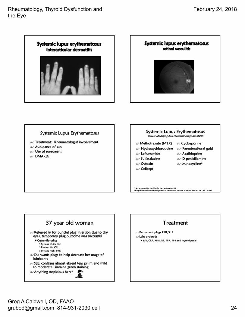
Systemic lupus erythematosus interarticular dermatitis
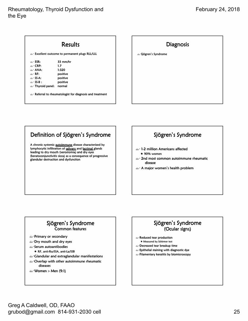
Systemic lupus erythematosus retinal vasculitis
Systemic Lupus Erythematosus
$ Treatment: Rheumatologist involvement$ Avoidance of sun$ Use of sunscreens$ DMARDs
Systemic Lupus ErythematosusDisease Modifying Anti-rheumatic Drugs /DMARDs
$Methotrexate (MTX)$ Hydroxychloroquine$ Leflunomide$ Sulfasalazine$ Cytoxin$ Cellcept
$Cyclosporine $ Parenteral/oral gold$ Azathioprine$ D-penicillamine $ Minocycline*
* Not approved by the FDA for the treatment of RA.ACR guidelines for the management of rheumatoid arthritis. Arthritis Rheum. 2002;46:328-346.
37 year old woman
$Referred in for punctal plug insertion due to dry eyes, temporary plug outcome was successful¬Currently using
2 Systane q1-2h OU2 Restasis bid OU2 Systane night PRN
$She wants plugs to help decrease her usage of lubricants
$SLE: confirms almost absent tear prism and mild to moderate Lisamine green staining
$Anything suspicious here?
Treatment
$Permanent plugs RUL/RLL$Labs ordered:
¬ ESR, CRP, ANA, RF, SS-A, SS-B and thyroid panel
Rheumatology, Thyroid Dysfunction and the Eye
February 24, 2018
Greg A Caldwell, OD, FAAO [email protected] 814-931-2030 cell 25
Results$ Excellent outcome to permanent plugs RLL/LLL
$ ESR: 33 mm/hr$ CRP: 1.7$ ANA: 1:320$ RF: positive$ SS-A: positive$ SS-B : positive$ Thyroid panel: normal
$ Referral to rheumatologist for diagnosis and treatment
Diagnosis
$Sjögren’s Syndrome
Definition of Sjögren’s Syndrome
A chronic systemic autoimmune disease characterized by lymphocytic infiltration of salivary and lacrimal glands leading to dry mouth (xerostomia) and dry eyes (keratoconjunctivitis sicca) as a consequence of progressive glandular destruction and dysfunction
$ 1-2 million Americans affected ¬ 90% women
$ 2nd most common autoimmune rheumatic disease
$ A major women’s health problem
Sjögren’s Syndrome
Sjögren’s SyndromeCommon features
$Primary or secondary$Dry mouth and dry eyes$Serum autoantibodies
¬ RF, anti-Ro/SSA, anti-La/SSB
$Glandular and extraglandular manifestations$Overlap with other autoimmune rheumatic
diseases$Women > Men (9:1)
$Reduced tear production¬ Measured by Schirmer test
$Decreased tear breakup time$Epithelial staining with diagnostic dye$Filamentary keratitis by biomicroscopy
Sjögren’s Syndrome (Ocular signs)
Rheumatology, Thyroid Dysfunction and the Eye
February 24, 2018
Greg A Caldwell, OD, FAAO [email protected] 814-931-2030 cell 26
$Dry mouth$Sore or burning mouth $Intolerance to acidic or spicy foods$Abnormalities of taste $Difficulty with chewing and swallowing dry
foods $Difficulty with phonation (speaking)$Difficulty wearing dentures
Sjögren’s Syndrome (Oral features)
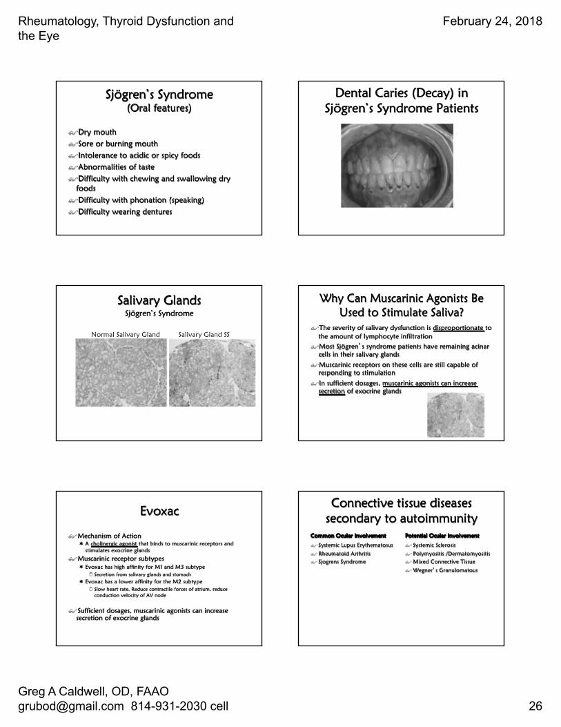
Dental Caries (Decay) in Sjögren’s Syndrome Patients
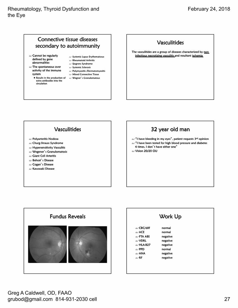
Salivary Glands Sjögren’s Syndrome
Normal Salivary Gland Salivary Gland SS
Why Can Muscarinic Agonists Be Used to Stimulate Saliva?
$The severity of salivary dysfunction is disproportionate to the amount of lymphocyte infiltration
$Most Sjögren�s syndrome patients have remaining acinar cells in their salivary glands
$Muscarinic receptors on these cells are still capable of responding to stimulation
$In sufficient dosages, muscarinic agonists can increase secretion of exocrine glands
$Mechanism of Action¬ A cholinergic agonist that binds to muscarinic receptors and
stimulates exocrine glands
$Muscarinic receptor subtypes¬ Evoxac has high affinity for M1 and M3 subtype
2 Secretion from salivary glands and stomach¬ Evoxac has a lower affinity for the M2 subtype
2 Slow heart rate, Reduce contractile forces of atrium, reduce conduction velocity of AV node
$Sufficient dosages, muscarinic agonists can increase secretion of exocrine glands
EvoxacConnective tissue diseases
secondary to autoimmunity Common Ocular Involvement
$ Systemic Lupus Erythematosus
$ Rheumatoid Arthritis$ Sjogrens Syndrome
Potential Ocular Involvement
$ Systemic Sclerosis
$ Polymyositis /Dermatomyositis$ Mixed Connective Tissue
$ Wegner�s Granulomatous
Rheumatology, Thyroid Dysfunction and the Eye
February 24, 2018
Greg A Caldwell, OD, FAAO [email protected] 814-931-2030 cell 27
Connective tissue diseases secondary to autoimmunity
$Cannot be regularly defined by gene abnormalities
$The spontaneous over activity of the immune system ¬ Results in the production of
extra antibodies into the circulation
$ Systemic Lupus Erythematosus
$ Rheumatoid Arthritis$ Sjogrens Syndrome
$ Systemic Sclerosis
$ Polymyositis /Dermatomyositis$ Mixed Connective Tissue
$ Wegner�s Granulomatous
Vasculitides
The vasculitides are a group of diseases characterized by non infectious necrotizing vasculitis and resultant ischemia
Vasculitides
$Polyarteritis Nodosa$Churg-Strauss Syndrome
$Hypersensitivity Vasculitis$Wegener�s Granulomatosis$Giant Cell Arteritis
$Behcet�s Disease$Cogan�s Disease
$Kawasaki Disease
32 year old man
$�I have bleeding in my eyes�, patient requests 3rd opinion$�I have been tested for high blood pressure and diabetes
4 times, I don�t have either one�$Vision 20/20 OU
Fundus Reveals Work Up
$CBC/diff normal$ACE normal
$FTA ABS negative$VDRL negative$HLA-B27 negative
$PPD normal$ANA negative
$RF negative
Rheumatology, Thyroid Dysfunction and the Eye
February 24, 2018
Greg A Caldwell, OD, FAAO [email protected] 814-931-2030 cell 28
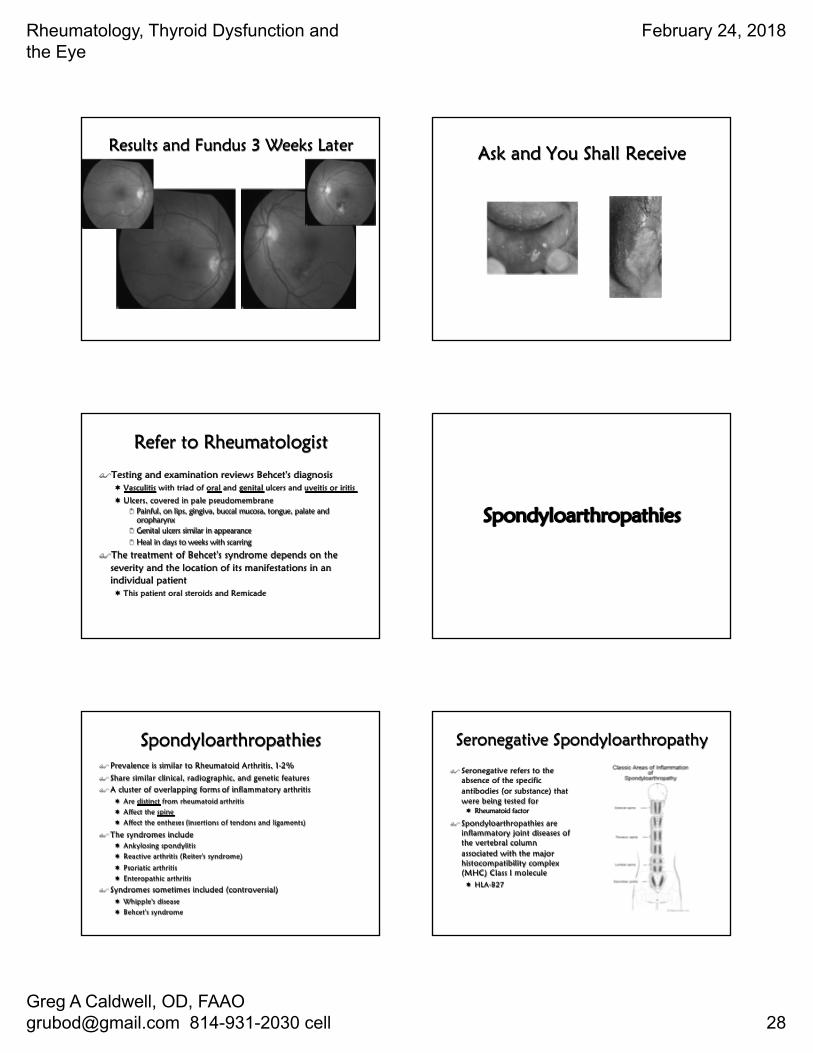
Results and Fundus 3 Weeks Later Ask and You Shall Receive
Refer to Rheumatologist
$Testing and examination reviews Behcet's diagnosis ¬ Vasculitis with triad of oral and genital ulcers and uveitis or iritis
¬ Ulcers, covered in pale pseudomembrane2 Painful, on lips, gingiva, buccal mucosa, tongue, palate and
oropharynx2 Genital ulcers similar in appearance2 Heal in days to weeks with scarring
$The treatment of Behcet's syndrome depends on the severity and the location of its manifestations in an individual patient¬ This patient oral steroids and Remicade
Spondyloarthropathies
Spondyloarthropathies$ Prevalence is similar to Rheumatoid Arthritis, 1-2%
$ Share similar clinical, radiographic, and genetic features$ A cluster of overlapping forms of inflammatory arthritis
¬ Are distinct from rheumatoid arthritis¬ Affect the spine¬ Affect the entheses (insertions of tendons and ligaments)
$ The syndromes include¬ Ankylosing spondylitis¬ Reactive arthritis (Reiter's syndrome)
¬ Psoriatic arthritis¬ Enteropathic arthritis
$ Syndromes sometimes included (controversial) ¬ Whipple's disease ¬ Behcet's syndrome
Seronegative Spondyloarthropathy
$ Seronegative refers to the absence of the specific antibodies (or substance) that were being tested for¬ Rheumatoid factor
$ Spondyloarthropathies are inflammatory joint diseases of the vertebral column associated with the major histocompatibility complex (MHC) Class I molecule ¬ HLA-B27
Rheumatology, Thyroid Dysfunction and the Eye
February 24, 2018
Greg A Caldwell, OD, FAAO [email protected] 814-931-2030 cell 29
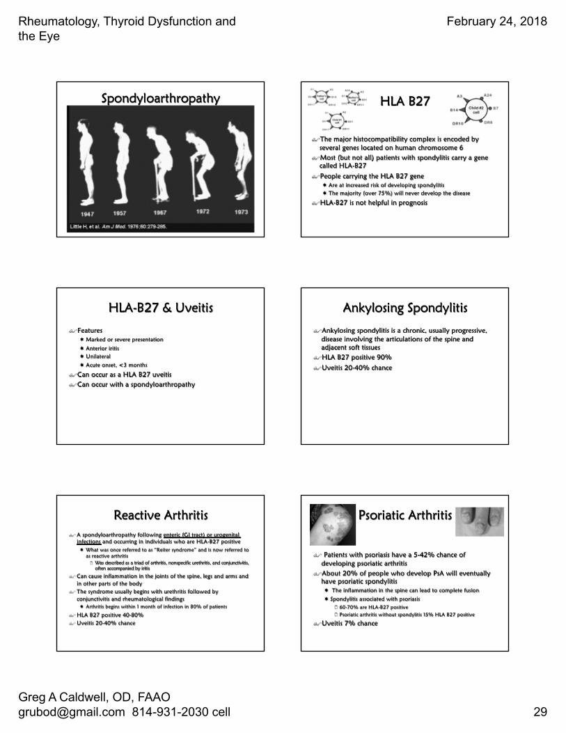
Spondyloarthropathy HLA B27
$The major histocompatibility complex is encoded by several genes located on human chromosome 6
$Most (but not all) patients with spondylitis carry a gene called HLA-B27
$People carrying the HLA B27 gene¬ Are at increased risk of developing spondylitis¬ The majority (over 75%) will never develop the disease
$HLA-B27 is not helpful in prognosis
HLA-B27 & Uveitis
$Features¬ Marked or severe presentation
¬ Anterior iritis¬ Unilateral
¬ Acute onset, <3 months
$Can occur as a HLA B27 uveitis$Can occur with a spondyloarthropathy
Ankylosing Spondylitis
$Ankylosing spondylitis is a chronic, usually progressive, disease involving the articulations of the spine and adjacent soft tissues
$HLA B27 positive 90%
$Uveitis 20-40% chance
Reactive Arthritis$ A spondyloarthropathy following enteric (GI tract) or urogenital
infections and occurring in individuals who are HLA-B27 positive¬ What was once referred to as “Reiter syndrome” and is now referred to
as reactive arthritis2 Was described as a triad of arthritis, nonspecific urethritis, and conjunctivitis,
often accompanied by iritis
$ Can cause inflammation in the joints of the spine, legs and arms and in other parts of the body
$ The syndrome usually begins with urethritis followed by conjunctivitis and rheumatological findings¬ Arthritis begins within 1 month of infection in 80% of patients
$ HLA B27 positive 40-80%$ Uveitis 20-40% chance
Psoriatic Arthritis
$ Patients with psoriasis have a 5-42% chance of developing psoriatic arthritis
$About 20% of people who develop PsA will eventually have psoriatic spondylitis¬ The inflammation in the spine can lead to complete fusion
¬ Spondylitis associated with psoriasis2 60-70% are HLA-B27 positive2 Psoriatic arthritis without spondylitis 15% HLA B27 positive
$Uveitis 7% chance
Rheumatology, Thyroid Dysfunction and the Eye
February 24, 2018
Greg A Caldwell, OD, FAAO [email protected] 814-931-2030 cell 30
Enteropathic Arthritis
$A form of chronic, inflammatory arthritis associated with the occurrence of an inflammatory bowel disease (IBD) ¬ Ulcerative colitis¬ Crohn's disease
$About one in five people with Crohn's or ulcerative colitis will develop enteropathic arthritis¬ Approximately 50-60% of patients with spondylitis in association
with IBD have HLA-B27
$The most common areas affected are the peripheral (limb) joints¬ In some cases, the entire spine can become involved as well
$Uveitis 3-11% chance
Undifferentiated Spondyloarthropathy (USpA)
$To describe symptoms and signs of spondylitis in someone who does not meet the criteria for a definitive diagnosis of AS or related disease¬ Unrecognized by many physicians
¬ Initial diagnosis of Spondyloarthropathy or Unclassified Spondyloarthropathy if certain symptoms are present but are not enough to make a specific diagnosis2 Over time, most people with USpA will develop a well-defined form
of spondylitis such as ankylosing spondylitis
What Drug Do Rheumatologists Use Quite Often?
Revised Recommendations on Screening for Chloroquine and Hydroxychloroquine Retinopathy
$ Recommendations were 2002 by the American Academy of Ophthalmology
$ Improved screening tools and new knowledge about prevalence of toxicity have prompt the change¬ 1% after 5-7 years of use or a cumulative
dose of 1000 grams (Plaquenil)
$ There is no treatment for this condition¬ Therefore must be caught early
$ Screening for the earliest hints of functional or anatomic change
$ Plaquenil toxicity is not well understood
Revised Again
Rheumatology, Thyroid Dysfunction and the Eye
February 24, 2018
Greg A Caldwell, OD, FAAO [email protected] 814-931-2030 cell 31
Plaquenil Toxicity
Oh Boy!
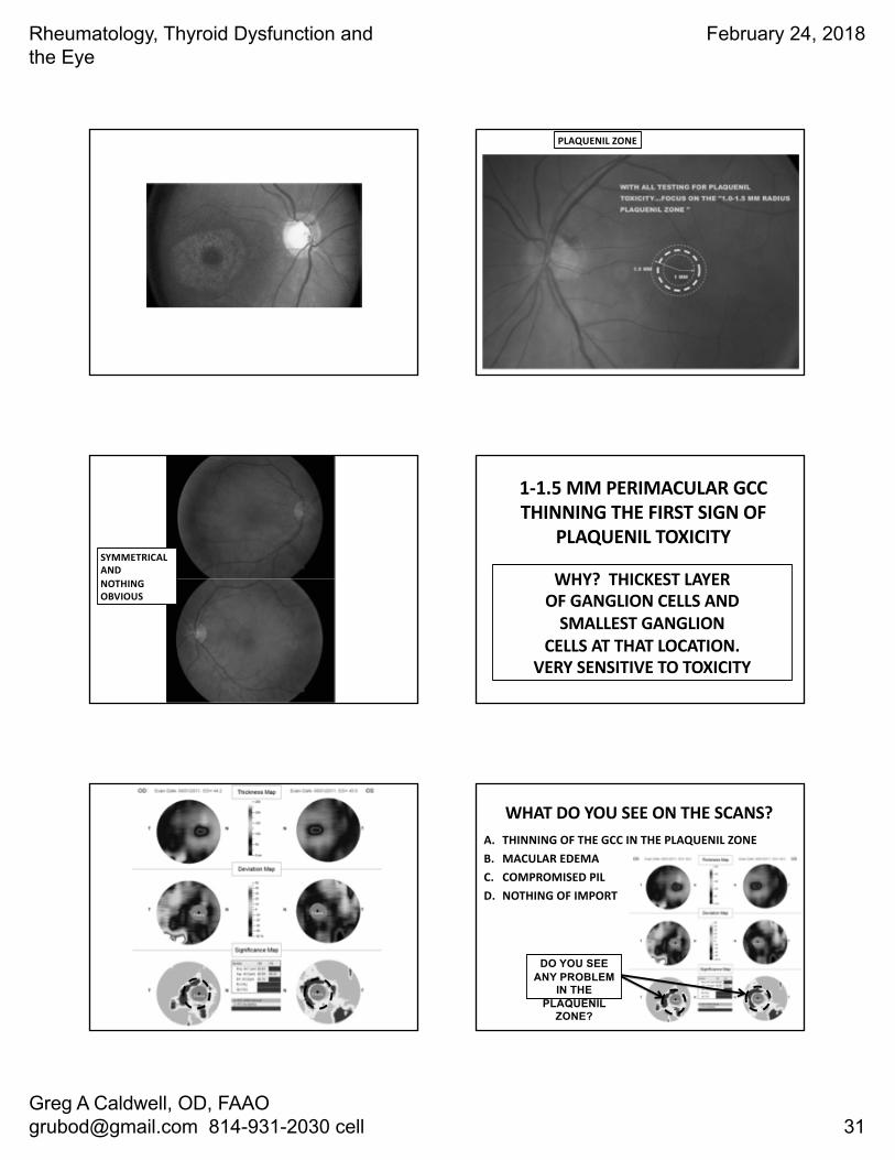
PLAQUENIL ZONE
SYMMETRICALANDNOTHING OBVIOUS
1-1.5 MM PERIMACULAR GCC THINNING THE FIRST SIGN OF
PLAQUENIL TOXICITY
WHY? THICKEST LAYEROF GANGLION CELLS AND
SMALLEST GANGLION CELLS AT THAT LOCATION.
VERY SENSITIVE TO TOXICITY
WHAT DO YOU SEE ON THE SCANS?A. THINNING OF THE GCC IN THE PLAQUENIL ZONEB. MACULAR EDEMAC. COMPROMISED PILD. NOTHING OF IMPORT
DO YOU SEE ANY PROBLEM
IN THE PLAQUENIL
ZONE?
Rheumatology, Thyroid Dysfunction and the Eye
February 24, 2018
Greg A Caldwell, OD, FAAO [email protected] 814-931-2030 cell 32
WHAT DO YOU SEE ON THE SCANS?A. THINNING OF THE GCC IN THE PLAQUENIL ZONEB. MACULAR EDEMAC. COMPROMISED PILD. NOTHING OF IMPORT
DO YOU SEE ANY PROBLEM
IN THE PLAQUENIL
ZONE?
AUGUST 2014 AUGUST 2014
WHAT DO YOU SEE ON THE SCANS?A. THE FLYING SAUCER SIGNB. MACULAR EDEMAC. INCREASED PERIMACULAR RETINAL THINNINGD. A AND C
WHAT DO YOU SEE ON THE SCANS?A. THE FLYING SAUCER SIGNB. MACULAR EDEMAC. INCREASED PERIMACULAR RETINAL THINNINGD. A AND C
A A
C C
Rheumatology, Thyroid Dysfunction and the Eye
February 24, 2018
Greg A Caldwell, OD, FAAO [email protected] 814-931-2030 cell 33
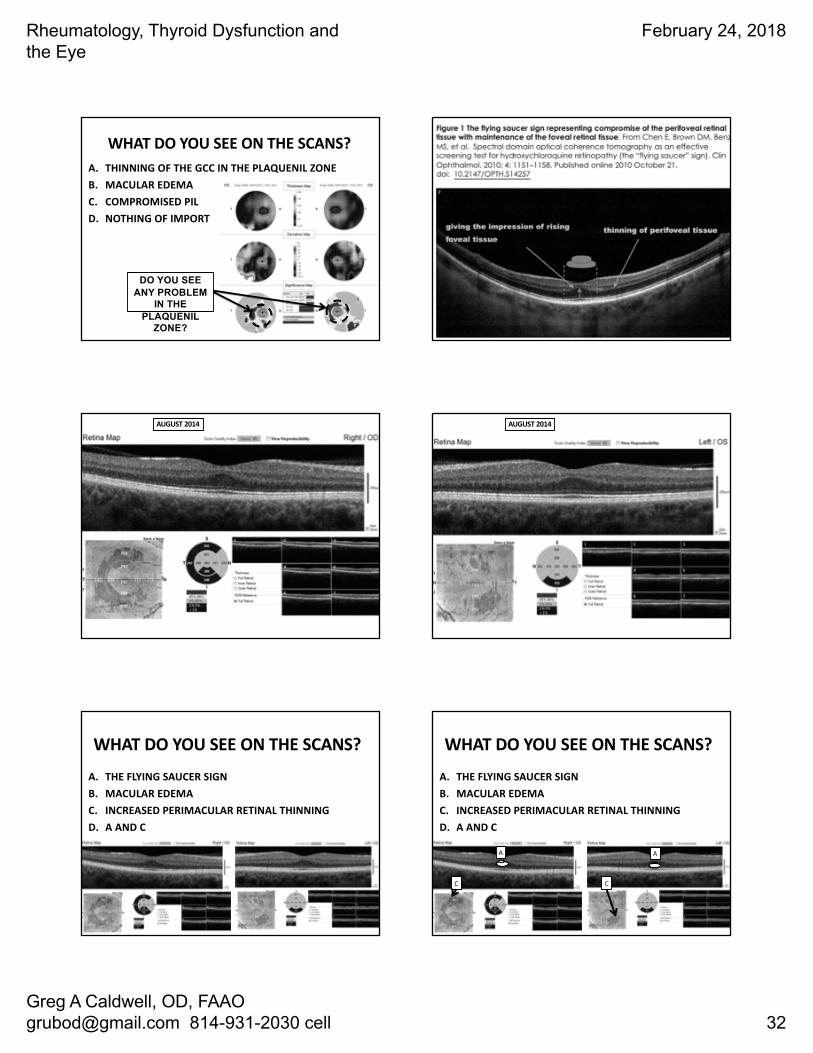
BILATERAL COMPROMISE OF THE PIL (WHITE ARROWS) AFTER COLLAPSE OF PERIFOVEAL RETINA (RED DASHED ARROWS) WITH FLYING SAUCER ATTACK (BLUE ARROWS)
THE END GAME…ONCE YOU DISCONTINUE PLAQUENIL IT STAYS AROUND A WHILE TO
CREATE DAMAGE..LONG ½ LIFE
WAY OUTTA THE BARN
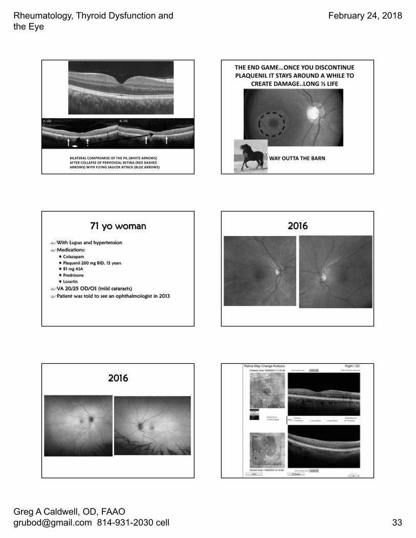
71 yo woman
$With Lupus and hypertension$Medications:
¬ Colazapam
¬ Plaquenil 200 mg BID, 15 years ¬ 81 mg ASA
¬ Prednisone
¬ Losartin
$VA 20/25 OD/OS (mild cataracts)
$Patient was told to see an ophthalmologist in 2013
2016
2016
Rheumatology, Thyroid Dysfunction and the Eye
February 24, 2018
Greg A Caldwell, OD, FAAO [email protected] 814-931-2030 cell 34
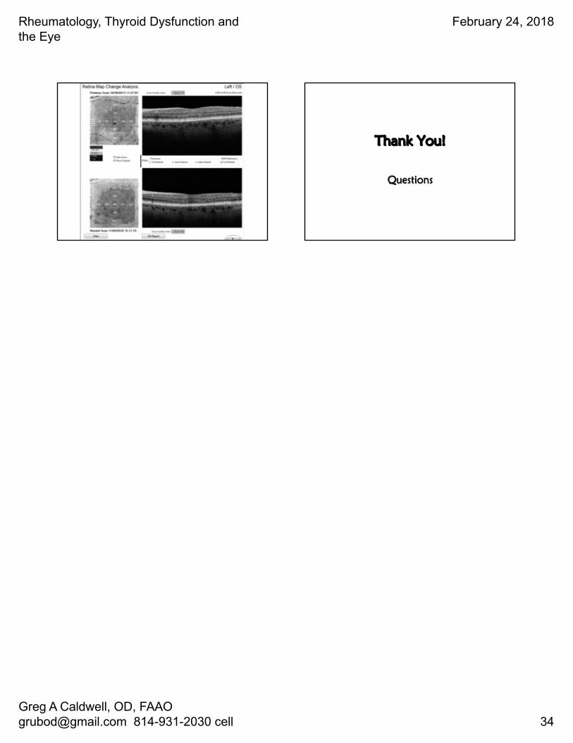
Thank You!
Questions