Embed Size (px)
Citation preview
Tissue Penetration of Antifungal Agents
Timothy Felton,a,b Peter F. Troke,c William W. Hopeb
The University of Manchester, Academic Health Science Centre, University Hospital of South Manchester NHS Foundation Trust, Manchester, United Kingdoma;Antimicrobial Pharmacodynamics and Therapeutics, Department of Molecular and Clinical Pharmacology, University of Liverpool, Liverpool, United Kingdomb;The Old Court, Kingsgate, Kent, United Kingdomc
SUMMARY . . . . . . . . . . . . . . . . . . . . . . . . . . . . . . . . . . . . . . . . . . . . . . . . . . . . . . . . . . . . . . . . . . . . . . . . . . . . . . . . . . . . . . . . . . . . . . . . . . . . . . . . . . . . . . . . . . . . . . . . . . . . . . . . . . . . . . . . . . . . . . . . . . .68INTRODUCTION . . . . . . . . . . . . . . . . . . . . . . . . . . . . . . . . . . . . . . . . . . . . . . . . . . . . . . . . . . . . . . . . . . . . . . . . . . . . . . . . . . . . . . . . . . . . . . . . . . . . . . . . . . . . . . . . . . . . . . . . . . . . . . . . . . . . . . . . . . . . . .68PENETRATION OF ANTIFUNGAL AGENTS INTO TISSUES: CONCEPTS, IMPORTANCE, AND CURRENT GAPS IN KNOWLEDGE . . . . . . . . . . . . . . . . . . . . . . . . . . .69
Importance of Tissue Concentrations for an Understanding of Antifungal Pharmacodynamics . . . . . . . . . . . . . . . . . . . . . . . . . . . . . . . . . . . . . . . . . . . . . . . . . . . . . . . . . .69Determinants of Distribution of Antifungal Agents into Tissues . . . . . . . . . . . . . . . . . . . . . . . . . . . . . . . . . . . . . . . . . . . . . . . . . . . . . . . . . . . . . . . . . . . . . . . . . . . . . . . . . . . . . . . . . . .69Limitations of Current Understanding and Approaches . . . . . . . . . . . . . . . . . . . . . . . . . . . . . . . . . . . . . . . . . . . . . . . . . . . . . . . . . . . . . . . . . . . . . . . . . . . . . . . . . . . . . . . . . . . . . . . . . . .72
ANTIFUNGAL DRUG CONCENTRATIONS IN ORGANS, TISSUES, AND BODY FLUIDS . . . . . . . . . . . . . . . . . . . . . . . . . . . . . . . . . . . . . . . . . . . . . . . . . . . . . . . . . . . . . . . . . . . . . . .72Brain and Cerebrospinal Fluid. . . . . . . . . . . . . . . . . . . . . . . . . . . . . . . . . . . . . . . . . . . . . . . . . . . . . . . . . . . . . . . . . . . . . . . . . . . . . . . . . . . . . . . . . . . . . . . . . . . . . . . . . . . . . . . . . . . . . . . . . . . . . .73Eye . . . . . . . . . . . . . . . . . . . . . . . . . . . . . . . . . . . . . . . . . . . . . . . . . . . . . . . . . . . . . . . . . . . . . . . . . . . . . . . . . . . . . . . . . . . . . . . . . . . . . . . . . . . . . . . . . . . . . . . . . . . . . . . . . . . . . . . . . . . . . . . . . . . . . . . . .74Lung . . . . . . . . . . . . . . . . . . . . . . . . . . . . . . . . . . . . . . . . . . . . . . . . . . . . . . . . . . . . . . . . . . . . . . . . . . . . . . . . . . . . . . . . . . . . . . . . . . . . . . . . . . . . . . . . . . . . . . . . . . . . . . . . . . . . . . . . . . . . . . . . . . . . . . .75Pulmonary Lymph Fluid . . . . . . . . . . . . . . . . . . . . . . . . . . . . . . . . . . . . . . . . . . . . . . . . . . . . . . . . . . . . . . . . . . . . . . . . . . . . . . . . . . . . . . . . . . . . . . . . . . . . . . . . . . . . . . . . . . . . . . . . . . . . . . . . . . .76Pleural Fluid. . . . . . . . . . . . . . . . . . . . . . . . . . . . . . . . . . . . . . . . . . . . . . . . . . . . . . . . . . . . . . . . . . . . . . . . . . . . . . . . . . . . . . . . . . . . . . . . . . . . . . . . . . . . . . . . . . . . . . . . . . . . . . . . . . . . . . . . . . . . . . . .76Bronchial Secretions . . . . . . . . . . . . . . . . . . . . . . . . . . . . . . . . . . . . . . . . . . . . . . . . . . . . . . . . . . . . . . . . . . . . . . . . . . . . . . . . . . . . . . . . . . . . . . . . . . . . . . . . . . . . . . . . . . . . . . . . . . . . . . . . . . . . . . .76Saliva, Sputum, Buccal Mucosa, and Esophagus . . . . . . . . . . . . . . . . . . . . . . . . . . . . . . . . . . . . . . . . . . . . . . . . . . . . . . . . . . . . . . . . . . . . . . . . . . . . . . . . . . . . . . . . . . . . . . . . . . . . . . . . . . .76Heart . . . . . . . . . . . . . . . . . . . . . . . . . . . . . . . . . . . . . . . . . . . . . . . . . . . . . . . . . . . . . . . . . . . . . . . . . . . . . . . . . . . . . . . . . . . . . . . . . . . . . . . . . . . . . . . . . . . . . . . . . . . . . . . . . . . . . . . . . . . . . . . . . . . . . . .76Liver. . . . . . . . . . . . . . . . . . . . . . . . . . . . . . . . . . . . . . . . . . . . . . . . . . . . . . . . . . . . . . . . . . . . . . . . . . . . . . . . . . . . . . . . . . . . . . . . . . . . . . . . . . . . . . . . . . . . . . . . . . . . . . . . . . . . . . . . . . . . . . . . . . . . . . . .77Kidney . . . . . . . . . . . . . . . . . . . . . . . . . . . . . . . . . . . . . . . . . . . . . . . . . . . . . . . . . . . . . . . . . . . . . . . . . . . . . . . . . . . . . . . . . . . . . . . . . . . . . . . . . . . . . . . . . . . . . . . . . . . . . . . . . . . . . . . . . . . . . . . . . . . . .77Spleen . . . . . . . . . . . . . . . . . . . . . . . . . . . . . . . . . . . . . . . . . . . . . . . . . . . . . . . . . . . . . . . . . . . . . . . . . . . . . . . . . . . . . . . . . . . . . . . . . . . . . . . . . . . . . . . . . . . . . . . . . . . . . . . . . . . . . . . . . . . . . . . . . . . . .77Pancreas . . . . . . . . . . . . . . . . . . . . . . . . . . . . . . . . . . . . . . . . . . . . . . . . . . . . . . . . . . . . . . . . . . . . . . . . . . . . . . . . . . . . . . . . . . . . . . . . . . . . . . . . . . . . . . . . . . . . . . . . . . . . . . . . . . . . . . . . . . . . . . . . . . .78Peritoneum . . . . . . . . . . . . . . . . . . . . . . . . . . . . . . . . . . . . . . . . . . . . . . . . . . . . . . . . . . . . . . . . . . . . . . . . . . . . . . . . . . . . . . . . . . . . . . . . . . . . . . . . . . . . . . . . . . . . . . . . . . . . . . . . . . . . . . . . . . . . . . . .78Genital System . . . . . . . . . . . . . . . . . . . . . . . . . . . . . . . . . . . . . . . . . . . . . . . . . . . . . . . . . . . . . . . . . . . . . . . . . . . . . . . . . . . . . . . . . . . . . . . . . . . . . . . . . . . . . . . . . . . . . . . . . . . . . . . . . . . . . . . . . . . . .78Bone . . . . . . . . . . . . . . . . . . . . . . . . . . . . . . . . . . . . . . . . . . . . . . . . . . . . . . . . . . . . . . . . . . . . . . . . . . . . . . . . . . . . . . . . . . . . . . . . . . . . . . . . . . . . . . . . . . . . . . . . . . . . . . . . . . . . . . . . . . . . . . . . . . . . . . .78Muscle . . . . . . . . . . . . . . . . . . . . . . . . . . . . . . . . . . . . . . . . . . . . . . . . . . . . . . . . . . . . . . . . . . . . . . . . . . . . . . . . . . . . . . . . . . . . . . . . . . . . . . . . . . . . . . . . . . . . . . . . . . . . . . . . . . . . . . . . . . . . . . . . . . . . .79Skin and Nails . . . . . . . . . . . . . . . . . . . . . . . . . . . . . . . . . . . . . . . . . . . . . . . . . . . . . . . . . . . . . . . . . . . . . . . . . . . . . . . . . . . . . . . . . . . . . . . . . . . . . . . . . . . . . . . . . . . . . . . . . . . . . . . . . . . . . . . . . . . . . .79
UNDERSTANDING TISSUE CONCENTRATIONS FOR OPTIMAL USE OF EXISTING AGENTS AND DEVELOPMENT OF NEWER ANTIFUNGAL AGENTS . . . .80Current State of the Art . . . . . . . . . . . . . . . . . . . . . . . . . . . . . . . . . . . . . . . . . . . . . . . . . . . . . . . . . . . . . . . . . . . . . . . . . . . . . . . . . . . . . . . . . . . . . . . . . . . . . . . . . . . . . . . . . . . . . . . . . . . . . . . . . . . .80Beyond State of the Art . . . . . . . . . . . . . . . . . . . . . . . . . . . . . . . . . . . . . . . . . . . . . . . . . . . . . . . . . . . . . . . . . . . . . . . . . . . . . . . . . . . . . . . . . . . . . . . . . . . . . . . . . . . . . . . . . . . . . . . . . . . . . . . . . . . .80
ACKNOWLEDGMENTS . . . . . . . . . . . . . . . . . . . . . . . . . . . . . . . . . . . . . . . . . . . . . . . . . . . . . . . . . . . . . . . . . . . . . . . . . . . . . . . . . . . . . . . . . . . . . . . . . . . . . . . . . . . . . . . . . . . . . . . . . . . . . . . . . . . . . . .80REFERENCES . . . . . . . . . . . . . . . . . . . . . . . . . . . . . . . . . . . . . . . . . . . . . . . . . . . . . . . . . . . . . . . . . . . . . . . . . . . . . . . . . . . . . . . . . . . . . . . . . . . . . . . . . . . . . . . . . . . . . . . . . . . . . . . . . . . . . . . . . . . . . . . . .80AUTHOR BIOS . . . . . . . . . . . . . . . . . . . . . . . . . . . . . . . . . . . . . . . . . . . . . . . . . . . . . . . . . . . . . . . . . . . . . . . . . . . . . . . . . . . . . . . . . . . . . . . . . . . . . . . . . . . . . . . . . . . . . . . . . . . . . . . . . . . . . . . . . . . . . . . .88
SUMMARY
Understanding the tissue penetration of systemically adminis-tered antifungal agents is critical for a proper appreciation of theirantifungal efficacy in animals and humans. Both the time courseof an antifungal drug and its absolute concentrations within tis-sues may differ significantly from those observed in the blood-stream. In addition, tissue concentrations must also be interpretedwithin the context of the pathogenesis of the various invasive fun-gal infections, which differ significantly. There are major technicalobstacles to the estimation of concentrations of antifungal agentsin various tissue subcompartments, yet these agents, even thosewithin the same class, may exhibit markedly different tissue dis-tributions. This review explores these issues and provides a sum-mary of tissue concentrations of 11 currently licensed systemicantifungal agents. It also explores the therapeutic implications oftheir distribution at various sites of infection.
INTRODUCTION
Despite recent advances in antifungal chemotherapy, invasivefungal infections (IFI) remain a significant cause of morbid-
ity and mortality (1). Candida species, Aspergillus fumigatus, andCryptococcus neoformans are the most common pathogens (2).
However, a wide range of other fungi, often with limited suscep-tibility to first-line antifungal agents, may also cause infection.Mortality from IFI remains high (e.g., that from aspergillosis is�50% [3, 4], and that from candidemia is 10 to 49% [5, 6, 7]). Anunderstanding of the pharmacological properties of any antifun-gal agent is crucial for optimizing patient outcomes for all theseinfections (8). This may be especially true for an increasingly rec-ognized group of patients who have not previously been consid-ered to be at high risk of IFI, such as critically ill patients and thosewith chronic obstructive pulmonary disease (COPD), who maydemonstrate marked pharmacokinetic (PK) variability (9, 10).
Penetration into the site of infection to achieve microbe-elim-inating concentrations is a key requirement for efficacy of all an-timicrobial agents (11, 12, 13, 14, 15). The importance of tissueconcentrations for the various classes of antibacterial agents hasbeen reviewed extensively, but relatively less attention has been
Address correspondence to William W. Hope, [email protected].
Copyright © 2014, American Society for Microbiology. All Rights Reserved.
doi:10.1128/CMR.00046-13
68 cmr.asm.org Clinical Microbiology Reviews p. 68 – 88 January 2014 Volume 27 Number 1
on June 16, 2019 by guesthttp://cm
r.asm.org/
Dow
nloaded from
paid to the currently available antifungal agents (12, 16, 17, 18,19). This review examines the tissue penetration of 11 commonlyused systemic antifungal agents (amphotericin B deoxycholate[AmBd], amphotericin B lipid complex [ABLC], liposomal am-photericin B [L-AMB], fluconazole, itraconazole, posaconazole,voriconazole, 5-fluorocytosine [5FC], anidulafungin, caspofun-gin, and micafungin) into the clinically relevant compartments forhuman infection and disease. All human data, ranging from casestudies through autopsies to small clinical studies in volunteers orpatients, were included. We also considered key laboratory animaldata, where relevant, especially if the respective information forhumans is absent. Because only free drug is considered to be bio-logically active (20, 21, 22), tissue and fluid concentrations areplaced in context with the key physicochemical properties of eachagent. The major organ systems covered include the lungs, liver,kidney, spleen, and heart. Attention has also been given to drugpenetration into sanctuary sites (e.g., brain and eye), with thecorresponding therapeutic implications. We have also reviewedthe data for key interstitial fluids, including bronchial secretions,epithelial lining fluid (ELF), pleural fluid, pericardial fluid, syno-vial fluid, prostatic fluid, and cerebrospinal fluid (CSF), andplaced these data in a clinical context (23).
PENETRATION OF ANTIFUNGAL AGENTS INTO TISSUES:CONCEPTS, IMPORTANCE, AND CURRENT GAPS INKNOWLEDGE
Importance of Tissue Concentrations for an Understandingof Antifungal Pharmacodynamics
The potential relevance of the tissue concentrations of any anti-infec-tive agent must be considered in context with the pathogenesis of theinvading fungal organism (24). There must be colocalization of“drug and bug” within tissue beds and tissue subcompartments.Such considerations are relevant at the level of the organ and tissuesubcompartments but may be elucidated further at the cellularand even molecular levels (25, 26, 27, 28, 29).
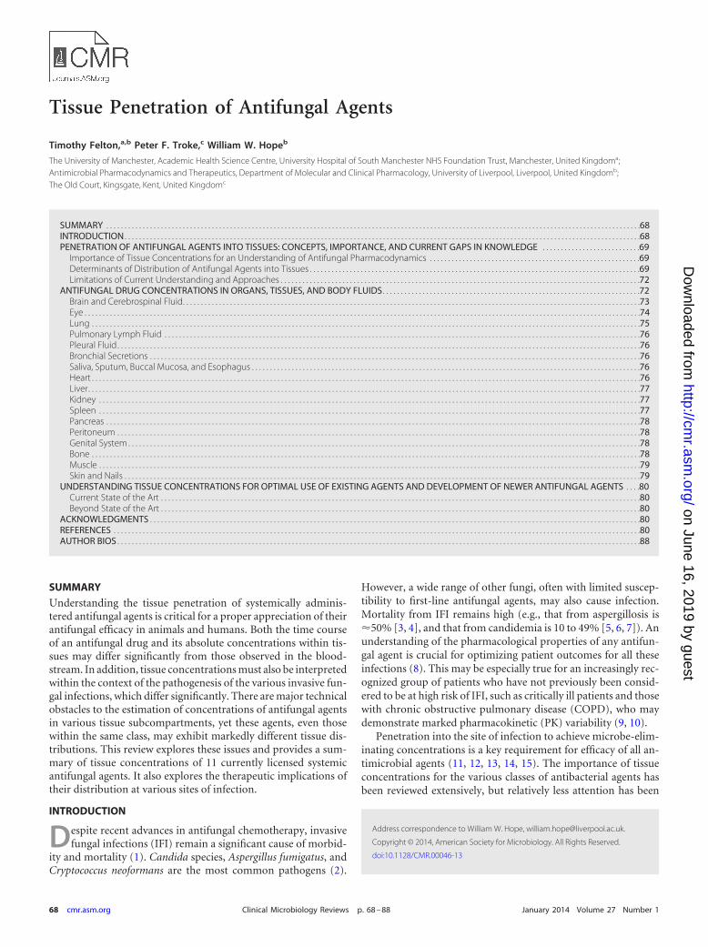
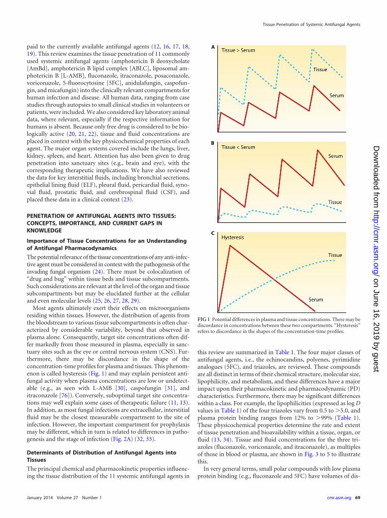
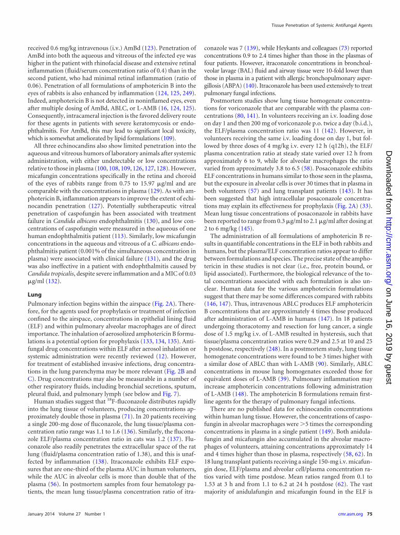
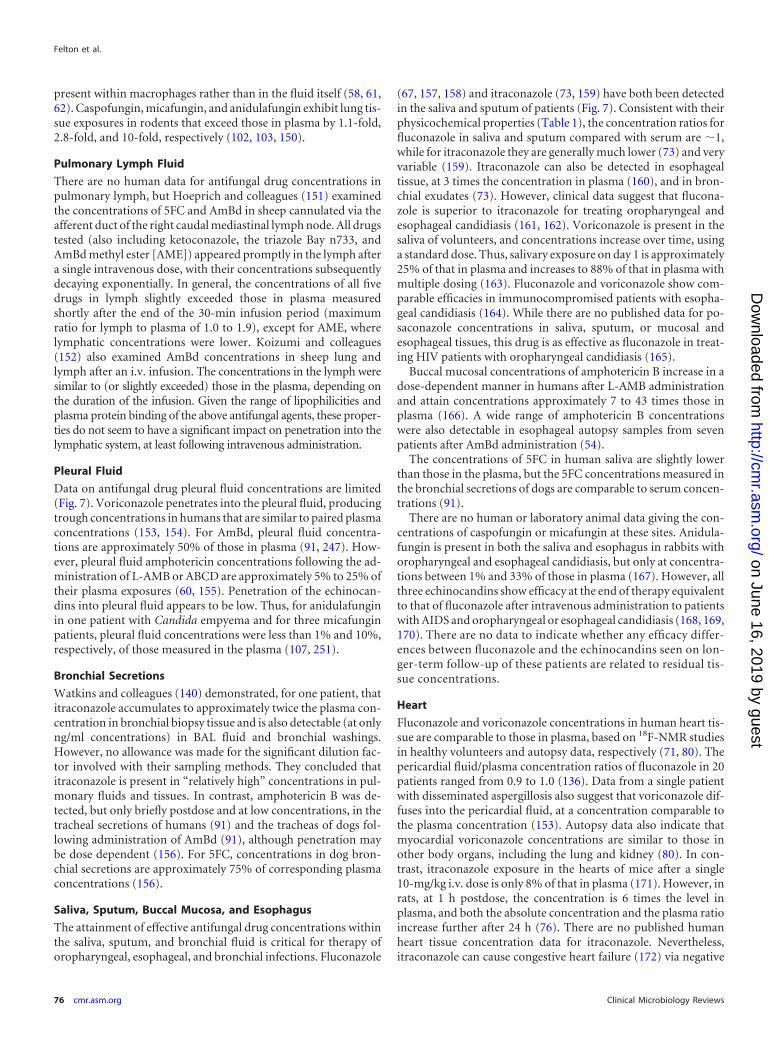
Most agents ultimately exert their effects on microorganismsresiding within tissues. However, the distribution of agents fromthe bloodstream to various tissue subcompartments is often char-acterized by considerable variability, beyond that observed inplasma alone. Consequently, target site concentrations often dif-fer markedly from those measured in plasma, especially in sanc-tuary sites such as the eye or central nervous system (CNS). Fur-thermore, there may be discordance in the shape of theconcentration-time profiles for plasma and tissues. This phenom-enon is called hysteresis (Fig. 1) and may explain persistent anti-fungal activity when plasma concentrations are low or undetect-able (e.g., as seen with L-AMB [30], caspofungin [31], anditraconazole [76]). Conversely, suboptimal target site concentra-tions may well explain some cases of therapeutic failure (11, 13).In addition, as most fungal infections are extracellular, interstitialfluid may be the closest measurable compartment to the site ofinfection. However, the important compartment for prophylaxismay be different, which in turn is related to differences in patho-genesis and the stage of infection (Fig. 2A) (32, 33).
Determinants of Distribution of Antifungal Agents intoTissues
The principal chemical and pharmacokinetic properties influenc-ing the tissue distribution of the 11 systemic antifungal agents in
this review are summarized in Table 1. The four major classes ofantifungal agents, i.e., the echinocandins, polyenes, pyrimidineanalogues (5FC), and triazoles, are reviewed. These compoundsare all distinct in terms of their chemical structure, molecular size,lipophilicity, and metabolism, and these differences have a majorimpact upon their pharmacokinetic and pharmacodynamic (PD)characteristics. Furthermore, there may be significant differenceswithin a class. For example, the lipophilicities (expressed as log Dvalues in Table 1) of the four triazoles vary from 0.5 to �5.0, andplasma protein binding ranges from 12% to �99% (Table 1).These physicochemical properties determine the rate and extentof tissue penetration and bioavailability within a tissue, organ, orfluid (13, 34). Tissue and fluid concentrations for the three tri-azoles (fluconazole, voriconazole, and itraconazole), as multiplesof those in blood or plasma, are shown in Fig. 3 to 5 to illustratethis.
In very general terms, small polar compounds with low plasmaprotein binding (e.g., fluconazole and 5FC) have volumes of dis-
FIG 1 Potential differences in plasma and tissue concentrations. There may bediscordance in concentrations between these two compartments. “Hysteresis”refers to discordance in the shapes of the concentration-time profiles.
Tissue Penetration of Systemic Antifungal Agents
January 2014 Volume 27 Number 1 cmr.asm.org 69
on June 16, 2019 by guesthttp://cm
r.asm.org/
Dow
nloaded from
tribution that approximate total body water (Table 1), achievebetter penetration into aqueous sites (e.g., CSF, synovial fluid, andanterior chamber of the eye), and generally have body fluid/plasma concentration ratios that are �1. A compound with an“intermediate” lipophilicity, volume of distribution, and plasmaprotein binding (e.g., voriconazole) is also predicted to distributeinto aqueous sites but to attain relatively higher tissue concentra-tions than those of fluconazole or 5FC. In contrast, more lipo-philic compounds (such as itraconazole and posaconazole) havemuch larger volumes of distribution (Table 1), tend to penetratepreferentially into tissues with high lipid content, and often ex-
hibit tissue/plasma concentration ratios that exceed 1. Despitethis, they may not necessarily penetrate well into sanctuary sitessuch as the brain, prostate, and eye. The polyenes (amphotericinB) and the echinocandins have variable tissue penetration but mayalso exhibit prolonged residence times.
A range of other factors may also have a significant impact upontissue penetration, including (i) pharmacologic factors, e.g., routeof drug administration, such as aerosol or parenteral therapy (35),or formulating drugs within lipids, e.g., amphotericin B colloidaldispersion (ABCD) and L-AMB (36), which may modify theirdistribution and alter their safety (37, 38) and potency (39); and
FIG 2 Different stages of invasive pulmonary aspergillosis (IPA) and the potential therapeutic importance of different tissue subcompartments. (A) In the veryearliest stages of disease, the relevant subcompartments include epithelial lining fluid, alveolar epithelial cells, pulmonary endothelial cells, and pulmonaryalveolar macrophages (PAMs). (B) In the early stages of established disease, a halo sign may be seen that consists of a nodule (n) surrounded by a halo (h), whichis caused by active infection and inflammation around the nodule. In this case, the relevant subcompartments are within the nodule and contiguous lung. (C) Inlate disease, an air crescent sign may be present, which represents an organizing sequestrum. (A pulmonary sequestrum [s] is surrounded by an air crescent [ac].)The therapeutic challenge in this case is the achievement of antifungal drug concentrations within a relatively avascular area. (Reprinted from reference 262 withpermission; imaging and details kindly provided by Reginald Greene.)
Felton et al.
70 cmr.asm.org Clinical Microbiology Reviews
on June 16, 2019 by guesthttp://cm
r.asm.org/
Dow
nloaded from
(ii) physiological factors, such as inflammation, which may in-crease tissue permeability, i.e., by disruption of normal physiolog-ical barriers such as the blood-brain barrier (29, 40); the underly-ing disease (41), which may result in a range of effects, including
modification of plasma protein composition and hence drugbinding (42, 43, 44); the recruitment of drug-containing phago-cytic cells, i.e., the “dump truck phenomenon,” which may in-crease drug concentrations at the site of infection (12, 13, 32, 45,46); drug export via pumps, e.g., for itraconazole and P-glycopro-
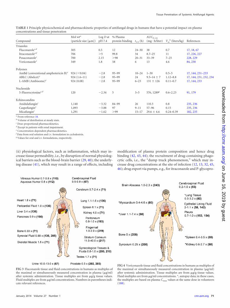
TABLE 1 Principle physicochemical and pharmacokinetic properties of antifungal drugs in humans that have a potential impact on plasmaconcentrations and tissue penetration
CompoundMol wta
(particle size [�m])Log D atpH 7.4
% Plasmaprotein binding t1/2 (h)
AUC0–24
(mg · h/liter) Vssb (liters/kg) References
TriazolesFluconazolec,d 305 0.5 12 24–30 38 0.7 17, 18, 67Itraconazolee,f 706 �5 99.8 34 8.7–25 11 17, 226, 227Posaconazolec 700 2.15 �98 20–31 33–39 7–25 228, 229Voriconazolee 349 1.8 58 6 13 4.6 84, 230
PolyenesAmBd (conventional amphotericin B)e 924 (�0.04) �2.8 95–99 10–24 1–30 0.5–5 17, 144, 231–233ABLC (Abelcet)e 924 (1.6–11) �2.8 95–99 24 9.5–14 � 7 1.12–8.8 17, 144, 231, 232, 234L-AMB (Ambisome)e 924 (0.08) �2.8 95–99 6–23 131 � 126 0.11–0.7 17, 144, 233
Nucleoside5-Fluorocytosinec,d 120 �2.34 5 3–5 576, 1289g 0.6–2.23 91, 179
EchinocandinsAnidulafunginc 1,140 �3.32 84–99 26 110.3 0.8 235, 236Caspofungine 1,093 �3.88 97 9–11 57–96 0.15 235, 236Micafunginc 1,291 �1.62 �99 15–17 29.6 � 4.6 0.24–0.39 182, 235
a From reference 18.b Volume of distribution at steady state.c Dose-proportional pharmacokinetics.d Except in patients with renal impairment.e Concentration-dependent pharmacokinetics.f Data from oral solution and i.v. formulation in cyclodextrin.g Values for oral and i.v. formulations, respectively.
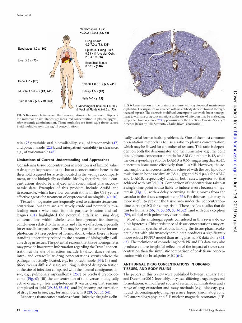
FIG 3 Fluconazole tissue and fluid concentrations in humans as multiples ofthe maximal or simultaneously measured concentration in plasma (�g/ml)after systemic administration. Tissue multiples are from �g/g tissue values.Fluid multiples are from �g/ml concentrations. Numbers in parentheses indi-cate relevant references.
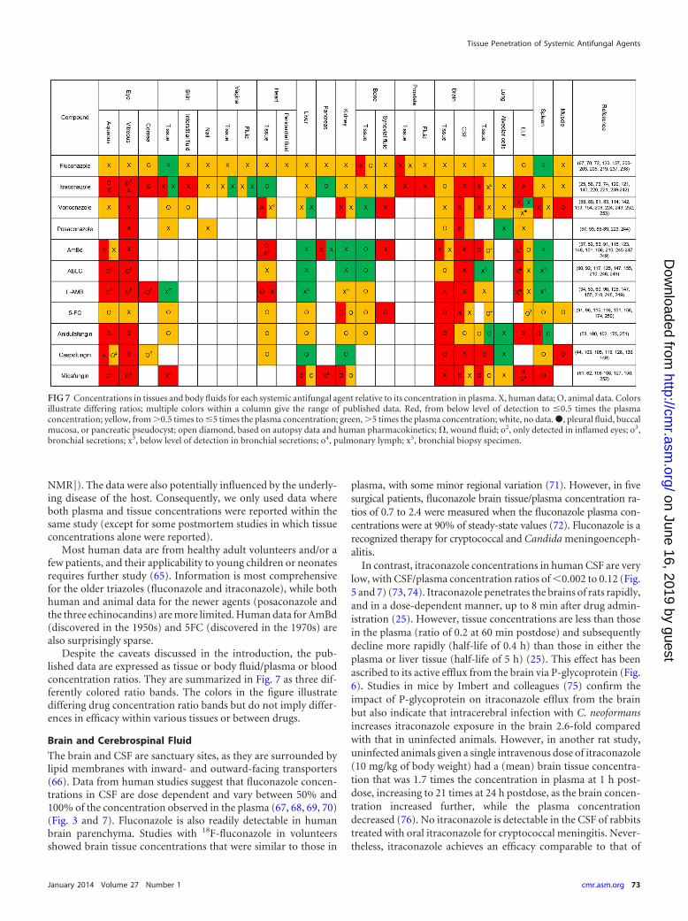
FIG 4 Voriconazole tissue and fluid concentrations in humans as multiples ofthe maximal or simultaneously measured concentration in plasma (�g/ml)after systemic administration. Tissue multiples are from �g/g tissue values.Fluid multiples are from �g/ml concentrations. *, autopsy data; in these cases,the multiples are based on plasma Cmax values at the same dose in volunteers(188).
Tissue Penetration of Systemic Antifungal Agents
January 2014 Volume 27 Number 1 cmr.asm.org 71
on June 16, 2019 by guesthttp://cm
r.asm.org/
Dow
nloaded from
tein (75); variable oral bioavailability, e.g., of itraconazole (47)and posaconazole (228); and interpatient variability in clearance,e.g., of voriconazole (48).
Limitations of Current Understanding and Approaches
Considering tissue concentrations in isolation is of limited value.A drug may be present at a site but at a concentration beneath thethreshold required for activity, located in the wrong subcompart-ment, or not biologically available. Ideally, therefore, tissue con-centrations should be analyzed with concomitant pharmacody-namic data. Examples of this problem include AmBd anditraconazole, which have low concentrations in the CSF yet areeffective agents for treatment of cryptococcal meningitis (49, 50).
Tissue homogenates are frequently used to estimate tissue con-centrations, but they are a relatively crude and potentially mis-leading matrix when used for this purpose. Mouton and col-leagues (51) highlighted the potential pitfalls in using drugconcentrations within whole-tissue homogenates for drawingconclusions related to the activity and efficacy of a drug, especiallyfor extracellular pathogens. This may be a particular issue for am-photericin B (irrespective of formulation), where there is long-standing uncertainty related to the amount of biologically avail-able drug in tissues. The potential reasons that tissue homogenatesmay provide inaccurate information regarding the “true” concen-tration at the site of infection include (i) discordance betweenintra- and extracellular drug concentrations versus where thepathogen is actually located, e.g., for posaconazole (33); (ii) mul-tifocal versus diffuse disease, resulting in altered drug penetrationat the site of infection compared with the normal contiguous tis-sue, e.g., pulmonary aspergilloma (257) or cerebral cryptococ-coma (Fig. 6); (iii) the concentration of total versus biologicallyactive drug, e.g., free amphotericin B versus drug that remainscomplexed to lipid (29, 52, 53, 54); and (iv) incomplete extractionof drug from tissue, e.g., for amphotericin B (29, 52, 53, 54).
Reporting tissue concentrations of anti-infective drugs in a clin-
ically useful format is also problematic. One of the most commonpresentation methods is to use a ratio to plasma concentration,which may be flawed for a number of reasons. This ratio is depen-dent on both the denominator and the numerator, e.g., the bonetissue/plasma concentration ratio for ABLC in rabbits is 42, whilethe corresponding ratio for L-AMB is 0.66, suggesting that ABLCpenetrates bone more effectively than L-AMB. However, the ac-tual amphotericin concentrations achieved with the two lipid for-mulations in bone are similar (35.4 �g/g and 39.5 �g/g for ABLCand L-AMB, respectively) and, in both cases, superior to thatachieved with AmBd (19). Comparison of concentrations taken ata single time point is also liable to induce errors because of hys-teresis (Fig. 1), with a delay occurring as drug moves from thevascular to the tissue compartment (55). For this reason, it may bemore useful to present the tissue area under the concentration-time curve (AUC) for comparison. There are few studies that dothis for humans (56, 57, 58, 59, 60, 61, 62), and with one exception(59), all deal with pulmonary distribution.
Most of the antifungal agents considered in this review do ex-hibit hysteresis. This persistence of tissue concentrations may ex-plain why, in specific situations, linking the tissue pharmacoki-netic data with pharmacodynamic data produces a significantlymore robust PK/PD model than using plasma PK data alone (31,63). The technique of comodeling both PK and PD data may alsoproduce a more insightful reflection of the impact of tissue con-centration than the simplistic comparison of peak tissue concen-tration with the breakpoint MIC (64).
ANTIFUNGAL DRUG CONCENTRATIONS IN ORGANS,TISSUES, AND BODY FLUIDS
The papers in this review were published between January 1965and December 2012. Inevitably, they used differing drug dosages andformulations, with different routes of systemic administration and arange of drug extraction and assay methods (e.g., bioassay, gas-liquid chromatography, high-pressure liquid chromatography,14C-autoradiography, and 18F-nuclear magnetic resonance [18F-
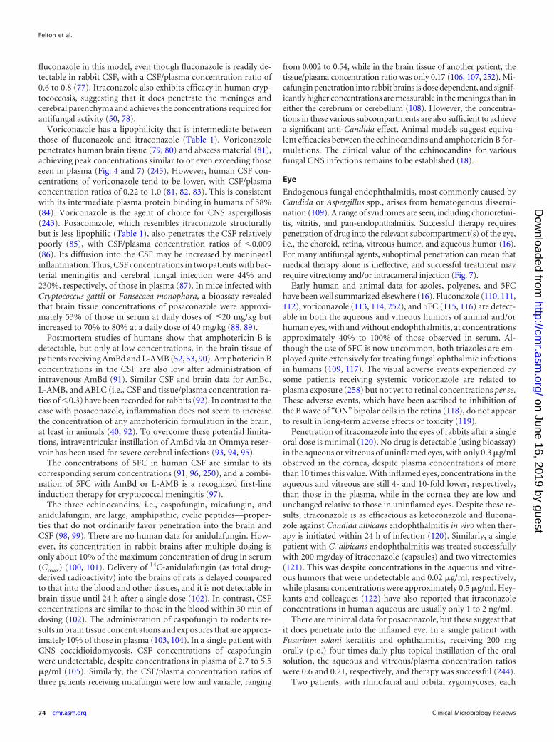
FIG 5 Itraconazole tissue and fluid concentrations in humans as multiples ofthe maximal or simultaneously measured concentration in plasma (�g/ml)after systemic administration. Tissue multiples are from �g/g tissue values.Fluid multiples are from �g/ml concentrations.
FIG 6 Cross section of the brain of a mouse with cryptococcal meningoen-cephalitis. The organism was stained with an antibody directed toward the cryp-tococcal capsule. The disease is multifocal. Attempts to use whole-brain homoge-nates to estimate drug concentrations at the site of infection may be misleading.(Reprinted from reference 263 by permission of the Infectious Diseases Society ofAmerica [taken by Julie Schwartz, Charles River Laboratories].)
Felton et al.
72 cmr.asm.org Clinical Microbiology Reviews
on June 16, 2019 by guesthttp://cm
r.asm.org/
Dow
nloaded from
NMR]). The data were also potentially influenced by the underly-ing disease of the host. Consequently, we only used data whereboth plasma and tissue concentrations were reported within thesame study (except for some postmortem studies in which tissueconcentrations alone were reported).
Most human data are from healthy adult volunteers and/or afew patients, and their applicability to young children or neonatesrequires further study (65). Information is most comprehensivefor the older triazoles (fluconazole and itraconazole), while bothhuman and animal data for the newer agents (posaconazole andthe three echinocandins) are more limited. Human data for AmBd(discovered in the 1950s) and 5FC (discovered in the 1970s) arealso surprisingly sparse.
Despite the caveats discussed in the introduction, the pub-lished data are expressed as tissue or body fluid/plasma or bloodconcentration ratios. They are summarized in Fig. 7 as three dif-ferently colored ratio bands. The colors in the figure illustratediffering drug concentration ratio bands but do not imply differ-ences in efficacy within various tissues or between drugs.
Brain and Cerebrospinal Fluid
The brain and CSF are sanctuary sites, as they are surrounded bylipid membranes with inward- and outward-facing transporters(66). Data from human studies suggest that fluconazole concen-trations in CSF are dose dependent and vary between 50% and100% of the concentration observed in the plasma (67, 68, 69, 70)(Fig. 3 and 7). Fluconazole is also readily detectable in humanbrain parenchyma. Studies with 18F-fluconazole in volunteersshowed brain tissue concentrations that were similar to those in
plasma, with some minor regional variation (71). However, in fivesurgical patients, fluconazole brain tissue/plasma concentration ra-tios of 0.7 to 2.4 were measured when the fluconazole plasma con-centrations were at 90% of steady-state values (72). Fluconazole is arecognized therapy for cryptococcal and Candida meningoenceph-alitis.
In contrast, itraconazole concentrations in human CSF are verylow, with CSF/plasma concentration ratios of �0.002 to 0.12 (Fig.5 and 7) (73, 74). Itraconazole penetrates the brains of rats rapidly,and in a dose-dependent manner, up to 8 min after drug admin-istration (25). However, tissue concentrations are less than thosein the plasma (ratio of 0.2 at 60 min postdose) and subsequentlydecline more rapidly (half-life of 0.4 h) than those in either theplasma or liver tissue (half-life of 5 h) (25). This effect has beenascribed to its active efflux from the brain via P-glycoprotein (Fig.6). Studies in mice by Imbert and colleagues (75) confirm theimpact of P-glycoprotein on itraconazole efflux from the brainbut also indicate that intracerebral infection with C. neoformansincreases itraconazole exposure in the brain 2.6-fold comparedwith that in uninfected animals. However, in another rat study,uninfected animals given a single intravenous dose of itraconazole(10 mg/kg of body weight) had a (mean) brain tissue concentra-tion that was 1.7 times the concentration in plasma at 1 h post-dose, increasing to 21 times at 24 h postdose, as the brain concen-tration increased further, while the plasma concentrationdecreased (76). No itraconazole is detectable in the CSF of rabbitstreated with oral itraconazole for cryptococcal meningitis. Never-theless, itraconazole achieves an efficacy comparable to that of
FIG 7 Concentrations in tissues and body fluids for each systemic antifungal agent relative to its concentration in plasma. X, human data; O, animal data. Colorsillustrate differing ratios; multiple colors within a column give the range of published data. Red, from below level of detection to �0.5 times the plasmaconcentration; yellow, from �0.5 times to �5 times the plasma concentration; green, �5 times the plasma concentration; white, no data.�, pleural fluid, buccalmucosa, or pancreatic pseudocyst; open diamond, based on autopsy data and human pharmacokinetics; , wound fluid; o2, only detected in inflamed eyes; o3,bronchial secretions; x3, below level of detection in bronchial secretions; o4, pulmonary lymph; x5, bronchial biopsy specimen.
Tissue Penetration of Systemic Antifungal Agents
January 2014 Volume 27 Number 1 cmr.asm.org 73
on June 16, 2019 by guesthttp://cm
r.asm.org/
Dow
nloaded from
fluconazole in this model, even though fluconazole is readily de-tectable in rabbit CSF, with a CSF/plasma concentration ratio of0.6 to 0.8 (77). Itraconazole also exhibits efficacy in human cryp-tococcosis, suggesting that it does penetrate the meninges andcerebral parenchyma and achieves the concentrations required forantifungal activity (50, 78).
Voriconazole has a lipophilicity that is intermediate betweenthose of fluconazole and itraconazole (Table 1). Voriconazolepenetrates human brain tissue (79, 80) and abscess material (81),achieving peak concentrations similar to or even exceeding thoseseen in plasma (Fig. 4 and 7) (243). However, human CSF con-centrations of voriconazole tend to be lower, with CSF/plasmaconcentration ratios of 0.22 to 1.0 (81, 82, 83). This is consistentwith its intermediate plasma protein binding in humans of 58%(84). Voriconazole is the agent of choice for CNS aspergillosis(243). Posaconazole, which resembles itraconazole structurallybut is less lipophilic (Table 1), also penetrates the CSF relativelypoorly (85), with CSF/plasma concentration ratios of �0.009(86). Its diffusion into the CSF may be increased by meningealinflammation. Thus, CSF concentrations in two patients with bac-terial meningitis and cerebral fungal infection were 44% and230%, respectively, of those in plasma (87). In mice infected withCryptococcus gattii or Fonsecaea monophora, a bioassay revealedthat brain tissue concentrations of posaconazole were approxi-mately 53% of those in serum at daily doses of �20 mg/kg butincreased to 70% to 80% at a daily dose of 40 mg/kg (88, 89).
Postmortem studies of humans show that amphotericin B isdetectable, but only at low concentrations, in the brain tissue ofpatients receiving AmBd and L-AMB (52, 53, 90). Amphotericin Bconcentrations in the CSF are also low after administration ofintravenous AmBd (91). Similar CSF and brain data for AmBd,L-AMB, and ABLC (i.e., CSF and tissue/plasma concentration ra-tios of �0.3) have been recorded for rabbits (92). In contrast to thecase with posaconazole, inflammation does not seem to increasethe concentration of any amphotericin formulation in the brain,at least in animals (40, 92). To overcome these potential limita-tions, intraventricular instillation of AmBd via an Ommya reser-voir has been used for severe cerebral infections (93, 94, 95).
The concentrations of 5FC in human CSF are similar to itscorresponding serum concentrations (91, 96, 250), and a combi-nation of 5FC with AmBd or L-AMB is a recognized first-lineinduction therapy for cryptococcal meningitis (97).
The three echinocandins, i.e., caspofungin, micafungin, andanidulafungin, are large, amphipathic, cyclic peptides—proper-ties that do not ordinarily favor penetration into the brain andCSF (98, 99). There are no human data for anidulafungin. How-ever, its concentration in rabbit brains after multiple dosing isonly about 10% of the maximum concentration of drug in serum(Cmax) (100, 101). Delivery of 14C-anidulafungin (as total drug-derived radioactivity) into the brains of rats is delayed comparedto that into the blood and other tissues, and it is not detectable inbrain tissue until 24 h after a single dose (102). In contrast, CSFconcentrations are similar to those in the blood within 30 min ofdosing (102). The administration of caspofungin to rodents re-sults in brain tissue concentrations and exposures that are approx-imately 10% of those in plasma (103, 104). In a single patient withCNS coccidioidomycosis, CSF concentrations of caspofunginwere undetectable, despite concentrations in plasma of 2.7 to 5.5�g/ml (105). Similarly, the CSF/plasma concentration ratios ofthree patients receiving micafungin were low and variable, ranging
from 0.002 to 0.54, while in the brain tissue of another patient, thetissue/plasma concentration ratio was only 0.17 (106, 107, 252). Mi-cafungin penetration into rabbit brains is dose dependent, and signif-icantly higher concentrations are measurable in the meninges than ineither the cerebrum or cerebellum (108). However, the concentra-tions in these various subcompartments are also sufficient to achievea significant anti-Candida effect. Animal models suggest equiva-lent efficacies between the echinocandins and amphotericin B for-mulations. The clinical value of the echinocandins for variousfungal CNS infections remains to be established (18).
Eye
Endogenous fungal endophthalmitis, most commonly caused byCandida or Aspergillus spp., arises from hematogenous dissemi-nation (109). A range of syndromes are seen, including chorioretini-tis, vitritis, and pan-endophthalmitis. Successful therapy requirespenetration of drug into the relevant subcompartment(s) of the eye,i.e., the choroid, retina, vitreous humor, and aqueous humor (16).For many antifungal agents, suboptimal penetration can mean thatmedical therapy alone is ineffective, and successful treatment mayrequire vitrectomy and/or intracameral injection (Fig. 7).
Early human and animal data for azoles, polyenes, and 5FChave been well summarized elsewhere (16). Fluconazole (110, 111,112), voriconazole (113, 114, 252), and 5FC (115, 116) are detect-able in both the aqueous and vitreous humors of animal and/orhuman eyes, with and without endophthalmitis, at concentrationsapproximately 40% to 100% of those observed in serum. Al-though the use of 5FC is now uncommon, both triazoles are em-ployed quite extensively for treating fungal ophthalmic infectionsin humans (109, 117). The visual adverse events experienced bysome patients receiving systemic voriconazole are related toplasma exposure (258) but not yet to retinal concentrations per se.These adverse events, which have been ascribed to inhibition ofthe B wave of “ON” bipolar cells in the retina (118), do not appearto result in long-term adverse effects or toxicity (119).
Penetration of itraconazole into the eyes of rabbits after a singleoral dose is minimal (120). No drug is detectable (using bioassay)in the aqueous or vitreous of uninflamed eyes, with only 0.3 �g/mlobserved in the cornea, despite plasma concentrations of morethan 10 times this value. With inflamed eyes, concentrations in theaqueous and vitreous are still 4- and 10-fold lower, respectively,than those in the plasma, while in the cornea they are low andunchanged relative to those in uninflamed eyes. Despite these re-sults, itraconazole is as efficacious as ketoconazole and flucona-zole against Candida albicans endophthalmitis in vivo when ther-apy is initiated within 24 h of infection (120). Similarly, a singlepatient with C. albicans endophthalmitis was treated successfullywith 200 mg/day of itraconazole (capsules) and two vitrectomies(121). This was despite concentrations in the aqueous and vitre-ous humors that were undetectable and 0.02 �g/ml, respectively,while plasma concentrations were approximately 0.5 �g/ml. Hey-kants and colleagues (122) have also reported that itraconazoleconcentrations in human aqueous are usually only 1 to 2 ng/ml.
There are minimal data for posaconazole, but these suggest thatit does penetrate into the inflamed eye. In a single patient withFusarium solani keratitis and ophthalmitis, receiving 200 mgorally (p.o.) four times daily plus topical instillation of the oralsolution, the aqueous and vitreous/plasma concentration ratioswere 0.6 and 0.21, respectively, and therapy was successful (244).
Two patients, with rhinofacial and orbital zygomycoses, each
Felton et al.
74 cmr.asm.org Clinical Microbiology Reviews
on June 16, 2019 by guesthttp://cm
r.asm.org/
Dow
nloaded from
received 0.6 mg/kg intravenous (i.v.) AmBd (123). Penetration ofAmBd into both the aqueous and vitreous of the infected eye washigher in the patient with rhinofacial disease and extensive retinalinflammation (fluid/serum concentration ratio of 0.4) than in thesecond patient, who had minimal retinal inflammation (ratio of0.06). Penetration of all formulations of amphotericin B into theeyes of rabbits is also enhanced by inflammation (124, 125, 249).Indeed, amphotericin B is not detected in noninflamed eyes, evenafter multiple dosing of AmBd, ABLC, or L-AMB (16, 124, 125).Consequently, intracameral injection is the favored delivery routefor these agents in patients with severe keratomycosis or endo-phthalmitis. For AmBd, this may lead to significant local toxicity,which is somewhat ameliorated by lipid formulations (109).
All three echinocandins also show limited penetration into theaqueous and vitreous humors of laboratory animals after systemicadministration, with either undetectable or low concentrationsrelative to those in plasma (100, 108, 109, 126, 127, 128). However,micafungin concentrations specifically in the retina and choroidof the eyes of rabbits range from 0.75 to 15.97 �g/ml and arecomparable with the concentrations in plasma (129). As with am-photericin B, inflammation appears to improve the extent of echi-nocandin penetration (127). Potentially subtherapeutic vitrealpenetration of caspofungin has been associated with treatmentfailure in Candida albicans endophthalmitis (130), and low con-centrations of caspofungin were measured in the aqueous of onehuman endophthalmitis patient (113). Similarly, low micafunginconcentrations in the aqueous and vitreous of a C. albicans endo-phthalmitis patient (0.001% of the simultaneous concentration inplasma) were associated with clinical failure (131), and the drugwas also ineffective in a patient with endophthalmitis caused byCandida tropicalis, despite severe inflammation and a MIC of 0.03�g/ml (132).
Lung
Pulmonary infection begins within the airspace (Fig. 2A). There-fore, for the agents used for prophylaxis or treatment of infectionconfined to the airspace, concentrations in epithelial lining fluid(ELF) and within pulmonary alveolar macrophages are of directimportance. The inhalation of aerosolized amphotericin B formu-lations is a potential option for prophylaxis (133, 134, 135). Anti-fungal drug concentrations within ELF after aerosol inhalation orsystemic administration were recently reviewed (12). However,for treatment of established invasive infections, drug concentra-tions in the lung parenchyma may be more relevant (Fig. 2B andC). Drug concentrations may also be measurable in a number ofother respiratory fluids, including bronchial secretions, sputum,pleural fluid, and pulmonary lymph (see below and Fig. 7).
Human studies suggest that 18F-fluconazole distributes rapidlyinto the lung tissue of volunteers, producing concentrations ap-proximately double those in plasma (71). In 20 patients receivinga single 200-mg dose of fluconazole, the lung tissue/plasma con-centration ratio range was 1.1 to 1.6 (136). Similarly, the flucona-zole ELF/plasma concentration ratio in cats was 1.2 (137). Flu-conazole also readily penetrates the extracellular space of the ratlung (fluid/plasma concentration ratio of 1.38), and this is unaf-fected by inflammation (138). Itraconazole exhibits ELF expo-sures that are one-third of the plasma AUC in human volunteers,while the AUC in alveolar cells is more than double that of theplasma (56). In postmortem samples from four hematology pa-tients, the mean lung tissue/plasma concentration ratio of itra-
conazole was 7 (139), while Heykants and colleagues (73) reportedconcentrations 0.9 to 2.4 times higher than those in the plasmas offour patients. However, itraconazole concentrations in bronchoal-veolar lavage (BAL) fluid and airway tissue were 10-fold lower thanthose in plasma in a patient with allergic bronchopulmonary asper-gillosis (ABPA) (140). Itraconazole has been used extensively to treatpulmonary fungal infections.
Postmortem studies show lung tissue homogenate concentra-tions for voriconazole that are comparable with the plasma con-centrations (80, 141). In volunteers receiving an i.v. loading doseon day 1 and then 200 mg of voriconazole p.o. twice a day (b.i.d.),the ELF/plasma concentration ratio was 11 (142). However, involunteers receiving the same i.v. loading dose on day 1, but fol-lowed by three doses of 4 mg/kg i.v. every 12 h (q12h), the ELF/plasma concentration ratio at steady state varied over 12 h fromapproximately 6 to 9, while for alveolar macrophages the ratiovaried from approximately 3.8 to 6.5 (58). Posaconazole exhibitsELF concentrations in humans similar to those seen in the plasma,but the exposure in alveolar cells is over 30 times that in plasma inboth volunteers (57) and lung transplant patients (143). It hasbeen suggested that high intracellular posaconazole concentra-tions may explain its effectiveness for prophylaxis (Fig. 2A) (33).Mean lung tissue concentrations of posaconazole in rabbits havebeen reported to range from 0.3 �g/ml to 2.1 �g/ml after dosing at2 to 6 mg/kg (145).
The administration of all formulations of amphotericin B re-sults in quantifiable concentrations in the ELF in both rabbits andhumans, but the plasma/ELF concentration ratios appear to differbetween formulations and species. The precise state of the ampho-tericin in these studies is not clear (i.e., free, protein bound, orlipid associated). Furthermore, the biological relevance of the to-tal concentrations associated with each formulation is also un-clear. Human data for the various amphotericin formulationssuggest that there may be some differences compared with rabbits(146, 147). Thus, intravenous ABLC produces ELF amphotericinB concentrations that are approximately 4 times those producedafter administration of L-AMB in humans (147). In 18 patientsundergoing thoracotomy and resection for lung cancer, a singledose of 1.5 mg/kg i.v. of L-AMB resulted in hysteresis, such thattissue/plasma concentration ratios were 0.29 and 2.5 at 10 and 25h postdose, respectively (248). In a postmortem study, lung tissuehomogenate concentrations were found to be 3 times higher witha similar dose of ABLC than with L-AMB (90). Similarly, ABLCconcentrations in mouse lung homogenates exceeded those forequivalent doses of L-AMB (39). Pulmonary inflammation mayincrease amphotericin concentrations following administrationof L-AMB (148). The amphotericin B formulations remain first-line agents for the therapy of pulmonary fungal infections.
There are no published data for echinocandin concentrationswithin human lung tissue. However, the concentrations of caspo-fungin in alveolar macrophages were �5 times the correspondingconcentrations in plasma in a single patient (149). Both anidula-fungin and micafungin also accumulated in the alveolar macro-phages of volunteers, attaining concentrations approximately 14and 4 times higher than those in plasma, respectively (58, 62). In18 lung transplant patients receiving a single 150-mg i.v. micafun-gin dose, ELF/plasma and alveolar cell/plasma concentration ra-tios varied with time postdose. Mean ratios ranged from 0.1 to1.53 at 3 h and from 1.1 to 6.2 at 24 h postdose (62). The vastmajority of anidulafungin and micafungin found in the ELF is
Tissue Penetration of Systemic Antifungal Agents
January 2014 Volume 27 Number 1 cmr.asm.org 75
on June 16, 2019 by guesthttp://cm
r.asm.org/
Dow
nloaded from
present within macrophages rather than in the fluid itself (58, 61,62). Caspofungin, micafungin, and anidulafungin exhibit lung tis-sue exposures in rodents that exceed those in plasma by 1.1-fold,2.8-fold, and 10-fold, respectively (102, 103, 150).
Pulmonary Lymph Fluid
There are no human data for antifungal drug concentrations inpulmonary lymph, but Hoeprich and colleagues (151) examinedthe concentrations of 5FC and AmBd in sheep cannulated via theafferent duct of the right caudal mediastinal lymph node. All drugstested (also including ketoconazole, the triazole Bay n733, andAmBd methyl ester [AME]) appeared promptly in the lymph aftera single intravenous dose, with their concentrations subsequentlydecaying exponentially. In general, the concentrations of all fivedrugs in lymph slightly exceeded those in plasma measuredshortly after the end of the 30-min infusion period (maximumratio for lymph to plasma of 1.0 to 1.9), except for AME, wherelymphatic concentrations were lower. Koizumi and colleagues(152) also examined AmBd concentrations in sheep lung andlymph after an i.v. infusion. The concentrations in the lymph weresimilar to (or slightly exceeded) those in the plasma, depending onthe duration of the infusion. Given the range of lipophilicities andplasma protein binding of the above antifungal agents, these proper-ties do not seem to have a significant impact on penetration into thelymphatic system, at least following intravenous administration.
Pleural Fluid
Data on antifungal drug pleural fluid concentrations are limited(Fig. 7). Voriconazole penetrates into the pleural fluid, producingtrough concentrations in humans that are similar to paired plasmaconcentrations (153, 154). For AmBd, pleural fluid concentra-tions are approximately 50% of those in plasma (91, 247). How-ever, pleural fluid amphotericin concentrations following the ad-ministration of L-AMB or ABCD are approximately 5% to 25% oftheir plasma exposures (60, 155). Penetration of the echinocan-dins into pleural fluid appears to be low. Thus, for anidulafunginin one patient with Candida empyema and for three micafunginpatients, pleural fluid concentrations were less than 1% and 10%,respectively, of those measured in the plasma (107, 251).
Bronchial Secretions
Watkins and colleagues (140) demonstrated, for one patient, thatitraconazole accumulates to approximately twice the plasma con-centration in bronchial biopsy tissue and is also detectable (at onlyng/ml concentrations) in BAL fluid and bronchial washings.However, no allowance was made for the significant dilution fac-tor involved with their sampling methods. They concluded thatitraconazole is present in “relatively high” concentrations in pul-monary fluids and tissues. In contrast, amphotericin B was de-tected, but only briefly postdose and at low concentrations, in thetracheal secretions of humans (91) and the tracheas of dogs fol-lowing administration of AmBd (91), although penetration maybe dose dependent (156). For 5FC, concentrations in dog bron-chial secretions are approximately 75% of corresponding plasmaconcentrations (156).
Saliva, Sputum, Buccal Mucosa, and Esophagus
The attainment of effective antifungal drug concentrations withinthe saliva, sputum, and bronchial fluid is critical for therapy oforopharyngeal, esophageal, and bronchial infections. Fluconazole
(67, 157, 158) and itraconazole (73, 159) have both been detectedin the saliva and sputum of patients (Fig. 7). Consistent with theirphysicochemical properties (Table 1), the concentration ratios forfluconazole in saliva and sputum compared with serum are �1,while for itraconazole they are generally much lower (73) and veryvariable (159). Itraconazole can also be detected in esophagealtissue, at 3 times the concentration in plasma (160), and in bron-chial exudates (73). However, clinical data suggest that flucona-zole is superior to itraconazole for treating oropharyngeal andesophageal candidiasis (161, 162). Voriconazole is present in thesaliva of volunteers, and concentrations increase over time, usinga standard dose. Thus, salivary exposure on day 1 is approximately25% of that in plasma and increases to 88% of that in plasma withmultiple dosing (163). Fluconazole and voriconazole show com-parable efficacies in immunocompromised patients with esopha-geal candidiasis (164). While there are no published data for po-saconazole concentrations in saliva, sputum, or mucosal andesophageal tissues, this drug is as effective as fluconazole in treat-ing HIV patients with oropharyngeal candidiasis (165).
Buccal mucosal concentrations of amphotericin B increase in adose-dependent manner in humans after L-AMB administrationand attain concentrations approximately 7 to 43 times those inplasma (166). A wide range of amphotericin B concentrationswere also detectable in esophageal autopsy samples from sevenpatients after AmBd administration (54).
The concentrations of 5FC in human saliva are slightly lowerthan those in the plasma, but the 5FC concentrations measured inthe bronchial secretions of dogs are comparable to serum concen-trations (91).
There are no human or laboratory animal data giving the con-centrations of caspofungin or micafungin at these sites. Anidula-fungin is present in both the saliva and esophagus in rabbits withoropharyngeal and esophageal candidiasis, but only at concentra-tions between 1% and 33% of those in plasma (167). However, allthree echinocandins show efficacy at the end of therapy equivalentto that of fluconazole after intravenous administration to patientswith AIDS and oropharyngeal or esophageal candidiasis (168, 169,170). There are no data to indicate whether any efficacy differ-ences between fluconazole and the echinocandins seen on lon-ger-term follow-up of these patients are related to residual tis-sue concentrations.
Heart
Fluconazole and voriconazole concentrations in human heart tis-sue are comparable to those in plasma, based on 18F-NMR studiesin healthy volunteers and autopsy data, respectively (71, 80). Thepericardial fluid/plasma concentration ratios of fluconazole in 20patients ranged from 0.9 to 1.0 (136). Data from a single patientwith disseminated aspergillosis also suggest that voriconazole dif-fuses into the pericardial fluid, at a concentration comparable tothe plasma concentration (153). Autopsy data also indicate thatmyocardial voriconazole concentrations are similar to those inother body organs, including the lung and kidney (80). In con-trast, itraconazole exposure in the hearts of mice after a single10-mg/kg i.v. dose is only 8% of that in plasma (171). However, inrats, at 1 h postdose, the concentration is 6 times the level inplasma, and both the absolute concentration and the plasma ratioincrease further after 24 h (76). There are no published humanheart tissue concentration data for itraconazole. Nevertheless,itraconazole can cause congestive heart failure (172) via negative
Felton et al.
76 cmr.asm.org Clinical Microbiology Reviews
on June 16, 2019 by guesthttp://cm
r.asm.org/
Dow
nloaded from
inotropic effects, although the precise mechanism is unknown(173).
Postmortem studies of patients following administration ofAmBd or L-AMB show a wide range of concentrations (�0.1 to9.1 �g/g) of amphotericin in heart tissue and myocardium (52,90). In the hearts of dogs, the AmBd concentration after 14 days ofdosing with 0.6 mg/kg/day is approximately 7 times the corre-sponding plasma value (37), while in rats given a single AmBddose of 1.0 mg/kg, it is approximately 3 times higher (225).
As with fluconazole, the concentration of 18F-5FC in rat hearttissue is similar to that in blood (174).
Caspofungin is detectable in the rodent heart after a single dose,at a concentration approximately 20% of the peak plasma concen-tration, which then declines at a lower rate than in the plasma(103, 104). In contrast, anidulafungin exposure in the heart tissueof neonatal rats increases to approximately 1.3 times the concen-tration in plasma after a single dose and 1.8 times after multipledosing (175).
Liver
Given its major role in metabolism and clearance, many xenobi-otics are likely to achieve higher concentrations in the liver than inthe plasma. Twenty minutes after intravenous administration, theconcentration of 18F-fluconazole in human livers is approxi-mately 3 times the paired plasma concentration, while in rabbits itis twice that in the plasma (71). Itraconazole also accumulates inthe liver (Fig. 7) (122), and it reached a concentration in onepatient that was over three times that in plasma (73). However, inthe livers of rats, itraconazole achieves concentrations that areapproximately 13 times those in plasma 1 h after a single intravenousdose, and this increases further over 24 h (76). The plasma concen-tration declines 9-fold over this period, resulting in a tissue/plasmaconcentration ratio exceeding 150 at 24 h postdose (76).
In contrast, the nucleoside 18F-5FC, which is even more polarthan fluconazole, attains concentrations in rat livers that are sim-ilar to those in plasma (174).
Hepatic concentrations of amphotericin are detectable fromtissue obtained at postmortem (52, 54). There is a relationshipbetween the plasma exposure of L-AMB and liver tissue concen-trations of amphotericin B in human autopsy samples. After L-AMB dosing, the mean amphotericin B concentration that wasachieved was 102 �g/g liver, but with substantial interpatient vari-ability (90). Amphotericin B has a long residence time in hepatictissue of mice. Concentrations (measured using bioassay) are detect-able 14 days after dosing with L-AMB (38). However, Andes andcolleagues (39) have shown that ABLC exhibits lower concentrationsin mouse liver homogenates than equivalent doses of AmBd or L-AMB (at least following intraperitoneal [i.p.] administration).
The exposures of anidulafungin and caspofungin in the livers ofrodents are raised approximately 10- and 16-fold, respectively,compared with plasma concentrations (102, 103). This is largelyrelated to delayed clearance from the liver. However, micafunginappears to behave differently, with a lower peak concentration inthe livers of rats and an AUC that is similar to that of the plasma(150). For caspofungin, specific hepatic transporters that mediateuptake into rat liver have been identified (26).
Kidney
Approximately 80% of a fluconazole dose is eliminated as un-changed drug in the urine. Consequently, urinary concentrations
are approximately 10 times those in human plasma (Fig. 7) (67).Fluconazole also readily penetrates kidney tissue, with peak tissueconcentrations of 18F-fluconazole that are approximately 4 timesthe peak in human plasma (71). Similar to fluconazole, voricona-zole is largely excreted via the urine (78%) and feces in humans,but mostly as metabolites, with less than 2% excreted as un-changed drug (84). Postmortem studies of eight patients showedthat voriconazole was detectable in kidney tissue, at a mean con-centration of 6.47 �g/g, but with significant interindividual vari-ability (80). In contrast to fluconazole, itraconazole concentra-tions in urine are very low due to its negligible renal excretion(122). When administered intravenously to rats, itraconazole at-tains concentrations in kidney tissue of 5.5 �g/g after 1 h (3 timesthe plasma concentration) and 5.9 �g/g (31 times the plasma con-centration) at 24 h postdose (76). However, a kidney tissue con-centration of only 0.5 �g/g (1.5 times the plasma concentration)was recorded in a single patient (73).
The kidneys are a primary site of toxicity for all polyenes. Post-mortem studies show that amphotericin B (from AmBd or L-AMB) is readily detectable in kidney tissue (52–54, 90). The renalconcentration of amphotericin B in rat kidneys after AmBd ad-ministration is 10 times that in the serum, while the correspond-ing renal concentration after L-AMB administration is one-thirdthat of AmBd and only 4 times the serum concentration (177).This is consistent with the reduction in amphotericin B-associatedrenal toxicity after its administration as L-AMB (or other lipidamphotericin formulations) rather than AmBd (178). The clear-ance of amphotericin B from the kidneys of rodents is prolonged,and the drug is detectable for at least 48 h after a single adminis-tration of AmBd (177) and at least 14 days after a prolongedcourse of L-AMB (38). In mouse kidney homogenates, concentra-tions of amphotericin B following administration of L-AMB orABLC at a dose of 80 mg/kg i.p. are comparable to those observedwith 20 mg/kg i.p. of AmBd (39).
Like fluconazole, 5FC is principally eliminated in the urine as un-changed drug (97%), and plasma clearance is closely related to creat-inine clearance (91, 179). The concentration of 18F-5FC in rat kidneysis 3 times that in blood 2 h after dosing, with very high concentrations(60 times the plasma concentration) in the urine (174).
All three echinocandins readily penetrate into the kidney tissueof laboratory animals. After a single dose, 14C-anidulafungin ex-posure in rat kidney tissue is approximately 10 times that inplasma (102). In addition, anidulafungin exhibits an extendedresidence time in the kidney, with a terminal half-life that is twicethat in plasma (102). Anidulafungin also accumulates in rabbitkidneys after multiple dosing (100). After a single dose adminis-tered to mice, caspofungin exhibits a longer mean residence timein the kidneys (31) and has a tissue/plasma concentration ratioover 24 h of approximately 7 (103). In contrast, micafungin con-centrations in rat kidneys exceed those in plasma 5 min after dos-ing, by 1.6-fold, but then decline in parallel with plasma concen-trations (150). All three echinocandins exhibit low concentrations(�2% of the dose) (104, 181, 182) of unchanged drug in humanurine. There are reported cases of the efficacy of the echinocandinsin patients with candiduria (183, 184), but this may reflect theattainment of high concentrations in renal parenchyma.
Spleen
Fluconazole penetrates into the spleen in both humans and rab-bits, although to different extents (71, 185, 213). Higher concen-
Tissue Penetration of Systemic Antifungal Agents
January 2014 Volume 27 Number 1 cmr.asm.org 77
on June 16, 2019 by guesthttp://cm
r.asm.org/
Dow
nloaded from
trations of 18F-fluconazole are seen in human spleens than in anyother organ, with a tissue/blood concentration ratio of approxi-mately 6 (Fig. 7). However, in rabbit spleens, concentrations aresimilar to those in the blood (71, 213) but slightly less than those inthe plasma (185). Human data are limited for itraconazole, butsplenic concentrations in two patients were 2- to 3-fold higherthan the plasma concentrations (73, 186). However, a study in ratsshowed a progressive accumulation of drug in the spleen over thedosing interval, to approximately 10 times the plasma concentra-tion (76). In contrast, mice receiving itraconazole at 20 mg/kg i.v.had splenic concentrations at 5 hours postdose that were 3 timesthose in the plasma, but they were similar to the plasma concen-trations by 24 h (187). There are no laboratory animal data forvoriconazole, but it is detectable in human splenic tissue at post-mortem (80, 141). The splenic concentration (mean, 5.6 �g/g) issimilar to the plasma steady-state concentrations in volunteersreceiving 200 mg b.i.d. p.o. (188).
Human postmortem studies indicate that AmBd and L-AMBare detectable in the spleen at concentrations exceeding those of allother organs except the liver (52–54). Furthermore, tissue con-centrations are dose dependent (53, 90). In dogs receiving 0.6mg/kg/day of AmBd for 14 days, splenic concentrations are �160times those in the plasma (37). After multiple dosing to mice, thesplenic concentrations of amphotericin B derived from the three for-mulations are in the rank order ABLC � L-AMB � AmBd (35, 189).
There is little published information for 5FC, but concentra-tions of 18F-5FC in rat spleens are similar to those in blood (174).
There are no human data for the echinocandins, but all threeagents are detectable in the spleens of laboratory animals (127). Inthe rat spleen, anidulafungin exposure is 10 times greater than thatin plasma after a single dose of 5 mg/kg, and peak splenic concentra-tions exceed those measured in rabbits following multiple dosing at10 mg/kg (100, 102). In contrast, the tissue/plasma concentrationratio of caspofungin is only �1 after a single dose in mice (103) or rats(104). Micafungin concentrations in rabbit spleens are also similar tothose in plasma, even after multiple dosing (127).
Pancreas
Pancreatic antifungal drug concentrations are rarely reported forlaboratory animals or humans. The most comprehensive data arefor fluconazole, where 15 patients undergoing pancreatic surgeryreceived a single fluconazole i.v. infusion of 400 mg (190). Pan-creatic tissue concentrations increased for up to 2 h postdose, andthe mean tissue/plasma concentration ratio at the time that tissuewas sampled was approximately 1.0 (Fig. 7). Fluconazole penetra-tion into pancreatic pseudocysts is slow, and concentrations at-tained in two cysts were lower than those in the plasma, at 0.4 and0.8 times the plasma concentrations (190). Fluconazole concen-trations in rat pancreatic tissue are similar to those in humans,with concentrations approximately 88% to 91% of those inplasma (190).
Penetration of AmBd into human pancreatic tissues has beendemonstrated only in autopsy samples. Tissue concentrations arehighly variable, ranging from �0.1 to 18.6 �g/g (52).
There are no data for caspofungin or anidulafungin, but a mi-cafungin pancreatic pseudocyst fluid concentration of 0.38 �g/mlwas recorded for a single patient 24 h after a prior dose (106).
Peritoneum
Intra-abdominal fungal infections are difficult to treat, particu-larly in patients requiring peritoneal dialysis (191). Fluconazole,5FC, and amphotericin B are typically used as primary therapy,although limited experience in patients suggests that voricona-zole, posaconazole, caspofungin, and micafungin could also beused for treating fungal peritoneal infection.
The polar agents fluconazole and 5FC achieve peritoneal con-centrations after i.v. administration to uninfected laboratory ani-mals of approximately 100% and 50% of those in serum, respec-tively (91, 192). Furthermore, in adults or children undergoingperitoneal dialysis, dialysate concentrations of fluconazole (fol-lowing systemic administration) are similar to or exceed those inthe plasma (193, 194). Limited clinical data suggest that the peri-toneal concentrations of 5FC in humans are approximately 65%to 100% of those in serum (195, 196).
Five patients receiving voriconazole for peritonitis complicat-ing peritoneal dialysis had concentrations in the peritoneal dialy-sate that were approximately 50% of those in the plasma after asingle oral voriconazole dose (197).
The peritoneal concentrations of amphotericin B followingAmBd administration are variable and less than 50% of serumconcentrations (91, 195, 198) and, on occasion, are undetectable(196). Weiler and colleagues (199) have demonstrated that similaramphotericin B ascitic fluid concentrations are attained followingadministration of either L-AMB or ABLC for 7 to 13 days at 3 to 5mg/kg/day.
A single patient receiving micafungin had a concentration inascitic fluid of 1.02 �g/ml, giving an ascites/plasma concentrationratio of 0.15 (107).
Genital System
Fungal infections of the genital system, particularly vaginal candi-diasis, are some of the most commonly experienced fungal infec-tions of humans. Fluconazole is used extensively for treating uro-genital infections caused by Candida spp. Consequently, there is arelative abundance of clinical data related to the concentrations offluconazole within gynecological tissues and secretions (67, 200,201), testicular (71) and prostatic (71, 202) tissues, and prostaticfluids (203) (Fig. 7). In the vagina and its secretions and in othergynecological tissues, the fluconazole tissue or secretion/plasmaconcentration ratio is at least 1. The tissue/plasma concentrationratio of fluconazole in the testicles of volunteers receiving 18F-fluconazole (71) is also �1. However, in the prostate, which is asanctuary site, the ratios range from 0.3 in prostatic hyperplasiapatients (202) to 2.0 in volunteers (71). In the prostatic fluid ofpatients with AIDS and cryptococcal meningitis, the fluconazolefluid/plasma concentration ratio range is 0.6 to 0.9 (203). Thehuman data for itraconazole indicate that its concentrations invaginal and other gynecological tissues and in cervical mucus arebetween 1.6 and 20 times those in plasma but that the vaginalfluid/plasma concentration ratio is �0.5 (73, 122). There are nopublished human or animal data for the other antifungal agentsfollowing systemic administration.
Bone
The concentration of 18F-fluconazole in bone is approximately33% of the plasma concentration in humans and 100% in rabbits(Fig. 7) (71). After two i.v. doses, fluconazole is also detectable inthe nucleus pulposus of the rabbit spine, but with a very wide
Felton et al.
78 cmr.asm.org Clinical Microbiology Reviews
on June 16, 2019 by guesthttp://cm
r.asm.org/
Dow
nloaded from
concentration range (0 to 63.5 �g/g) that is apparently unrelatedto concentrations in plasma (204). In three patients, fluconazolesynovial fluid concentrations were 0.88 to 1.0 times those inplasma (136, 205, 206). Similarly, the mean synovial fluid/plasmaconcentration ratio of fluconazole after 10 days of dosing to horseswas 0.5 (207). Itraconazole may accumulate in bone, and thebone/plasma concentration ratio in a single patient was 4.7 (73).Voriconazole is detectable in human medullary and cortical bone,with especially high concentrations (approximately 5 times theplasma concentration) in the former (208). The concentration ofvoriconazole in synovial fluid from a single patient was approxi-mately one-third the plasma concentration (208), while in horses,the mean voriconazole synovial fluid/plasma concentration ratiowas 0.6 (209).
Amphotericin concentrations are high in the bone marrow ofdogs and rabbits following administration of any of the currentlyavailable formulations (37, 210). Certainly the administration ofamphotericin B in lipid formulations is considered an example ofdrug targeting, with particular respect to the kidneys and bonemarrow (36). The lowest bone marrow concentrations are ob-served following administration of AmBd, but concentrations arestill approximately 5 times those in plasma (37). In human syno-vial fluid, the measured AmBd fluid/plasma concentration ratio isapproximately 0.4 (91), while in a single neonate with C. albicansosteoarthritis, the synovial fluid/plasma amphotericin concentra-tion ratio of a random sample following 35 days of AmBd and 10days of L-AMB was 1.4 (211).
There are limited data available on bone and synovial fluid con-centrations of 5FC in humans and animals. Polak (91) reportedbone and synovial fluid concentrations of 30% and 41% of thosein plasma, respectively. However, in a premature infant with Can-dida arthritis, the synovial fluid concentration was approximately83% of that in the plasma (212). In rats given 18F-5FC, bone andblood concentrations are comparable (174).
Anidulafungin concentrations in the bone of neonatal rats aftera single dose are less than those in plasma, with a bone/plasmaconcentration ratio of 0.21 (175). No data are available for caspo-fungin or micafungin, although these drugs have been used totreat a few patients with bone/joint infections, in combinationwith AmBd or a triazole.
Muscle
The concentrations of 18F-fluconazole in human skeletal muscleare similar to those in the myocardium, both of which have aconcentration ratio to blood of 1.8 (71). However, in rats andrabbits, the ratio is somewhat lower (0.58 to 0.74) (138, 185, 213).In contrast, itraconazole accumulates in skeletal muscle relative toplasma, attaining a muscle/tissue concentration ratio of 2.4 in asingle patient (73) but one of over 7 in rats (76) (Fig. 7).
In human autopsy samples, skeletal muscle concentrations ofamphotericin following the administration of AmBd ranged from0 to 1.2 �g/g and were lower than those in any other tissue (54). Inthe rat, concentrations of amphotericin B in muscle (0.21 to 0.27�g/g) were also lower than those in other tissues but were stillapproximately 10-fold higher than plasma concentrations follow-ing multiple dosages of AmBd (214). Simultaneously collectedheart muscle tissue concentrations were approximately 20-foldhigher than those in plasma. In autopsy samples from patientsreceiving L-AMB, the mean myocardial amphotericin concentra-tion was 3.18 �g/g (90).
There are no human data for 5FC, but in rats receiving 18F-5FC,the skeletal muscle/blood concentration ratio is 1.1 (174).
Human data are also lacking for the echinocandins. However,skeletal muscle concentrations of anidulafungin in rats are com-parable to those in plasma (102), whereas for caspofungin, skeletalmuscle concentrations in mice are less than 50% of those inplasma (103, 104).
Skin and Nails
The prolonged exposure of antifungal agents within the skin, nail,and nail bed is an important factor determining the outcome oftreatment of dermatomycosis (215). Fluconazole concentrationswithin the dermis are similar to those in plasma (216, 217), butconcentrations in the stratum corneum are up to 40 times those inplasma (217, 218) (Fig. 7). The clearance of fluconazole from thestratum corneum is also significantly slower than that from theplasma and other skin layers, with concentrations that decline 2 to3 times more slowly than the plasma concentrations (215, 217,218). Interestingly, once-weekly oral dosing of 150 mg for 2 weeksresults in higher fluconazole concentrations in the stratum cor-neum relative to those in the epidermis/dermis, sweat, and serumthan those obtained by daily dosing at 50 mg for 12 days (217). Infingernails, fluconazole concentrations are dose proportional and,at steady state, are approximately twice those in the plasma. Flu-conazole is also detectable in nails up to 4 months after cessationof therapy (219). Slow clearance from both skin and nails is alsoseen for itraconazole. It binds tightly in the stratum corneum anddoes not readily distribute back to the plasma compartment (215,220). The drug also accumulates in sebum. Consequently, thoseareas of skin with active sebaceous glands contain higher concen-trations of itraconazole (e.g., the back, with twice the plasma con-centration) than those that do not (e.g., the palm, with less thanthe plasma concentration) (122). Concentrations of itraconazolein blister fluid increase more slowly than those in the plasma,attaining a maximal concentration approximately 0.7 times thatin the plasma (221). Itraconazole also has a very long residencetime in nails after the cessation of therapy (122). Maximal concen-trations of itraconazole in fingernails and toenails are 0.95 �g/gand 1.5 �g/g, respectively, 4 and 6 months after cessation of pulsetherapy (222). The concentration of posaconazole within the hu-man dermis is comparable to that in plasma (59). However, intoenails, its concentration is both dose and time dependent, at-taining a maximum approximately 3 times greater than that inplasma after 24 weeks of therapy (223). There are no human datafor voriconazole, but in guinea pigs, voriconazole skin concentra-tions are approximately twice those in blood, while in skin micro-dialysates, the voriconazole concentrations are only 50% of thosein blood (224). Patients receiving voriconazole therapy have beenshown to suffer from significant phototoxicity on exposure tosunlight, although a relationship to the voriconazole concentra-tion or retinol levels in skin remains to be established (254). In afew patients, long-term voriconazole exposure may result in skincancer (255, 256).
AmBd skin concentrations in rats receiving a single intravenousdose of 1.0 mg/kg are approximately 30% to 50% of those inplasma and decrease with time in parallel with the plasma concen-trations (225).
Laboratory animal studies show that clearance of anidulafunginand caspofungin from rat skin is delayed compared to that fromplasma, but these drugs never attain the peak concentrations mea-
Tissue Penetration of Systemic Antifungal Agents
January 2014 Volume 27 Number 1 cmr.asm.org 79
on June 16, 2019 by guesthttp://cm
r.asm.org/
Dow
nloaded from
sured in plasma (102, 104). After a single i.v. dose of anidulafungin,peak skin concentrations are approximately 80% of those in plasma,while for caspofungin, skin concentrations in rats peak at some 2 hpostdose, but with a skin/plasma concentration ratio of only 0.3.However, caspofungin clearance from the skin is such that by 288 hpostdose, residual skin concentrations, while only 15% of their peak,are 4 times those remaining in the plasma (104).
UNDERSTANDING TISSUE CONCENTRATIONS FOR OPTIMALUSE OF EXISTING AGENTS AND DEVELOPMENT OF NEWERANTIFUNGAL AGENTS
Current State of the Art
This review provides a summary of tissue concentration data for keyantifungal drugs in humans and some animals. While there is a size-able body of literature on this topic, many of the data are of variablequality, and the implications for the clinical care of patients withinvasive fungal infections are frequently unclear. In addition, the hu-man data are almost exclusively from adults, meaning that the impli-cations for young children and neonates remain uncertain (65). Theinterpretation of many studies is further compounded by a multitudeof different methodological approaches. Nevertheless, the followinggeneral conclusions seem reasonable.
First, small polar agents with low protein binding (e.g., flucona-zole and 5FC) distribute more evenly and into a wider range oftissues than the larger, more lipophilic (itraconazole) or amphi-pathic (e.g., amphotericin B and echinocandins) agents.
Second, the more lipophilic or amphipathic agents may havelonger residence times within tissues and may also accumulate toconcentrations that exceed those in the plasma.
Third, agents with relatively low molecular weights, such asfluconazole, 5FC, and voriconazole, penetrate more readily intotissue beds.
Fourth, the formulation may have a significant impact on se-rum and tissue pharmacokinetics, although the pharmacody-namic implications of these differences frequently remain unclear.
Fifth, the measurable concentration of a drug within a tissuemay not necessarily be an indication of its biological activity inthat compartment.
Sixth, within a single drug class and with apparently closelyrelated structures, there may be marked differences in tissue dis-tribution (e.g., the triazoles).
Finally, a degree of caution is always advisable in extrapolatingdata from laboratory animals to humans.
Beyond State of the Art
A detailed understanding of tissue concentrations is an importantcomponent of drug development (13). In this regard, the follow-ing are worthy of consideration.
First, comodeling both PK and PD data (if possible) provideskey insights into the importance of tissue concentrations (64).
Second, single point estimates of tissue concentrations are ofrelatively limited value. Estimating concentration-time profiles(and thereby calculating the AUC in tissues) is possible using pop-ulation pharmacokinetic modeling techniques. Relatively fewstudies have done this for humans to date (56–62), and all but one(59) deal with pulmonary distribution.
Third, designing antifungal regimens that optimize exposure atthe site of infection rather than plasma exposure requires further
consideration and study but may be pivotal in the design of opti-mum regimens for new antifungal agents (259).
Fourth, as has long been understood (51), tissue homogenatesare not the ideal matrix for estimating tissue concentrations. Non-invasive methods such as magnetic resonance spectroscopy withspectroscopic imaging (e.g., 18F-5FC [260]) or positron emissiontomography (e.g., 18F-fluconazole [71, 213]) can be used in labo-ratory animals or humans. Direct molecular analysis of whole-body animal tissue or isolated organs by matrix-assisted laser de-sorption ionization (MALDI) mass spectroscopy also represents apromising approach, without the requirement for radiolabeleddrug (261).
ACKNOWLEDGMENTS
Timothy Felton is an MRC Clinical Training Fellow supported by theNorth West England Medical Research Council Fellowship Scheme inClinical Pharmacology and Therapeutics, which is funded by the MedicalResearch Council (grant G1000417/94909), ICON, GlaxoSmithKline, As-traZeneca, and the Medical Evaluation Unit.
Peter F. Troke was previously an employee of and a consultant toPfizer and owns shares in Pfizer and other pharmaceutical companies.William W. Hope has acted as a consultant and received research supportfrom Gilead Sciences, Pfizer Inc., Astellas, Merck, and F2G.
REFERENCES1. Pfaller MA, Diekema DJ. 2010. Epidemiology of invasive mycoses in
North America. Crit. Rev. Microbiol. 36:1–53. http://dx.doi.org/10.3109/10408410903241444.
2. Pfaller MA, Pappas PG, Wingard JR. 2006. Invasive fungal pathogens:current epidemiological trends. Clin. Infect. Dis. 43:S3–S14. http://dx.doi.org/10.1086/504490.
3. Lortholary O, Gangneux JP, Sitbon K, Lebeau B, de Monbrison F, LeStrat Y, Coignard B, Dromer F, Bretagne S. 2011. Epidemiologicaltrends in invasive aspergillosis in France: the SAIF network (2005–2007).Clin. Microbiol. Infect. 17:1882–1889. http://dx.doi.org/10.1111/j.1469-0691.2011.03548.x.
4. Nivoix Y, Velten M, Letscher-Bru V, Moghaddam A, Natarajan-AmeS, Fohrer C, Lioure B, Bilger K, Lutun P, Marcellin L, Launoy A, FreysG, Bergerat JP, Herbrecht R. 2008. Factors associated with overall andattributable mortality in invasive aspergillosis. Clin. Infect. Dis. 47:1176 –1184. http://dx.doi.org/10.1086/592255.
5. Andes DR, Safdar N, Baddley JW, Playford G, Reboli AC, Rex JH,Sobel JD, Pappas PG, Kullberg BJ. 2012. Impact of treatment strategyon outcomes in patients with candidemia and other forms of invasivecandidiasis: a patient-level quantitative review of randomized trials. Clin.Infect. Dis. 54:1110 –1122. http://dx.doi.org/10.1093/cid/cis021.
6. Pfaller MA, Diekema DJ. 2007. Epidemiology of invasive candidiasis: apersistent public health problem. Clin. Microbiol. Rev. 20:133–163. http://dx.doi.org/10.1128/CMR.00029-06.
7. Wenzel RP, Gennings C. 2005. Bloodstream infections due to Candidaspecies in the intensive care unit: identifying especially high-risk patients todetermine prevention strategies. Clin. Infect. Dis. 41:S389–S393. http://dx.doi.org/10.1086/430923.
8. Hope WW, Drusano GL. 2009. Antifungal pharmacokinetics and pharma-codynamics: bridging from the bench to bedside. Clin. Microbiol. Infect.15:602–612. http://dx.doi.org/10.1111/j.1469-0691.2009.02913.x.
9. Guinea J, Torres-Narbona M, Gijon P, Munoz P, Pozo F, Pelaez T, deMiguel J, Bouza E. 2010. Pulmonary aspergillosis in patients withchronic obstructive pulmonary disease: incidence, risk factors, and out-come. Clin. Microbiol. Infect. 16:870 – 877. http://dx.doi.org/10.1111/j.1469-0691.2009.03015.x.
10. Meersseman W, Vandecasteele SJ, Wilmer A, Verbeken E, PeetermansWE, Van Wijngaerden E. 2004. Invasive aspergillosis in critically illpatients without malignancy. Am. J. Respir. Crit. Care Med. 170:621–625. http://dx.doi.org/10.1164/rccm.200401-093OC.
11. Drusano GL. 2005. Infection site concentrations: their therapeutic im-portance and the macrolide and macrolide-like class of antibiotics. Phar-macotherapy 25:150S–158S. http://dx.doi.org/10.1592/phco.2005.25.12part2.150S.
Felton et al.
80 cmr.asm.org Clinical Microbiology Reviews
on June 16, 2019 by guesthttp://cm
r.asm.org/
Dow
nloaded from
12. Rodvold KA, Yoo L, George JM. 2011. Penetration of anti-infectiveagents into pulmonary epithelial lining fluid: focus on antifungal, anti-tubercular and miscellaneous anti-infective agents. Clin. Pharmacokinet. 50:689–704. http://dx.doi.org/10.2165/11592900-000000000-00000.
13. Muller M, de la Pena A, Derendorf H. 2004. Issues in pharmacokineticsand pharmacodynamics of anti-infective agents: distribution in tissue.Antimicrob. Agents Chemother. 48:1441–1453. http://dx.doi.org/10.1128/AAC.48.5.1441-1453.2004.
14. Stein GE, Wells EM. 2010. The importance of tissue penetration inachieving successful antimicrobial treatment of nosocomial pneumoniaand complicated skin and soft-tissue infections caused by methicillin-resistant Staphylococcus aureus: vancomycin and linezolid. Curr. Med.Res. Opin. 26:571–588. http://dx.doi.org/10.1185/03007990903512057.
15. Theuretzbacher U. 2007. Tissue penetration of antibacterial agents: howshould this be incorporated into pharmacodynamic analyses? Curr. Opin.Pharmacol. 7:498–504. http://dx.doi.org/10.1016/j.coph.2007.05.003.
16. O’Day DM, Head SW. 2006. Ocular pharmacology of antifungal drugs,p 24. In Tasman W, Jaeger EA (ed), Duane’s ophthalmology, vol 2. Foun-dations. Lippincott Williams & Wilkins, Philadelphia, PA.
17. Bellmann R. 2007. Clinical pharmacokinetics of systemically adminis-tered antimycotics. Curr. Clin. Pharmacol. 2:37–58. http://dx.doi.org/10.2174/157488407779422311.
18. Kethireddy S, Andes D. 2007. CNS pharmacokinetics of antifungalagents. Expert Opin. Drug Metab. Toxicol. 3:573–581. http://dx.doi.org/10.1517/17425255.3.4.573.
19. Eltoukhy N, Crank C. 2010. Antifungal distribution into cerebrospinalfluid, vitreous humour, bone and other difficult sites. Curr. Fungal In-fect. Rep. 4:111–119. http://dx.doi.org/10.1007/s12281-010-0016-8.
20. Andes D, Craig WA. 2002. Animal model pharmacokinetics and phar-macodynamics: a critical review. Int. J. Antimicrob. Agents 19:261–268.http://dx.doi.org/10.1016/S0924-8579(02)00022-5.
21. Zeitlinger MA, Derendorf H, Mouton JW, Cars O, Craig WA, AndesD, Theuretzbacher U. 2011. Protein binding: do we ever learn? Antimi-crob. Agents Chemother. 55:3067–3074. http://dx.doi.org/10.1128/AAC.01433-10.
22. Lignell A, Lowdin E, Cars O, Chryssanthou E, Sjolin J. 2011. Posacona-zole in human serum: a greater pharmacodynamic effect than predictedby the non-protein-bound serum concentration. Antimicrob. AgentsChemother. 55:3099 –3104. http://dx.doi.org/10.1128/AAC.01671-10.
23. Kiem S, Schentag JJ. 2008. Interpretation of antibiotic concentrationratios measured in epithelial lining fluid. Antimicrob. Agents Che-mother. 52:24 –36. http://dx.doi.org/10.1128/AAC.00133-06.
24. Liu P, Muller M, Derendorf H. 2002. Rational dosing of antibiotics:the use of plasma concentrations versus tissue concentrations. Int. J.Antimicrob. Agents 19:285–290. http://dx.doi.org/10.1016/S0924-8579(02)00024-9.
25. Miyama T, Takanaga H, Matsuo H, Yamano K, Yamamoto K, Iga T,Naito M, Tsuruo T, Ishizuka H, Kawahara Y, Sawada Y. 1998. P-gly-coprotein-mediated transport of itraconazole across the blood-brainbarrier. Antimicrob. Agents Chemother. 42:1738 –1744.
26. Sandhu P, Lee W, Xu X, Leake BF, Yamazaki M, Stone JA, Lin JH,Pearson PG, Kim RB. 2005. Hepatic uptake of the novel antifungal agentcaspofungin. Drug Metab. Dispos. 33:676 – 682. http://dx.doi.org/10.1124/dmd.104.003244.
27. Bazzoli C, Jullien V, Le Tiec C, Rey E, Mentre F, Taburet AM. 2010.Intracellular pharmacokinetics of antiretroviral drugs in HIV-infectedpatients, and their correlation with drug action. Clin. Pharmacokinet.49:17– 45. http://dx.doi.org/10.2165/11318110-000000000-00000.
28. Lestner JM, Howard SJ, Goodwin J, Gregson L, Majithiya J, Walsh TJ,Jensen GM, Hope WW. 2010. Pharmacokinetics and pharmacodynam-ics of amphotericin B deoxycholate, liposomal amphotericin B, and am-photericin B lipid complex in an in vitro model of invasive pulmonaryaspergillosis. Antimicrob. Agents Chemother. 54:3432–3441. http://dx.doi.org/10.1128/AAC.01586-09.
29. Clemons KV, Schwartz JA, Stevens DA. 2012. Experimental centralnervous system aspergillosis therapy: efficacy, drug levels and localiza-tion, immunohistopathology, and toxicity. Antimicrob. Agents Che-mother. 56:4439 – 4449. http://dx.doi.org/10.1128/AAC.06015-11.
30. Bekersky I, Boswell GW, Hiles R, Fielding RM, Buell D, Walsh TJ.2000. Safety, toxicokinetics and tissue distribution of long-term intravenousliposomal amphotericin B (AmBisome): a 91-day study in rats. Pharm. Res.17:1494–1502. http://dx.doi.org/10.1023/A:1007605024942.
31. Louie A, Deziel M, Liu W, Drusano MF, Gumbo T, Drusano GL. 2005.
Pharmacodynamics of caspofungin in a murine model of systemic can-didiasis: importance of persistence of caspofungin in tissues to under-standing drug activity. Antimicrob. Agents Chemother. 49:5058 –5068.http://dx.doi.org/10.1128/AAC.49.12.5058-5068.2005.
32. Jahn B, Rampp A, Dick C, Jahn A, Palmer M, Bhakdi S. 1998. Accumu-lation of amphotericin B in human macrophages enhances activity againstAspergillus fumigatus conidia: quantification of conidial kill at the single-celllevel. Antimicrob. Agents Chemother. 42:2569–2575.
33. Campoli P, Al Abdallah Q, Robitaille R, Solis NV, Fielhaber JA,Kristof AS, Laverdiere M, Filler SG, Sheppard DC. 2011. Concentra-tion of antifungal agents within host cell membranes: a new paradigmgoverning the efficacy of prophylaxis. Antimicrob. Agents Chemother.55:5732–5739. http://dx.doi.org/10.1128/AAC.00637-11.
34. Andes D. 2003. Pharmacokinetics and pharmacodynamics in the devel-opment of antifungal compounds. Curr. Opin. Invest. Drugs 4:991–998.
35. Chang T, Olson JA, Proffitt RT, Adler-Moore JP. 2010. Differences intissue drug concentrations following intravenous versus intraperitonealtreatment with amphotericin B deoxycholate or liposomal amphotericin B.Med. Mycol. 48:430–435. http://dx.doi.org/10.3109/13693780903208249.
36. Torrado JJ, Espada R, Ballesteros MP, Torrado-Santiago S. 2008.Amphotericin B formulations and drug targeting. J. Pharm. Sci. 97:2405–2425. http://dx.doi.org/10.1002/jps.21179.
37. Fielding RM, Singer AW, Wang LH, Babbar S, Guo LS. 1992. Relationshipof pharmacokinetics and drug distribution in tissue to increased safety ofamphotericin B colloidal dispersion in dogs. Antimicrob. Agents Che-mother. 36:299–307. http://dx.doi.org/10.1128/AAC.36.2.299.
38. Smith PJ, Olson JA, Constable D, Schwartz J, Proffitt RT, Adler-Moore JP. 2007. Effects of dosing regimen on accumulation, retentionand prophylactic efficacy of liposomal amphotericin B. J. Antimicrob.Chemother. 59:941–951. http://dx.doi.org/10.1093/jac/dkm077.
39. Andes D, Safdar N, Marchillo K, Conklin R. 2006. Pharmacokinetic-pharmacodynamic comparison of amphotericin B (AMB) and two lipid-associated AMB preparations, liposomal AMB and AMB lipid complex,in murine candidiasis models. Antimicrob. Agents Chemother. 50:674 –684. http://dx.doi.org/10.1128/AAC.50.2.674-684.2006.
40. Pyrgos V, Mickiene D, Sein T, Cotton M, Fransesconi A, Mizrahi I,Donoghue M, Bundrant N, Kim SY, Hardwick M, Shoham S, WalshTJ. 2010. Effects of immunomodulatory and organism-associated mol-ecules on the permeability of an in vitro blood-brain barrier model toamphotericin B and fluconazole. Antimicrob. Agents Chemother. 54:1305–1310. http://dx.doi.org/10.1128/AAC.01263-09.
41. Pea F. 2013. Plasma pharmacokinetics of antimicrobial agents in criti-cally ill patients. Curr. Clin. Pharmacol. 8:5–12. http://dx.doi.org/10.2174/1574884711308010003.
42. Nguyen TH, Hoppe-Tichy T, Geiss HK, Rastall AC, Swoboda S,Schmidt J, Weigand MA. 2007. Factors influencing caspofungin plasmaconcentrations in patients of a surgical intensive care unit. J. Antimicrob.Chemother. 60:100 –106. http://dx.doi.org/10.1093/jac/dkm125.
43. Kennedy AL, Wasan KM. 1999. Preferential distribution of amphoter-icin B lipid complex into human HDL3 is a consequence of high densitylipoprotein coat lipid content. J. Pharm. Sci. 88:1149 –1155. http://dx.doi.org/10.1021/js990101z.
44. Wasan KM, Kennedy AL, Cassidy SM, Ramaswamy M, Holtorf L,Chou JW, Pritchard PH. 1998. Pharmacokinetics, distribution in serumlipoproteins and tissues, and renal toxicities of amphotericin B and am-photericin B lipid complex in a hypercholesterolemic rabbit model: sin-gle-dose studies. Antimicrob. Agents Chemother. 42:3146 –3152.
45. Wildfeuer A, Reisert I, Laufen H. 1992. Subcellular distribution andantifungal effects of fluconazole in human phagocytic cells. Demonstra-tion of the antifungal agent in neutrophil polymorphonuclear leucocytesand monocytes by autoradiography and electron micrography. Ar-zneimittelforschung 42:1049 –1052.
46. Perfect JR, Savani DV, Durack DT. 1993. Uptake of itraconazole byalveolar macrophages. Antimicrob. Agents Chemother. 37:903–904.http://dx.doi.org/10.1128/AAC.37.4.903.
47. Willems L, van der Geest R, de Beule K. 2001. Itraconazole oralsolution and intravenous formulations: a review of pharmacokineticsand pharmacodynamics. J. Clin. Pharm. Ther. 26:159 –169. http://dx.doi.org/10.1046/j.1365-2710.2001.00338.x.
48. Hope WW. 2012. Population pharmacokinetics of voriconazole inadults. Antimicrob. Agents Chemother. 56:526 –531. http://dx.doi.org/10.1128/AAC.00702-11.
49. Dromer F, Mathoulin S, Dupont B, Brugiere O, Letenneur L. 1996.
Tissue Penetration of Systemic Antifungal Agents
January 2014 Volume 27 Number 1 cmr.asm.org 81
on June 16, 2019 by guesthttp://cm
r.asm.org/
Dow
nloaded from
Comparison of the efficacy of amphotericin B and fluconazole in thetreatment of cryptococcosis in human immunodeficiency virus-negativepatients: retrospective analysis of 83 cases. French Cryptococcosis StudyGroup. Clin. Infect. Dis. 22(Suppl 2):S154 –S160.
50. Denning DW, Tucker RM, Hanson LH, Hamilton JR, Stevens DA.1989. Itraconazole therapy for cryptococcal meningitis and cryptococcosis.Arch. Intern. Med. 149:2301–2308. http://dx.doi.org/10.1001/archinte.1989.00390100107024.
51. Mouton JW, Theuretzbacher U, Craig WA, Tulkens PM, Derendorf H,Cars O. 2008. Tissue concentrations: do we ever learn? J. Antimicrob.Chemother. 61:235–237. http://dx.doi.org/10.1093/jac/dkm476.
52. Collette N, van der Auwera P, Lopez AP, Heymans C, Meunier F.1989. Tissue concentrations and bioactivity of amphotericin B in cancerpatients treated with amphotericin B-deoxycholate. Antimicrob. AgentsChemother. 33:362–368. http://dx.doi.org/10.1128/AAC.33.3.362.
53. Collette N, Van der Auwera P, Meunier F, Lambert C, Sculier JP,Coune A. 1991. Tissue distribution and bioactivity of amphotericin Badministered in liposomes to cancer patients. J. Antimicrob. Chemother.27:535–548. http://dx.doi.org/10.1093/jac/27.4.535.
54. Christiansen KJ, Bernard EM, Gold JW, Armstrong D. 1985. Distri-bution and activity of amphotericin B in humans. J. Infect. Dis. 152:1037–1043. http://dx.doi.org/10.1093/infdis/152.5.1037.
55. Joukhadar C, Thallinger C, Poppl W, Kovar F, Konz KH, JoukhadarSM, Traunmuller F. 2009. Concentrations of voriconazole in healthyand inflamed lung in rats. Antimicrob. Agents Chemother. 53:2684 –2686. http://dx.doi.org/10.1128/AAC.00885-08.
56. Conte JE, Jr, Golden JA, Kipps J, McIver M, Zurlinden E. 2004.Intrapulmonary pharmacokinetics and pharmacodynamics of itracona-zole and 14-hydroxyitraconazole at steady state. Antimicrob. AgentsChemother. 48:3823–3827. http://dx.doi.org/10.1128/AAC.48.10.3823-3827.2004.
57. Conte JE, Jr, Golden JA, Krishna G, McIver M, Little E, Zurlinden E.2009. Intrapulmonary pharmacokinetics and pharmacodynamics of po-saconazole at steady state in healthy subjects. Antimicrob. Agents Che-mother. 53:703–707. http://dx.doi.org/10.1128/AAC.00663-08.
58. Crandon JL, Banevicius MA, Fang AF, Crownover PH, Knauft RF,Pope JS, Russomanno JH, Shore E, Nicolau DP, Kuti JL. 2009. Bron-chopulmonary disposition of intravenous voriconazole and anidulafun-gin given in combination to healthy adults. Antimicrob. Agents Che-mother. 53:5102–5107. http://dx.doi.org/10.1128/AAC.01042-09.
59. Krishna G, Ma L, Beresford E, Tavakkol A, Martinho M, Yu X,Komjathy S. 2010. Skin concentrations and pharmacokinetics of po-saconazole after oral administration. Antimicrob. Agents Chemother.54:1807–1810. http://dx.doi.org/10.1128/AAC.01616-09.
60. Moriyama B, Torabi-Parizi P, Pratt AK, Henning SA, Pennick G, SheaYR, Roy Chowdhuri S, Rinaldi MG, Barrett AJ, Walsh TJ. 2010.Pharmacokinetics of liposomal amphotericin B in pleural fluid. Antimi-crob. Agents Chemother. 54:1633–1635. http://dx.doi.org/10.1128/AAC.01438-09.
61. Nicasio AM, Tessier PR, Nicolau DP, Knauft RF, Russomanno J,Shore E, Kuti JL. 2009. Bronchopulmonary disposition of micafungin inhealthy adult volunteers. Antimicrob. Agents Chemother. 53:1218 –1220. http://dx.doi.org/10.1128/AAC.01386-08.
62. Walsh TJ, Goutelle S, Jelliffe RW, Golden JA, Little EA, DeVoe C,Mickiene D, Hayes M, Conte JE, Jr. 2010. Intrapulmonary pharmaco-kinetics and pharmacodynamics of micafungin in adult lung transplantpatients. Antimicrob. Agents Chemother. 54:3451–3459. http://dx.doi.org/10.1128/AAC.01647-09.
63. Warn PA, Sharp A, Parmar A, Majithiya J, Denning DW, Hope WW.2009. Pharmacokinetics and pharmacodynamics of a novel triazole, isavu-conazole: mathematical modeling, importance of tissue concentrations, andimpact of immune status on antifungal effect. Antimicrob. Agents Che-mother. 53:3453–3461. http://dx.doi.org/10.1128/AAC.01601-08.
64. Soon RL, Ly NS, Rao G, Wollenberg L, Yang K, Tsuji B, Forrest A.2013. Pharmacodynamic variability beyond that explained by MICs. An-timicrob. Agents Chemother. 57:1730 –1735. http://dx.doi.org/10.1128/AAC.01224-12.
65. Regazzi M, Billaud EM, Lefeuvre S, Stronati M. 2012. Pharmacokinet-ics of antifungal agents in neonates and young infants. Curr. Med. Chem.19:4621– 4632. http://dx.doi.org/10.2174/092986712803306402.
66. Djukic M, Munz M, Sörgel F, Holzgrabe U, Eiffert H, Nau R. 2012.Overton’s rule helps to estimate the penetration of anti-infectives into
patients’ cerebrospinal fluid. Antimicrob. Agents Chemother. 56:979 –988. http://dx.doi.org/10.1128/AAC.00437-11.
67. Brammer KW, Farrow PR, Faulkner JK. 1990. Pharmacokinetics andtissue penetration of fluconazole in humans. Rev. Infect. Dis. 12(Suppl 3):S318–S326. http://dx.doi.org/10.1093/clinids/12.Supplement_3.S318.
68. Tucker RM, Williams PL, Arathoon EG, Levine BE, Hartstein AI,Hanson LH, Stevens DA. 1988. Pharmacokinetics of fluconazole incerebrospinal fluid and serum in human coccidioidal meningitis. Anti-microb. Agents Chemother. 32:369 –373. http://dx.doi.org/10.1128/AAC.32.3.369.
69. Foulds G, Brennan DR, Wajszczuk C, Catanzaro A, Garg DC, KnopfW, Rinaldi M, Weidler DJ. 1988. Fluconazole penetration into cerebro-spinal fluid in humans. J. Clin. Pharmacol. 28:363–366. http://dx.doi.org/10.1002/j.1552-4604.1988.tb03159.x.
70. Manosuthi W, Chetchotisakd P, Nolen TL, Wallace D, Sungkanu-parph S, Anekthananon T, Supparatpinyo K, Pappas PG, Larsen RA,Filler SG, Andes D. 2010. Monitoring and impact of fluconazole serumand cerebrospinal fluid concentration in HIV-associated cryptococcalmeningitis-infected patients. HIV Med. 11:276 –281. http://dx.doi.org/10.1111/j.1468-1293.2009.00778.x.
71. Fischman AJ, Alpert NM, Livni E, Ray S, Sinclair I, Callahan RJ,Correia JA, Webb D, Strauss HW, Rubin RH. 1993. Pharmacokineticsof 18F-labeled fluconazole in healthy human subjects by positron emis-sion tomography. Antimicrob. Agents Chemother. 37:1270 –1277. http://dx.doi.org/10.1128/AAC.37.6.1270.
72. Thaler F, Bernard B, Tod M, Jedynak CP, Petitjean O, Derome P,Loirat P. 1995. Fluconazole penetration in cerebral parenchyma in hu-mans at steady state. Antimicrob. Agents Chemother. 39:1154 –1156.http://dx.doi.org/10.1128/AAC.39.5.1154.
73. Heykants J, Micheils M, Mueldermans W, Monbaliu J, Lavrijsen K,Van Peer A, Levron J, Woestenborghs R, Cauwenbergh G. 1987. Thepharmacokinetics of itraconazole in animals and man: an overview, p223–259. In Fromtling RA (ed), Recent trends in the discovery, develop-ment and evaluation of antifungal agents. J R Prous, Barcelona, Spain.
74. Verweij PE, Brinkman K, Kremer HP, Kullberg BJ, Meis JF. 1999.Aspergillus meningitis: diagnosis by non-culture-based microbiologicalmethods and management. J. Clin. Microbiol. 37:1186 –1189.
75. Imbert F, Jardin M, Fernandez C, Gantier JC, Dromer F, Baron G,Mentre F, Van Beijsterveldt L, Singlas E, Gimenez F. 2003. Effect ofefflux inhibition on brain uptake of itraconazole in mice infected withCryptococcus neoformans. Drug Metab. Dispos. 31:319 –325. http://dx.doi.org/10.1124/dmd.31.3.319.
76. Lee JH, Shin JH, Lee MG. 2004. Pharmacokinetics, blood partition, andtissue distribution of itraconazole. Res. Commun. Mol. Pathol. Pharma-col. 115–116:203–215.
77. Perfect JR, Savani DV, Durack DT. 1986. Comparison of itraconazoleand fluconazole in treatment of cryptococcal meningitis and Candidapyelonephritis in rabbits. Antimicrob. Agents Chemother. 29:579 –583.http://dx.doi.org/10.1128/AAC.29.4.579.
78. Cauwenbergh G. 1993. Cryptococcal meningitis: the place of itracona-zole. Mycoses 36:221–228.
79. Elter T, Sieniawski M, Gossmann A, Wickenhauser C, Schroder U,Seifert H, Kuchta J, Burhenne J, Riedel KD, Fatkenheuer G, CornelyOA. 2006. Voriconazole brain tissue levels in rhinocerebral aspergillosisin a successfully treated young woman. Int. J. Antimicrob. Agents 28:262–265. http://dx.doi.org/10.1016/j.ijantimicag.2006.04.006.
80. Weiler S, Fiegl D, MacFarland R, Stienecke E, Bellmann-Weiler R,Dunzendorfer S, Joannidis M, Bellmann R. 2011. Human tissue distri-bution of voriconazole. Antimicrob. Agents Chemother. 55:925–928.http://dx.doi.org/10.1128/AAC.00949-10.
81. Denes E, Pichon N, Debette-Gratien M, Bouteille B, Gaulier JM. 2004.Pharmacokinetics of voriconazole in the cerebrospinal fluid of an immu-nocompromised patient with a brain abscess due to Aspergillus fumiga-tus. Clin. Infect. Dis. 39:603– 604. http://dx.doi.org/10.1086/422526.
82. Schwartz S, Milatovic D, Thiel E. 1997. Successful treatment of cerebralaspergillosis with a novel triazole (voriconazole) in a patient with acuteleukaemia. Br. J. Haematol. 97:663– 665.
83. Lutsar I, Roffey S, Troke P. 2003. Voriconazole concentrations in thecerebrospinal fluid and brain tissue of guinea pigs and immunocompro-mised patients. Clin. Infect. Dis. 37:728 –732. http://dx.doi.org/10.1086/377131.
84. Roffey SJ, Cole S, Comby P, Gibson D, Jezequel SG, Nedderman AN,Smith DA, Walker DK, Wood N. 2003. The disposition of voriconazole
Felton et al.
82 cmr.asm.org Clinical Microbiology Reviews
on June 16, 2019 by guesthttp://cm
r.asm.org/
Dow
nloaded from
in mouse, rat, rabbit, guinea pig, dog, and human. Drug Metab. Dispos.31:731–741. http://dx.doi.org/10.1124/dmd.31.6.731.
85. Calcagno A, Baietto L, De Rosa FG, Tettoni MC, Libanore V, BertucciR, D’Avolio A, Di Perri G. 2011. Posaconazole cerebrospinal concen-trations in an HIV-infected patient with brain mucormycosis. J. Antimi-crob. Chemother. 66:224 –225. http://dx.doi.org/10.1093/jac/dkq385.
86. Reinwald M, Uharek L, Lampe D, Grobosch T, Thiel E, Schwartz S. 2009.Limited penetration of posaconazole into cerebrospinal fluid in an alloge-neic stem cell recipient with invasive pulmonary aspergillosis. Bone MarrowTranspl. 44:269–270. http://dx.doi.org/10.1038/bmt.2009.17.
87. Ruping MJ, Albermann N, Ebinger F, Burckhardt I, Beisel C, MullerC, Vehreschild JJ, Kochanek M, Fatkenheuer G, Bangard C, UllmannAJ, Herr W, Kolbe K, Hallek M, Cornely OA. 2008. Posaconazoleconcentrations in the central nervous system. J. Antimicrob. Chemother.62:1468 –1470. http://dx.doi.org/10.1093/jac/dkn409.
88. Calvo E, Pastor FJ, Rodriguez MM, Pujol I, Guarro J. 2010. Antifungaltherapy in a murine model of disseminated infection by Cryptococcusgattii. Antimicrob. Agents Chemother. 54:4074 – 4077. http://dx.doi.org/10.1128/AAC.00172-10.
89. Calvo E, Pastor FJ, Rodriguez MM, Mayayo E, Salas V, Guarro J. 2010.Murine model of a disseminated infection by the novel fungus Fonsecaeamonophora and successful treatment with posaconazole. Antimicrob.Agents Chemother. 54:919–923. http://dx.doi.org/10.1128/AAC.01284-09.
90. Vogelsinger H, Weiler S, Djanani A, Kountchev J, Bellmann-Weiler R,Wiedermann CJ, Bellmann R. 2006. Amphotericin B tissue distributionin autopsy material after treatment with liposomal amphotericin B andamphotericin B colloidal dispersion. J. Antimicrob. Chemother. 57:1153–1160. http://dx.doi.org/10.1093/jac/dkl141.
91. Polak A. 1979. Pharmacokinetics of amphotericin B and flucytosine. Post-grad. Med. J. 55:667–670. http://dx.doi.org/10.1136/pgmj.55.647.667.
92. Groll AH, Giri N, Petraitis V, Petraitiene R, Candelario M, Bacher JS,Piscitelli SC, Walsh TJ. 2000. Comparative efficacy and distribution oflipid formulations of amphotericin B in experimental Candida albicansinfection of the central nervous system. J. Infect. Dis. 182:274 –282. http://dx.doi.org/10.1086/315643.
93. Young RF, Gade G, Grinnell V. 1985. Surgical treatment for fungalinfections in the central nervous system. J. Neurosurg. 63:371–381. http://dx.doi.org/10.3171/jns.1985.63.3.0371.
94. LePage E. 1993. Using a ventricular reservoir to instill amphotericinB. J. Neurosci. Nurs. 25:212–217. http://dx.doi.org/10.1097/01376517-199308000-00004.
95. Shen H, Huo Z, Liu L, Lin Z. 2011. Stereotatic implantation of Om-maya reservoir in the management of brain abscesses. Br. J. Neurosurg.25:636 – 640. http://dx.doi.org/10.3109/02688697.2010.534201.
96. Block ER, Bennett JE. 1972. Pharmacological studies with 5-fluorocy-tosine. Antimicrob. Agents Chemother. 1:476 – 482. http://dx.doi.org/10.1128/AAC.1.6.476.
97. Perfect JR, Dismukes WE, Dromer F, Goldman DL, Graybill JR,Hamill RJ, Harrison TS, Larsen RA, Lortholary O, Nguyen MH,Pappas PG, Powderly WG, Singh N, Sobel JD, Sorrell TC. 2010.Clinical practice guidelines for the management of cryptococcal disease:2010 update by the Infectious Diseases Society of America. Clin. Infect.Dis. 50:291–322. http://dx.doi.org/10.1086/649858.
98. Begley DJ, Squires LK, Zlokovic BV, Mitrovic DM, Hughes CC, RevestPA, Greenwood J. 1990. Permeability of the blood-brain barrier to theimmunosuppressive cyclic peptide cyclosporin A. J. Neurochem. 55:1222–1230. http://dx.doi.org/10.1111/j.1471-4159.1990.tb03128.x.
99. Su Y, Sinko PJ. 2006. Drug delivery across the blood-brain barrier: whyis it difficult? How to measure and improve it? Expert Opin. Drug Deliv.3:419 – 435. http://dx.doi.org/10.1517/17425247.3.3.419.
100. Groll AH, Mickiene D, Petraitiene R, Petraitis V, Lyman CA, BacherJS, Piscitelli SC, Walsh TJ. 2001. Pharmacokinetic and pharmacody-namic modeling of anidulafungin (LY303366): reappraisal of its efficacyin neutropenic animal models of opportunistic mycoses using optimalplasma sampling. Antimicrob. Agents Chemother. 45:2845–2855. http://dx.doi.org/10.1128/AAC.45.10.2845-2855.2001.
101. Warn PA, Livermore J, Howard S, Felton TW, Sharp A, Gregson L,Goodwin J, Petraitiene R, Petraitis V, Cohen-Wolkowiez M, Walsh TJ,Benjamin DK, Jr, Hope WW. 2012. Anidulafungin for neonatal hema-togenous Candida meningoencephalitis: identification of candidate reg-imens for humans using a translational pharmacological approach. An-timicrob. Agents Chemother. 56:708 –714. http://dx.doi.org/10.1128/AAC.05826-11.
102. Damle B, Stogniew M, Dowell J. 2008. Pharmacokinetics and tissuedistribution of anidulafungin in rats. Antimicrob. Agents Chemother.52:2673–2676. http://dx.doi.org/10.1128/AAC.01596-07.
103. Hajdu R, Thompson R, Sundelof JG, Pelak BA, Bouffard FA, Dropin-ski JF, Kropp H. 1997. Preliminary animal pharmacokinetics of theparenteral antifungal agent MK-0991 (L-743,872). Antimicrob. AgentsChemother. 41:2339 –2344.
104. Stone JA, Xu X, Winchell GA, Deutsch PJ, Pearson PG, Migoya EM,Mistry GC, Xi L, Miller A, Sandhu P, Singh R, deLuna F, Dilzer SC,Lasseter KC. 2004. Disposition of caspofungin: role of distribution in de-termining pharmacokinetics in plasma. Antimicrob. Agents Chemother. 48:815–823. http://dx.doi.org/10.1128/AAC.48.3.815-823.2004.
105. Hsue G, Napier JT, Prince RA, Chi J, Hospenthal DR. 2004. Treatmentof meningeal coccidioidomycosis with caspofungin. J. Antimicrob. Che-mother. 54:292–294. http://dx.doi.org/10.1093/jac/dkh306.
106. Lat A, Thompson GR, 3rd, Rinaldi MG, Dorsey SA, Pennick G, LewisJS, 2nd. 2010. Micafungin concentrations from brain tissue and pancre-atic pseudocyst fluid. Antimicrob. Agents Chemother. 54:943–944. http://dx.doi.org/10.1128/AAC.01294-09.
107. Yamada N, Kumada K, Kishino S, Mochizuki N, Ohno K, Ogura S.2011. Distribution of micafungin in the tissue fluids of patients withinvasive fungal infections. J. Infect. Chemother. 17:731–734. http://dx.doi.org/10.1007/s10156-011-0240-3.
108. Hope WW, Mickiene D, Petraitis V, Petraitiene R, Kelaher AM,Hughes JE, Cotton MP, Bacher J, Keirns JJ, Buell D, Heresi G,Benjamin DK, Jr, Groll AH, Drusano GL, Walsh TJ. 2008. The phar-macokinetics and pharmacodynamics of micafungin in experimental he-matogenous Candida meningoencephalitis: implications for echinocan-din therapy in neonates. J. Infect. Dis. 197:163–171. http://dx.doi.org/10.1086/524063.
109. Riddell J, IV, Comer GM, Kauffman CA. 2011. Treatment of endoge-nous fungal endophthalmitis: focus on new antifungal agents. Clin. In-fect. Dis. 52:648 – 653. http://dx.doi.org/10.1093/cid/ciq204.
110. Tod M, Lortholary O, Padoin C, Chaine G. 1997. Intravenous pene-tration of fluconazole during endophthalmitis. Clin. Microbiol. Infect.3:143-144. http://dx.doi.org/10.1111/j.1469-0691.1997.tb00269.x.
111. Urbak SF, Degn T. 1994. Fluconazole in the management of fungalocular infections. Ophthalmologica 208:147–156. http://dx.doi.org/10.1159/000310473.
112. Aust R, Kruse FE, Wildfeuer A, Pfaff G, Rohrschneider K, Volcker HE.1995. Fluconazole level in aqueous humor after oral drug administrationin humans. Ophthalmologe 92:829 – 832.
113. Spriet I, Delaere L, Lagrou K, Peetermans WE, Maertens J, Willems L.2009. Intraocular penetration of voriconazole and caspofungin in a pa-tient with fungal endophthalmitis. J. Antimicrob. Chemother. 64:877–878. http://dx.doi.org/10.1093/jac/dkp306.
114. Hariprasad SM, Mieler WF, Holz ER, Gao H, Kim JE, Chi J, PrinceRA. 2004. Determination of vitreous, aqueous, and plasma concentra-tion of orally administered voriconazole in humans. Arch. Ophthalmol.122:42– 47. http://dx.doi.org/10.1001/archopht.122.1.42.
115. O’Day DM, Head WS, Robinson RD, Stern WH, Freeman JM. 1985.Intraocular penetration of systemically administered antifungal agents.Curr. Eye Res. 4:131–134. http://dx.doi.org/10.3109/02713688508999978.
116. Walsh A, Haft DA, Miller MH, Loran MR, Friedman AH. 1978. Ocularpenetration of 5-fluorocytosine. Invest. Ophthalmol. Vis. Sci. 17:691–694.
117. Hariprasad SM, Mieler WF, Lin TK, Sponsel WE, Graybill JR. 2008.Voriconazole in the treatment of fungal eye infections: a review of cur-rent literature. Br. J. Ophthalmol. 92:871– 878. http://dx.doi.org/10.1136/bjo.2007.136515.
118. Kinoshita J, Iwata N, Ohba M, Kimotsuki T, Yasuda M. 2011. Mech-anism of voriconazole-induced transient visual disturbance: reversibledysfunction of retinal ON-bipolar cells in monkeys. Invest. Ophthalmol.Vis. Sci. 52:5058 –5063. http://dx.doi.org/10.1167/iovs.11-7183.
119. Laties AM, Fraunfelder FT, Tomaszewski K, Goodrich J, Moreira AT,Sato MT, de Queiroz-Telles F. 2010. Long-term visual safety of vori-conazole in adult patients with paracoccidioidomycosis. Clin. Ther. 32:2207–2217. http://dx.doi.org/10.1016/S0149-2918(10)80024-4.
120. Savani DV, Perfect JR, Cobo LM, Durack DT. 1987. Penetration of newazole compounds into the eye and efficacy in experimental Candida en-dophthalmitis. Antimicrob. Agents Chemother. 31:6 –10. http://dx.doi.org/10.1128/AAC.31.1.6.
121. Mochizuki K, Niwa Y, Ishida K, Kawakami H. 2013. Intraocular pen-
Tissue Penetration of Systemic Antifungal Agents
January 2014 Volume 27 Number 1 cmr.asm.org 83
on June 16, 2019 by guesthttp://cm
r.asm.org/
Dow
nloaded from
etration of itraconazole in patient with fungal endophthalmitis. Int. Oph-thalmol. 33:579–581. http://dx.doi.org/10.1007/s10792-012-9696-0.
122. Heykants J, Van Peer A, Van de Velde V, Van Rooy P, MeuldermansW, Lavrijsen K, Woestenborghs R, Van Cutsem J, Cauwenbergh G.1989. The clinical pharmacokinetics of itraconazole: an overview. Myco-ses 32:67– 87.
123. Fisher JF, Taylor AT, Clark J, Rao R, Espinel-Ingroff A. 1983. Pene-tration of amphotericin B into the human eye. J. Infect. Dis. 147:164.http://dx.doi.org/10.1093/infdis/147.1.164.
124. Green WR, Bennett JE, Goos RD. 1965. Ocular penetration of ampho-tericin B: a report of laboratory studies and a case peport of postsurgicalCephalosporium endophthalmitis. Arch. Ophthalmol. 73:769 –775.http://dx.doi.org/10.1001/archopht.1965.00970030771004.
125. Goldblum D, Rohrer K, Frueh BE, Theurillat R, Thormann W, Zim-merli S. 2002. Ocular distribution of intravenously administered lipidformulations of amphotericin B in a rabbit model. Antimicrob. AgentsChemother. 46:3719 –3723. http://dx.doi.org/10.1128/AAC.46.12.3719-3723.2002.
126. Goldblum D, Fausch K, Frueh BE, Theurillat R, Thormann W, Zim-merli S. 2007. Ocular penetration of caspofungin in a rabbit uveitismodel. Graefes Arch. Clin. Exp. Ophthalmol. 245:825– 833. http://dx.doi.org/10.1007/s00417-006-0460-x.
127. Groll AH, Mickiene D, Petraitis V, Petraitiene R, Ibrahim KH, Pisci-telli SC, Bekersky I, Walsh TJ. 2001. Compartmental pharmacokineticsand tissue distribution of the antifungal echinocandin lipopeptide mica-fungin (FK463) in rabbits. Antimicrob. Agents Chemother. 45:3322–3327. http://dx.doi.org/10.1128/AAC.45.12.3322-3327.2001.
128. Livermore JL, Felton TW, Abbott J, Sharp A, Goodwin J, Gregson L,Warn PA, Howard SJ, Hope WW. 2013. Pharmacokinetics and phar-macodynamics of anidulafungin for experimental Candida endophthal-mitis: insights into the utility of echinocandins for treatment of a poten-tially sight-threatening infection. Antimicrob. Agents Chemother. 57:281–288. http://dx.doi.org/10.1128/AAC.01387-12.
129. Suzuki T, Uno T, Chen G, Ohashi Y. 2008. Ocular distribution ofintravenously administered micafungin in rabbits. J. Infect. Chemother.14:204 –207. http://dx.doi.org/10.1007/s10156-008-0612-5.
130. Gauthier GM, Nork TM, Prince R, Andes D. 2005. Subtherapeuticocular penetration of caspofungin and associated treatment failure inCandida albicans endophthalmitis. Clin. Infect. Dis. 41:e27– e28. http://dx.doi.org/10.1086/431761.
131. Mochizuki K, Suemori S, Udo K, Komori S, Ohkusu K, Yamada N,Ogura S. 2011. Intraocular penetration of micafungin in patient withCandida albicans endophthalmitis. J. Ocul. Pharmacol. Ther. 27:531–533. http://dx.doi.org/10.1089/jop.2011.0026.
132. Mochizuki K, Murase H, Yasuda Y, Suematsu H, Yamagishi Y, Mi-kamo H. 2012. Discrepancy of in-vitro data and clinical efficacy of mi-cafungin against Candida tropicalis endophthalmitis. J. Infect. Che-mother. 18:786 –789. http://dx.doi.org/10.1007/s10156-012-0370-2.
133. Ho KM, Duff O, Chambers D, Murray R. 2008. Meta-analysis ofnebulized amphotericin B to prevent or treat pulmonary aspergillosis inimmunosuppressed animals. Trans. Infect. Dis. 10:168 –176. http://dx.doi.org/10.1111/j.1399-3062.2007.00290.x.
134. Kuiper L, Ruijgrok EJ. 2009. A review on the clinical use of inhaledamphotericin B. J. Aerosol. Med. Pulm. Drug Deliv. 22:213–227. http://dx.doi.org/10.1089/jamp.2008.0715.
135. Monforte V, Ussetti P, Gavalda J, Bravo C, Laporta R, Len O, Garcia-Gallo CL, Tenorio L, Sole J, Roman A. 2010. Feasibility, tolerability,and outcomes of nebulized liposomal amphotericin B for Aspergillusinfection prevention in lung transplantation. J. Heart Lung Transplant.29:523–530. http://dx.doi.org/10.1016/j.healun.2009.11.603.
136. Wildfeuer A, Laufen H, Schmalreck AF, Yeates RA, Zimmermann T.1997. Fluconazole: comparison of pharmacokinetics, therapy and invitro susceptibility. Mycoses 40:259 –265. http://dx.doi.org/10.1111/j.1439-0507.1997.tb00230.x.
137. Vaden SL, Heit MC, Hawkins EC, Manaugh C, Riviere JE. 1997.Fluconazole in cats: pharmacokinetics following intravenous and oraladministration and penetration into cerebrospinal fluid, aqueous hu-mour and pulmonary epithelial lining fluid. J. Vet. Pharmacol. Ther.20:181–186. http://dx.doi.org/10.1111/j.1365-2885.1997.tb00093.x.
138. Mauric O, Thallinger C, Kugler SA, Joukhadar SM, Kovar FM, KonzKH, Graninger W, Joukhadar C. 2011. The ability of fluconazole topenetrate into ventilated, healthy and inflamed lung tissue in a model of
severe sepsis in rats. Pharmacology 87:130 –134. http://dx.doi.org/10.1159/000323738.
139. Coronel B, Levron JC, Dorez D, Van Devenne A, Archimbaud E, Mer-catello A. 2000. Itraconazole lung concentrations in haematological pa-tients. Mycoses 43:125–127. http://dx.doi.org/10.1046/j.1439-0507.2000.00565.x.
140. Watkins DN, Badcock NR, Thompson PJ. 1992. Itraconazole concen-trations in airway fluid and tissue. Br. J. Clin. Pharmacol. 33:206 –207.http://dx.doi.org/10.1111/j.1365-2125.1992.tb04030.x.
141. Weiler S, Bellmann-Weiler R, Dunzendorfer S, Joannidis M, BellmannR. 2008. Voriconazole and target-site penetration into human tissue.BMC Pharmacol. 8:A57. http://dx.doi.org/10.1186/1471-2210-8-S1-A57.
142. Capitano BP, Husain B, Zhang S, Paterson S, Studer D, McCurry S, K.Venkataramanan R. 2006. Intrapulmonary penetration of voriconazolein patients receiving an oral prophylactic regimen. Antimicrob. AgentsChemother. 50:1878 –1880. http://dx.doi.org/10.1128/AAC.50.5.1878-1880.2006.
143. Conte JE, DeVoe C, Little E, Golden JA. 2010. Steady-state intrapul-monary pharmacokinetics and pharmacodynamics of posaconazole inlung transplant recipients. Antimicrob. Agents Chemother. 54:3609 –3613. http://dx.doi.org/10.1128/AAC.01396-09.
144. Bellmann R, Egger P, Wiedermann CJ. 2003. Differences in pharma-cokinetics of amphotericin B lipid formulations despite clinical equiva-lence. Clin. Infect. Dis. 36:1500–1501. http://dx.doi.org/10.1086/374876.
145. Groll AH, Walsh TJ. 2006. Antifungal efficacy and pharmacodynamicsof posaconazole in experimental models of invasive fungal infections.Mycoses 49(Suppl 1):7–16. http://dx.doi.org/10.1111/j.1439-0507.2006.01296.x.
146. Groll AH, Lyman CA, Petraitis V, Petraitiene R, Armstrong D, Mick-iene D, Alfaro RM, Schaufele RL, Sein T, Bacher J, Walsh TJ. 2006.Compartmentalized intrapulmonary pharmacokinetics of amphotericinB and its lipid formulations. Antimicrob. Agents Chemother. 50:3418 –3423. http://dx.doi.org/10.1128/AAC.00241-06.
147. Weiler S, Falkensammer G, Hammerer-Lercher A, Anliker M, Vogel-singer H, Joannidis M, Dunzendorfer S, Stein M, Bellmann R. 2009.Pulmonary epithelial lining fluid concentrations after use of systemicamphotericin B lipid formulations. Antimicrob. Agents Chemother. 53:4934 – 4937. http://dx.doi.org/10.1128/AAC.00796-09.
148. Watanabe A, Matsumoto K, Igari H, Uesato M, Yoshida S, NakamuraY, Morita K, Shibuya K, Matsubara H, Yoshino I, Kamei K. 2010.Comparison between concentrations of amphotericin B in infected lunglesion and in uninfected lung tissue in a patient treated with liposomalamphotericin B (AmBisome). Int. J. Infect. Dis. 14(Suppl 3):e220 – e223.http://dx.doi.org/10.1016/j.ijid.2009.07.020.
149. Burkhardt O, Ellis S, Burhenne H, Kaever V, Hadem J, Kielstein JT,Welte T. 2009. High caspofungin levels in alveolar cells of a lung trans-plant patient with suspected pulmonary aspergillosis. Int. J. Antimicrob.Agents 34:491–492. http://dx.doi.org/10.1016/j.ijantimicag.2009.04.002.
150. Niwa T, Yokota Y, Tokunaga A, Yamato Y, Kagayama A, Fujiwara T,Hatakeyama J, Anezaki M, Ohtsuka Y, Takagi A. 2004. Tissue distri-bution after intravenous dosing of micafungin, an antifungal drug, torats. Biol. Pharm. Bull. 27:1154 –1156. http://dx.doi.org/10.1248/bpb.27.1154.
151. Hoeprich PD, Merry JM, Gunther R, Franti CE. 1987. Entry of fiveantifungal agents into the ovine lung. Antimicrob. Agents Chemother.31:1234 –1237. http://dx.doi.org/10.1128/AAC.31.8.1234.
152. Koizumi T, Kubo K, Kaneki T, Hanaoka M, Hayano T, Miyahara T,Okada K, Fujimoto K, Yamamoto H, Kobayashi T, Sekiguchi M. 1998.Pharmacokinetic evaluation of amphotericin B in lung tissue: lunglymph distribution after intravenous injection and airspace distributionafter aerosolization and inhalation of amphotericin B. Antimicrob.Agents Chemother. 42:1597–1600.
153. Poupelin JC, Philit F, Richard JC, Badet M, Lemasson S, Bayle F,Guerin C. 2006. Pericardial and pleural diffusion of voriconazole duringdisseminated invasive aspergillosis: report of a case with successful out-come. Intensive Care Med. 32:939 –940. http://dx.doi.org/10.1007/s00134-006-0113-x.
154. Stern JB, Girard P, Caliandro R. 2004. Pleural diffusion of voriconazolein a patient with Aspergillus fumigatus empyema thoracis. Antimicrob.Agents Chemother. 48:1065. http://dx.doi.org/10.1128/AAC.48.3.1065.2004.
155. Weiler S, Bellmann-Weiler R, Joannidis M, Bellmann R. 2007. Pene-tration of amphotericin B lipid formulations into pleural effusion. Anti-
Felton et al.
84 cmr.asm.org Clinical Microbiology Reviews
on June 16, 2019 by guesthttp://cm
r.asm.org/
Dow
nloaded from
microb. Agents Chemother. 51:4211– 4213. http://dx.doi.org/10.1128/AAC.01087-07.
156. Pennington JE, Block ER, Reynolds HY. 1974. 5-Fluorocytosine andamphotericin B in bronchial secretions. Antimicrob. Agents Chemother.6:324 –326. http://dx.doi.org/10.1128/AAC.6.3.324.
157. Ebden P, Neill P, Farrow PR. 1989. Sputum levels of fluconazole inhumans. Antimicrob. Agents Chemother. 33:963–964. http://dx.doi.org/10.1128/AAC.33.6.963.
158. Force RW, Nahata MC. 1995. Salivary concentrations of ketoconazoleand fluconazole: implications for drug efficacy in oropharyngeal andesophageal candidiasis. Ann. Pharmacother. 29:10 –15.
159. Sermet-Gaudelus I, Lesne-Hulin A, Lenoir G, Singlas E, Berche P,Hennequin C. 2001. Sputum itraconazole concentrations in cystic fibro-sis patients. Antimicrob. Agents Chemother. 45:1937–1938. http://dx.doi.org/10.1128/AAC.45.6.1937-1938.2001.
160. Darouiche RO, Setoodeh A, Anaissie EJ. 1995. Potential use of a sim-plified method for determination of itraconazole levels in plasma andesophageal tissue by using high-performance liquid chromatography.Antimicrob. Agents Chemother. 39:757–759. http://dx.doi.org/10.1128/AAC.39.3.757.
161. Oude Lashof AM, De Bock R, Herbrecht R, de Pauw BE, KrcmeryV, Aoun M, Akova M, Cohen J, Siffnerova H, Egyed M, Ellis M,Marinus A, Sylvester R, Kullberg BJ. 2004. An open multicentrecomparative study of the efficacy, safety and tolerance of fluconazoleand itraconazole in the treatment of cancer patients with oropharyn-geal candidiasis. Eur. J. Cancer 40:1314 –1319. http://dx.doi.org/10.1016/j.ejca.2004.03.003.
162. Barbaro G, Di Lorenzo G. 1995. Comparison of therapeutic activity offluconazole and itraconazole in the treatment of oesophageal candidiasisin AIDS patients: a double-blind, randomized, controlled clinical study.Ital. J. Gastroenterol. 27:175–180.
163. Purkins L, Wood N, Greenhalgh K, Eve MD, Oliver SD, Nichols D.2003. The pharmacokinetics and safety of intravenous voriconazole—anovel wide-spectrum antifungal agent. Br. J. Clin. Pharmacol. 56:2–9.http://dx.doi.org/10.1046/j.1365-2125.2003.01992.x.
164. Ally R, Schurmann D, Kreisel W, Carosi G, Aguirrebengoa K, DupontB, Hodges M, Troke P, Romero AJ. 2001. A randomized, double-blind,double-dummy, multicenter trial of voriconazole and fluconazole in thetreatment of esophageal candidiasis in immunocompromised patients.Clin. Infect. Dis. 33:1447–1454. http://dx.doi.org/10.1086/322653.
165. Vazquez JA, Skiest DJ, Nieto L, Northland R, Sanne I, Gogate J,Greaves W, Isaacs R. 2006. A multicenter randomized trial evaluatingposaconazole versus fluconazole for the treatment of oropharyngeal can-didiasis in subjects with HIV/AIDS. Clin. Infect. Dis. 42:1179 –1186.http://dx.doi.org/10.1086/501457.
166. Gubbins PO, Amsden JR, McConnell SA, Anaissie EJ. 2009. Pharma-cokinetics and buccal mucosal concentrations of a 15 milligram per ki-logram of body weight total dose of liposomal amphotericin B adminis-tered as a single dose (15 mg/kg), weekly dose (7.5 mg/kg), or daily dose(1 mg/kg) in peripheral stem cell transplant patients. Antimicrob. AgentsChemother. 53:3664 –3674. http://dx.doi.org/10.1128/AAC.01448-08.
167. Petraitis V, Petraitiene R, Groll AH, Sein T, Schaufele RL, Lyman CA,Francesconi A, Bacher J, Piscitelli SC, Walsh TJ. 2001. Dosage-dependent antifungal efficacy of V-echinocandin (LY303366) against ex-perimental fluconazole-resistant oropharyngeal and esophageal candidi-asis. Antimicrob. Agents Chemother. 45:471– 479. http://dx.doi.org/10.1128/AAC.45.2.471-479.2001.
168. Krause DS, Simjee AE, van Rensburg C, Viljoen J, Walsh TJ, GoldsteinBP, Wible M, Henkel T. 2004. A randomized, double-blind trial ofanidulafungin versus fluconazole for the treatment of esophageal candi-diasis. Clin. Infect. Dis. 39:770 –775. http://dx.doi.org/10.1086/423378.
169. Arathoon EG, Gotuzzo E, Noriega LM, Berman RS, DiNubile MJ,Sable CA. 2002. Randomized, double-blind, multicenter study of caspo-fungin versus amphotericin B for treatment of oropharyngeal and esopha-geal candidiases. Antimicrob. Agents Chemother. 46:451– 457. http://dx.doi.org/10.1128/AAC.46.2.451-457.2002.
170. de Wet N, Llanos-Cuentas A, Suleiman J, Baraldi E, Krantz EF, DellaNegra M, Diekmann-Berndt H. 2004. A randomized, double-blind,parallel-group, dose-response study of micafungin compared with flucona-zole for the treatment of esophageal candidiasis in HIV-positive patients.Clin. Infect. Dis. 39:842–849. http://dx.doi.org/10.1086/423377.
171. Tang J, Wei H, Liu H, Ji H, Dong D, Zhu D, Wu L. 2010. Pharma-cokinetics and biodistribution of itraconazole in rats and mice following
intravenous administration in a novel liposome formulation. Drug De-liv. 17:223–230. http://dx.doi.org/10.3109/10717541003667822.
172. Ahmad SR, Singer SJ, Leissa BG. 2001. Congestive heart failure associ-ated with itraconazole. Lancet 357:1766 –1767. http://dx.doi.org/10.1016/S0140-6736(00)04891-1.
173. Qu Y, Fang M, Gao B, Amouzadeh HR, Li N, Narayanan P, Acton P,Lawrence J, Vargas HM. 2013. Itraconazole decreases left ventricularcontractility in isolated rabbit heart: mechanism of action. Toxicol. Appl.Pharmacol. 268:113–122. http://dx.doi.org/10.1016/j.taap.2013.01.029.
174. Visser GW, Boele S, Knops GH, Herscheid JD, Hoekstra A. 1985.Synthesis and biodistribution of [18F]-5-fluorocytosine. Nucl. Med.Commun. 6:455– 459. http://dx.doi.org/10.1097/00006231-198508000-00005.
175. Ripp SL, Aram JA, Bowman CJ, Chmielewski G, Conte U, Cross DM,Gao H, Lewis EM, Lin J, Liu P, Schlamm HT. 2012. Tissue distributionof anidulafungin in neonatal rats. Birth Defects Res. B Dev. Reprod.Toxicol. 95:89 –94. http://dx.doi.org/10.1002/bdrb.20347.
176. Reference deleted.177. Wasan KM, Grossie VB, Lopez-Berestein G. 1994. Concentrations in
serum and distribution in tissue of free and liposomal amphotericin B inrats during continuous intralipid infusion. Antimicrob. Agents Che-mother. 38:2224 –2226. http://dx.doi.org/10.1128/AAC.38.9.2224.
178. Saliba F, Dupont B. 2008. Renal impairment and amphotericin B for-mulations in patients with invasive fungal infections. Med. Mycol. 46:97–112. http://dx.doi.org/10.1080/13693780701730469.
179. Vermes A, Guchelaar HJ, Dankert J. 2000. Flucytosine: a review of itspharmacology, clinical indications, pharmacokinetics, toxicity and druginteractions. J. Antimicrob. Chemother. 46:171–179. http://dx.doi.org/10.1093/jac/46.2.171.
180. Reference deleted.181. Damle BD, Dowell JA, Walsky RL, Weber GL, Stogniew M, Inskeep
PB. 2009. In vitro and in vivo studies to characterize the clearance mech-anism and potential cytochrome P450 interactions of anidulafungin. An-timicrob. Agents Chemother. 53:1149 –1156. http://dx.doi.org/10.1128/AAC.01279-08.
182. Groll AH, Stergiopoulou T, Roilides E, Walsh TJ. 2005. Micafungin:pharmacology, experimental therapeutics and clinical applications. ExpertOpin. Invest. Drugs 14:489–509. http://dx.doi.org/10.1517/13543784.14.4.489.
183. Sobel JD, Bradshaw SK, Lipka CJ, Kartsonis NA. 2007. Caspofungin inthe treatment of symptomatic candiduria. Clin. Infect. Dis. 44:e46 – e49.http://dx.doi.org/10.1086/510432.
184. Lagrotteria D, Rotstein C, Lee CH. 2007. Treatment of candiduria withmicafungin: a case series. Can. J. Infect. Dis. Med. Microbiol. 18:149–150.
185. Walsh TJ, Foulds G, Pizzo PA. 1989. Pharmacokinetics and tissuepenetration of fluconazole in rabbits. Antimicrob. Agents Chemother.33:467– 469. http://dx.doi.org/10.1128/AAC.33.4.467.
186. Viviani MA, Cogliati M, Esposto MC, Prigitano A, Tortorano AM.2006. Four-year persistence of a single Candida albicans genotype caus-ing bloodstream infections in a surgical ward proven by multilocus se-quence typing. J. Clin. Microbiol. 44:218 –221. http://dx.doi.org/10.1128/JCM.44.1.218-221.2006.
187. Chen W, Gu B, Wang H, Pan J, Lu W, Hou H. 2008. Development andevaluation of novel itraconazole-loaded intravenous nanoparticles. Int. J.Pharm. 362:133–140. http://dx.doi.org/10.1016/j.ijpharm.2008.05.039.
188. Purkins L, Wood N, Ghahramani P, Greenhalgh K, Allen MJ, Klein-ermans D. 2002. Pharmacokinetics and safety of voriconazole followingintravenous- to oral-dose escalation regimens. Antimicrob. Agents Che-mother. 46:2546 –2553. http://dx.doi.org/10.1128/AAC.46.8.2546-2553.2002.
189. Olson JA, Adler-Moore JP, Schwartz J, Jensen GM, Proffitt RT. 2006.Comparative efficacies, toxicities, and tissue concentrations of ampho-tericin B lipid formulations in a murine pulmonary aspergillosis model.Antimicrob. Agents Chemother. 50:2122–2131. http://dx.doi.org/10.1128/AAC.00315-06.
190. Shrikhande S, Friess H, Issenegger C, Martignoni ME, Yong H, GloorB, Yeates R, Kleeff J, Büchler MW. 2000. Fluconazole penetration intothe pancreas. Antimicrob. Agents Chemother. 44:2569 –2571. http://dx.doi.org/10.1128/AAC.44.9.2569-2571.2000.
191. Matuszkiewicz-Rowinska J. 2009. Update on fungal peritonitis and itstreatment. Perit. Dial. Int. 29(Suppl 2):S161–S165.
192. Aoyama T, Ogata K, Shimizu M, Hatta S, Masuhara K, Shima Y,Kimura K, Matsumoto Y. 2005. Pharmacokinetics of fluconazole and
Tissue Penetration of Systemic Antifungal Agents
January 2014 Volume 27 Number 1 cmr.asm.org 85
on June 16, 2019 by guesthttp://cm
r.asm.org/
Dow
nloaded from
fosfluconazole after intraperitoneal administration to peritoneal dialysisrats. Drug Metab. Pharmacokinet. 20:485– 490. http://dx.doi.org/10.2133/dmpk.20.485.
193. Debruyne D, Ryckelynck JP. 1992. Fluconazole serum, urine, and dia-lysate levels in CAPD patients. Perit. Dial. Int. 12:328 –329.
194. Wong SF, Leung MP, Chan MY. 1997. Pharmacokinetics of fluconazolein children requiring peritoneal dialysis. Clin. Ther. 19:1039 –1047. http://dx.doi.org/10.1016/S0149-2918(97)80056-2.
195. Muther RS, Bennett WM. 1980. Peritoneal clearance of amphotericin Band 5-fluorocytosine. West. J. Med. 133:157–160.
196. van der Voort PH, Boerma EC, Yska JP. 2007. Serum and intraperito-neal levels of amphotericin B and flucytosine during intravenous treat-ment of critically ill patients with Candida peritonitis. J. Antimicrob.Chemother. 59:952–956. http://dx.doi.org/10.1093/jac/dkm074.
197. Peng LW, Lien YH. 2005. Pharmacokinetics of single, oral-dose vori-conazole in peritoneal dialysis patients. Am. J. Kidney Dis. 45:162–166.http://dx.doi.org/10.1053/j.ajkd.2004.09.017.
198. Levin RM, Jason C, Black S, Nickolai D, Potter D, Grossman M. 1982.Management of Candida peritonitis with intravenous amphotericin:peritoneal fluid antibiotic levels. Clin. Pediatr. (Phila.) 21:693– 695. http://dx.doi.org/10.1177/000992288202101110.
199. Weiler S, Bellmann-Weiler R, Dunzendorfer S, Joannidis M, Bellmann R.2008. Levels of amphotericin B lipid formulations in ascites. J. Antimicrob.Chemother. 62:1163–1164. http://dx.doi.org/10.1093/jac/dkn306.
200. Houang ET, Chappatte O, Byrne D, Macrae PV, Thorpe JE. 1990.Fluconazole levels in plasma and vaginal secretions of patients after a150-milligram single oral dose and rate of eradication of infection invaginal candidiasis. Antimicrob. Agents Chemother. 34:909 –910. http://dx.doi.org/10.1128/AAC.34.5.909.
201. Mikamo H, Kawazoe K, Sato Y, Izumi K, Ito T, Ito K, Tamaya T. 1999.Penetration of oral fluconazole into gynecological tissues. Antimicrob.Agents Chemother. 43:148 –151.
202. Finley RW, Cleary JD, Goolsby J, Chapman SW. 1995. Fluconazolepenetration into the human prostate. Antimicrob. Agents Chemother.39:553–555. http://dx.doi.org/10.1128/AAC.39.2.553.
203. Luzzati R, Gatti G, Lazzarini L, Limonta D, Vento S, Concia E. 1998.Fluconazole penetration into the prostatic fluid of patients with AIDS-associated cryptococcal meningitis. J. Antimicrob. Chemother. 41:423–424. http://dx.doi.org/10.1093/jac/41.3.423.
204. Conaughty JM, Khurana S, Banovac K, Martinez OV, Eismont FJ. 2004.Antifungal penetration into normal rabbit nucleus pulposus. Spine 29:E289–E293. http://dx.doi.org/10.1097/01.BRS.0000131210.59316.2D.
205. Weers-Pothoff G, Havermans JF, Kamphuis J, Sinnige HA, Meis JF.1997. Candida tropicalis arthritis in a patient with acute myeloid leukemiasuccessfully treated with fluconazole: case report and review of the literature.Infection 25:109–111. http://dx.doi.org/10.1007/BF02113588.
206. Cushing RD, Fulgenzi WR. 1997. Synovial fluid levels of fluconazole ina patient with Candida parapsilosis prosthetic joint infection who had anexcellent clinical response. J. Arthroplasty 12:950. http://dx.doi.org/10.1016/S0883-5403(97)90166-2.
207. Latimer FG, Colitz CM, Campbell NB, Papich MG. 2001. Pharmaco-kinetics of fluconazole following intravenous and oral administrationand body fluid concentrations of fluconazole following repeated oraldosing in horses. Am. J. Vet. Res. 62:1606 –1611. http://dx.doi.org/10.2460/ajvr.2001.62.1606.
208. Denes E, Boumediene A, Durox H, Oksman A, Saint-Marcoux F,Darde ML, Gaulier JM. 2007. Voriconazole concentrations in synovialfluid and bone tissues. J. Antimicrob. Chemother. 59:818 – 819. http://dx.doi.org/10.1093/jac/dkm023.
209. Colitz CM, Latimer FG, Cheng H, Chan KK, Reed SM, Pennick GJ.2007. Pharmacokinetics of voriconazole following intravenous and oraladministration and body fluid concentrations of voriconazole followingrepeated oral administration in horses. Am. J. Vet. Res. 68:1115–1121.http://dx.doi.org/10.2460/ajvr.68.10.1115.
210. Groll AH, Mickiene D, Piscitelli SC, Walsh TJ. 2000. Distribution oflipid formulations of amphotericin B into bone marrow and fat tissue inrabbits. Antimicrob. Agents Chemother. 44:408 – 410. http://dx.doi.org/10.1128/AAC.44.2.408-410.2000.
211. Evdoridou J, Roilides E, Bibashi E, Kremenopoulos G. 1997. Multifo-cal osteoarthritis due to Candida albicans in a neonate: serum level mon-itoring of liposomal amphotericin B and literature review. Infection 25:112–116. http://dx.doi.org/10.1007/BF02113589.
212. Weisse ME, Person DA, Berkenbaugh JT, Jr. 1993. Treatment of Candidaarthritis with flucytosine and amphotericin B. J. Perinatol. 13:402–404.
213. Fischman AJ, Alpert NM, Livni E, Ray S, Sinclair I, Elmaleh DR, WeissS, Correia JA, Webb D, Liss R, Strauss W, Rubin RH. 1991. Pharma-cokinetics of 18F-labeled fluconazole in rabbits with candidal infectionsstudied with positron emission tomography. J. Pharmacol. Exp. Ther.259:1351–1359.
214. Wang LH, Fielding RM, Smith PC, Guo LS. 1995. Comparative tissuedistribution and elimination of amphotericin B colloidal dispersion(Amphocil) and Fungizone after repeated dosing in rats. Pharm. Res.12:275–283. http://dx.doi.org/10.1023/A:1016243313027.
215. Sobue S, Sekiguchi K, Nabeshima T. 2004. Intracutaneous distributionsof fluconazole, itraconazole, and griseofulvin in guinea pigs and bindingto human stratum corneum. Antimicrob. Agents Chemother. 48:216 –223. http://dx.doi.org/10.1128/AAC.48.1.216-223.2004.
216. Sasongko L, Williams KM, Day RO, McLachlan AJ. 2003. Humansubcutaneous tissue distribution of fluconazole: comparison of microdi-alysis and suction blister techniques. Br. J. Clin. Pharmacol. 56:551–561.http://dx.doi.org/10.1046/j.1365-2125.2003.01930.x.
217. Faergemann J, Laufen H. 1993. Levels of fluconazole in serum, stratumcorneum, epidermis-dermis (without stratum corneum) and eccrinesweat. Clin. Exp. Dermatol. 18:102–106. http://dx.doi.org/10.1111/j.1365-2230.1993.tb00987.x.
218. Wildfeuer A, Faergemann J, Laufen H, Pfaff G, Zimmermann T, SeidlHP, Lach P. 1994. Bioavailability of fluconazole in the skin after oralmedication. Mycoses 37:127–130.
219. Savin RC, Drake L, Babel D, Stewart DM, Rich P, Ling MR,Breneman D, Scher RK, Martin AG, Pariser DM, Pariser RJ, EllisCN, Kang S, Friedman D, Katz HI, McDonald CJ, Muglia J,Webster G, Elewski BE, Leyden JJ, Bucko AD, Tschen EH, HanifinJM, Morman MR, Shupack JL, Levine N, Lowe NJ, Bergfeld WF,Camisa C, Feingold DS, Konnikov N, Odom RB, Aly R, Greer DL,Hilbert J. 1998. Pharmacokinetics of three once-weekly dosages offluconazole (150, 300, or 450 mg) in distal subungual onychomycosis ofthe fingernail. J. Am. Acad. Dermatol. 38:S110–S116. http://dx.doi.org/10.1016/S0190-9622(98)70494-3.
220. Cauwenbergh G, Degreef H, Heykants J, Woestenborghs R, Van RooyP, Haeverans K. 1988. Pharmacokinetic profile of orally administereditraconazole in human skin. J. Am. Acad. Dermatol. 18:263–268. http://dx.doi.org/10.1016/S0190-9622(88)70037-7.
221. Schafer-Korting M, Korting HC, Lukacs A, Heykants J, Behrendt H.1990. Levels of itraconazole in skin blister fluid after a single oral dose andduring repetitive administration. J. Am. Acad. Dermatol. 22:211–215.http://dx.doi.org/10.1016/0190-9622(90)70026-E.
222. De Doncker P, Decroix J, Pierard GE, Roelant D, Woestenborghs R,Jacqmin P, Odds F, Heremans A, Dockx P, Roseeuw D. 1996. Anti-fungal pulse therapy for onychomycosis. A pharmacokinetic and phar-macodynamic investigation of monthly cycles of 1-week pulse therapywith itraconazole. Arch. Dermatol. 132:34 – 41.
223. Krishna G, Ma L, Martinho M, Prasad P, Wahl J, Tavakkol A. 2011.Determination of posaconazole levels in toenails of adults with onycho-mycosis following oral treatment with four regimens of posaconazole for12 or 24 weeks. Antimicrob. Agents Chemother. 55:4424 – 4426. http://dx.doi.org/10.1128/AAC.01302-10.
224. Saunte DM, Simmel F, Frimodt-Moller N, Stolle LB, Svejgaard EL,Haedersdal M, Kloft C, Arendrup MC. 2007. In vivo efficacy andpharmacokinetics of voriconazole in an animal model of dermatophytosis.Antimicrob. Agents Chemother. 51:3317–3321. http://dx.doi.org/10.1128/AAC.01185-06.
225. Fielding RM, Smith PC, Wang LH, Porter J, Guo LS. 1991. Compar-ative pharmacokinetics of amphotericin B after administration of a novelcolloidal delivery system, ABCD, and a conventional formulation to rats.Antimicrob. Agents Chemother. 35:1208 –1213. http://dx.doi.org/10.1128/AAC.35.6.1208.
226. Peeters J, Neeskens P, Tollenaere JP, Van Remoortere P, Brewster ME.2002. Characterization of the interaction of 2-hydroxypropyl--cyclodextrin with itraconazole at pH 2, 4, and 7. J. Pharm. Sci. 91:1414 –1422. http://dx.doi.org/10.1002/jps.10126.
227. Prentice AG, Glasmacher A. 2005. Making sense of itraconazole phar-macokinetics. J. Antimicrob. Chemother. 56(Suppl 1):i17–i22. http://dx.doi.org/10.1093/jac/dki220.
228. Keating GM. 2005. Posaconazole. Drugs 65:1553–1567. http://dx.doi.org/10.2165/00003495-200565110-00007.
Felton et al.
86 cmr.asm.org Clinical Microbiology Reviews
on June 16, 2019 by guesthttp://cm
r.asm.org/
Dow
nloaded from
229. Sandherr M, Maschmeyer G. 2011. Pharmacology and metabolism ofvoriconazole and posaconazole in the treatment of invasive aspergillo-sis—review of the literature. Eur. J. Med. Res. 16:139 –144. http://dx.doi.org/10.1186/2047-783X-16-4-139.
230. Theuretzbacher U, Ihle F, Derendorf H. 2006. Pharmacokinetic/pharmacodynamic profile of voriconazole. Clin. Pharmacokinet. 45:649 – 663. http://dx.doi.org/10.2165/00003088-200645070-00002.
231. Adedoyin A, Bernardo JF, Swenson CE, Bolsack LE, Horwith G,DeWit S, Kelly E, Klasterksy J, Sculier JP, DeValeriola D, Anaissie E,Lopez-Berestein G, Llanos-Cuentas A, Boyle A, Branch RA. 1997.Pharmacokinetic profile of ABELCET (amphotericin B lipid complexinjection): combined experience from phase I and phase II studies. An-timicrob. Agents Chemother. 41:2201–2208.
232. Linden PK. 2003. Amphotericin B lipid complex for the treatment ofinvasive fungal infections. Expert Opin. Pharmacother. 4:2099 –2110.http://dx.doi.org/10.1517/14656566.4.11.2099.
233. Bekersky I, Fielding RM, Dressler DE, Lee JW, Buell DN, Walsh TJ.2002. Pharmacokinetics, excretion, and mass balance of liposomal am-photericin B (AmBisome) and amphotericin B deoxycholate in humans.Antimicrob. Agents Chemother. 46:828 – 833. http://dx.doi.org/10.1128/AAC.46.3.828-833.2002.
234. Sanders SW, Buchi KN, Goddard MS, Lang JK, Tolman KG. 1991.Single-dose pharmacokinetics and tolerance of a cholesteryl sulfate complexof amphotericin B administered to healthy volunteers. Antimicrob. AgentsChemother. 35:1029–1034. http://dx.doi.org/10.1128/AAC.35.6.1029.
235. Kim R, Khachikian D, Reboli AC. 2007. A comparative evaluation ofproperties and clinical efficacy of the echinocandins. Expert Opin. Phar-macother. 8:1479 –1492. http://dx.doi.org/10.1517/14656566.8.10.1479.
236. Theuretzbacher U. 2004. Pharmacokinetics/pharmacodynamics ofechinocandins. Eur. J. Clin. Microbiol. Infect. Dis. 23:805– 812. http://dx.doi.org/10.1007/s10096-004-1228-z.
237. Dellenbach P. 1989. Penetration of fluconazole into vaginal tissues andsecretions, p 19 –22. In Richardson RG (ed), Fluconazole and its role invaginal candidiasis. Royal Society of Medicine congress and symposiumseries, vol 160. RSM Services Ltd, London, United Kingdom.
238. Rieder-Nelissen CM, Hasse J, Yeates RA, Sarnow E. 1997. Fluconazoleconcentrations in pulmonary tissue and pericardial fluid. Infection 25:192–194. http://dx.doi.org/10.1007/BF02113615.
239. Seishima M, Oyama Z, Oda M, Ishigo S. 2004. Distribution of anantifungal drug, itraconazole, in pathological and non-pathological tis-sues. Eur. J. Dermatol. 14:24 –27.
240. Larosa E, Cauwenbergh G, Cilli P, Woestenborghs R, Heykants J.1986. Itraconazole pharmacokinetics in the female genital tract: plasmaand tissue levels in patients undergoing hysterectomy after a single doseof 200 mg itraconazole. Eur. J. Obstet. Gynecol. Reprod. Biol. 23:85– 89.http://dx.doi.org/10.1016/0028-2243(86)90109-7.
241. Viviani MA, Tortorano AM, Woenstenbroghs R, Cauwenbergh G.1987. Experience with itraconazole in deep mycoses in northern Italy.Mykosen 30:233–244.
242. Davis JL, Salmon JH, Papich MG. 2005. Pharmacokinetics and tissuedistribution of itraconazole after oral and intravenous administration tohorses. Am. J. Vet. Res. 66:1694 –1701. http://dx.doi.org/10.2460/ajvr.2005.66.1694.
243. Schwartz S, Runhke M. 2008. Aspergillus sinusitis and cerebral asper-gillosis, p 301–322. In Latge JP, Steinbach WJ (ed), Aspergillus fumigatusand aspergillosis, 1st ed, vol 1. ASM Press, Washington, DC.
244. Sponsel WE, Graybill JR, Nevarez HL, Dang D. 2002. Ocular andsystemic posaconazole (SCH-56592) treatment of invasive Fusarium so-lani keratitis and endophthalmitis. Br. J. Ophthalmol. 86:829 – 830. http://dx.doi.org/10.1136/bjo.86.7.829-a.
245. Reference deleted.246. Ramaswamy M, Peteherych KD, Kennedy AL, Wasan KM. 2001.
Amphotericin B lipid complex or amphotericin B multiple-dose admin-istration to rabbits with elevated plasma cholesterol levels: pharmacoki-netics in plasma and blood, plasma lipoprotein levels, distribution intissues, and renal toxicities. Antimicrob. Agents Chemother. 45:1184 –1191. http://dx.doi.org/10.1128/AAC.45.4.1184-1191.2001.
247. Kutty K, Neicheril JC. 1987. Treatment of pleural blastomycosis: pen-etration of amphotericin B into the pleural fluid. J. Infect. Dis. 156:689 –690. http://dx.doi.org/10.1093/infdis/156.4.689.
248. Demartini G, Lequaglie C, Brega Massone P, Scaglione F, Fraschini F.2005. Penetration of amphotericin B in human lung tissue after singleliposomal amphotericin B (AmBisome) infusion. J. Chemother. 17:82–85. http://dx.doi.org/10.1287/ijoc.1030.0050.
249. Goldblum D, Rohrer K, Frueh BE, Theurillat R, Thormann W, Zim-merli S. 2004. Corneal concentrations following systemic administrationof amphotericin B and its lipid preparations in a rabbit model. Ophthal-mol. Res. 36:172–176. http://dx.doi.org/10.1159/000077331.
250. Shadomy S. 1970. Further in vitro studies with 5-fluorocytosine. Infect.Immun. 2:484 – 488.
251. Moriyama B, Ditullio M, Wilson E, Henning SA, Penzak SR, Danner RL,Pennick G, Rinaldi MG, Zelazny AM, Gea-Banacloche J, Barrett AJ,Walsh TJ. 2011. Pharmacokinetics of anidulafungin in pleural fluid duringthe treatment of a patient with Candida empyema. Antimicrob. Agents Che-mother. 55:2478–2480. http://dx.doi.org/10.1128/AAC.01560-10.
252. Okugawa S, Ota Y, Tatsuno K, Tsukada K, Kishino S, Koike K. 2007.A case of invasive central nervous system aspergillosis treated with mica-fungin with monitoring of micafungin concentrations in the cerebrospi-nal fluid. Scand. J. Infect. Dis. 39:344 –346. http://dx.doi.org/10.1080/00365540600951333.
253. Logan S, Rajan M, Graham E, Johnson E, Klein J. 2010. A case ofaspergillus endophthalmitis in an immunocompetent woman: intra-ocular penetration of oral voriconazole: a case report. Cases J. 3:31–32.http://dx.doi.org/10.1186/1757-1626-3-31.
254. Epaulard O, Leccia MT, Blanche S, Chosidow O, Mamzer-Bruneel MF,Ravaud P, Thiebaut A, Villier C, Lortholary O. 2011. Phototoxicity andphotocarcinogenesis associated with voriconazole. Med. Mal. Infect. 41:639 – 645. http://dx.doi.org/10.1016/j.medmal.2011.09.016.
255. Zwald FO, Spratt M, Lemos BD, Veledar E, Lawrence C, MarshallLyon G, Chen SC. 2012. Duration of voriconazole exposure: an indepen-dent risk factor for skin cancer after lung transplantation. Dermatol. Surg.38:1369–1374. http://dx.doi.org/10.1111/j.1524-4725.2012.02418.x.
256. McLaughlin JM, Equils O, Somerville KT, Aram JA, Schlamm HT,Welch VL, Mardekian J, Barbers RG. 12 March 2013. Risk-adjustedrelationship between voriconazole utilization and non-melanoma skincancer among lung and heart/lung transplant patients. Transpl. Infect.Dis. http://dx.doi.org/10.1111/tid.12063.
257. Kravitz JN, Berry MW, Schabel SI, Judson MA. 2013. A modern seriesof percutaneous intracavitary instillation of amphotericin B for the treat-ment of severe hemoptysis from pulmonary aspergilloma. Chest 143:1414 –1421. http://dx.doi.org/10.1378/chest.12-1784.
258. Tan K, Brayshaw N, Tomaszewski K, Troke P, Wood N. 2006. Investiga-tion of the potential relationships between plasma voriconazole concentra-tions and visual adverse events or liver function test abnormalities. J. Clin.Pharmacol. 46:235–243. http://dx.doi.org/10.1177/0091270005283837.
259. Rodvold KA, Nicolau DP, Lodise TP, Khashab M, Noel GJ, Kahn JB,Gotfried M, Murray SA, Nicholson S, Laohavaleeson S, Tessier PR,Drusano GL. 2009. Identifying exposure targets for treatment of staph-ylococcal pneumonia with ceftobiprole. Antimicrob. Agents Chemother.53:3294 –3301. http://dx.doi.org/10.1128/AAC.00144-09.
260. Aboagye EO, Artemov D, Senter PD, Bhujwalla ZM. 1998. Intratu-moral conversion of 5-fluorocytosine to 5-fluorouracil by monoclonalantibody-cytosine deaminase conjugates: noninvasive detection of pro-drug activation by magnetic resonance spectroscopy and spectroscopicimaging. Cancer Res. 58:4075– 4078.
261. Khatib-Shahidi S, Andersson M, Herman JL, Gillespie TA, CaprioliRM. 2006. Direct molecular analysis of whole-body animal tissue sec-tions by imaging MALDI mass spectrometry. Anal. Chem. 78:6448 –6456. http://dx.doi.org/10.1021/ac060788p.
262. Hope WW, Petraitis V, Petraitiene R, Aghamolla T, Bacher J, Walsh TJ.2010. The initial 96 hours of invasive pulmonary aspergillosis: histopathol-ogy, comparative kinetics of galactomannan and (1 ¡ 3)--D-glucan, andconsequences of delayed antifungal therapy. Antimicrob. Agents Che-mother. 54:4879–4886. http://dx.doi.org/10.1128/AAC.00673-10.
263. O’Connor L, Livermore J, Sharp AD, Goodwin J, Gregson L, HowardSJ, Felton TW, Schwartz JA, Neely MN, Harrison TS, Perfect JR,Hope WW. 2013. Pharmacodynamics of liposomal amphotericin B andflucytosine for cryptococcal meningoencephalitis: safe and effective reg-imens for immunocompromised patients. J. Infect. Dis. 208:351–361.http://dx.doi.org/10.1093/infdis/jit164.
Continued next page
Tissue Penetration of Systemic Antifungal Agents
January 2014 Volume 27 Number 1 cmr.asm.org 87
on June 16, 2019 by guesthttp://cm
r.asm.org/
Dow
nloaded from
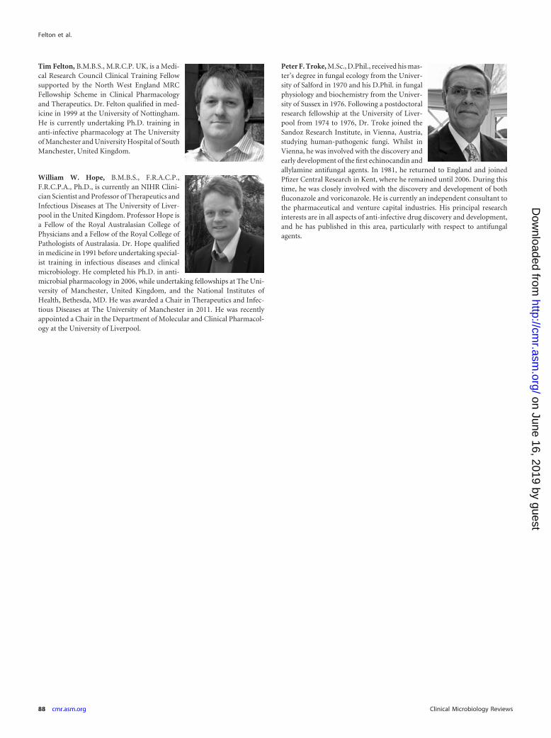
Tim Felton, B.M.B.S., M.R.C.P. UK, is a Medi-cal Research Council Clinical Training Fellowsupported by the North West England MRCFellowship Scheme in Clinical Pharmacologyand Therapeutics. Dr. Felton qualified in med-icine in 1999 at the University of Nottingham.He is currently undertaking Ph.D. training inanti-infective pharmacology at The Universityof Manchester and University Hospital of SouthManchester, United Kingdom.
William W. Hope, B.M.B.S., F.R.A.C.P.,F.R.C.P.A., Ph.D., is currently an NIHR Clini-cian Scientist and Professor of Therapeutics andInfectious Diseases at The University of Liver-pool in the United Kingdom. Professor Hope isa Fellow of the Royal Australasian College ofPhysicians and a Fellow of the Royal College ofPathologists of Australasia. Dr. Hope qualifiedin medicine in 1991 before undertaking special-ist training in infectious diseases and clinicalmicrobiology. He completed his Ph.D. in anti-microbial pharmacology in 2006, while undertaking fellowships at The Uni-versity of Manchester, United Kingdom, and the National Institutes ofHealth, Bethesda, MD. He was awarded a Chair in Therapeutics and Infec-tious Diseases at The University of Manchester in 2011. He was recentlyappointed a Chair in the Department of Molecular and Clinical Pharmacol-ogy at the University of Liverpool.
Peter F. Troke, M.Sc., D.Phil., received his mas-ter’s degree in fungal ecology from the Univer-sity of Salford in 1970 and his D.Phil. in fungalphysiology and biochemistry from the Univer-sity of Sussex in 1976. Following a postdoctoralresearch fellowship at the University of Liver-pool from 1974 to 1976, Dr. Troke joined theSandoz Research Institute, in Vienna, Austria,studying human-pathogenic fungi. Whilst inVienna, he was involved with the discovery andearly development of the first echinocandin andallylamine antifungal agents. In 1981, he returned to England and joinedPfizer Central Research in Kent, where he remained until 2006. During thistime, he was closely involved with the discovery and development of bothfluconazole and voriconazole. He is currently an independent consultant tothe pharmaceutical and venture capital industries. His principal researchinterests are in all aspects of anti-infective drug discovery and development,and he has published in this area, particularly with respect to antifungalagents.
Felton et al.
88 cmr.asm.org Clinical Microbiology Reviews
on June 16, 2019 by guesthttp://cm
r.asm.org/
Dow
nloaded from