Embed Size (px)
Citation preview

Harvard-MIT Division of Health Sciences and TechnologyHST.035: Principle and Practice of Human PathologyDr. Badizadegan
Tissue Repair, Fibrosis, and Healing
HST.035
Spring 2003

Tissue Repair (Healing)
• Regeneration of injured tissue (replacement by normal cells of the same kind)
• Replacement by fibrous tissue (fibrosis, scarring)
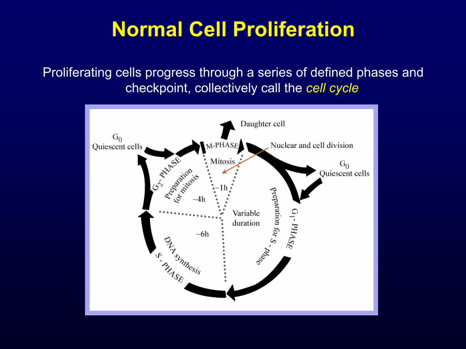
Normal Cell Proliferation
Proliferating cells progress through a series of defined phases and checkpoint, collectively call the cell cycle

Control of Cell Cycle
• Progression through the cell cycle is controlled at specific checkpoints (restriction point in G1, mitosis entry and mitosis exit)
• Transition between stages of mitosis is triggered by increased activity of cyclin-dependet kinases (CDK)
• Each CDK modulates the activity of a subset of cellular targets specific for progression through individual transitions with the cell cycle

Control of Cell Cycle

Control of Cell Cycle
CDK activity is controlled by constant synthesis and cyclic breakdown of specific cyclins

Control of Cell Cycle
• Cyclin-CDK complexes are also regulated by the binding of CDK-inhibitors
• CDK-inhibitors are particularly critical at at G1 → S and G2 → M checkpoints, at which time adequacy and fidelity of DNA synthesis and replication are monitored
• When DNA is found to be damaged, TP53 (p53) is stabilized and induces transcription of CDKN1A (p21), an inhibitor which arrests cells in G1 or G2
• If DNA damage is too extensive, TP53 will initiate apoptosis
Cell growth and differentiation are dependent on extracellular signals from soluble
polypeptide growth factors and the ECM

Polypeptide Growth Factors
• Soluble growth factors are transported either via the Gap junctions, or by autocrine, paracrine, endocrine, or synaptic transmission
• For intracellular receptors, ligand binding leads to formation of complexes that directly associate with nuclear DNA and activate transcription
• For cell surface receptors, ligands bind to a variety of receptor types that ultimately lead to activation of nuclear transcription factors

Major Types of Cell Surface Receptors

ECM: Interstitial Matrix and Basement Membrane
Please see Kumar et al. Robbins Basic Pathology. 7th edition. WB Saunders 2003. ISBN: 0721692745.

Biological Roles of the ECM
• Mechanical support
• Determination of cell polarity
• Control of cell growth
• Control/maintenance of cell differentiation
• Scaffolding for tissue renewal
• Establishment of tissue microenvironment
• Storage and presentation of regulatory proteins

Major Components of the ECM
• Collagen
• Elastic fibers
• Proteoglycans and hyaluronan
• Fibronectin
• Laminin
• Integrins
Fibrillar Collagen: Tensile Strength
Please see figures 5-17 and 5-18 of Junqueira & Carneiro. Basic Histology: Text and Atlas. 10th edition. McGraw Hill. 2003. ISBN: 0071378294.

Collagen Fibrills
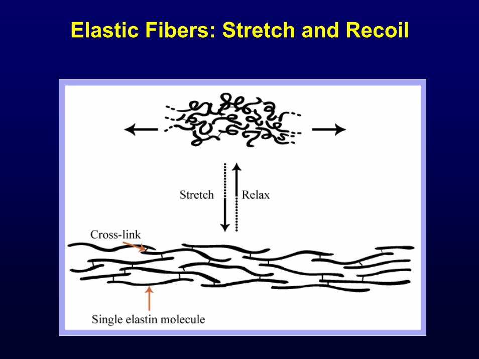
Elastic Fibers: Stretch and Recoil
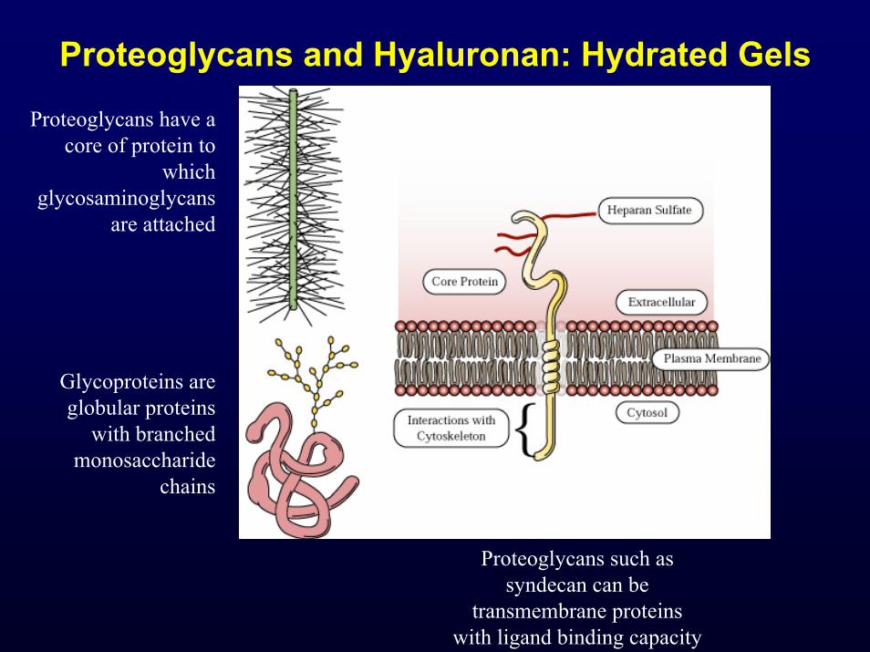
Proteoglycans and Hyaluronan: Hydrated Gels
Proteoglycans have a core of protein to
which glycosaminoglycans
are attached
Glycoproteins are globular proteins
with branched monosaccharide
chains
Proteoglycans such as syndecan can be
transmembrane proteins with ligand binding capacity

Fibronectin and Laminin
• Fibronectin bind to a wide spectrum of ECM components and can attach to cell surface integrins
• Laminin is a key glycoprotein in the basement membrane that binds underlying ECM components such as type IV collagen
• Laminin also modulates cell survival, proliferation and differentiation

Integrins link the ECM to Actin Cytoskeleton through Focal Adhesion Complexes

ECM and Growth Control

Repair by Connective Tissue (Fibrosis/Scarring)
• Occurs when severe cell injury and damage to ECM framework precludes regeneration of native tissue
• Fibrosis progresses through four main stages:
– Angiogenesis
– Migration and proliferation of fibroblasts
– Deposition of ECM
– Remodeling of ECM

Fibrosis/Scarring
Please see figure 3-10 of Kumar et al. Robbins Basic Pathology. 7th edition. WB Saunders 2003. ISBN: 0721692745.

Overview of Tissue Response to Injury