Embed Size (px)
Citation preview
Instructions for use
Title Lymph Node Metastasis in T4 Maxillary Sinus Squamous Cell Carcinoma : Incidence and Treatment Outcome
Author(s)Homma, Akihiro; Hayashi, Ryuichi; Matsuura, Kazuto; Kato, Kengo; Kawabata, Kazuyoshi; Monden, Nobuya;Hasegawa, Yasuhisa; Onitsuka, Tetsuro; Fujimoto, Yasushi; Iwae, Shigemichi; Okami, Kenji; Matsuzuka, Takashi;Yoshino, Kunitoshi; Nibu, Ken-ichi; Kato, Takakuni; Nishino, Hiroshi; Asakage, Takahiro; Ota, Ichiro; Kitamura,Morimasa; Kubota, Akira; Ueda, Tsutomu; Ikebuchi, Kaichiro; Watanabe, Akihito; Fujii, Masato
Citation Annals of Surgical Oncology, 21(5), 1706-1710https://doi.org/10.1245/s10434-014-3544-6
Issue Date 2014-05
Doc URL http://hdl.handle.net/2115/59107
Rights The original publication is available at www.springerlink.com
Type article (author version)
File Information Ann Surg Oncol_21(5)_1706-1710.pdf
Hokkaido University Collection of Scholarly and Academic Papers : HUSCAP

1
Lymph node metastasis in T4 maxillary sinus squamous cell carcinoma: Incidence and
treatment outcome
Akihiro Homma, MD1, Ryuichi Hayashi, MD
2, Kazuto Matsuura, MD
3, Kengo Kato, MD
4,
Kazuyoshi Kawabata, MD 5
, Nobuya Monden, MD 6
, Yasuhisa Hasegawa, MD 7
, Tetsuro Onitsuka,
MD 8
, Yasushi Fujimoto, MD 9
, Shigemichi Iwae, MD 10
, Kenji Okami, MD 11
, Takashi Matsuzuka,
MD 12
, Kunitoshi Yoshino, MD 13
, Ken-ichi Nibu, MD 14
, Kato, Takakuni, MD 15
, Hiroshi Nishino,
MD 16,
Takahiro Asakage, MD 17
, Ichiro Ota, MD 18
, Morimasa Kitamura, MD 19
, Akira Kubota, MD
20, Tsutomu Ueda, MD
21, Kaichiro Ikebuchi, MD
22, Akihito Watanabe, MD
23, Masato Fujii, MD
24
1 Hokkaido University Graduate School of Medicine, Otolaryngology- Head and Neck Surgery
Sapporo, Hokkaido, Japan
2 National Cancer Center Hospital East, Head and Neck Surgery, Kashiwa, Japan
3 Miyagi Cancer Center, Head and Neck Surgery, Sendai, Japan
4 Tohoku University School of Medicine, Otolaryngology-Head and Neck Surgery, Sendai, Japan
5 Cancer Institute Hospital, Japanese Foundation for Cancer Research, Head and Neck Oncology,
Tokyo, Japan
6 National Shikoku Cancer Center, Matsuyama, Otorhinolaryngology-Head and Neck Surgery
Matsuyama, Japan

2
7 Aichi Cancer Center, Head and Neck Surgery, Nagoya, Japan
8 Shizuoka Cancer Center, and Neck Surgery, Shizuoka, Japan
9 Nagoya University Graduate School of Medicine, Otolaryngology, Nagoya, Japan
10 Hyogo Cancer Center, Head and Neck Surgery, Akashi, Japan
11 Tokai University School of Medicine, Otolaryngology, Isehara, Japan
12 Fukushima Medical University School of Medicine, Otolaryngology, Fukushima, Japan
13 Osaka Medical Center for Cancer and Cardiovascular Diseases, Otolaryngology-Head and Neck
Surgery, Osaka, Japan
14Kobe University Graduate School of Medicine, Otolaryngology-Head and Neck Surgery
Kobe, Japan
15 Jikei University School of Medicine, Otorhinolaryngology, Tokyo, Japan
16 Jichi Medical University School of Medicine, Otolaryngology-Head and Neck Surgery, Tochigi,
Japan
17 Tokyo University School of Medicine, Otolaryngology-Head and Neck Surgery, Tokyo, Japan
18 Nara Medical University, Otolaryngology-Head and Neck Surgery, Nara, Japan
19 Kyoto University Graduate School of Medicine, Otolaryngology-Head and Neck Surgery, Kyoto,
Japan
20 Kanagawa Cancer Center, Head and Neck Surgery, Yokohama, Japan
21 Hiroshima University School of Medicine, Otolaryngology, Hiroshima, Japan
22 Kyoto Prefectural University of Medicine, Otolaryngology-Head and Neck Surgery

3
kyoto, Japan
23 Keiyukai Sapporo Hospital, Otolaryngology, Sapporo, Japan
24 National Tokyo Medical Center, Otorhinolaryngology, Tokyo, Japan
Running title: Node metastasis in T4 maxillary sinus SCC
*Corresponding author: Akihiro Homma; Akihiro Homma
Department of Otolaryngology – Head & Neck Surgery, Hokkaido University Graduate
School of Medicine
Kita 15, Nishi 7, Kita-ku, Sapporo 060-8638, Japan
Phone: +81-11-706-5958; Fax: +81-11-717-7566;
E-mail: [email protected]
4
SYNOPSIS
This study identified the incidence of lymph node metastasis among patients with T4
maxillary sinus squamous cell carcinoma (MS-SCC) as well as the delayed metastasis
rate and the treatment outcome for untreated N0 neck in patients with T4 MS-SCC.
5
ABSTRACT
PURPOSE
To evaluate the incidence of lymph node metastasis among patients with T4 maxillary
sinus squamous cell carcinoma (MS-SCC) as well as the delayed metastasis rate and the
treatment outcome for untreated N0 neck in patients with T4 MS-SCC.
PATIENTS AND METHODS
Consecutive series of all patients (n=128) with previously untreated T4 maxillary sinus
SCC between 2006 and 2007 were obtained from 28 institutions belonging to or
cooperating in the Head and Neck Cancer Study Group of the Japan Clinical Oncology
Group.
RESULTS
Twenty-eight (21.9%) of the 128 patients had lymph node metastasis, and 6 patients
(4.7%) had distant metastasis at diagnosis. Among the 111 patients who were treated
with curative intent, 98 had clinically N0 neck disease and did not receive prophylactic
neck irradiation. A total of 11 patients (11.2%) subsequently developed evidence of
lymph node metastasis, of whom 8 were among the 83 patients with an N0 neck and had
not received elective neck treatment. There were 15 patients who received an elective
neck dissection as part of the initial treatment, of whom 3 had pathologically positive
for lymph node metastases. Of 11 patients, 6 patients with non-lateral retropharyngeal
6
lymph node metastasis without primary or distant disease were successfully salvaged.
CONCLUSION
This study identified the incidence of lymph node metastasis among patients with T4
MS-SCC as well as the delayed metastasis rate and the treatment outcome for untreated
N0 neck in patients with T4 MS-SCC. These results will be of assistance in selecting
treatment strategy for T4 MS-SCC in the future.
7
Maxillary sinus cancer is the most common form is sinonasal cancer. But the incidence
of it has been considered to be decreasing gradually. According to vital statistics
obtained from the Ministry of Health, Labour and Welfare, Japan, the number of deaths
due to the maxillary sinus cancer was 1,051, 643, and 175, in 1971, 1991, and 2011,
respectively. Some investigators in Japan consider this decrease to be correlated with
the decrease in sinusitis, which is considered to be one of the risk factors of maxillary
sinus cancer.
The Japanese head and neck community has been gaining experience in the
treatment of patients with maxillary sinus cancer due to the many opportunities we have
to treat them, and most head and neck surgeons and radiation oncologists in Japan
prefer to take a common sense “wait-and-see” approach in the management of patients
with clinically negative neck as the incidence of delayed neck metastasis is considered
to be low. However, whether clinically negative neck in patients with the maxillary
sinus cancer should be irradiated prophylactically or not is controversial in Europe and
the United States1, 2
.
In addition, the incidence of neck metastasis in cases of maxillary sinus cancer
has not been well defined, although it is currently believed to be low. To help clarify the
situation, a multi-institutional joint research program for maxillary sinus cancer was
undertaken in Japan3. This study was aimed at evaluating the incidence of lymph node
metastasis among patients with T4 maxillary sinus squamous cell carcinoma (MS-SCC)
8
as well as the delayed metastasis rate and the treatment outcome for untreated N0 neck
in patients with T4 MS-SCC.
MATERIALS AND METHODS
Consecutive series of all patients with previously untreated T4 MS-SCC between
January 2006 and December 2007 were obtained from 28 institutions belonging to or
cooperating in the Head and Neck Cancer Study Group of the Japan Clinical Oncology
Group. This study was a retrospective analysis. Therefore, the selection criteria for
therapeutic modality were decided according to the policy of each institution or
individual patient preference. This multi-institutional joint research has been
representatively approved by the appropriate ethical committee in the National Hospital
Organization Tokyo Medical Center, Tokyo, Japan.
Initial treatment of the primary tumor
The initial therapeutic strategy was classified in the treatment for primary tumor.
Surgical treatment was classified into total maxillectomy and partial maxillectomy. The
classification of total maxillectomy included extended total maxillectomy
simultaneously orbital exenteration and skull base surgery. “Trimodality therapy”,
consisting of partial maxillectomy, intra-arterial chemotherapy and radiotherapy, was
classified as partial maxillectomy. Surgery in which the anterior wall of the maxillary
9
sinus was opened and the necrotic tumor tissue therein was curetted was also classified
as partial maxillectomy. The superselective intra-arterial infusion of high-dose cisplatin
with concomitant radiotherapy was defined as RADPLAT, while intravenous
chemotherapy with concomitant radiotherapy was defined as IV-CRT. All patients
undergoing any form of surgical intervention as part of the initial treatment were
classified into either the total or partial maxillectomy group, even if radiotherapy and/or
chemotherapy was performed as pre- or post-surgical therapy. No patients with
clinically N0 received elective radiation therapy to the neck lymph nodes.
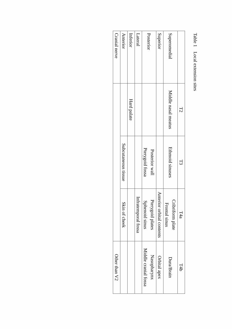
Local extension sites
In this study, the anatomical sites of the primary tumor has developed were
evaluated in detail using CT and/or MR imaging. The local extension sites were
classified according to the 7th
Edition of the Union for International Cancer Control
staging system (Table 1). As only 2 cases had invasion into the brain, with both
involving the dura, they were included as extension into the dura/brain. No cases
showed involvement of the clivus.
Statistical analysis
The median follow-up period for the survivors was 4.3 years (range 0.2-5.9 years).
Correlations between neck metastasis and variables including age, sex, T-stage, tumor
10
differentiation and local extension site were tested using Pearson's Chi-square test or
Fisher's exact test using JMP Pro 10.0.0 statistical software (SAS Institute, Cary, NC). A
2-tailed p value <0.05 was considered to be statistically significant.
RESULTS
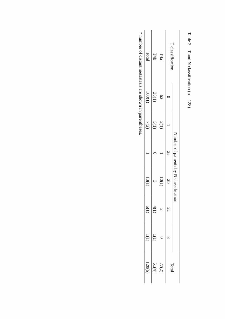
A total of 128 patients enrolled. T and N classifications of the 128 patients are
shown in Table 2. Ninety-six patients were male and 32 were female. The median age
was 64 years (range 30-84 years). Seventy-seven patients (60.1%) had T4a disease, and
51(39.8%) had T4b disease. Twenty-eight patients (21.9%) had lymph node metastasis
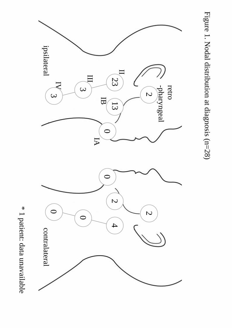
and 6 patients (4.7%) had distant metastasis at diagnosis. The distribution of clinically
diagnosed lymph node metastasis is shown in Figure 1. All patients showed clinically N
positive but one patient had ipsilateral level Ib or II metastasis. One patient had a
solitary metastasis in his contralateral level II node. Apart from this patient, all patients
with metastasis to other sites had level Ib and/or II metastasis. Regarding correlations
between neck metastasis and variables such as age, sex, T-stage, tumor differentiation
and local extension site at diagnosis, the nasopharynx (p= 0.046) and the hard palate
(p<0.001) were the only sites that was correlated with neck lymph node metastasis.
Of the 128 patients, 6 patients underwent palliative therapy due to distant
metastasis. Three patients chose to be treated at other institutions, and one patient
refused any therapy. The initial treatment for the remaining 118 patients was classified
11
by treatment for primary tumor. A total of 39 (33%) of the 118 patients were categorized
into the total maxillectomy group, while 25 patients (21%) underwent partial
maxillectomy, 22 patients (19%) underwent RADPLAT, 19 patients (16%) underwent
IV-CRT, and 13 patients (11%) underwent other therapies, such as radiation alone.
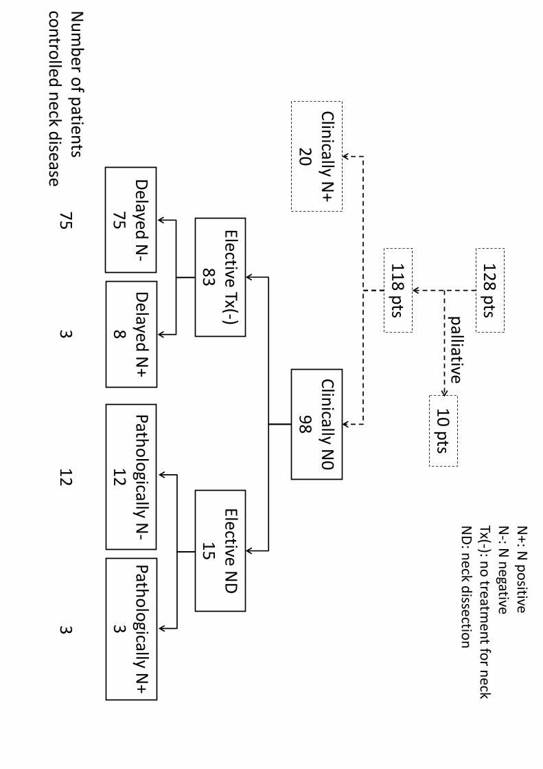
Among the 111 patients who were treated with curative intent, 98 had clinically
N0 neck disease and did not receive prophylactic neck irradiation (Figure 2). A total of
11 patients (11.2%) subsequently developed evidence of lymph node metastasis, of
whom 8 were among the 83 patients with an N0 neck and had not received elective neck
treatment. There were 15 patients who received an elective neck dissection as part of the
initial treatment, of whom 3 had pathologically positive for lymph node metastases.
Delayed neck recurrence was observed at a median 6 months (average; 10 months,
range: 1-39 months) after the completion of RADPLAT.
Among the 8 patients who had no elective neck treatment and developed delayed
neck metastasis, 3 patients were successfully salvaged by neck dissection. But neck
disease could not be controlled in 2 patients with lateral retropharyngeal lymph node
(RPLN) metastasis and 3 patients with residual or recurrence of primary or distant
disease. And neck disease was successfully controlled in 3 patients who had
pathologically positive for lymph node metastasis after elective neck dissection.
Sixty-three patients with N0 neck disease at diagnosis and who were monitored
for neck disease for more than 2 years were analyzed for late neck metastasis. Of the
12
initial 128 patients, 28 patients with clinical neck metastasis at diagnosis, 35 patients
with N0 neck disease at diagnosis who died within 2 years due to primary and/or distant
disease without neck recurrence, and 2 patients who died of other causes without neck
recurrence were excluded. Of the remaining 63 patients, 11 (17.5%) had late neck
metastasis, as mentioned above. With regard to correlations between delayed neck
metastasis and variables such as age, sex, T-stage, tumor differentiation and local
extension sites among the 63 patients, no factor was found to be correlated with neck
lymph node metastasis. Moreover, the factors related to a delayed neck metastasis rate
of more than 25 % were female gender (4/16=25%), T4b (6/23=26.1%), low-grade
tumor (6/17=35.3%), nasopharyngeal invasion (2/5=40%), middle cranial fossa invasion
(3/10=30%), and invasion of a cranial nerve other than V2 (2/4=50%).
DISCUSSION
The prognosis for MS-SCC is significantly related to local tumor control.
Therefore, lymph node metastasis in MS-SCC has received little attention to date. The
incidence and distribution of lymph node metastasis and the percentage of delayed
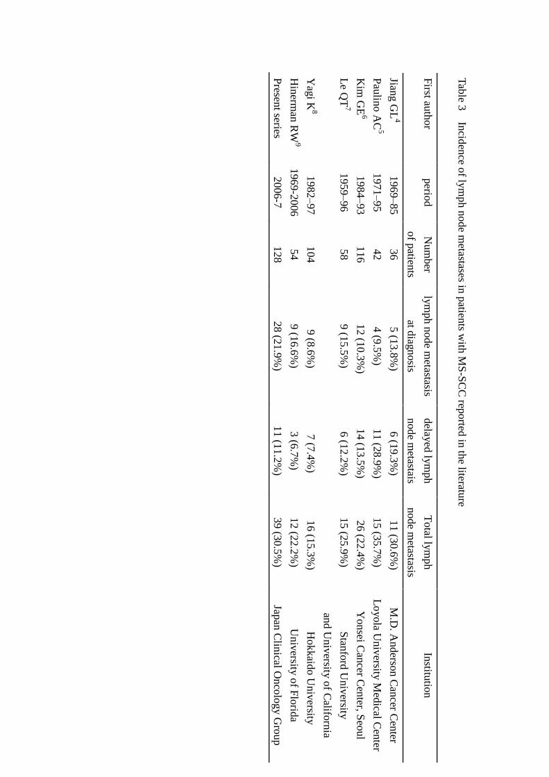
metastasis in cases of maxillary sinus SCC are reported to range widely (Table 3)4-9
as
MS-SCC is a rare neoplasm and the number of patients treated at a single center is small.
In addition, some reports have included patients from several decades ago. Time factor
must have influence on pretreatment diagnosis and treatment outcome.
13
The retrospective data in this study were limited to patients with T4 MS-SCC
who were treated between 2006 and 2007. Thus, the cases represent a very limited stage
treated within a limited period, affording homogeneity to the data. The modality for
diagnosis was not checked, but most of the patients were examined by physical
examination as well as CT scan and/or MRI at that time. FDG-PET, ultrasound, and fine
needle aspiration cytology were used at the attending physician’s discretion.
Regarding the correlation between local extension site and neck lymph node
metastasis at diagnosis, the nasopharynx and hard palate were both correlated with
lymph node metastasis. There was a report that the rate of neck metastasis is much
higher in T2 tumors than in T3 or T4 tumors10
. The reason for this was suspected to be
that cases with extension to the hard palate diagnosed as T2 are more likely to develop
lymph node metastasis. This study supports this speculation. Cases with invasion to
areas known to be rich in lymphatics, such as the nasopharynx and hard palate, are
considered more likely to develop lymph node metastasis11
.
The risk of RPLN metastasis in cases of MS-SCC has been discussed
previously1,12, 13
. In 1993, in a series of 25 patients with maxillary sinus cancer, Watarai
et al. found that RPLNs were involved in 16% of the patients12
. In this study, only 3
patients had RPLN metastasis at diagnosis (one patient had bilateral RPLN metastasis)
(Figure 1). And 2 patients who received en bloc tumor resection and radiotherapy as an
initial treatment showed delayed RPLN metastasis. The incidence of delayed RPLN
14
metastasis is considered to be low. Therefore, whether the radiotherapy plan should
include the RPLN area or not needs to be discussed carefully.
Delayed neck metastasis developed in 11 patients with clinically N0. The
possibility of delayed neck metastasis was calculated to be 11.2%, based on the 98
patients with clinically N0 neck disease among the 118 patients treated with curative
intent. This result was comparable with those of previous reports4-9
. In addition, the
delayed neck metastasis rate was 17.5%, based on the 63 patients with N0 neck disease
at diagnosis who were monitored for neck disease for more than 2 years.
The reason why elective neck irradiation was not done for patients with clinically
N0 neck in this retrospective multi-institutional study was that we could identify neck
metastasis at an early stage and control it effectively as patients were followed up
closely. Indeed, all 3 patients with non-RPLN delayed neck metastasis without residual
or recurrent primary or distant disease were successfully salvaged.
Delayed neck metastasis is an unfavorable prognostic factor; therefore, some
investigators have recommended prophylactic neck irradiation7,5,9,14. In general, elective
treatment of the neck is recommended for patients with squamous cell carcinoma of the upper
aerodigestive tract when the anticipated risk of occult metastasis is greater than 15-20%15,16
.
According to this idea, patients with T4N0 MS-SCC should be candidates for elective
neck irradiation. However, if close follow-up is possible, we consider that prophylactic
neck irradiation is unnecessary, particularly due to the risk of adverse effects of elective
15
neck irradiation, such as mucositis and osteoradionecrosis of the mandible.
In conclusion, this study revealed the incidence of lymph node metastasis among
patients with T4 MS-SCC as well as the proportion of cases with delayed metastasis and
the treatment outcome for untreated N0 neck disease in patients with T4 MS-SCC. We
expect these result to be of assistance in selecting treatment strategies for T4 MS-SCC
in the future.
ACKNOWLEDGMENTS
This study was supported in part by a Health and Labour Sciences Research Grant for
Clinical Cancer Research (H22-Gannrinshou-Ippan-017) from the Ministry of Health,
Labour and Welfare of Japan, the National Cancer Center Research and Development
Fund (23-A-21) of Japan, and a grant-in-aid for Scientific Research (C) (KAKENHI
24592587) from the Ministry of Education, Culture, Sports, Science, and Technology of
Japan.
APPENDIX
In addition to the authors, the following investigators participated in this study: National
Cancer Center Hospital East, Kashiwa - T. Shinohara; Miyagi Cancer Center, Sendai -
Y. Asada; Cancer Institute Hospital, Japanese Foundation for Cancer Research, Tokyo -
T. Sasaki; Aichi Cancer Center, Nagoya - H. Hirakawa; Osaka Medical Center for
16
Cancer and Cardiovascular Diseases, Osaka - T. Fujii; Kobe University Graduate
School of Medicine - N. Otsuki; Tokyo University - Y. Saito; Aichi Medical University,
Nagakute - A. Ikeda; Iwate Prefectural Central Hospital, Morioka - S. Kato; Kochi
Health Sciences Center, Kochi - K. Kozakura; Japanese Red Cross Nagoya Daiichi
Hospital, Nagoya - K. Kawata and A. Terada
REFERENCES
1. Takes RP, Ferlito A, Silver CE, et al. The controversy in the management of the N0
neck for squamous cell carcinoma of the maxillary sinus. Eur Arch Otorhinolaryngol (in
press)
2. Rinaldo A, Ferlito A, Shaha AR, Wei WI. Is elective neck treatment indicated in
patients with squamous cell carcinoma of the maxillary sinus? Acta Otolaryngol
2002;122:443-447.
3. Kano S, Hayashi R, Homma A, et al. Local extension sites affect survival in locally
advanced maxillary sinus cancer. Head Neck DOI: 10.1002/hed.23483.
4. Jiang GL, Ang KK, Peters LJ, Wendt CD, Oswald MJ, Goepfert H. Maxillary sinus
carcinomas: natural history and results of postoperative radiotherapy. Radiother Oncol
1991;21:193-200.
5. Paulino AC, Fisher SG, Marks JE. Is prophylactic neck irradiation
indicated in patients with squamous cell carcinoma of the maxillary
17
sinus? Int J Radiat Oncol Biol Phys 1997;39:283-9.
6. Kim GE, Chung EJ, Lim JJ, et al. Clinical significance of neck node metastasis in
squamous cell carcinoma of the maxillary antrum. Am J Otolaryngol 1999;20:383-390.
7. Le QT, Fu KK, Kaplan MJ, Terris DJ, Fee WE, Goffinet DR. Lymph node metastasis
in maxillary sinus carcinoma. Int J Radiat Oncol Biol Phys 2000;46:541-549.
8. Yagi K, Fukuda S, Furuta Y, Oridate N, Homma A, Nagahashi T, Inuyama Y. A
clinical study on the cervical lymph node metastasis of maxillary sinus carcinoma. Auris
Nasus Larynx 2001;28:S77-81.
9. Hinerman RW, Indelicato DJ, Morris CG, Kirwan JM, Werning JW, Vaysberg M,
Mendenhall WM. Radiotherapy With or Without Surgery for Maxillary Sinus Squamous
Cell Carcinoma: Should the Clinical N0 Neck be Treated?
Am J Clin Oncol 2011;34:483-487.
10. Cantu G, Bimbi G, Miceli R, et al. Lymph node metastases in malignant tumors of
the paranasal sinuses: prognostic value and treatment. Arch Otolaryngol Head Neck
Surg 2008;134:170-177.
11. Jeremic B, Nguyen-Tan PF, Bamberg M. Elective neck irradiation in locally
advanced squamous cell carcinoma of the maxillary sinus: a review. J Cancer Res Clin
Oncol 2002;128:235-238.
12. Watarai J, Seino Y, Kobayashi M, Shindo M, Kato T. CT of retropharyngeal lymph
node metastasis from maxillary carcinoma. Acta Radiol 1993;34:492-495.
18
13. Coskun HH, Ferlito A, Medina JE, et al. Retropharyngeal lymph node metastases in
head and neck malignancies. Head Neck 2011 Oct;33:1520-1529.
14. Jeremic B, Shibamoto Y, Milicic B, et al. Elective ipsilateral neck irradiation of
patients with locally advanced maxillary sinus carcinoma. Cancer 2000;88:2246-2251.
15. Pillsbury HC 3rd, Clark M. A rationale for therapy of the N0 neck. Laryngoscope
1997;107:1294-1315.
16. Weiss MH, Harrison LB, Isaacs RS. Use of decision analysis in planning a
management strategy for the stage N0 neck. Arch Otolaryngol Head Neck Surg
1994;120:699-702.
19
FIGURE LEGEND
Figure 1
Nodal distribution at diagnosis (n=28)
Figure 2
Clinical course in 98 patients with Clinically N0 neck disease.
13
0
2
2
23
3 3
ip
silateral co
ntralateral
2
4
0
0
0
retro
-phary
ngeal
IA
IB
II III IV
* 1
patien
t: data u
nav
ailable
Fig
ure 1
. Nodal d
istributio
n at d
iagnosis (n
=28)
Nu
mb
er of p
atients
con
trolled
neck d
isease
11
8 p
ts
Clin
ically N+
20
C
linically N
0
98
Delayed
N+
8
12
8 p
ts
10
pts
Patho
logically N
+
3
Patho
logically N
- 1
2
Delayed
N-
75
palliative
N+: N
po
sitive N
-: N n
egative Tx(-): n
o treatm
ent fo
r neck
ND
: neck d
issection
75
3
1
2
3
Elective Tx(-)
83
Ele
ctive ND
1
5
Tab
le 1
Local ex
tensio
n sites
T
2
T3
T
4a
T4b
Supero
med
ial M
iddle n
asal meatu
s E
thm
oid
sinuses
Crib
riform
plate
Fro
ntal sin
us
Dura/B
rain
Superio
r
A
nterio
r orb
ital conten
ts O
rbital ap
ex
Posterio
r
Posterio
r wall
Ptery
goid
fossa
Ptery
goid
plates
Sphen
oid
sinus
Naso
phary
nx
Mid
dle cran
ial fossa
Lateral
Infratem
po
ral fossa
Inferio
r H
ard p
alate
Anterio
r
Subcu
taneo
us tissu
e S
kin
of ch
eek
Cran
ial nerv
e
Oth
er than
V2
Tab
le 2 T
and N
classification (n
= 1
28)
T classificatio
n
Num
ber o
f patien
ts by N
classification
T
otal
0
1
2a
2b
2c
3
T4a
62
2(1
) 1
10(1
) 2
0
77(2
)
T4b
38
(1)
5(1
) 0
3
4(1
) 1(1
) 51(4
)
Total
100
(1)
7(2
) 1
13(1
) 6(1
) 1(1
) 128(6
)
* n
um
ber o
f distan
t metastasis are sh
ow
n in
paren
theses.
Tab
le 3
Incid
ence o
f lym
ph n
ode m
etastases in p
atients w
ith M
S-S
CC
reported
in th
e literature
First au
thor
perio
d
Num
ber
of p
atients
lym
ph n
ode m
etastasis
at diag
nosis
dela
yed
lym
ph
node m
etastais
Total ly
mph
node m
etastasis
Institu
tion
Jiang G
L4
1969–85
36
5 (1
3.8
%)
6 (1
9.3
%)
11 (3
0.6
%)
M.D
. Anderso
n C
ancer C
enter
Pau
lino A
C5
1971–95
42
4 (9
.5%
) 11 (2
8.9
%)
15 (3
5.7
%)
Lo
yola U
niv
ersity M
edical C
enter
Kim
GE
6 1984–93
116
12 (1
0.3
%)
14 (1
3.5
%)
26 (2
2.4
%)
Yonsei C
ancer C
enter, S
eoul
Le Q
T7
1959–96
58
9 (1
5.5
%)
6 (1
2.2
%)
15 (2
5.9
%)
Stan
ford
Univ
ersity
and U
niv
ersity o
f Califo
rnia
Yag
i K8
1982–97
104
9 (8
.6%
) 7 (7
.4%
) 16 (1
5.3
%)
H
okkaid
o U
niv
ersity
Hin
erman
RW
9 1969-2
006
54
9 (1
6.6
%)
3 (6
.7%
) 12 (2
2.2
%)
Univ
ersity o
f Flo
rida
Presen
t series 2006-7
128
28 (2
1.9
%)
11 (1
1.2
%)
39 (3
0.5
%)
Japan
Clin
ical Onco
log
y G
roup