Embed Size (px)
Citation preview
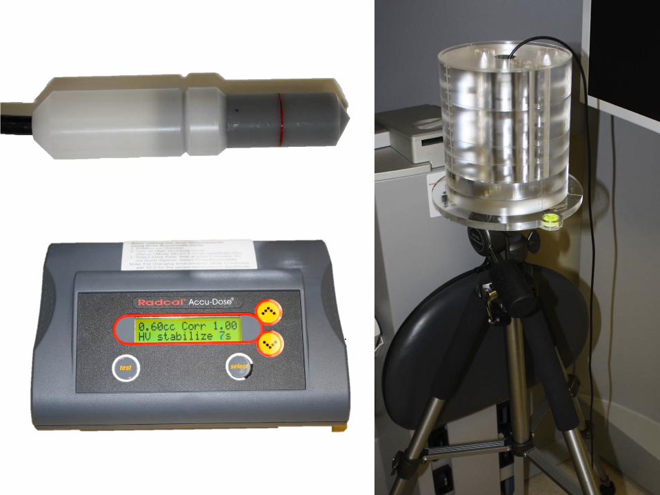
Title: CBCT Absorbed Radiation Dose in Pediatric Patients Authors: Ella Choi, DDS and Nancy Ford, PhD Objectives: To study the characteristics of the patients who received CBCT at British Columbia Children’s Hospital (BCCH), and to compare the absorbed radiation dose using adult and pediatric phantoms Methods: The patients under the age of 18 years who received iCAT CBCT scans at BCCH between 2010 and 2011 met the inclusion criteria. Their soft and hard tissue head dimensions were measured. The information on ethnicity, age, gender, co-morbidities, CBCT protocol, and indications for CBCT were obtained. A Sedendex DI adult head phantom and a custom-made pediatric head phantom were used with Radcal thimble ionization chamber (0.6 cm3) and AccuDose Dosimeter to measure the absorbed dose in different locations at the two most common protocols. The two most common protocols were at 60 mm and 130 mm field of view with kVp 120, mA 5, 0.4 voxel size, and exposure time of 4 seconds. Results: Thirty-two patients aged 5 to 17 at BCCH met the inclusion criteria. The most common indications for CBCT were for orthodontic treatment (39%), craniofacial abnormality (25%), and cleft lip and palate (19%). The soft and hard head dimensions varied depending on ethnic background and craniofacial abnormality. The absorbed radiation dose varied depending on the CBCT protocol and location. In the adult phantom, the absorbed dose ranged from 1.1 mGy at the surface to 1.5 mGy at the centre; in the pediatric phantom, it ranged from 1.7 mGy at the surface to 2.2 mGy at the centre. Conclusions: Children from a wide variety of ethnic and medical backgrounds received CBCT at BCCH. Higher absorbed radiation was measured in all locations in pediatric head phantom compared to the adult phantom. The highest measurement of absorbed radiation was at the edge of the phantom, the second highest at the centre, and the lowest at the surface. UBC Faculty of Dentistry Research Equipment Grant
Use of Cone Beam Computed Tomography in the Pediatric Pa8ent
Ella Choi, D.D.S. Graduate Pediatric Den.stry
UBC, Vancouver
Nancy Ford, Ph.D. Research Supervisor UBC, Vancouver
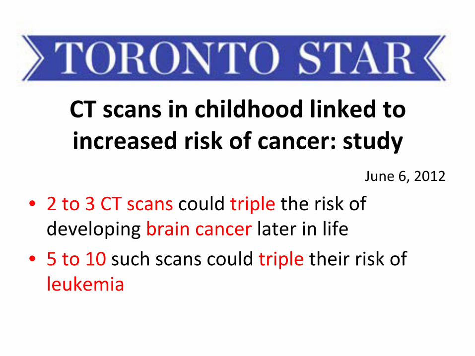
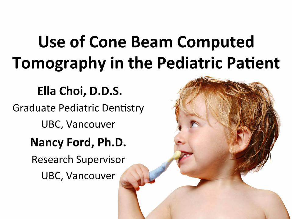
CT scans in childhood linked to increased risk of cancer: study
• 2 to 3 CT scans could triple the risk of developing brain cancer later in life
• 5 to 10 such scans could triple their risk of leukemia
June 6, 2012
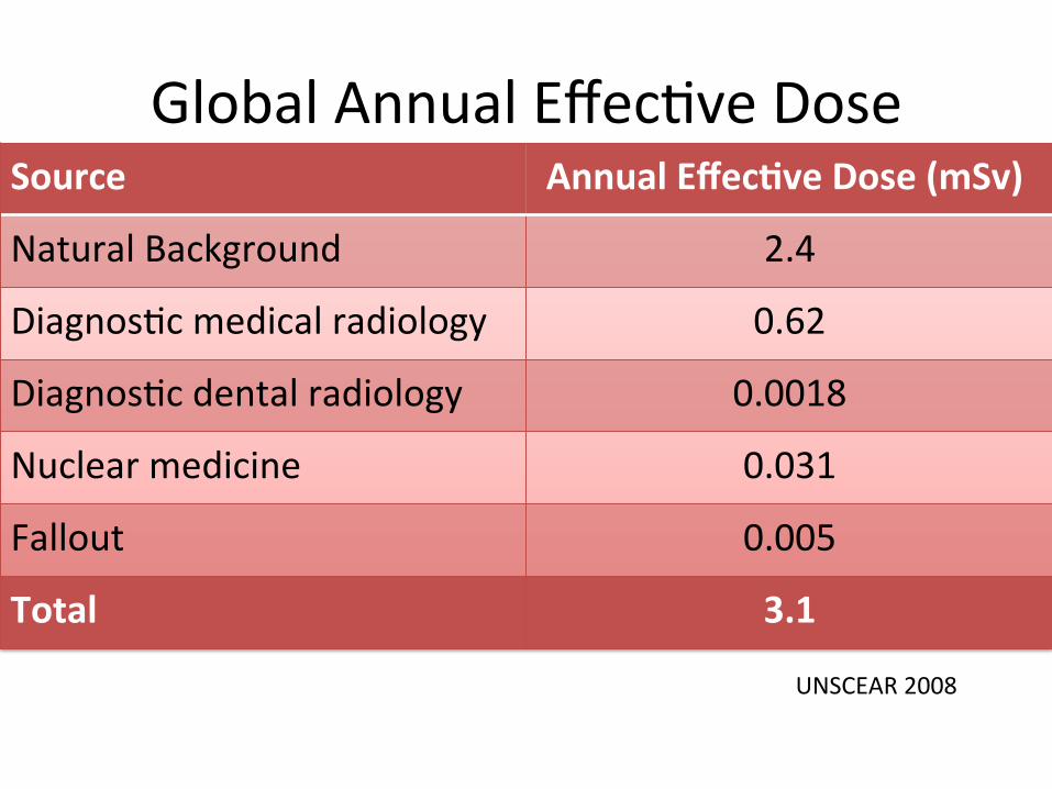
Global Annual Effec.ve Dose Source Annual Effec8ve Dose (mSv)
Natural Background 2.4
Diagnos.c medical radiology 0.62
Diagnos.c dental radiology 0.0018
Nuclear medicine 0.031
Fallout 0.005
Total 3.1
UNSCEAR 2008
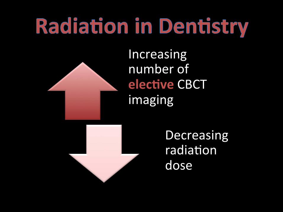
Increasing number of elec8ve CBCT imaging
Decreasing radia.on dose
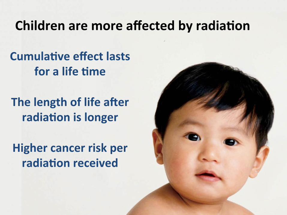
Cumula8ve effect lasts for a life 8me
The length of life aJer radia8on is longer
Higher cancer risk per radia8on received
Children are more affected by radia8on
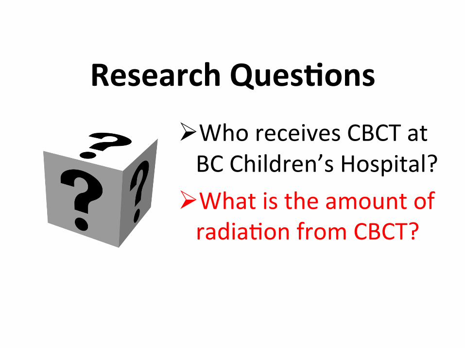
Research Ques8ons
Ø Who receives CBCT at BC Children’s Hospital?
Ø What is the amount of radia.on from CBCT?
Research Ques8ons
Ø Who receives CBCT at BC Children’s Hospital?
Ø What is the amount of radia.on from CBCT?
• Retrospec.ve Study: 2010 and 2011 • Ethics approved • Inclusion criteria
– Under the age of 18 – CBCT
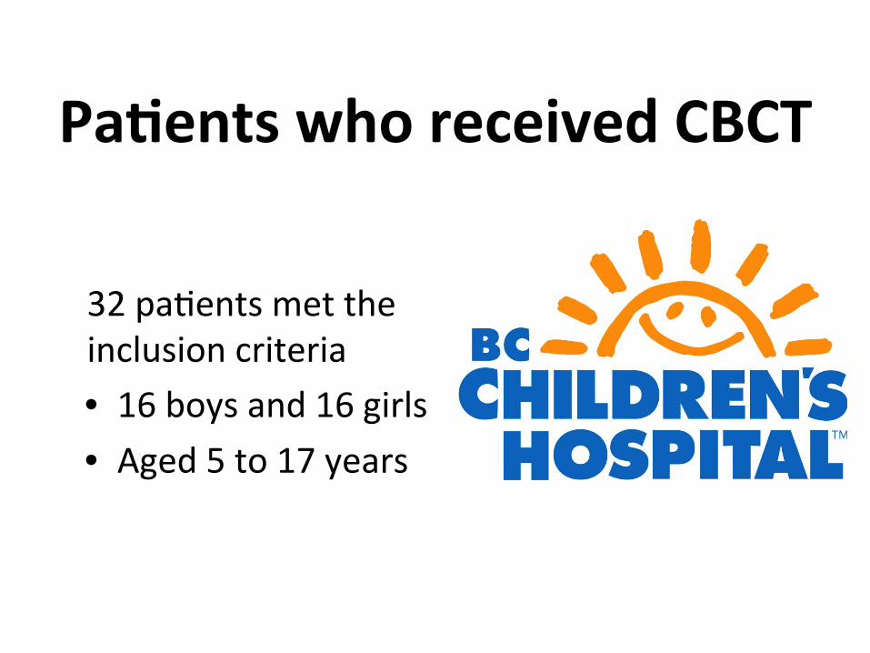
Pa8ents who received CBCT
32 pa.ents met the inclusion criteria • 16 boys and 16 girls • Aged 5 to 17 years
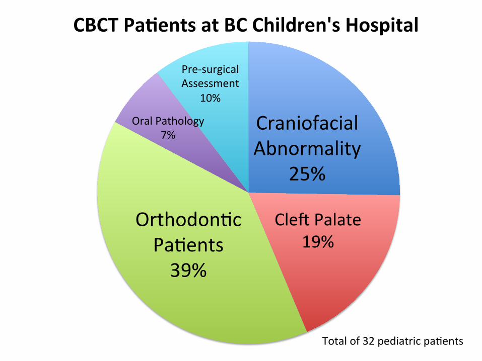
Craniofacial Abnormality
25%
CleZ Palate 19%
Orthodon.c Pa.ents 39%
Oral Pathology 7%
Pre-‐surgical Assessment
10%
CBCT Pa8ents at BC Children's Hospital
Total of 32 pediatric pa.ents
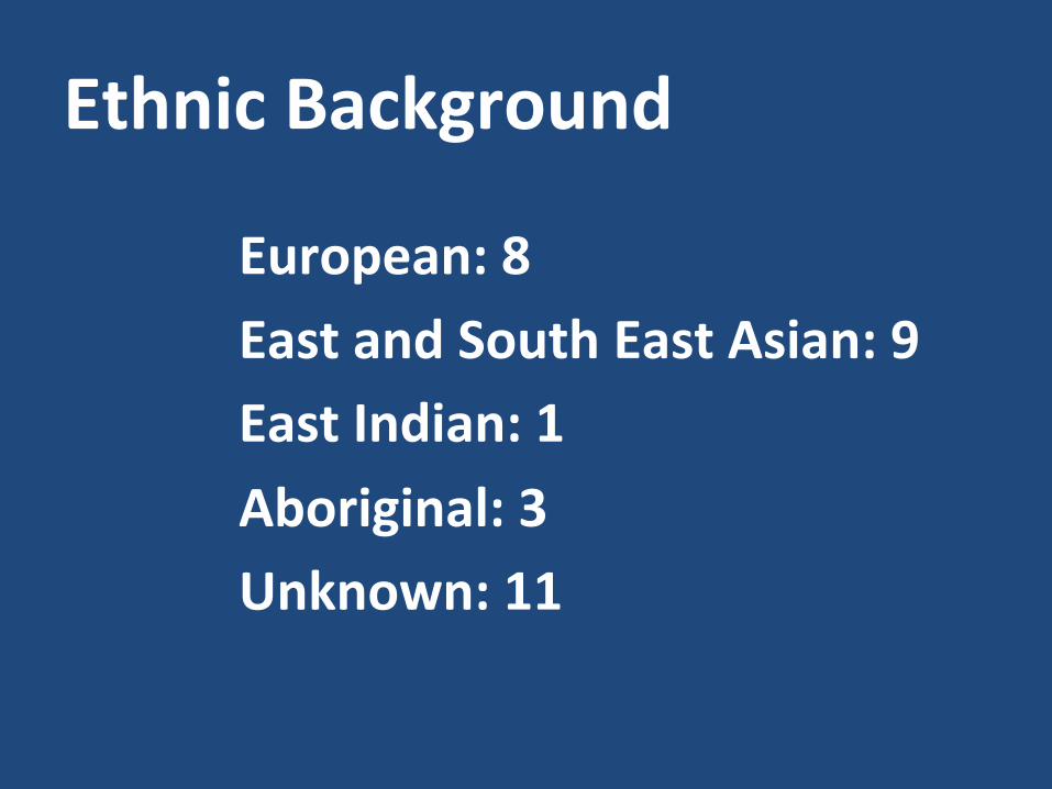
Ethnic Background
European: 8 East and South East Asian: 9 East Indian: 1 Aboriginal: 3 Unknown: 11
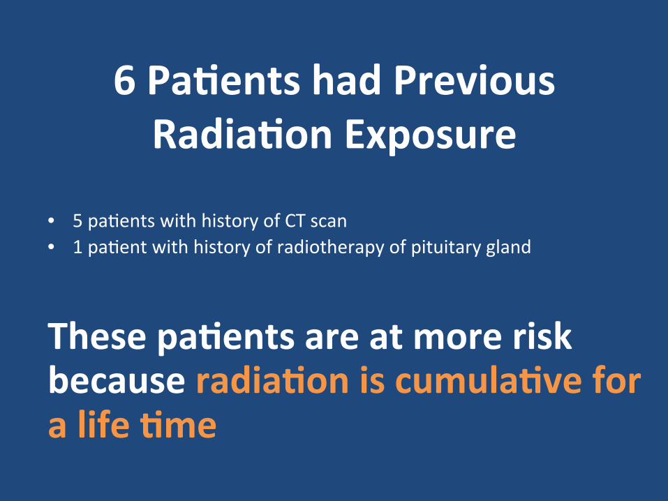
6 Pa8ents had Previous Radia8on Exposure
• 5 pa.ents with history of CT scan • 1 pa.ent with history of radiotherapy of pituitary gland
These pa8ents are at more risk because radia8on is cumula8ve for a life 8me
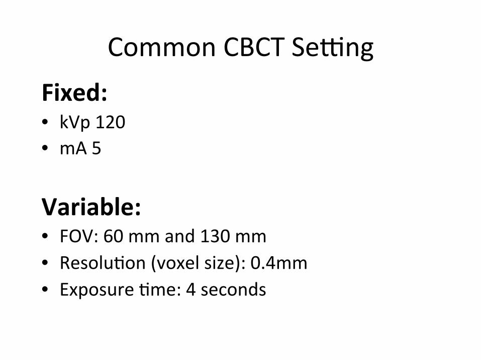
Common CBCT Se_ng Fixed: • kVp 120 • mA 5
Variable: • FOV: 60 mm and 130 mm • Resolu.on (voxel size): 0.4mm • Exposure .me: 4 seconds
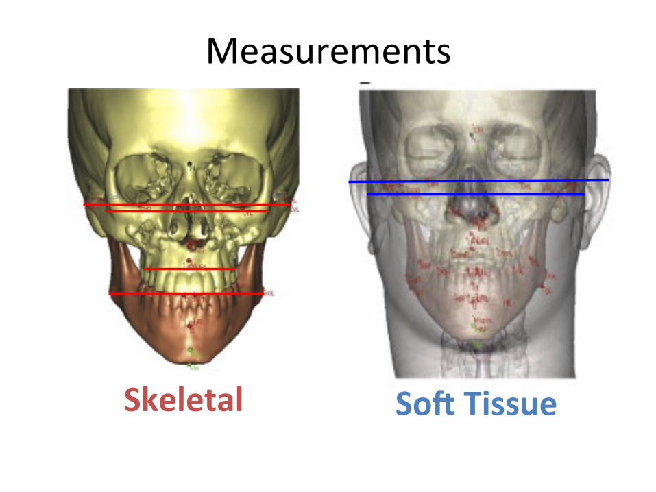
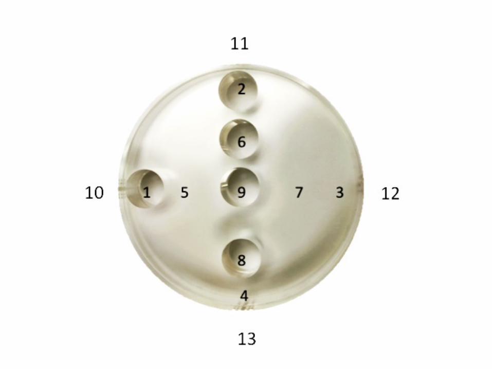
Measurements
Skeletal
SoJ Tissue
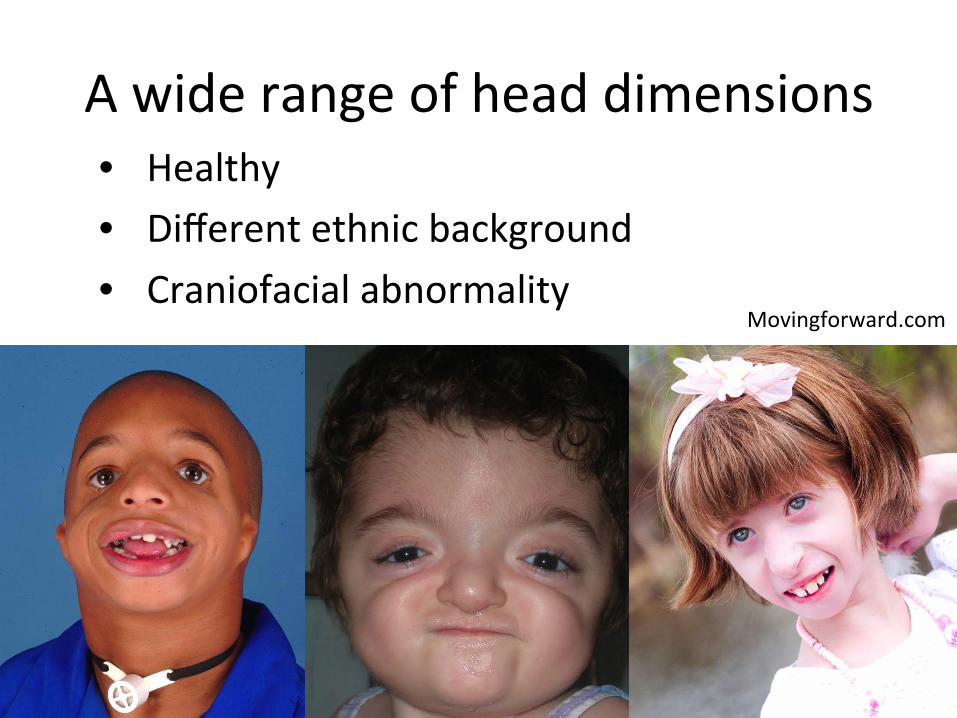
A wide range of head dimensions • Healthy • Different ethnic background • Craniofacial abnormality
Movingforward.com
Research Ques8ons
Ø Who receives CBCT at BC Children’s Hospital?
Ø What is the amount of radia.on from CBCT?
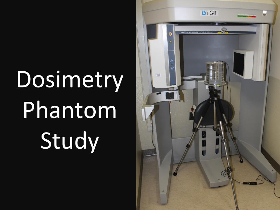
Dosimetry Phantom Study
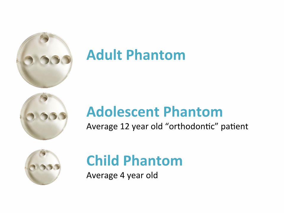
Adult Phantom Adolescent Phantom Average 12 year old “orthodon.c” pa.ent
Child Phantom Average 4 year old
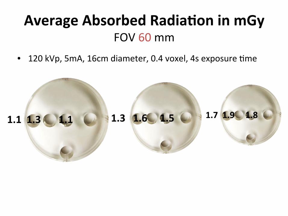
• 120 kVp, 5mA, 16cm diameter, 0.4 voxel, 4s exposure .me
1.1 1.3 1.1 1.7 1.9 1.8 1.3 1.6 1.5
Average Absorbed Radia8on in mGy FOV 60 mm
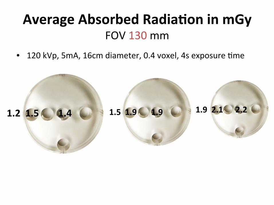
Average Absorbed Radia8on in mGy FOV 130 mm
• 120 kVp, 5mA, 16cm diameter, 0.4 voxel, 4s exposure .me
1.2 1.5 1.4 1.5 1.9 1.9 1.9 2.1 2.2
Conclusion 1. Children from a wide variety of ethnic and
medical backgrounds receive CBCT at BC Children’s Hospital
2. Pediatric phantom absorbed more radia.on than adult phantom in all loca.ons
3. Highest absorbed radia.on is at the edge of the phantom, second highest in the centre, and the lowest on the surface.
What can you do? • Lowest possible dose se_ng • Posi.oning + Shielding • Buy CBCTs that allows you to customize • Familiarize and Develop guidelines • More research • Request informa.on from manufacturers
Thank you! • Dr. Nancy Ford, Research Supervisor • Research Commikee: Dr. Rosamund Harrison and Dr. David MacDonald
• Dr. Robin Coope at BC Cancer Agency for fabrica.ng the pediatric phantom
• Pierre Deman • Funding from Faculty of Den.stry Research Equipment Grant
References • Safety Inves.ga.on of CT Brain Perfusion Scans (2009) hkp://www.fda.gov/MedicalDevices/
Safety/AlertsandNo.ces/ucm185898.htm • Bogdanich, W. (2010). THE RADIATION BOOM. HTTP://WWW.NYTIMES.COM/2010/08/01/
HEALTH/01RADIATION.HTML?PAGEWANTED=ALL • Oglivie, M. (2012). CT scans in childhood linked to increased risk of cancer: study Toronto
Star. hkp://www.thestar.com/news/canada/ar.cle/1207147-‐-‐ct-‐scans-‐in-‐childhood-‐linked-‐to-‐increased-‐risk-‐of-‐cancer-‐study
• Pearce, M., et al. Radia.on exposure from CT scans in childhood and subsequent risk of leukaemia and brain tumours: a retrospec.ve cohort study. Lancet. 2012 Aug 4;380(9840):499-‐505
• De Vos, W., J. Casselman, et al. (2009). "Cone-‐beam computerized tomography (CBCT) imaging of the oral and maxillofacial region: a systema.c review of the literature." Int J Oral Maxillofac Surg 38(6): 609-‐625.
• UNSCEAR 2008 Report: Sources and effects of ionizing radia.on • Movingforward.com • Absorp.on in medium • Ludlow JB, Davies-‐Ludlow LE, Brooks SL. Dosimetry of two extraoral direct digital imaging
devices: NewTom cone beam CT and Orthophos Plus DS panoramic unit. Dentomaxillofac Radiol 2003: 32: 229–234.
• Imagewisely.org • Imagegently.org