Embed Size (px)
Citation preview
Copy Right ©2010, Delia Vazquez, All Rights Reserved Page 1
Title: Long -term Changes in the Adrenocortical Response to Stress and in the Serotonin System following Postnatal Exposure to Glucocorticoids: Implications for the Treatment of Premature Infants Authors: *Delia M. Vázquez1, 2, Charles R. Neal, Jr.4, Paresh D. Patel 2, 3, Juan F. López2, 3 Affiliations: 1Department of Pediatrics and Communicable Diseases, University of Michigan, Ann Arbor, MI 48109, USA 2Department of Psychiatry, University of Michigan, Ann Arbor, MI 48109, USA 3Molecular and Behavioral Neuroscience Institute, University of Michigan, Ann Arbor, MI 48109, USA 4Department of Pediatrics and Communicable Diseases, John A. Burns School of Medicine, University of Hawaii, Honolulu, HI 96813 USA *corresponding author
Abstract (limit to 170 words) Potent glucocorticoids (GC) administered early in life has improved premature infant survival dramatically.
However, these agents may increase the risk for physical, neurological and behavior alterations. Anxiety,
depression and attention difficulties are commonly described in adolescent and young adult survivors of
prematurity. In the present study we administered vehicle, dexamethasone, or hydrocortisone to rat pups
on postnatal days 5 and 6, mimicking a short term clinical protocol commonly used in human premature
infants. Two systems that are implicated in the regulation of stress and behavior were assessed: the
limbic-hypothalamic-pituitary-adrenal axis [LHPA, glucocorticoid and mineralocorticoid receptors within]
and the Serotonin (5-HT) system. We found that as adults, pups treated with GC showed agent specific
altered growth, changes in corticoid response to novelty, anxious behavior and gene expression changes
within LHPA and 5-HT–related circuitry. The data suggest that prolonged GC-receptor occupation during
the early neonatal period can contribute to the development of individual differences in stress response
and anxiety behavior later in life.
Keywords Dexamethasone/*administration & dosage/*adverse effects Hydrocortisone/ *administration & dosage/*adverse effects Body Weight/drug effects Animals, Newborn/growth & development Anxiety Behavior/Attention Stress response Adult Rat
Copy Right ©2010, Delia Vazquez, All Rights Reserved Page 2
Introduction
Premature birth defined as birth prior to 37th week of gestation, accounts for approximately 11% of all
births in the USA (Centers for Disease Control and Prevention 2007). In 2007 this was equivalent to over
500,000 newborns (Centers for Disease Control and Prevention 2007). Over the last two decades the
survival of the most premature infants in this group has increased to approximately 40% at 24 weeks
gestation and 95% at 28 weeks (Hack and Fanaroff 1999; Lorenz 2001). Improvements in survival have
created a population of extremely low birth weight premature infant survivors with unique physical and
behavioral challenges (Hack and Fanaroff 1999; Hack et al 2000). A significant aspect of prematurity is
the fact that brain maturation after birth is very complex, corresponding to that which is occurring in utero
during the late second and entire third trimester of human pregnancy. This “ex-utero” brain development
remains the least understood in terms of its role in long-term neurodevelopmental outcome. Complicating
this process, the brains of critically ill premature infants must develop while simultaneously experiencing
multiple stressors (e.g. cold, light, noise, pain), as well as exposure to neuroactive agents, such as
glucocorticoids (GC), and opiates.
The glucocorticoids dexamethasone (DEX) and hydrocortisone (HC) are likely the most frequently used
neuroactive agents in the neonatal intensive care setting for multiple reasons, the most prominent of which
are lung disease (O'Shea et al 1999); (Cummings et al 1989) and refractory hypotension (Martens et al
2003). For the past two decades, DEX has been the glucocorticoid agent most often used in this clinical
setting. Unfortunately, DEX administration has been associated with neurodevelopmental impairments
among preterm infants (American Academy of Pediatrics 2002). Neurodevelopmental difficulties described
in the premature infant survivors who are not neurologically impaired vary depending on the age of the
child. At pre-school age (2–3 y), several reports indicate delayed psychomotor development and
anxious/depressed and/or withdrawn behavior (Sajaniemi et al 2001; Stoelhorst et al 2003a; Stoelhorst et
al 2003b). At school age and adolescence, increased incidences of social competence problems,
Copy Right ©2010, Delia Vazquez, All Rights Reserved Page 3
attention disorders and hyperactivity have been described (Botting et al 1997; Hille et al 2001; Pharoah et
al 1994; Saigal 2000; Schothorst and van Engeland 1996; Stjernqvist and Svenningsen 1995). A
systematic review of the DEX experience in neonates (Barrington 2001; Doyle and Davis 2000) led to a
consensus statement from American Academy of Pediatrics which recommended limited use of DEX in
premature infants (American Academy of Pediatrics 2002). During the past decade, hydrocortisone (HC),
a less potent glucocorticoid, has become a preferred agent in neonatal medicine for the treatment of
hypotension and chronic lung disease (van der Heide-Jalving et al 2003). Premature infant survivors
treated with HC during their neonatal period are at early school age now so less is known regarding the
neurodevelopmental effects of hydrocortisone use in this population, compared to those children who are
now adolescent and young adults that received DEX early in life. Immediate effects on neurodevelopment
appear to be benign (Benders et al 2009), but dosing regimens with HC are markedly variable and there
are mixed reports of the long term consequences (Lodygensky et al 2005; Thompson et al 2008).
The brain is clearly a target for GC action (De Kloet et al 1988; Vazquez 1998) and specific brain systems
expressing receptor molecules that are under organization at the time of GC exposure may be at the root
of the long term consequences observed in survivors of premature birth. Pre-clinical animal models have
been developed to study the long term effects of early life GC exposure on neurodevelopment, eliminating
confounding perinatal and postnatal factors found in human studies (Felszeghy et al 2000; Felszeghy et al
1996; Ferguson and Holson 1999; Heine et al 2009; Kauffman et al 1994; Kurosawa et al 1980; Slotkin et
al 1982). However, few animal studies have administered GC agents in the same manner as performed in
neonatal intensive units across the nation. Most recently, our laboratory reported neonatal DEX effects on
neonatal neurodevelopment, adolescent rat behavior, and adult limbic neurochemistry using a pre-clinical
animal model that mimics the prolonged DEX exposure that until 2002 was common in neonatal intensive
care settings (Bhatt-Mehta et al 2009; Flagel et al 2002a; Neal et al 2004a; Neal et al 2004b). Long-term
effects of neonatal treatments in any of these developmental animal models are interpreted under the
assumption that, although timing differs significantly between species, the general sequence of brain
Copy Right ©2010, Delia Vazquez, All Rights Reserved Page 4
growth is similar (Dobbing 1981; Flagel et al 2002a). While much caution is necessary when extrapolating
from animal models to the human condition, one can still take advantage of similarities in sequence and
timing of brain development between species. In humans, excluding the cerebellum and hippocampus,
neuronal proliferation is essentially completed before 24 wks gestation (Dobbing 1974). Beyond 24 wks,
glia continues to proliferate and oligodendroglia maintain ongoing myelination, with a peak in brain growth
occurring near term. In contrast to humans, rodents experience their brain growth spurt after birth. It is
estimated that on postnatal day 10 the rodent brain is roughly equivalent to that of the full term human
brain of 38 to 40 weeks post-conception (Dobbing 1974; Dobbing 1981; Hagberg et al 1997). Extrapolating
from this model, the brain of a rodent pup at birth (postnatal day 1 or PD 1) corresponds to that of a
human fetal brain at or near 19–21 weeks gestation (Dobbing 1981; Whitelaw and Thoresen 2000). The
PD 3 corresponds with that of a 24-26 week human, and PD 5-6 approximates a 28-32 week human.
Given these parallels, the neonatal rodent is an ideal model in which to investigate effects of a tapering
course of neonatal GC exposure on the developing brain.
In the present study, our objective was to compare the effects of the two leading GC agents under clinical
use in premature neonates over the last two decades (DEX and HC) on the developing central nervous
system. We chose to focus on a short course of GC treatment given on PD 5 and PD 6 for several
reasons. First, the majority of infants receiving GC for chronic lung disease will be those born at 23-25
weeks gestation (Hack and Fanaroff 1999; Lorenz 2001). These infants will receive steroids for lung
disease usually around 3-4 weeks of life. In addition, HC is often given to infants who are born at 24-28
weeks gestation for refractory hypotension. Consequently, a relatively short course of HC treatment is
used extensively in neonatal intensive units for non-pulmonary reasons. Second, the timing of GC
treatment is critical, because normal development of the human limbic-hypothalamic-pituitary axis (LHPA)
provides a low GC milieu during a period when elevated GC levels may negatively impact neuronal circuits
(Watterberg et al 2001). Third, we wished to focus on specific structures of the developing limbic system
and brainstem, locations where there is a high density of corticoid and serotonin (5-HT) receptors and 5-
Copy Right ©2010, Delia Vazquez, All Rights Reserved Page 5
HT producing cells, that have roles in the development of stress and emotional regulation (Benesova and
Pavlik 1989; Gunnar and Vázquez 2006; McEwen 1987; Weinstock et al 1992). Thus, in the present study
our objectives were three fold: 1) to provide GC during a postnatal age in the rat that corresponds to the
neurodevelopmental time point at which human premature infants commonly receive GC therapy, 2) to
provide tapering doses for both agents at a particular time after birth (from PD 5 to PD 6) with the specific
goal to more closely mimic and compare GC protocols provided in the neonatal intensive care setting,
where 3-7 day courses are administered (Halliday et al 2003), and 3) to ascertain the long term effect of
each of these agents on: a) growth, b) hormonal response to stress, c) anxiogenic behavior and d)
molecules relevant to GC action and the serotonin (5-HT) system in the adult animal. We hypothesized
that somatic effects and emotional instability will be present in animals treated early in life with both
glucocorticoid agents, with DEX having the greatest effect. In addition, we hypothesized that the
behavioral phenotype will be associated with developmental adaptations of both the limbic-corticoid and
serotonin receptor systems, to early life exposure of GC.
Material and Methods
Animals: Litter management and animal handling of neonatal rats in this study was similar to that
reported from our laboratories previously (Flagel et al 2002b). Adult Sprague-Dawley rats (Charles Rivers,
Wilmington, MA) were housed and treated according to Guide for the Care and Use of Laboratory
Animals. All animals were kept under constant temperature (25 + 2oC) and photoperiodicity (14:10h light-
dark cycle) and provided with food and water ad libitum. Animals were mated using one to one mating
system (1F:1M). Assuming a 21-day gestation, pregnant females were housed separately, starting on day
18. They were then checked twice daily until pups were born. The day of the birth of the pups was
designated as postnatal day one (PD1).
On PD2, each litter was sexed and culled to 12 pups (6M:6F) that were randomly selected in male: female
pairs from 3 different dams giving birth the same day. This ensured both genetic diversity, equality in
nutrition and maternal care within litters. On PD3, 4 pups representing both sexes were assigned to one of
Copy Right ©2010, Delia Vazquez, All Rights Reserved Page 6
three treatment groups within each litter (4 pups x 3 treatment groups= 12 pups/litter): 1) Vehicle Controls
(VEH), 2) Dexamethasone (DEX) and 3) Hydrocortisone (HC). Therefore, a total of 144 animals obtained
from 12 different litters were utilized in the study. All animals were studied in the early ages of PD 5,6,7,14
20, and 33. At day 33, female animals were sacrificed and brains collected, as part of another experiment.
Seventy-two males were studied at pre adolescence and adulthood on days PD 33, 60, 70 and 140 (n=24
for each).
Drug Treatment: On the day of treatments, cages with the dam and pups were brought into a room
adjacent to the animal colony and placed in a warm pad (temperature 30-35º C). Mothers were removed,
placed in a holding cage and moved to a separate room sheltered from the noise that may have been
present in the treatment room. Pups were sexed, weigh and treated between the hours of 1100 and 1300.
Animals in the DEX group received an intramuscular (IM) injection of DEX in a tapering dose of 0.5 mg/kg
on PD 5 and 0.1 mg/kg on PD 6. Animals in the HC group also received an IM injection of HC in a
tapering dose of 5.0 mg/kg on PD 5 and 1.0 mg/kg PD 6. Animals in the vehicle (VEH) group received
equivalent volume of IM sterile normal saline as the DEX and HC animals. The dam was then reunited
with the pups. The procedure time was 5 minutes. The study protocol and days of treatment are depicted
in Table 1.
Weaning: Weaning was performed at PD 21. Females were grouped housed 6 animals per cage, then
sacrificed on PD33. Males were grouped housed 6 animals per cage from PD 21 to PD 45, then
separated to single housing due to their weight at PD 45 until the end of the experiments. This was done
following the "Guidelines for the Care and Use of Laboratory Animals", which bases the number of animals
in a cage on the size of the animals and expected growth without food restriction.
Somatic Growth: Lengths and weights were measured for each pup in each treatment group before
treatments on PD 5, 6. All animals were also measured and weighed on PD 7, 14, 20, 33. Male animals
were weighed when 33, 60 and 140 days old. Length was not obtained at 60 days in the males due to a
lab error. The weight and length of the female animals up to PD 33 is reported here for comparison
Copy Right ©2010, Delia Vazquez, All Rights Reserved Page 7
purposes with the males (Figure 1). At each age, length was measured from the nose to the base of the
tail (head-rump length).
Behavioral Testing: Light-Dark Preference Box- The light-dark preference procedure, was used to
evaluate the locomotion and investigatory behavior of the animals when 33 and 70 days old. Preference
for darkness and decreased activity are gross measurements of anxiety (Bourin and Hascoet 2003; File
1990; Ramos 2008). Three days prior to testing (PD 30 or 67), animals were acclimated to the handling
needed before the start of the procedure. Handling consisted in transporting the cage to the test room and
removal from the cage at the expected time of testing. This was done for three consecutive days. The
testing apparatus was a covered 30 x 60 x 30-cm Plexiglas shuttle-box with a computerized monitor. It has
two equal sized compartments (light and dark) with a 12-cm wide opening and stainless steel grid floor
suspended above the corncob bedding. The light compartment was constructed of white Plexiglas and
brightly illuminated. The dark compartment was constructed of black Plexiglas and minimally illuminated.
Fluorescent lights above the box provided the illumination. To start the session, each animal was placed in
the dark compartment and the timer set to start. Each animal‟s locomotor activity, the time spent in each
compartment, the number of transitions and the latency to leave the dark were scored. Locomotor activity
as well as time spent in each compartment was monitored by photocells located on the wall of each box,
with the number of photocell beams interrupted per unit time recorded with microprocessor. The number
of transitions was recorded manually. Total testing time was 5 minutes (Flagel et al 2002b).
Adrenocortical Response to Novelty Stress: Immediately after light-dark preference testing was
completed on PD 70, animals underwent a tail nick procedure to collect blood. Blood was collected from
the tail vein at 15, 30 and 60 min after placing the rat in the dark compartment of the light-dark box. The
last blood sample was collected 120 min after the start of the test at which time the animals were
decapitated. A pre-stress blood sample was obtained the day prior to the light-dark box as the animal was
acclimated to handling. The time of the pre-stress sample corresponded to the same time of the start of
the procedure on the following day. Blood samples were collected in pre chilled tubes containing EDTA,
Copy Right ©2010, Delia Vazquez, All Rights Reserved Page 8
placed on ice and subsequently spun at 2000rpm for 7 min. The plasma was separated and stored at -200
until assayed for corticosterone hormone concentrations.
Hormonal Assay: Corticosterone (CORT) levels were measured using a commercially available
corticosterone I125 radioimmunoassay kit (Cat. #07-120102, MP Biomedicals LLC, Diagnostic Division,
Orangeburg, New York). Un-extracted plasma samples were diluted to 1:200 in Phosphosaline gelatin
buffer (pH 7.0). The intra- and inter assay CVs for corticosterone were 4.4% and 6.5%, respectively.
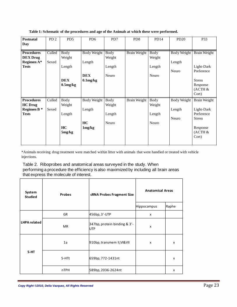
Brain Sectioning: Six animals per group were randomly selected for brain sectioning and anatomical
analyses that followed. For this purpose, the blocks between Bregma +5.9mm to -2mm (“forebrain”) and
between Bregma -2mm to Bregma -7mm (“raphe”, with DR cell groups typically between Bregma -4.1mm
and -5.2mm) were frozen in dry ice-isopentane at -20ºC followed by storage at -80ºC. We generated two
brain blocks per animal. The raphe block was sectioned at 12 um and collected in sets of 5 deep (sections
1-5 on slides 1-5, respectively; sections 6-10 on slides on 1-5 again, etc, repeated until full, then on
subsequent groups of 5 slides). This generates 5 “sets” of slides, each of which surveys the region at ~60
µm intervals. The forebrain block (containing dorsal hippocampus and amygdala in the same coronal
plane) were acquired 8 sections deep. This sampling scheme generated approximately 250 sections per
animal for the rostral raphe group, generating 9 slides per animal per set (x 5 sets), with each slide
containing six sections. Three set of slides surveying raphe were analyzed separately by in situ
hybridization (ISH) for 1) nTPH, 2) 5-HTT, 3) 5-HT1a receptor mRNAs. Sections through hippocampus
were processed for 1) 5-HT1a mRNA, 2) MR mRNA, and 3) GR mRNA. Table 2 depicts the riboprobes
used for these analyses. The anatomical areas of interest for each of theses are also shown.
Hybridization: Sections were removed from storage at −80°C and placed directly into 4% buffered
paraformaldehyde at room temperature. After 60 min, slides were rinsed in isotonic phosphate-buffered
saline and treated with proteinase K (1 μg/mL in 100 mmol/L TRIS/HCl, pH 8.0) for 10 min at 37°C.
Subsequently, sections underwent successive washes in water (1 min), 0.1 mol/L triethanolamine (pH 8.0,
Copy Right ©2010, Delia Vazquez, All Rights Reserved Page 9
plus 0.25% acetic anhydride) for 10 min and 2X SSC (0.3 mmol/L NaCl, 0.03 mmol/L sodium citrate, pH
7.2) for 5 min. Sections were then dehydrated through graded alcohols and air dried.
Postfixed sections were hybridized with 1.0 × 10⁶ dpm [35S]UTP-labeled riboprobes in hybridization buffer
containing 50% formamide, 10% dextran sulphate, 3X SSC, 50 mmol/L sodium phosphate buffer (pH 7.4),
1X Denhardt‟s solution, 0.1 mg/mL yeast transfer RNA (tRNA), and 10 mmol/L dithiothreitol in a total
volume of 25 μL. The probe was applied to sections on a glass coverslip and hybridized overnight at 55°C.
Next day the sections were washed in 2X SSC for 5 min and then treated with RNase A (200 μg/mL in 10
mmol/L TRIS/HCl, pH 8.0, containing 0.5 mol/L NaCl) for 60 min at 37°C. Subsequently, sections were
washed in 2X SSC for 5 min, 1X SSC for 5 min, and 0.5X SSC for 60 min at hybridization temperature,
and 0.5X SSC at room temperature for 5 min, and then dehydrated in graded alcohols and air dried. For
signal detection, sections were placed on Kodak XAR-5 X-ray film and exposed for 2 days at room
temperature.
Microdensitometric Analysis: Autoradiograms generated from the IHS were analyzed using an
automated image analysis system (Dage camera, Scion Image Beta 4.03; Scion Corporation). Anatomical
regions of interest were interactively selected and mean optical density measurements for each region
were determined from at least six coronal sections in a single animal. This single data point was utilized in
the statistical analyses. Hippocampus subfields were determined with reference to Nissla-stained sections
and the anatomical atlas of (Paxinos and Watson 1986). Nonspecific labeling of [35S]-riboprobes was
determined from an area of section exhibiting apparent lack of hybridization signal.
Statistical Analyses: Statistical differences were determined by analysis of variance (ANOVA).
Significance was indicated by a p value p<0.05. Once significance was observed by ANOVA, the Fisher‟s
least significant difference (Fisher‟s PLSD) method was utilized for further pair wise comparisons.
Copy Right ©2010, Delia Vazquez, All Rights Reserved Page 10
Results
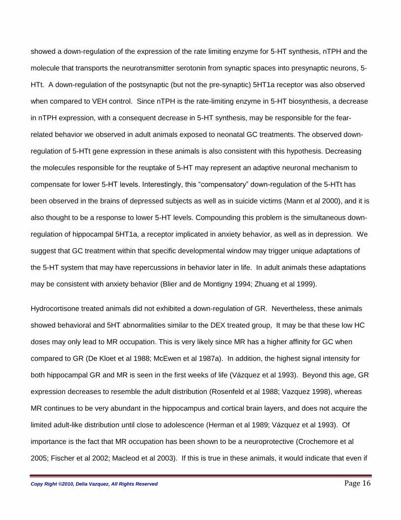
Somatic Growth: ANOVA analyses revealed significant sex, treatment and age effect (sex, treatment and
age, each p< 0.0001). Figure 1, Panels A and B shows weight and length progression on females from
PD 5 to PD 33; Panels C and D depicts these on the males. Compared to VEH animals the DEX-treated
rats had a decrease in somatic growth at all early ages. The adult DEX-treated males were not different
from VEH treated males for both weight and length at PD60 and 140. In contrast, HC-treated animals
were not different from VEH at early ages until adulthood when both weight and length were significantly
greater in the males.
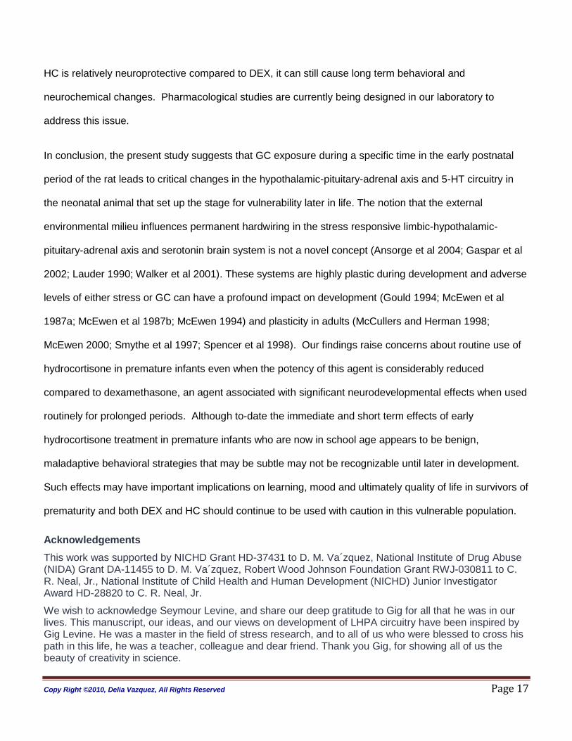
Anxiety Behaviors (Figure 2): A treatment effect was evident on the parameters assessed with the L-D box
test on PD 70 animals only (time to light p <0.05, time in light p<0.002, activity in light p<0.0005, time in
dark p<0.002). Testing performed at PD 33 showed no significance for any of the parameters (data not
shown). Post hoc analyses revealed that as young adults (PD70), DEX- and HC- treated animals show
altered behaviors in the L-D box that were similar. Compared to VEH controls, the DEX-treated animals
showed significantly decreased time and exploratory behavior in the light compartment. DEX-treated
animals spent an increased amount of time in the dark compartment when compared to VEH. The HC
treated animal also had significantly decreased time and exploratory behavior in the light compartment
and they spent an increased amount of time in the dark compartment when compared to VEH controls.
The decreased time and activity in the light compartment of the HC-treated animals was somewhat less
marked when compared to the DEX-treated animals.
Animals are placed in the dark compartment upon initiation of this test and HC-treated animals moved
from the dark to the light compartment significantly faster when compared to VEH-treated animals (see
Figure 3, time to light). This behavior was not observed in the DEX-treated animals.
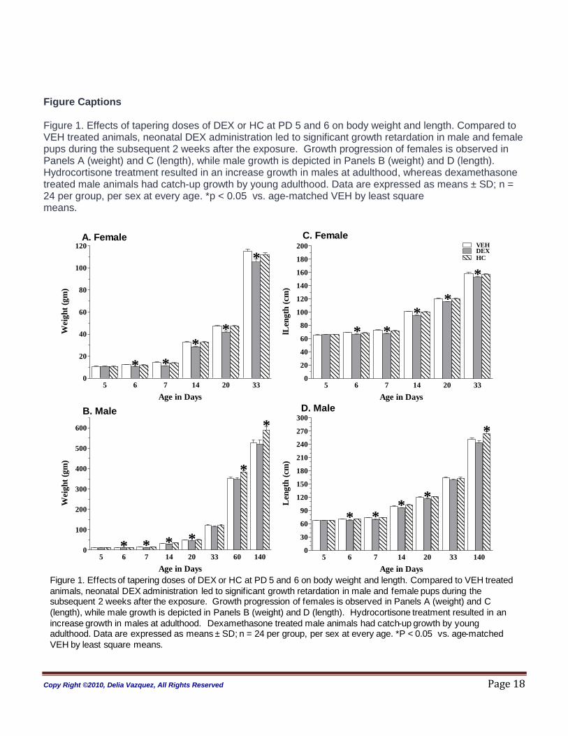
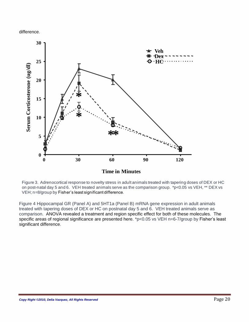
Stress Response (Figure 3): A significant effect for treatment and time, with a treatment by time interaction
was observed in the ANOVA analysis (treatment p < 0.0001; time p<0.0001; treatment:time, p<0.001).
DEX-treated adult rats had a blunted adrenocortical response (CORT) to novelty stress compared to all
Copy Right ©2010, Delia Vazquez, All Rights Reserved Page 11
groups. HC-treated animals were also different from VEH-treated animals. HC -treated animals had a
lower peak response and faster CORT inhibition when compared to VEH.
Corticoid Receptors [glucocorticoid receptor (GR) and mineralocorticoid receptor (MR)) and Serotonin (5-
HT] Molecules (Figure 4 and 5)
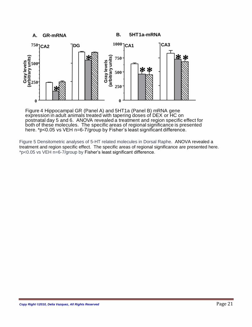
Hippocampus: ANOVA revealed a treatment (p<0.01) and region (p<0.001) effect on GR gene
expression. In the hippocampus, significance for GR mRNA the ANOVA analysis was split by region. We
observed that adult DEX-treated rats had significantly decreased GR mRNA expression in areas CA1 and
CA2 of Ammon‟s horn, and the dentate gyrus (DG) when compared to HC and VEH animals (CA1 p=0.05;
CA2 p<0.0001; CA3, p=0.13; DG, p=0.01). Post hoc analysis demonstrated that DEX treated animals had
down-regulation when compared to VEH control. No effect of DEX was found on the MR gene expression
(ANOVA p=0.345) and no GR or MR mRNA changes were detected in the HC-treated animals compared
to VEH controls.
The analysis of variance (ANOVA) of the 5-HT1a receptor (5-HT1a) mRNA levels in the hippocampus
revealed a treatment (p<0.0001) and region effect (p=0.001). When split by region the effect was in CA1
and CA3 (CA1 p=0.006; CA2 p=0.62; CA3 p=0.05; DG p=0.63). Post hoc analysis using Fisher protected
least significance difference (Fisher PLSD) showed significant down-regulation in the DEX and HC treated
animals when compared to vehicle (VEH) [see Figure 4].
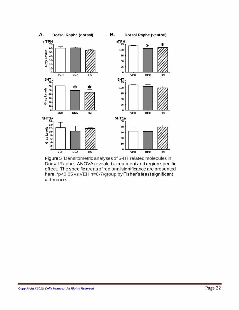
Raphe: In the raphe nucleus, ANOVA revealed no treatment (p=0.84) or region effects for 5-HT1a
receptor gene expression (dorsal raphe dorsal, p=0.8 and dorsal raphe ventral, p=0.93; see Figure 5,
Panels A and B). Treatment and regional effects were observed in this structure for neuronal tryptophan
hydroxylase (nTPH) and serotonin transporter (5-HTt) gene expression measurements (ANOVA nTPH
treatment= p< 0.0001; region= p< 0.05; ANOVA 5-HTt treatment= p< 0.0001; region= p< 0.005). In the
DEX and HC -treated animals nTPH mRNA was significantly decreased in the ventral part of the Dorsal
Raphe nucleus (DRN). HC -treated animals showed a trend towards down-regulation in the dorsal part of
the DRN (p=0.06). Both DEX and HC treated animals also showed a modest but significant down
Copy Right ©2010, Delia Vazquez, All Rights Reserved Page 12
regulation of the 5HTt mRNA levels in the dorsal area of the DRN when compared to VEH controls.
Notice that compared to VEH-treated animals the 5-HT1a auto-receptor gene expression was found to be
unchanged in the DRN of the DEX and HC treated animals.
Discussion
In the present study, we have investigated long-term effects of a DEX and HC treatment regimen given
during the first week of life in the infant rat. We administered tapering doses of these agents on postnatal
days 5 and 6 in an attempt to mimic short treatment regimens of DEX and HC that are commonly used in
the neonatal intensive care setting in premature infants. The timing of drug administration is critical in our
rat model, since it corresponds to late third trimester of human pregnancy (28 to 32 weeks gestation), a
period of growth and development that renders the brain highly vulnerable to insult in the human neonate
(Dobbing 1981). We found that our treatments had significant effects on somatic growth, affecting both
length and weight in the developing organism. Beyond these somatic observations, we found that a short
course of either DEX or HC administration in the postnatal period affects stress response, and results in
anxiogenic behavior in the adult animal. Changes in the gene expression of corticoid receptors and
serotonin related molecules were also observed in hippocampus and raphe of those animals experiencing
anxiety-like behavior. These findings suggest that early short-term exposure to either DEX or HC may
have long-lasting consequences even when a tapering dose regimen is implemented.
DEX or HC administered during early in life has a lasting impact on somatic growth. The effects were
specific for each of the agents used, both in terms of time at which the growth was affected and the
persistence of the change. Consistent with previous reports, we found that DEX treatment induced
significant decreases in length and weight gain in the rat pup that persisted for up to two weeks after
treatment (Flagel et al 2002a; He et al 2004; Kanagawa et al 2006; Kreider et al 2006; Neal et al 2004b;
Slotkin et al 2006). Catch up growth was consistently achieved by adulthood. In contrast, HC-treated
animals were not affected by this regimen during infancy, but were significantly larger, for both weight and
Copy Right ©2010, Delia Vazquez, All Rights Reserved Page 13
length, than VEH and DEX treated animals at adulthood. The decreased somatic growth observed in the
DEX-treated pups may be a result of inadequate nutritional intake during the postnatal period, due to
inability of the pup to attach to the mother‟s nipples, or poor suckling. Though this is a possibility, it has
been shown that the dam spends more time providing nutrition, stimulation, and warmth to a litter that is
perceived to have poor health (Brunelli et al 1994; Lynch 1976; Stern 1997; Wiener et al 1977; Wiener and
Levine 1978). Direct effects of DEX on protein catabolism that result in reduced growth and lean body
mass have also been described (Leret et al 2004; Weiler et al 1997). Thus, it is likely that increased
protein catabolism beyond the capacity for an anabolic state needed for growth is responsible for the
growth deficits observed during the first 3 weeks of life in the DEX-treated animals. The adverse effects
on weight gain in early neonatal stages likely contribute to long-term risks of GC use, as has been
postulated in the „Barker Hypothesis‟, relating early growth failure in human infants to subsequent elevated
risk of cardiovascular and metabolic disorders (Baker 2000; Breier et al 2001; Lucas 1991). In both animal
and human literature there is evidence to suggest that early life DEX treatment similarly increases the
incidence of cardiovascular and metabolic disorders (Barrington 2001; He et al 2004; Seckl 2008), despite
the eventual recovery of body weight, as seen in our study.
Animals treated with postnatal HC have a unique pattern of growth when compared to DEX-treated
animals: they do not have altered growth early in life, but show weight gain in adulthood. A thorough
review of the postnatal HC literature in humans or animals treated with HC early in life revealed that this
phenomena has received little attention (Peltoniemi et al 2009). Since weight gain is associated with
increased adiposity and insulin resistance (Brindley 1995; Cameron and Demerath 2002; London et al
2009; Seckl et al 2004; Slieker et al 1996; Walker et al 2003), it possible that early life HC treatment also
increases the risk for later metabolic disorders. Further research on this aspect of early life HC exposure
is warranted.
The adrenocortical stress response was altered by early life GC treatments. Basal corticosterone levels
were not different between groups. In response to novelty stress, the male animals exposed to HC or to
Copy Right ©2010, Delia Vazquez, All Rights Reserved Page 14
DEX-early in life had a blunted adrenocortical response, with an adequate termination of the stress
response. These patterns of stress response are consistent with other animal and human reports that
indicate lifetime altered responses with early life DEX treatments (Felszeghy et al 2000; Flagel et al
2002b; Karemaker et al 2008; Ng et al 2008). Surprisingly, no other animal studies have investigated
long-term response to early HC treatment. Modulation of glucocorticoid receptors in hippocampus are
linked to baseline corticosterone levels and the quality of the adrenocortical response [reviewed in (De
Kloet et al 1998)]. Our findings of unchanged MR mRNA expression in the hippocampus are consistent
with the basal corticosterone levels observed in both treatment groups. However, decreased GR mRNA
expression but no change in MR mRNA expression in adult rat hippocampus is associated with a delayed
return to baseline in serum corticosterone after stress (reviewed in (De Kloet et al 1998)). This
combination of corticoid receptor balanced was found in our DEX-treated (but not the HC-treated) animals;
however an adequate inhibition of corticosterone secretion was present. One possible explanation is that
a much stronger stressor is needed to challenge the DEX treated animals in order for this to result in a
loss of regulatory feedback inhibition of corticosterone secretion. Neal and co-workers showed that peak
corticosterone response to crowding stress in adult animals exposed to tapering doses of DEX from
postnatal day 3 to 6 is no different from VEH control animals, but delayed return to baseline is noted in the
DEX-exposed cohort (Neal et al 2004b). Taken together, these data indicate either that the type of
stressor is important for the adrenocortical response observed or that the timing of GC exposure early in
life is an important factor modulating long-term effects of neonatal exposure.
In the present study, increased anxiety-like behaviors were observed in both DEX and HC –treated
animals when tested in the light-dark test. Of importance is the fact that all males were housed alone
starting at adolescence. This factor may constitute an environmental modification; therefore there are a
number of ways to interpret our study findings. One is that the behavioral manifestations (and stress
response) seen in the GC treated animals emerges during the transition from adolescence to adulthood. It
is also possible that our animals may have been affected by their single-housing environment at
Copy Right ©2010, Delia Vazquez, All Rights Reserved Page 15
adolescence after PD 45. This raises the possibility that GC exposure early in life creates a vulnerable
state in the animal, leading to alterations in limbic-hypothalamic-pituitary-adrenal function only after
experiencing what could be construed as a prolonged social isolation. There is significant literature from
the 1960‟s that support the possibility of “isolation stress syndrome” in rodents. Animals exhibiting this
syndrome are excessively reactive to handling, timid, or fearful (Ader et al 1960; Hatch et al 1965; Stern et
al 1960). However, in 1990, Holson and co-workers were able to show that the isolation syndrome was
dependent on environmental factors and not the isolation itself (Holson et al 1991). Animals isolated in
hanging metal cages showed the behavioral and stress responses of profound fear, but this was not the
case for littermates that were isolated and reared singly in plastic cages. The main difference was that
animals in hanging metal cages were never touched by human caretakers, whereas rats reared in plastic
cages were picked up and put in clean cages twice weekly as part of their usual animal care. In support of
this observation, handling animals that were isolated in hanging-cages twice weekly to model the handling
associated with the plastic cage changes completely protected against the “isolation stress syndrome”.
Our animals were isolated in plastic cages but received care as described in Holson‟s study. Despite this
reassuring report, there is a recent publication that shows a lower level of exploratory behavior following a
mild shock in 65 and 130-day-old rats isolated during the juvenile stage, but not in rats isolated after
puberty (Arakawa 2007). Although further studies comparing single and group housing are currently being
performed in our laboratory to elucidate the importance of these housing conditions, we interpret the adult
anxiogenic phenotype observed in our early life GC treated groups as one that is related to specific
changes occurring early in life as a result of HC exposure. It is possible that this exposure renders
vulnerability to the later exposure to an isolated environment, at puberty. Brain receptor findings support
this conclusion.
Both DEX and HC cause long-term repercussions in behavior and in the serotonin (5-HT) system.
Animals treated with our GC tapering paradigm exhibited, as adults (but not pre-adolescence), anxiety-like
behavior in the Light-Dark test. When compared to VEH control, both DEX and HC treated animals also
Copy Right ©2010, Delia Vazquez, All Rights Reserved Page 16
showed a down-regulation of the expression of the rate limiting enzyme for 5-HT synthesis, nTPH and the
molecule that transports the neurotransmitter serotonin from synaptic spaces into presynaptic neurons, 5-
HTt. A down-regulation of the postsynaptic (but not the pre-synaptic) 5HT1a receptor was also observed
when compared to VEH control. Since nTPH is the rate-limiting enzyme in 5-HT biosynthesis, a decrease
in nTPH expression, with a consequent decrease in 5-HT synthesis, may be responsible for the fear-
related behavior we observed in adult animals exposed to neonatal GC treatments. The observed down-
regulation of 5-HTt gene expression in these animals is also consistent with this hypothesis. Decreasing
the molecules responsible for the reuptake of 5-HT may represent an adaptive neuronal mechanism to
compensate for lower 5-HT levels. Interestingly, this “compensatory” down-regulation of the 5-HTt has
been observed in the brains of depressed subjects as well as in suicide victims (Mann et al 2000), and it is
also thought to be a response to lower 5-HT levels. Compounding this problem is the simultaneous down-
regulation of hippocampal 5HT1a, a receptor implicated in anxiety behavior, as well as in depression. We
suggest that GC treatment within that specific developmental window may trigger unique adaptations of
the 5-HT system that may have repercussions in behavior later in life. In adult animals these adaptations
may be consistent with anxiety behavior (Blier and de Montigny 1994; Zhuang et al 1999).
Hydrocortisone treated animals did not exhibited a down-regulation of GR. Nevertheless, these animals
showed behavioral and 5HT abnormalities similar to the DEX treated group, It may be that these low HC
doses may only lead to MR occupation. This is very likely since MR has a higher affinity for GC when
compared to GR (De Kloet et al 1988; McEwen et al 1987a). In addition, the highest signal intensity for
both hippocampal GR and MR is seen in the first weeks of life (Vázquez et al 1993). Beyond this age, GR
expression decreases to resemble the adult distribution (Rosenfeld et al 1988; Vazquez 1998), whereas
MR continues to be very abundant in the hippocampus and cortical brain layers, and does not acquire the
limited adult-like distribution until close to adolescence (Herman et al 1989; Vázquez et al 1993). Of
importance is the fact that MR occupation has been shown to be a neuroprotective (Crochemore et al
2005; Fischer et al 2002; Macleod et al 2003). If this is true in these animals, it would indicate that even if
Copy Right ©2010, Delia Vazquez, All Rights Reserved Page 17
HC is relatively neuroprotective compared to DEX, it can still cause long term behavioral and
neurochemical changes. Pharmacological studies are currently being designed in our laboratory to
address this issue.
In conclusion, the present study suggests that GC exposure during a specific time in the early postnatal
period of the rat leads to critical changes in the hypothalamic-pituitary-adrenal axis and 5-HT circuitry in
the neonatal animal that set up the stage for vulnerability later in life. The notion that the external
environmental milieu influences permanent hardwiring in the stress responsive limbic-hypothalamic-
pituitary-adrenal axis and serotonin brain system is not a novel concept (Ansorge et al 2004; Gaspar et al
2002; Lauder 1990; Walker et al 2001). These systems are highly plastic during development and adverse
levels of either stress or GC can have a profound impact on development (Gould 1994; McEwen et al
1987a; McEwen et al 1987b; McEwen 1994) and plasticity in adults (McCullers and Herman 1998;
McEwen 2000; Smythe et al 1997; Spencer et al 1998). Our findings raise concerns about routine use of
hydrocortisone in premature infants even when the potency of this agent is considerably reduced
compared to dexamethasone, an agent associated with significant neurodevelopmental effects when used
routinely for prolonged periods. Although to-date the immediate and short term effects of early
hydrocortisone treatment in premature infants who are now in school age appears to be benign,
maladaptive behavioral strategies that may be subtle may not be recognizable until later in development.
Such effects may have important implications on learning, mood and ultimately quality of life in survivors of
prematurity and both DEX and HC should continue to be used with caution in this vulnerable population.
Acknowledgements
This work was supported by NICHD Grant HD-37431 to D. M. Va´zquez, National Institute of Drug Abuse (NIDA) Grant DA-11455 to D. M. Va´zquez, Robert Wood Johnson Foundation Grant RWJ-030811 to C. R. Neal, Jr., National Institute of Child Health and Human Development (NICHD) Junior Investigator Award HD-28820 to C. R. Neal, Jr.
We wish to acknowledge Seymour Levine, and share our deep gratitude to Gig for all that he was in our lives. This manuscript, our ideas, and our views on development of LHPA circuitry have been inspired by Gig Levine. He was a master in the field of stress research, and to all of us who were blessed to cross his path in this life, he was a teacher, colleague and dear friend. Thank you Gig, for showing all of us the beauty of creativity in science.
Copy Right ©2010, Delia Vazquez, All Rights Reserved Page 18
Figure Captions Figure 1. Effects of tapering doses of DEX or HC at PD 5 and 6 on body weight and length. Compared to VEH treated animals, neonatal DEX administration led to significant growth retardation in male and female pups during the subsequent 2 weeks after the exposure. Growth progression of females is observed in Panels A (weight) and C (length), while male growth is depicted in Panels B (weight) and D (length). Hydrocortisone treatment resulted in an increase growth in males at adulthood, whereas dexamethasone treated male animals had catch-up growth by young adulthood. Data are expressed as means ± SD; n = 24 per group, per sex at every age. *p < 0.05 vs. age-matched VEH by least square means.
Figure 1. Effects of tapering doses of DEX or HC at PD 5 and 6 on body weight and length. Compared to VEH treated
animals, neonatal DEX administration led to significant growth retardation in male and female pups during the subsequent 2 weeks after the exposure. Growth progression of females is observed in Panels A (weight) and C
(length), while male growth is depicted in Panels B (weight) and D (length). Hydrocortisone treatment resulted in an
increase growth in males at adulthood. Dexamethasone treated male animals had catch-up growth by young adulthood. Data are expressed as means ± SD; n = 24 per group, per sex at every age. *P < 0.05 vs. age-matched
VEH by least square means.
5 6 7 14 20 330
20
40
60
80
100
120
*
**
*
*
A. Female
Age in Days
Wei
gh
t (g
m)
5 6 7 14 20 330
20
40
60
80
100
120
140
160
180
200DEXVEH
HC
*
**
*
*
C. Female
Age in Days
lLen
gth
(cm
)
5 6 7 14 20 33 60 1400
100
200
300
400
500
600
* *
*
*
*B. Male
*
Age in Days
Wei
gh
t (g
m)
5 6 7 14 20 33 1400
30
60
90
120
150
180
210
240
270
300
**
*
*
*
D. Male
Age in Days
Len
gth
(cm
)
Copy Right ©2010, Delia Vazquez, All Rights Reserved Page 19
Figure 2. Adult behavior in Light:Dark preference box. Dex and HC -treated animals spent significantly less time in the light compartment and were less active in a test of adaptation to a novel environment. In addition, the HC-treated young adult animals latency to move from the light to the dark compartment was significantly less when compared to all other groups. Data are expressed as means ± SD; n = 8 per group. *p < 0.05, vs age-matched VEH by Fisher‟s least significant difference.
Time to Light Light Time Light Activity Dark Time Dark Activity0
30
60
90
120
150
180
210
240
270
300
VEHDEXHC
*
*
*
*
**
Tim
e i
n S
eco
nd
s
*
Figure 2. Adult behavior in Light:Dark preference box. Dex and HC -treated animals spent significantly less time in the light
compartment and were less active in a test of adaptation to a novel environment. In addition, the HC-treated young adult animals latency to move from the light to the dark compartment was significantly less when compared to all other groups.
Data are expressed as means ± SD; n = 24 per group. *P < 0.05, vs age-matched VEH by Fisher‟s least significant
difference. Figure 3. Adrenocortical response to novelty stress in adult animals treated with tapering doses of DEX or HC on post-natal day 5 and 6. VEH treated animals serve as the comparison group. *p<0.05 vs VEH and HC, ** vs VEH; n=8/group by Fisher‟s least significant
Copy Right ©2010, Delia Vazquez, All Rights Reserved Page 20
difference.
Figure 3. Adrenocortical response to novelty stress in adult animals treated with tapering doses of DEX or HC
on post-natal day 5 and 6. VEH treated animals serve as the comparison group. *p<0.05 vs VEH, ** DEX vs
VEH; n=8/group by Fisher‟s least significant difference.
0 30 60 90 1200
5
10
15
20
25
30
DexVeh
Time in Minutes
HC
*
**
Ser
um
Co
rtic
ost
ero
ne
(ug
/dl)
*
Figure 4 Hippocampal GR (Panel A) and 5HT1a (Panel B) mRNA gene expression in adult animals treated with tapering doses of DEX or HC on postnatal day 5 and 6. VEH treated animals serve as comparison. ANOVA revealed a treatment and region specific effect for both of these molecules. The specific areas of regional significance are presented here. *p<0.05 vs VEH n=6-7/group by Fisher‟s least significant difference.
Copy Right ©2010, Delia Vazquez, All Rights Reserved Page 21
A. GR-mRNA
0
250
500
750
*
*
CA2 DG
Gra
y le
ve
ls(a
rbit
rary
un
its
)
B. 5HT1a-mRNA
0
250
500
750
1000
**
CA1 CA3
**
Gra
y le
ve
ls(a
rbit
rary
un
its
)Figure 4 Hippocampal GR (Panel A) and 5HT1a (Panel B) mRNA gene expression in adult animals treated with tapering doses of DEX or HC on postnatal day 5 and 6. ANOVA revealed a treatment and region specific effect for both of these molecules. The specific areas of regional significance is presented here. *p<0.05 vs VEH n=6-7/group by Fisher‟s least significant difference.
Figure 5 Densitometric analyses of 5-HT related molecules in Dorsal Raphe. ANOVA revealed a treatment and region specific effect. The specific areas of regional significance are presented here. *p<0.05 vs VEH n=6-7/group by Fisher‟s least significant difference.
Copy Right ©2010, Delia Vazquez, All Rights Reserved Page 22
Figure 5 Densitometric analyses of 5-HT related molecules in
Dorsal Raphe. ANOVA revealed a treatment and region specific effect. The specific areas of regional significance are presented here. *p<0.05 vs VEH n=6-7/group by Fisher‟s least significant
difference.
VEH DEX HC0
10
20
30
40
50
60
70 nTPH
Gra
y L
ev
els
VEH DEX HC0
25
50
75
100
125
* * nTPH
VEH DEX HC0
10
20
30
40
50
60
70 5HTt
* *
Gra
y L
ev
els
VEH DEX HC0
25
50
75
100
125 5HTt
VEH DEX HC0
2
4
6
8
10
12
14
165HT1a
Gra
y L
ev
els
VEH DEX HC0
10
20
30
40
505HT1a
A. Dorsal Raphe (dorsal) B. Dorsal Raphe (ventral)
Copy Right ©2010, Delia Vazquez, All Rights Reserved Page 23
Postnatal
Day
PD 2 PD5 PD6 PD7 PD8 PD14 PD20 P33
Procedures
DEX Drug
Regimen A*
Tests
Culled
Sexed
Body
Weight
Length
DEX
0.5mg/kg
Body Weight
Length
DEX
0.1mg/kg
Body
Weight
Length
Neuro
Brain Weight Body
Weight
Length
Neuro
Body Weight
Length
Neuro
Brain Weight
Light-Dark
Preference
Stress
Response
(ACTH &
Cort)
Procedures
HC Drug
Regimen B *
Tests
Culled
Sexed
Body
Weight
Length
HC
5mg/kg
Body Weight
Length
HC
1mg/kg
Body
Weight
Length
Neuro
Brain Weight Body
Weight
Length
Neuro
Body Weight
Length
Neuro
Brain Weight
Light-Dark
Preference
Stress
Response
(ACTH &
Cort)
Table 1: Schematic of the procedures and age of the Animals at which these were performed.
*Animals receiving drug treatment were matched within litter with animals that were handled or treated with vehicle
injections.
RapheHippocampus
x
x
x
Anatomical Areas
x589bp, 2036-2624ntnTPH
5-HT
659bp, 772-1431nt
910bp, transmem II,VI&VII
347bp, protein binding & 3'-UTP
456bp, 3'-UTP
cRNA Probes Fragment SizeSystem Studied
Probes
LHPA related
GR
MR
1a x
5-HTt x
Table 2. Riboprobes and anatomical areas surveyed in the study. When
performing a procedure the efficiency is also maximized by including all brain areas that express the molecule of interest.
Copy Right ©2010, Delia Vazquez, All Rights Reserved Page 24
References Ader R, Beels CC, Tatum R (1960): Social factors affecting emotinality and resistance to disease in
animals: II. Susceptibility to gastric ulceration as a function of interruptions in social interactions and the time at which they occur. Journal of comparative and physiological psychology 53:455-458.
American Academy of Pediatrics TF (2002): Postnatal corticosteroids to treat or prevent chronic lung disease in preterm infants. Pediatrics 109:330-338.
Ansorge MS, Zhou M, Lira A, Hen R, Gingrich JA (2004): Early-Life Blockade of the 5-HT Transporter Alters Emotional Behavior in Adult Mice. Science 306:879-881.
Arakawa H (2007): Age-dependent change in exploratory behavior of male rats following exposure to threat stimulus: effect of juvenile experience. Dev Psychobiol 49:522-530.
Baker DJ (2000): In utero programming of cardiovascular disease. Theriogenology 53:555-574. Barrington KJ (2001): Postnatal steroids and neurodevelopmental outcomes: a problem in the making.
Pediatrics 107:1425-1426. Benders MJ, Groenendaal F, van Bel F, Ha Vinh R, Dubois J, Lazeyras F, Warfield SK, Huppi PS, de
Vries LS (2009): Title: Brain development of the preterm neonate after neonatal hydrocortisone treatment for chronic lung disease. Pediatr Res.
Benesova O, Pavlik A (1989): Perinatal treatment with glucocorticoids and the risk of maldevelopment of the brain. Neuropharmacology 28:89-97.
Bhatt-Mehta V, Neal CR, Osetek A, Huang L, Pawar M, Vazquez DM (2009): Comparison of Early Life Tapering Dose of Hydrocortisone and Dexamethasone in Neonatal Rats: Effect on the HPA Stress Response, Somatic and Neurological Development. Submitted.
Blier P, de Montigny C (1994): Current advances and trends in the treatment of depression. Trends in pharmacological sciences 15:220-226.
Botting N, Powls A, Cooke RW, Marlow N (1997): Attention deficit hyperactivity disorders and other psychiatric outcomes in very low birthweight children at 12 years. Journal of child psychology and psychiatry, and allied disciplines 38:931-941.
Bourin M, Hascoet M (2003): The mouse light/dark box test. European journal of pharmacology 463:55-65. Breier BH, Vickers MH, Ikenasio BA, Chan KY, Wong WPS (2001): Fetal programming of appetite and
obesity. Molecular and cellular endocrinology 185:73-79. Brindley DN (1995): Role of glucocorticoids and fatty acids in the impairment of lipid metabolism observed
in the metabolic syndrome. Int J Obes Relat Metab Disord 19 Suppl 1:S69-75. Brunelli SA, Shair HN, Hofer MA (1994): Hypothermic vocalizations of rat pups (Rattus norvegicus) elicit
and direct maternal search behavior. J Comp Psychol 108:298-303. Cameron N, Demerath EW (2002): Critical periods in human growth and their relationship to diseases of
aging. Am J Phys Anthropol Suppl 35:159-184. Centers for Disease Control and Prevention (2007): QuickStats: Distribution of Births, by Gestational Age
–United States. . MMWR 344. Crochemore C, Lu J, Wu Y, Liposits Z, Sousa N, Holsboer F, Almeida OF (2005): Direct targeting of
hippocampal neurons for apoptosis by glucocorticoids is reversible by mineralocorticoid receptor activation. Molecular psychiatry 10:790-798.
Cummings JJ, D'Eugenio DB, Gross SJ (1989): A controlled trial of dexamethasone in preterm infants at high risk for bronchopulmonary dysplasia. N Engl J Med 320:1505-1510.
De Kloet ER, Rosenfeld P, Van Eekelen JA, Sutanto W, Levine S (1988): Stress, glucocorticoids and development. Prog Brain Res 73:101-120.
De Kloet ER, Vreugdenhil E, Oitzl MS, Joels M (1998): Brain corticosteroid receptor balance in health and disease. Endocr Rev 19:269-301.
Dobbing J (1974): The later development of the brain and its vulnerability. London: Heinemann Medical Books.
Copy Right ©2010, Delia Vazquez, All Rights Reserved Page 25
Dobbing J (1981): The later development of the brain and its vulnerability. London: Heinemann Medical Books.
Doyle L, Davis P (2000): Postnatal corticosteroids in preterm infants: systematic review of effects on mortality and motor function. Journal of paediatrics and child health 36:101-107.
Felszeghy K, Bagdy G, Nyakas C (2000): Blunted pituitary-adrenocortical stress response in adult rats following neonatal dexamethasone treatment. J Neuroendocrinol 12:1014-1021.
Felszeghy K, Gaspar E, Nyakas C (1996): Long-term selective down-regulation of brain glucocorticoid receptors after neonatal dexamethasone treatment in rats. J Neuroendocrinol 8:493-499.
Ferguson SA, Holson RR (1999): Neonatal dexamethasone on day 7 causes mild hyperactivity and cerebellar stunting. Neurotoxicol Teratol 21:71-76.
File SE (1990): New strategies in the search for anxiolytics. Drug design and delivery 5:195-201. Fischer AK, von Rosenstiel P, Fuchs E, Goula D, Almeida OF, Czeh B (2002): The prototypic
mineralocorticoid receptor agonist aldosterone influences neurogenesis in the dentate gyrus of the adrenalectomized rat. Brain research 947:290-293.
Flagel SB, Neal JCR, Watson JSJ, Vázquez DM (2002a): Effects of tapering neonatal dexamethasone on rat growth, neurodevelopment, and stress response. Am J Physiol Regulatory Integrative Comp Physiol 282:R55-63.
Flagel SB, Vazquez DM, Watson SJ, Jr., Neal CR, Jr. (2002b): Effects of tapering neonatal dexamethasone on rat growth, neurodevelopment, and stress response. Am J Physiol Regul Integr Comp Physiol 282:R55-63.
Gaspar P, Cases O, Maroteaux L (2002): The developmental role of serotonin: news from mouse molecular genetics. [Review] [142 refs]. Nature Reviews Neuroscience 4:1002-1012.
Gould E (1994): The effects of adrenal steroids and excitatory input on neuronal birth and survival. Annals of the New York Academy of Sciences 743:73-92; discussion 92-73.
Gunnar M, Vázquez DM (2006): Stress neurobiology and developmental psychopathology. In: Cicchetti D, Cohen D editors. Developmental Neuroscience, Vol 2. New York Wiley, pp 533-577.
Hack M, Fanaroff AA (1999): Outcomes of children of extremely low birthweight and gestational age in the 1990's. Early Hum Dev 53:193-218.
Hack M, Wilson-Costello D, Friedman H, Taylor GH, Schluchter M, Fanaroff AA (2000): Neurodevelopment and predictors of outcomes of children with birth weights of less than 1000 g: 1992-1995. Arch Pediatr Adolesc Med 154:725-731.
Hagberg H, Bona E, Gilland E, Puka-Sundvall M (1997): Hypoxia-ischaemia model in the 7-day-old rat: possibilities and shortcomings. Acta Paediatr Suppl 422:85-88.
Halliday HL, Ehrenkranz RA, Doyle LW (2003): Moderately early (7-14 days) postnatal corticosteroids for preventing chronic lung disease in preterm infants. Cochrane database of systematic reviews (Online):CD001144.
Hatch AM, Wiberg GS, Zawidzka Z, Cann M, Airth JM, Grice HC (1965): Isolation syndrome in the rat. Toxicology and applied pharmacology 7:737-745.
He J, Varma A, Weissfeld LA, Devaskar SU (2004): Postnatal glucocorticoid exposure alters the adult phenotype. Am J Physiol Regul Integr Comp Physiol 287:R198-208.
Heine VM, Rowitch DH, Heine VM, Rowitch DH (2009): Hedgehog signaling has a protective effect in glucocorticoid-induced mouse neonatal brain injury through an 11betaHSD2-dependent mechanism.[see comment]. Journal of Clinical Investigation 119:267-277.
Herman JP, Patel PD, Akil H, Watson SJ (1989): Localization and regulation of glucocorticoid and mineralocorticoid receptor messenger RNAs in the hippocampal formation of the rat. Mol Endocrinol 3:1886-1894.
Hille ET, den Ouden AL, Saigal S, Wolke D, Lambert M, Whitaker A, Pinto-Martin JA, Hoult L, Meyer R, Feldman JF, Verloove-Vanhorick SP, Paneth N (2001): Behavioural problems in children who weigh 1000 g or less at birth in four countries. Lancet 357:1641-1643.
Holson RR, Scallet AC, Ali SF, Turner BB (1991): "Isolation stress" revisited: isolation-rearing effects depend on animal care methods. Physiology & behavior 49:1107-1118.
Copy Right ©2010, Delia Vazquez, All Rights Reserved Page 26
Kanagawa T, Tomimatsu T, Hayashi S, Shioji M, Fukuda H, Shimoya K, Murata Y (2006): The effects of repeated corticosteroid administration on the neurogenesis in the neonatal rat. American journal of obstetrics and gynecology 194:231-238.
Karemaker R, Kavelaars A, ter Wolbeek M, Tersteeg-Kamperman M, Baerts W, Veen S, Samsom JF, Visser GH, van Bel F, Heijnen CJ, Karemaker R, Kavelaars A, ter Wolbeek M, Tersteeg-Kamperman M, Baerts W, Veen S, Samsom JF, Visser GHA, van Bel F, Heijnen CJ (2008): Neonatal dexamethasone treatment for chronic lung disease of prematurity alters the hypothalamus-pituitary-adrenal axis and immune system activity at school age. Pediatrics 121:e870-878.
Kauffman KS, Seidler FJ, Slotkin TA (1994): Prenatal dexamethasone exposure causes loss of neonatal hypoxia tolerance: cellular mechanisms. Pediatr Res 35:515-522.
Kreider ML, Tate CA, Cousins MM, Oliver CA, Seidler FJ, Slotkin TA (2006): Lasting effects of developmental dexamethasone treatment on neural cell number and size, synaptic activity, and cell signaling: critical periods of vulnerability, dose-effect relationships, regional targets, and sex selectivity. Neuropsychopharmacology 31:12-35.
Kurosawa A, Kageyama H, John TM, Hirota R, Itoh S (1980): Effect of neonatal hydrocortisone treatment on brain monoamines in developing rats. Jpn J Pharmacol 30:213-220.
Lauder JM (1990): Ontogeny of the serotonergic system in the rat: serotonin as a developmental signal. Annals of the New York Academy of Sciences 600:297-313; discussion 314.
Leret ML, Peinado V, Suarez LM, Tecedor L, Gamallo A, Gonzalez JC (2004): Role of maternal adrenal glands on the developing serotoninergic and aminoacidergic systems of the postnatal rat brain. International Journal of Developmental Neuroscience 22:87-93.
Lodygensky GA, Rademaker K, Zimine S, Gex-Fabry M, Lieftink AF, Lazeyras F, Groenendaal F, de Vries LS, Huppi PS (2005): Structural and functional brain development after hydrocortisone treatment for neonatal chronic lung disease. Pediatrics 116:1-7.
London E, Castonguay TW, London E, Castonguay TW (2009): Diet and the role of 11beta-hydroxysteroid dehydrogenase-1 on obesity. Journal of Nutritional Biochemistry 20:485-493.
Lorenz JM (2001): The outcome of extreme prematurity. Semin Perinatol 25:348-359. Lucas A (1991): Programming by early nutrition in man. In: Block GR, Whelan J editors. The childhood
environment and adult disease. Chichester: John Wiley and Sons, pp 38-55. Lynch A (1976): Postnatal undernutrition: an alternative method. Dev Psychobiol 9:39-48. Macleod MR, Johansson IM, Soderstrom I, Lai M, Gido G, Wieloch T, Seckl JR, Olsson T (2003):
Mineralocorticoid receptor expression and increased survival following neuronal injury. The European journal of neuroscience 17:1549-1555.
Mann JJ, Huang YY, Underwood MD, Kassir SA, Oppenheim S, Kelly TM, Dwork AJ, Arango V (2000): A serotonin transporter gene promoter polymorphism (5-HTTLPR) and prefrontal cortical binding in major depression and suicide. Archives of general psychiatry 57:729-738.
Martens SE, Rijken M, Stoelhorst GM, van Zwieten PH, Zwinderman AH, Wit JM, Hadders-Algra M, Veen S (2003): Is hypotension a major risk factor for neurological morbidity at term age in very preterm infants? Early Hum Dev 75:79-89.
McCullers DL, Herman JP (1998): Mineralocorticoid receptors regulate bcl-2 and p53 mRNA expression in hippocampus. Neuroreport 9:3085-3089.
McEwen B, Brinton R, Harrelson A, Rostene W (1987a): Modulatory interactions between steroid hormones, neurotransmitters and neuropeptides in hippocampus. Adv Biochem Psychopharmacol 43:87-102.
McEwen B, Chao H, Spencer R, Brinton R, Macisaac L, Harrelson A (1987b): Corticosteroid receptors in brain: relationship of receptors to effects in stress and aging. Annals of the New York Academy of Sciences 512:394-401.
McEwen BS (1987): Glucocorticoid-biogenic amine interactions in relation to mood and behavior. Biochem Pharmacol 36:1755-1763.
Copy Right ©2010, Delia Vazquez, All Rights Reserved Page 27
McEwen BS (1994): Corticosteroids and hippocampal plasticity. Annals of the New York Academy of Sciences 746:134-142; discussion 142-134, 178-139.
McEwen BS (2000): Effects of adverse experiences for brain structure and function. Biological psychiatry 48:721-731.
Neal CRJ, VanderBeek BL, Vázquez DM, Watson SJJIPA (2004a): Dexamethasone Exposure During the Neonatal Period Alters ORL1 mRNA Expression in the Hypothalamic Paraventricular Nucleus and Hippocampus of the Adult Rat. Developmental Brain Research 146:15-24.
Neal CRJ, Weidemann G, Kabbaj M, Vázquez DM (2004b): Effect of Neonatal Dexamethasone Exposure on Growth and Neurological Development in the Adult Rat. Am J Physiol Regul Integr Comp Physiol 287:R375-385.
Ng PC, Lam CW, Lee CH, Chan IH, Wong SP, Fok TF, Lam CWK, Chan IHS, Wong SPS (2008): Suppression and recovery of the hypothalamic function after high-dose corticosteroid treatment in preterm infants. Neonatology 94:170-175.
O'Shea TM, Kothadia JM, Klinepeter KL, Goldstein DJ, Jackson BG, Weaver RG, 3rd, Dillard RG (1999): Randomized placebo-controlled trial of a 42-day tapering course of dexamethasone to reduce the duration of ventilator dependency in very low birth weight infants: outcome of study participants at 1-year adjusted age. Pediatrics 104:15-21.
Paxinos G, Watson C (1986): The rat brain in stereotaxic coordinates,, Second ed. Orlando, FL: Academic.
Peltoniemi OM, Lano A, Puosi R, Yliherva A, Bonsante F, Kari MA, Hallman M, Neonatal Hydrocortisone Working G, Peltoniemi OM, Lano A, Puosi R, Yliherva A, Bonsante F, Kari MA, Hallman M (2009): Trial of early neonatal hydrocortisone: two-year follow-up. Neonatology 95:240-247.
Pharoah PO, Stevenson CJ, Cooke RW, Stevenson RC (1994): Prevalence of behaviour disorders in low birthweight infants. Archives of disease in childhood 70:271-274.
Ramos A (2008): Animal models of anxiety: do I need multiple tests? Trends in pharmacological sciences 29:493-498.
Rosenfeld P, Sutanto W, Levine S, De Kloet ER (1988): Ontogeny of type I and type II corticosteroid receptors in the rat hippocampus. Brain research 470:113-118.
Saigal S (2000): Follow-up of very low birthweight bavies to adolescence. Semin Neonatol 5:107-118. Sajaniemi N, Hakamies-Blomqvist L, Makela J, Avellan A, Rita H, von Wendt L (2001): Cognitive
development, temperament and behavior at 2 years as indicative of language development at 4 years in pre-term infants. Child psychiatry and human development 31:329-346.
Schothorst PF, van Engeland H (1996): Long-term behavioral sequelae of prematurity. Journal of the American Academy of Child and Adolescent Psychiatry 35:175-183.
Seckl JR (2008): Glucocorticoids, developmental 'programming' and the risk of affective dysfunction. Progress in Brain Research 167:17-34.
Seckl JR, Morton NM, Chapman KE, Walker BR (2004): Glucocorticoids and 11beta-hydroxysteroid dehydrogenase in adipose tissue. Recent Prog Horm Res 59:359-393.
Slieker LJ, Sloop KW, Surface PL, Kriauciunas A, LaQuier F, Manetta J, Bue-Valleskey J, Stephens TW (1996): Regulation of expression of ob mRNA and protein by glucocorticoids and cAMP. Journal of Biological Chemistry 271:5301-5304.
Slotkin TA, Barnes G, Lau C, Seidler FJ, Trepanier P, Weigel SJ, Whitmore WL (1982): Development of polyamine and biogenic amine systems in brains and hearts of neonatal rats given dexamethasone: role of biochemical alterations in cellular maturation for producing deficits in ontogeny of neurotransmitter levels, uptake, storage and turnover. The Journal of pharmacology and experimental therapeutics 221:686-693.
Slotkin TA, Kreider ML, Tate CA, Seidler FJ (2006): Critical prenatal and postnatal periods for persistent effects of dexamethasone on serotonergic and dopaminergic systems. Neuropsychopharmacology 31:904-911.
Copy Right ©2010, Delia Vazquez, All Rights Reserved Page 28
Smythe JW, Murphy D, Timothy C, Costall B (1997): Hippocampal mineralocorticoid, but not glucocorticoid, receptors modulate anxiety-like behavior in rats. Pharmacology, biochemistry, and behavior 56:507-513.
Spencer RL, Kim PJ, Kalman BA, Cole MA (1998): Evidence for mineralocorticoid receptor facilitation of glucocorticoid receptor-dependent regulation of hypothalamic-pituitary-adrenal axis activity. Endocrinology 139:2718-2726.
Stern JA, Winokur G, Eisenstein A, Taylor R, Sly M (1960): The effect of group vs. individual housing on behaviour and physiological responses to stress in the albino rat. Journal of psychosomatic research 4:185-190.
Stern JM (1997): Offspring-induced nurturance: animal-human parallels. Dev Psychobiol 31:19-37. Stjernqvist K, Svenningsen NW (1995): Extremely low-birth-weight infants less than 901 g: development
and behaviour after 4 years of life. Acta Paediatr 84:500-506. Stoelhorst GM, Martens SE, Rijken M, van Zwieten PH, Zwinderman AH, Wit JM, Veen S (2003a):
Behaviour at 2 years of age in very preterm infants (gestational age < 32 weeks). Acta Paediatr 92:595-601.
Stoelhorst GM, Rijken M, Martens SE, van Zwieten PH, Feenstra J, Zwinderman AH, Wit JM, Veen S, Leiden Follow-Up Project on P, Stoelhorst GMSJ, Rijken M, Martens SE, van Zwieten PHT, Feenstra J, Zwinderman AH, Wit JM, Veen S, Leiden Follow-Up Project on P (2003b): Developmental outcome at 18 and 24 months of age in very preterm children: a cohort study from 1996 to 1997. Early Human Development 72:83-95.
Thompson DK, Wood SJ, Doyle LW, Warfield SK, Lodygensky GA, Anderson PJ, Egan GF, Inder TE (2008): Neonate hippocampal volumes: prematurity, perinatal predictors, and 2-year outcome. Annals of neurology 63:642-651.
van der Heide-Jalving M, Kamphuis PJ, van der Laan MJ, Bakker JM, Wiegant VM, Heijnen CJ, Veen S, van Bel F (2003): Short- and long-term effects of neonatal glucocorticoid therapy: is hydrocortisone an alternative to dexamethasone? Acta Paediatr 92:827-835.
Vazquez DM (1998): Stress and the developing limbic-hypothalamic-pituitary-adrenal axis. Psychoneuroendocrinology 23:663-700.
Vázquez DM, López JF, Morano MI, Akil H (1993): Short-term adrenalectomy increases glucocorticoid and mineralocorticoid receptor mRNA in selective areas of the developing hippocampus. Molecular and Cellular Neuroscience 4:455-471.
Walker BR, Seckl JR, Walker BR, Seckl JR (2003): 11beta-hydroxysteroid dehydrogenase type 1 as a novel therapeutic target in metabolic and neurodegenerative disease. Expert Opin Ther Targets 7:771-783.
Walker CD, Anand KJS, Plotsky PM (2001): Development of the hypothalamic-pituitary-adrenal axis and the stress response. Coping with the Environment, McEwen, BS, Editor. New York: Oxford University Press.
Watterberg KL, Gerdes JS, Cook KL (2001): Impaired glucocorticoid synthesis in premature infants developing chronic lung disease. Pediatr Res 50:190-195.
Weiler HA, Wang Z, Atkinson SA (1997): Whole body lean mass is altered by dexamethasone treatment through reductions in protein and energy utilization in piglets. Biology of the neonate 71:53-59.
Weinstock M, Matlina E, Maor GI, Rosen H, McEwen BS (1992): Prenatal stress selectively alters the reactivity of the hypothalamic-pituitary adrenal system in the female rat. Brain research 595:195-200.
Whitelaw A, Thoresen M (2000): Antenatal steroids and the developing brain. Arch Dis Child Fetal Neonatal Ed 83:F154-157.
Wiener SG, Fitzpatrick KM, Levin R, Smotherman WP, Levine S (1977): Alterations in the maternal behavior of rats rearing malnourished offspring. Dev Psychobiol 10:243-254.
Wiener SG, Levine S (1978): Perinatal malnutrition and early handling: interactive effects on the development of the pituitary-adrenal system. Dev Psychobiol 11:335-352.

Copy Right ©2010, Delia Vazquez, All Rights Reserved Page 29
Zhuang X, Gross C, Santarelli L, Compan V, Trillat AC, Hen R (1999): Altered emotional states in knockout mice lacking 5-HT1A or 5-HT1B receptors. Neuropsychopharmacology 21:52S-60S.