Embed Size (px)
Citation preview
Provided by the author(s) and NUI Galway in accordance with publisher policies. Please cite the published
version when available.
Downloaded 2022-03-22T02:11:37Z
Some rights reserved. For more information, please see the item record link above.
Title Regulation and assembly of the associated network in humancells
Author(s) Quinn, Nadine
PublicationDate 2011-10
Item record http://hdl.handle.net/10379/2598
Regulation and assembly of the constitutive centromere
associated network in human cells
A thesis presented to the National University of Ireland for the degree of Doctor of
Philosophy
by
Nadine Quinn B.Sc.,
Discipline of Biochemistry, School of Natural Sciences, College of Science, National
University of Ireland, Galway
Head of School: Dr. Heinz Peter Nasheuer
Head of Discipline: Dr. Michael Carty
Supervisor: Professor Kevin F. Sullivan
October 2011
Table of Contents
Table of Contents
Chapter Title Page
Title page i
Table of Contents ii
List of Figures viii
List of Tables x
List of Abbreviations xi
Author abstract xvii
Acknowledgements xviii
Chapter 1: Introduction
1.1 Overview 1
1.2 Epigenetics 2
1.2.1 Inheritance of DNA methylation during DNA replication 3
1.2.2 Inheritance of histones and their Post translational modifications 4
1.3 Specifying centromere identity 6
ii
Table of Contents
1.3.1 Centromeric DNA 6
1.3.2 Epigenetic behaviour at centromeres 8
1.4 Chromatin and centromere determination 11
1.4.1 CENP-A 11
1.4.2 CENP-A tetramers 12
1.4.3 Composition of centromeric nucleosomes 13
1.4.4 Centromeric chromatin 14
1.4.4.1 Heterochromatin 14
1.4.4.2 Organisation of centromeric chromatin 16
1.4.4.3 Histone modifications at centromeric chromatin 17
1.5 Assembly of CENP-A and the constitutive centromere associated network 17
1.5.1 Assembly of CENP-A during the cell cycle 17
1.5.2 Inheritance of centromere identity 19
1.5.3 Targeting CENP-A to centromeres 21
1.6 Constitutive centromere associated network (CCAN) 24
1.6.1 CENP-A/-B/-C complex 27
1.6.2 CENP-H/-I/-K group 28
1.6.3 CENP-L/ -M/-N group 29
1.6.4 CENP-O/-P/-Q/-R/-U group 29
1.6.5 CENP-T/-W group 30
iii
Table of Contents
1.6.6 CENP-S/-X group 31
1.7 Heritability and assembly dynamics of centromere protein 33
Chapter 2: Materials and Methods
2.1 Materials 37
2.1. 1 Chemical reagents and consumables 37
2.1.2 Molecular biology reagents and equipment 37
2.1.3 Antibodies 39
2.1.4 SDS PAGE and western blotting reagents 40
2.1.5 Tissue culture reagents 41
2.2 Methods 42
2.2.1 Cell Culture 42
2.2.1.1 Culturing cells 42
2.2.1.2 Freezing cells 42
2.2.1.3 Resuscitation of cells 42
2.2.2 Transfection 42
2.2.3 Cell Cycle fractionation of cells 43
2.2.3.1 Synchronisation of HeLa cells by double thymidine block 43
2.2.3.2 Fractionation of K652 suspension cell line by elutriation 44
2.2.4 Preparation of samples for flow cytometry, RNA and protein analysis 45
2.2.5 Flow cytometry analysis 45
iv
Table of Contents
2.2.6 Purification of Total RNA from Human cells using Qiagen RNeasy Mini
Kit 46
2.2.6.1 Formaldehyde Gels 46
2.2.7 cDNA synthesis 47
2.2.8 Real time PCR 47
2.2.8.1 Design of primers 48
2.2.8.2 Validation of primers for real-time PCR 48
2.2.8.3 Calculation of primer efficiency by standard curve method 48
2.2.8.4 Selection of a reference gene 49
2.2.8.5 Real-time PCR calculations 49
2.2.9 Protein sample preparation 50
2.2.10 Subcellular fractionation 50
2.2.11 Sodium dodecyl sulphate polyacrylamide gel electrophoresis (SDS-PAGE)
50
2.2.12 Western immunoblotting 51
2.2.13 Protein densitometry 52
2.2.14 Gateway Cloning 52
2.2.15 Bacterial transformations 52
2.2.16 Plasmid DNA preparation 53
2.2.17 Immunofluorescence Microscopy 53
2.2.17.1 Methanol Fixation 53
2.2.17.2 Immunofluorescence Staining 53
2.2.17.3 Microscopy 54
2.2.18 CLIP Labelling 54
2.2.18.1 CLIP Quench 54
2.2.18.2 CLIP Pulse Labelling 55
2.2.19 Fluorescence resonance energy transfer (FRET) 55
2.2.20 Fluorescence cross correlation spectroscopy (FCCS) 56
2.2.21 Statistical Analysis of Data 57
v
Table of Contents
Chapter 3: Regulation of CCAN component abundance during the cell cycle
3.1 Introduction 59
3.2 Cell cycle synchronisation by double thymidine block 60
3.3 Relative quantitation of transcript abundance of CCAN members during the HeLa cell
cycle by qPCR 62
3.3.1 Selection of endogenous control for qRT-PCR 62
3.3.2 Relative transcript abundance of the genes encoding known cell cycle markers and
outer kinetochore proteins. 65
3.3.3 Relative transcript abundance of CCAN members during HeLa cell cycle 66
3.4 Relative protein abundance of selected CCAN members during the HeLa cell cycle
69
3.5 Cell cycle fractionation by elutriation 73
3.6 Relative protein abundance of selected CCAN members in elutriated human lymphoid
cells 75
3.7 Just in Time Assembly 77
3.8 Discussion 81
Chapter 4: Chapter 4: Assembly of the CENP-S/CENP-X histone fold complex
4.1 Introduction 86
vi
Table of Contents
4.2 Relative abundance of CENP-S and CENP-X at centromeres during the cell cycle
89
4.3 Assembly of CENP-S and CENP-X during the cell cycle 94
4.4 Persistence of CENP-S and CENP-X at centromeres across cell generations 96
4.5 Fluorescence recovery after photobleaching (FRAP) 97
4.6 Subcellular distribution of CENP-S and CENP-X 101
4.7 Fluorescence cross correlation spectroscopy (FCCS) 102
4.8 Half Life of CENP-S and CENP-X 106
4.9 Detection of molecular interactions of CENP-S and CENP-X within the CCAN 108
4.10 Discussion 112
Chapter 5: Concluding remarks
5.1 General Discussion 116
Chapter 6: Bibliography 124
Appendix: Author Publications
vii
List of Figures
List of Figures
Figure Title Page
Figure 1 Overview of the cell cycle 2
Figure 2 Point and regional centromeres 7
Figure 3 Inheritance of centromere identity 20
Figure 4 Schematic of the proteins that comprise the centromere and kinetochore 27
Figure 5 Schematic of localisation dependencies of CCAN members. 32
Figure 6 Validation of HeLa cell cycle fractionation 61
Figure 7 Selection of endogenous controls for real-time PCR 63
Figure 8 Relative transcript abundance of the genes encoding cell cycle markers and
outer kinetochore proteins during the HeLa cell cycle 66
Figure 9 Relative quantitation of transcript abundance of the members of the CCAN
68
Figure 10 Determination of the relative protein abundance of selected CCAN members
across the HeLa cell cycle 72
Figure 11 Validation of the fractionation of K-562 suspension cells by centrifugal
elutriation 74
Figure 12 Selection of endogenous controls for real-time PCR 75
Figure 13 Expression levels of CCAN members in elutriated K-562 cells 77
viii
List of Figures
Figure 14 Schematic of JIT assembly in the budding yeast kinetochore 80
Figure 15 Schematic of strategies used in the CLIP-tag based experiments 90
Figure 16 CLIP pulse and quench-pulse labelling 91
Figure 17 Cell cycle classification 92
Figure 18 The relative abundance of centromere associated CENP-S and CENP-X during
the cell cycle 93
Figure 19 The assembly of CENP-S and CENP-X during the cell cycle 95
Figure 20 CENP-S and CENP-X exhibit persistence at centromeres 97
Figure 21 Short-term FRAP experiments with CENP-X 98
Figure 22 Short-term FRAP experiment with CENP-S during the cell cycle 99
Figure 23 Long-term FRAP experiments with CENP-S during the cell cycle 100
Figure 24 Immunoblots demonstrating the subcellular distribution of histone fold domain
containing proteins CENP-S and CENP-X 102
Figure 25 Cross correlation between GFP-mRFP fusion protein (positive control) 104
Figure 26 Cross correlation between CENP-S and CENP-X 105
Figure 27 Cross correlation between GFP and RFP (negative control). 105
Figure 28 Determining the half life of CENP-S CLIP 107
Figure 29 Molecular interactions between CENP-S, CENP-X, CENP-M and CENP-T
111
Figure 30 Cell cycle of centromeric chromatin 121
ix
List of Tables
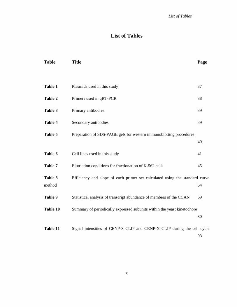
List of Tables
Table Title Page
Table 1 Plasmids used in this study 37
Table 2 Primers used in qRT-PCR 38
Table 3 Primary antibodies 39
Table 4 Secondary antibodies 39
Table 5 Preparation of SDS-PAGE gels for western immunoblotting procedures
40
Table 6 Cell lines used in this study 41
Table 7 Elutriation conditions for fractionation of K-562 cells 45
Table 8 Efficiency and slope of each primer set calculated using the standard curve
method 64
Table 9 Statistical analysis of transcript abundance of members of the CCAN 69
Table 10 Summary of periodically expressed subunits within the yeast kinetochore
80
Table 11 Signal intensities of CENP-S CLIP and CENP-X CLIP during the cell cycle
93
x
Abbreviations
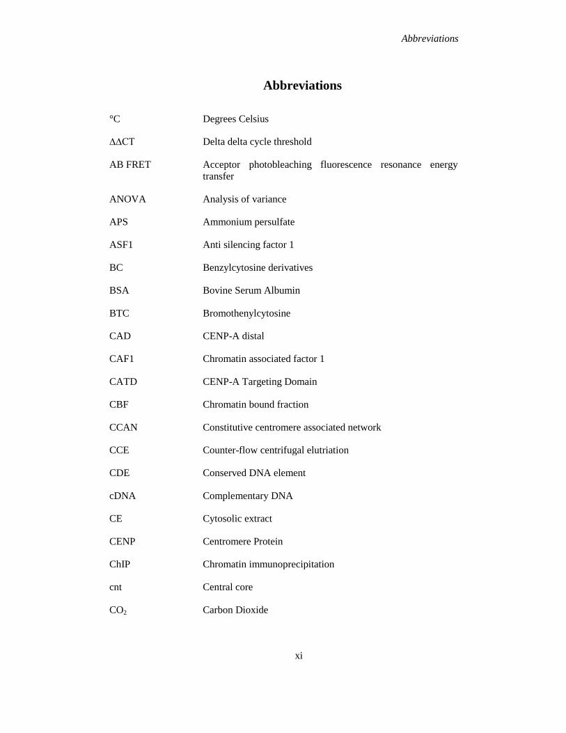
Abbreviations
°C Degrees Celsius
∆∆CT Delta delta cycle threshold
AB FRET Acceptor photobleaching fluorescence resonance energy
transfer
ANOVA Analysis of variance
APS Ammonium persulfate
ASF1 Anti silencing factor 1
BC Benzylcytosine derivatives
BSA Bovine Serum Albumin
BTC Bromothenylcytosine
CAD CENP-A distal
CAF1 Chromatin associated factor 1
CATD CENP-A Targeting Domain
CBF Chromatin bound fraction
CCAN Constitutive centromere associated network
CCE Counter-flow centrifugal elutriation
CDE Conserved DNA element
cDNA Complementary DNA
CE Cytosolic extract
CENP Centromere Protein
ChIP Chromatin immunoprecipitation
cnt Central core
CO2 Carbon Dioxide
xi
Abbreviations
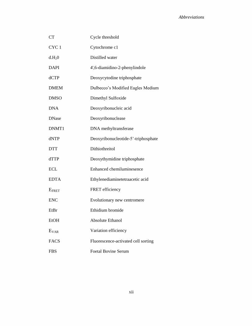
CT Cycle threshold
CYC 1 Cytochrome c1
d.H20 Distilled water
DAPI 4',6-diamidino-2-phenylindole
dCTP Deoxycytodine triphosphate
DMEM Dulbecco’s Modified Eagles Medium
DMSO Dimethyl Sulfoxide
DNA Deoxyribonucleic acid
DNase Deoxyribonuclease
DNMT1 DNA methyltransferase
dNTP Deoxyribonucleotide-5’-triphosphate
DTT Dithiothreitol
dTTP Deoxythymidine triphosphate
ECL Enhanced chemiluminesence
EDTA Ethylenediaminetetraacetic acid
EFRET FRET efficiency
ENC Evolutionary new centromere
EtBr Ethidium bromide
EtOH Absolute Ethanol
EVAR Variation efficiency
FACS Fluorescence-activated cell sorting
FBS Foetal Bovine Serum
xii
Abbreviations
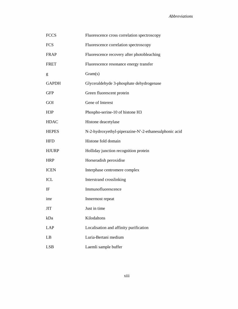
FCCS Fluorescence cross correlation spectroscopy
FCS Fluorescence correlation spectroscopy
FRAP Fluorescence recovery after photobleaching
FRET Fluorescence resonance energy transfer
g Gram(s)
GAPDH Glyceraldehyde 3-phosphate dehydrogenase
GFP Green fluorescent protein
GOI Gene of Interest
H3P Phospho-serine-10 of histone H3
HDAC Histone deacetylase
HEPES N-2-hydroxyethyl-piperazine-N′-2-ethanesulphonic acid
HFD Histone fold domain
HJURP Holliday junction recognition protein
HRP Horseradish peroxidise
ICEN Interphase centromere complex
ICL
IF
Interstrand crosslinking
Immunofluorescence
imr Innermost repeat
JIT Just in time
kDa Kilodaltons
LAP Localisation and affinity purification
LB Luria-Bertani medium
LSB Laemli sample buffer
xiii
Abbreviations
mAb Monoclonal antibody
MeOH Methanol
MGMT Methylguanine-DNA methyltransferase
mL Millilitre
mM Millimolar
Mnase Micrococcal Nuclease
mRNA messenger RNA
Na Pyr Sodium Pyruvate
NAC Nucleosome associated complex
NaCl Sodium chloride
NE Nuclear extract
NEAA Non-essential amino acids
nm Nanometre(s)
ns Non-significant
otr Outer repeat
PBS Phosphate Buffered Saline
PBS-T PBS-Tween
PCNA Proliferating Cell Nuclear Antigen
PCR Polymerase chain reaction
PEV Position Effect Variegation
PFA Paraformaldehyde
PI Propidium Iodide
PS Penicillin-Streptomycin
xiv
Abbreviations
#PTM Post-translational modification
PVDF Polyvinyllidene fluoride
qRT-PCR Quantitative Real-time PCR
RCF/ rcf Relative centrifugal force
RFP Red fluorescence protein
RNA Ribonucleic acid
RNAi RNA interference
RNase Ribonuclease
ROI Region of interest
RPM/ rpm Revolutions per minute
RQ Relative quantification
RT Room temperature
RT-PCR Reverse transcription PCR
S.D Standard deviation
SAC Spindle Assembly Checkpoint
SDS Sodium Dodecyl Sulfate
SDS-PAGE SDS-polyacrylamide gel electrophoresis
SEM Standard Error of the Mean
SGD Saccharomyces genome database
siRNA Small interfering RNA
TAE Tris-acetate-EDTA Buffer
TAP Tandem affinity purification
TE Trypsin-EDTA
TEMED N, N, N', N'-tetramethylethylenediamine
xv
Abbreviations
xvi
TG Tris-Glycine
Trichostatin A TSA
Tris Tris(hydroxymethyl)aminomethane
UV Ultraviolet
V
WB
Volts
Western blot
WT Wild type
Abstract
Abstract
Centromeres are the chromosomal loci which are responsible for the equal segregation of sister
chromatids to daughter cells during mitosis, by directing the assembly of kinetochores. Despite
being involved in the faithful inheritance of the genome, centromeres are themselves
epigenetically determined. Centromeres of higher organisms are determined by their
centromeric chromatin which consists of unique nucleosomes, in which canonical histone H3 is
replaced by the histone H3 variant CENP-A. A large group of additional proteins associate with
the centromere throughout the cell cycle, collectively known as the constitutive centromere
associated network (CCAN), and these are required for kinetochore function. In this thesis I
have investigated the regulation and assembly of these CCAN proteins in human cells.
Chapter 3 focuses on the regulation of the abundance of CCAN components during the cell
cycle. Our analysis of the relative transcript abundance of CCAN members using real-time PCR
did not reveal strong regulation of gene expression for the CCAN as a whole. However our
results were suggestive of the centromere and kinetochore being regulated in a "just-in-time"
manner, with the robust periodic expression of CENP-C and CENP-E and –F during G2/M,
making them ideal candidates to act as the “pacesetters” for assembly of the inner and outer
kinetochore respectively.
Chapter 4 examines the assembly and inheritance of the histone fold domain proteins CENP-S
and CENP-X at centromeres. Both proteins were found to assemble late in the cell cycle during
late S/G2 phase similar to the CENP-T/-W, and to exhibit multi-generational persistence. FCCS
and FRET analysis confirmed that CENP-S and CENP-X form a complex with one another. In
addition we found CENP-S and CENP-T to be in close proximity to each other using FRET.
These results with CENP-S/-X are consistent with the complex playing a role in kinetochore
assembly before cells enter mitosis, coordinate with CENP-T/-W.
xvii
Acknowledgements
Acknowledgements
I would sincerely like to thank a number of people for the help, support and understanding that I
have received during the course of my Ph.D.
Firstly, I wish to thank my project supervisor, Professor Kevin Sullivan, for giving me the
opportunity to join his research group. His guidance, support and encouragement during this
research have been invaluable. I wish also to thank Dr. Michael P. Carty (Head of Discipline)
and Dr. Heinz Peter Nasheuer (Head of School) for allowing me access to all the facilities
available in the department of Biochemistry. Many thanks also to all the research and technical
staff in Biochemistry who ensured our lab ran smoothly. Thank you to all members of the CCB
for their support and advice over the years.
Many thanks to Professor Stephan Diekmann and all members of his research group, at the
Leibniz Institute for Age Research, especially Carsten and Christian, for their collaborative
efforts with the FRAP, FRET and FCCS experiments.
To all the members of the Sullivan group- I cannot even begin to tell you all how much I have
enjoyed your company, friendship, tea-breaks and nights in The Roisin over the years! A sincere
thank you to Mac and Lisa, our two wonderful post-docs, for your constant support and
supervision during the course of my experimental work. Thank you to Chelly for all her help
with the microscope and for always cheering me up with her smile! And last but by no means
least, a huge thank you to Aga “my partner in crime”. I could never have done this without all
the laughs, tears and jagerbombs! Special thank you to all members of the Lahue Lab for the
reagents (including chocolate supplies) and laughs over the years.
Thank you to my fabulous friends and housemates, Aisling, Deborah and Sarah. I honestly
would not have made it through the last four years without all of you. Thank you for your
encouragement through the years as well as the much needed wine nights and not-so scientific
chats!
I wish to extend a sincere and heartfelt thank you to my parents, Gary and Agnes, and brothers,
Stephen and Andrew, without your love, support and continuous encouragement, I certainly
xviii

Acknowledgements
would not be here today. A special thank you to my boyfriend Tomás for his patience and
encouragement during the course of my Ph.D. and for always being there.
xix

Chapter 1
Introduction
Chapter 1
1.1 Overview
The eukaryotic cell cycle involves a series of controlled events which are responsible for the
faithful duplication of the entire genome and its equal distribution to daughter cells to ensure the
maintenance of genome stability (Norbury and Nurse, 1992). In eukaryotes the cell cycle can be
divided into two distinct stages: interphase and mitosis. The mitotic phase consists of mitosis
and cytokinesis. Mitosis can be sub-divided into 5 different stages. Prophase is the first stage of
mitosis during which the replicated chromosomes condense and centrosomes move to opposite
sides of the cell to organise the spindle microtubules. Cells then enter prometaphase during
which the nuclear envelope breaks down, and the microtubules elongate and attach kinetochores.
Tension is then applied by these spindles and the chromosomes bi-orient during metaphase. The
spindles then shorten and sister chromatids begin to move towards opposite poles of the cell
during anaphase. Following anaphase, cells enter telophase and the sister chromatids arrive at
opposite poles of the cell. During cytokinesis, the cytoplasm and cell membrane are divided
between the daughter cells. Interphase consists of three phases: gap-1 phase, synthesis phase and
gap-2 phase. Following mitosis, cells enter the first gap phase (G1), where they prepare for
DNA replication, cells then proceed into the synthesis phase (S phase) where DNA replication
occurs. Following S phase, cells enter G2 phase and prepare for mitosis.
Centromeres are the chromosomal loci which are responsible for the correct segregation of sister
chromatids during mitosis (Cleveland et al., 2003). During G2 phase of the cell cycle,
kinetochores assemble upon centromeres, and these proteinaceous structures serve as the
attachment point for microtubules on each sister chromatid. Kinetochores mediate the
microtubule dependent movement of chromosomes during mitosis (Cheeseman and Desai,
2008). Kinetochores also function in sensing errors in chromosome attachments and activate the
spindle assembly checkpoint (SAC), which delays cells at the metaphase-anaphase transition
until correct bipolar attachment is achieved (Carroll and Straight, 2006).
1
Chapter 1
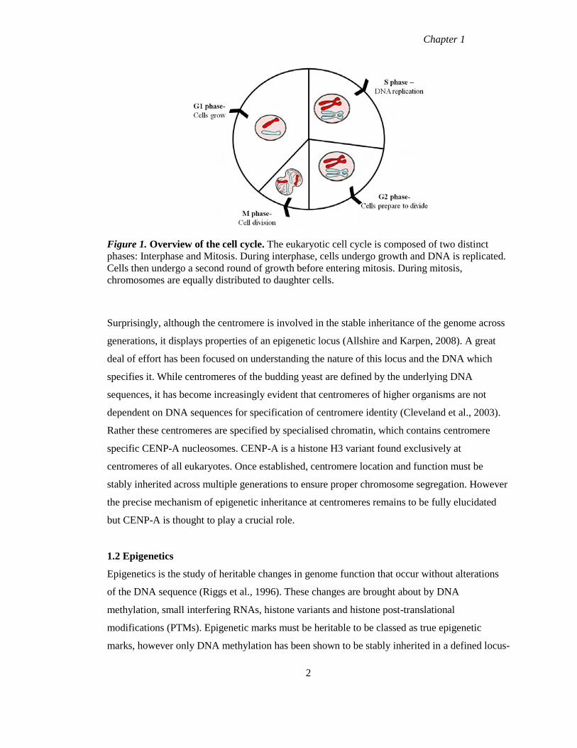
Figure 1. Overview of the cell cycle. The eukaryotic cell cycle is composed of two distinct
phases: Interphase and Mitosis. During interphase, cells undergo growth and DNA is replicated.
Cells then undergo a second round of growth before entering mitosis. During mitosis,
chromosomes are equally distributed to daughter cells.
Surprisingly, although the centromere is involved in the stable inheritance of the genome across
generations, it displays properties of an epigenetic locus (Allshire and Karpen, 2008). A great
deal of effort has been focused on understanding the nature of this locus and the DNA which
specifies it. While centromeres of the budding yeast are defined by the underlying DNA
sequences, it has become increasingly evident that centromeres of higher organisms are not
dependent on DNA sequences for specification of centromere identity (Cleveland et al., 2003).
Rather these centromeres are specified by specialised chromatin, which contains centromere
specific CENP-A nucleosomes. CENP-A is a histone H3 variant found exclusively at
centromeres of all eukaryotes. Once established, centromere location and function must be
stably inherited across multiple generations to ensure proper chromosome segregation. However
the precise mechanism of epigenetic inheritance at centromeres remains to be fully elucidated
but CENP-A is thought to play a crucial role.
1.2 Epigenetics
Epigenetics is the study of heritable changes in genome function that occur without alterations
of the DNA sequence (Riggs et al., 1996). These changes are brought about by DNA
methylation, small interfering RNAs, histone variants and histone post-translational
modifications (PTMs). Epigenetic marks must be heritable to be classed as true epigenetic
marks, however only DNA methylation has been shown to be stably inherited in a defined locus-
2
Chapter 1
associated manner across cell generations (Wigler et al., 1981). The process of DNA replication
causes the disruption of both the genetic and epigenetic information as the chromatin must be
disassembled and then reassembled following passage of the replication fork. While a great deal
of information is known regarding the inheritance of the DNA based genetic material, the
processes involved in the inheritance of the non DNA based epigenetic material remains unclear.
1.2.1 Inheritance of DNA methylation during DNA replication
DNA replication occurs in an asymmetrical fashion, with continuous synthesis on the leading
strand but discontinuous synthesis on the lagging strand (Probst et al., 2009). The synthesis is
catalysed by DNA polymerases (Kunkel and Burgers, 2008) and these are assisted by the DNA
processivity factor, proliferating cell nuclear antigen (PCNA) on both strands (Moldovan et al.,
2007). As well as acting as the link between the leading and lagging strand, PCNA is thought to
link DNA synthesis and the inheritance of epigenetic marks as suggested by experiments in
Drosophila that found mutations in PCNA caused suppression in position effect variegation
(PEV) (Henderson et al., 1994). PCNA has also been shown to interact with chromatin assembly
and modifying factors (Groth et al., 2007). DNA methylation has been shown to be stably
inherited in a similar fashion to the semi conservative inheritance of DNA, as result of the
symmetrical pattern of DNA methylation at CpG sites on both DNA strands (Probst et al.,
2009). DNA methylation is maintained at the replication fork by the DNA methyltransferase 1
(DNMT1) as a result of its affinity for hemi-methylated DNA (Hermann et al., 2004), as well as
its interaction with PCNA (Chuang et al., 1997). The mechanism which ensures the maintenance
of methylation remained unclear until the discovery of the protein NP95, which provided a
mechanistic link between hemi-methylated DNA and DNMT1 (Sharif et al., 2007). NP95
preferentially binds to hemi-methylated DNA and also interacts with DNMT1. Depletion of
NP95 caused methylation defects similar to those observed following depletion of DNMT1 (Li
et al., 1992), suggesting NP95 plays a role in tethering maintenance methyltransferase activity to
the newly replicated DNA (Probst et al., 2009). Together these mechanisms ensure the stable
inheritance of DNA methylation patterns, which is necessary for DNA methylation to function
as an epigenetic mark.
3

Chapter 1
1.2.2 Inheritance of histones and their post-translational modifications
As previously discussed, the inheritance of DNA methylation involves a semi-conservative
mechanism with the hemi-methylated DNA serving as the template. During DNA replication
nucleosomes and their modifications must be disassembled ahead of the replication fork and
reassembled behind it, however no obvious template has been found for this reassembly process.
H3 and H4 have emerged as the most likely candidates to carry information across generations,
as unlike H2A and H2B, H3 and H4 exhibit very little exchange outside of S phase (Jackson and
Chalkley, 1981, Kimura and Cook, 2001). It has therefore been suggested that nucleosome
inheritance involves the coordination of assembly of parental H3:H4 and their modifications
with the incorporation of newly synthesised histones (Groth et al., 2007).
Nucleosome reassembly after replication involves the deposition of parental histones on both
daughter strands, with H3:H4 deposited before the H2A:H2B dimers (Campos and Reinberg,
2009). H3:H4 was always thought to be deposited as a tetramer, however more recent evidence
has emerged for the existence of H3:H4 dimers which are associated with the histone chaperone
anti silencing factor 1 (ASF1) (Campos and Reinberg, 2009, Tagami et al., 2004). Histone
chaperones are associated with soluble histone complexes, and these are involved in the
deposition/assembly of newly synthesised nucleosomes as well as in the disruption/reassembly
that occurs during nucleosome exchange reactions. CAF1 (chromatin associated factor 1) is a
chaperone specific for H3:H4 and is recruited to the replication fork through its interaction with
PCNA and other chromatin modifiers including histone deacetylases (HDACs) (Zhang et al.,
2000, Shibahara and Stillman, 1999). ASF1 is another H3:H4 chaperone, and this directly
interacts with a subunit of CAF1, together these chaperones function in replication coupled
nucleosome assembly. HIRA is a histone chaperone specific for the histone variant H3.3 and
functions in the DNA synthesis-independent nucleosome assembly pathway (Tagami et al.,
2004, Ray-Gallet et al., 2002). Newly synthesised histones have also been found to be modified
post translation (acetylation of K5 and K15 on H4) before deposition and this acetylation is
removed upon nucleosome maturation (Loyola et al., 2006, Sobel et al., 1995).
Two models have been suggested to explain how parental histones with their modifications and
newly synthesised histones are distributed to daughter strands following DNA replication. The
first model suggests that this distribution occurs in a random fashion. This mode of distribution
would cause dilution of the PTMs and the distribution of PTMs relative to DNA sequences
4
Chapter 1
would also be modified (Margueron and Reinberg, 2010, Probst et al., 2009). Random
distribution would only be an effective mode for transmitting histone PTMs if they were
enriched on several adjacent nucleosomes, whereas those PTMs which were present in few
copies would get diluted which would result in the loss of this PTM’s influence on chromatin
(Margueron and Reinberg, 2010, Probst et al., 2009). The second model suggests that parental
H3:H4 molecules are equally distributed to both strands following DNA replication in a semi-
conservative manner (Margueron and Reinberg, 2010, Probst et al., 2009). The propagation of
the histone modifications would require machinery to duplicate marks either between
corresponding nucleosomes (if H3:H4 deposited as a dimer) or between adjacent nucleosomes
(if deposited as a tetramer) (Probst et al., 2009). While the precise mechanism of histone
segregation remains to be elucidated, it is clear that following replication the chromatin only
contains part of the epigenetic information and this must to be rapidly duplicated to ensure the
stability of chromatin structures. Whatever the mechanism for distribution of the nucleosomes to
daughter strands, the parental histone modification is proposed to be recognised by a chromatin
binding protein, and this protein in turn recruits a chromatin modifying enzyme (Probst et al.,
2009). Therefore the histone modifications on parental histones serve as the template for
modifying the newly incorporated histones.
PCNA plays a role in the inheritance of these modifications, as it has been found to recruit
chromatin modifying proteins to replication forks (Moldovan et al., 2007). PCNA along with
CAF1 remains at the replication fork for approximately 20 minutes, during which time newly
replicated chromatin, undergoes PTMs including the removal of K5 and K12 acetylation from
newly synthesised H4 (Sobel et al., 1995, Loyola et al., 2006). The methyl CpG binding protein
(MBD1) was found to interact with CAF1 during replication at regions enriched in DNA
methylation (Reese et al., 2003), which links histone deposition with establishment of a
modification on newly incorporated histones, while DNMT1 was found to interact with histone
deacetylases (Fuks et al., 2000, Rountree et al., 2000). These results indicate that propagation of
PTMs of newly incorporated histones at regions enriched in DNA methylation could be
facilitated by DNA methylation machinery (Probst et al., 2009). While these mechanisms help to
explain the inheritance of replication dependent histones, the mechanisms involved in the
inheritance of replication independent histones, including CENP-A, remain unclear.
5
Chapter 1
1.3 Specifying centromere identity
1.3.1 Centromeric DNA
The size and composition of centromeric DNA varies among different eukaryotes (Cleveland et
al., 2003). The simplest centromere is found in S. cerevisiae and is termed a point centromere.
These are between 116 and 120bp in length and comprised of three conserved DNA elements
(CDE); CDEI (8bp), the AT-rich CDEII (78-86bp) and CDEIII (26bp) (Fitzgerald-Hayes et al.,
1982). The CDEII region is the site at which CENP-ACse4
containing nucleosome is assembled
(Stoler et al., 1995), while the CBF3 complex binds to the CDEIII region and is required for
targeting of CENP-ACse4
nucleosomes to centromeres (Gardner et al., 2001). Regional
centromeres are associated with a discrete chromosomal locus and are found in a variety of
organisms from fission yeast to humans. The centromere of fission yeast is comprised of a
central core (cnt) which is flanked by inverted inner repeats (imr) and outer repeats (Pluta et al.,
1995, Clarke, 1998). CENP-ACnp1
nucleosomes are found in the central core and surrounded by
H3 containing regions. Regional centromeres of higher eukaryotes are composed of long
stretches of tandemly arranged repetitive DNA sequences which span hundreds to thousands of
kilobase pairs (Lee et al., 1997). Human centromeres are typical of metazoans and built upon
AT-rich complex satellite repeat DNA termed alpha satellite in primates, originally identified
from the African green monkey (Maio, 1971, Gruss and Sauer, 1975). Each chromosome is
characterised by a higher order array of alpha satellite DNA, which is composed of hundreds to
thousands of the 171 base-pair repeat subunit which can span over 3-5 Mb (Willard, 1985). This
higher order structure observed in vertebrates is also present in plants, which is surprising
considering how divergent the two groups are from each other. The centromere of Arabidopsis
consists of 178bp repeat of AT-rich satellite DNA. However despite the structural similarity
centromeric DNA is evolving rapidly even among closely related species (Malik and Henikoff,
2002). The holocentric centromeres of nematodes are more unusual, in that the centromere is
diffuse along the length of the chromosome (Maddox et al., 2004).While centromere function in
budding yeast is specified by the underlying DNA sequences this is not the case for higher
eukaryotes. In higher eukaryotes only a small 17bp binding site has been found to be conserved
for the sequence specific binding protein CENP-B (Masumoto et al., 2004). While mutations in
this binding site caused reduced efficiency in the formation of artificial centromeres, it is a non
essential protein and it does not play a role in the maintenance of centromeres
6
Chapter 1
(Jun-ichirouOhzeki, 2002, Okada et al., 2007). This coupled with the apparent lack of
conservation of centromeric DNA sequences suggests that epigenetics may play a role in
centromere specification.
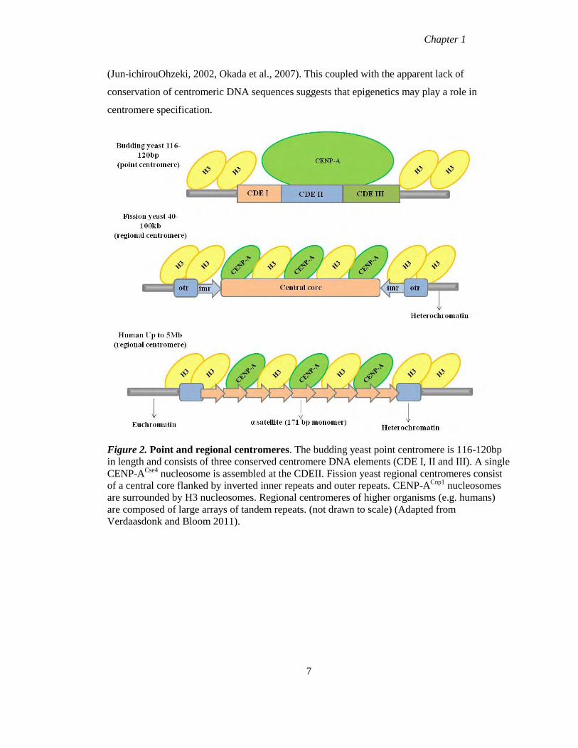
Figure 2. Point and regional centromeres. The budding yeast point centromere is 116-120bp
in length and consists of three conserved centromere DNA elements (CDE I, II and III). A single
CENP-ACse4
nucleosome is assembled at the CDEII. Fission yeast regional centromeres consist
of a central core flanked by inverted inner repeats and outer repeats. CENP-ACnp1
nucleosomes
are surrounded by H3 nucleosomes. Regional centromeres of higher organisms (e.g. humans)
are composed of large arrays of tandem repeats. (not drawn to scale) (Adapted from
Verdaasdonk and Bloom 2011).
7
Chapter 1
1.3.2 Epigenetic behaviour at centromeres
The many examples of epigenetic behaviour at centromeres support a model in which
epigenetics plays an important role in the stable inheritance of centromeres. Position effect
variegation (PEV) has been observed at centromeres and causes the inheritance of differential
expression states of genes inserted close to heterochromatin (Ekwall, 2007). In fission yeast
PEV has been demonstrated to play a role in centromeric silencing of nearby genes, with
mutations of the centromere protein Swi6 (heterochromatin protein 1 homologue) effecting
centromeric silencing (Ekwall et al., 1995).
Centromere inactivation/silencing is another epigenetic feature of centromeres. In humans,
Robertsonian translocations (which occur as result of fusion of the long arms of non-
homologous arcocentric chromosomes) results in dicentric chromosomes which are stably
inherited (Earnshaw and Migeon, 1985). In some cases these dicentrics are close enough to each
other that both centromeres can functionally cooperate to ensure mitotic stability (Sullivan and
Willard, 1998). Many examples of dicentric chromosomes exist however which comprise of one
functionally active centromere while the other has been silenced through centromere
inactivation and this no longer recruits centromere proteins (Earnshaw and Migeon, 1985,
Sullivan and Schwartz, 1995)
Neocentromeres are ectopic centromeres that form on non-centromeric DNA which lack α
satellite DNA, suggesting that centromere formation is determined by epigenetic mechanisms
and does not depend strictly on the primary DNA sequence (Lo et al., 2001). These
neocentromeres contain all the necessary centromere proteins including CENP-A, required to
form a fully functioning kinetochore (Warburton, 2004). Approximately 90 neocentromeres
have been identified in humans by cytologists to date, with examination of their genetic location
revealing that they are found on 21 different chromosomes (Alonso et al., 2010). Some
chromosomes show a higher frequency than others for neocentromere formation e.g.
chromosome 13q has 16 described cases of neocentromeres, however analysis of these sites of
neocentromere formation did not detect any sequence similarity (Alonso et al., 2003). Typically
human neocentromeres occur in association with chromosomal rearrangement and karyotypic
imbalance which result in birth defects or developmental delays (Amor and Choo, 2002).
8
Chapter 1
Centromere repositioning is another apparently epigenetic phenomenon associated with
vertebrate karyotype evolution. Unlike with neocentromeres, centromere repositioning occurs
without chromosomal rearrangements and does not result in aberrant phenotypes (Amor et al.,
2004). It was first identified in primate evolution as changes in centromere position that are not
associated with apparent chromosome rearrangement (Montefalcone et al., 1999). Subsequently,
centromere repositioning and the appearance of so-called evolutionarily new centromeres
(ENCs) have been observed in other vertebrates and plants (Marshall et al., 2008a). In these
examples centromeric satellite DNA is observed at ENCs, prompting the hypothesis that
centromere activity first relocates as observed for neocentromeres and later the centromeric
satellite DNA is recruited to the site. A remarkable phenomenon that may be analogous to the
early stages of centromere repositioning has been described in one family, with a centromere on
chromosome 4 relocating from the alpha satellite DNA to a non-centromeric location (Amor et
al., 2004). As result of this repositioning, a pseudodicentric chromosome was formed with an
active neocentromere and an inactive alphoid centromere (Amor et al., 2004).
Further evidence for epigenetics playing a role in centromere specification was demonstrated in
the horse. Sequencing of the horse genome revealed the unexpected feature of an evolutionary
new centromere (ENC) on chromosome 11 (ECA11) (Wade et al., 2009). As previously
outlined, mammalian centromeres are usually assembled upon satellite tandem repeats. The
centromere of ECA11was found in a chromosomal region which is conserved among mammals
but the horse is the only species to have a centromere in this location, suggesting it is
evolutionarily new (Wade et al., 2009). This centromere was also the only centromere of the
horse to lack any hybridisation signal with horse satellite sequences, indicating that the ENC has
not yet acquired its satellite DNA which further indicates it is an evolutionarily new centromere.
ECA11 was also demonstrated to possess centromeric function, this region of chromosome 11
immunoprecipiated with antibodies against CENP-A and CENP-C (Wade et al., 2009). Taken
together these results implicate an epigenetic mechanism in specifying centromere identity and
function and do not involve the primary DNA sequences.
Further analysis of 4 additional members of the Equus species and the distribution of their
satellite tandem repeats revealed that several centromeres lack satellite DNA and satellite DNA
was found at non-centromeric termini (Piras et al., 2010).
9
Chapter 1
Using two major satellite families from the Equus species, they found that both forms of satellite
DNA are found at centromeric locations in all chromosomes of the horse apart from
chromosome 11. However in the other species (donkey and two zebra species), several
centromeres lacked any detectable satellite DNA while satellite DNA was present at non
centromeric locations on some chromosomes (Piras et al., 2010). These data indicate that like
ECA11 of the horse, the function of centromeres in donkey and zebra can be uncoupled from
satellite DNA (Piras et al., 2010). The absence of satellite DNA at some centromeres and its
presence at non-centromeric sites is in agreement with previous reports that suggested several
repositioning events occurred during the evolution of the Equus species (Carbone et al., 2006,
Piras et al., 2009). These functional centromeres which are devoid of satellite DNA provide an
excellent model to study the epigenetic factors which are responsible for specifying centromere
identity and function.
Sequencing of the orang-utan genome revealed the presence of a neocentromere on chromosome
12 (Locke et al., 2011). Previously this had been described as a polymorphic chromosome
(Seuanez et al., 1976); surprisingly however, fluorescence in-situ hybridisation (FISH)
demonstrated that both forms of the chromosome showed no difference in marker order despite
the different centromere positions (Locke et al., 2011). Chromatin immunoprecipitation with
antibodies against CENP-A and CENP-C confirmed centromere function despite the lack of
alpha satellite DNA (Locke et al., 2011).
An experimental indication of the sensitivity of centromeres to epigenetic events comes from the
analysis of histone deacetylases (HDACs). Inhibitors of HDACs have been used to demonstrate
that histone acetylation controls the epigenetic state of centromeres in S. pombe (Ekwall, 1997).
In human cells experiments with Trichostatin A (TSA) (inhibitor of HDACs) have suggested a
role for histone acetylation in neocentromere formation (Craig et al., 2003). These examples of
epigenetic behaviour coupled with the lack of conservation of centromeric DNA sequence
among higher eukaryotes suggest that centromere function is not determined primarily by the
underlying DNA sequences but through epigenetic mechanisms.
10
Chapter 1
1.4 Chromatin and centromere determination
As previously outlined, the DNA sequences of eukaryotic centromeres varies greatly through
evolution. One common feature of all centromeres is the presence of the histone H3 variant,
CENP-A. CENP-A is the foundation upon which the centromere is assembled and is thought to
be the epigenetic mark which specifies centromere identity and function.
1.4.1 CENP-A
CENP-A was originally identified as an autoantigen recognised by anti-centromere antibodies in
patients with limited systemic sclerosis (CREST) along with CENP-B and CENP-C (Earnshaw
and Rothfield, 1985). CENP-B was shown to associate with alpha satellite DNA (Masumoto et
al., 1989) while CENP-C was identified as a component of the inner kinetochore (Saitoh et al.,
1992). CENP-A was isolated with nucleosomes following MNase digestion (Palmer and
Margolis, 1985) and found to co-purify with core nucleosome components (Palmer et al., 1987).
Analysis of partial amino acid sequence of CENP-A suggested that it shared homology with
histone H3 (Palmer et al., 1991) and this was confirmed by isolation of full length cDNA for
human CENP-A (Sullivan et al., 1994). The C terminal domain of CENP-A which contains the
histone fold domain (HFD) shares approximately 60% homology with histone H3, while the N
terminal domain does not share any homology (Sullivan et al., 1994). This C terminal HFD is
comprised of three alpha helices separated by loops, and it is through these helices that CENP-A
and histone H4 form tetramers. The HFD of CENP-A was also shown to be necessary and
sufficient for targeting of the protein to centromeres (Sullivan et al., 1994). The region within
the HFD of CENP-A responsible for targeting the protein was identified as the CENP-A
targeting domain (CATD) (Shelby et al., 1997, Black et al., 2004). The CATD is composed of a
22 amino acid region within the classic HFD of histone CENP-A and when this was substituted
to H3, the resulting H3CATD
chimeric protein was targeted to centromeres and capable of
maintaining centromere function in the absence of CENP-A (Black et al., 2007, Black et al.,
2004).The (CENP-A: H4)2 tetramer is more compact and rigid than that of the (H3:H4)2
tetramer, a property which is conferred by the CATD of CENP-A (Black et al., 2007).
11
Chapter 1
1.4.2 CENP-A tetramers
Physical differences between (H3:H4)2 tetramers and (CENP-A: H4)2 tetramers reveal that
CENP-A generates unique chromatin features which may physically mark centromere locations.
The crystal structure of CENP-A containing tetramers revealed three important physical
differences between CENP-A and H3 containing tetramers, all of which are mediated by amino
acid substitutions specific to CENP-A within the CATD (Sekulic et al., 2010). An overlay of
CENP-A containing tetramer with a H3 containing tetramer revealed that the CENP-A-CENP-A
interface is rotated relative to the H3-H3 interface. This rotation results in differences in the
relative positioning of residues within the tetramer which could cause compaction within the
nucleosome (Sekulic et al., 2010). The crystal structure of CENP-A tetramers also revealed an
extended positively charged bulge at the N terminal of the L1 loop of CENP-A which is not
present on H3 (Sekulic et al., 2010). This part of the tetramer remains exposed at the surface
following nucleosome assembly and is the site of many PTMs. This coupled with the amino acid
substitutions including replacing an aspartate with a lysine and the addition of an arginine
generate a shape and charge of CENP-A which is dramatically different from canonical H3
(Sekulic et al., 2010). Finally CENP-A tetramers are more rigid than their H3 counterparts. This
was first observed in hydrogen/deuterium exchange mass spectrometry (X/DX-MS), which
revealed that (CENP-A: H4)2 tetramers are protected from exchange which is consistent with
conformational rigidity (Black et al., 2004). Mapping of the CENP-A crystal structure onto the
H/DX-MS data revealed that the 10 fold slower exchange rates observed for CENP-A
containing tetramers in comparison to H3 occurred at the interface of the α2 helix of CENP-A
and the α2 and α3 helices of H4 (Sekulic et al., 2010). This increased rigidity is proposed to be
due to an increase in hydrophobicity in comparison to (H3:H4)2 tetramers (Sekulic et al., 2010).
The crystal structure of CENP-A revealed amino acid substitutions at either end of the CENP-A:
H4 interface, that result in the interface being stitched together with increased hydrophobicity
and this can explain the slow deuterium exchange observed for (CENP-A:H4)2 tetramers
(Sekulic et al., 2010). These data indicate that CENP-A may be involved in specifying
centromere location by physically altering the chromatin onto which it assembles.
Although the majority of studies carried out with CENP-A have focused on the C terminal of the
protein, some interest in the N terminal has emerged. Although the N terminal of CENP-A is not
12
Chapter 1
required for centromere targeting (Shelby et al., 1997), deletion of this tail of CENP-ACse4
of
budding yeast is lethal to cells (Morey et al., 2004).
1.4.3 Composition of centromeric nucleosomes
The composition of CENP-A nucleoprotein particles was long thought to be that of a classical
octamer with CENP-A replacing the histone H3, with several lines of evidence in several
species supporting this interpretation. In vitro assembly studies demonstrated that CENP-A can
replace histone H3 in octamers containing H4, H2A and H2B (Yoda et al., 2000, Furuyama et
al., 2006). Experiments carried out by Shelby et al (1997) demonstrated that epitope tagged
CENP-A co-immunoprecipitated in eqimolar amounts with endogenous CENP-A. This result
suggests that CENP-A self associates in vivo, indicating CENP-A forms homotypic nucleosomes
(Shelby et al., 1997). Tandem affinity purifications with CENP-A found CENP-A, H4, H2A and
H2B to be present in eqimolar amounts, however H3 nucleosomes were also present (Foltz et
al., 2006). These H3 nucleosomes could represent the blocks of H3 nucleosomes which are
interspersed with blocks of CENP-A nucleosomes (Blower et al., 2002) or CENP-A may form
heterotypic nucleosomes with one copy of CENP-A and H3 (Foltz et al., 2006).
The structural features of the CATD of CENP-A discussed above (Black et al., 2007, Black et
al., 2004) may be responsible for distinguishing CENP-A nucleosomes from H3 containing
nucleosomes. Recent studies have emerged suggesting that CENP-A nucleosomes exist in
different forms other than octamers including; tetrasomes (2 copies of CENP-A and H4),
hemisomes (1 copy of CENP-A, H4, H2A and H2B) or hexamers (nucleosomes in which H2A
and H2B have been replaced by Scm3). Studies carried out in Drosophila and humans have lead
to the suggestion that hemisomes are a key feature of centromeric chromatin (Dimitriadis et al.,
2010, Dalal et al., 2007). Atomic force microscopy of reconstituted centromeric nucleosomes
revealed centromeric chromatin was half the height of an octamer which suggests the existence
of CENP-A hemisomes in Drosophila cells, which would consist of 1 copy of CENP-A, H4,
H2A and H2B (Dimitriadis et al., 2010). Evidence that CENP-A could exist as a tetramer came
from both budding and fission yeast which found that histones H2A and H2B interacted very
weakly with centromeric DNA sequences (Mizuguchi et al., 2007, Williams et al., 2009). In
budding yeast CENP-A nucleosomes have been described in which H2A and H2B have been
replaced by the non-histone protein Scm3 to form a hexameric complex which consists of 2
copies of CENP-A, H4 and Scm3 (Mizuguchi et al., 2007). Surprisingly however, depletion
13
Chapter 1
of CENP-A and Scm3 did not restore H2A or H2B to comparable levels in other genomic
regions in either study as would be expected, suggesting that the absence of H2A or H2B may
be a feature of the method rather than the existence of a CENP-A tetramer. Contrary to these
results obtained from Mizuguchi et al (2007), other groups have co-purified CENP-A with H2A,
H2B and H4 in budding yeast and have reported that CENP-A octamers represent the major
form of CENP-A containing chromatin (Camahort et al., 2009). The precise nature of CENP-A
nucleosomes and the extent to which they are conserved across evolution remains to be fully
elucidated.
1.4.4 Centromeric chromatin
Although the size and sequence of centromeric DNA varies between species as previously
outlined, the composition of centromeric chromatin is quite similar between species. All
functional centromeres contain CENP-A nucleosomes upon which the kinetochore is built and
this chromatin is embedded in highly condensed heterochromatin which is characterised by
methylation of histone H3 on lysine 9 (H3K9me), and this heterochromatin is involved in
centromere chromatid cohesion (Bernard et al., 2001).
1.4.4.1 Heterochromatin
In S.pombe, this heterochromatin, as well as functioning in accurate segregation of
chromosomes, is thought to play a role in establishing centromeric chromatin (Folco et al.,
2008). Fission yeast, like Drosophila and humans, consist of regional centromeres, with a
central core of DNA sequences flanked on either side by heterochromatin (Pluta et al., 1995).
These regional centromeres consist of two distinct DNA domains; the outer repeats (otr) which
surround the central kinetochore domain (comprised of the innermost repeats (imr) and the
central core (cnt)). The heterochromatin is assembled on the repetitive otr sequences and
contains histones which are hypo-acetylated and methylated on lysine 9 of H3 (H3K9me2) by
the methyltransferase Clr4, which in turn provides a binding site for Swi6 (HP1 orthologue)
(Rea et al., 2000, Bannister et al., 2001). Swi6 then mediates the binding of cohesin to
centromeric heterochromatin which is turn facilitates accurate chromosome segregation by sister
centromere cohesion (Ekwall et al., 1995, Bernard et al., 2001).
The role of this heterochromatin in establishing CENP-ACnp1
chromatin at the central domain
was investigated in mini-chromosomes of S.pombe. Delivery of naked DNA template with
14
Chapter 1
centromere sequences (otr, imr and cnt sequences) into wild type cells resulted in the
establishment of CENP-ACnp1
chromatin and flanking heterochromatin (Folco et al., 2008).
Mutant strains such as Chlr4Δ, revealed that the H3K9me2 modification of the flanking
heterochromatin is essential for establishment of CENP-ACnp1
at the central domain but not for
the maintenance of the CENP-ACnp1
chromatin across generations (Folco et al., 2008). This
heterochromatin is maintained by non-coding transcripts from the otr, which are processed into
small interfering RNAs (siRNA) and are then incorporated into the RITS effector complex to
target homologous chromatin (Durand-Dubief and Ekwall, 2008). The RNA interference
(RNAi) pathway recruits Clr4 which leads to the H3K9me2 modification and Swi6 binding
(Ekwall, 2004). This RNAi pathway has also been found to play a role in the establishment of
CENP-ACnp1
chromatin de novo in S.pombe (Folco et al., 2008).
To examine the role of the RNAi pathway in establishment of CENP-ACnp1
chromatin, S. pombe
cells lacking two components of the RNAi pathway (chp1 and dcr1), were found to lack the
H3K9me2 modification of heterochromatin and therefore CENP-ACnp1
was absent from the
central domain (Folco et al., 2008). S.pombe cells with mutant Swi6 were demonstrated to lack
CENP-ACnp1
at the central domain (Folco et al., 2008). This result was surprising as these cells
produced siRNAs and the H3K9me2 modification of heterochromatin was present suggesting
that the RNAi pathway also plays a direct role in establishment of CENP-ACnp1
chromatin (Folco
et al., 2008).
While it appears that heterochromatin does in fact play a role in establishing CENP-ACnp1
chromatin de novo in S.pombe, it remains unclear whether this role is conserved.
Heterochromatin has been observed near neocentromeres in human cells, while CENP-B the
heterochromatin protein promotes de novo CENP-A chromatin on human artificial
chromosomes (Okada et al., 2007). ChIP on ChIP analysis of 3 neocentromeres however,
revealed a lack of significant domains of heterochromatin in two of the three neocentromeres
examined, which is in contrast to all other eukaryotic centromeres (Alonso et al., 2010).
Consistent with a lack of heterochromatin, neocentric sister centromeres separate prematurely as
compared with endogenous centromeres, suggesting that centromeric cohesion is not well
established at these neocentromeres (Alonso et al., 2010).
15
Chapter 1
1.4.4.2 Organisation of centromeric chromatin
The organisation of centromeric chromatin has been the subject of much investigation. One of
first models suggested that kinetochores are assembled from condensed functional subunits
which are arranged as discontinuous repeats along a continuous DNA fibre (Zinkowski et al.,
1991). Examination of stretched chromatin fibres revealed the presence of repetitive subunits as
detected by CREST antibodies arranged along centromeric DNA fibres which are interspersed
with DNA linkers (Zinkowski et al., 1991). Another study which examined extended chromatin
fibres in humans and Drosophila found similar results with blocks of CENP-A nucleosomes
interspersed with blocks of H3 nucleosomes (solenoid model) (Blower et al., 2002).
However a different model of centromeric chromatin organisation has been proposed by
Marshall et al (2008b). Using electron microscopy, they found that CENP-A occupies a
surprisingly discrete proportion of the constriction (2/3 of the length and 1/3 of the width and
height of the constriction) (Marshall et al., 2008b). More surprising however was the finding
that at the Mardel (10) neocentromere CENP-A occupies the same proportion of the constriction
despite the lower levels of CENP-A found at neocentromeres (Irvine et al., 2004). However if
the CENP-A of this neocentromere was arranged according to the repeat subunit along a linear
coiled DNA fibre as suggested by Zinkowski et al (1991) and Blower et al (2002), CENP-A
would only occupy 1/10 of the constriction length. Marshall et al (2008b) have proposed that the
cylindrical coiled DNA fibre, rather than being linear, is folded/looped back on itself multiple
times to form a higher order structure (Marshall et al., 2008b).
Ribeiro et al (2010) have proposed a sinusoidal patch model to explain centromeric chromatin
organisation based on the locations of CCAN proteins within the centromere. Super-resolution
microscopy revealed that CENP-C was positioned external to but in very close proximity with
CENP-A nucleosomes, with CENP-T then positioned external to CENP-C (Ribeiro et al., 2010).
The positioning of CENP-T relative to CENP-A nucleosomes coupled with the fact that CENP-
T is found in association with H3 nucleosomes (Hori et al., 2008a) means that the model which
proposes that CENP-A nucleosomes faces the outer kinetochore and H3 nucleosomes are
embedded within the centromere cannot be fully correct. Based on their results, Ribeiro et al
(2010) have proposed a new model for centromeric chromatin organisation with alternating
blocks of CENP-A and H3 nucleosomes folded into a planar sinusoidal patch, with several
patches stacked on top of each other and these are linked at both ends by heterochromatin.
16
Chapter 1
1.4.4.3 Histone modifications at centromeric chromatin
The H3 nucleosomes within the centromeric region have modifications which are distinct from
both euchromatin and heterochromatin and are referred to as “centrochromatin” (Sullivan and
Karpen, 2004). These H3 nucleosomes contain, H3K4me2 which is normally associated with
euchromatin but lack acetylation of H3 or H4 which is also a marker for euchromatin (Sullivan
and Karpen, 2004). They also lack H3K9me which is a marker of silent heterochromatin
(Sullivan and Karpen, 2004). A more recent study using super resolution microscopy found
H3K9me3 between CENP-A domains of chicken cells (Ribeiro et al., 2010). Using a non-
essential human artificial chromosome (HAC), Bergmann et al (2011) found a new
centrochromatin modification H3K36me2 which they also found at an endogenous centromere
on chromosome 21. This modification is normally associated with transcription elongation and
was found to span the centromere and overlap with CENP-A/H3K4me2 domains, suggesting
that centromeres are transcriptionally active (Bergmann et al., 2011). This study also found the
H3K4me2 modification plays a role in CENP-A maintenance (Bergmann et al., 2011).
Depletion of H3K4me2 at HAC using a histone demethylase caused a reduction of CENP-A
incorporation levels as result of loss of the CENP-A chaperone HJURP at the centromere,
suggesting the modification is involved in the recruitment of HJURP to centromeres (Bergmann
et al., 2011). These distinct patterns of histone modifications associated with centrochromatin
distinguish it from bulk chromatin, thereby facilitating the centromere specific deposition of
CENP-A.
1.5 Assembly of CENP-A and the constitutive centromere associated network
1.5.1 Assembly of CENP-A during the cell cycle
The assembly of centromeric chromatin does not occur during S phase like canonical histones
(Shelby et al., 2000). In human cells, CENP-A synthesis is not coupled with DNA replication,
with mRNA and protein accumulating during the G2 phase of the cell cycle suggesting a
separation of CENP-A and histone H3 assembly (Shelby et al., 1997, Shelby et al., 2000). SNAP
labelling, a novel conditional protein labelling method, was used to examine the timing of
assembly of CENP-A to centromeres (Jansen et al., 2007). This method allowed for the
examination of incorporation of newly synthesised CENP-A to centromeres. Although CENP-A
is normally synthesised during G2 phase of the cell cycle, a pulse labelled population was found
17
Chapter 1
to localise to centromeres only after passage through mitosis (Jansen et al., 2007). Assembly of
the newly synthesised CENP-A was first detected in late telophase cells and continued into G1
phase but was not seen at any other stage of the cell cycle (Jansen et al., 2007). Photobleaching
experiments provided further insight into the assembly dynamics of CENP-A. FRAP
(fluorescence recovery after photobleaching) revealed that the loading of CENP-A occurred
exclusively during late telophase/early G1 (Hemmerich et al., 2008). Fluorescent centromeres
which were bleached during late telophase/early G1phase, showed recovery after 30min with a
slow but steady increase observed over a 2 hour period. All bleached centromeres acquired new
CENP-A GFP, but the maximum recovery of signal intensity was 50% of pre-bleach signals,
indicating that CENP-A is incorporated without dynamic exchange of already loaded molecules
(Hemmerich et al., 2008). To assess the dynamics of CENP-A assembly during G1, sister
telophase cells were used to measure FRAP. All centromeres were bleached in one of the sister
cells and these recovered to ~50% of pre-bleach signal intensities following a 2 hour period.
These centromeres were then bleached for a second time but no recovery was observed.
Photobleaching of the second sister cell showed recovery, indicating that incorporation of
CENP-A was still ongoing. These results demonstrate that in human cells, CENP-A is
assembled late in telophase/early G1 without dynamic exchange (Hemmerich et al., 2008). A
complementary study was carried out using live cell microscopy and demonstrated that loading
of YFP tagged CENP-A began 10 minutes following anaphase and continued for 10 hours into
late G1 phase (Lagana et al., 2010). This study also identified MgcRacGAP (a Rho family
GTPase activating protein) to be involved in the maintenance of newly loaded CENP-A at
centromeres (Lagana et al., 2010).
While assembly of CENP-A in human cells has been demonstrated to require exit from mitosis,
CENP-ACID
of syncytial embryos of Drosophila is assembled at centromeres earlier in the cell
cycle during anaphase (these cells cycle between S and M and do not contain G1or G2 phases)
(Schuh et al., 2007). These data indicate that like in human cells, there is a separation of DNA
replication and assembly of CENP-ACID
to centromeres in Drosophila, and therefore
centromeres carry out their main functions of kinetochore assembly and chromosome
segregation with half the maximal CENP-A levels (Schuh et al., 2007). Further studies carried
out in S2 embryo cells of Drosophila, using similar methods to the conditional labelling method
used by Jansen et al (2007), found that CENP-ACID
assembly occurred during metaphase of
18
Chapter 1
mitosis (earlier than human cells) (Mellone et al., 2011). Examination of the assembly of
additional centromeric proteins CENP-C and CAL1 (both of which are essential for CENP-ACID
localisation) (Erhardt et al., 2008), showed that CENP-C assembly occurred at all stages of the
cell cycle while CAL1 assembled before CENP-ACID
in prophase of the cell cycle (Mellone et
al., 2011). These results coupled with the demonstration that CAL1 is required for CENP-ACID
deposition (Erhardt et al., 2008), indicate that CAL1 executes its function prior to new CENP-
ACID
deposition.
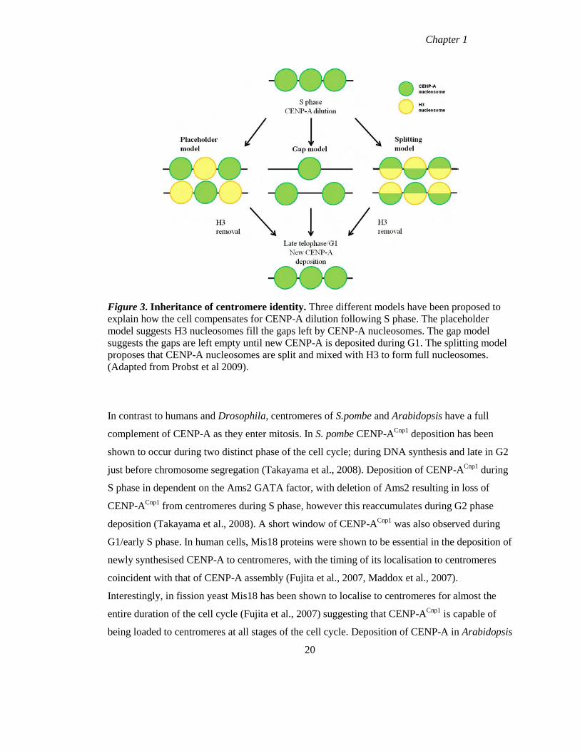
1.5.2 Inheritance of centromere identity
The timing of deposition of CENP-A in both humans and Drosophila results in the cell entering
mitosis with half the maximal CENP-A complement. Different models have been proposed to
explain how the cell compensates for the dilution of CENP-A during DNA replication. H3
nucleosomes have been suggested to temporarily replace the gaps left by CENP-A following
DNA replication, or the gaps left by CENP-A remain empty until new CENP-A is assembled
(Sullivan, 2001, Probst et al., 2009). Finally, parental CENP-A nucleosomes have been
suggested to split during DNA replication and then mix with H3 to form full nucleosomes
(Dunleavy et al., 2011). Experiments carried out by Dunleavy et al (2011), investigated the
composition of centromeric chromatin in S and G1 phases of the cell cycle on extended
chromatin fibres. These experiments demonstrated that both histone H3.1 and H3.3 localise
between CENP-A domains and within the CENP-A domains, with newly synthesised H3.1 and
H3.3 deposited during S phase (Dunleavy et al., 2011). If these H3 nucleosomes are acting as
“placeholders” to fill the gaps left by CENP-A, then their levels should decrease during G1
when CENP-A is assembled to centromeres. Examination of the newly incorporated H3.1 levels
at centromeric regions during G1 phase of the cell cycle revealed similar abundance as observed
during S phase; however the levels of H3.3 were reduced in G1 in comparison to S phase. These
results support a model in which H3.3 nucleosomes temporarily fill the gaps left by CENP-A
following DNA replication (Dunleavy et al., 2011). Consistent with H3.3 filling the gaps left by
CENP-A, CENP-ACnp1
mutants in fission yeast have increased levels of H3.3 while depletion of
CENP-ACID
in Drosophila results in H3 occupying a larger area of centromeric chromatin
(Castillo et al., 2007, Blower et al., 2002).
19
Chapter 1
Figure 3. Inheritance of centromere identity. Three different models have been proposed to
explain how the cell compensates for CENP-A dilution following S phase. The placeholder
model suggests H3 nucleosomes fill the gaps left by CENP-A nucleosomes. The gap model
suggests the gaps are left empty until new CENP-A is deposited during G1. The splitting model
proposes that CENP-A nucleosomes are split and mixed with H3 to form full nucleosomes.
(Adapted from Probst et al 2009).
In contrast to humans and Drosophila, centromeres of S.pombe and Arabidopsis have a full
complement of CENP-A as they enter mitosis. In S. pombe CENP-ACnp1
deposition has been
shown to occur during two distinct phase of the cell cycle; during DNA synthesis and late in G2
just before chromosome segregation (Takayama et al., 2008). Deposition of CENP-ACnp1
during
S phase in dependent on the Ams2 GATA factor, with deletion of Ams2 resulting in loss of
CENP-ACnp1
from centromeres during S phase, however this reaccumulates during G2 phase
deposition (Takayama et al., 2008). A short window of CENP-ACnp1
was also observed during
G1/early S phase. In human cells, Mis18 proteins were shown to be essential in the deposition of
newly synthesised CENP-A to centromeres, with the timing of its localisation to centromeres
coincident with that of CENP-A assembly (Fujita et al., 2007, Maddox et al., 2007).
Interestingly, in fission yeast Mis18 has been shown to localise to centromeres for almost the
entire duration of the cell cycle (Fujita et al., 2007) suggesting that CENP-ACnp1
is capable of
being loaded to centromeres at all stages of the cell cycle. Deposition of CENP-A in Arabidopsis
20
Chapter 1
was found to occur late in G2 phase of the cell cycle, and therefore while cells will not contain
the maximal complement of CENP-A as they progress from S phase onto G2, these levels will
be replenished before cells enter mitosis (Lermontova et al., 2006). CENP-ACse4
deposition in
budding yeast occurs during early DNA replication (Pearson et al., 2004). FRAP measurements
revealed significant recovery of CENP-ACse4
at the time of bud emergence, but very low levels
of recovery at all other stages of the cell cycle, suggesting once deposited CENP-ACse4
is stably
bound (Pearson et al., 2004). Newly synthesised CENP-ACse4
of budding yeast completely
replaces parental CENP-ACse4
during deposition, which is in contrast to higher eukaryotes where
centromere identity is epigenetically defined. In higher organisms parental CENP-A is
redistributed to daughter cells and this parental CENP-A marks centromere location in the
following cell cycle. There does not appear to be a conserved restriction on the timing of CENP-
A deposition at centromeres, rather other mechanisms must be involved in directing the
deposition of newly synthesised CENP-A to centromeres, rather the data are consistent with
mechanisms that clearly segregate it from bulk histone assembly in S phase.
1.5.3 Targeting CENP-A to centromeres
Newly synthesised histones rapidly bind their partners; H3 to H4 and H2A to H2B. To prevent
the non-specific association of these highly basic proteins with acidic nucleic acids, chaperones
bind to these histone proteins prior to their assembly (Ransom et al., 2010). Examination of the
proteins found in soluble complexes with human CENP-A, identified three potential CENP-A
chaperones: HJURP, Npm1 and RbAp48 (Foltz et al., 2006, Foltz et al., 2009, Dunleavy et al.,
2009). HJURP appears to be a specific CENP-A chaperone, while RbAp48 is also involved in
chaperoning histone H3. HJURP specifically interacts with the CATD of the CENP-A: H4
tetramer (Shuaib et al., 2010) and localises to centromeres late in mitosis/G1 coincident with
CENP-A deposition (Foltz et al., 2009, Dunleavy et al., 2009). Depletion of HJURP by siRNA
caused a reduction in the overall levels of CENP-A and also the loading of newly synthesised
CENP-A was greatly diminished following this depletion (Foltz et al., 2009, Dunleavy et al.,
2009). These results indicate that HJURP is a CENP-A specific chaperone which is responsible
for targeting CENP-A to centromeres, a hypothesis which was supported by HJURP tethering
experiments (Barnhart et al., 2011). Barnhart et al (2011) demonstrated that targeting HJURP to
an ectopic location was sufficient for the deposition of CENP-A nucleosomes, and to
21
Chapter 1
subsequently direct the assembly of a functional kinetochore. In addition, HJURP was found to
be capable of assembling CENP-A nucleosomes in-vitro, but not H3 nucleosomes, confirming
HJURP’s specificity for CENP-A (Barnhart et al., 2011).
Apart from HJURP, RbAp46/RbAp48 has also been found to be essential for CENP-A
deposition at centromeres (Hayashi et al., 2004). Depletion of either RbAp46 or RbAp68 had no
effect on recruitment of CENP-A to centromeres, while the simultaneous depletion of both
dramatically reduced CENP-A‟s ability to localise to centromeres (Hayashi et al., 2004).
Components of the Mis18 complex has also been shown to play a role in the deposition of
CENP-A at centromeres. The complex accumulates at the centromere late in mitosis/G1, the
same time as CENP-A is assembled (Fujita et al., 2007). It has been suggested that the Mis18
complex may play a role in priming/licensing of centromeric chromatin through histone
acetylation. Depletion of Mis18α lead to a loss of CENP-A from centromeres, this phenotype
could be overcome by treating cells with the histone deacetylase TSA (Fujita et al., 2007).
Recently, the Mis18 complex has been found to be required for the recruitment of HJURP to
centromeres (Barnhart et al., 2011). The fact that HJURP localisation to centromeres is
dependent on the Mis18 complex coupled with the fact that the H3K4me2 modification has also
been shown to be required for recruitment of HJURP to centromeres, suggest a complex
interplay between chromatin modifications and CENP-A assembly factors.
Analysis of budding and fission yeast has revealed that Scm3 (a homologue of HJURP), also
acts as a chaperone for CENP-A deposition. In S.pombe, Scm3 localises to centromeres and is
present at the central domain of centromeric chromatin. Targeting of CENP-A to centromeres is
abolished in Scm3 mutant cells, indicating it is essential for the deposition of CENP-A (Pidoux
et al., 2009). Targeting of Scm3 to centromeres is in turn dependent on the centromeric complex
Mis6-Sim4, as well as Mis16-18 (Pidoux et al., 2009, Williams et al., 2009). These proteins in
addition to Ams2 are also essential for targeting CENP-A to centromeres in fission yeast (Chen
et al., 2003, Hayashi et al., 2004, Takahashi et al., 2000).
In budding yeast Scm3 has been demonstrated to be essential for the centromere localisation of
both CENP-ACse4
and Ndc10, while it is itself dependent on Ndc10 for its centromeric
localisation (Mizuguchi et al., 2007). A genome wide screen in Drosophila identified CENP-C
22
Chapter 1
and CAL1 as being essential for the assembly of newly synthesised CENP-ACID
at centromeres
(Erhardt et al., 2008). These studies have highlighted the large number of proteins in different
species which are essential for the assembly and maintenance of CENP-A nucleosomes at
centromeres.
The early experiments with human CENP-A which found mRNA and protein abundances were
up-regulated during G2 phase of the cell cycle, suggested that regulation of gene expression may
play a role in organising the mechanism of centromere assembly/replication. The majority of
cellular processes are controlled by multi-subunit complexes, with the activity of these
complexes controlled at the transcript level by transcription and mRNA degradation and at the
protein level by translation and protein degradation (Webb and Westhead, 2009).
Transcriptional control is one of the first stages at which protein complexes can be regulated and
this control is a feature of all eukaryotic cells (Breeden, 2003). Transcriptional regulation has
been extensively studied in S. cerevisiae with the observation that between 10 and 20% of genes
are periodically transcribed. Initially, transcription of these cell cycle regulated genes was
thought to be turned on just before the protein products were needed. However the nucleosome
is one of the few complexes in which all subunits are synthesised “just in time”, with
transcription of histone genes tightly coupled with S phase (de Lichtenberg et al., 2005). Rather,
analysis of the yeast transcriptome revealed that protein complexes are composed of both static
and periodically expressed subunits, with transcription of the periodic subunits controlling the
assembly/activity of the complex (de Lichtenberg et al., 2005). This just-in-time mode of
assembly (named after streamlined inventory processes in industry) was found to be conserved
from yeast to humans but the identity and timing of the transcriptionally regulated subunit was
not conserved (Jensen et al., 2006).
The publication of comprehensive proteomic analyses of CENP-A and the centromere
associated proteins led to a sudden expansion in the inventory of proteins relevant to the
understanding of centromere replication (Obuse et al., 2004, Foltz et al., 2006, Okada et al.,
2006). Given that CENP-A showed a distinct cell cycle regulation that appears to be relevant to
its mechanism of assembly we asked whether there was a discernable program of cell cycle
regulation across the collection of centromere associated proteins. This work forms the basis for
Chapter 3 of this thesis.
23
Chapter 1
1.6 Constitutive centromere associated network (CCAN)
Proteomic studies yielded a large increase in the number of proteins known to reside at
centromeres. In particular those proteins that are constitutively associated with centromeres are
of direct interest for the question of how centromeric chromatin is replicated. These proteins
have been referred to as the constitutive centromere associated network of proteins, or CCAN
(Hori et al., 2008a). As previously discussed, CENP-A,-B and –C were the first centromere
proteins to be identified and were originally identified as autoantigens recognised by
anticentromere antibodies (Earnshaw and Rothfield, 1985). CENP-A, a histone H3 variant
which localises exclusively to centromeres at the inner kinetochore plate (Warburton et al.,
1997), is thought to be the key epigenetic mark which specifies centromere identity. CENP-B a
constitutive centromere protein, binds a 17 base-pair motif present in human alpha satellite
DNA, but is not present at neocentromeres which are devoid of this alpha satellite DNA
(Masumoto et al., 1989). CENP-C is also present at centromeres at all stages of the cell cycle
with CENP-A at the inner kinetochore plate (Saitoh et al., 1992), and is required for kinetochore
assembly (Tomkiel et al., 1994). CENP-E and CENP-F were identified as facultative centromere
proteins which are present during G2 and M phases of the cell cycle (Yen et al., 1991, Liao et
al., 1995). CENP-G was identified as a constitutive centromere protein which localises with
CENP-A, -C and –H at inner kinetochores (He et al., 1998). Like CENP-B, as well as being
present at active centromeres CENP-G has also been detected at inactive centromeres, however
the molecular identity of this autoantigen was never established, and it has been “lost” from the
CCAN (Gimelli et al., 2000). CENP-H was identified as a constitutive centromere protein which
co-localised with CENP-A and CENP-C at the inner kinetochore (Sugata et al., 2000, Sugata et
al., 1999). Nishihashi et al (2002) identified CENP-I (homologue of fission yeast Mis6) as a
constitutive centromere protein which co-localises with CENP-A, -C and CENP-H. CENP-H
and CENP-I were found to be required for centromere localisation of CENP-C (Fukagawa et al.,
2001, Nishihashi et al., 2002). In addition CENP-50 was identified as a constitutive centromere
protein which colocalises with CENP-A and CENP-H at all stages of the cell cycle and was
found to be required for recovery from spindle damage (Minoshima et al., 2005). CENP-50 was
later renamed as CENP-U.
A large group of proteins have been identified in addition to the 6 constitutive centromere
proteins discussed above, which assemble onto centromeric chromatin in a CENP-A dependent
manner but which also influence assembly of CENP-A (Obuse et al., 2004, Foltz et al., 2006,
24
Chapter 1
Okada et al., 2006). Obuse et al (2004) isolated CENP-A chromatin from HeLa interphase
nuclei by chromatin immunoprecipitation and identified the interphase centromere complex
(ICEN complex), which was composed of previously known centromere proteins CENP-A, -B, -
C, -H and human Mis 12 as well and approximately 40 proteins of unknown function.
Foltz et al (2006) demonstrated using tandem affinity purification (TAP) in conjunction with
CENP-A in HeLa cells, a set of tightly bound proteins which they termed the CENP-A
nucleosome associated complex (NAC). The CENP-A NAC was found to be comprised of the
previously identified CENP-C, -H and –U as well as the previously unidentified centromere
proteins CENP-M, -N and –T (Foltz et al., 2006). Iterative TAP tag purifications with CENP-M,
-N and –U identified a second set of proteins termed the CENP-A distal complex (CAD) (Foltz
et al., 2006). The CAD was found to contain CENP-I, -K, -L, -O, -P, -Q, -R, and –S. Depletion
of NAC components by siRNA found CENP-M and CENP-N to be interdependent on each other
for their centromere localisation and depletion of either resulted in the loss of CENP-H from
centromeres. Depletion of CENP-T resulted in loss of CENP-M and CENP-N and therefore
presumably CENP-H (Foltz et al., 2006). Depletion of CENP-U by siRNA did not affect the
localisation of other NAC members but did result in the loss of CENP-O and CENP-P from
centromeres (Foltz et al., 2006). Depletion of CENP-M, -N or –T caused mitotic defects,
including mitotic delay, failure in chromosome congression as well as failure in cytokinesis as
indicated by an increase in cells with 4N DNA content (Foltz et al., 2006).
Okada et al (2006) took a complementary approach, focusing on the proteins which were
associated with the inner kinetochore proteins, CENP-H and CENP-I and identified a multi-
subunit complex, which they termed the CENP-H-I complex. FLAG/GFP tagged CENP-H and
CENP-I cell lines were generated in DT40 cells, in which the endogenous protein was
completely replaced (Okada et al., 2006). Affinity purifications with these tagged proteins from
chromatin fractions identified 5 previously unidentified centromere proteins (CENP-K, -L, -M, -
O and –P). Simultaneous experiments in HeLa cells with LAP tagged CENP-H, -O and –P,
identified four additional proteins to those identified in DT40 cells; CENP-N, -Q, -R and –U
(Okada et al., 2006). In total the purifications from chicken and human cells identified a multi-
subunit complex which consists of 11 interacting proteins (CENP-H, -I, -K to –R and CENP-U)
(Okada et al., 2006). Okada et al (2006) divided this complex into 3 functional and physical
groups, based on phenotypic analysis and localisation dependencies (Okada et al., 2006). The
25
Chapter 1
essential CENP-H class is comprised of CENP-H, -I, -K and –L. Depletion of any member of
this class results in a severe mitotic delay and the localisation of this group depends on the
CENP-M class. The CENP-M class consists of CENP-M alone, and like the CENP-H class, it is
an essential protein. Depletion of CENP-M results in mitotic delay, while its localisation is
dependent on both the CENP-H and CENP-O class proteins. Finally the CENP-O class proteins
consist of CENP-O, -P, -Q, -R and –U and are a non-essential group. Depletion of these proteins
results in mild mitotic delay and this class depend on CENP-H and CENP-M class proteins for
its centromere localisation. CENP-W was identified later as an interacting partner of CENP-T
(Hori et al., 2008a), while CENP-X was found in complex with CENP-S (Amano et al., 2009).
Collectively these centromere proteins are referred to as the constitutive centromere associated
network (CCAN) (Hori et al., 2008a). More recent studies which have integrated quantitative
proteomics and bioinformatics analysis have predicted the existence of nearly 100 additional
centromere and kinetochore associated proteins (Ohta et al., 2010)
In conclusion, to date 16 centromere proteins have been identified as members of the CCAN.
The role that these proteins play in the establishment of centromeric chromatin is an active area
of investigation. Although all CCAN members assemble downstream of CENP-A, several have
been implicated in the establishment of CENP-A chromatin as well as in kinetochore function
and microtubule modulation. The CCAN can be divided into subgroups based principally on the
nomenclature used by Okada et al (2006). The classification of groups is based on
targeting/recruiting interdependencies, knockout phenotypes, and some biochemical data.
26
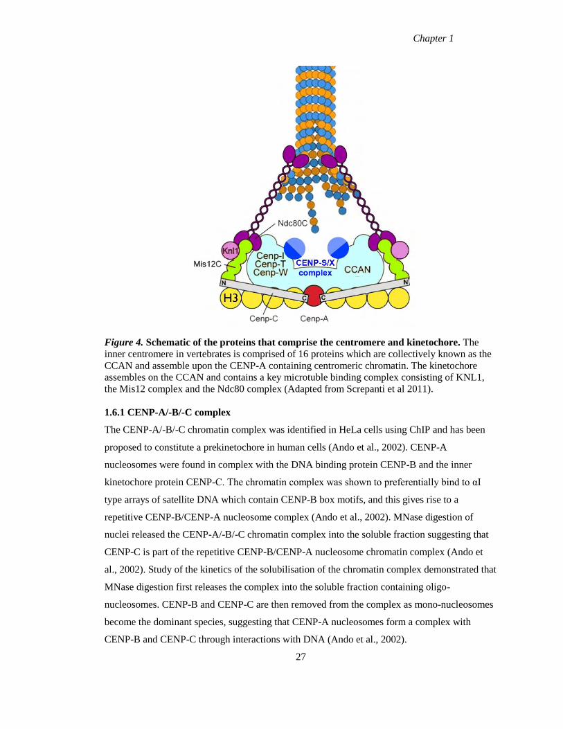
Chapter 1
Figure 4. Schematic of the proteins that comprise the centromere and kinetochore. The
inner centromere in vertebrates is comprised of 16 proteins which are collectively known as the
CCAN and assemble upon the CENP-A containing centromeric chromatin. The kinetochore
assembles on the CCAN and contains a key microtuble binding complex consisting of KNL1,
the Mis12 complex and the Ndc80 complex (Adapted from Screpanti et al 2011).
1.6.1 CENP-A/-B/-C complex
The CENP-A/-B/-C chromatin complex was identified in HeLa cells using ChIP and has been
proposed to constitute a prekinetochore in human cells (Ando et al., 2002). CENP-A
nucleosomes were found in complex with the DNA binding protein CENP-B and the inner
kinetochore protein CENP-C. The chromatin complex was shown to preferentially bind to αI
type arrays of satellite DNA which contain CENP-B box motifs, and this gives rise to a
repetitive CENP-B/CENP-A nucleosome complex (Ando et al., 2002). MNase digestion of
nuclei released the CENP-A/-B/-C chromatin complex into the soluble fraction suggesting that
CENP-C is part of the repetitive CENP-B/CENP-A nucleosome chromatin complex (Ando et
al., 2002). Study of the kinetics of the solubilisation of the chromatin complex demonstrated that
MNase digestion first releases the complex into the soluble fraction containing oligo-
nucleosomes. CENP-B and CENP-C are then removed from the complex as mono-nucleosomes
become the dominant species, suggesting that CENP-A nucleosomes form a complex with
CENP-B and CENP-C through interactions with DNA (Ando et al., 2002).
27
Chapter 1
While the function of CENP-B remains obscure, much progress has been made towards
elucidating the functions of CENP-C. CENP-C has been demonstrated along with CENP-T/-W
to directly associate with H3 nucleosomes but not CENP-A, but was shown to be in close
proximity with CENP-A nucleosomes (Hori et al., 2008a). However another study has
demonstrated that CENP-C directly binds to CENP-A nucleosomes at the C terminus (Carroll et
al., 2010). CENP-C has been demonstrated along with CENP-T to be capable of directing the
assembly of a functional kinetochore in the absence of CENP-A nucleosomes (Gascoigne et al.,
2011). Additional studies have demonstrated that CENP-C directly binds to the Mis12 complex
thus providing a link between centromeric chromatin and the outer kinetochore (Przewloka et
al., 2011, Screpanti et al., 2011).
1.6.2 CENP-H/-I/-K group
The CENP-H/-I/-K group is a major group of the CCAN, which is involved in ensuring proper
chromosome congression as well as playing a role in the deposition of newly synthesised CENP-
A at centromeres (Okada et al., 2006). CENP-H is a constitutive centromere coil-coiled protein
(Sugata et al., 1999) which is over-expressed in a variety of cancers including lung, colon and
oral (Liao et al., 2009, Shigeishi et al., 2006, Tomonaga et al., 2003). CENP-I is also a
constitutive centromere protein and the binding partner of CENP-H. Depletion of Mis6, the
fission yeast homologue of CENP-I, affects the localisation CENP-A (Takahashi et al., 2000).
CENP-K, like CENP-H and CENP-I, localises to centromeres at all stages of the cell cycle and
contains a leucine zipper motif (Okada et al., 2006). All three proteins are interdependent for
their centromere localisation, and depletion of either also results in the loss of CENP-L, -M, -O,
-P and –Q (Okada et al., 2006).
CENP-H along with CENP-I is necessary for the deposition of newly synthesised CENP-A at
centromeres. Incorporation rates of CENP-A GFP were drastically reduced in CENP-H/-I
mutant cells (rates of between 15-30%) when compared to incorporation in wild-type cells (rates
of approximately 80%), indicating CENP-H/-I play a role in the incorporation of nascent CENP-
A (Okada et al., 2006). This was surprising as CENP-H/-I depend on CENP-A for their
centromere localisation and lead to the hypothesis that incorporation of nascent CENP-A
requires the presence of pre-existing CENP-A nucleosomes with CENP-H/-I acting as an
intermediary (Okada et al., 2006). CENP-K in association with KNL-1 is involved in the
recruitment of the Ndc80 complex to kinetochores during mitosis (Cheeseman et al., 2008).
28

Chapter 1
Simultaneous depletion of both these proteins severely disrupts kinetochore assembly and
results in chromosome segregation defects (Cheeseman et al., 2008).
1.6.3 CENP-L/ -M/-N group
CENP-L/-M/-N were co-purified with the CENP-H/-I/-K group but the details of this interaction
remain unclear (Perpelescu and Fukagawa, 2011). CENP-L was identified as a member of the
CAD complex (Foltz et al., 2006), while Okada et al included it in the CENP-H class proteins
(CENP-H, -I and –K) based on functional studies. CENP-L is interdependent with CENP-H, -I
and –K for its centromere localisation (Okada et al., 2006). CENP-L is proposed to play a role in
bipolar spindle assembly as depletion by siRNA results in monopolar spindles (McClelland et
al., 2007). CENP-M was first identified as a member of the CENP-A NAC along with CENP-N
and CENP-T, with depletion of CENP-M (-N or –T) disrupting the recruitment of the CENP-A
NAC (Foltz et al., 2006). Depletion of CENP-M resulted in a loss of CENP-H, -I, K and –L,
while depletion of members from either the CENP-H or CENP-O class resulted in the loss of
CENP-M centromere localisation (Okada et al., 2006). Loss of CENP-M resulted in mitotic
defects including chromosome misalignment, accumulation of G2/M cells and some cells
exiting mitosis without undergoing cytokinesis (Okada et al., 2006, Foltz et al., 2006). CENP-N
has been demonstrated to directly interact with CENP-A nucleosomes and this interaction is
dependent on the CATD of CENP-A (Carroll et al., 2009). CENP-N interacts with CENP-A
nucleosomes via its N terminal while the C terminal is required for CENP-N targeting to
centromeres (Carroll et al., 2009). Depletion of CENP-N by siRNA results in a reduction of
CENP-A incorporation which is turn reduces the levels of CENP-A, -H, -K and –L at
centromeres (Carroll et al., 2009).
1.6.4 CENP-O/-P/-Q/-R/-U group
This group of proteins has been shown to be non-essential but depletion results in a G2/M
accumulation when compared to wild type DT40 cells (Hori et al., 2008b). CENP-O, -P –Q and
–U are interdependent for their centromere localisation, but CENP-R is recruited downstream of
these proteins, while all are dependent on the CENP-H/-I/-K group (Okada et al., 2006, Hori et
al., 2008b). Biochemical experiments in bacteria and chicken cells demonstrated that CENP-O, -
P, -Q and –U form a stable complex and CENP-R can associate with this complex (Hori et al.,
2008b). This CENP-O class plays an important role in the recovery of cells from spindle damage
and have been proposed to protect cells from premature sister chromatid separation until all
29
Chapter 1
microtubules have correctly attached to kinetochores (Hori et al., 2008b). In support of this
hypothesis CENP-Q was found to bind to microtubules in vitro (Amaro et al., 2010).
1.6.5 CENP-T/-W group
CENP-T was originally identified as a member of the CENP-A NAC (Foltz et al., 2006). This
putative histone fold domain (HFD) containing protein was identified as a distinct member of
the CCAN as it was not isolated as a member of the CENP-H-I complex. A novel HFD
containing centromere protein CENP-W was found in association with CENP-T; these proteins
form a complex through their histone fold domains and associate with centromeric DNA (Hori
et al., 2008a). The HFD of CENP-T is necessary to target the protein to centromeres. Neither
CENP-T nor CENP-W associate with CENP-A nucleosomes, rather the complex associates with
the H3 nucleosomes which are interspersed with CENP-A nucleosomes at centromeric
chromatin, similar to CENP-C (Hori et al., 2008a). Knockout experiments in DT40 cells
revealed that CENP-T is recruited upstream of CENP-H and CENP-O class CCAN members but
parallel to CENP-C in centromere assembly (Hori et al., 2008a). CENP-T/-W has been
suggested to be the connection between the centromere and the outer kinetochore in vertebrate
cells (Hori et al., 2008a). While CENP-A nucleosomes have been demonstrated to be essential
for specifying centromere identity as previously discussed, over-expression of CENP-A alone is
not sufficient to direct kinetochore assembly in humans (Van Hooser et al., 2001). In
Drosophila, ectopic expression of CENP-ACID results in kinetochore assembly (Heun et al.,
2006), while in humans ectopic expression of CENP-A only results in the mis-localisation of
three proteins, CENP-C, CENP-N and Mis18 (Gascoigne et al., 2011). Gascoigne et al (2011)
demonstrated that CENP-T along with CENP-C can direct kinetochore assembly in the absence
of CENP-A nucleosomes. Ectopic co-localisation of CENP-C and CENP-T resulted in
functional kinetochores which were capable of interacting with microtubules directing
chromosome assembly (Gascoigne et al., 2011). CENP-C along with CENP-T/-W have been
suggested to provide the platform for outer kinetochore assembly in vertebrate cells (Gascoigne
et al., 2011).
30
Chapter 1
1.6.6 CENP-S/-X group
CENP-S was originally identified as a member of the CENP-A distal (CAD) complex, found in
affinity purifications with CENP-M and –U (Foltz et al., 2006). Like CENP-T, CENP-S was
found to be a distinct member of the CCAN, as purifications with CENP-S did not contain
members of the H or O class proteins, but did reveal small amounts of CENP-T as well as a
novel centromere protein, CENP-X (Amano et al., 2009). Sequence analysis of CENP-S and
CENP-X revealed that both proteins contain putative HFDs. Both proteins are interdependent on
each other for their centromere localisation but are recruited downstream of H class proteins.
Depletion of either protein resulted in mitotic abnormalities in both human and chicken cells,
smaller outer kinetochore plates and they are required for the assembly of outer kinetochore
proteins including; KNL1, Ndc80 and Hec1 (Amano et al., 2009).
Apart from their role at centromeres, CENP-S and -X have been implicated in the Fanconi
anemia pathway. Fanconi anemia is a disease of cancer predisposition which is characterised by
bone marrow failure, developmental defects, and an increased sensitivity to DNA interstrand
crosslinking (ICLs) agents (Thompson and Jones, 2010). FANC and the associated proteins
function at stalled and broken replication forks produced by ICLs. CENP-S and CENP-X were
found to exist as a heterodimer in association with the FANCM protein (Yan et al., 2010).
FANCM is an ATP dependent branch-point translocase which acts on Holliday junctions and
replication forks (Yan et al., 2010, Singh et al., 2010). The CENP-S/-X heterodimer enhances
the branch-point translocase activity of FANCM as well as its binding to model replication forks
(Yan et al., 2010, Singh et al., 2010). Depletion of CENP-S/-X in HeLa cells caused an increase
in chromosomal aberrations and an increased sensitivity to ICLs (Yan et al., 2010, Singh et al.,
2010). The role of CENP-S/-X in the assembly of outer kinetochore proteins coupled with
FANCM depleted cells exhibiting supernumerary centrosomes has lead to the hypothesis that
the CENP-S/-X/FANCM association could play a role in chromosome segregation, possibly by
ensuring the faithful replication of structures associated with centromeric DNA (Thompson and
Jones, 2010).
31
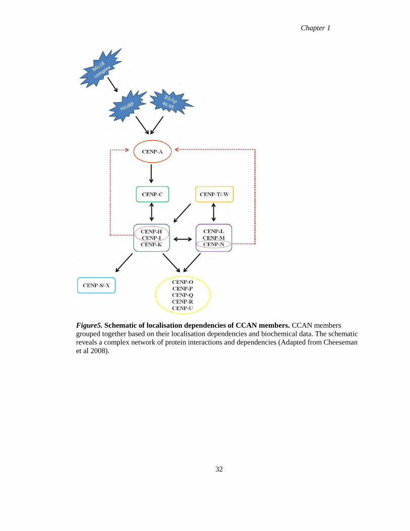
Chapter 1
Figure5. Schematic of localisation dependencies of CCAN members. CCAN members
grouped together based on their localisation dependencies and biochemical data. The schematic
reveals a complex network of protein interactions and dependencies (Adapted from Cheeseman
et al 2008).
32
Chapter 1
1.7 Heritability and assembly dynamics of centromere proteins
While CENP-A had been suggested to play a role in specifying centromere identity and
function, the mechanism for the maintenance of this locus is not fully elucidated. CENP-A was
previously shown to exhibit a slow turnover rate at centromeres (Shelby et al., 2000). It was
found to decrease by 50% per generation on a per cell basis and could be detected 4 days
following repression of the epitope tagged CENP-A, suggesting that parental CENP-A is equally
partitioned to daughter cells following DNA replication (Shelby et al., 2000). Complementary
experiments were carried out using the SNAP conditional labelling method and showed that
CENP-A remained stably associated with centromeres all through the cell cycle including during
DNA synthesis (Jansen et al., 2007). Following centromere duplication, there was an
approximate 50% reduction in CENP-A signal intensities at individual centromeres,
demonstrating that CENP-A which is loaded onto centromeric chromatin during G1 is equally
distributed to and retained by daughter cells (Jansen et al., 2007). Thus CENP-A exhibits
persistent association with centromeres across multiple cell cycles. This multi-generational
inheritance of the protein in-situ is thought to be important for the epigenetic maintenance of
centromere locus identity.
Similar SNAP experiments were carried out in Drosophila to examine the heritability of CENP-
ACID
, CENP-C and CAL1 (both of which are essential for CENP-ACID
assembly) (Erhardt et al.,
2008). Examination of the persistence of these proteins demonstrated that both CENP-ACID
and
CENP-C are retained at centromeres, with 50% signal remaining at centromeres after one cell
cycle suggesting that both proteins are equally distributed to daughter cells, similar to results
obtained for CENP-A in human cells (Mellone et al., 2011). CAL1 however was reduced to
~30% of initial signal intensities after one cell cycle, suggesting that this protein does not persist
across multiple generations. The lack of persistence of CAL1 coupled with its earlier assembly
time in comparison to CENP-ACID
has lead to the hypothesis that CAL1 is assembled de novo in
each cell cycle with pre-existing CENP-ACID
and CENP-C acting to recruit the protein to
centromeres, and this in turn acts to recruit newly synthesised CENP-ACID
(Mellone et al.,
2011).
Although extensive studies have been carried out to investigate the functional roles and
localisation dependencies of the CCAN components, very little is currently known regarding the
timing of assembly and heritability of these proteins during the cell cycle. The assembly
33
Chapter 1
dynamics and turnover rates of the HFD containing complex CENP-T/-W were assayed using
SNAP and FRAP (fluorescence recovery after photobleaching) techniques. Examination of
persistence of CENP-T and CENP-W revealed that neither protein persisted across multiple cell
cycles and can therefore not contribute as a heritable marker of centromere identity (Prendergast
et al., 2011). The timing of assembly of these two proteins was also examined, and in contrast to
CENP-A both proteins assembled before mitosis (late S/G2 phase of the cell cycle) (Prendergast
et al., 2011). FRAP experiments also demonstrated that there was no recovery observed during
G1 or early S phase but recovery was observed during late S phase and unlike CENP-A the
loading of CENP-T and CENP-W occurred with dynamic exchange (Prendergast et al., 2011).
The CENP-T/-W complex has been shown to interact with H3 nucleosomes and is required for
kinetochore assembly (Hori et al., 2008a). Taken together these data suggest that while CENP-A
is required to specify centromere identity of multiple generations, the CENP-T/-W complex
along with H3 nucleosomes is involved in kinetochore assembly before cells enter mitosis
(Prendergast et al., 2011).
The assembly dynamics of selected CCAN members (CENP-A,-B,-C,-H and –I) were examined
using FRAP. The analysis revealed that all centromere proteins examined were stably bound to
the centromere during mitosis, with some proteins stably bound (CENP-A and –I) during the
cell cycle while others exhibited dynamic exchange (CENP-B, -C and –H) (Hemmerich et al.,
2008).
FRAP revealed two populations of CENP-B existed with different residence times at
centromeres which reflect different retention modes of the protein during G1 and S phase of the
cell cycle (Hemmerich et al., 2008). The differences in residence time of the two populations
could be accounted for by the binding of CENP-B to the CENP-B box motifs (which the protein
has a great affinity for) versus binding of the protein to centromeric DNA adjacent to these
motifs when the motifs have become saturated with the protein (Hemmerich et al., 2008). FRAP
experiments with CENP-C revealed a fast exchanging population during G1 and G2 phases of
cell cycle which may act to attract the freely diffusing downstream CCAN members
(Hemmerich et al., 2008). CENP-C becomes stably bound to centromeres during mid and late S
phase which coincides with duplication of the genome and replication of centromeric DNA. The
change in dynamic behaviour of CENP-C at this stage of the cell cycle has lead to the
suggestion that CENP-C acts as a mediator for a functional interaction between centromeric
34
Chapter 1
DNA and the replication machinery possibly by providing a stable platform (Hemmerich et al.,
2008).
Examination of CENP-H revealed cell cycle dependent amounts of the protein that slowly
exchanged at centromeres while approximately 20% of the protein remained stably bound. The
stable fraction of the protein may act to stabilise the inner kinetochore, while the mobile fraction
may function in the dynamic assembly of downstream CCAN members similar to CENP-C
(Hemmerich et al., 2008) . The increased stability observed for CENP-H during S phase also
resembles the behaviour of CENP-C suggesting CENP-H may also act to connect centromeric
DNA to the replication machinery.
CENP-I did not exhibit FRAP during G1 and G2 phases of the cell cycle, but did during S
phase, recovering to 50% of pre-bleach signals (Hemmerich et al., 2008). These data suggest
that CENP-I is loaded exclusively during S phase and immediately fills the gaps created during
DNA synthesis (Hemmerich et al., 2008). Like CENP-A, CENP-I is loaded onto centromeres
without dynamic exchange of already loaded molecules. These data suggest that CENP-I may
play a role in supporting CENP-A in propagating and maintaining centromere identity
(Hemmerich et al., 2008).
To date, examination of the behaviour of centromere proteins at the centromere has identified
CENP-A and possibly CENP-I in maintaining centromere identity, while CENP-T/-W have been
implicated in playing a role in kinetochore formation. To fully understand centromere assembly
and inheritance will require a detailed knowledge of the remaining CCAN members with respect
to their assembly, persistence and dynamic behaviour at centromeres.
In the research presented here, the regulation and assembly of CCAN members was
investigated. The regulation of mRNA and protein levels of the CCAN was measured during the
human cell cycle using quantitative real-time PCR and western blotting respectively. The timing
of assembly and multigenerational persistence of the putative HFD proteins CENP-S and CENP-
X was examined in cell based analyses using conditional labelling methods. In addition, the
proximity of CENP-S and CENP-X to each other and other CCAN members was examined
using FRET, while complex formation between CENP-S and CENP-X was also analysed using
FCCS.
35
Chapter 2
Materials and Methods
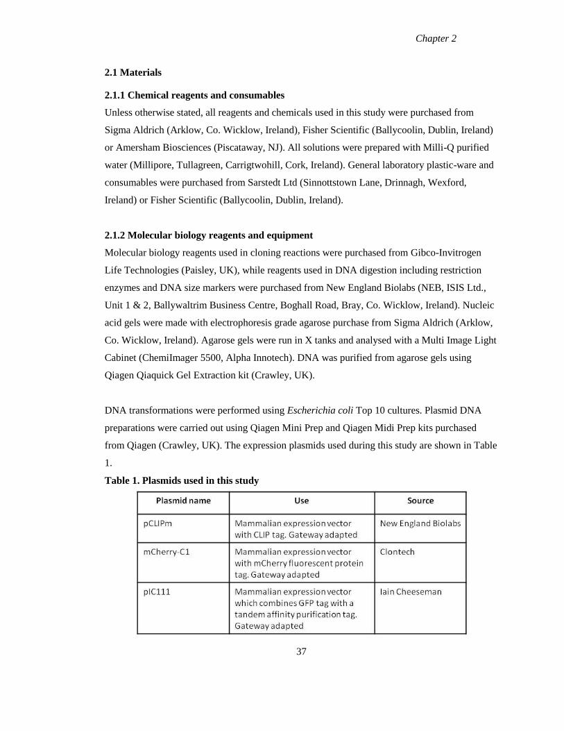
Chapter 2
2.1 Materials
2.1.1 Chemical reagents and consumables
Unless otherwise stated, all reagents and chemicals used in this study were purchased from
Sigma Aldrich (Arklow, Co. Wicklow, Ireland), Fisher Scientific (Ballycoolin, Dublin, Ireland)
or Amersham Biosciences (Piscataway, NJ). All solutions were prepared with Milli-Q purified
water (Millipore, Tullagreen, Carrigtwohill, Cork, Ireland). General laboratory plastic-ware and
consumables were purchased from Sarstedt Ltd (Sinnottstown Lane, Drinnagh, Wexford,
Ireland) or Fisher Scientific (Ballycoolin, Dublin, Ireland).
2.1.2 Molecular biology reagents and equipment
Molecular biology reagents used in cloning reactions were purchased from Gibco-Invitrogen
Life Technologies (Paisley, UK), while reagents used in DNA digestion including restriction
enzymes and DNA size markers were purchased from New England Biolabs (NEB, ISIS Ltd.,
Unit 1 & 2, Ballywaltrim Business Centre, Boghall Road, Bray, Co. Wicklow, Ireland). Nucleic
acid gels were made with electrophoresis grade agarose purchase from Sigma Aldrich (Arklow,
Co. Wicklow, Ireland). Agarose gels were run in X tanks and analysed with a Multi Image Light
Cabinet (ChemiImager 5500, Alpha Innotech). DNA was purified from agarose gels using
Qiagen Qiaquick Gel Extraction kit (Crawley, UK).
DNA transformations were performed using Escherichia coli Top 10 cultures. Plasmid DNA
preparations were carried out using Qiagen Mini Prep and Qiagen Midi Prep kits purchased
from Qiagen (Crawley, UK). The expression plasmids used during this study are shown in Table
1.
Table 1. Plasmids used in this study
37
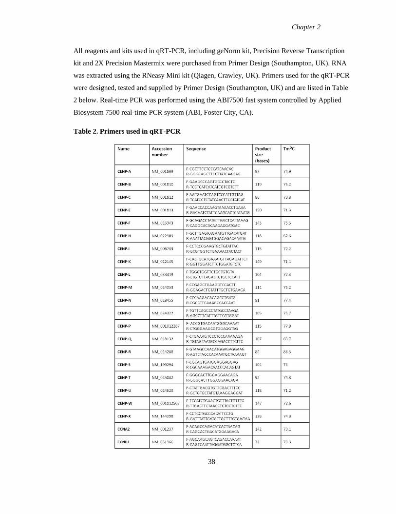
Chapter 2
All reagents and kits used in qRT-PCR, including geNorm kit, Precision Reverse Transcription
kit and 2X Precision Mastermix were purchased from Primer Design (Southampton, UK). RNA
was extracted using the RNeasy Mini kit (Qiagen, Crawley, UK). Primers used for the qRT-PCR
were designed, tested and supplied by Primer Design (Southampton, UK) and are listed in Table
2 below. Real-time PCR was performed using the ABI7500 fast system controlled by Applied
Biosystem 7500 real-time PCR system (ABI, Foster City, CA).
Table 2. Primers used in qRT-PCR
38
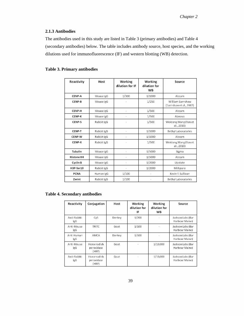
Chapter 2
2.1.3 Antibodies
The antibodies used in this study are listed in Table 3 (primary antibodies) and Table 4
(secondary antibodies) below. The table includes antibody source, host species, and the working
dilutions used for immunofluorescence (IF) and western blotting (WB) detection.
Table 3. Primary antibodies
Table 4. Secondary antibodies
39
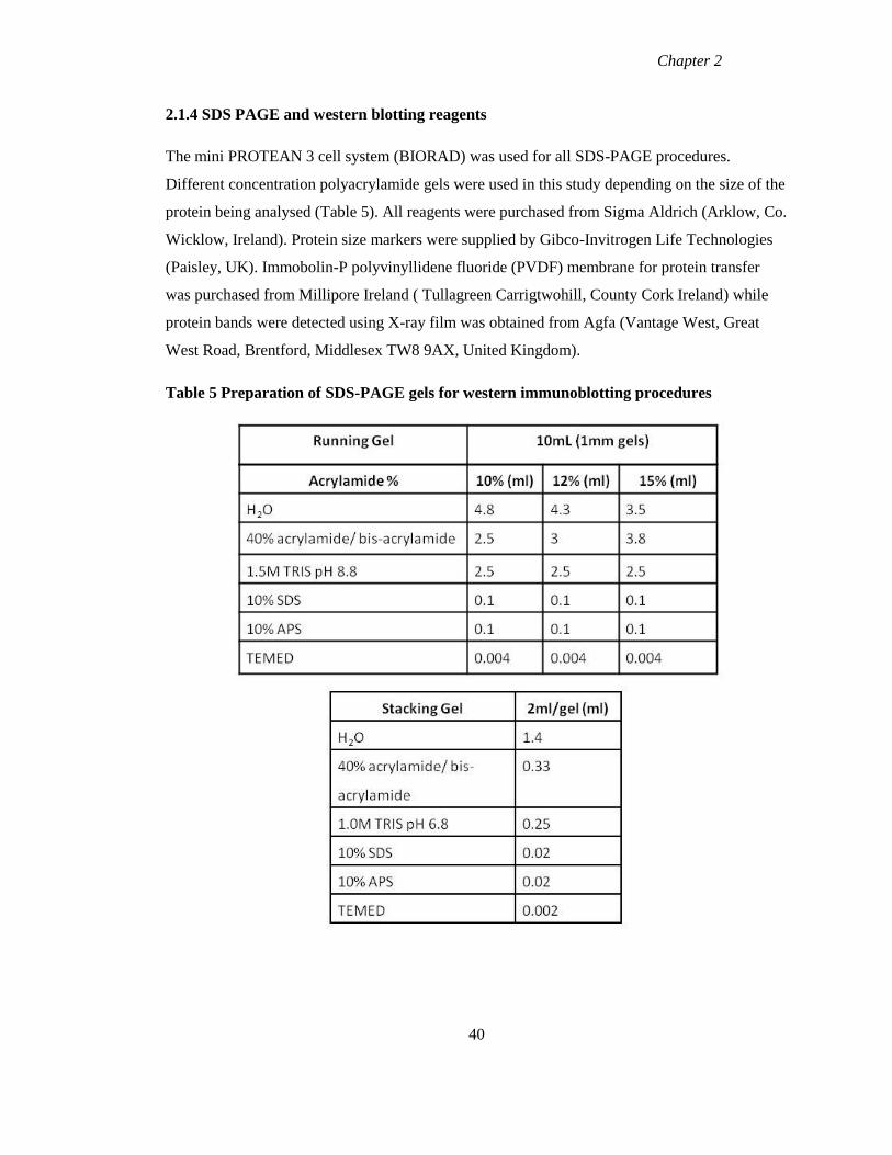
Chapter 2
2.1.4 SDS PAGE and western blotting reagents
The mini PROTEAN 3 cell system (BIORAD) was used for all SDS-PAGE procedures.
Different concentration polyacrylamide gels were used in this study depending on the size of the
protein being analysed (Table 5). All reagents were purchased from Sigma Aldrich (Arklow, Co.
Wicklow, Ireland). Protein size markers were supplied by Gibco-Invitrogen Life Technologies
(Paisley, UK). Immobolin-P polyvinyllidene fluoride (PVDF) membrane for protein transfer
was purchased from Millipore Ireland ( Tullagreen Carrigtwohill, County Cork Ireland) while
protein bands were detected using X-ray film was obtained from Agfa (Vantage West, Great
West Road, Brentford, Middlesex TW8 9AX, United Kingdom).
Table 5 Preparation of SDS-PAGE gels for western immunoblotting procedures
40
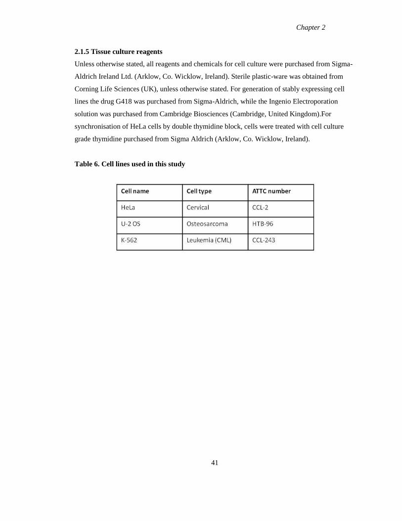
Chapter 2
2.1.5 Tissue culture reagents
Unless otherwise stated, all reagents and chemicals for cell culture were purchased from Sigma-
Aldrich Ireland Ltd. (Arklow, Co. Wicklow, Ireland). Sterile plastic-ware was obtained from
Corning Life Sciences (UK), unless otherwise stated. For generation of stably expressing cell
lines the drug G418 was purchased from Sigma-Aldrich, while the Ingenio Electroporation
solution was purchased from Cambridge Biosciences (Cambridge, United Kingdom).For
synchronisation of HeLa cells by double thymidine block, cells were treated with cell culture
grade thymidine purchased from Sigma Aldrich (Arklow, Co. Wicklow, Ireland).
Table 6. Cell lines used in this study
41
Chapter 2
2.2 Methods
2.2.1 Cell culture
2.2.1.1 Culturing cells
HeLa cells were grown in Dulbecco’s Modified Eagle Medium (DMEM) with 4.5g/L glucose
(Lonza Biologics, Slough, United Kingdom) stable L-glutamine, 10% fetal calf serum (FCS),
non essential amino acids (NEAA) and 1% penicillin streptomycin. Culturing of cells was
carried out in a Class III Bio-safety cabinet. Cells were typically grown in 100mm x 20mm or
150mm x 25mm culture dishes and incubated in 5% CO2 at 37oC. Cells were subcultured when
they reached approximately 80% confluency; cells were first washed in phosphate-buffered
saline (1X PBS) and then treated with 1X Trypsin EDTA (Gibco-Invitrogen Life Technologies,
Paisley, UK) for 5min at 37oC to detach the cells. The cells were then resuspended in 8mL fresh
medium and centrifuged at 1,200 r.p.m (300g) for 5min. Cells were then resuspended in 10mL
fresh medium, counted using a hemocytometer and an appropriate number of cells plated.
2.2.1.2 Freezing cells
Cells were detached as outlined detach the cells. Cells were resuspended in 8mL complete
medium and centrifuged at 1,200 r.p.m (300g) for 5min. Cells were resuspended in 1mL of
freezing medium (90% FCS with 10% DMSO) and transferred to a cryovial (Nunc GMBH and
Co., KG, Wiesbaden, Germany). Cells were subsequently transferred to the -80oC freezer and
the next day to the liquid nitrogen storage facility (Jencons-PLS, UK).
2.2.1.3 Resuscitation of cells
Cells were thawed rapidly at 37oC, added to 9mL complete medium and incubated in 5% CO2 at
37oC. The medium was changed the following day.
2.2.2 Transfection
Transfection of plasmid DNAs was carried out by electroporation using a NucleofectorTM
II
device (Lonza Biologics,Slough, United Kingdom). HeLa cells were trypsinised and counted as
outlined in section 2.2.1.1. 1x10⁶ cells were spun in a centrifuge at 1200 r.p.m for 5min. The
supernatant was removed from cell pellet, 4ug of plasmid DNA was pipetted directly on top of
42
Chapter 2
the cell pellet followed by 100ul of Ingenio electroporation solution (Mirus supplied through
Cambridge Bioscience, Cambridge, United Kingdom). The cell pellet was resuspended in this
solution and transferred to an Ingenio cuvette (Cambridge Bioscience, Cambridge United
Kingdom). The Amaxa program I-013 was selected and cells were transfected. After
transfection, the sample was removed from the cuvette and transferred to a dish with pre-
warmed medium and placed back in 37oC incubator. Transiently transfected cells were then
analysed 48hr later by fluorescence microscopy.
For stable transfections, the same procedure was used as for transient transfections. Specifically
4ug of CENP-S and CENP-X CLIP plasmid was transfected into HeLa cells. 48hr following
transfection, cells were trypsinised and plated in 10cm dishes at different dilutions to obtain
clones derived from single cells in medium containing G418 (300mg/mL) (Melford
Laboratories, Suffolk, IP7 7LE U.K.). When individual colonies were identified the media was
removed, the plate was washed with 1X PBS and sterile greased glass cloning rings were
positioned around the colonies. The colony was then trypsinised and plated in a 24 well plate.
When cells had sufficiently expanded they were moved to a 6 well plate and screened by
immuno-fluorescence microscopy.
2.2.3 Cell cycle fractionation
2.2.3.1 Synchronisation of HeLa cells by double thymidine block
A double thymidine block synchronises cells by arresting them at the G1/S transition (Bootsma
et al., 1964). Cells are exposed to high concentrations of thymidine which increases the
thymidine triphosphate (dTTP) pools. This increase has an inhibitory effect on the
ribonucleotide reductase enzymes (involved in the biosynthesis of pyrimidines) which causes a
decrease in the production of deoxycytodine-5-triphosphate (dCTP). The reduced levels of
dCTP inhibit DNA synthesis and causes cells to arrest at the G1/S transition.
HeLa cells were plated in 100mm x 20mm dishes and grown to 50% confluence and treated with
5mM thymidine (200mM stock concentration) for 16hr. This first treatment causes some cells to
be arrested at the G1/S transition while other cells will be at a later stage of S phase. Cells were
43
Chapter 2
washed twice with 1X PBS for 5min followed by one 5min wash with fresh medium. Cells
were then released into fresh medium for a 10hr release during which cells will progress through
the cell cycle. Following the 10hr release, cells were blocked again for 16hr with 5mM
thymidine. The second block arrests cells already in the G1 phase at the G1/S transition, with
later cells allowed to progress through G2/M and then blocked at the G1/S transition. Cells were
released from the block as previously outlined and samples harvested over a 24hr period for
analysis by flow cytometry, western blotting and qRT-PCR.
2.2.3.2 Fractionation of K-562 suspension cell line by elutriation
The human chronic myelogenous cell line K-562 was fractionated by centrifugal elutriation on
the basis of cell size. Cells are separated based on their sedimentation rate in a gravitational
field, with the medium containing cells flowing against the force of gravity. This method of cell
cycle fractionation is less invasive than other methods and does not have any effects on the
metabolism of the cells. Cells are loaded in a chamber and spun at a constant centrifugal force
and pump speed, and are then sedimented in the chamber according to their sedimentation
velocity. This parameter (sedimentation velocity) depends on cell size, cell density, pump and
rotor speed. Cells of specific size are then eluted by increasing or decreasing either the pump or
rotor speed.
The day before elutriation was carried out the rotor was assembled and the pump was calibrated.
On the day of fractionation, cells were counted and 1.5 x10⁸ cells were resuspended in 10mL of
RPMI-1640 medium (Lonza Biologics,Slough, United Kingdom) which was supplemented with
5% FCS. Cells were passed through an 18 gauge needle (Becton Dickson U.K. Ltd, England) to
prevent cell clumps from forming. Cells were injected to the mixing chamber via the inlet which
was then closed and the flow to the elutriation chamber was opened. Cells were loaded at a flow
rate of 19mL/min and a rotor speed of 2500 r.p.m. Cell fractions were collected once the mixing
chamber was cleared of cells with 100mL collected for each fraction. The pump was then set to
22mL/min to collect the second fraction and this was continued according to the Table 7. All
cell fractions were kept on ice until samples were processed with each fraction analysed by flow
cytometry, western blotting and qRT-PCR.
44
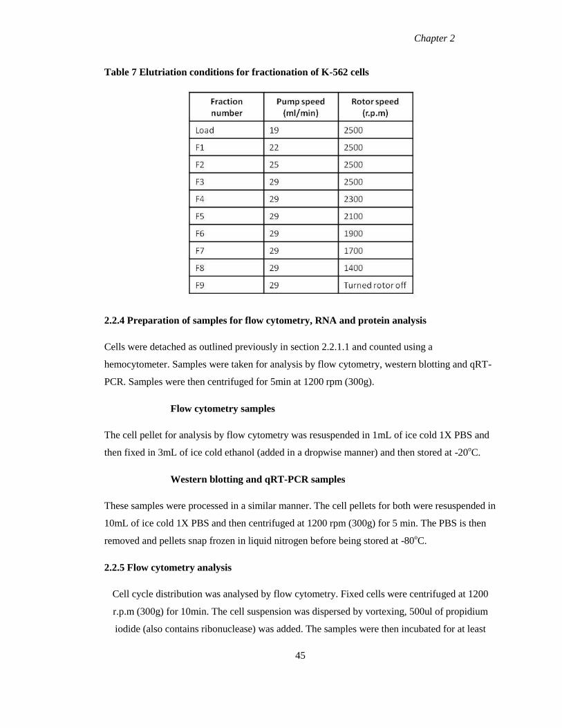
Chapter 2
Table 7 Elutriation conditions for fractionation of K-562 cells
2.2.4 Preparation of samples for flow cytometry, RNA and protein analysis
Cells were detached as outlined previously in section 2.2.1.1 and counted using a
hemocytometer. Samples were taken for analysis by flow cytometry, western blotting and qRT-
PCR. Samples were then centrifuged for 5min at 1200 rpm (300g).
Flow cytometry samples
The cell pellet for analysis by flow cytometry was resuspended in 1mL of ice cold 1X PBS and
then fixed in 3mL of ice cold ethanol (added in a dropwise manner) and then stored at -20oC.
Western blotting and qRT-PCR samples
These samples were processed in a similar manner. The cell pellets for both were resuspended in
10mL of ice cold 1X PBS and then centrifuged at 1200 rpm (300g) for 5 min. The PBS is then
removed and pellets snap frozen in liquid nitrogen before being stored at -80oC.
2.2.5 Flow cytometry analysis
Cell cycle distribution was analysed by flow cytometry. Fixed cells were centrifuged at 1200
r.p.m (300g) for 10min. The cell suspension was dispersed by vortexing, 500ul of propidium
iodide (also contains ribonuclease) was added. The samples were then incubated for at least
45
Chapter 2
30min in the dark before analysis was carried out on the FACS calibur flow cytometer (Becton
Dickinson Biosciences, San Jose, California, USA). The data were analysed using the WinMDI
v2.9 software. Scatter plots were generated to gate out cell doublets and DNA content was
visualised by generating a histogram plotting cell count versus PI content and thus showing the
progression of cells through the cell cycle.
2.2.6 Purification of Total RNA from Human cells using Qiagen RNeasy Mini Kit
Total RNA was isolated from human cells as per the manual from the Qiagen RNeasy Mini Kit
(Qiagen, Crawley, West Sussex, RH10 9NQ). All tubes and tips used in the protocol were first
treated with Diethylpyrocarbonate (DEPC) purchased from Sigma-Aldrich Ireland Ltd. (Arklow,
Co. Wicklow, Ireland) (inactivates the RNase enzymes) and then autoclaved to ensure no
contamination of the RNA samples with DNA. Frozen pellets were thawed on ice before being
disrupted by Buffer RLT and homogenised by the Qiashredder to reduce sample viscosity. 70%
ethanol (Sigma-Aldrich) was added and the sample was applied to the spin column. Total RNA
binds to the membrane with contaminants being washed away and separated from RNA. An on-
column DNase digestion was carried out using RNase free DNase (Qiagen) to ensure removal of
all contaminating DNA which may interfere with downstream processes. All traces of DNase
were removed by subsequent washes. Total RNA was eluted in 50ul of RNase free water.
Quality of the RNA was assessed by analysing the absorbance readings (A260/280) of each
sample using a Nanodrop 1000 spectrophotometer. RNA was also run on a formaldehyde gel to
visualise it and further ensure no degradation or contamination of samples. All RNA was stored
at -80oC.
2.2.6.1 Formaldehyde Gels
RNA was electrophoretically separated on 1.2% gels which comprised of 1.2g of agarose
dissolved in 10X formaldehyde gel buffer (200mM MOPS, 50mM Sodium Acetate, 10mM
EDTA) and this was made up to 100mL with RNase free H20. The mixture was heated in the
microwave until the agarose was completely dissolved; the mixture was then cooled to 60oC in a
water bath. The gel was submerged in a gel tank in 1X formaldehyde gel running buffer (1X
formaldehyde gel buffer, 37% (12.3M) formaldehyde) and this was made up to one litre with
RNase free H20 .One volume of 5X RNA loading buffer was added to four volumes of RNA
(0.5ug of RNA) and the samples were incubated at 65oC for 5min and then chilled on ice before
46
Chapter 2
being loaded onto the equilibrated gel. The gel was run at five volts/cm until the
bromophenolblue (BPB) had run 2/3 down the gel. The gel was stained by placing the gel in an
RNase free dish and soaking the gel in 0.5M ammonium acetate for 20min. The ammonium
acetate was then removed and replaced with fresh solution and the gel was left to soak for an
additional 20min. 0.5ug/mL of ethidium bromide (EtBr) was added to the 0.5M ammonium
acetate and left for 30min. The gel was finally visualised using the alpha imager.
2.2.7 cDNA synthesis
Total RNA was reverse transcribed using random nonamer primers to generate cDNA using the
Precision qScript Reverse Transcript Kit supplied by Primer Design (Southampton, UK). 1ug of
total RNA was combined with 1ul of random nonamer primers and were annealed at 65oC for
5min. Samples were placed on ice to chill following the incubation period. 2ul of 10X qScript
Buffer, 1ul of dNTP mix (10mM each), 2ul of 100mM DTT, 1ul of qScript enzyme and 4ul of
DNase/RNase free water was then added and the mixture was vortexed followed by a pulse spin.
The mixture was incubated at 25oC for 5min followed by 20min incubation at 55
oC. The
extension reaction was then stopped by heat inactivation for 15min at 75oC. cDNA was then
stored at -20oC.
2.2.8 Real-time PCR
Relative transcript abundance was measured using real-time PCR (qPCR) in conjunction with
SYBR green dye. qPCR measures the amount of amplified product at the end of each cycle. The
changes in product concentration at the end of each cycle are calculated by measuring the levels
of SYBR green fluorescence, which is proportional to the amount of product formed. SYBR
green dye binds to double stranded DNA which results in an increase in fluorescence. The qPCR
were carried out in 96 well optical plates on a StepOnePlus™ Real-Time PCR System (Applied
Biosystems supplied through Life Technologies, Paisley, PA4 9RF United Kingdom). Primers
for specific genes (see Table 2) were resuspended in RNase/DNase free water. cDNA as
prepared in section 2.2.7 was used as template for each reaction. Each 20ul reaction contained
5ul cDNA, 1ul primer mix (300nM, 150nM, and 75nM), 10ul SYBR Green master mix (Primer
Design, Southampton, UK) and 4ul of RNase free water. The cycling conditions were as follows
95oC for 10min, 1 cycle, and then 95
oC for 15sec, 60
oC for 1min, 40 cycles.
47
Chapter 2
2.2.8 .1 Design of primers
All primers used in the gene expression study were designed by Primer Design Ltd, real-time
PCR specialists who custom design real-time PCR primers. Primers were designed using a
modified version of the Beacon Designer programme with the details of the primers outlined in
Table 2. All primers have guaranteed priming specificity, minimised secondary structures, and
minimised primer dimer formation
2.2.8.2 Validation of primers for real-time PCR
The major limitation with the use of SYBR green chemistry with qRT-PCR is SYBR green
binds to all double stranded DNA, including non-specific products and primer dimers. To
eliminate this problem, a dissociation curve for each primer is generated. This reaction involves
the temperature being increased to 95oC for 1s, followed by a decrease in temperature between
65oC -70
oC. The reaction is then heated back up again slowly (0.5
oC per second) with the
fluorescence measured constantly. The increase in temperature is coupled with a decrease in
fluorescence. Once the melting temperature has been reached, the double stranded DNA
dissociates and the fluorescence levels will decrease. If the primers are specific for one
particular product, a single peak will be observed in the dissociation curve, while additional
peaks indicate the presence of non specific products or primer dimers.
2.2.8.3 Calculation of primer efficiency by standard curve method
Real-time PCR primer efficiencies were validated using the standard curve method with a
standard curve constructed for each primer set. In order for qRT-PCR to work optimally, the
primers must be working with an efficiency of 100% and a slope of -3.32, meaning that after
every cycle the PCR product doubles in quantity. Efficiencies within the range of 90-110% and
the corresponding slopes between -3.58 and -3.1 are generally acceptable values. The set of 22
primers used in this gene expression study were evaluated and the efficiency and slope of each
primer are listed in Table 8. The priming efficiency for each primer set was then calculated with
the following formula
Efficiency (E) = 10 (-1/slope)
48
Chapter 2
2.2.8.4 Selection of a reference gene
An important step in the calculation of the relative transcript abundance is the selection of the
correct reference gene. The relative expression level for each gene examined is obtained by
normalising the relative expression to a stably expressed endogenous control. The endogenous
control used in this study (GAPDH) was chosen with the help of the GeNorm Kit (Primer
Design) GeNorm software was developed by Vandesompele et al and analyses a set of candidate
reference genes and measures the relative expression by real-time PCR. The CT values obtained
are converted to relative expression values by subtracting the highest CT value from all other
CT values for each gene measured, the following formula is then applied
Relative Expression= 2-∆CT
All data is then expressed relative to the expression of the least stable gene. The data are then
analysed by the GeNorm software and a graph is constructed showing the stability of the
expressed genes with the least stable genes on the left and the most stably expressed genes on
the right. This software was used for the purpose of this study and demonstrated that
Cytochrome C1 and GAPDH are the most suitable reference genes as they show the highest
level of stability as shown in Figure 7 and 12.
2.2.8.5 Real-time PCR calculations
The Pfaffel method was used to quantify the results obtained from the qPCR (Pfaffl, 2001).
Unlike the delta delta CT method, this method does not assume the priming efficiency of each
primer set is 100%. Instead the priming efficiency is calculated (as outlined above) and
incorporated into the relative quantitation calculation. The cycle threshold (CT) of the gene of
interest (CTGOI) was measured in the test samples and the calibrator sample as well as for the
reference gene (CTREF) in both the test and calibrator samples. The CTGOI was normalised to
CTREF to give delta CT values (ΔCT).
ΔCT= CTGOI - CTREF
The ΔCTGOI value of the in test sample was then further normalised to the ΔCT GOI of the in
calibrator sample.
ΔΔCT=ΔCT GOI test sample - ΔCT GOI calibrator sample
49
Chapter 2
The relative fold change (relative quantitation RQ) was then calculated by the formula, E -∆∆CT
(where E is the primer efficiency). This calculation gives the ratio of the gene of interest in the
test sample to the calibrator sample normalised to expression levels of the reference gene.
2.2.9 Protein sample preparation
Protein samples were thawed on ice before being prepared in 2X LSB with 1M DTT (final
concentration 100uM DTT), the samples were then boiled for 10min to denature sample and
facilitate separation. Typically 100,000 cells of each protein sample was loaded per well
2.2.10 Subcellular Fractionation
Cells were grown to 90% confluence on 150mm dishes. All steps of the procedure were carried
out on ice, in the cold room unless otherwise stated. Cells were washed twice in cold 1X PBS,
PBS was removed and 15mL of LS buffer was added and cells were allowed to swell for 10min.
LS buffer was removed and cells were scraped before being transferred to a dounce
homogeniser and were dounced 25 times with a loose dounce. Cells were then transferred to a
siliconised 1.5mL tube and centrifuged at 2100g for 3min at 4oC. This centrifugation separated
the cytosolic fraction (supernatant) from the nuclei pellet. The cytosolic fraction was transferred
to a new 1.5mL tube before being centrifuged at 21,000g for 20min to remove all debris. Half
the nuclei pellet volume of LS buffer was added to the nuclei pellet, the sample was then placed
on a rotator for 9 min at 4oC. The sample was then centrifuged at 21,000g for 20min; the nuclear
fraction (supernatant) was transferred to a new 1.5mL tube. One pellet volume of LS buffer was
added to the chromatin bound fraction (pellet), followed by 1/100 volume of Benzonase (an
endonuclease that removes nucleic acids from protein samples), the sample was then incubated
at 30oC for 30min. Following the incubation, the sample was sonicated 4 times for 30sec each
with 30sec between each sonication. The chromatin bound fraction was then centrifuged for
21,000g for 20min; the supernatant was removed to a clean tube. All three samples were snap
frozen in liquid nitrogen for 1min and then stored at-80oC.
2.2.11 Sodium dodecyl sulphate polyacrylamide gel electrophoresis (SDS-PAGE)
SDS-PAGE gels were prepared according to Table 5. HeLa cell protein extract was prepared as
previously described in 2.2.4. The extract was denatured in 1X Laemmli SDS buffer (10ul of
50
Chapter 2
LSB was added to every 100,000 cells) with a final concentration of100mM dithiothreitol
(DTT). Samples were boiled for 10min and then centrifuged at 13,000 r.p.m for 5min. 100,000
cells were loaded per well as well as a marker in an adjacent well. The buffer tanks were filled
with 1X running buffer (Glycine 19.2mM, TrisBase 2.5mM, 0.01% SDS) and the gel was run at
constant voltage (100 volts) for between one and two hr until the desired separation was
achieved.
2.2.12 Western Immunoblotting
The Mini Trans-Blot Cell system (BIORAD) was used for all procedures in western
immunoblotting. Immobolin-P polyvinyllidene fluoride (PVDF) membrane was cut to size and
submerged in methanol for 3min, followed by 3min in H20 to activate the membrane. It was then
submerged in 1X transfer buffer (19.2mM glycine, 2.5mM TRIS, 20% methanol) along with the
sponges and filter paper, until being assembled in transfer cassette. The transfer was assembled
with the PVDF membrane on the cathode side and the gel on the anode. Three pieces of filter
paper were placed on either side of the membrane and sponges either side of the filter paper. The
transfer was assembled in the transfer tank, along with ice pack and stir bar, the tank was filled
with 1X transfer buffer. Protein transfer was carried out at constant voltage (100v) for 90-
120min at 4oC. After transfer the apparatus was disassembled and each membrane was stained
in 0.1% ponceau S in 5% acetic acid for 3min to visualise protein transfer. Ponceau stain is
removed by washing three times in H20 on a platform rocker.
The membrane was then blocked in 5% blocking solution (100mM TRIS, 68mM NaCl, 0.1%
Tween -20 (TBS-T), and 5% non-fat dried milk (Marvel) for 1 hr at room temperature on a
platform rocker. The membrane was probed with the primary antibody diluted in the 5%
blocking solution at the appropriate dilution. The membrane was then heat sealed in a plastic
bag and incubated at 4oC on a platform rocker overnight. After the primary antibody incubation
period the membrane was washed three times for 10min in TBS-T to remove any unbound
primary antibody. The primary antibody was then detected with the addition of a secondary
antibody linked to horseradish peroxidise (HRP). The secondary antibody was diluted 1/10,000
in 2.5% blocking solution and the membrane was incubated 1hr at room temperature on a
platform shaker. Following the secondary antibody incubation the membrane was washed three
times for 10min each in TBS-T to remove any unbound secondary antibody.
51
Chapter 2
The activity of the HRP was then detected with the addition of ECL + Western Blotting
Chemoluminescent detection system from Millipore Ireland B.V. (Tullagreen Carrigtwohill,
County Cork Ireland). The solutions were brought to room temperature and Solution A was
mixed with Solution B in a ratio of 1:1. 2mL of the ECL was prepared for each membrane and
the membrane was incubated for 5min at room temperature. The membrane was then transferred
to a film cassette; the remainder of the detection was carried out in a dark room. X-RAY
medical film (Konica Minolta) was used to detect the light emitted by ECL. The detection was
carried out using a CP1000 Automatic Film processor with Devalex X Ray Developer and
FixaPlus X Ray Fixer.
2.2.13 Protein densitometry
Chemiluminscent images were captured using the Gene Snap software from Syngene. Band
intensities were determined using the Image J freeware (http://rsb.info.nih.gov/ij/). The
densitometry was calculated as the mean intensity normalized to the loading control (Tubulin)
and presented as the mean ± SEM (n=3).
2.2.14 Gateway Cloning
Gateway recombination cloning was used to generate all CENP-S and CENP-X constructs. PCR
products of CENP-S and CENP-X were generated from cDNA with each product flanked by
recombination sites attB. The PCR product was then recombined into the entry vector pDonr-
221 which is also flanked by two recombination sites attL1 and attL2. The entry clone was then
recombined into a destination vector (also contains two recombination sites attR1 and attR2) and
the construct is formed. The directionality of the insert is maintained as recombination site
attL1 will only recombine with attR1 and cannot recombine with attR2.Destination vectors used
in this study were gateway adapted and included pCLIPm (NEB), pIC111, pCherry and PAGFP.
2.2.15 Bacterial transformations
For transformations, 50ul of competent E.coli cells were thawed on ice for each transformation
reaction. DNA was combined with competent E.coli cells and then incubated on ice for 30min
and then subjected to heat shock for 1min at 42oC and then placed back on ice for 2min. 250ul
of LB broth was added to competent cells under sterile conditions and bacteria were incubated at
52
Chapter 2
37oC for 1hr with shaking. Dilutions of this were then plated on agar plates with the appropriate
antibiotic selection for the particular vector and plates were incubated over night at 37oC upside
down. 12-16hr later single colonies were picked and grown in 5/100mL cultures.
2.2.16 Plasmid DNA preparation
Mini and midi plasmid DNA preparations were carried out using Qiagen Mini and Midi Prep
kits respectively according to manufacturers’ instructions. Bacterial cultures were grown over
night at 37oC on a shaking platform in the presence of selective antibiotics. For mini preps,
DNA was isolated from 5mL E. coli culture and resusupended in 50ul of elution buffer (EB).
For midi preps, DNA was isolated form 100mL E.coli culture and resuspended in 100-300ul of
EB.
2.2.17 Immunofluorescence Microscopy
2.2.17.1 Methanol Fixation
Hela cells were grown on glass coverslips in 4 well dishes. At appropriate time points, the
medium was removed and cells were washed in 1X PBS. Cells were then fixed with pre-chilled
methanol and stored at -200C until ready for immunofluorescence staining.
2.2.17.2 Immunofluorescence Staining
Fixed cells were washed 2 times for 3min each in 1X PBS, followed by 2 washes in 1X PBS-TX
for 3min each. Cells were then blocked in 1X PBS-TX with 1% BSA at room temperature for
30min, cells were washed 2 times in 1X PBS-TX. Cells were incubated with primary antibodies
(see Table 3 for appropriate dilutions) diluted in 1% BSA in 1X PBS-TX in a 37oC incubator for
1hr. Cells were washed twice in 1X PBS-TX for 10min each. Cells were then incubated with
fluorescently conjugated secondary antibodies (see Table 4 for appropriate dilutions) diluted in
1% BSA in 1X PBS-TX in the dark in a 370C incubator for 1hr. Cells were washed once in 1X
PBS-TX for 10min, followed by 1 wash in 1X PBS for 10min. Finally cells were washed once
in distilled H20 for 1min. Coverslips were then allowed to completely dry before being mounted
in slowfade with DAPI. Coverslips were sealed with nail varnish and stored at 4oC in the dark.
53
Chapter 2
2.2.17.3 Microscopy
Unless otherwise stated all microscopy was carried out using a Deltavision Core system
(Applied Precision) controlling an interline charge-coupled device camera mounted on an
inverted microscope (Olympus). For each sample images were collected at 2x2 binning using a
100x oil objective at 0.2um z sections. Immunofluorescence images were subject to iterative
constrained deconvolution and maximum intensity projection using the SoftWoRx software
(Applied Precision).
Deltavision files were used in quantitation. Image Pro software was used to identifiy
centromeres, by segmentation thresholding of the CENP-A signal and object definitions were
saved as a mask. This mask was applied to the CLIP images and CLIP label within centromeres
was recorded. Background correction was applied by averaging the intensities in 3 background
areas within nuclei and devoid of centromeres and subtracting the mean value from individual
centromere. Measurements are reported as mean (+/- SEM).
2.2.18 CLIP Labelling
The CLIP tag is a modified variant of the mammalian DNA repair enzyme O6 alkylguanine
DNA alkyltransferase whose normal function is in DNA repair. During the labelling reaction the
CLIP tag is covalently and irreversibly modified through the acceptance of a benzylcytosine
derivative. The CLIP substrate is CLIP cell 505 a green fluorescent dye which is cell permeable
and a maximum excitation at 504nm and maximum emission at 532nm and is therefore suitable
for use with fluorescein filters.
2.2.18.1 CLIP Quench
CLIP cell block (New England Biolabs, ISIS Ltd., Unit 1 & 2, Ballywaltrim Business Centre,
Boghall Road, Bray, Co. Wicklow, Ireland) (bromothenylcytosine BTC) a non-fluorescent
molecule, blocks the reactivity of the CLIP tag and irreversibly inactivates all CLIP tagged
proteins for further labelling steps. CLIP cell block was dissolved in 50ul of DMSO to give a
2mM stock and mixed for 10min by vortexing to ensure all CLIP cell block was dissolved. The
CLIP cell clock was diluted 1/200 in complete medium to give a final concentration of 10uM
quench labelling medium. Medium was removed from cells expressing the CLIP tag fusion
proteins and replaced with the quench medium. The cells were incubated at 37OC with 5% CO2
54
Chapter 2
for 30min. Cells were then washed 2 times for 5min each in pre-warmed PBS followed by a
5min wash in pre-warmed medium. Cells were then placed in complete medium and incubated
at 37OC with 5% CO2 for 8hr to allow cells time to synthesise new CLIP tag fusion proteins.
Cells were then labelled with CLIP cell 505 as outlined below.
2.2.18.2 CLIP Pulse Labelling
The CLIP cell 505 (New England Biolabs, ISIS Ltd., Unit 1 & 2, Ballywaltrim Business Centre,
Boghall Road, Bray, Co. Wicklow, Ireland) was dissolved in 50ul of DMSO to give a stock
concentration of 1mM CLIP substrate and was mixed for 10min by vortexing to ensure all
substrate was dissolved. Before labelling cells, the stock CLIP substrate was diluted 1/200 in 1%
BSA in complete DMEM and mixed thoroughly by pipetting up and down 10 times to reduce
background. Medium was removed from cells expressing the CLIP tag fusion proteins and
replaced with the CLIP tag labelling medium. The cells were incubated at 37OC with 5% CO2
for 45min. Cells were washed 2 times for 5min each in pre-warmed PBS followed by a 5min
wash in pre-warmed medium. Medium was changed on cells and cells were incubated at 37oC
with 5% CO2 for 30min. Cells were washed 2 times for 5min each in pre-warmed PBS and
either placed in complete medium and incubated at 37oC with 5% CO2 or fixed in methanol.
2.2.19 Fluorescence resonance energy transfer (FRET)
Fluorescence resonance energy transfer (FRET) is used to measure the interaction between two
proteins which have been tagged with two spectrally distinct fluorophores. It is a nonradiative
transfer of energy from an excited donor state to a nearby acceptor through intramolecular
dipole-dipole coupling (Forster 1948). The efficiency of the energy transfer is related to the
distance between the donor-acceptor pair and the overlap of the donor molecule emission and
the acceptor molecule excitation spectra. Acceptor photobleaching FRET involves the acceptor
molecule being bleached which results in an increase of fluorescence intensity of the donor if
the pair are in close proximity.
FRET experiments were conducted on a LSM710 Confocor3 microscope (Zeiss®) using a Plan
Apochromat 63x objective. Acceptor photobleaching was performed here to determine the
interaction between CENP-S and CENP-X and other CCAN members. eGFP and mCherry
constructs were used to analyse the proximity of these proteins to each other. Cells were
transfected with fluorescently tagged constructs by electroporation as outlined in section 2.2.2.
55
Chapter 2
AB FRET was performed by scanning a 4-10um2 ROI, which contained between 2-5
centromeres, 7 times using the 561nm laser at 100% intensity. Four images were taken in the
donor and acceptor channels before acceptor bleaching (pre-bleach). The acceptor was then
irreversibly bleached with the laser. Four images were then taken post-bleach to measure
fluorescence intensity of donor to determine whether FRET occurred. 2-5 ROIs were selected at
bleached and unbleached (control) regions. The donor intensity of the first time-point after
photobleaching was compared to an average of three values before photobleaching. Typically
between 30 and 50 ROIs were measured in bleached and unbleached regions and were compared
and evaluated for statistically significant differences using Sigma Plot (ver. 11.0) software.
At the bleached spot FRET efficiency was measured using the following equation:
EFRET =1-(IDA/ID)
where IDA and ID are the donor fluorescence intensities in the presence and absence of the intact
acceptor. IDA is the average of the donor fluorescence intensity of 3 pre-bleached images in the
presence of an intact acceptor while ID is the donor fluorescence intensity measured in the first
image after destroying the acceptor by irreversibly photobleaching.
At the unbleached control region the donor fluorescence change was calculated using the
following equation:
EVAR=1-(IDA before/IDA after)
which gives a corresponding value for the variation efficiency (EVAR ), which can be directly
compared to FRET efficiency. Values for EFRET and EVAR were classified into a 4% deviating
categories and the number of counts in each category is shown as a bar chart (Figure 29).
Statistically significant differences between EFRET and EVAR were assessed using the Student t-
test or Mann-Whitney Rank Sum test
2.2.20 Fluorescence cross correlation spectroscopy (FCCS)
FCCS experiments were carried out at 37OC on a LSM 710 Confocor3 microscope (Zeiss®),
using a C-Apochromat infinity corrected 1.2 NA 40x water objective. U-2 OS cells were doubly
transfected with eGFP and mCherry constructs by electroporation as outlined in Section 2.2.2.
Following transfection, cells which were expressing both constructs at low and comparable
56
Chapter 2
levels were selected in both the cytoplasm and nucleoplasm in a centromere free region. The
488nm laser of the 25 mW Argon/2-laser (Zeiss®) was used for illumination of the eGFP tagged
proteins while the DPSS 561-10 laser (Zeiss®) was used for illumination of mCherry tagged
proteins. Both lasers were used at between 0.2 and 0.5% intensity, while the pinhole was set at
40um. Following passage of the dichroic beam-splitter for APD (avalange photodiode detector;
Zeiss®), the emission of mCherry was recorded in channel 1 through a BP-IR 615-680 nm
bandpath by an APD, whereas eGFP was recorded in channel 2 through a BP-IR 505-540 nm
bandpath filter by a second APD. Before each measurement was made, cross talk between the
eGFP and mCherry channels was calculated, only cells with very little/no cross-talk were used.
10X10 time series of 10sec were measured simultaneously for both channels. These data were
then averaged and superimposed for fitting with the Fit-3Dfree-1C-1Tnw model of the
ZEN2010-software (Zeiss®), a diffusion model in three dimensions with triplet function. From
this software autocorrelation curves for both eGFP and mCherry were obtained as well as a
cross correlation curve of eGFP versus mCherry.
2.2.21 Statistical analysis of data
All values are expressed as the mean ± standard error of the mean (SEM). Statistical
significance was tested using the one way ANOVA using Prism software (ver 5.04). Differences
were considered statistically significant at the 95% confidence interval (p>0.01).
57
Chapter 3
Regulation of CCAN component
abundance during the cell cycle
Chapter 3
3.1 Introduction
Epigenetic features play an essential role in specifying centromere identity (Allshire and
Karpen, 2008). These epigenetically determined loci are not defined by the underlying DNA
sequences; rather they are specified by the CENP-A containing chromatin. A set of constitutive
centromere associated network proteins (CCAN) assemble upon this specialised chromatin.
Currently there is very little known about this assembly process (Jansen et al., 2007, Hemmerich
et al., 2008, Prendergast et al., 2011). We hypothesised that the replication of centromere
identity is likely to involve cell cycle regulated synthesis and assembly of the CCAN proteins.
This chapter reports analysis of CCAN mRNA and protein levels as a function of the human cell
cycle.
The assembly of CENP-A has been studied extensively (Shelby et al., 2000, Jansen et al., 2007,
Hemmerich et al., 2008). CENP-A, which is the foundation upon which the CCAN assembles,
has been previously shown to be regulated at the level of mRNA abundance with levels peaking
in the G2 phase of the human cell cycle (Shelby et al., 1997). Analysis of the protein abundance
of CENP-A demonstrated that, unlike canonical histones whose synthesis is tightly coupled with
DNA synthesis, CENP-A protein accumulated during the in G2 phase of the cell cycle (Shelby
et al., 2000). Jansen et al (2007) demonstrated that although CENP-A is made during late S/G2
phase of the cell cycle, the protein is not assembled at centromeres until telophase/early G1 of
the following cell cycle and the protein is retained at centromeres across multiple generations.
CENP-ACnp1
mRNA levels were also examined in fission yeast and were found again to be
uncoupled from DNA replication during S phase (Takahashi et al., 2000). CENP-ACnp1
mRNA
levels accumulated late in mitosis to the G1/S boundary of the cell cycle, whereas H3 levels
peaked with the onset of S phase. Examination of the deposition of CENP-ACnp1
revealed that it
can place in two phases, during S and late G2 phase (Takayama et al., 2008). Examination of
CENP-ACID
incorporation in Drosophila demonstrated that like in human cells, CENP-ACID
incorporation at centromeres is independent of DNA replication (Sullivan and Karpen, 2001).
A critical theme that has emerged from the examination of CENP-A expression and assembly is
that in most organisms these processes are uncoupled from DNA replication, indicating a
distinctive mode of chromatin assembly during the cell cycle. The centromere contains
59
Chapter 3
additional tightly bound chromatin components in the form of the CCAN, however very little is
known about how these proteins are regulated during the cell cycle. These considerations led us
to ask how these constitutive centromere proteins are regulated in the human cell cycle.
3.2 Cell cycle synchronisation of HeLa cells by double thymidine block
In this study we examined the relative transcript abundance of the genes encoding centromere
proteins to determine whether a program of gene expression could provide insights into the
cellular regulation of centromere replication. To investigate this, HeLa cells were selected as the
primary cell system. These cells, a highly transformed adenocarcinoma (Scherer et al., 1953),
were chosen because they have been extensively used for both cell cycle analysis, analysis of
centromere and kinetochore assembly and centromere/kinetochore function in mitosis
(Prendergast et al., 2011, Carroll et al., 2009, Foltz et al., 2009). For cell synchrony, replicate
dishes were each seeded with approximately 1x10⁶ cells from a uniform suspension and
synchronised by a double thymidine arrest/release protocol (Bootsma et al., 1964). This protocol
synchronises cells by arresting the cells at the G1/S transition by exposing cells to high
concentrations of thymidine, which causes an increase in deoxythymidine triphosphate (dTTP)
pools. The increase in the dTTP pool has an inhibitory effect on ribonucleotide reductase, the
enzyme involved in the biosynthesis of pyrimidines, which results in a reduction in the
production of deoxycytodine triphosphate (dCTP). This decrease inhibits DNA synthesis, which
causes cells to arrest at the G1/S transition. The cells were released from the G1/S boundary and
samples were harvested at time points following release.
RNA was extracted from the fractionated samples and the relative transcript abundance of
CCAN members was measured using quantitative real-time RT-PCR (qRT-PCR). We examined
the relative transcript abundance of the 18 members of the CCAN (including the four HFDs;
CENP –T, -W, -S and –X) (Table 2). The relative transcript abundance of known cell cycle
markers histone H2A (Sittman et al., 1983), cyclin A and cyclin B (Pines and Hunter, 1989,
Pines and Hunter, 1990) was determined to assess the level of synchrony achieved in the
samples. In addition, the abundance of transcripts for CENP-E and CENP-F, outer kinetochore
proteins that assemble in G2 phase (Thrower et al., 1995, Liao et al., 1995), were also
determined.
60
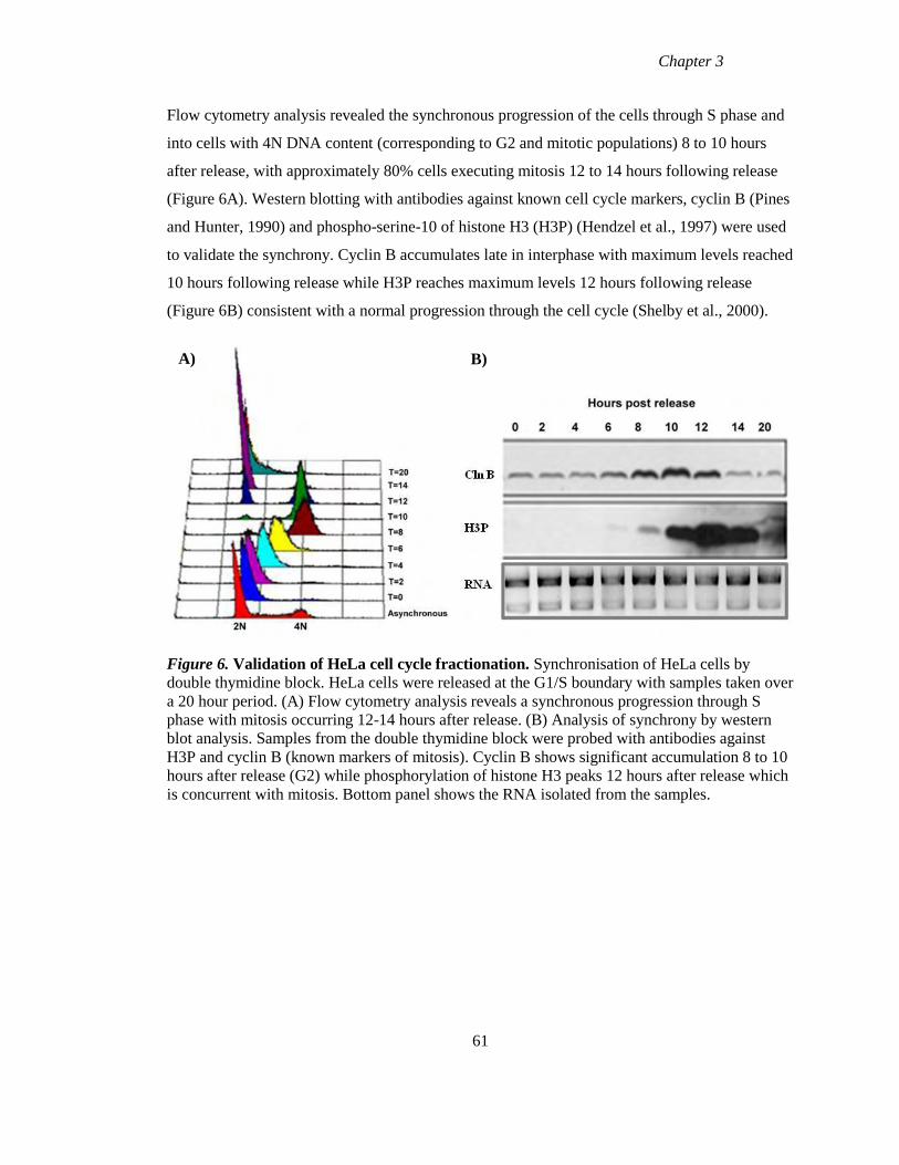
Chapter 3
Flow cytometry analysis revealed the synchronous progression of the cells through S phase and
into cells with 4N DNA content (corresponding to G2 and mitotic populations) 8 to 10 hours
after release, with approximately 80% cells executing mitosis 12 to 14 hours following release
(Figure 6A). Western blotting with antibodies against known cell cycle markers, cyclin B (Pines
and Hunter, 1990) and phospho-serine-10 of histone H3 (H3P) (Hendzel et al., 1997) were used
to validate the synchrony. Cyclin B accumulates late in interphase with maximum levels reached
10 hours following release while H3P reaches maximum levels 12 hours following release
(Figure 6B) consistent with a normal progression through the cell cycle (Shelby et al., 2000).
Figure 6. Validation of HeLa cell cycle fractionation. Synchronisation of HeLa cells by
double thymidine block. HeLa cells were released at the G1/S boundary with samples taken over
a 20 hour period. (A) Flow cytometry analysis reveals a synchronous progression through S
phase with mitosis occurring 12-14 hours after release. (B) Analysis of synchrony by western
blot analysis. Samples from the double thymidine block were probed with antibodies against
H3P and cyclin B (known markers of mitosis). Cyclin B shows significant accumulation 8 to 10
hours after release (G2) while phosphorylation of histone H3 peaks 12 hours after release which
is concurrent with mitosis. Bottom panel shows the RNA isolated from the samples.
61
A) B)
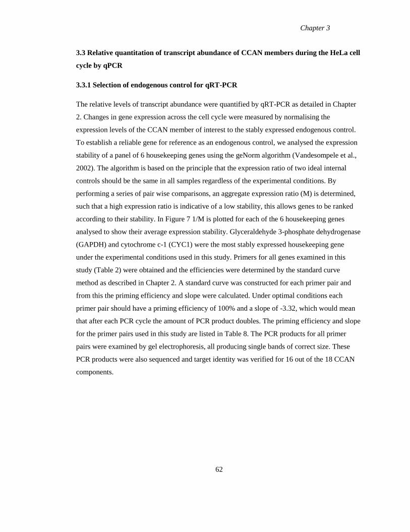
Chapter 3
3.3 Relative quantitation of transcript abundance of CCAN members during the HeLa cell
cycle by qPCR
3.3.1 Selection of endogenous control for qRT-PCR
The relative levels of transcript abundance were quantified by qRT-PCR as detailed in Chapter
2. Changes in gene expression across the cell cycle were measured by normalising the
expression levels of the CCAN member of interest to the stably expressed endogenous control.
To establish a reliable gene for reference as an endogenous control, we analysed the expression
stability of a panel of 6 housekeeping genes using the geNorm algorithm (Vandesompele et al.,
2002). The algorithm is based on the principle that the expression ratio of two ideal internal
controls should be the same in all samples regardless of the experimental conditions. By
performing a series of pair wise comparisons, an aggregate expression ratio (M) is determined,
such that a high expression ratio is indicative of a low stability, this allows genes to be ranked
according to their stability. In Figure 7 1/M is plotted for each of the 6 housekeeping genes
analysed to show their average expression stability. Glyceraldehyde 3-phosphate dehydrogenase
(GAPDH) and cytochrome c-1 (CYC1) were the most stably expressed housekeeping gene
under the experimental conditions used in this study. Primers for all genes examined in this
study (Table 2) were obtained and the efficiencies were determined by the standard curve
method as described in Chapter 2. A standard curve was constructed for each primer pair and
from this the priming efficiency and slope were calculated. Under optimal conditions each
primer pair should have a priming efficiency of 100% and a slope of -3.32, which would mean
that after each PCR cycle the amount of PCR product doubles. The priming efficiency and slope
for the primer pairs used in this study are listed in Table 8. The PCR products for all primer
pairs were examined by gel electrophoresis, all producing single bands of correct size. These
PCR products were also sequenced and target identity was verified for 16 out of the 18 CCAN
components.
62
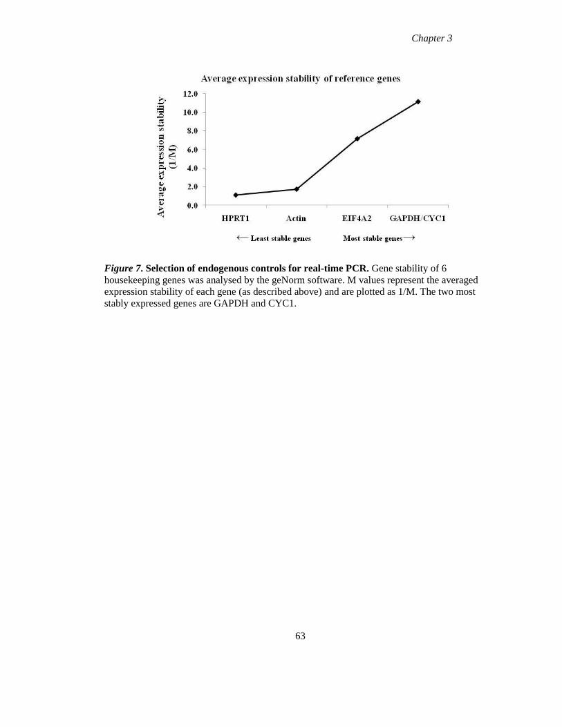
Chapter 3
Figure 7. Selection of endogenous controls for real-time PCR. Gene stability of 6
housekeeping genes was analysed by the geNorm software. M values represent the averaged
expression stability of each gene (as described above) and are plotted as 1/M. The two most
stably expressed genes are GAPDH and CYC1.
63
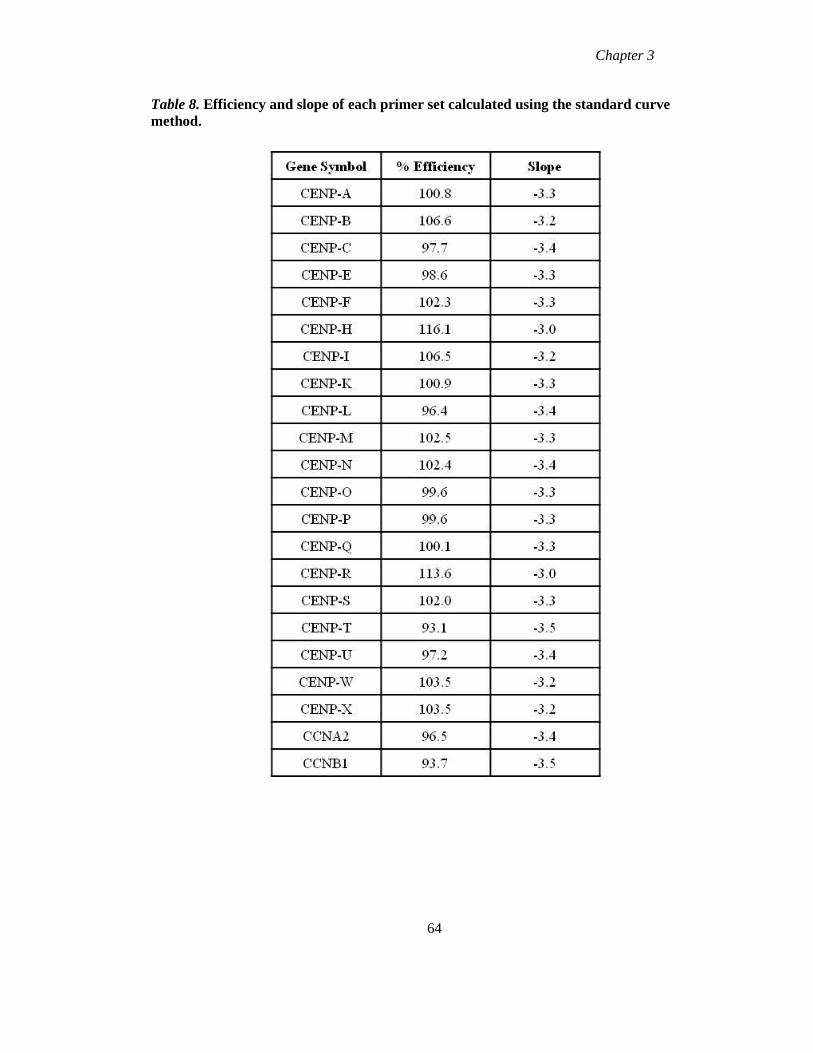
Chapter 3
Table 8. Efficiency and slope of each primer set calculated using the standard curve
method.
64
Chapter 3
The expression profile of each gene was measured in the synchronised HeLa cell RNA samples
characterised above. RNA from each time-point was reverse transcribed into cDNA and the
relative transcript abundance of each gene was examined by qRT-PCR. The relative transcript
abundance of each centromere gene across the cell cycle was measured using the ∆∆CT method,
which involved first normalising the expression level of the gene of interest to the endogenous
control (GAPDH) and then normalising these values to the expression levels from a reference
point within the sample set, chosen as t=4 in this study. The standard ∆∆CT method assumes the
priming efficiency for each reaction is 100%. In order to improve the accuracy of the
measurement, the priming efficiency of each primer pair was incorporated into the calculation
(as outlined in Chapter 2) (Pfaffl, 2001). Data from three independent experiments were then
averaged (± SEM) and plotted in a graph versus time (Figure 9). These data were then analysed
for statistically significant changes in relative transcript abundance across the HeLa cell cycle
using ANOVA (Table 9).
3.3.2 Relative transcript abundance of the genes encoding known cell cycle markers and
outer kinetochore proteins.
The relative transcript abundance of known cell cycle markers was measured to evaluate the
correspondence between the qRT-PCR analysis and the biochemical and flow cytometric
analysis in the synchronised HeLa cells. The replication dependent histone H2A showed up-
regulation between 4 and 8 hours after release (Figure 8A) (Sittman et al., 1983). Cyclin A
abundance began to increase 2 hours after release with maximum levels reached at 12 hours
while cyclin B accumulation began 8 hours after release (Figure 8A), also consistent with
previous reports (Pines and Hunter, 1989, Pines and Hunter, 1990). Both cyclin A and cyclin B
showed a rapid decrease in abundance between 12 and 14 hours after release which is
concurrent with the appearance of 2N (G1) cells following mitosis. The transcript abundance of
outer kinetochore proteins CENP-E and CENP-F was also examined. CENP-E accumulation
began late in S phase (6 hours after release) and continued until the cells had entered mitosis,
while CENP-F accumulation began during late S phase and peaked 10 hours after release
consistent with previous reports (Figure 8B) (Brown et al., 1994). Similar to the cyclins, CENP-
E and CENP-F show a rapid return to baseline levels following mitosis. The normal expression
profile of these known cell cycle markers demonstrated a high level of synchrony was achieved,
and documented normal patterns of cell cycle dependent gene expression in these experiments.
65
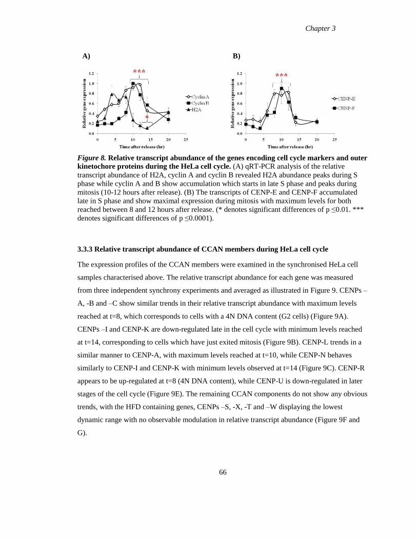
Chapter 3
Figure 8. Relative transcript abundance of the genes encoding cell cycle markers and outer
kinetochore proteins during the HeLa cell cycle. (A) qRT-PCR analysis of the relative
transcript abundance of H2A, cyclin A and cyclin B revealed H2A abundance peaks during S
phase while cyclin A and B show accumulation which starts in late S phase and peaks during
mitosis (10-12 hours after release). (B) The transcripts of CENP-E and CENP-F accumulated
late in S phase and show maximal expression during mitosis with maximum levels for both
reached between 8 and 12 hours after release. (* denotes significant differences of p ≤0.01. ***
denotes significant differences of p ≤0.0001).
3.3.3 Relative transcript abundance of CCAN members during HeLa cell cycle
The expression profiles of the CCAN members were examined in the synchronised HeLa cell
samples characterised above. The relative transcript abundance for each gene was measured
from three independent synchrony experiments and averaged as illustrated in Figure 9. CENPs –
A, -B and –C show similar trends in their relative transcript abundance with maximum levels
reached at t=8, which corresponds to cells with a 4N DNA content (G2 cells) (Figure 9A).
CENPs –I and CENP-K are down-regulated late in the cell cycle with minimum levels reached
at t=14, corresponding to cells which have just exited mitosis (Figure 9B). CENP-L trends in a
similar manner to CENP-A, with maximum levels reached at t=10, while CENP-N behaves
similarly to CENP-I and CENP-K with minimum levels observed at t=14 (Figure 9C). CENP-R
appears to be up-regulated at t=8 (4N DNA content), while CENP-U is down-regulated in later
stages of the cell cycle (Figure 9E). The remaining CCAN components do not show any obvious
trends, with the HFD containing genes, CENPs –S, -X, -T and –W displaying the lowest
dynamic range with no observable modulation in relative transcript abundance (Figure 9F and
G).
66
A) B)
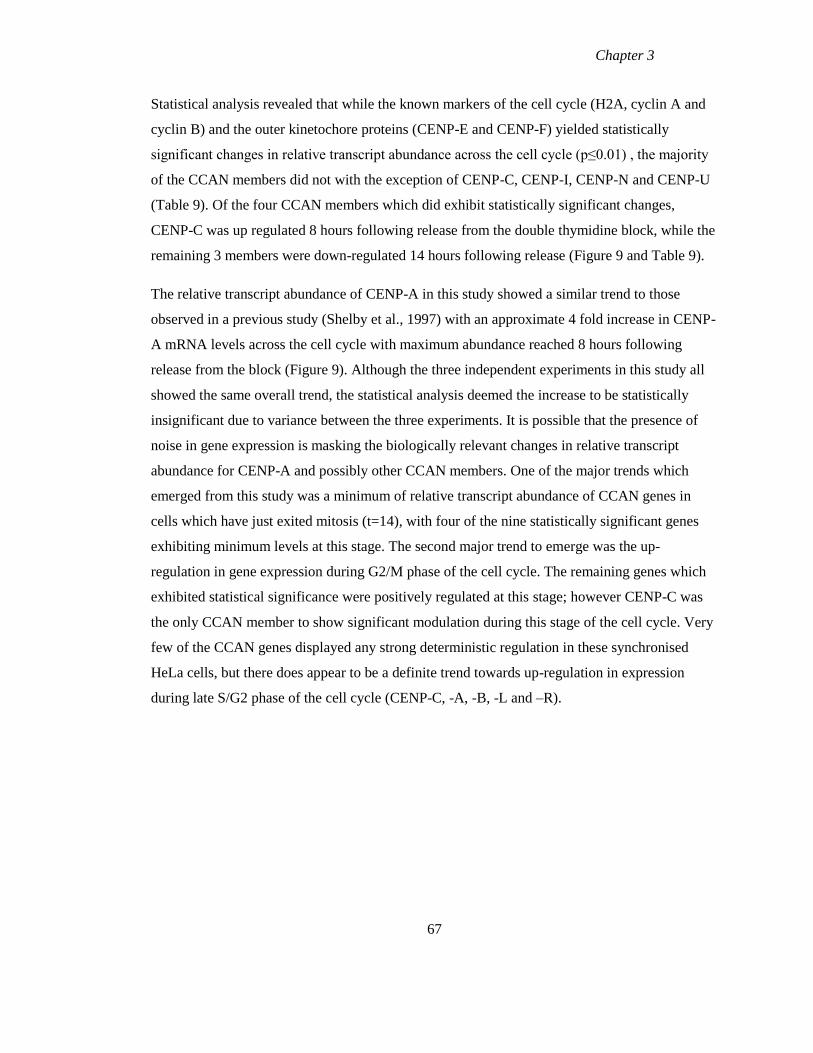
Chapter 3
Statistical analysis revealed that while the known markers of the cell cycle (H2A, cyclin A and
cyclin B) and the outer kinetochore proteins (CENP-E and CENP-F) yielded statistically
significant changes in relative transcript abundance across the cell cycle (p≤0.01) , the majority
of the CCAN members did not with the exception of CENP-C, CENP-I, CENP-N and CENP-U
(Table 9). Of the four CCAN members which did exhibit statistically significant changes,
CENP-C was up regulated 8 hours following release from the double thymidine block, while the
remaining 3 members were down-regulated 14 hours following release (Figure 9 and Table 9).
The relative transcript abundance of CENP-A in this study showed a similar trend to those
observed in a previous study (Shelby et al., 1997) with an approximate 4 fold increase in CENP-
A mRNA levels across the cell cycle with maximum abundance reached 8 hours following
release from the block (Figure 9). Although the three independent experiments in this study all
showed the same overall trend, the statistical analysis deemed the increase to be statistically
insignificant due to variance between the three experiments. It is possible that the presence of
noise in gene expression is masking the biologically relevant changes in relative transcript
abundance for CENP-A and possibly other CCAN members. One of the major trends which
emerged from this study was a minimum of relative transcript abundance of CCAN genes in
cells which have just exited mitosis (t=14), with four of the nine statistically significant genes
exhibiting minimum levels at this stage. The second major trend to emerge was the up-
regulation in gene expression during G2/M phase of the cell cycle. The remaining genes which
exhibited statistical significance were positively regulated at this stage; however CENP-C was
the only CCAN member to show significant modulation during this stage of the cell cycle. Very
few of the CCAN genes displayed any strong deterministic regulation in these synchronised
HeLa cells, but there does appear to be a definite trend towards up-regulation in expression
during late S/G2 phase of the cell cycle (CENP-C, -A, -B, -L and –R).
67
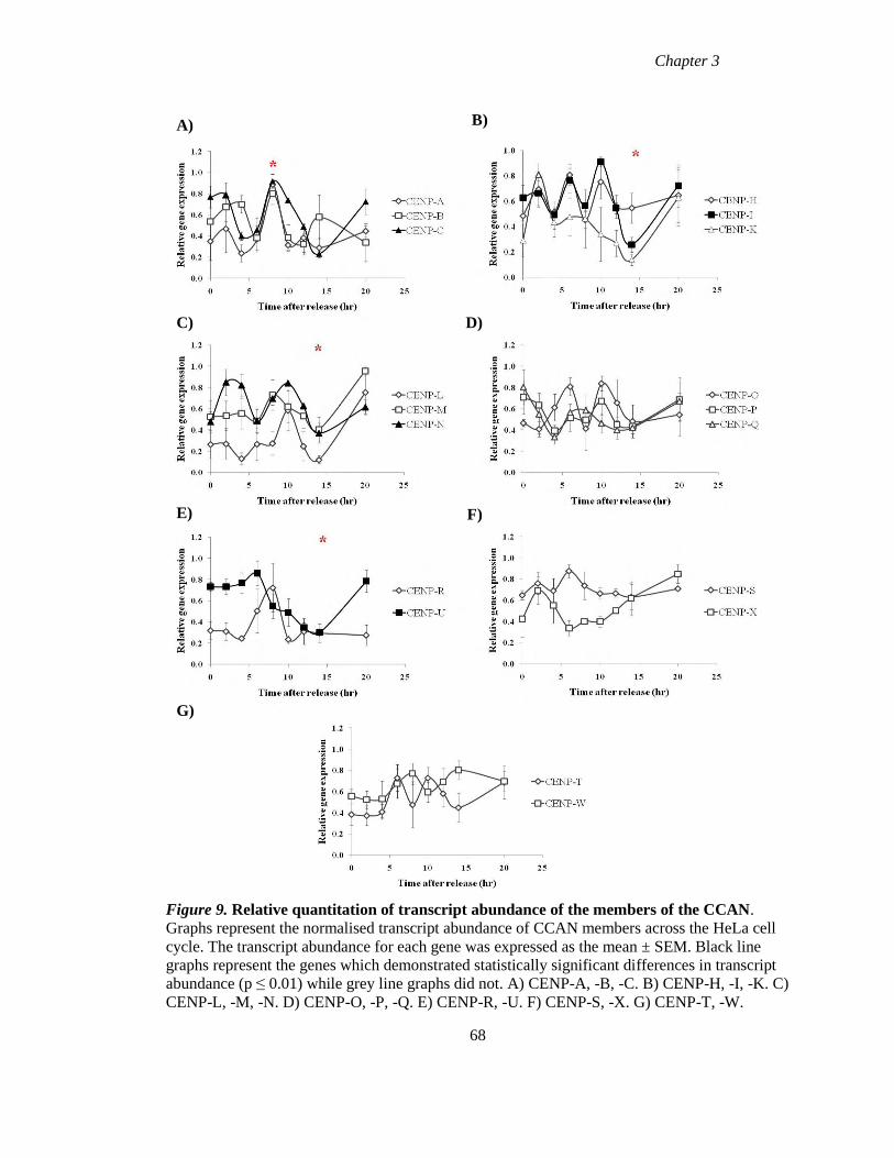
Chapter 3
Figure 9. Relative quantitation of transcript abundance of the members of the CCAN.
Graphs represent the normalised transcript abundance of CCAN members across the HeLa cell
cycle. The transcript abundance for each gene was expressed as the mean ± SEM. Black line
graphs represent the genes which demonstrated statistically significant differences in transcript
abundance (p ≤ 0.01) while grey line graphs did not. A) CENP-A, -B, -C. B) CENP-H, -I, -K. C)
CENP-L, -M, -N. D) CENP-O, -P, -Q. E) CENP-R, -U. F) CENP-S, -X. G) CENP-T, -W.
68
A) B)
D) C)
E) F)
G)
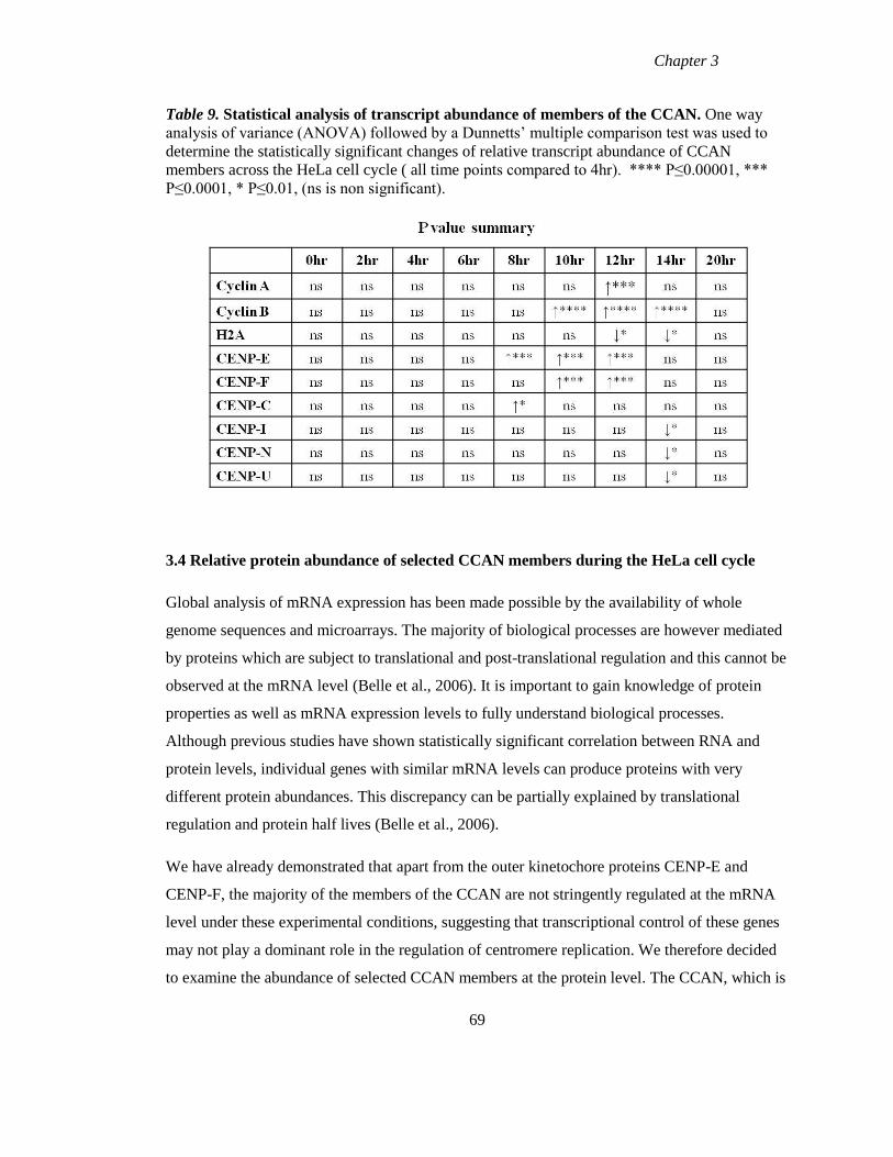
Chapter 3
Table 9. Statistical analysis of transcript abundance of members of the CCAN. One way
analysis of variance (ANOVA) followed by a Dunnetts’ multiple comparison test was used to
determine the statistically significant changes of relative transcript abundance of CCAN
members across the HeLa cell cycle ( all time points compared to 4hr). **** P≤0.00001, ***
P≤0.0001, * P≤0.01, (ns is non significant).
3.4 Relative protein abundance of selected CCAN members during the HeLa cell cycle
Global analysis of mRNA expression has been made possible by the availability of whole
genome sequences and microarrays. The majority of biological processes are however mediated
by proteins which are subject to translational and post-translational regulation and this cannot be
observed at the mRNA level (Belle et al., 2006). It is important to gain knowledge of protein
properties as well as mRNA expression levels to fully understand biological processes.
Although previous studies have shown statistically significant correlation between RNA and
protein levels, individual genes with similar mRNA levels can produce proteins with very
different protein abundances. This discrepancy can be partially explained by translational
regulation and protein half lives (Belle et al., 2006).
We have already demonstrated that apart from the outer kinetochore proteins CENP-E and
CENP-F, the majority of the members of the CCAN are not stringently regulated at the mRNA
level under these experimental conditions, suggesting that transcriptional control of these genes
may not play a dominant role in the regulation of centromere replication. We therefore decided
to examine the abundance of selected CCAN members at the protein level. The CCAN, which is
69
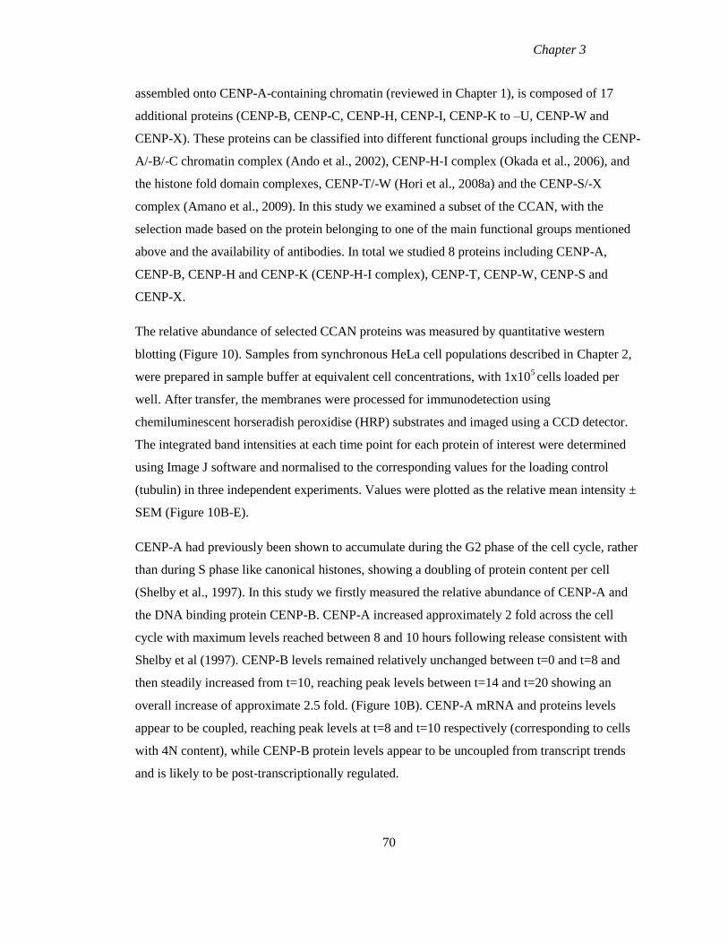
Chapter 3
assembled onto CENP-A-containing chromatin (reviewed in Chapter 1), is composed of 17
additional proteins (CENP-B, CENP-C, CENP-H, CENP-I, CENP-K to –U, CENP-W and
CENP-X). These proteins can be classified into different functional groups including the CENP-
A/-B/-C chromatin complex (Ando et al., 2002), CENP-H-I complex (Okada et al., 2006), and
the histone fold domain complexes, CENP-T/-W (Hori et al., 2008a) and the CENP-S/-X
complex (Amano et al., 2009). In this study we examined a subset of the CCAN, with the
selection made based on the protein belonging to one of the main functional groups mentioned
above and the availability of antibodies. In total we studied 8 proteins including CENP-A,
CENP-B, CENP-H and CENP-K (CENP-H-I complex), CENP-T, CENP-W, CENP-S and
CENP-X.
The relative abundance of selected CCAN proteins was measured by quantitative western
blotting (Figure 10). Samples from synchronous HeLa cell populations described in Chapter 2,
were prepared in sample buffer at equivalent cell concentrations, with 1x10⁵ cells loaded per
well. After transfer, the membranes were processed for immunodetection using
chemiluminescent horseradish peroxidise (HRP) substrates and imaged using a CCD detector.
The integrated band intensities at each time point for each protein of interest were determined
using Image J software and normalised to the corresponding values for the loading control
(tubulin) in three independent experiments. Values were plotted as the relative mean intensity ±
SEM (Figure 10B-E).
CENP-A had previously been shown to accumulate during the G2 phase of the cell cycle, rather
than during S phase like canonical histones, showing a doubling of protein content per cell
(Shelby et al., 1997). In this study we firstly measured the relative abundance of CENP-A and
the DNA binding protein CENP-B. CENP-A increased approximately 2 fold across the cell
cycle with maximum levels reached between 8 and 10 hours following release consistent with
Shelby et al (1997). CENP-B levels remained relatively unchanged between t=0 and t=8 and
then steadily increased from t=10, reaching peak levels between t=14 and t=20 showing an
overall increase of approximate 2.5 fold. (Figure 10B). CENP-A mRNA and proteins levels
appear to be coupled, reaching peak levels at t=8 and t=10 respectively (corresponding to cells
with 4N content), while CENP-B protein levels appear to be uncoupled from transcript trends
and is likely to be post-transcriptionally regulated.
70
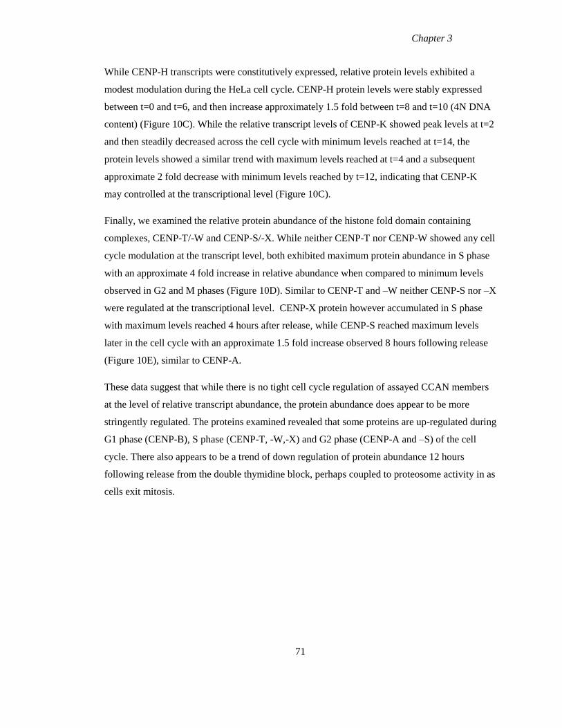
Chapter 3
While CENP-H transcripts were constitutively expressed, relative protein levels exhibited a
modest modulation during the HeLa cell cycle. CENP-H protein levels were stably expressed
between t=0 and t=6, and then increase approximately 1.5 fold between t=8 and t=10 (4N DNA
content) (Figure 10C). While the relative transcript levels of CENP-K showed peak levels at t=2
and then steadily decreased across the cell cycle with minimum levels reached at t=14, the
protein levels showed a similar trend with maximum levels reached at t=4 and a subsequent
approximate 2 fold decrease with minimum levels reached by t=12, indicating that CENP-K
may controlled at the transcriptional level (Figure 10C).
Finally, we examined the relative protein abundance of the histone fold domain containing
complexes, CENP-T/-W and CENP-S/-X. While neither CENP-T nor CENP-W showed any cell
cycle modulation at the transcript level, both exhibited maximum protein abundance in S phase
with an approximate 4 fold increase in relative abundance when compared to minimum levels
observed in G2 and M phases (Figure 10D). Similar to CENP-T and –W neither CENP-S nor –X
were regulated at the transcriptional level. CENP-X protein however accumulated in S phase
with maximum levels reached 4 hours after release, while CENP-S reached maximum levels
later in the cell cycle with an approximate 1.5 fold increase observed 8 hours following release
(Figure 10E), similar to CENP-A.
These data suggest that while there is no tight cell cycle regulation of assayed CCAN members
at the level of relative transcript abundance, the protein abundance does appear to be more
stringently regulated. The proteins examined revealed that some proteins are up-regulated during
G1 phase (CENP-B), S phase (CENP-T, -W,-X) and G2 phase (CENP-A and –S) of the cell
cycle. There also appears to be a trend of down regulation of protein abundance 12 hours
following release from the double thymidine block, perhaps coupled to proteosome activity in as
cells exit mitosis.
71
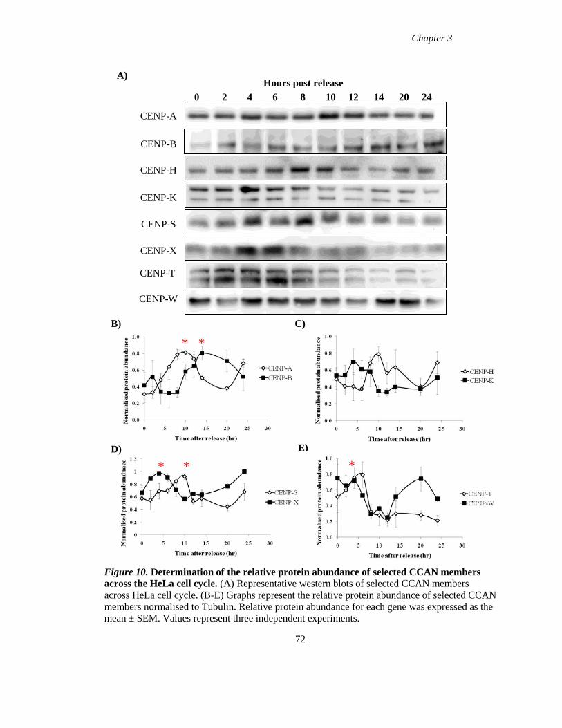
Chapter 3
Figure 10. Determination of the relative protein abundance of selected CCAN members
across the HeLa cell cycle. (A) Representative western blots of selected CCAN members
across HeLa cell cycle. (B-E) Graphs represent the relative protein abundance of selected CCAN
members normalised to Tubulin. Relative protein abundance for each gene was expressed as the
mean ± SEM. Values represent three independent experiments.
72
0 2 4 6 8 10 12 14 20 24
Hours post release
CENP-W
CENP-A
CENP-B
CENP-K
CENP-H
CENP-S
CENP-X
CENP-T
A)
B)
* *
C)
E)
* * *
D)
Chapter 3
3.5 Cell cycle fractionation of K-562 cells by elutriation
The double thymidine block protocol requires a physiological perturbation of cells to induce
synchrony. An alternative method is to fractionate an unperturbed population of cells which can
be done using counter-flow centrifugal elutriation (CCE) (Bauer, 1999). CCE separates cells on
the basis of cell size; it takes place in culture medium and therefore results in the characteristics
of the elutriated samples closely reflecting those of cell populations before elutriation, however
it does yield lower resolution in cell cycle fractionation. CCE was initially applied to HeLa cells
in order to maintain continuity of the cell system. However this was unsuccessful due to the fact
that these cells formed clumps very quickly, limiting the resolution of the size based
fractionation. The human lymphoid K-562 cell line, which grows in suspension, was instead
used as they are more suited to CCE.
Elutriation was performed and fractions were collected as outlined in Chapter 2, cell
concentrations were determined and samples were analysed by flow cytometry and western blot.
Flow cytometry analysis revealed good fractionation across the eight samples collected.
Fractions 1 and 2 were enriched in G1 cells, fractions 4 and 5 were enriched in S phase cells
while fractions 6, 7 and 8 were enriched in 4N cells corresponding to G2 and mitotic
populations (Figure 11A). Western blot analysis with antibodies against the known mitotic
markers, cyclin B and H3P showed that both proteins have maximum abundance in the G2 and
M enriched fractions (Figure 11B).
73
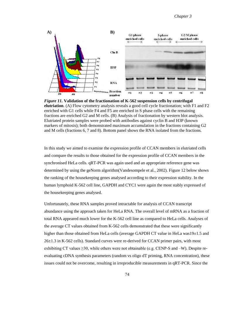
Chapter 3
Figure 11. Validation of the fractionation of K-562 suspension cells by centrifugal
elutriation. (A) Flow cytometry analysis reveals a good cell cycle fractionation; with F1 and F2
enriched with G1 cells while F4 and F5 are enriched in S phase cells with the remaining
fractions are enriched G2 and M cells. (B) Analysis of fractionation by western blot analysis.
Elutriated protein samples were probed with antibodies against cyclin B and H3P (known
markers of mitosis); both demonstrated maximum accumulation in the fractions containing G2
and M cells (fractions 6, 7 and 8). Bottom panel shows the RNA isolated from the fractions.
In this study we aimed to examine the expression profile of CCAN members in elutriated cells
and compare the results to those obtained for the expression profile of CCAN members in the
synchronised HeLa cells. qRT-PCR was again used and an appropriate reference gene was
determined by using the geNorm algorithm(Vandesompele et al., 2002). Figure 12 below shows
the ranking of the housekeeping genes analysed according to their expression stability. In the
human lymphoid K-562 cell line, GAPDH and CYC1 were again the most stably expressed of
the housekeeping genes analysed.
Unfortunately, these RNA samples proved intractable for analysis of CCAN transcript
abundance using the approach taken for HeLa RNA. The overall level of mRNA as a fraction of
total RNA appeared much lower for the K-562 cell line as compared to HeLa cells. Analyses of
the average CT values obtained from K-562 cells demonstrated that these were significantly
higher than those obtained from HeLa cells (average GAPDH CT value in HeLa was19±1.5 and
26±1.3 in K-562 cells). Standard curves were re-derived for CCAN primer pairs, with most
exhibiting CT values ≥30, while others were not obtainable (e.g. CENP-S and –W). Despite re-
evaluating cDNA synthesis parameters (random vs oligo dT priming, RNA concentration), these
issues could not be overcome, resulting in irreproducible measurements in qRT-PCR. Since the
74
A)
)
B)
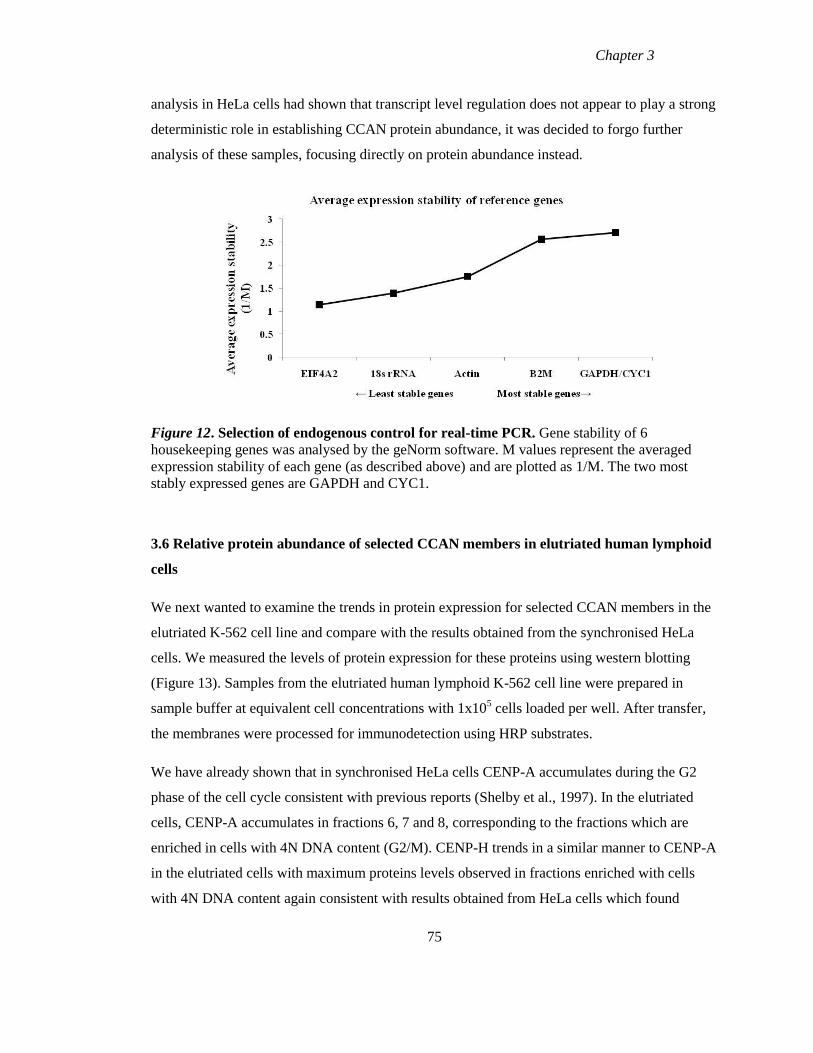
Chapter 3
analysis in HeLa cells had shown that transcript level regulation does not appear to play a strong
deterministic role in establishing CCAN protein abundance, it was decided to forgo further
analysis of these samples, focusing directly on protein abundance instead.
Figure 12. Selection of endogenous control for real-time PCR. Gene stability of 6
housekeeping genes was analysed by the geNorm software. M values represent the averaged
expression stability of each gene (as described above) and are plotted as 1/M. The two most
stably expressed genes are GAPDH and CYC1.
3.6 Relative protein abundance of selected CCAN members in elutriated human lymphoid
cells
We next wanted to examine the trends in protein expression for selected CCAN members in the
elutriated K-562 cell line and compare with the results obtained from the synchronised HeLa
cells. We measured the levels of protein expression for these proteins using western blotting
(Figure 13). Samples from the elutriated human lymphoid K-562 cell line were prepared in
sample buffer at equivalent cell concentrations with 1x10⁵ cells loaded per well. After transfer,
the membranes were processed for immunodetection using HRP substrates.
We have already shown that in synchronised HeLa cells CENP-A accumulates during the G2
phase of the cell cycle consistent with previous reports (Shelby et al., 1997). In the elutriated
cells, CENP-A accumulates in fractions 6, 7 and 8, corresponding to the fractions which are
enriched in cells with 4N DNA content (G2/M). CENP-H trends in a similar manner to CENP-A
in the elutriated cells with maximum proteins levels observed in fractions enriched with cells
with 4N DNA content again consistent with results obtained from HeLa cells which found
75
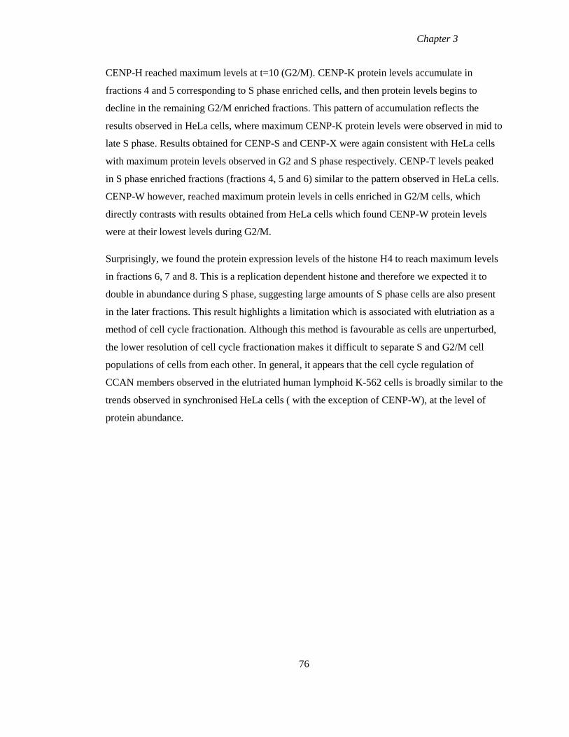
Chapter 3
CENP-H reached maximum levels at t=10 (G2/M). CENP-K protein levels accumulate in
fractions 4 and 5 corresponding to S phase enriched cells, and then protein levels begins to
decline in the remaining G2/M enriched fractions. This pattern of accumulation reflects the
results observed in HeLa cells, where maximum CENP-K protein levels were observed in mid to
late S phase. Results obtained for CENP-S and CENP-X were again consistent with HeLa cells
with maximum protein levels observed in G2 and S phase respectively. CENP-T levels peaked
in S phase enriched fractions (fractions 4, 5 and 6) similar to the pattern observed in HeLa cells.
CENP-W however, reached maximum protein levels in cells enriched in G2/M cells, which
directly contrasts with results obtained from HeLa cells which found CENP-W protein levels
were at their lowest levels during G2/M.
Surprisingly, we found the protein expression levels of the histone H4 to reach maximum levels
in fractions 6, 7 and 8. This is a replication dependent histone and therefore we expected it to
double in abundance during S phase, suggesting large amounts of S phase cells are also present
in the later fractions. This result highlights a limitation which is associated with elutriation as a
method of cell cycle fractionation. Although this method is favourable as cells are unperturbed,
the lower resolution of cell cycle fractionation makes it difficult to separate S and G2/M cell
populations of cells from each other. In general, it appears that the cell cycle regulation of
CCAN members observed in the elutriated human lymphoid K-562 cells is broadly similar to the
trends observed in synchronised HeLa cells ( with the exception of CENP-W), at the level of
protein abundance.
76
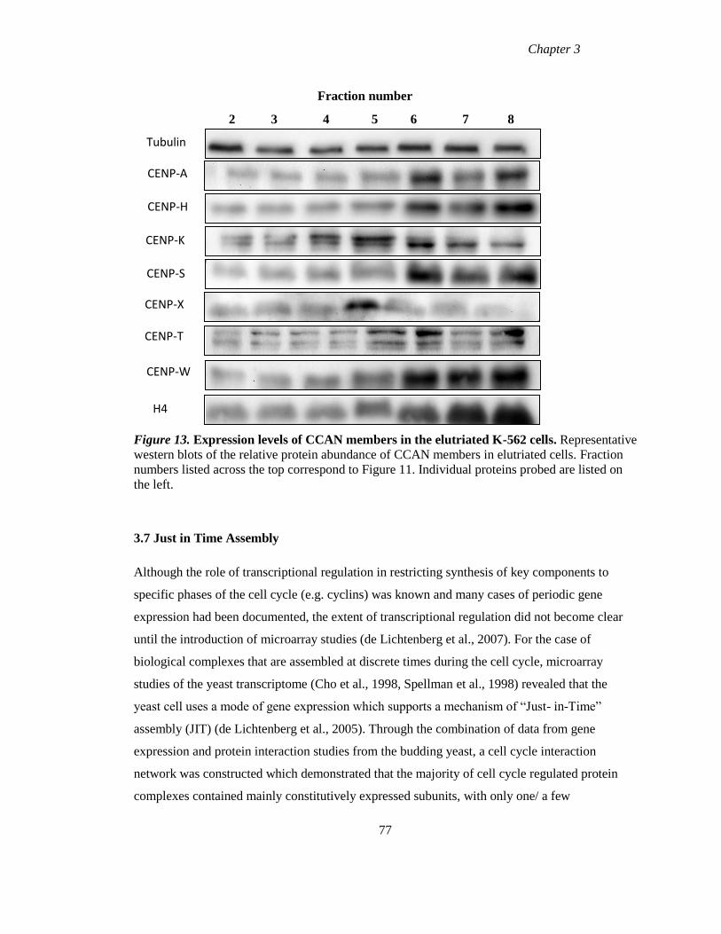
Chapter 3
Figure 13. Expression levels of CCAN members in the elutriated K-562 cells. Representative
western blots of the relative protein abundance of CCAN members in elutriated cells. Fraction
numbers listed across the top correspond to Figure 11. Individual proteins probed are listed on
the left.
3.7 Just in Time Assembly
Although the role of transcriptional regulation in restricting synthesis of key components to
specific phases of the cell cycle (e.g. cyclins) was known and many cases of periodic gene
expression had been documented, the extent of transcriptional regulation did not become clear
until the introduction of microarray studies (de Lichtenberg et al., 2007). For the case of
biological complexes that are assembled at discrete times during the cell cycle, microarray
studies of the yeast transcriptome (Cho et al., 1998, Spellman et al., 1998) revealed that the
yeast cell uses a mode of gene expression which supports a mechanism of “Just- in-Time”
assembly (JIT) (de Lichtenberg et al., 2005). Through the combination of data from gene
expression and protein interaction studies from the budding yeast, a cell cycle interaction
network was constructed which demonstrated that the majority of cell cycle regulated protein
complexes contained mainly constitutively expressed subunits, with only one/ a few
77
Fraction number
2 3 4 5 6 7 8
Tubulin
CENP-A
CENP-K
CENP-S
CENP-X
CENP-T
CENP-H
CENP-W
H4
Chapter 3
dynamically expressed subunits (de Lichtenberg et al., 2005). Transcription of the periodically
expressed subunit was found to peak just before the protein complex functions, which suggests a
link between the timing of transcription and protein complex activation (de Lichtenberg et al.,
2005). Further microarray data from humans, Arabidopsis thaliana and fission yeast extended
this finding, showing that JIT assembly is an evolutionary conserved feature of the eukaryotic
cell cycle (Jensen et al., 2006). Although transcriptional regulation is widely used to control the
synthesis of key proteins during the cell cycle, each species regulated a different set of proteins
(Jensen et al., 2006).
The proteome of eukaryotic cells is organised into multi-protein complexes which act as the
molecular machinery of the cell (Aloy et al., 2004). These multi-protein complexes are
composed of both periodically and constitutively expressed subunits. Typically only one or a
few subunits are transcriptionally regulated and these subunits are responsible for the activity of
the protein complex. Analysis of the transcript abundance of CCAN members by qRT-PCR
(section 3.3.3) revealed that apart from the outer kinetochore proteins CENP-E and CENP-F,
only CENP-C showed periodic expression, while the majority of CCAN members did not
exhibit strong modulation in relative transcript levels. The relative transcript abundance of
CENP-C, CENP-E and CENP-F is up-regulated during G2 and M phases of the cell cycle,
which is coincident with centromere/kinetochore activity which led us to ask whether the JIT
mode of assembly could be responsible for the centromere and kinetochore assembly.
If JIT assembly is an important feature of centromere replication, this mode of assembly should
be evolutionary conserved. The Saccharomyces Genome Database (SGD) which contains
extensive gene expression data, allowed us to ask whether JIT assembly is a feature of the
budding yeast kinetochore by examining the expression profile of the components of the
kinetochore. The budding yeast kinetochore is the simplest of all kinetochores, with each
kinetochore binding a single microtubule. These kinetochores are comprised of approximately
60 proteins which belong to seven different complexes, which in turn are associated with
chromatin proteins (Figure 14). Pramila et al (2006) generated two sets of microarray data from
the budding yeast cell cycle and combined this data with data from an additional three data sets.
This meta analysis compiled data from the 5 cell cycle sets, and genes were ranked according to
their dynamic range and periodicity. The transcript dynamics were then expressed in terms of
their maximal abundance using a 0-100% scale, with the M/G1 boundary set as 0%.
78
Chapter 3
Using this data from the SGD, we identified the components of the budding yeast kinetochore
which were periodically expressed and when during the cell cycle these subunits peak in
expression (summarised in Table 10). The schematic in Figure 14 represents the complexes
within the kinetochore of the budding yeast Saccharomyces cerevisiae. The inner kinetochore
consists of 8 subunits, 3 of which are cell cycle regulated, while the remaining components are
constitutively expressed (coloured in grey). Mif2 (CENP-C homologue) displays cell cycle
regulation, with maximum expression levels occurring at 21% of the cell cycle which
corresponds to the G1 phase of yeast cell cycle (yellow colour). The Ndc10 component of the
CBF3 complex also exhibits cell cycle regulation with gene expression up regulated at 34% of
the cell cycle which represents the G1/S boundary (orange colour). The final component of the
inner kinetochore to exhibit periodic gene expression is Cse4 (CENP-A homologue) which
exhibits maximum levels of gene expression during S phase (red colour). Of the complexes
which comprise the central kinetochore only the Ndc80 complex displays components with
periodic expression. Spc25 and Nuf2 display maximum expression levels during S phase of the
cell cycle (red colour), while the remaining complexes are composed of constitutively expressed
subunits. Examination of the components of the outer kinetochore shows a similar pattern of
expression consistent with JIT assembly in that only four components of the Dam1 complex are
transcriptionally regulated (G1/S phase of cell cycle) while the remaining components are stably
expressed.
The kinetochore of the budding yeast is therefore composed of both periodically and
constitutively expressed subunits, with only a small number of these subunits under
transcriptional regulation. Of the 45 subunits which were examined, only 10 of these were
transcriptionally regulated, with an increase in transcript abundance observed between 21 and 60
% of the cell cycle, which corresponds to late G1 continuing into S phase, which is consistent
with the JIT assembly model. Taken together with the results from the gene expression analysis
of CCAN members in HeLa cells which found that only CENP-C of the CCAN and the outer
kinetochore proteins CENP-E and CENP-F were under tight transcriptional control, it appears
that centromere/kinetochore complex assembly is via a JIT mechanism.
79
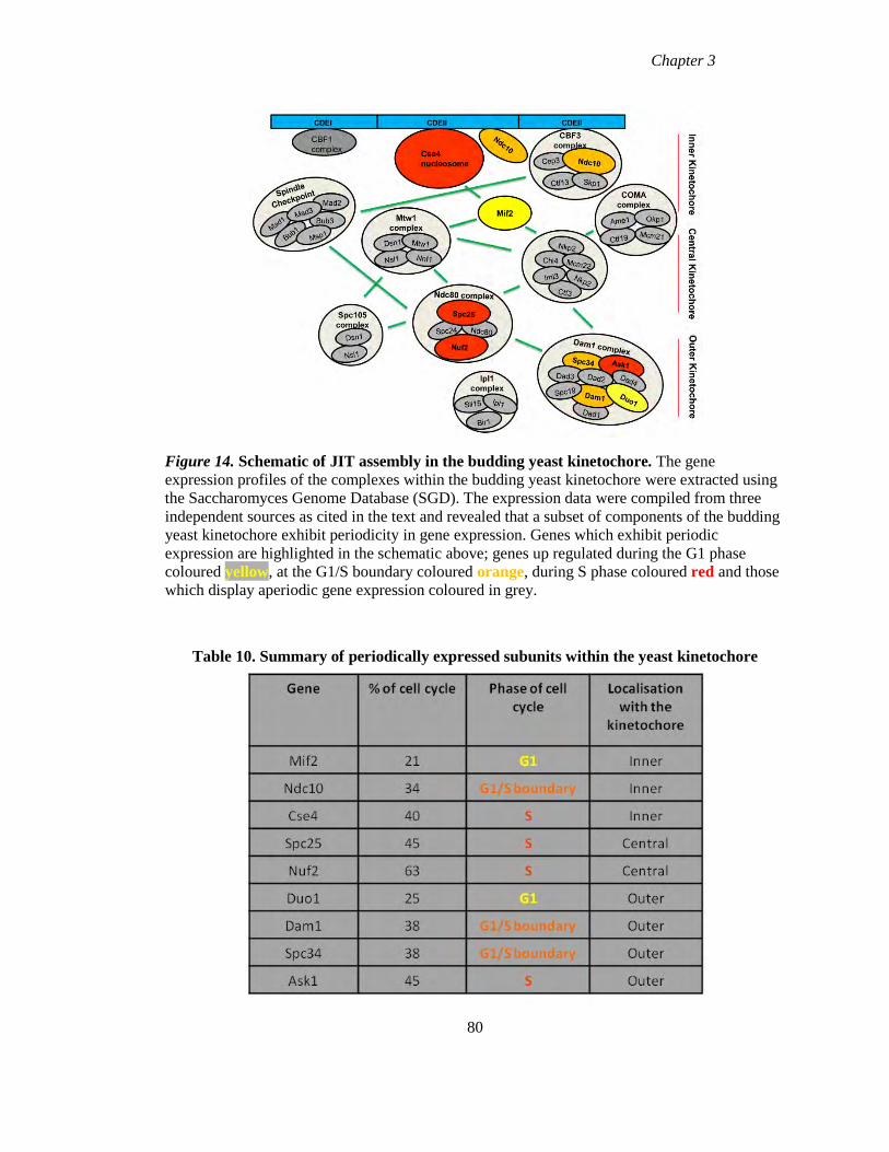
Chapter 3
Figure 14. Schematic of JIT assembly in the budding yeast kinetochore. The gene
expression profiles of the complexes within the budding yeast kinetochore were extracted using
the Saccharomyces Genome Database (SGD). The expression data were compiled from three
independent sources as cited in the text and revealed that a subset of components of the budding
yeast kinetochore exhibit periodicity in gene expression. Genes which exhibit periodic
expression are highlighted in the schematic above; genes up regulated during the G1 phase
coloured yellow, at the G1/S boundary coloured orange, during S phase coloured red and those
which display aperiodic gene expression coloured in grey.
Table 10. Summary of periodically expressed subunits within the yeast kinetochore
80
Chapter 3
3.8 Discussion
The aim of this study was to determine whether a programme of cell cycle regulation of gene
expression for CCAN components existed which could provide insight into the cellular
regulation of centromere replication. Previous work with CENP-A reported that the histone H3
variant had a distinct mRNA expression profile, which peaked during G2 phase, with the
assembly of the protein at centromeres occurring in a replication independent manner (Shelby et
al., 1997, Shelby et al., 2000). This uncoupling of mRNA expression and assembly of CENP-A
at centromeres from DNA replication appears to be a feature of many organisms including
fission yeast and Drosophila (Takahashi et al., 2000, Takayama et al., 2008, Sullivan and
Karpen, 2001), and suggests the existence of a distinctive chromatin assembly pathway for
centromeres. Examination of the expression profiles of the human genes encoding CCAN
members did not in fact reveal a programme of regulated expression for most genes; rather
protein abundance appeared to be modulated during the cell cycle for selected proteins. Thus
like other systems transcription levels are not tightly coupled with protein abundance. Overall
our results are consistent with a JIT mode of assembly and our analysis of expression levels of
yeast kinetochore components suggested that this JIT assembly may be an evolutionarily
conserved mechanism for centromere/kinetochore assembly.
Examination of the regulation of gene expression of CCAN members during the HeLa cell cycle
revealed that few genes exhibited tight transcript level control, apart from the CCAN component
CENP-C and the genes encoding outer kinetochore proteins CENP-E and CENP-F. With the
exception of CENP-C the expression profile of the CCAN as a whole appears to be much noisier
across the cell cycle, with the majority of CCAN members not exhibiting statistically significant
modulation. These results are consistent with a JIT mode of assembly. JIT assembly involves
one or a few subunits of a multi-subunit complex being periodically expressed at the time at
which the complex is required while the remaining subunits are constitutively expressed (de
Lichtenberg et al., 2005). Thus the periodically expressed subunits control the final
assembly/activity of the multi-subunit complex. CENP-C was the only CCAN component that
exhibited significant positive modulation during the cell cycle, peaking 8hr following release,
corresponding to late S/G2 phase of the cell cycle, while CENP-E and CENP-F are periodically
expressed during G2/M coincident with kinetochore assembly therefore implicating these genes
as the “pacesetters” for kinetochore assembly. CENP-C is a key driver of kinetochore function
81
Chapter 3
(Przewloka et al., 2011, Screpanti et al., 2011, Gascoigne et al., 2011), that acts at the level of
the inner kinetochore. Thus, centromere and kinetochore assembly late in the cell cycle may be a
two part process.
While JIT assembly is evolutionarily conserved from yeast to humans, which we demonstrated
with our analysis of the budding yeast kinetochore, the identity and/or timing of the periodically
expressed subunit is often not (Jensen et al., 2006). The noisier gene expression patterns
observed for the CCAN components may reflect this sort of evolutionary plasticity, either as a
consequence of the changing identity of the periodically expressed subunit or through some
advantage in maintaining a pool of weakly modulated genes that can take on a pacesetter role as
transcriptional regulation evolves. Thus while strong regulation of gene expression does not
appear to be a major feature of the CCAN, two trends did emerge: several genes exhibited
moderate up-regulation during late S/G2 phase and several also exhibited pronounced down-
regulation correlated with exit from mitosis.
A recent study which examined the timing of assembly of the HFD proteins CENP-T and
CENP-W, found that both proteins assemble at centromeres late S/G2 phase of the cell cycle
(Prendergast et al., 2011). Unlike CENP-A which is required to specify centromere identity,
assembly of CENP-T/-W has been proposed to act as a trigger for kinetochore assembly before
cells enter mitosis (Prendergast et al., 2011). Support for this model comes from experiments in
which CENP-T was artificially tethered to a lac operator array along with CENP-C, creating
artificial sites of kinetochore assembly (Gascoigne et al., 2011). CENP-B has also been observed
to become stably bound to centromeres during this phase of the cell cycle (Hemmerich et al.,
2008), also pointing to an alteration in the behaviour of some CCAN proteins in the period
during or just after DNA replication. Thus, the trend towards up-regulation in gene expression
during late S/G2 phase of the cell cycle of certain CCAN members is consistent with CCAN
components playing a role in kinetochore assembly/function. We therefore revised our original
hypothesis and rather than cell cycle regulation of gene expression providing insight into
centromere replication, this regulation may be contributing to kinetochore assembly before cells
enter mitosis.
Analysis of the relationship between mRNA and protein levels has been hampered by the
technical difficulties involved in large scale protein identification and quantification. The
82
Chapter 3
availability in S.cerevisiae of two yeast strains expression epitope tagged fusion proteins
(tandem affinity purification (TAP) and GFP) (Ghaemmaghami et al., 2003, Huh et al., 2003)
made examining the relationship between mRNA and protein levels possible (Belle et al., 2006).
While a correlation was observed between mRNA and protein levels, examination of individual
genes revealed that genes with similar mRNA levels can produce proteins with very different
abundances (Belle et al., 2006). These differences suggest that apart from transcription
regulation, other process must be involved in controlling protein abundance; including
translational regulation and protein degradation and that these influences dominate over
transcriptional regulation for many genes (Belle et al., 2006). Integration of mRNA levels,
translation rates, protein abundance and protein half life revealed a correlation between
transcriptional regulation and protein half lives, indicating that cells may combine both
processes to control protein abundance (Belle et al., 2006).
Due to the relatively low level of correlation between mRNA and proteins levels which have
been reported for individual genes and proteins, we also examined the relative protein
abundance of selected CCAN members across the cell cycle. Unlike the transcriptional
regulation, the relative protein abundances of CCAN members exhibited clear
compartmentalisation in different stages of the cell cycle. Our analysis revealed that CENP-B
reached maximum protein levels during G1, CENP-K, -T and –X peaked during S phase, while
CENP-A, -H and –S achieved maximum levels during G2/M phases. CENP-W appeared to be
down-regulated during G2/M before returning to original levels as cells exited mitosis. Thus, the
CCAN components appear to be regulated through different processes at different stages of the
cell cycle. However, there does not appear to be a direct correlation between maximum protein
abundance of these proteins and the timing of their assembly at centromeres during the cell
cycle. Previous studies with CENP-A demonstrated that while the protein accumulated during
G2 phase, it was not assembled at centromeres until late telophase/G1 of the following cell cycle
(Jansen et al., 2007, Hemmerich et al., 2008). In this study we found that CENP-B accumulates
during G1 phase of the cell cycle, while FRAP experiments have demonstrated that the protein
becomes stably bound at centromeres during G2 phase (Hemmerich et al., 2008). Similarly
while CENP-H protein levels accumulate during G2/M, results from FRAP experiments
demonstrated an increase in stability at centromeres during S phase (Hemmerich et al., 2008).
Both CENP-T and CENP-W show maximum protein levels during S phase and both have been
shown to assemble at centromeres during late S/G2 (Prendergast et al., 2011).
83
Chapter 3
Within the context of the centromere/kinetochore complex there are two main events related to
chromatin assembly, CENP-A replenishment during G1 and kinetochore assembly during G2
phase of the cell cycle. Our data are consistent with the regulation of expression of the genes
encoding CCAN components playing a principle role in kinetochore assembly rather than
CENP-A replenishment. While a general trend of up-regulation of gene expression during G2/M
was observed for many components, CENP-C showed significant up-regulation. Outer
kinetochore proteins CENP-E and CENP-F also exhibited statistically significant up-regulation,
but after maximal CENP-C expression. We suggest that assembly of the kinetochore occurs
through a “just in time” mode of assembly that may take place in two steps, with the
transcriptional regulation of the genes encoding CCAN elements preceding that of outer
kinetochore components CENP-E and CENP-F, and these may control the final assembly of the
complex. Examination of the relative protein abundances of CCAN components did reveal
tighter cell cycle modulation than the regulation of gene expression, however in order to gain
understanding of the regulation of the assembly processes involved in centromere replication
more direct approaches at the level of protein assembly are required.
84

Chapter 4
Assembly of the CENP-S/CENP-X
histone fold complex
Chapter 4
4.1 Introduction
The centromere displays properties of an epigenetic locus with its identity and function specified
by unique centromeric chromatin proteins and not the underlying DNA sequences (Allshire and
Karpen, 2008). At the core of centromeric chromatin are CENP-A nucleosomes; these
nucleosomes contain the histone H3 variant CENP-A in the place of histone H3 and are found in
all eukaryotes. The assembly of CENP-A into centromeric chromatin is thought to be the
process that maintains the epigenetic mark that specifies centromere identity (Dunleavy et al.,
2009, Foltz et al., 2009). CENP-A was found to be equally distributed to daughter cells
following cell division and persists at centromeres across multiple cell cycles, with 50% CENP-
A signal per centromere remaining after each cell cycle (Sullivan, 2001, Jansen et al., 2007).
This multigenerational persistence is consistent with CENP-A playing a role as an epigenetic
marker by maintaining centromere identity across multiple generations (Jansen et al., 2007).
However misincorporated CENP-A nucleosomes are not sufficient for full centromere assembly
(Van Hooser et al., 2001). Overexpression of CENP-A does increase the frequency of ectopic
centromere formation in Drosophila, although this remains a low frequency event (Heun et al.,
2006) and the tethering of HJURP, a CENP-A assembly factor does recruit CENP-A and lead to
ectopic kinetochore formation (Foltz et al., 2009). Taken together we have interpreted these
findings as indicating that other chromatin proteins may be required to establish and maintain
centromeric specific chromatin, forming a recognition complex with CENP-A to guide the
CENP-A replenishment process.
Our results from Chapter 3 highlighted the need to focus on direct analysis of assembly of
CCAN members in order to gain insight into the cellular regulation of centromere replication. In
this study we chose to focus on the CCAN members, CENP-S and CENP-X. These two proteins,
along with CENP-T and CENP-W, are histone fold domain containing proteins like CENP-A,
and therefore have the potential to be tightly associated with centromeric chromatin. The timing
of assembly of CENP-A at centromeres has been examined using different approaches including
inducible labelling methods and FRAP (fluorescence recovery after photobleaching). CENP-A
assembly was first examined by labelling centromeres using epitope tagged CENP-A (Shelby et
al., 2000). This method demonstrated that CENP-A is assembled at centromeres outside of S
phase and can occur in the absence of DNA replication. These results indicate that unlike most
histones, CENP-A synthesis and assembly is uncoupled from DNA replication (Shelby et al.,
2000). A second inducible labelling method, SNAP labelling, demonstrated that CENP-A is
86
Chapter 4
assembled during late telophase/G1 phase of the cell cycle, with exit from mitosis a pre-requisite
for assembly (Jansen et al., 2007). Furthermore, FRAP experiments showed that CENP-A is
assembled at centromeres during a 3 hour window in late telophase/early G1 and this assembly
process occurs without dynamic exchange (Hemmerich et al., 2008). Direct observation of
CENP-A-YFP intensity through the cell cycle suggests that the assembly of CENP-A continues
throughout G1 (Lagana et al., 2010).
The CENP-T/-W complex has been shown to interact with H3 nucleosomes and to be required
for kinetochore formation (Hori et al., 2008a). Assembly and turnover of both CENP-T and
CENP-W were assayed using a conditional chemical labelling method (SNAP/CLIP labelling),
similar to those carried out with CENP-A (Prendergast et al., 2011). Unlike CENP-A, neither
CENP-T nor CENP-W were found to persist at centromeres across multiple cell cycles, while
both assembled prior to mitosis through a dynamic exchange mechanism. FRAP also revealed
that the CENP-T and CENP-W in contrast to CENP-A, are loaded late in S phase/G2 of the cell
cycle with dynamic exchange (Prendergast et al., 2011). These data suggest that while CENP-A
plays a role in specification of centromere identity over multiple generations, the CENP-T/-W
complex along with H3 nucleosomes is involved in kinetochore formation before cells enter
mitosis (Prendergast et al., 2011).
In this study we investigated the behaviour of the two remaining HFD containing proteins
CENP-S and CENP-X at centromeres. CENP-S was originally identified as a member of the
CENP-A distal complex (CAD) through purifications with CENP-M and CENP-U (Foltz et al.,
2006). Biochemical studies with CENP-S revealed a previously unidentified centromere protein,
CENP-X, which was found in association with CENP-S and is also predicted to be comprised of
a histone fold domain (Amano et al., 2009). Both proteins are mutually interdependent for their
centromere localisation and depletion of either caused a reduction in outer kinetochore plate size
as well as an increase in intrakinetochore distances, suggesting these proteins play a role in outer
kinetochore formation (Amano et al., 2009). In addition to their roles as centromere proteins,
CENP-S and –X also function in the Fanconi anemia pathway (Yan et al., 2010, Singh et al.,
2010). Using the conditional chemical labelling method, we assayed the timing of assembly and
persistence properties of these HFD proteins at the centromere. Secondly we focused on the
dynamic behaviour and structural architecture of CENP-S and CENP-X. Fluorescence recovery
87
Chapter 4
after photobleaching (FRAP) experiments carried out by the collaborative group of Prof.
Stephan Diekmann were used to assess the dynamic behaviour of CENP-S and CENP-X at
centromeres. We investigated whether CENP-S and CENP-X form complexes, in the soluble
phase using fluorescence cross correlation spectroscopy (FCCS) and at centromeres using
fluorescence resonance energy transfer (FRET). We also used FRET to examine the interactions
between CENP-S and CENP-X to other CCAN members.
88
Chapter 4
4.2. Relative abundance of CENP-S and CENP-X at centromeres during the cell cycle.
Neither CENP-S nor CENP-X exhibited significant changes at the transcript level while both
exhibited regulation at the protein level, as previously outlined in Chapter 3. Both CENP-S and
CENP-X proteins demonstrated maximal abundance late in S phase and G2/M, 1.5 fold greater
than the minimum levels observed after mitosis (Chapter 3). To determine the abundance of
both CENP-S and CENP-X at centromeres a CLIP labelling strategy was employed as
antibodies obtained against the proteins were not suitable for use in immunofluorescence (Singh
et al., 2010, Yan et al., 2010).Therefore, we used a conditional chemical labelling method
(SNAP/CLIP) , which had been used previously to examine the abundance of tagged derivatives
of CENP-T and CENP-W stably expressed in HeLa cells (Prendergast et al., 2011).
The SNAP tag is a modified variant of the O6 alkylguanine DNA alkyltransferase enzyme
whose normal function is in DNA repair (Keppler et al., 2003). The protein has been adapted to
covalently and irreversibly modify itself through the acceptance of an O6 benzylguanine
derivative, a so-called suicide substrate. A CLIP tag has also been engineered; this is a mutated
form of the SNAP tag which reacts specifically with O2 benzylcytosine derivatives (BC)
(Gautier et al., 2008). Pulse-chase experiments allow us to track the behaviour of labelled
molecules during the cell cycle and across cell generations. Classical pulse-chase experiments
usually involve exposing cells to radiolabelled molecules for a short period of time (pulse). The
radiolabelled molecules are then replaced with the non labelled form during the “chase” period.
The fate of the radiolabelled molecules made during the “pulse” can then be monitored. The
SNAP /CLIP labelling system allows us to track proteins by chemically labelling (pulsing) the
proteins with fluorochromes. The turnover of CENP-S and CENP-X at centromeres was assayed
using a “CLIP pulse-chase” experiment (illustrated in Figure 15A), which involves labelling
CLIP tagged protein with the CLIP fluorescent substrate (pulse) (Figure 16A) and then chasing
the signal over time. The timing of assembly of these proteins at centromeres was assayed using
a “quench-chase-pulse” experiment as illustrated in Figure 15B. This method can be used to
examine the behaviour of a newly synthesised pool of protein. CLIP tagged proteins are
incubated with benzylcytosine block (non fluorescent CLIP substrate) which quenches all
existing CLIP protein (Figure 16B). Cells are allowed to recover and newly synthesised protein
is then labelled with fluorescent CLIP substrate (Jansen et al., 2007).
89
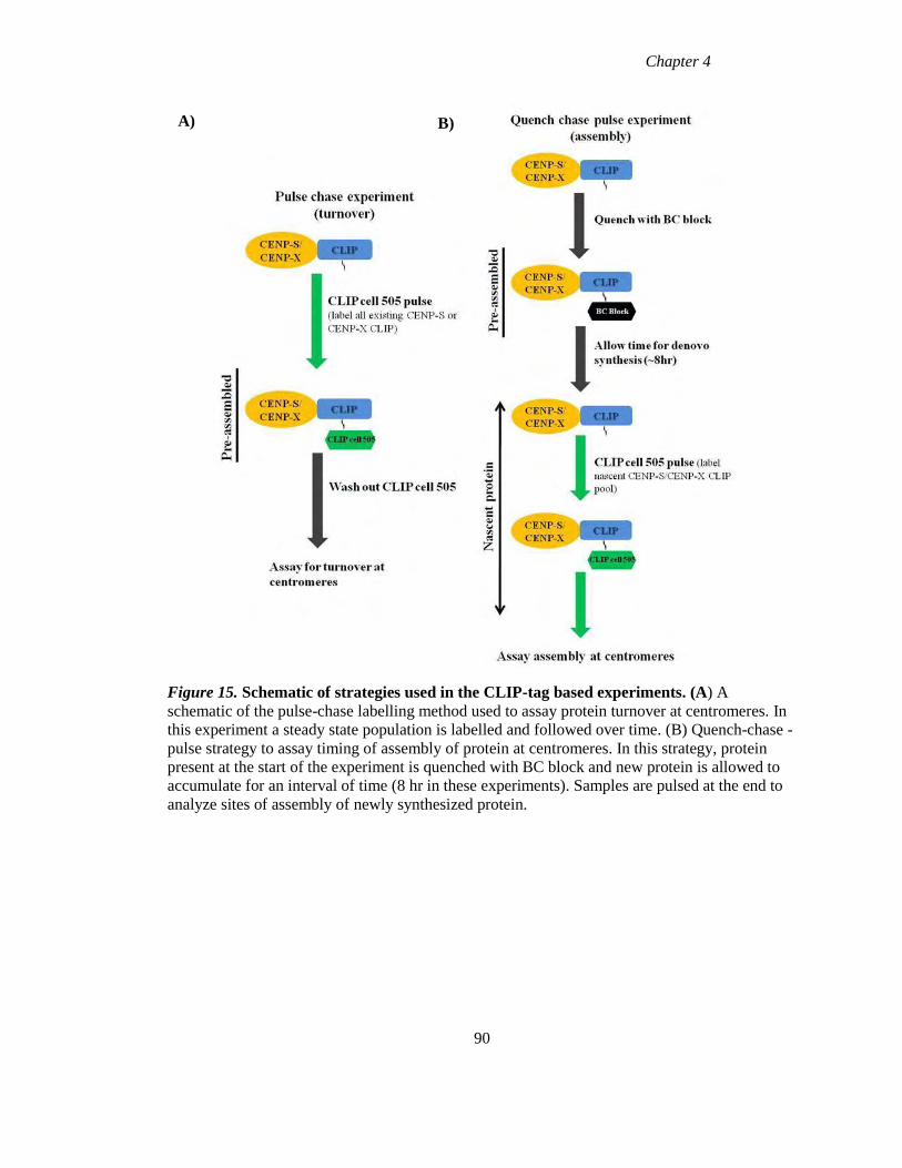
Chapter 4
Figure 15. Schematic of strategies used in the CLIP-tag based experiments. (A) A
schematic of the pulse-chase labelling method used to assay protein turnover at centromeres. In
this experiment a steady state population is labelled and followed over time. (B) Quench-chase -
pulse strategy to assay timing of assembly of protein at centromeres. In this strategy, protein
present at the start of the experiment is quenched with BC block and new protein is allowed to
accumulate for an interval of time (8 hr in these experiments). Samples are pulsed at the end to
analyze sites of assembly of newly synthesized protein.
90
A) B)
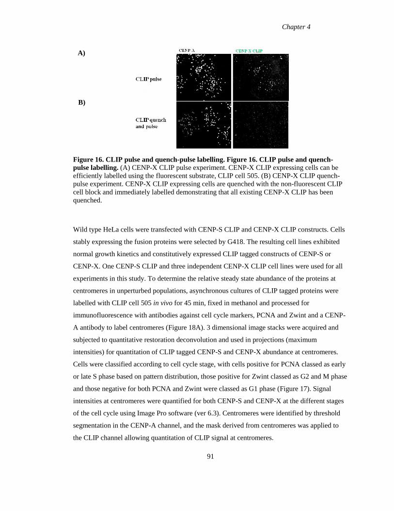
Chapter 4
Figure 16. CLIP pulse and quench-pulse labelling. Figure 16. CLIP pulse and quench-
pulse labelling. (A) CENP-X CLIP pulse experiment. CENP-X CLIP expressing cells can be
efficiently labelled using the fluorescent substrate, CLIP cell 505. (B) CENP-X CLIP quench-
pulse experiment. CENP-X CLIP expressing cells are quenched with the non-fluorescent CLIP
cell block and immediately labelled demonstrating that all existing CENP-X CLIP has been
quenched.
Wild type HeLa cells were transfected with CENP-S CLIP and CENP-X CLIP constructs. Cells
stably expressing the fusion proteins were selected by G418. The resulting cell lines exhibited
normal growth kinetics and constitutively expressed CLIP tagged constructs of CENP-S or
CENP-X. One CENP-S CLIP and three independent CENP-X CLIP cell lines were used for all
experiments in this study. To determine the relative steady state abundance of the proteins at
centromeres in unperturbed populations, asynchronous cultures of CLIP tagged proteins were
labelled with CLIP cell 505 in vivo for 45 min, fixed in methanol and processed for
immunofluorescence with antibodies against cell cycle markers, PCNA and Zwint and a CENP-
A antibody to label centromeres (Figure 18A). 3 dimensional image stacks were acquired and
subjected to quantitative restoration deconvolution and used in projections (maximum
intensities) for quantitation of CLIP tagged CENP-S and CENP-X abundance at centromeres.
Cells were classified according to cell cycle stage, with cells positive for PCNA classed as early
or late S phase based on pattern distribution, those positive for Zwint classed as G2 and M phase
and those negative for both PCNA and Zwint were classed as G1 phase (Figure 17). Signal
intensities at centromeres were quantified for both CENP-S and CENP-X at the different stages
of the cell cycle using Image Pro software (ver 6.3). Centromeres were identified by threshold
segmentation in the CENP-A channel, and the mask derived from centromeres was applied to
the CLIP channel allowing quantitation of CLIP signal at centromeres.
91
A)
B)
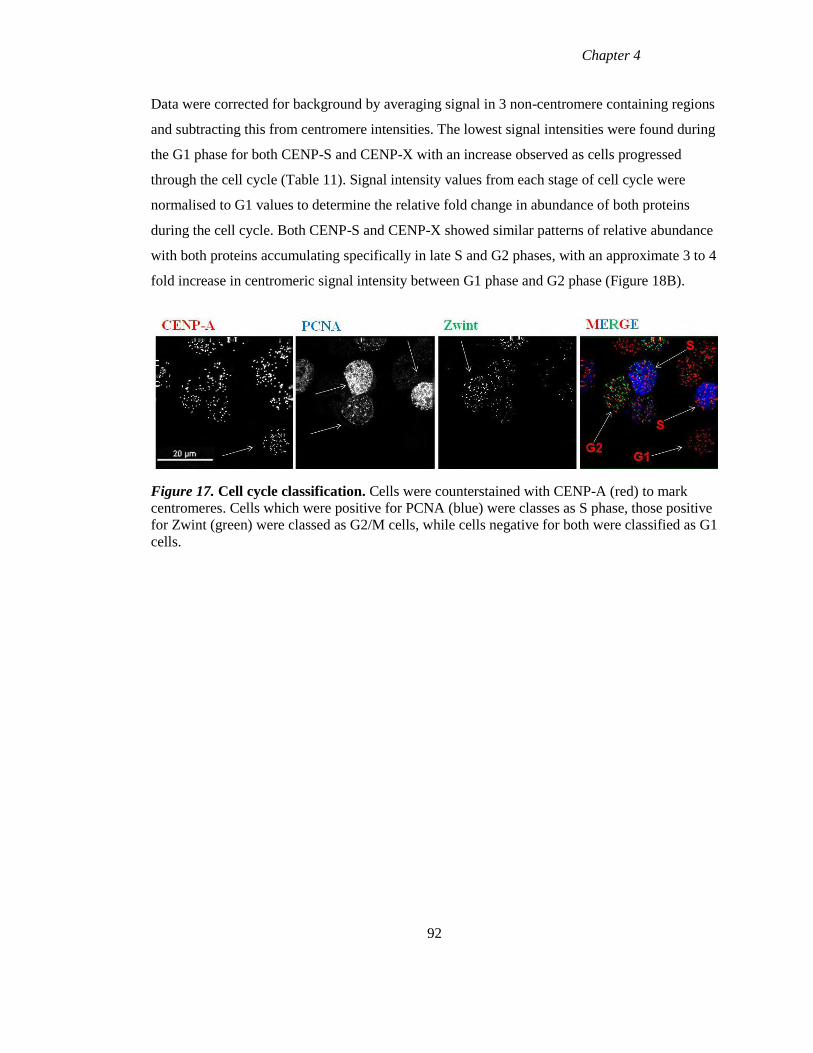
Chapter 4
Data were corrected for background by averaging signal in 3 non-centromere containing regions
and subtracting this from centromere intensities. The lowest signal intensities were found during
the G1 phase for both CENP-S and CENP-X with an increase observed as cells progressed
through the cell cycle (Table 11). Signal intensity values from each stage of cell cycle were
normalised to G1 values to determine the relative fold change in abundance of both proteins
during the cell cycle. Both CENP-S and CENP-X showed similar patterns of relative abundance
with both proteins accumulating specifically in late S and G2 phases, with an approximate 3 to 4
fold increase in centromeric signal intensity between G1 phase and G2 phase (Figure 18B).
Figure 17. Cell cycle classification. Cells were counterstained with CENP-A (red) to mark
centromeres. Cells which were positive for PCNA (blue) were classes as S phase, those positive
for Zwint (green) were classed as G2/M cells, while cells negative for both were classified as G1
cells.
92
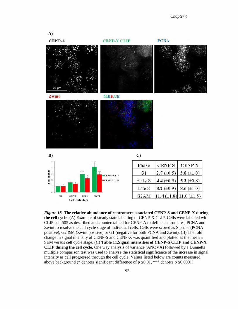
Chapter 4
A)
B)
Figure 18. The relative abundance of centromere associated CENP-S and CENP-X during
the cell cycle. (A) Example of steady state labelling of CENP-X CLIP. Cells were labelled with
CLIP cell 505 as described and counterstained for CENP-A to define centromeres, PCNA and
Zwint to resolve the cell cycle stage of individual cells. Cells were scored as S phase (PCNA
positive), G2 &M (Zwint positive) or G1 (negative for both PCNA and Zwint). (B) The fold
change in signal intensity of CENP-S and CENP-X was quantified and plotted as the mean ±
SEM versus cell cycle stage. (C) Table 11.Signal intensities of CENP-S CLIP and CENP-X
CLIP during the cell cycle. One way analysis of variance (ANOVA) followed by a Dunnetts
multiple comparison test was used to analyse the statistical significance of the increase in signal
intensity as cell progressed through the cell cycle. Values listed below are counts measured
above background (* denotes significant difference of p ≤0.01, *** denotes p ≤0.0001).
93
C)
Chapter 4
4.3 Assembly of CENP-S and CENP-X during the cell cycle
To examine the cell cycle dependent assembly of CENP-S and CENP-X at centromeres, a
conditional CLIP labelling strategy was employed, similar to that used determine the timing of
CENP-T and CENP-W assembly (Prendergast et al., 2011). A quench chase pulse experiment
(as illustrated in Figure 15B) was conducted with asynchronous HeLa cells stably expressing
CLIP tagged CENP-S and CENP-X. The existing CLIP tagged proteins were first blocked with
the CLIP cell block, bromothenylcytosine (BTC), cells were cultured for 8 hr to allow synthesis
of new protein, which was then labeled with CLIP cell 505 for 45 min. Cells were fixed in
methanol and counterstained with the centromere marker CENP-A and antibodies against cell
cycle markers, PCNA and Zwint (Figure 19A). Cells were then classified as G1, early S, late S
or G2/M phase. Assembly of CENP-S or CENP-X at centromeres was scored by inspection,
with robust CLIP signal detected during late S and G2/M phases of the cell cycle, with
approximately 50% of these cells being CLIP positive. Less than 5% of G1 cells analysed were
positive for CENP-S/CENP-X assembly, while none of the early S phase cells analysed were
positive for either protein (Figure 19B). The assembly of the CENP-S/CENP-X complex
appears to be similar to that of the CENP-T/CENP-W complex as both assemble late in the cell
cycle (late S and G2/M).
94
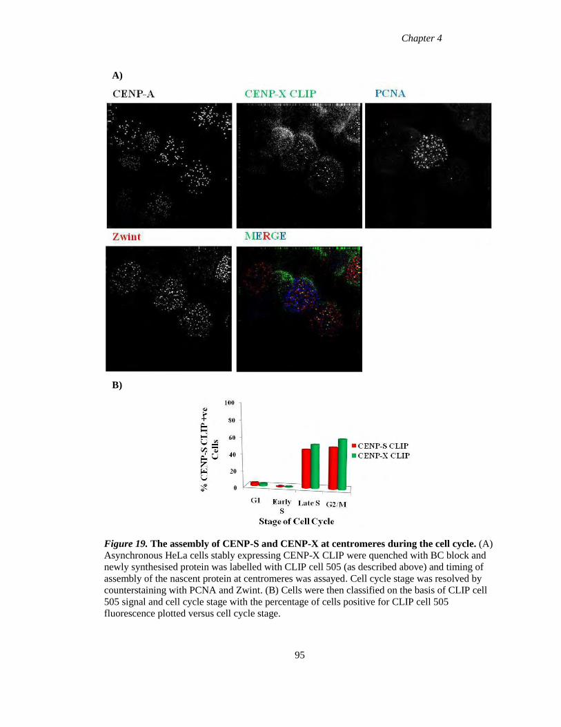
Chapter 4
A)
B)
Figure 19. The assembly of CENP-S and CENP-X at centromeres during the cell cycle. (A)
Asynchronous HeLa cells stably expressing CENP-X CLIP were quenched with BC block and
newly synthesised protein was labelled with CLIP cell 505 (as described above) and timing of
assembly of the nascent protein at centromeres was assayed. Cell cycle stage was resolved by
counterstaining with PCNA and Zwint. (B) Cells were then classified on the basis of CLIP cell
505 signal and cell cycle stage with the percentage of cells positive for CLIP cell 505
fluorescence plotted versus cell cycle stage.
95
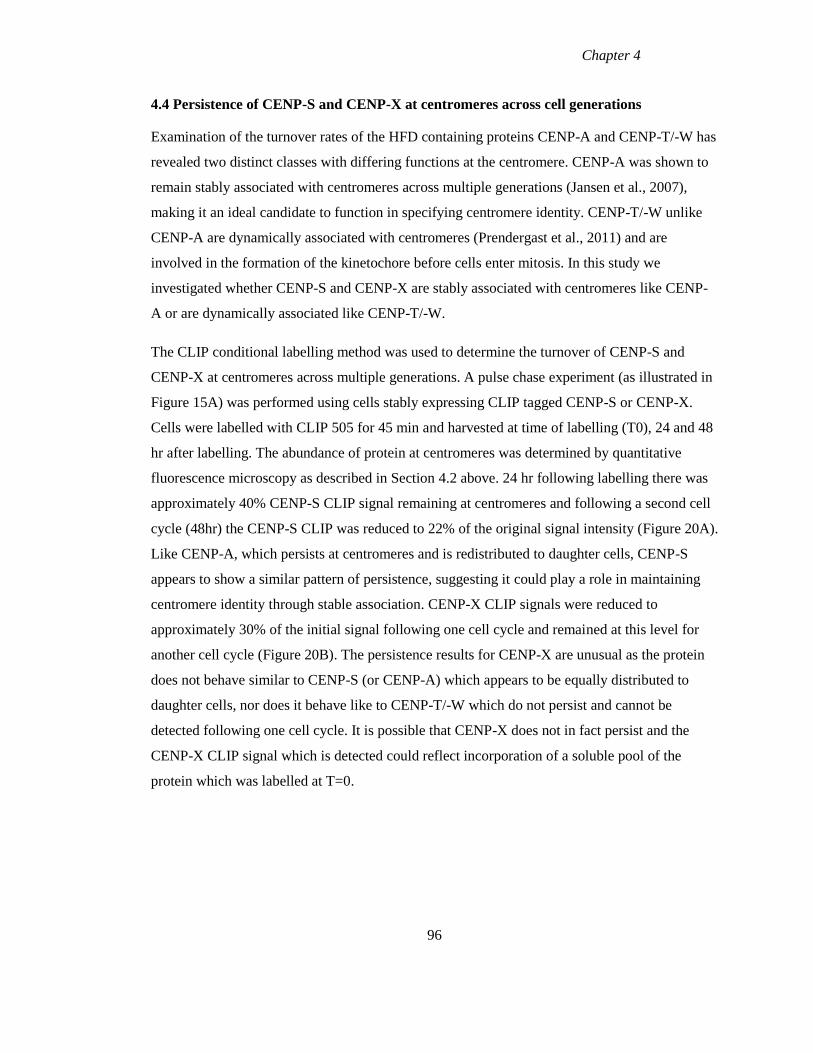
Chapter 4
4.4 Persistence of CENP-S and CENP-X at centromeres across cell generations
Examination of the turnover rates of the HFD containing proteins CENP-A and CENP-T/-W has
revealed two distinct classes with differing functions at the centromere. CENP-A was shown to
remain stably associated with centromeres across multiple generations (Jansen et al., 2007),
making it an ideal candidate to function in specifying centromere identity. CENP-T/-W unlike
CENP-A are dynamically associated with centromeres (Prendergast et al., 2011) and are
involved in the formation of the kinetochore before cells enter mitosis. In this study we
investigated whether CENP-S and CENP-X are stably associated with centromeres like CENP-
A or are dynamically associated like CENP-T/-W.
The CLIP conditional labelling method was used to determine the turnover of CENP-S and
CENP-X at centromeres across multiple generations. A pulse chase experiment (as illustrated in
Figure 15A) was performed using cells stably expressing CLIP tagged CENP-S or CENP-X.
Cells were labelled with CLIP 505 for 45 min and harvested at time of labelling (T0), 24 and 48
hr after labelling. The abundance of protein at centromeres was determined by quantitative
fluorescence microscopy as described in Section 4.2 above. 24 hr following labelling there was
approximately 40% CENP-S CLIP signal remaining at centromeres and following a second cell
cycle (48hr) the CENP-S CLIP was reduced to 22% of the original signal intensity (Figure 20A).
Like CENP-A, which persists at centromeres and is redistributed to daughter cells, CENP-S
appears to show a similar pattern of persistence, suggesting it could play a role in maintaining
centromere identity through stable association. CENP-X CLIP signals were reduced to
approximately 30% of the initial signal following one cell cycle and remained at this level for
another cell cycle (Figure 20B). The persistence results for CENP-X are unusual as the protein
does not behave similar to CENP-S (or CENP-A) which appears to be equally distributed to
daughter cells, nor does it behave like to CENP-T/-W which do not persist and cannot be
detected following one cell cycle. It is possible that CENP-X does not in fact persist and the
CENP-X CLIP signal which is detected could reflect incorporation of a soluble pool of the
protein which was labelled at T=0.
96
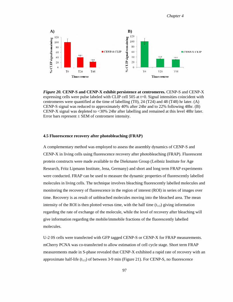
Chapter 4
A) B)
Figure 20. CENP-S and CENP-X exhibit persistence at centromeres. CENP-S and CENP-X
expressing cells were pulse labeled with CLIP cell 505 at t=0. Signal intensities coincident with
centromeres were quantified at the time of labelling (T0), 24 (T24) and 48 (T48) hr later. (A)
CENP-S signal was reduced to approximately 40% after 24hr and to 22% following 48hr. (B)
CENP-X signal was depleted to <30% 24hr after labelling and remained at this level 48hr later.
Error bars represent ± SEM of centromere intensity.
4.5 Fluorescence recovery after photobleaching (FRAP)
A complementary method was employed to assess the assembly dynamics of CENP-S and
CENP-X in living cells using fluorescence recovery after photobleaching (FRAP). Fluorescent
protein constructs were made available to the Diekmann Group (Leibniz Institute for Age
Research, Fritz Lipmann Institute, Jena, Germany) and short and long term FRAP experiments
were conducted. FRAP can be used to measure the dynamic properties of fluorescently labelled
molecules in living cells. The technique involves bleaching fluorescently labelled molecules and
monitoring the recovery of fluorescence in the region of interest (ROI) in series of images over
time. Recovery is as result of unbleached molecules moving into the bleached area. The mean
intensity of the ROI is then plotted versus time, with the half time (t1/2) giving information
regarding the rate of exchange of the molecule, while the level of recovery after bleaching will
give information regarding the mobile/immobile fractions of the fluorescently labelled
molecules.
U-2 0S cells were transfected with GFP tagged CENP-S or CENP-X for FRAP measurements.
mCherry PCNA was co-transfected to allow estimation of cell cycle stage. Short term FRAP
measurements made in S-phase revealed that CENP-X exhibited a rapid rate of recovery with an
approximate half-life (t1/2) of between 3-9 min (Figure 21). For CENP-S, no fluorescence
97
Chapter 4
recovery was observed during the course of the short term FRAP experiment (Figure 22). Long
term FRAP measurements for CENP-S revealed that the protein recovered at all stages of the
cell cycle (Figure 23). The approximate half time of recovery (t1/2) was faster in G1 (~75 min)
than later cell cycle phases where recovery rates showed half times of 110-120 min. Thus, these
HFD CCAN components exhibit dynamic exchange, unlike CENP-A.
Figure 21. Short-term FRAP experiments with CENP-X. FRAP with GFP tagged CENP-X
revealed the protein undergoes a rapid rate of recovery during S phase with a an approximate t1/2
of between 3-9 min.
98
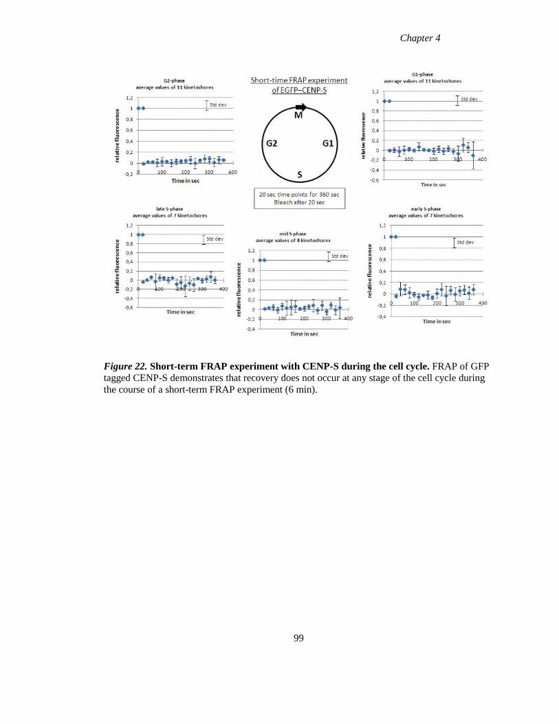
Chapter 4
Figure 22. Short-term FRAP experiment with CENP-S during the cell cycle. FRAP of GFP
tagged CENP-S demonstrates that recovery does not occur at any stage of the cell cycle during
the course of a short-term FRAP experiment (6 min).
99
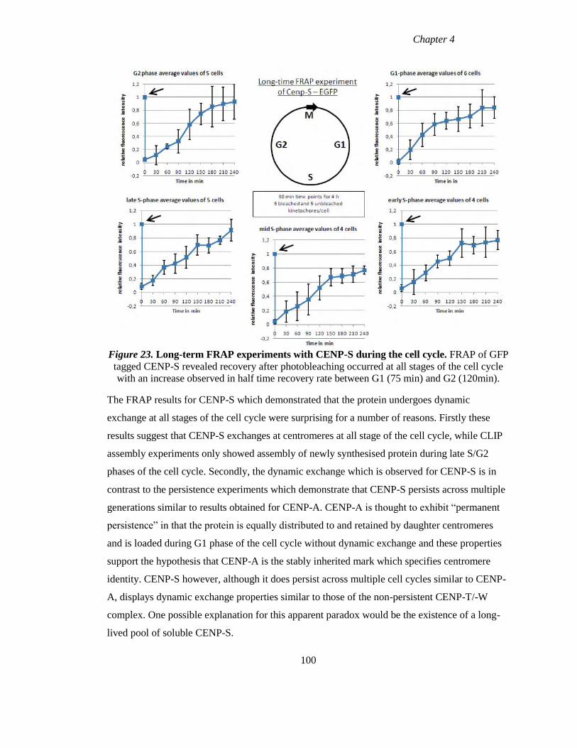
Chapter 4
Figure 23. Long-term FRAP experiments with CENP-S during the cell cycle. FRAP of GFP
tagged CENP-S revealed recovery after photobleaching occurred at all stages of the cell cycle
with an increase observed in half time recovery rate between G1 (75 min) and G2 (120min).
The FRAP results for CENP-S which demonstrated that the protein undergoes dynamic
exchange at all stages of the cell cycle were surprising for a number of reasons. Firstly these
results suggest that CENP-S exchanges at centromeres at all stage of the cell cycle, while CLIP
assembly experiments only showed assembly of newly synthesised protein during late S/G2
phases of the cell cycle. Secondly, the dynamic exchange which is observed for CENP-S is in
contrast to the persistence experiments which demonstrate that CENP-S persists across multiple
generations similar to results obtained for CENP-A. CENP-A is thought to exhibit “permanent
persistence” in that the protein is equally distributed to and retained by daughter centromeres
and is loaded during G1 phase of the cell cycle without dynamic exchange and these properties
support the hypothesis that CENP-A is the stably inherited mark which specifies centromere
identity. CENP-S however, although it does persist across multiple cell cycles similar to CENP-
A, displays dynamic exchange properties similar to those of the non-persistent CENP-T/-W
complex. One possible explanation for this apparent paradox would be the existence of a long-
lived pool of soluble CENP-S.
100
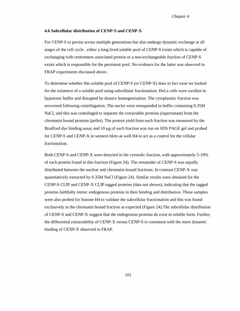
Chapter 4
4.6 Subcellular distribution of CENP-S and CENP-X
For CENP-S to persist across multiple generations but also undergo dynamic exchange at all
stages of the cell cycle , either a long lived soluble pool of CENP-S exists which is capable of
exchanging with centromere associated protein or a non-exchangeable fraction of CENP-S
exists which is responsible for the persistent pool. No evidence for the latter was observed in
FRAP experiments discussed above.
To determine whether this soluble pool of CENP-S (or CENP-X) does in fact exist we looked
for the existence of a soluble pool using subcellular fractionation. HeLa cells were swollen in
hypotonic buffer and disrupted by dounce homogenisation. The cytoplasmic fraction was
recovered following centrifugation. The nuclei were resuspended in buffer containing 0.35M
NaCl, and this was centrifuged to separate the extractable proteins (supernatant) from the
chromatin bound proteins (pellet). The protein yield from each fraction was measured by the
Bradford dye binding assay and 10 g of each fraction was run on SDS PAGE gel and probed
for CENP-S and CENP-X in western blots as well H4 to act as a control for the cellular
fractionation.
Both CENP-S and CENP-X were detected in the cytosolic fraction, with approximately 5-10%
of each protein found in this fraction (Figure 24). The remainder of CENP-S was equally
distributed between the nuclear and chromatin bound fractions. In contrast CENP-X was
quantitatively extracted by 0.35M NaCl (Figure 24). Similar results were obtained for the
CENP-S CLIP and CENP-X CLIP tagged proteins (data not shown), indicating that the tagged
proteins faithfully mimic endogenous proteins in their binding and distribution. These samples
were also probed for histone H4 to validate the subcellular fractionation and this was found
exclusively in the chromatin bound fraction as expected (Figure 24).The subcellular distribution
of CENP-S and CENP-X suggest that the endogenous proteins do exist in soluble form. Further,
the differential extractability of CENP-X versus CENP-S is consistent with the more dynamic
binding of CENP-X observed in FRAP.
101
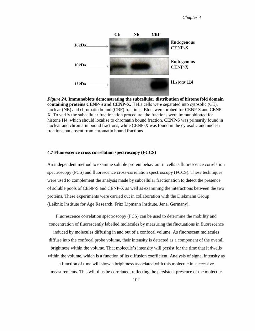
Chapter 4
Figure 24. Immunoblots demonstrating the subcellular distribution of histone fold domain
containing proteins CENP-S and CENP-X. HeLa cells were separated into cytosolic (CE),
nuclear (NE) and chromatin bound (CBF) fractions. Blots were probed for CENP-S and CENP-
X. To verify the subcellular fractionation procedure, the fractions were immunoblotted for
histone H4, which should localise to chromatin bound fraction. CENP-S was primarily found in
nuclear and chromatin bound fractions, while CENP-X was found in the cytosolic and nuclear
fractions but absent from chromatin bound fractions.
4.7 Fluorescence cross correlation spectroscopy (FCCS)
An independent method to examine soluble protein behaviour in cells is fluorescence correlation
spectroscopy (FCS) and fluorescence cross-correlation spectroscopy (FCCS). These techniques
were used to complement the analysis made by subcellular fractionation to detect the presence
of soluble pools of CENP-S and CENP-X as well as examining the interactions between the two
proteins. These experiments were carried out in collaboration with the Diekmann Group
(Leibniz Institute for Age Research, Fritz Lipmann Institute, Jena, Germany).
Fluorescence correlation spectroscopy (FCS) can be used to determine the mobility and
concentration of fluorescently labelled molecules by measuring the fluctuations in fluorescence
induced by molecules diffusing in and out of a confocal volume. As fluorescent molecules
diffuse into the confocal probe volume, their intensity is detected as a component of the overall
brightness within the volume. That molecule’s intensity will persist for the time that it dwells
within the volume, which is a function of its diffusion coefficient. Analysis of signal intensity as
a function of time will show a brightness associated with this molecule in successive
measurements. This will thus be correlated, reflecting the persistent presence of the molecule
102
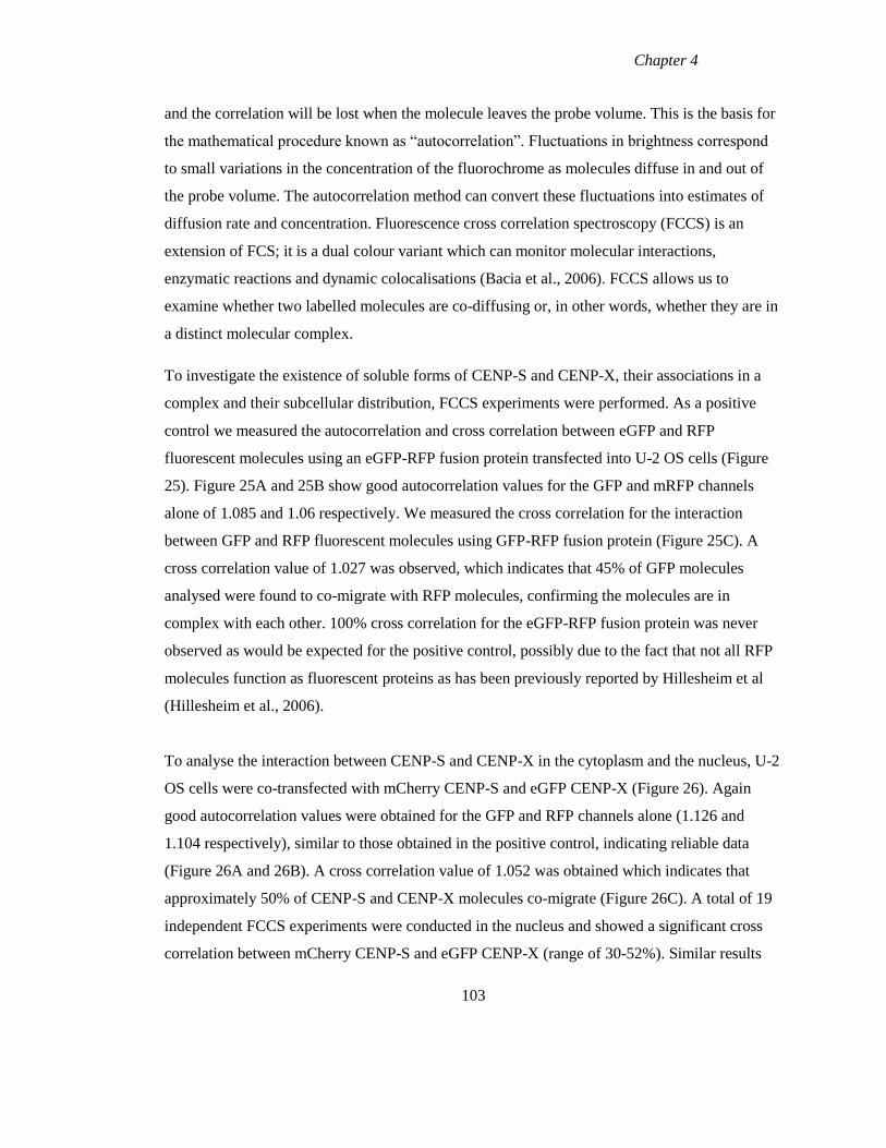
Chapter 4
and the correlation will be lost when the molecule leaves the probe volume. This is the basis for
the mathematical procedure known as “autocorrelation”. Fluctuations in brightness correspond
to small variations in the concentration of the fluorochrome as molecules diffuse in and out of
the probe volume. The autocorrelation method can convert these fluctuations into estimates of
diffusion rate and concentration. Fluorescence cross correlation spectroscopy (FCCS) is an
extension of FCS; it is a dual colour variant which can monitor molecular interactions,
enzymatic reactions and dynamic colocalisations (Bacia et al., 2006). FCCS allows us to
examine whether two labelled molecules are co-diffusing or, in other words, whether they are in
a distinct molecular complex.
To investigate the existence of soluble forms of CENP-S and CENP-X, their associations in a
complex and their subcellular distribution, FCCS experiments were performed. As a positive
control we measured the autocorrelation and cross correlation between eGFP and RFP
fluorescent molecules using an eGFP-RFP fusion protein transfected into U-2 OS cells (Figure
25). Figure 25A and 25B show good autocorrelation values for the GFP and mRFP channels
alone of 1.085 and 1.06 respectively. We measured the cross correlation for the interaction
between GFP and RFP fluorescent molecules using GFP-RFP fusion protein (Figure 25C). A
cross correlation value of 1.027 was observed, which indicates that 45% of GFP molecules
analysed were found to co-migrate with RFP molecules, confirming the molecules are in
complex with each other. 100% cross correlation for the eGFP-RFP fusion protein was never
observed as would be expected for the positive control, possibly due to the fact that not all RFP
molecules function as fluorescent proteins as has been previously reported by Hillesheim et al
(Hillesheim et al., 2006).
To analyse the interaction between CENP-S and CENP-X in the cytoplasm and the nucleus, U-2
OS cells were co-transfected with mCherry CENP-S and eGFP CENP-X (Figure 26). Again
good autocorrelation values were obtained for the GFP and RFP channels alone (1.126 and
1.104 respectively), similar to those obtained in the positive control, indicating reliable data
(Figure 26A and 26B). A cross correlation value of 1.052 was obtained which indicates that
approximately 50% of CENP-S and CENP-X molecules co-migrate (Figure 26C). A total of 19
independent FCCS experiments were conducted in the nucleus and showed a significant cross
correlation between mCherry CENP-S and eGFP CENP-X (range of 30-52%). Similar results
103
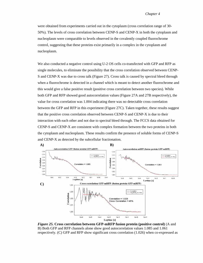
Chapter 4
were obtained from experiments carried out in the cytoplasm (cross correlation range of 30-
50%). The levels of cross correlation between CENP-S and CENP-X in both the cytoplasm and
nucleoplasm were comparable to levels observed in the covalently coupled fluorochrome
control, suggesting that these proteins exist primarily in a complex in the cytoplasm and
nucleoplasm.
We also conducted a negative control using U-2 OS cells co-transfected with GFP and RFP as
single molecules, to eliminate the possibility that the cross correlation observed between CENP-
S and CENP-X was due to cross talk (Figure 27). Cross talk is caused by spectral bleed through
when a fluorochrome is detected in a channel which is meant to detect another fluorochrome and
this would give a false positive result (positive cross correlation between two species). While
both GFP and RFP showed good autocorrelation values (Figure 27A and 27B respectively), the
value for cross correlation was 1.004 indicating there was no detectable cross correlation
between the GFP and RFP in this experiment (Figure 27C). Taken together, these results suggest
that the positive cross correlation observed between CENP-S and CENP-X is due to their
interaction with each other and not due to spectral bleed through. The FCCS data obtained for
CENP-S and CENP-X are consistent with complex formation between the two proteins in both
the cytoplasm and nucleoplasm. These results confirm the presence of soluble forms of CENP-S
and CENP-X as detected by the subcellular fractionation.
Figure 25. Cross correlation between GFP-mRFP fusion protein (positive control) (A and
B) Both GFP and RFP channels alone show good autocorrelation values 1.085 and 1.061
respectively. (C) GFP and RFP show significant cross correlation (1.026) when co-expressed as
A) B)
C)
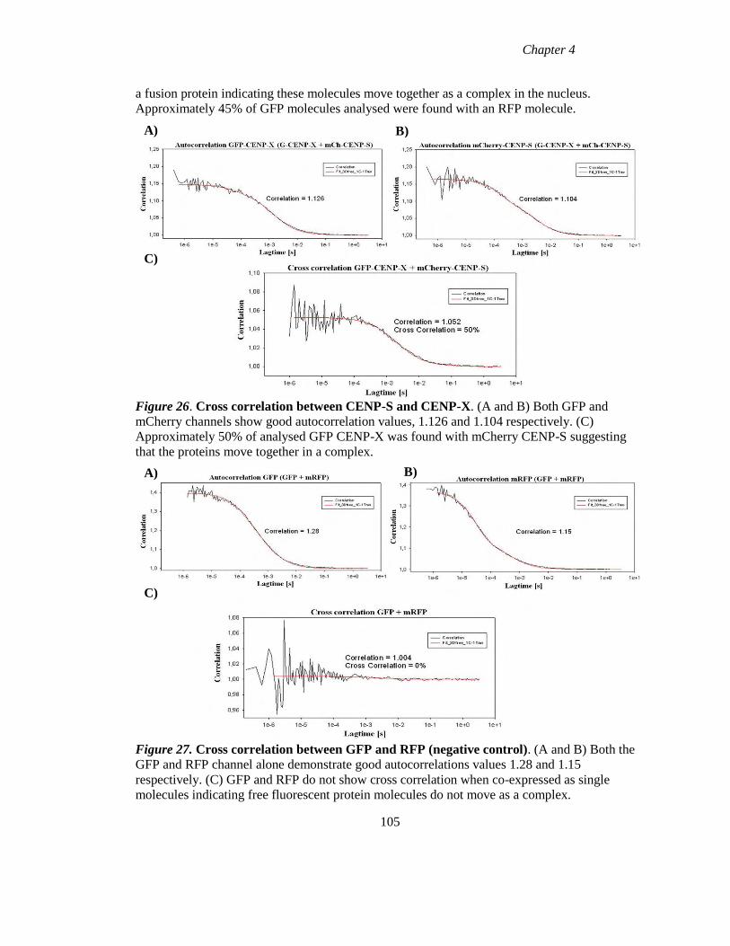
Chapter 4
a fusion protein indicating these molecules move together as a complex in the nucleus.
Approximately 45% of GFP molecules analysed were found with an RFP molecule.
Figure 26. Cross correlation between CENP-S and CENP-X. (A and B) Both GFP and
mCherry channels show good autocorrelation values, 1.126 and 1.104 respectively. (C)
Approximately 50% of analysed GFP CENP-X was found with mCherry CENP-S suggesting
that the proteins move together in a complex.
Figure 27. Cross correlation between GFP and RFP (negative control). (A and B) Both the
GFP and RFP channel alone demonstrate good autocorrelations values 1.28 and 1.15
respectively. (C) GFP and RFP do not show cross correlation when co-expressed as single
molecules indicating free fluorescent protein molecules do not move as a complex.
105
B) A)
C)
A) B)
C)
Chapter 4
4.8 Half Life of CENP-S and CENP-X
The subcellular fractionation and FCCS experiments demonstrated the presence of a soluble
pool of CENP-S. The existence of this soluble pool could help to explain the apparent opposing
properties of persistence and dynamic exchange associated with CENP-S if the half life of the
protein is a significant fraction of the cell cycle. To address the question of the half life of
CENP-S and CENP-X, a CLIP biotin experiment was conducted. CLIP biotin is a cell
permeable substrate which is based on biotin linked via an amidocaproyl linkage to the
benzylguanine scaffold. The CLIP biotin is suitable for the labelling of CLIP tag fusion proteins
in living cells so they can be detected with streptavidin conjugates in combination western blot
analysis. The protein half life would be estimated by measuring the reduction of biotinylated
CLIP tagged fusion protein across multiple cell cycles. The CENP-S and CENP-X CLIP tag
fusion proteins were labelled with CLIP biotin and the samples were collected at time of
labelling (T0), 24 and 48 hr later. These samples were run on SDS PAGE gels and biotin
labelled proteins were detected using HRP conjugated streptavidin (Figure 28A).
Due to the binding of the streptavidin to cellular proteins, detection of a specific signal for the
biotin labelled CLIP tag proteins was not possible as illustrated for CENP-S (Figure 28A),
(CENP-X data not shown).Wild type HeLa cells were used as a negative control. As illustrated
in Figure 28A, a band is present in wild type samples at 36kDa which could be obscuring the
biotin labelled CLIP tagged protein in the CENP-S CLIP samples. It was therefore not possible
to calculate the t1/2 for either CENP-S or CENP-X using this method. To ensure that the inability
to detect the biotin labelled protein was not due to the absence of CLIP tagged protein, the
samples were also probed with MGMT antibody, which detects the O6 methylguanine DNA
methyltransferase domain of the CLIP tag fusion proteins. Figure 28B clearly shows a band
detected at 20kDa (endogenous MGMT) and another band at ~36kDa which corresponds to
CLIP tagged CENP-S, confirming expression of the CLIP tagged fusion protein in this
experiment. In conclusion, the CLIP biotin experiment is not a suitable method to determine the
protein half-life of CENP-S or CENP-X using whole cell extracts.
106
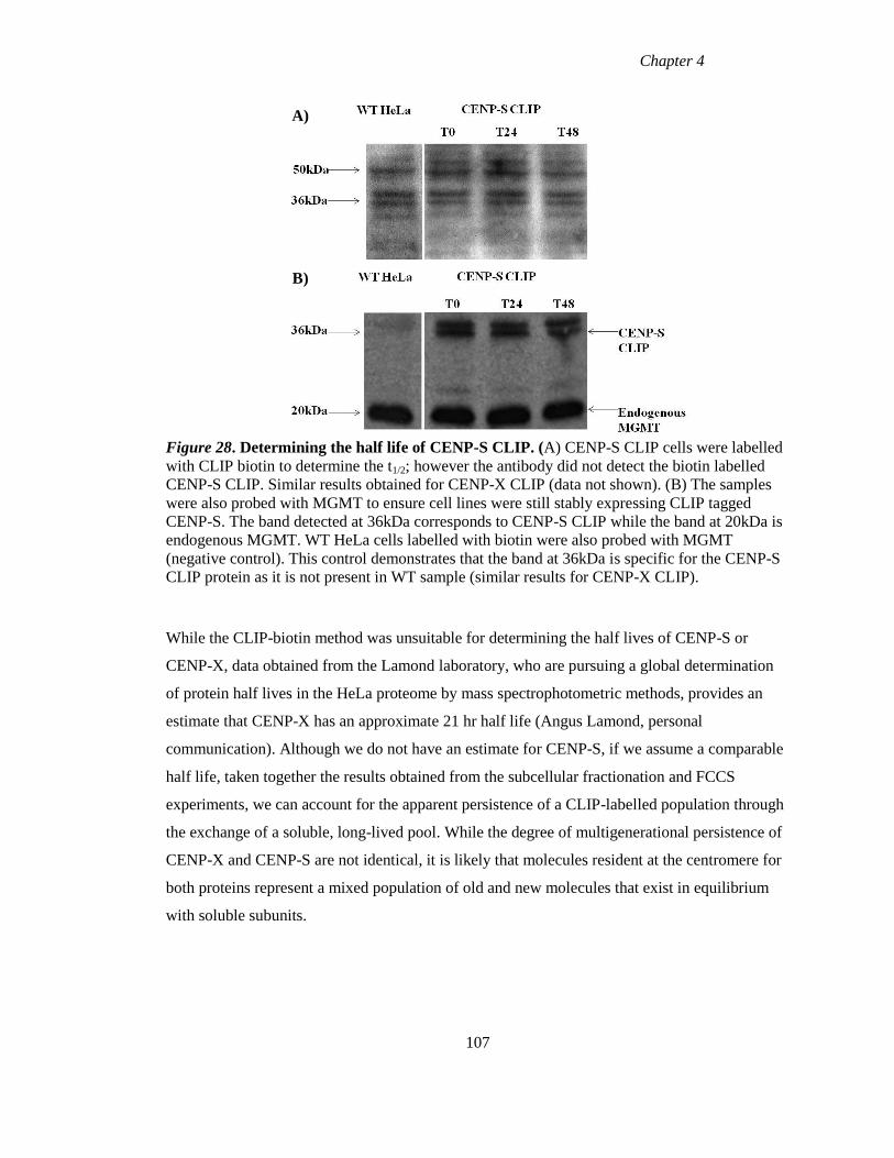
Chapter 4
Figure 28. Determining the half life of CENP-S CLIP. (A) CENP-S CLIP cells were labelled
with CLIP biotin to determine the t1/2; however the antibody did not detect the biotin labelled
CENP-S CLIP. Similar results obtained for CENP-X CLIP (data not shown). (B) The samples
were also probed with MGMT to ensure cell lines were still stably expressing CLIP tagged
CENP-S. The band detected at 36kDa corresponds to CENP-S CLIP while the band at 20kDa is
endogenous MGMT. WT HeLa cells labelled with biotin were also probed with MGMT
(negative control). This control demonstrates that the band at 36kDa is specific for the CENP-S
CLIP protein as it is not present in WT sample (similar results for CENP-X CLIP).
While the CLIP-biotin method was unsuitable for determining the half lives of CENP-S or
CENP-X, data obtained from the Lamond laboratory, who are pursuing a global determination
of protein half lives in the HeLa proteome by mass spectrophotometric methods, provides an
estimate that CENP-X has an approximate 21 hr half life (Angus Lamond, personal
communication). Although we do not have an estimate for CENP-S, if we assume a comparable
half life, taken together the results obtained from the subcellular fractionation and FCCS
experiments, we can account for the apparent persistence of a CLIP-labelled population through
the exchange of a soluble, long-lived pool. While the degree of multigenerational persistence of
CENP-X and CENP-S are not identical, it is likely that molecules resident at the centromere for
both proteins represent a mixed population of old and new molecules that exist in equilibrium
with soluble subunits.
107
A)
B)
Chapter 4
4.9 Detection of molecular interactions of CENP-S and CENP-X within the CCAN
Biochemical studies revealed that CENP-S was a distinct member of the CCAN based on its
variable presence in co-immunoprecipitation experiments performed with different CCAN
subunits. It was initially identified in association with CENP-M and –U and classified as a
CENP- A distal complex component (Foltz et al., 2006) and was not found in association of with
any members of the CENP-H-I complex (Okada et al., 2006). Subsequent TAP-tag purification
showed that its major co-purification partner was another novel HFD centromere protein,
CENP-X and to a lesser extent CENP-T (Amano et al., 2009). If tagged CENP-S and CENP-X
form complexes as expected from this biochemical data and from the FCCS data shown here,
then the proximity of these proteins should be close within the CCAN at fully assembled
centromeres. FRET analysis was carried out to examine the proximity of the HFD proteins
CENP-S and CENP-X to each other and to other centromere proteins.
Fluorescence resonance energy transfer (FRET) is a technique which is used to measure the
interaction between two molecules labelled with two different fluorophores (donor and
acceptor), by the transfer of energy from the excited donor to the acceptor. The technique is
widely used to examine protein-protein interactions and to measure the distance between donor
and acceptor molecules. The process involves the transfer of energy from a fluorophore in an
excited state (donor) to a neighbouring molecule (acceptor) by nonradiative dipole-dipole
interactions (Periasamy and Day, 1999). Two different approaches can be employed to measure
FRET using standard confocal microscopy: emission measurement and acceptor photobleaching.
Emission measurement involves the excitation of donor molecules and the detection of light
emitted by the donor and/or the acceptor in the presence of the other fluorophore. A positive
FRET result causes a decrease in donor emission coupled with an increase in acceptor emission.
Acceptor photobleaching involves the excitation of the donor molecule and the detection of the
light it emits before and after bleaching of the acceptor molecule. Energy transfer between the
donor and the acceptor is eliminated as the acceptor is irreversibly photobleached which results
in an increase in donor fluorescence intensity.
The proximity of CENP-S and CENP-X to each other and to other CCAN members at
centromeres was examined in fixed U-2 OS cells by acceptor photobleaching FRET (AB FRET)
in collaboration with the Diekmann Group (Leibniz Institute for Age Research, Fritz Lipmann
108
Chapter 4
Institute, Jena, Germany). U-2 OS cells were co-transfected with two plasmid constructs
containing two different genes, each coding for different centromere proteins fused to
fluorescent proteins of different colours (FRET pair) for the AB FRET. In this study eGFP and
mCherry were used as the FRET pair. The change in fluorescent intensity in the bleached area
was recorded in the donor and acceptor channels.
In each image analysed, two centromere locations were identified and the acceptor was bleached
at one of these locations. Bleaching of the acceptor results in an increase in donor fluorescence,
if the donor and acceptor molecules are in close proximity and FRET has quenched primary
donor emission. The second location is not bleached acts as a control and measures the donor
fluorescence in the presence of the FRET acceptor. The efficiency of FRET (EFRET) is given by
the ratio of signal pre and post quenching at the quenched site and the efficiency variation
(EVAR) is given by the ratio of signal at the reference site. Data are recorded by measuring donor
fluorescence intensity in four images before and after the photobleaching event. Positive FRET
is identified when EFRET values are greater than that of EVAR.
To examine FRET between CENP-S mCherry and eGFP CENP-X, 37 bleached and 36 control
(unbleached) centromeres were measured (Figure 29A). The EVAR values from unbleached
centromeres and EFRET values from the bleached centromeres were calculated. These values
were classified into 4% deviating categories and plotted as a histogram with the number of
events (y axis) plotted against E value bins (x axis). The grey bars represent the frequency of the
calculated efficiency of control centromeres (EVAR) and black bars represent the frequency of
FRET efficiency (EFRET) of bleached centromeres. The mean EFRET value was calculated as 4.5,
while the mean EVAR value was calculated as 0.9 with a difference of 5.4 calculated between the
two. The measurements failed the normality of distribution and could not be analysed by a
Paired T test, thus the Mann Whitney Sum Rank test was used and gave a p value of ≥0.001
demonstrating statistically significant differences between EVAR and EFRET. This is a positive
FRET result and indicates the C terminus of CENP-S is in close proximity to the N terminus of
CENP-X (<10nm apart).
109
Chapter 4
Next we examined the proximity of CENP-S and CENP-T to each other. There was no FRET
between eGFP CENP-S and mCherry CENP-T (Figure 29C). However CENP-S mCherry was
demonstrated to be in close proximity to CENP-T eGFP (Figure 29D). The mean EVAR value
was 4.2 while the mean EFRET value was -0.34 resulting in a difference of 4.5. The Mann
Whitney rank sum test gave a p value of 0.001, indicating that the difference between EFRET and
EVAR is statistically significant (Figure 29E). This positive FRET result between CENP-S and
CENP-T is consistent with results from Amano et al (2009) which indicated an interaction
between the two proteins.
Finally, the proximity of CENP-S to CENP-M was analysed by examining FRET between
mCherry CENP-S and eGFP CENP-M (Figure 29E). In total 37 bleached and unbleached
centromeres were measured, and a difference of 0.8 was calculated between EVAR and EFRET
values. The Paired T test showed that there was no FRET between the N terminus of CENP-S
and N terminus of CENP-M (p value 0.064) indicating the distance between the N termini of
both proteins is further apart than 10nm. FRET analysis of 42 bleached and 41 control
centromeres of the FRET pair mCherry CENP-S and CENP-M eGFP showed FRET (Figure
29F). EVAR values ranged from -10 to +6% while EFRET values ranged between -6 to +14%. The
difference between EVAR and EFRET is statistically significant (p value 0.001) indicating that
although the N termini of the proteins are not close together, the N terminus of CENP-S is in
close proximity to the C terminus of CENP-M.
110
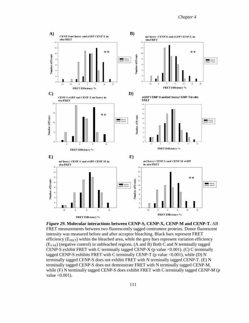
Chapter 4
Figure 29. Molecular interactions between CENP-S, CENP-X, CENP-M and CENP-T. AB
FRET measurements between two fluorescently tagged centromere proteins. Donor fluorescent
intensity was measured before and after acceptor bleaching. Black bars represent FRET
efficiency (EFRET) within the bleached area, while the grey bars represent variation efficiency
(EVAR) (negative control) in unbleached regions. (A and B) Both C and N terminally tagged
CENP-S exhibit FRET with C terminally tagged CENP-X (p value <0.001). (C) C terminally
tagged CENP-S exhibits FRET with C terminally CENP-T (p value <0.001), while (D) N
terminally tagged CENP-S does not exhibit FRET with N terminally tagged CENP-T. (E) N
terminally tagged CENP-S does not demonstrate FRET with N terminally tagged CENP-M,
while (F) N terminally tagged CENP-S does exhibit FRET with C terminally tagged CENP-M (p
value <0.001).
111
F) E)
B) A)
C)
E) F)
D)
Chapter 4
4.10 Discussion
The aim of this study was to investigate the assembly and inheritance of the HFD centromere
proteins CENP-S and CENP-X. In previous studies of other CCAN proteins, two types of
behaviour have been seen. Jansen et al (2007), demonstrated that CENP-A was assembled
during late telophase/G1 and this assembly was observed to occur without dynamic exchange
(Hemmerich et al., 2008). CENP-A was also found to persist across multiple generations in a
manner consistent with conservation of parental CENP-A and equal distribution to daughter
cells (Jansen et al., 2007). These observations are consistent with CENP-A being an epigenetic
mark which specifies centromere identity. In contrast to CENP-A, CENP-T/-W were found to
assemble later in the cell cycle during late S/G2 and this assembly was shown to occur via a
dynamic exchange mechanism, while neither protein persisted across multiple generations
(Prendergast et al., 2011). Rather than playing a primary role in propagating centromere identity,
CENP-T/-W have been proposed to be involved in kinetochore assembly/function before cells
enter mitosis (Prendergast et al., 2011). FRAP experiments with additional CCAN members
have demonstrated that CENP-I, like CENP-A, is stably bound at all stages of the cell cycle with
some recovery observed in S phase coincident with assembly of the protein (Hemmerich et al.,
2008). CENP-B, -C and –H exhibited dynamic exchange during the majority of the cell cycle,
with CENP-B becoming stably bound during G2 while CENP-C and CENP-H demonstrated
increased stability at centromeres during S phase (Hemmerich et al., 2008). In this present study
we found that CENP-S and CENP-X are assembled during late S/G2 similar to CENP-T/-W. We
also found however, that both proteins exhibit persistence across multiple generations. In
addition FCCS and FRET provide evidence that CENP-S and CENP-X form a complex both in
the cytoplasm and nucleoplasm, as well as being in close proximity to CENP-M and CENP-T.
Examination of the timing of assembly of CENP-S and CENP-X revealed that both proteins are
assembled during late S/G2 phase of the cell cycle in a similar manner to CENP-T/-W. However
unlike CENP-T/-W both CENP-S and CENP-X appeared to persist across multiple cell cycles.
CENP-S in particular behaved almost identical to CENP-A, with an approximate 50% dilution
in signal following each cell cycle. Surprisingly despite this apparent persistence, CENP-S also
exhibited dynamic exchange at all stages of the cell cycle. How can an apparently persistent
protein undergo dynamic exchange? We found evidence for the existence of a soluble pool of
CENP-S (and CENP-X) and we propose that this population of CENP-S contributes to the
persistence. Thus while the persistence exhibited by CENP-A is as result of the protein being
112
Chapter 4
retained at centromeres and redistributed to daughter cells, we propose that CENP-S persistence
is mediated through a long lived soluble pool which undergoes exchange with centromere bound
protein.
FRAP recovery of CCAN components at centromeres is usually coupled with assembly at
centromeres e.g. CENP-A and CENP-T/-W. However, this was not the case for CENP-S and
CENP-X. Both proteins exhibited dynamic exchange at all stages of the cell cycle, however de-
novo assembly was only observed, using CLIP-tagging, during S late/G2 phase. While we
cannot rule out the possibility of some assembly occurring for these proteins outside of late
S/G2, it is clear that the most de novo assembly occurs during this window. The dynamic
exchange which is occurring during G1 and early S phase could be with the long lived soluble
pool of parental CENP-S and not the newly synthesised pool of protein. While CENP-S
recovery is seen at all stages of the cycle there is a change in this recovery during late S phase,
coincident with de novo assembly of the protein as seen with CLIP labelling. At all other stages
of the cell cycle, a plateau in recovery was observed at approximately 80%, while during late S
phase this was no plateau in recovery, suggesting the newly synthesised protein behaves
differently to the rest of the protein population. One possibility for the observed difference in
recovery rates for CENP-S could be a histone like deposition marking, similar to that observed
for histone H4. Newly synthesised H4 is acetylated on K5 and K12 before its deposition (the
function of the modification is unknown), and then this PTM is removed upon nucleosome
maturation (Loyola et al., 2006, Sobel et al., 1995). A similar modification could also be
associated with CENP-S just before its assembly during late S phase, which may account for the
difference observed in recovery rates. Finally the FRAP data with CENP-S suggest the protein
becomes more stably bound at the centromere outside of G1phase. Previous studies with CENP-
S have shown that the protein along with CENP-X act downstream of H class proteins (Amano
et al., 2009) and these proteins are interdependent with CENP-T/-W for their localisation. FRAP
and assembly studies have shown that these proteins are assembled at centromeres later in the
cell cycle (from S phase to G2) which results in a more “mature‟ centromeric chromatin and this
could be responsible for the increase in CENP-S stability observed.
Our FCCS and FRET experiments support the finding that CENP-S and CENP-X form a sub-
complex (Amano et al., 2009). FCCS experiments provided evidence for the existence of a pre-
complex forming between CENP-S and CENP-X before these proteins localise to centromeres.
113
Chapter 4
FRET experiments found that CENP-S and CENP-X are in close proximity to each other at
centromeres, consistent with complex formation. In addition CENP-S was found to be in close
proximity with CENP-M and CENP-T. The proximity of CENP-S to CENP-T is particularly
interesting when we also consider the similar timing of assembly of CENP-S/-X and CENP-T/-
W. CENP-T/-W, which have been shown to associate with H3 nucleosomes, have been
proposed to assemble on these H3 nucleosomes which fill the gaps left following CENP-A
dilution after DNA replication suggesting a role for CENP-T/-W in provide a platform for
kinetochore assembly (Prendergast et al., 2011). This hypothesis was supported by the finding
that CENP-T with CENP-C is capable of assembling a functional kinetochore in the absence of
CENP-A (Gascoigne et al., 2011). The similar timing of CENP-S/-X assembly, coupled with the
close proximity between CENP-S and CENP-T have lead us to propose that CENP-S/-X may
also contribute to kinetochore assembly during late S/G2 phase, a hypothesis which is further
strengthened by the fact that depletion of CENP-S/-X caused a reduction in outer kinetochore
proteins KNL-1/Ndc80 (Amano et al., 2009). In conclusion we propose that the assembly of
CENP-T/-W on H3 nucleosomes during late S/G2 is responsible for stabilising the de-novo
assembly of CENP-S/-X.
114
Chapter 5
Concluding Remarks
Chapter 5
5.1 General Discussion
Centromeres are specialised chromosomal regions which ensure the accurate segregation of
chromosomes during mitosis by directing the assembly of kinetochores. Although involved in
the faithful transmission of the genome, centromeres are themselves epigenetically determined
(Allshire and Karpen, 2008). However the mechanisms involved in maintaining the epigenetic
information encoded in chromatin during DNA replication are poorly understood. CENP-A, a
histone H3 variant, is thought to be the key epigenetic mark which is involved in the
propagation of centromere identity and is deposited at centromeres during G1 phase (Jansen et
al., 2007, Hemmerich et al., 2008, Lagana et al., 2010). A major question remaining in
centromere biology is how CENP-A is retained at centromeres during DNA replication when
nucleosomes are disrupted and then reassembled on daughter DNA strands following
replication. In addition, the uncoupling of CENP-A deposition from DNA replication results in
cells entering mitosis with half the maximal CENP-A complement. H3 nucleosomes have been
suggested to fill the gaps left by the dilution of CENP-A following DNA replication. The
finding that CENP-T/-W associate with these H3 nucleosomes (Hori et al., 2008a), coupled with
the fact CENP-T has been shown to be sufficient for de-novo kinetochore assembly in the
absence of CENP-A (Gascoigne et al., 2011), have lead to a hypothesis that there are two
distinct phases in the centromeric chromatin cell cycle: The CENP-A phase during G1 which
results in replication competent chromatin and the CENP-T/-W phase during late S/G2 which
provides the platform upon which kinetochores can assemble (Prendergast et al., 2011). In this
thesis we investigated whether regulation of gene expression of CCAN members plays a role in
the replication of centromere identity. We also examined the assembly and persistence
properties of the HFD proteins, CENP-S and CENP-X, during the cell cycle.
Examination of the expression and assembly of CENP-A in most organisms has revealed that
these processes are uncoupled from DNA replication. In humans, CENP-A mRNA and protein
levels peak during G2 phase (Shelby et al., 1997, Shelby et al., 2000), while the protein is
assembled at centromeres during G1 phase of the cell cycle (Jansen et al., 2007, Hemmerich et
al., 2008, Lagana et al., 2010). Similarly in Drosophila, CENP-ACID
incorporation at
centromeres was also found to be uncoupled from DNA replication, occurring during metaphase
of mitosis (Sullivan and Karpen, 2001, Mellone et al., 2011).
116
Chapter 5
In addition fission yeast CENP-ACnp1
mRNA levels were found to peak at the G1/S transition
while histone H3 levels peak during S phase (Takahashi et al., 2000), with assembly of CENP-
ACnp1
found to be bi-phasic occurring during S and G2 phases (Takayama et al., 2008). This
unique regulation exhibited by CENP-A lead us to ask whether the remaining CCAN members
also exhibit a clear pattern gene regulation and if this regulation could shed light on the
mechanism of replication of centromere identity. Analysis of the gene expression of the CCAN
showed that the majority of CCAN members did not exhibit robust regulation. Rather, only a
subset of the CCAN did. CENP-C was the only CCAN member to exhibit robust positive
regulation during the cell cycle (G2/M), while CENP-I, -N and -U were down-regulated as cells
exited mitosis. The outer kinetochore proteins CENP-E and CENP-F also demonstrated strong
up-regulation during the G2/M phase of the cell cycle. Our results are consistent with a just-in-
time mode of assembly, with a subset of components of a multi-subunit complex being
periodically expressed at the time the complex is needed; therefore these components control the
assembly/activity of the complex (de Lichtenberg et al., 2005). Rather than being involved in the
replication of centromere identity as we had initially hypothesised, the timing of the periodic
expression of these subunits is instead consistent with them being expressed just-in-time for
kinetochore assembly/function. Analysis of the gene expression regulation of the components of
the yeast kinetochore was also found to be consistent with the JIT mode of assembly, indicating
that a general mode of centromere and kinetochore is conserved from yeast to humans.
Comparison of the periodically expressed subunits from the yeast and human analyses revealed
that in both organisms the JIT components belonged to both the inner (CCAN) and outer
kinetochore, suggesting that the JIT assembly of the centromere/kinetochore is a multi-step
process. In both organisms, CENP-C (Mif2) is periodically expressed and therefore may be the
“pacesetter” for assembly of the inner kinetochore (CCAN) consistent with its role in
kinetochore formation in vertebrate and Drosophila cells (Gascoigne et al., 2011, Screpanti et
al., 2011, Przewloka et al., 2011). The strong positive regulation of the periodically expressed
outer kinetochore subunits, CENP-E and CENP-F during G2/M, make these “pacesetter”
candidates for the assembly of the remainder of the kinetochore complex.
117
Chapter 5
Analysis of the CCAN members revealed very few clearly regulated components and a loose
coupling between transcript levels and protein abundance during the cell cycle. In order to
understand the pathways involved in centromere replication and kinetochore assembly a more
direct analysis of protein assembly events in cells is essential. We therefore focused directly on
the assembly and inheritance of CENP-S and CENP-X during the cell cycle. Both proteins
contain putative histone fold domains and possess DNA binding activity (Singh et al., 2010, Yan
et al., 2010), making them ideal candidates to facilitate CENP-A in propagating centromere
identity. However both proteins were found to assembly during late S/G2 phase of the cell cycle
similar to CENP-T/-W, suggesting that these proteins may also function in kinetochore
assembly as opposed to playing a direct role in propagating centromere identity. Both CENP-S
and CENP-X appeared to persist across multiple generations, however they also exhibited
dynamic exchange at all stages of the cell cycle. Thus the “dynamic‟ persistence exhibited by
CENP-S and CENP-X is in stark contrast to that of the “permanent‟ persistence observed for
CENP-A. CENP-A molecules do not undergo dynamic exchange at any stage of the cell cycle,
even during DNA replication the protein is retained at centromeres and equally redistributed to
daughter cells following replication (Shelby et al., 2000, Jansen et al., 2007, Hemmerich et al.,
2008). We propose that the persistence observed for both CENP-S and CENP-X is as result of
dynamic exchange between the protein at centromeres and the long-lived soluble pools of both
proteins, which were found to exist in subcellular fractionation and FCCS experiments.
Therefore despite exhibiting apparent persistence neither CENP-S nor CENP-X are likely to
contribute to propagation of centromere identity as the parental protein populations do not
permanently reside at centromeres like CENP-A. These observations clearly distinguish
different modes of protein association with loci: “permanent” and dynamic. Determining
whether and how dynamically associated subunits contribute to the propagation of epigenetic
information across cell generations will be important for understanding the complete mechanism
of centromere replication and its integration with kinetochore function.
Analysis of relative protein abundance of CENP-S and CENP-X demonstrated that these
proteins reach maximal levels during G2 and mid S phase respectively, consistent with
measurements of the relative abundance of CLIP-tagged proteins at centromeres as well as the
timing of de-novo assembly of both proteins. However, the FRAP results appear to be
inconsistent with these observations, as dynamic exchange for CENP-S is observed during all
118
Chapter 5
stages of the cell cycle and this exchange is indicative of assembly of the protein at centromeres.
The FRAP and CLIP-based assembly experiments for CENP-S and CENP-X however, are not
looking at the same protein populations. While the CLIP experiments exclusively examine the
newly synthesised protein, the FRAP experiment does not distinguish between old and new
protein populations. We demonstrated that both CENP-S and CENP-X also exist in soluble form
and that CENP-X (and presumably CENP-S) has a long half life, thus the dynamic exchange
occurring during G1 and early S phase may represent exchange of the soluble parental pool of
the proteins. Indeed consistent with the observation of de-novo assembly of CENP-S occurring
during late S/G2 is the difference observed in recovery rates of the protein during late S phase.
We hypothesise that the new protein population behaves differently to the parental possible due
to a histone-like deposition mark (Loyola et al., 2006, Sobel et al., 1995). We propose that in
addition to CENP-T/-W, CENP-S/-X also contribute to kinetochore assembly, which is
supported by the fact that both proteins were shown to be necessary for the stable assembly of
the outer kinetochore (Amano et al., 2009).
The results presented here are in agreement with the hypothesis proposed by Prendergast et al
(2011), which suggested that centromeric chromatin exists in two distinct phases which are
responsible for segregating centromere and kinetochore activities. During the G1 phase of the
cell cycle, CENP-A is assembled at centromeres which results in replication competent
chromatin with the maximal CENP-A complement, and this chromatin state is involved in the
propagation of centromere identity (Prendergast et al., 2011). Following replication, CENP-A is
diluted as it is equally distributed to daughter DNA strands and H3 nucleosomes have been
proposed to fill the gaps left by this dilution. CENP-T/-W have been proposed to assemble on
these H3 nucleosomes during late S/G2 phase of the cell cycle which gives rise to the post-
replicative chromatin and this acts as the trigger for kinetochore assemble (Prendergast et al.,
2011). We propose that CENP-S/-X also contribute to this post-replicative chromatin and
therefore contribute to kinetochore assembly.
These data have highlighted the importance of focusing directly on the assembly and function of
proteins in order to gain understanding of the assembly of functional complexes. Our
comparison of CLIP and FRAP experiments highlight the importance of using different
approaches to ask a biological question. Comparisons of these methods led us to hypothesise
119
Chapter 5
that parental and newly synthesised CENP-S may exhibit different properties possibly due to a
deposition-like histone mark. Thus, mass spectrometric analysis of CENP-S and the remaining
CCAN components for post-translational modifications may be insightful in understanding the
assembly of these proteins. Comparison of the CLIP- and FRAP based results also raises
questions regarding the differences in behaviour of the probes used (GFP versus OMGT fusion
proteins) as well as the differing levels of sensitivity exhibited by these probes thus an
independent method to measure protein exchange at centromeres should be employed. A
fluorescence loss after photoactivation (FLAP) experiment is one such method which involves
labelling centromeres with a photoactivatable GFP derivative of CENP-S along with mCherry
PCNA as a cell cycle marker. Photoactivation of CENP-S at different stages of the cell cycle
specifically measuring the differences in the loss of fluorescence intensity over time in G1 cells
versus G2 cells. In addition this experiment could be carried out with a mCherry derivative of
CENP-S and thus the kinetics of the “on” versus the “off” rate for CENP-S at centromeres could
be measured simultaneously in G1 cells versus G2 cells. This may clarify the differences
observed between these two approaches in the measurements of the assembly of proteins at the
centromere. While the original goal of this study was to conduct a „systems‟-like approach to
examine the regulation of gene and protein expression of CCAN members, a direct examination
of CCAN assembly at centromeres has guided us to a more pragmatic approach. Direct analysis
of candidate assembly in vivo allows the progressive identification of critical protein behaviours
which then guides the specific key measurements to make for understanding distinct functional
situations as summarised in Figure 30.
The initial aim of this study was to identify the CCAN members which exhibit properties of a
stable epigenetic mark and thus contribute to centromere replication. Instead, we found evidence
for a novel set of chromatin assembly processes (CENP-S/-X) which could contribute to
kinetochore assembly and function. Propagation of centromere identity has been proposed to be
regulated at some level by kinetochore function in mitosis but the mode of this regulation
remains unclear (Mellone and Allshire, 2003). Understanding the de-novo assembly of CCAN
components during late S/G2 of the cell cycle could prove insightful in deciphering the
relationship between the kinetochore assembly pathway (during late S/G2) and centromere
replication pathway (during G1).
120
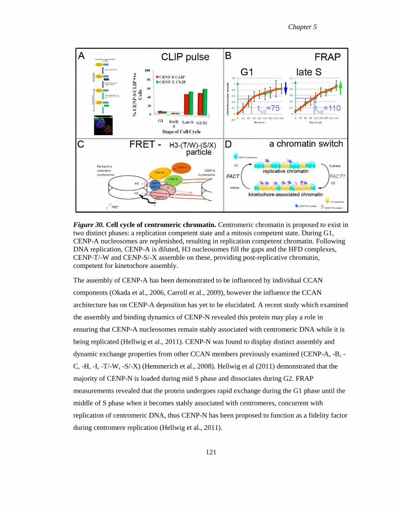
Chapter 5
Figure 30. Cell cycle of centromeric chromatin. Centromeric chromatin is proposed to exist in
two distinct phases: a replication competent state and a mitosis competent state. During G1,
CENP-A nucleosomes are replenished, resulting in replication competent chromatin. Following
DNA replication, CENP-A is diluted, H3 nucleosomes fill the gaps and the HFD complexes,
CENP-T/-W and CENP-S/-X assemble on these, providing post-replicative chromatin,
competent for kinetochore assembly.
The assembly of CENP-A has been demonstrated to be influenced by individual CCAN
components (Okada et al., 2006, Carroll et al., 2009), however the influence the CCAN
architecture has on CENP-A deposition has yet to be elucidated. A recent study which examined
the assembly and binding dynamics of CENP-N revealed this protein may play a role in
ensuring that CENP-A nucleosomes remain stably associated with centromeric DNA while it is
being replicated (Hellwig et al., 2011). CENP-N was found to display distinct assembly and
dynamic exchange properties from other CCAN members previously examined (CENP-A, -B, -
C, -H, -I, -T/-W, -S/-X) (Hemmerich et al., 2008). Hellwig et al (2011) demonstrated that the
majority of CENP-N is loaded during mid S phase and dissociates during G2. FRAP
measurements revealed that the protein undergoes rapid exchange during the G1 phase until the
middle of S phase when it becomes stably associated with centromeres, concurrent with
replication of centromeric DNA, thus CENP-N has been proposed to function as a fidelity factor
during centromere replication (Hellwig et al., 2011).
121
Chapter 5
It will be interesting in future experiments to examine the assembly and binding
dynamics of the remaining CCAN members at centromeres in an effort to elucidate the
roles these play in either propagation of centromere identity (G1 phase), maintenance of
centromeric chromatin structure during replication (mid S phase) or in the establishment
of kinetochore function (late S/G2) and the contribution of all these processes to the
chromatin based inheritance.
122

Chapter 6
Bibliography
Chapter 6
Bibliography
ALLSHIRE, R. C. & KARPEN, G. H. (2008) Epigenetic regulation of centromeric chromatin:
dogs, new tricks? Nat Rev Genet, 9, 923-37.
ALONSO, A., HASSON, D., CHEUNG, F. & WARBURTON, P. E. (2010) A paucity of
heterochromatin at functional human neocentromeres. Epigenetics Chromatin, 3, 6.
ALONSO, A., MAHMOOD, R., LI, S., CHEUNG, F., YODA, K. & WARBURTON, P. E.
(2003) Genomic microarray analysis reveals distinct locations for the CENP-A binding
domains in three human chromosome 13q32 neocentromeres. Hum Mol Genet, 12,
2711-21.
ALOY, P., BOTTCHER, B., CEULEMANS, H., LEUTWEIN, C., MELLWIG, C., FISCHER,
S., GAVIN, A. C., BORK, P., SUPERTI-FURGA, G., SERRANO, L. & RUSSELL, R.
B. (2004) Structure-based assembly of protein complexes in yeast. Science, 303, 2026-9.
AMANO, M., SUZUKI, A., HORI, T., BACKER, C., OKAWA, K., CHEESEMAN, I. M. &
FUKAGAWA, T. (2009) The CENP-S complex is essential for the stable assembly of
outer kinetochore structure. J Cell Biol, 186, 173-82.
AMARO, A. C., SAMORA, C. P., HOLTACKERS, R., WANG, E., KINGSTON, I. J.,
ALONSO, M., LAMPSON, M., MCAINSH, A. D. & MERALDI, P. (2010) Molecular
control of kinetochore-microtubule dynamics and chromosome oscillations. Nat Cell
Biol, 12, 319-29.
AMOR, D. J., BENTLEY, K., RYAN, J., PERRY, J., WONG, L., SLATER, H. & CHOO, K. H.
(2004) Human centromere repositioning "in progress". Proc Natl Acad Sci U S A, 101,
6542-7.
AMOR, D. J. & CHOO, K. H. (2002) Neocentromeres: role in human disease, evolution, and
centromere study. Am J Hum Genet, 71, 695-714.
ANDO, S., YANG, H., NOZAKI, N., OKAZAKI, T. & YODA, K. (2002) CENP-A, -B, and -C
chromatin complex that contains the I-type alpha-satellite array constitutes the
prekinetochore in HeLa cells. Mol Cell Biol, 22, 2229-41.
BACIA, K., KIM, S. A. & SCHWILLE, P. (2006) Fluorescence cross-correlation spectroscopy
in living cells. Nat Methods, 3, 83-9.
BANNISTER, A. J., ZEGERMAN, P., PARTRIDGE, J. F., MISKA, E. A., THOMAS, J. O.,
ALLSHIRE, R. C. & KOUZARIDES, T. (2001) Selective recognition of methylated
lysine 9 on histone H3 by the HP1 chromo domain. Nature, 410, 120-4.
BARNHART, M. C., KUICH, P. H., STELLFOX, M. E., WARD, J. A., BASSETT, E. A.,
BLACK, B. E. & FOLTZ, D. R. (2011) HJURP is a CENP-A chromatin assembly factor
sufficient to form a functional de novo kinetochore. J Cell Biol, 194, 229-43.
BAUER, J. (1999) Advances in cell separation: recent developments in counterflow centrifugal
elutriation and continuous flow cell separation. J Chromatogr B Biomed Sci Appl, 722,
55-69.
BELLE, A., TANAY, A., BITINCKA, L., SHAMIR, R. & O'SHEA, E. K. (2006)
Quantification of protein half-lives in the budding yeast proteome. Proc Natl Acad Sci U
S A, 103, 13004-9.
BERGMANN, J. H., RODRIGUEZ, M. G., MARTINS, N. M., KIMURA, H., KELLY, D. A.,
MASUMOTO, H., LARIONOV, V., JANSEN, L. E. & EARNSHAW, W. C. (2011)
Epigenetic engineering shows H3K4me2 is required for HJURP targeting and CENP-A
assembly on a synthetic human kinetochore. EMBO J, 30, 328-40.
124
Chapter 6
BERNARD, P., MAURE, J. F., PARTRIDGE, J. F., GENIER, S., JAVERZAT, J. P. &
ALLSHIRE, R. C. (2001) Requirement of heterochromatin for cohesion at centromeres.
Science, 294, 2539-42.
BLACK, B. E., BROCK, M. A., BEDARD, S., WOODS, V. L., JR. & CLEVELAND, D. W.
(2007) An epigenetic mark generated by the incorporation of CENP-A into centromeric
nucleosomes. Proc Natl Acad Sci U S A, 104, 5008-13.
BLACK, B. E., FOLTZ, D. R., CHAKRAVARTHY, S., LUGER, K., WOODS, V. L., JR. &
CLEVELAND, D. W. (2004) Structural determinants for generating centromeric
chromatin. Nature, 430, 578-82.
BLOWER, M. D., SULLIVAN, B. A. & KARPEN, G. H. (2002) Conserved organization of
centromeric chromatin in flies and humans. Dev Cell, 2, 319-30.
BOOTSMA, D., BUDKE, L. & VOS, O. (1964) Studies on Synchronous Division of Tissue
Culture Cells Initiated by Excess Thymidine. Exp Cell Res, 33, 301-9.
BREEDEN, L. L. (2003) Periodic transcription: a cycle within a cycle. Curr Biol, 13, R31-8.
BROWN, K. D., COULSON, R. M., YEN, T. J. & CLEVELAND, D. W. (1994) Cyclin-like
accumulation and loss of the putative kinetochore motor CENP-E results from coupling
continuous synthesis with specific degradation at the end of mitosis. J Cell Biol, 125,
1303-12.
CAMAHORT, R., SHIVARAJU, M., MATTINGLY, M., LI, B., NAKANISHI, S., ZHU, D.,
SHILATIFARD, A., WORKMAN, J. L. & GERTON, J. L. (2009) Cse4 is part of an
octameric nucleosome in budding yeast. Mol Cell, 35, 794-805.
CAMPOS, E. I. & REINBERG, D. (2009) Histones: annotating chromatin. Annu Rev Genet, 43,
559-99.
CARBONE, L., NERGADZE, S. G., MAGNANI, E., MISCEO, D., FRANCESCA CARDONE,
M., ROBERTO, R., BERTONI, L., ATTOLINI, C., FRANCESCA PIRAS, M., DE
JONG, P., RAUDSEPP, T., CHOWDHARY, B. P., GUERIN, G., ARCHIDIACONO,
N., ROCCHI, M. & GIULOTTO, E. (2006) Evolutionary movement of centromeres in
horse, donkey, and zebra. Genomics, 87, 777-82.
CARROLL, C. W., MILKS, K. J. & STRAIGHT, A. F. (2010) Dual recognition of CENP-A
nucleosomes is required for centromere assembly. J Cell Biol, 189, 1143-55.
CARROLL, C. W., SILVA, M. C., GODEK, K. M., JANSEN, L. E. & STRAIGHT, A. F.
(2009) Centromere assembly requires the direct recognition of CENP-A nucleosomes by
CENP-N. Nat Cell Biol, 11, 896-902.
CARROLL, C. W. & STRAIGHT, A. F. (2006) Centromere formation: from epigenetics to self-
assembly. Trends Cell Biol, 16, 70-8.
CASTILLO, A. G., MELLONE, B. G., PARTRIDGE, J. F., RICHARDSON, W., HAMILTON,
G. L., ALLSHIRE, R. C. & PIDOUX, A. L. (2007) Plasticity of fission yeast CENP-A
chromatin driven by relative levels of histone H3 and H4. PLoS Genet, 3, e121.
CHEESEMAN, I. M. & DESAI, A. (2008) Molecular architecture of the kinetochore-
microtubule interface. Nat Rev Mol Cell Biol, 9, 33-46.
CHEESEMAN, I. M., HORI, T., FUKAGAWA, T. & DESAI, A. (2008) KNL1 and the CENP-
H/I/K complex coordinately direct kinetochore assembly in vertebrates. Mol Biol Cell,
19, 587-94.
CHEN, E. S., SAITOH, S., YANAGIDA, M. & TAKAHASHI, K. (2003) A cell cycle-regulated
GATA factor promotes centromeric localization of CENP-A in fission yeast. Mol Cell,
11, 175-87.
CHO, R. J., CAMPBELL, M. J., WINZELER, E. A., STEINMETZ, L., CONWAY, A.,
WODICKA, L., WOLFSBERG, T. G., GABRIELIAN, A. E., LANDSMAN, D.,
LOCKHART, D. J. & DAVIS, R. W. (1998) A genome-wide transcriptional analysis of
the mitotic cell cycle. Mol Cell, 2, 65-73.
125
Chapter 6
CHUANG, L. S., IAN, H. I., KOH, T. W., NG, H. H., XU, G. & LI, B. F. (1997) Human DNA-
(cytosine-5) methyltransferase-PCNA complex as a target for p21WAF1. Science, 277,
1996-2000.
CLARKE, L. (1998) Centromeres: proteins, protein complexes, and repeated domains at
centromeres of simple eukaryotes. Curr Opin Genet Dev, 8, 212-8.
CLEVELAND, D. W., MAO, Y. & SULLIVAN, K. F. (2003) Centromeres and kinetochores:
from epigenetics to mitotic checkpoint signaling. Cell, 112, 407-21.
CRAIG, J. M., WONG, L. H., LO, A. W., EARLE, E. & CHOO, K. H. (2003) Centromeric
chromatin pliability and memory at a human neocentromere. EMBO J, 22, 2495-504.
DALAL, Y., WANG, H., LINDSAY, S. & HENIKOFF, S. (2007) Tetrameric structure of
centromeric nucleosomes in interphase Drosophila cells. PLoS Biol, 5, e218.
DE LICHTENBERG, U., JENSEN, L. J., BRUNAK, S. & BORK, P. (2005) Dynamic complex
formation during the yeast cell cycle. Science, 307, 724-7.
DE LICHTENBERG, U., JENSEN, T. S., BRUNAK, S., BORK, P. & JENSEN, L. J. (2007)
Evolution of cell cycle control: same molecular machines, different regulation. Cell
Cycle, 6, 1819-25.
DIMITRIADIS, E. K., WEBER, C., GILL, R. K., DIEKMANN, S. & DALAL, Y. (2010)
Tetrameric organization of vertebrate centromeric nucleosomes. Proc Natl Acad Sci U S
A, 107, 20317-22.
DUNLEAVY, E. M., ALMOUZNI, G. & KARPEN, G. H. (2011) H3.3 is deposited at
centromeres in S phase as a placeholder for newly assembled CENP-A in G(1) phase.
Nucleus, 2, 146-157.
DUNLEAVY, E. M., ROCHE, D., TAGAMI, H., LACOSTE, N., RAY-GALLET, D.,
NAKAMURA, Y., DAIGO, Y., NAKATANI, Y. & ALMOUZNI-PETTINOTTI, G.
(2009) HJURP is a cell-cycle-dependent maintenance and deposition factor of CENP-A
at centromeres. Cell, 137, 485-97.
DURAND-DUBIEF, M. & EKWALL, K. (2008) Heterochromatin tells CENP-A where to go.
Bioessays, 30, 526-9.
EARNSHAW, W. C. & MIGEON, B. R. (1985) Three related centromere proteins are absent
from the inactive centromere of a stable isodicentric chromosome. Chromosoma, 92,
290-6.
EARNSHAW, W. C. & ROTHFIELD, N. (1985) Identification of a family of human
centromere proteins using autoimmune sera from patients with scleroderma.
Chromosoma, 91, 313-21.
EKWALL, K. (2004) The roles of histone modifications and small RNA in centromere function.
Chromosome Res, 12, 535-42.
EKWALL, K. (2007) Epigenetic Control of Centromere Behavior. Annual Review of Genetics,
41, 18.
EKWALL, K., JAVERZAT, J. P., LORENTZ, A., SCHMIDT, H., CRANSTON, G. &
ALLSHIRE, R. (1995) The chromodomain protein Swi6: a key component at fission
yeast centromeres. Science, 269, 1429-31.
EKWALL, K., OLSSON,T., TURNER,B.M., CRANSTON,G. AND ALLSHIRE,R.C. (1997)
Transient Inhibition of Histone Deacetylation Alters the Structural and Functional
Imprint at Fission Yeast Centromeres. Cell, 91, 11.
ERHARDT, S., MELLONE, B. G., BETTS, C. M., ZHANG, W., KARPEN, G. H. &
STRAIGHT, A. F. (2008) Genome-wide analysis reveals a cell cycle-dependent
mechanism controlling centromere propagation. J Cell Biol, 183, 805-18.
FITZGERALD-HAYES, M., CLARKE, L. & CARBON, J. (1982) Nucleotide sequence
comparisons and functional analysis of yeast centromere DNAs. Cell, 29, 235-44.
126
Chapter 6
FOLCO, H. D., PIDOUX, A. L., URANO, T. & ALLSHIRE, R. C. (2008) Heterochromatin and
RNAi are required to establish CENP-A chromatin at centromeres. Science, 319, 94-7.
FOLTZ, D. R., JANSEN, L. E., BAILEY, A. O., YATES, J. R., 3RD, BASSETT, E. A.,
WOOD, S., BLACK, B. E. & CLEVELAND, D. W. (2009) Centromere-specific
assembly of CENP-a nucleosomes is mediated by HJURP. Cell, 137, 472-84.
FOLTZ, D. R., JANSEN, L. E., BLACK, B. E., BAILEY, A. O., YATES, J. R., 3RD &
CLEVELAND, D. W. (2006) The human CENP-A centromeric nucleosome-associated
complex. Nat Cell Biol, 8, 458-69.
FUJITA, Y., HAYASHI, T., KIYOMITSU, T., TOYODA, Y., KOKUBU, A., OBUSE, C. &
YANAGIDA, M. (2007) Priming of centromere for CENP-A recruitment by human
hMis18alpha, hMis18beta, and M18BP1. Dev Cell, 12, 17-30.
FUKAGAWA, T., MIKAMI, Y., NISHIHASHI, A., REGNIER, V., HARAGUCHI, T.,
HIRAOKA, Y., SUGATA, N., TODOKORO, K., BROWN, W. & IKEMURA, T.
(2001) CENP-H, a constitutive centromere component, is required for centromere
targeting of CENP-C in vertebrate cells. EMBO J, 20, 4603-17.
FUKS, F., BURGERS, W. A., BREHM, A., HUGHES-DAVIES, L. & KOUZARIDES, T.
(2000) DNA methyltransferase Dnmt1 associates with histone deacetylase activity. Nat
Genet, 24, 88-91.
FURUYAMA, T., DALAL, Y. & HENIKOFF, S. (2006) Chaperone-mediated assembly of
centromeric chromatin in vitro. Proc Natl Acad Sci U S A, 103, 6172-7.
GARDNER, R. D., PODDAR, A., YELLMAN, C., TAVORMINA, P. A., MONTEAGUDO,
M. C. & BURKE, D. J. (2001) The spindle checkpoint of the yeast Saccharomyces
cerevisiae requires kinetochore function and maps to the CBF3 domain. Genetics, 157,
1493-502.
GASCOIGNE, K. E., TAKEUCHI, K., SUZUKI, A., HORI, T., FUKAGAWA, T. &
CHEESEMAN, I. M. (2011) Induced ectopic kinetochore assembly bypasses the
requirement for CENP-A nucleosomes. Cell, 145, 410-22.
GAUTIER, A., JUILLERAT, A., HEINIS, C., CORREA, I. R., JR., KINDERMANN, M.,
BEAUFILS, F. & JOHNSSON, K. (2008) An engineered protein tag for multiprotein
labeling in living cells. Chem Biol, 15, 128-36.
GHAEMMAGHAMI, S., HUH, W. K., BOWER, K., HOWSON, R. W., BELLE, A.,
DEPHOURE, N., O'SHEA, E. K. & WEISSMAN, J. S. (2003) Global analysis of
protein expression in yeast. Nature, 425, 737-41.
GIMELLI, G., ZUFFARDI, O., GIGLIO, S., ZENG, C. & HE, D. (2000) CENP-G in
neocentromeres and inactive centromeres. Chromosoma, 109, 328-33.
GROTH, A., ROCHA, W., VERREAULT, A. & ALMOUZNI, G. (2007) Chromatin challenges
during DNA replication and repair. Cell, 128, 721-33.
GRUSS, P. & SAUER, G. (1975) Repetitive primate DNA containing the recognition sequences
for two restriction endonucleases which generate cohesive ends. FEBS Lett, 60, 85-8.
HAYASHI, T., FUJITA, Y., IWASAKI, O., ADACHI, Y., TAKAHASHI, K. & YANAGIDA,
M. (2004) Mis16 and Mis18 are required for CENP-A loading and histone deacetylation
at centromeres. Cell, 118, 715-29.
HE, D., ZENG, C., WOODS, K., ZHONG, L., TURNER, D., BUSCH, R. K., BRINKLEY, B.
R. & BUSCH, H. (1998) CENP-G: a new centromeric protein that is associated with the
alpha-1 satellite DNA subfamily. Chromosoma, 107, 189-97.
HELLWIG, D., EMMERTH, S., ULBRICHT, T., DORING, V., HOISCHEN, C., MARTIN, R.,
SAMORA, C. P., MCAINSH, A. D., CARROLL, C. W., STRAIGHT, A. F.,
MERALDI, P. & DIEKMANN, S. (2011) Dynamics of CENP-N kinetochore binding
during the cell cycle. J Cell Sci, 124, 3871-83.
127
Chapter 6
HEMMERICH, P., WEIDTKAMP-PETERS, S., HOISCHEN, C., SCHMIEDEBERG, L.,
ERLIANDRI, I. & DIEKMANN, S. (2008) Dynamics of inner kinetochore assembly
and maintenance in living cells. J Cell Biol, 180, 1101-14.
HENDERSON, D. S., BANGA, S. S., GRIGLIATTI, T. A. & BOYD, J. B. (1994) Mutagen
sensitivity and suppression of position-effect variegation result from mutations in
mus209, the Drosophila gene encoding PCNA. EMBO J, 13, 1450-9.
HENDZEL, M. J., WEI, Y., MANCINI, M. A., VAN HOOSER, A., RANALLI, T.,
BRINKLEY, B. R., BAZETT-JONES, D. P. & ALLIS, C. D. (1997) Mitosis-specific
phosphorylation of histone H3 initiates primarily within pericentromeric
heterochromatin during G2 and spreads in an ordered fashion coincident with mitotic
chromosome condensation. Chromosoma, 106, 348-60.
HERMANN, A., GOYAL, R. & JELTSCH, A. (2004) The Dnmt1 DNA-(cytosine-C5)-
methyltransferase methylates DNA processively with high preference for
hemimethylated target sites. J Biol Chem, 279, 48350-9.
HEUN, P., ERHARDT, S., BLOWER, M. D., WEISS, S., SKORA, A. D. & KARPEN, G. H.
(2006) Mislocalization of the Drosophila centromere-specific histone CID promotes
formation of functional ectopic kinetochores. Dev Cell, 10, 303-15.
HILLESHEIM, L. N., CHEN, Y. & MULLER, J. D. (2006) Dual-color photon counting
histogram analysis of mRFP1 and EGFP in living cells. Biophys J, 91, 4273-84.
HORI, T., AMANO, M., SUZUKI, A., BACKER, C. B., WELBURN, J. P., DONG, Y.,
MCEWEN, B. F., SHANG, W. H., SUZUKI, E., OKAWA, K., CHEESEMAN, I. M. &
FUKAGAWA, T. (2008a) CCAN makes multiple contacts with centromeric DNA to
provide distinct pathways to the outer kinetochore. Cell, 135, 1039-52.
HORI, T., OKADA, M., MAENAKA, K. & FUKAGAWA, T. (2008b) CENP-O class proteins
form a stable complex and are required for proper kinetochore function. Mol Biol Cell,
19, 843-54.
HUH, W. K., FALVO, J. V., GERKE, L. C., CARROLL, A. S., HOWSON, R. W.,
WEISSMAN, J. S. & O'SHEA, E. K. (2003) Global analysis of protein localization in
budding yeast. Nature, 425, 686-91.
IRVINE, D. V., AMOR, D. J., PERRY, J., SIRVENT, N., PEDEUTOUR, F., CHOO, K. H. &
SAFFERY, R. (2004) Chromosome size and origin as determinants of the level of
CENP-A incorporation into human centromeres. Chromosome Res, 12, 805-15.
JACKSON, V. & CHALKLEY, R. (1981) A reevaluation of new histone deposition on
replicating chromatin. J Biol Chem, 256, 5095-103.
JANSEN, L. E., BLACK, B. E., FOLTZ, D. R. & CLEVELAND, D. W. (2007) Propagation of
centromeric chromatin requires exit from mitosis. J Cell Biol, 176, 795-805.
JENSEN, L. J., JENSEN, T. S., DE LICHTENBERG, U., BRUNAK, S. & BORK, P. (2006)
Co-evolution of transcriptional and post-translational cell-cycle regulation. Nature, 443,
594-7.
JUN-ICHIROU OHZEKI, M. N., TERUAKI OKADA AND HIROSHI MASUMOTO (2002)
CENP-B box is required for de novo centromere chromatin assembly on human alphoid
DNA The Journal of Cell Biology, 159, 10.
KEPPLER, A., GENDREIZIG, S., GRONEMEYER, T., PICK, H., VOGEL, H. & JOHNSSON,
K. (2003) A general method for the covalent labeling of fusion proteins with small
molecules in vivo. Nat Biotechnol, 21, 86-9.
KIMURA, H. & COOK, P. R. (2001) Kinetics of core histones in living human cells: little
exchange of H3 and H4 and some rapid exchange of H2B. J Cell Biol, 153, 1341-53.
KUNKEL, T. A. & BURGERS, P. M. (2008) Dividing the workload at a eukaryotic replication
fork. Trends Cell Biol, 18, 521-7.
128
Chapter 6
LAGANA, A., DORN, J. F., DE ROP, V., LADOUCEUR, A. M., MADDOX, A. S. &
MADDOX, P. S. (2010) A small GTPase molecular switch regulates epigenetic
centromere maintenance by stabilizing newly incorporated CENP-A. Nat Cell Biol, 12,
1186-93.
LEE, C., WEVRICK, R., FISHER, R. B., FERGUSON-SMITH, M. A. & LIN, C. C. (1997)
Human centromeric DNAs. Hum Genet, 100, 291-304.
LERMONTOVA, I., SCHUBERT, V., FUCHS, J., KLATTE, S., MACAS, J. & SCHUBERT, I.
(2006) Loading of Arabidopsis centromeric histone CENH3 occurs mainly during G2
and requires the presence of the histone fold domain. Plant Cell, 18, 2443-51.
LI, E., BESTOR, T. H. & JAENISCH, R. (1992) Targeted mutation of the DNA
methyltransferase gene results in embryonic lethality. Cell, 69, 915-26.
LIAO, H., WINKFEIN, R. J., MACK, G., RATTNER, J. B. & YEN, T. J. (1995) CENP-F is a
protein of the nuclear matrix that assembles onto kinetochores at late G2 and is rapidly
degraded after mitosis. J Cell Biol, 130, 507-18.
LIAO, W. T., YU, C. P., WU, D. H., ZHANG, L., XU, L. H., WENG, G. X., ZENG, M. S.,
SONG, L. B. & LI, J. S. (2009) Upregulation of CENP-H in tongue cancer correlates
with poor prognosis and progression. J Exp Clin Cancer Res, 28, 74.
LO, A. W., MAGLIANO, D. J., SIBSON, M. C., KALITSIS, P., CRAIG, J. M. & CHOO, K. H.
(2001) A novel chromatin immunoprecipitation and array (CIA) analysis identifies a
460-kb CENP-A-binding neocentromere DNA. Genome Res, 11, 448-57.
LOCKE, D. P., HILLIER, L. W., WARREN, W. C., WORLEY, K. C., NAZARETH, L. V.,
MUZNY, D. M., YANG, S. P., WANG, Z., CHINWALLA, A. T., MINX, P.,
MITREVA, M., COOK, L., DELEHAUNTY, K. D., FRONICK, C., SCHMIDT, H.,
FULTON, L. A., FULTON, R. S., NELSON, J. O., MAGRINI, V., POHL, C.,
GRAVES, T. A., MARKOVIC, C., CREE, A., DINH, H. H., HUME, J., KOVAR, C.
L., FOWLER, G. R., LUNTER, G., MEADER, S., HEGER, A., PONTING, C. P.,
MARQUES-BONET, T., ALKAN, C., CHEN, L., CHENG, Z., KIDD, J. M.,
EICHLER, E. E., WHITE, S., SEARLE, S., VILELLA, A. J., CHEN, Y., FLICEK, P.,
MA, J., RANEY, B., SUH, B., BURHANS, R., HERRERO, J., HAUSSLER, D.,
FARIA, R., FERNANDO, O., DARRE, F., FARRE, D., GAZAVE, E., OLIVA, M.,
NAVARRO, A., ROBERTO, R., CAPOZZI, O., ARCHIDIACONO, N., DELLA
VALLE, G., PURGATO, S., ROCCHI, M., KONKEL, M. K., WALKER, J. A.,
ULLMER, B., BATZER, M. A., SMIT, A. F., HUBLEY, R., CASOLA, C.,
SCHRIDER, D. R., HAHN, M. W., QUESADA, V., PUENTE, X. S., ORDONEZ, G.
R., LOPEZ-OTIN, C., VINAR, T., BREJOVA, B., RATAN, A., HARRIS, R. S.,
MILLER, W., KOSIOL, C., LAWSON, H. A., TALIWAL, V., MARTINS, A. L.,
SIEPEL, A., ROYCHOUDHURY, A., MA, X., DEGENHARDT, J., BUSTAMANTE,
C. D., GUTENKUNST, R. N., MAILUND, T., DUTHEIL, J. Y., HOBOLTH, A.,
SCHIERUP, M. H., RYDER, O. A., YOSHINAGA, Y., DE JONG, P. J.,
WEINSTOCK, G. M., ROGERS, J., MARDIS, E. R., GIBBS, R. A., et al. (2011)
Comparative and demographic analysis of orang-utan genomes. Nature, 469, 529-33.
LOYOLA, A., BONALDI, T., ROCHE, D., IMHOF, A. & ALMOUZNI, G. (2006) PTMs on
H3 variants before chromatin assembly potentiate their final epigenetic state. Mol Cell,
24, 309-16.
MADDOX, P. S., HYNDMAN, F., MONEN, J., OEGEMA, K. & DESAI, A. (2007) Functional
genomics identifies a Myb domain-containing protein family required for assembly of
CENP-A chromatin. J Cell Biol, 176, 757-63.
MADDOX, P. S., OEGEMA, K., DESAI, A. & CHEESEMAN, I. M. (2004) "Holo"er than
thou: chromosome segregation and kinetochore function in C. elegans. Chromosome
Res, 12, 641-53.
129
Chapter 6
MAIO, J. J. (1971) DNA strand reassociation and polyribonucleotide binding in the African
green monkey, Cercopithecus aethiops. J Mol Biol, 56, 579-95.
MALIK, H. S. & HENIKOFF, S. (2002) Conflict begets complexity: the evolution of
centromeres. Curr Opin Genet Dev, 12, 711-8.
MARGUERON, R. & REINBERG, D. (2010) Chromatin structure and the inheritance of
epigenetic information. Nat Rev Genet, 11, 285-96.
MARSHALL, O., CHUEH, A., WONG, L. & CHOO, K. (2008a) Neocentromeres: new insights
into centromere structure, disease development, and karyotype evolution. The American
Journal of Human Genetics, 82, 261-282.
MARSHALL, O. J., MARSHALL, A. T. & CHOO, K. H. (2008b) Three-dimensional
localization of CENP-A suggests a complex higher order structure of centromeric
chromatin. J Cell Biol, 183, 1193-202.
MASUMOTO, H., MASUKATA, H., MURO, Y., NOZAKI, N. & OKAZAKI, T. (1989) A
human centromere antigen (CENP-B) interacts with a short specific sequence in alphoid
DNA, a human centromeric satellite. J Cell Biol, 109, 1963-73.
MASUMOTO, H., NAKANO, M. & OHZEKI, J. (2004) The role of CENP-B and a-satellite
DNA: de novo assembly and epigenetic maintenance of human centromeres.
Chromosome Research, 12, 543-556.
MCCLELLAND, S. E., BORUSU, S., AMARO, A. C., WINTER, J. R., BELWAL, M.,
MCAINSH, A. D. & MERALDI, P. (2007) The CENP-A NAC/CAD kinetochore
complex controls chromosome congression and spindle bipolarity. EMBO J, 26, 5033-
47.
MELLONE, B. G. & ALLSHIRE, R. C. (2003) Stretching it: putting the CEN(P-A) in
centromere. Curr Opin Genet Dev, 13, 191-8.
MELLONE, B. G., GRIVE, K. J., SHTEYN, V., BOWERS, S. R., ODERBERG, I. &
KARPEN, G. H. (2011) Assembly of Drosophila centromeric chromatin proteins during
mitosis. PLoS Genet, 7, e1002068.
MINOSHIMA, Y., HORI, T., OKADA, M., KIMURA, H., HARAGUCHI, T., HIRAOKA, Y.,
BAO, Y. C., KAWASHIMA, T., KITAMURA, T. & FUKAGAWA, T. (2005) The
constitutive centromere component CENP-50 is required for recovery from spindle
damage. Mol Cell Biol, 25, 10315-28.
MIZUGUCHI, G., XIAO, H., WISNIEWSKI, J., SMITH, M. M. & WU, C. (2007) Nonhistone
Scm3 and histones CenH3-H4 assemble the core of centromere-specific nucleosomes.
Cell, 129, 1153-64.
MOLDOVAN, G. L., PFANDER, B. & JENTSCH, S. (2007) PCNA, the maestro of the
replication fork. Cell, 129, 665-79.
MONTEFALCONE, G., TEMPESTA, S., ROCCHI, M. & ARCHIDIACONO, N. (1999)
Centromere repositioning. Genome Res, 9, 1184-8.
MOREY, L., BARNES, K., CHEN, Y., FITZGERALD-HAYES, M. & BAKER, R. E. (2004)
The histone fold domain of Cse4 is sufficient for CEN targeting and propagation of
active centromeres in budding yeast. Eukaryot Cell, 3, 1533-43.
NISHIHASHI, A., HARAGUCHI, T., HIRAOKA, Y., IKEMURA, T., REGNIER, V.,
DODSON, H., EARNSHAW, W. C. & FUKAGAWA, T. (2002) CENP-I is essential
for centromere function in vertebrate cells. Dev Cell, 2, 463-76.
NORBURY, C. & NURSE, P. (1992) Animal cell cycles and their control. Annu Rev Biochem,
61, 441-70.
130
Chapter 6
OBUSE, C., YANG, H., NOZAKI, N., GOTO, S., OKAZAKI, T. & YODA, K. (2004)
Proteomics analysis of the centromere complex from HeLa interphase cells: UV-
damaged DNA bindingprotein 1 (DDB-1) is a component of the CEN-complex, while
BMI-1 is transiently co-localized with the centromeric region in interphase. Genes
Cells, 9, 105-20.
OHTA, S., BUKOWSKI-WILLS, J. C., SANCHEZ-PULIDO, L., ALVES FDE, L., WOOD, L.,
CHEN, Z. A., PLATANI, M., FISCHER, L., HUDSON, D. F., PONTING, C. P.,
FUKAGAWA, T., EARNSHAW, W. C. & RAPPSILBER, J. (2010) The protein
composition of mitotic chromosomes determined using multiclassifier combinatorial
proteomics. Cell, 142, 810-21.
OKADA, M., CHEESEMAN, I. M., HORI, T., OKAWA, K., MCLEOD, I. X., YATES, J. R.,
3RD, DESAI, A. & FUKAGAWA, T. (2006) The CENP-H-I complex is required for
the efficient incorporation of newly synthesized CENP-A into centromeres. Nat Cell
Biol, 8, 446-57.
OKADA, T., OHZEKI, J., NAKANO, M., YODA, K., BRINKLEY, W. R., LARIONOV, V. &
MASUMOTO, H. (2007) CENP-B controls centromere formation depending on the
chromatin context. Cell, 131, 1287-300.
PALMER, D. K. & MARGOLIS, R. L. (1985) Kinetochore components recognized by human
autoantibodies are present on mononucleosomes. Mol Cell Biol, 5, 173-86.
PALMER, D. K., O'DAY, K., TRONG, H. L., CHARBONNEAU, H. & MARGOLIS, R. L.
(1991) Purification of the centromere-specific protein CENP-A and demonstration that
it is a distinctive histone. Proc Natl Acad Sci U S A, 88, 3734-8.
PALMER, D. K., O'DAY, K., WENER, M. H., ANDREWS, B. S. & MARGOLIS, R. L. (1987)
A 17-kD centromere protein (CENP-A) copurifies with nucleosome core particles and
with histones. J Cell Biol, 104, 805-15.
PEARSON, C. G., YEH, E., GARDNER, M., ODDE, D., SALMON, E. D. & BLOOM, K.
(2004) Stable kinetochore-microtubule attachment constrains centromere positioning in
metaphase. Curr Biol, 14, 1962-7.
PERIASAMY, A. & DAY, R. N. (1999) Visualizing protein interactions in living cells using
digitized GFP imaging and FRET microscopy. Methods Cell Biol, 58, 293-314.
PERPELESCU, M. & FUKAGAWA, T. (2011) The ABCs of CENPs. Chromosoma.
PFAFFL, M. W. (2001) A new mathematical model for relative quantification in real-time RT-
PCR. Nucleic Acids Res, 29, e45.
PIDOUX, A. L., CHOI, E. S., ABBOTT, J. K., LIU, X., KAGANSKY, A., CASTILLO, A. G.,
HAMILTON, G. L., RICHARDSON, W., RAPPSILBER, J., HE, X. & ALLSHIRE, R.
C. (2009) Fission yeast Scm3: A CENP-A receptor required for integrity of
subkinetochore chromatin. Mol Cell, 33, 299-311.
PINES, J. & HUNTER, T. (1989) Isolation of a human cyclin cDNA: evidence for cyclin
mRNA and protein regulation in the cell cycle and for interaction with p34cdc2. Cell,
58, 833-46.
PINES, J. & HUNTER, T. (1990) Human cyclin A is adenovirus E1A-associated protein p60
and behaves differently from cyclin B. Nature, 346, 760-3.
PIRAS, F. M., NERGADZE, S. G., MAGNANI, E., BERTONI, L., ATTOLINI, C.,
KHORIAULI, L., RAIMONDI, E. & GIULOTTO, E. (2010) Uncoupling of satellite
DNA and centromeric function in the genus Equus. PLoS Genet, 6, e1000845.
PIRAS, F. M., NERGADZE, S. G., POLETTO, V., CERUTTI, F., RYDER, O. A., LEEB, T.,
RAIMONDI, E. & GIULOTTO, E. (2009) Phylogeny of horse chromosome 5q in the
genus Equus and centromere repositioning. Cytogenet Genome Res, 126, 165-72.
PLUTA, A. F., MACKAY, A. M., AINSZTEIN, A. M., GOLDBERG, I. G. & EARNSHAW,
W. C. (1995) The centromere: hub of chromosomal activities. Science, 270, 1591-4.
131
Chapter 6
PRENDERGAST, L., VAN VUUREN, C., KACZMARCZYK, A., DOERING, V., HELLWIG,
D., QUINN, N., HOISCHEN, C., DIEKMANN, S. & SULLIVAN, K. F. (2011)
Premitotic assembly of human CENPs -T and -W switches centromeric chromatin to a
mitotic state. PLoS Biol, 9, e1001082.
PROBST, A. V., DUNLEAVY, E. & ALMOUZNI, G. (2009) Epigenetic inheritance during the
cell cycle. Nat Rev Mol Cell Biol, 10, 192-206.
PRZEWLOKA, M. R., VENKEI, Z., BOLANOS-GARCIA, V. M., DEBSKI, J., DADLEZ, M.
& GLOVER, D. M. (2011) CENP-C is a structural platform for kinetochore assembly.
Curr Biol, 21, 399-405.
RANSOM, M., DENNEHEY, B. K. & TYLER, J. K. (2010) Chaperoning histones during DNA
replication and repair. Cell, 140, 183-95.
RAY-GALLET, D., QUIVY, J. P., SCAMPS, C., MARTINI, E. M., LIPINSKI, M. &
ALMOUZNI, G. (2002) HIRA is critical for a nucleosome assembly pathway
independent of DNA synthesis. Mol Cell, 9, 1091-100.
REA, S., EISENHABER, F., O'CARROLL, D., STRAHL, B. D., SUN, Z. W., SCHMID, M.,
OPRAVIL, S., MECHTLER, K., PONTING, C. P., ALLIS, C. D. & JENUWEIN, T.
(2000) Regulation of chromatin structure by site-specific histone H3 methyltransferases.
Nature, 406, 593-9.
REESE, B. E., BACHMAN, K. E., BAYLIN, S. B. & ROUNTREE, M. R. (2003) The methyl-
CpG binding protein MBD1 interacts with the p150 subunit of chromatin assembly
factor 1. Mol Cell Biol, 23, 3226-36.
RIBEIRO, S. A., VAGNARELLI, P., DONG, Y., HORI, T., MCEWEN, B. F., FUKAGAWA,
T., FLORS, C. & EARNSHAW, W. C. (2010) A super-resolution map of the vertebrate
kinetochore. Proc Natl Acad Sci U S A, 107, 10484-9.
ROUNTREE, M. R., BACHMAN, K. E. & BAYLIN, S. B. (2000) DNMT1 binds HDAC2 and
a new co-repressor, DMAP1, to form a complex at replication foci. Nat Genet, 25, 269-
77.
SAITOH, H., TOMKIEL, J., COOKE, C. A., RATRIE, H., 3RD, MAURER, M., ROTHFIELD,
N. F. & EARNSHAW, W. C. (1992) CENP-C, an autoantigen in scleroderma, is a
component of the human inner kinetochore plate. Cell, 70, 115-25.
SCHERER, W. F., SYVERTON, J. T. & GEY, G. O. (1953) Studies on the propagation in vitro
of poliomyelitis viruses. IV. Viral multiplication in a stable strain of human malignant
epithelial cells (strain HeLa) derived from an epidermoid carcinoma of the cervix. J Exp
Med, 97, 695-710.
SCHUH, M., LEHNER, C. F. & HEIDMANN, S. (2007) Incorporation of Drosophila
CID/CENP-A and CENP-C into centromeres during early embryonic anaphase. Curr
Biol, 17, 237-43.
SCREPANTI, E., DE ANTONI, A., ALUSHIN, G. M., PETROVIC, A., MELIS, T.,
NOGALES, E. & MUSACCHIO, A. (2011) Direct binding of Cenp-C to the Mis12
complex joins the inner and outer kinetochore. Curr Biol, 21, 391-8.
SEKULIC, N., BASSETT, E. A., ROGERS, D. J. & BLACK, B. E. (2010) The structure of
(CENP-A-H4)(2) reveals physical features that mark centromeres. Nature, 467, 347-51.
SEUANEZ, H., FLETCHER, J., EVANS, H. J. & MARTIN, D. E. (1976) A chromosome
rearrangement in orangutan studied with Q-, C-, and G-banding techniques. Cytogenet
Cell Genet, 17, 26-34.
SHARIF, J., MUTO, M., TAKEBAYASHI, S., SUETAKE, I., IWAMATSU, A., ENDO, T. A.,
SHINGA, J., MIZUTANI-KOSEKI, Y., TOYODA, T., OKAMURA, K., TAJIMA, S.,
MITSUYA, K., OKANO, M. & KOSEKI, H. (2007) The SRA protein Np95 mediates
epigenetic inheritance by recruiting Dnmt1 to methylated DNA. Nature, 450, 908-12.
132
Chapter 6
SHELBY, R. D., MONIER, K. & SULLIVAN, K. F. (2000) Chromatin assembly at
kinetochores is uncoupled from DNA replication. J Cell Biol, 151, 1113-8.
SHELBY, R. D., VAFA, O. & SULLIVAN, K. F. (1997) Assembly of CENP-A into
centromeric chromatin requires a cooperative array of nucleosomal DNA contact sites. J
Cell Biol, 136, 501-13.
SHIBAHARA, K. & STILLMAN, B. (1999) Replication-dependent marking of DNA by PCNA
facilitates CAF-1-coupled inheritance of chromatin. Cell, 96, 575-85.
SHIGEISHI, H., HIGASHIKAWA, K., ONO, S., MIZUTA, K., NINOMIYA, Y., YONEDA,
S., TAKI, M. & KAMATA, N. (2006) Increased expression of CENP-H gene in human
oral squamous cell carcinomas harboring high-proliferative activity. Oncol Rep, 16,
1071-5.
SHUAIB, M., OUARARHNI, K., DIMITROV, S. & HAMICHE, A. (2010) HJURP binds
CENP-A via a highly conserved N-terminal domain and mediates its deposition at
centromeres. Proc Natl Acad Sci U S A, 107, 1349-54.
SINGH, T. R., SARO, D., ALI, A. M., ZHENG, X. F., DU, C. H., KILLEN, M. W.,
SACHPATZIDIS, A., WAHENGBAM, K., PIERCE, A. J., XIONG, Y., SUNG, P. &
MEETEI, A. R. (2010) MHF1-MHF2, a histone-fold-containing protein complex,
participates in the Fanconi anemia pathway via FANCM. Mol Cell, 37, 879-86.
SITTMAN, D. B., GRAVES, R. A. & MARZLUFF, W. F. (1983) Histone mRNA
concentrations are regulated at the level of transcription and mRNA degradation. Proc
Natl Acad Sci U S A, 80, 1849-53.
SOBEL, R. E., COOK, R. G., PERRY, C. A., ANNUNZIATO, A. T. & ALLIS, C. D. (1995)
Conservation of deposition-related acetylation sites in newly synthesized histones H3
and H4. Proc Natl Acad Sci U S A, 92, 1237-41.
SPELLMAN, P. T., SHERLOCK, G., ZHANG, M. Q., IYER, V. R., ANDERS, K., EISEN, M.
B., BROWN, P. O., BOTSTEIN, D. & FUTCHER, B. (1998) Comprehensive
identification of cell cycle-regulated genes of the yeast Saccharomyces cerevisiae by
microarray hybridization. Mol Biol Cell, 9, 3273-97.
STOLER, S., KEITH, K. C., CURNICK, K. E. & FITZGERALD-HAYES, M. (1995) A
mutation in CSE4, an essential gene encoding a novel chromatin-associated protein in
yeast, causes chromosome nondisjunction and cell cycle arrest at mitosis. Genes Dev, 9,
573-86.
SUGATA, N., LI, S., EARNSHAW, W. C., YEN, T. J., YODA, K., MASUMOTO, H.,
MUNEKATA, E., WARBURTON, P. E. & TODOKORO, K. (2000) Human CENP-H
multimers colocalize with CENP-A and CENP-C at active centromere--kinetochore
complexes. Hum Mol Genet, 9, 2919-26.
SUGATA, N., MUNEKATA, E. & TODOKORO, K. (1999) Characterization of a novel
kinetochore protein, CENP-H. J Biol Chem, 274, 27343-6.
SULLIVAN, B. & KARPEN, G. (2001) Centromere identity in Drosophila is not determined in
vivo by replication timing. J Cell Biol, 154, 683-90.
SULLIVAN, B. A. & KARPEN, G. H. (2004) Centromeric chromatin exhibits a histone
modification pattern that is distinct from both euchromatin and heterochromatin. Nat
Struct Mol Biol, 11, 1076-83.
SULLIVAN, B. A. & SCHWARTZ, S. (1995) Identification of centromeric antigens in dicentric
Robertsonian translocations: CENP-C and CENP-E are necessary components of
functional centromeres. Hum Mol Genet, 4, 2189-97.
SULLIVAN, B. A. & WILLARD, H. F. (1998) Stable dicentric X chromosomes with two
functional centromeres. Nat Genet, 20, 227-8.
SULLIVAN, K. F. (2001) A solid foundation: functional specialization of centromeric
chromatin. Curr Opin Genet Dev, 11, 182-8.
133
Chapter 6
SULLIVAN, K. F., HECHENBERGER, M. & MASRI, K. (1994) Human CENP-A contains a
histone H3 related histone fold domain that is required for targeting to the centromere. J
Cell Biol, 127, 581-92.
TAGAMI, H., RAY-GALLET, D., ALMOUZNI, G. & NAKATANI, Y. (2004) Histone H3.1
and H3.3 complexes mediate nucleosome assembly pathways dependent or independent
of DNA synthesis. Cell, 116, 51-61.
TAKAHASHI, K., CHEN, E. S. & YANAGIDA, M. (2000) Requirement of Mis6 centromere
connector for localizing a CENP-A-like protein in fission yeast. Science, 288, 2215-9.
TAKAYAMA, Y., SATO, H., SAITOH, S., OGIYAMA, Y., MASUDA, F. & TAKAHASHI,
K. (2008) Biphasic incorporation of centromeric histone CENP-A in fission yeast. Mol
Biol Cell, 19, 682-90.
THOMPSON, L. H. & JONES, N. J. (2010) Stabilizing and remodeling the blocked DNA
replication fork: anchoring FANCM and the Fanconi anemia damage response. Mol
Cell, 37, 749-51.
THROWER, D. A., JORDAN, M. A., SCHAAR, B. T., YEN, T. J. & WILSON, L. (1995)
Mitotic HeLa cells contain a CENP-E-associated minus end-directed microtubule motor.
EMBO J, 14, 918-26.
TOMKIEL, J., COOKE, C. A., SAITOH, H., BERNAT, R. L. & EARNSHAW, W. C. (1994)
CENP-C is required for maintaining proper kinetochore size and for a timely transition
to anaphase. J Cell Biol, 125, 531-45.
TOMONAGA, T., MATSUSHITA, K., YAMAGUCHI, S., OOHASHI, T., SHIMADA, H.,
OCHIAI, T., YODA, K. & NOMURA, F. (2003) Overexpression and mistargeting of
centromere protein-A in human primary colorectal cancer. Cancer Res, 63, 3511-6.
VAN HOOSER, A. A., OUSPENSKI, II, GREGSON, H. C., STARR, D. A., YEN, T. J.,
GOLDBERG, M. L., YOKOMORI, K., EARNSHAW, W. C., SULLIVAN, K. F. &
BRINKLEY, B. R. (2001) Specification of kinetochore-forming chromatin by the
histone H3 variant CENP-A. J Cell Sci, 114, 3529-42.
VANDESOMPELE, J., DE PRETER, K., PATTYN, F., POPPE, B., VAN ROY, N., DE
PAEPE, A. & SPELEMAN, F. (2002) Accurate normalization of real-time quantitative
RT-PCR data by geometric averaging of multiple internal control genes. Genome Biol,
3, RESEARCH0034.
WADE, C. M., GIULOTTO, E., SIGURDSSON, S., ZOLI, M., GNERRE, S., IMSLAND, F.,
LEAR, T. L., ADELSON, D. L., BAILEY, E., BELLONE, R. R., BLOCKER, H.,
DISTL, O., EDGAR, R. C., GARBER, M., LEEB, T., MAUCELI, E., MACLEOD, J.
N., PENEDO, M. C., RAISON, J. M., SHARPE, T., VOGEL, J., ANDERSSON, L.,
ANTCZAK, D. F., BIAGI, T., BINNS, M. M., CHOWDHARY, B. P., COLEMAN, S.
J., DELLA VALLE, G., FRYC, S., GUERIN, G., HASEGAWA, T., HILL, E. W.,
JURKA, J., KIIALAINEN, A., LINDGREN, G., LIU, J., MAGNANI, E.,
MICKELSON, J. R., MURRAY, J., NERGADZE, S. G., ONOFRIO, R., PEDRONI, S.,
PIRAS, M. F., RAUDSEPP, T., ROCCHI, M., ROED, K. H., RYDER, O. A., SEARLE,
S., SKOW, L., SWINBURNE, J. E., SYVANEN, A. C., TOZAKI, T., VALBERG, S. J.,
VAUDIN, M., WHITE, J. R., ZODY, M. C., LANDER, E. S. & LINDBLAD-TOH, K.
(2009) Genome sequence, comparative analysis, and population genetics of the
domestic horse. Science, 326, 865-7.
WARBURTON, P. E. (2004) Chromosomal dynamics of human neocentromere formation.
Chromosome Research, 12, 9.
134
Chapter 6
WARBURTON, P. E., COOKE, C. A., BOURASSA, S., VAFA, O., SULLIVAN, B. A.,
STETTEN, G., GIMELLI, G., WARBURTON, D., TYLER-SMITH, C., SULLIVAN,
K. F., POIRIER, G. G. &EARNSHAW, W. C. (1997) Immunolocalization of CENP-A
suggests a distinct nucleosome structure at the inner kinetochore plate of active
centromeres. Curr Biol, 7, 901-4.
WEBB, E. C. & WESTHEAD, D. R. (2009) The transcriptional regulation of protein
complexes; a cross-species perspective. Genomics, 94, 369-76.
WIGLER, M., LEVY, D. & PERUCHO, M. (1981) The somatic replication of DNA
methylation. Cell, 24, 33-40.
WILLARD, H. F. (1985) Chromosome-specific organization of human alpha satellite DNA. Am
J Hum Genet, 37, 524-32.
WILLIAMS, J. S., HAYASHI, T., YANAGIDA, M. & RUSSELL, P. (2009) Fission yeast
Scm3 mediates stable assembly of Cnp1/CENP-A into centromeric chromatin. Mol Cell,
33, 287-98.
YAN, Z., DELANNOY, M., LING, C., DAEE, D., OSMAN, F., MUNIANDY, P. A., SHEN,
X., OOSTRA, A. B., DU, H., STELTENPOOL, J., LIN, T., SCHUSTER, B.,
DECAILLET, C., STASIAK, A., STASIAK, A. Z., STONE, S., HOATLIN, M. E.,
SCHINDLER, D., WOODCOCK, C. L., JOENJE, H., SEN, R., DE WINTER, J. P., LI,
L., SEIDMAN, M. M., WHITBY, M. C., MYUNG, K., CONSTANTINOU, A. &
WANG, W. (2010) A histone-fold complex and FANCM form a conserved DNA-
remodeling complex to maintain genome stability. Mol Cell, 37, 865-78.
YEN, T. J., COMPTON, D. A., WISE, D., ZINKOWSKI, R. P., BRINKLEY, B. R.,
EARNSHAW, W. C. & CLEVELAND, D. W. (1991) CENP-E, a novel human
centromere-associated protein required for progression from metaphase to anaphase.
EMBO J, 10, 1245-54.
YODA, K., ANDO, S., MORISHITA, S., HOUMURA, K., HASHIMOTO, K., TAKEYASU,
K. & OKAZAKI, T. (2000) Human centromere protein A (CENP-A) can replace
histone H3 in nucleosome reconstitution in vitro. Proc Natl Acad Sci U S A, 97, 7266-
71.
ZHANG, Z., SHIBAHARA, K. & STILLMAN, B. (2000) PCNA connects DNA replication to
epigenetic inheritance in yeast. Nature, 408, 221-5.
ZINKOWSKI, R. P., MEYNE, J. & BRINKLEY, B. R. (1991) The centromere-kinetochore
complex: a repeat subunit model. J Cell Biol, 113, 1091-110.
135
Appendix A
Authors Publications
GLYNN, M., KACZMARCZYK, A., PRENDERGAST, L., QUINN, N. & SULLIVAN, K. F.
(2010) Centromeres: assembling and propagating epigenetic function. Subcell Biochem,
50, 223-49.
Centromeres: Assembling and PropagatingEpigenetic Function
Macdara Glynn, Agnieszka Kaczmarczyk, Lisa Prendergast, Nadine Quinn,and Kevin F. Sullivan
Abstract The faithful replication of DNA and the accurate segregation of genomicmaterial from one generation to the next is critical in the maintenance of genomicstability. This chapter will describe the structure and assembly of an epigeneticallyinherited locus, the centromere, and its role in the processes by which sister chro-matids are evenly segregated to daughter cells. During the G2 phase of the cell cyclekinetochores are assembled upon the chromatids. During mitosis, kinetochoresattach chromosome(s) to the mitotic spindle. The kinetochore structure serves asthe interface between the mitotic spindle and the chromatids and it is at the kine-tochore where the forces that drive chromatid separation are generated. Unattachedchromosomes fail to satisfy the spindle assembly checkpoint (SAC), resulting in cellcycle arrest. The centromere is the locus upon which the kinetochore assembles, andcentromeres themselves are determined by their unique protein composition. Apartfrom budding yeast, centromeres are not specified simply by DNA sequence, butrather through chromatin composition and architecture and are thus epigeneticallydetermined. Centromeres are built on a specific nucleosome not found elsewherein the genome, in which histone H3 is replaced with a homologue – CENP-Aor CenH3. This domain is flanked by heterochromatin and is folded to provide a3-dimensional cylinder-like structure at metaphase that establishes the kinetochoreon the surface of the mitotic chromosomes. A large family of CENtromere Proteins(CENPs) associates with centromeric chromatin throughout the cell cycle and arerequired for kinetochore function. Unlike the bulk of histones, CENP-A is notassembled concurrently with DNA synthesis in S-phase but rather assembles intothe centromere in the subsequent G1 phase. The assembly of CENP-A chromatinfollowing DNA replication and the re-establishment of this network of constitutiveproteins have emerged as critical mechanisms for understanding how the centromereis replicated during the cell cycle.
K.F. Sullivan (B)Centre for Chromosome Biology, School of Natural Sciences, National University of Ireland,Galway, University Rd, Galway, Irelande-mail: [email protected]
223H.P. Nasheuer (ed.), Genome Stability and Human Diseases, SubcellularBiochemistry 50, DOI 10.1007/978-90-481-3471-7_12,C© Springer Science+Business Media B.V. 2010
224 M. Glynn et al.
Keywords Centromere · Mitosis · Epigenetics · Chromatin
Abbreviations
APC/C Anaphase Promoting Complex-CyclosomeBUB Budding Uninhibited by BenzimidazoleCAD CENP-A Distal ComplexCAF1 Chromatin Assembly FactorCATD CENP-A Targeting DomainCCAN Constitutive Centromere-Associated NetworkCENP Centromere ProteinHAT1 Histone AcetylTransferase catalytic subunitHFD Histone Fold DomainhFLEG1 human Fetal Liver Expressing Gene 1Hir1 HIRA like histone chaperoneKMN Knl1-Mis12-Ndc80 complexMAD Mitotic-Arrested DeficientMCC Mitotic Checkpoint ComplexMPS1 Multipolar Spindle 1NAC Nucleosome Associated ComplexPEV Position Effect VariegationPRC2 Polycomb Repressive Complex 2RZZ ROD-ZW10-Zwilch ComplexSAC Spindle Assembly Checkpoint
Introduction
The centromere is a chromosomal locus present in a single copy per chromosome.It is essential for the stable inheritance of the genome but it is itself epigeneticallyinherited. The functional identity of the centromere derives from interplay of spe-cific and global chromatin components that provide unique centromere functionand also specify a mechanism for replicating centromeres in the cell cycle. Thisis accomplished through the assembly and maintenance of a core set of constitu-tive centromere-associated proteins that are anchored to the DNA fiber through aspecialized nucleosome that contains a centromere-specific histone H3 homologue,CENP-A or CenH3.
For higher organisms the centromere displays properties of an epigenetic locuswith the information stably passed from one cell cycle to the next without beingdirectly encoded or templated by the underlying DNA sequence. The essence ofcentromere identity and function is the specialized chromatin that forms it. Thedetails of many of the issues in centromere biology have been recently summa-rized in several excellent reviews (Cleveland et al., 2003; Allshire and Karpen,2008; Cheeseman and Desai, 2008b; Musacchio and Salmon, 2007b; Henikoff andAhmad, 2005). In this chapter, we aim to provide an overview of the factors that
Centromeres: Assembling and Propagating Epigenetic Function 225
specify centromeres as genomic loci, the specific biochemistry of the specializedchromatin that comprises it and a summary of the functions of the kinetochore, themitotic product of centromere function, in chromosome movement and cell cyclecontrol.
Specifying the Centromere
Centromeric DNA
Centromere DNA organization varies widely among eukaryotes (see Cleveland et al.2003 for review). The simplest centromere is found in the budding yeast S. cere-visiae, a so-called point centromere built upon a small 125 bases of centromericDNA (Fitzgerald-Hayes et al., 1982). A diffuse centromere has been found to existin nematodes like C. elegans, where the kinetochore forms along the entire lengthof the mitotic chromosome. Such chromosomes are said to be holocentric. Moretypical is the regional centromere, in which the centromere is associated with a dis-crete DNA locus. The best studied example of this type of centromere are those ofS. pombe where a central core that specifies centromere function is flanked by longarrays of repetitive DNA (Clarke, 1998). In higher eukaryotes regional centromeresform on long stretches of repetitive DNA sequences that span several hundreds tothousands of kilobase pairs (Gieni et al., 2008). Centromeres in humans are com-prised of arrays of repetitive satellite DNA that is characteristic of multicellularorganisms. The primate centromeric repeat is termed alpha satellite DNA (or alphoidDNA) and consists of a basic repeating unit of 171 bp. Individual alpha satelliterepeats or monomers show variation around a consensus sequence of up to 60%across the entire genome. Higher order repeats of 2–16 alpha satellite monomers arefound in chromosome specific families and these higher order repeats are arrangedin tandem to form arrays ranging from ~200–5,000 kb (Schueler et al., 2001). Whilesuch organization is typical of both plant and animal centromeres, the sequencesthat make up centromeric DNA are rapidly evolving and diverge extensively evenamong closely related species (Henikoff et al., 2001). Indeed, centromere DNA inDrosophila is organized as islands of transposable elements in simple satellite DNAwith no specific sequences detectable as common to all centromeres of the karyotype(Sun et al., 1997).
Does the centromeric DNA sequence specify centromere function? In the bud-ding yeast, centromere function is dependent on specific DNA binding proteins suchthat single point mutations can abolish centromere activity (Bouck and Bloom,2005). Only one conserved DNA binding sequence has been identified in cen-tromeres of higher organisms, a small 17 base pair binding site for the sequencespecific DNA binding protein, CENP-B (Masumoto et al., 2004). Mutations ofCENP-B Boxes caused reduced efficiency of artificial chromosomes to form onalphoid DNA and those de novo events were shown to be dependent on CENP-B
226 M. Glynn et al.
(Ohzeki et al., 2002; Okada et al., 2007). However, CENP-B is nonessential,demonstrating that it does not play a role in the maintenance or normal replica-tion of centromeres (Hudson et al., 1998). Rather, the model that has emerged forcentromere formation in higher cells has focused on epigenetic determinants asdiscussed below.
Evidence for Epigenetic Behavior at the Centromere
Strong evidence for epigenetic inheritance at the centromere comes from the factthat the centromere displays various epigenetic behaviors. Position effect varie-gation (PEV) is an effect observed when a gene or reporter is positioned next toheterochromatin which leads to gene expression states being stochastically inher-ited (Ekwall, 2007). In fission yeast there is striking evidence of PEV in the formof centromeric silencing of nearby genes. Swi6 was one of the first centromericproteins identified in the fission yeast and when mutations of this were introducedit had an effect on centromeric silencing (Ekwall et al., 1995). Extreme cases ofsilencing of a centromere locus itself, observed in human cells, demonstrate thatcentromeric DNA is not sufficient to specify centromere formation. Certain deriva-tives of Robertsonian translocation (a type of translocation in which the long armsof non-homologous arcocentric chromosomes fuse at the centromeres) in humansmaintain two loci of centromeric DNA (known as a dicentric chromosome) butexhibit stable segregation at mitosis (Earnshaw and Migeon, 1985). In some ofthese cases, the centromeres are close enough to functionally cooperate (Sullivanand Willard, 1998). However, in numerous examples, one of the sites will be inac-tivated, allowing chromatid separation to occur normally (Sullivan and Schwartz,1995; Sullivan and Willard, 1998). A complementary example demonstrates thatcentromeric DNA is not necessary to form centromeres on human chromosomes– the formation of neocentromeres (Marshall et al., 2008). This involves the for-mation of a new centromere on non-centromeric DNA. These centromeres havebeen observed in both humans and plants. Studies of neocentromeres have foundthat these loci possess full complements of functional centromeric proteins (Safferyet al., 2000; Warburton, 2004). Repositioning of centromeres is another epige-netic phenomenon which is found at the centromere. This involves two epigeneticmechanisms, the first being inactivation of the original centromere and the for-mation of a neocentromere. Finally the inhibitor of histone deacetylase HDACTrichostatin A (TSA) has been used to demonstrated epigenetic effects at cen-tromeres in S. pombe (Ekwall et al., 1997). These experiments document epigeneticinfluences on the centromere which are mediated by histone modification. Humancell studies with TSA have pointed to a role for histone acetylation in the forma-tion of neocentromeres (Nakano et al., 2003). Taken together, these observationsargue that centromeric DNA itself is not the dominant determinant of centromerefunction but, rather, epigenetic mechanisms determine the functional identity of thelocus.
Centromeres: Assembling and Propagating Epigenetic Function 227
Chromatin and Centromere Determination
While the DNA sequences of eukaryotic centromeres vary widely, one common fea-ture found in all is a distinctive, centromere-specific nucleosome based on a histoneH3 variant known as CENP-A, or CenH3 (Earnshaw and Rothfield, 1985; Meluhet al., 1998; Palmer et al., 1991; Sullivan et al., 2001; Henikoff et al., 2000; Oegemaet al., 2001). This essential centromere component lies at the foundation of a dis-tinctive chromatin structure that is thought to underlie the functional properties andthe epigenetic determination of the centromere.
CENP-A, The Centromere Specific Histone
CENP-A was originally identified from auto antisera derived from patients with lim-ited systemic sclerosis – CREST syndrome (Tan et al., 1980). These sera recognizedthree centromere specific antigens: CENP-A, -B and -C (Earnshaw and Rothfield,1985). CENP-B and CENP-C were cloned using autoantibodies and shown to beproteins that associate with alpha-satellite DNA and the inner kinetochore platerespectively (Earnshaw, 1987; Saitoh et al., 1992). CENP-A, however, was iso-lated from nucleosomal preparations following digestion with micrococcal nuclease(Palmer and Margolis, 1985), and was shown to co-purify with nucleosomal coreparticles (Palmer, 1987). Partial amino acid sequence of bovine CENP-A suggestedhomology to histone H3 (Palmer et al., 1991) which was confirmed by direct isola-tion of the cDNA for CENP-A (Sullivan, 1994) and generalized as a common featureof eukaryotes by identification of Cse4 as the yeast homologue of the centromereprotein (Stoler et al., 1995).
Human CENP-A contains a histone H3-related histone fold domain (HFD) thatis required for targeting CENP-A to the centromere (Sullivan, 1994). In histoneH3, this HFD forms an interface with histone H4 to assemble the canonical het-erotetrameric core of nucleosome structure. The HFD, contact with DNA andhomodimerization are equally necessary for correct CENP-A localization (Keithet al., 1999; Shelby et al., 1997; Vermaak et al., 2002). A region of the CENP-A HFDwas found to confer centromeric targeting to histone H3, and was termed the CENP-A targeting domain (CATD) (Black et al., 2004; Black et al., 2007b). This CATDis essential for CENP-A function (Black et al., 2007a). Drosophila CENP-ACID-targeting domain is even smaller, consisting of the C-terminus of α1, all of loop 1and the N-terminus of α2 (15 amino acids) (Vermaak et al., 2002). Thus assemblyof centromere-specific CENP-A nucleosomes requires core histone structures ratherthan unique sequence elements of CENP-A’s divergent N-terminus.
The N-terminal tail of CENP-A exhibits a surprisingly high level of divergencebetween species in terms of both length and composition (Henikoff and Dalal,2005). Analysis of centromere protein evolution indicates that CENP-A and othercomponents of centromeric chromatin are rapidly and adaptively evolving (Meraldiet al., 2006; Talbert et al., 2004). A meiotic drive mechanism, in which centromeres
228 M. Glynn et al.
of different strengths affect their heritability at the level of positioning within thespindle in female meiosis, has been proposed to account for the rapid co-evolutionof centromere proteins and DNA in higher organisms (Talbert et al., 2004). TheN-terminal domain of Cse4, the budding yeast CENP-A homologue, contributesan essential role in interaction with other centromere proteins (Chen et al., 2000)and in human cells CENP-A is phosphorylated by Aurora kinases in G2 and M(Kunitoku et al., 2003; Zeitlin et al., 2001), though little is known about what role(s)the N-terminus plays in most organisms.
CENP-A Nucleosomes
In vitro nucleosome assembly studies have shown that CENP-A can replace histoneH3 in octamers containing H4, H2A and H2B (Yoda et al., 2000; Furuyama et al.,2006). Concordantly, purification of CENP-A containing nucleosomes from bothhuman and Drosophila cells have shown that the majority of CENP-A is located innucleosomes comprised as [CENP-A – H4 – H2A – H2B]2 octamers (Blower et al.,2002; Foltz et al., 2006). Atomic force microscopy of reconstituted centromericnucleosomes from Drosophila suggests that individual tetramers of [CENP-A – H4– H2A – H2B] may exist (Dalal et al., 2007). Reconstituted CENP-A containingnucleosomes have a more compact binding surface with H4 than H3 nucleosomes,observed in both [CENP-A – H4]2 tetramers and [CENP-A – H4 – H2A – H2B]2octamers (Black et al., 2004). CENP-A nucleosomes are also more readily destabi-lized than canonical nucleosomes, a property that has been proposed to ensure thatmisplaced CENP-A nucleosomes are easily removed (Conde e Silva et al., 2007). Inbudding yeast Cse4-containing nucleosomes are found in which the H2A and H2Bhistones are replaced by a non-histone protein (Scm3) that co-localizes with CENP-ACse4 at centromeres (Mizuguchi et al., 2007). The precise nature of the CENP-Anucleosomes and nucleosome like complexes that exist, and the extent to which theyare conserved evolutionarily, remains to be fully elucidated.
Organization of CENP-A Within Centromeres
CENP-A containing nucleosomes are the marker for the location and functional-ity of the centromere, but what is the context in which they are found? Usingstretched chromatin fibers from human and Drosophila cells, CENP-A and H3nucleosomes are found to occupy mutually exclusive domains, alternating acrossseveral hundred kb at the locus (Blower et al., 2002). Core histones H4, H2B andH2A are distributed throughout the domain, indicating their common use betweenthe CENP-A- and H3-containing nucleosomes. Recently, interspersion motifs havebeen proposed in both fission and budding yeast (Gregan et al., 2007; Yeh et al.,2008), suggesting that alternating conventional and centromeric histones are a con-served motif. ChIP experiments using neocentromeres demonstrate that CENP-Adomains in human chromosomes discontinuously span >100kb domains (Alonsoet al., 2007; Lo et al., 2001). The interspersed CENP-A domains are thought to fold
Centromeres: Assembling and Propagating Epigenetic Function 229
or coil into a cylindrical-like structure, with the CENP-A containing nucleosomesorientated to chromosome surface and the H3 domains facing the inter-chromatidspace (Sullivan et al., 2001).
The formation of centromeric chromatin is dependent on its association withflanking domains of heterochromatin. Classically associated with morphologicallydefined heterochromatin, a centromere-specific role for the molecular mechanismsthat mediate heterochromatic silencing of chromosomal domains has been mostclearly documented by studies in S. pombe (Ekwall, 2007). In S. pombe, cen-tromere function was shown to be dependent on the RNA interference pathway,which is required for silencing and modification of the heterochromatin that flanksthe CENP-A nucleosome-containing central core domain of the centromere (Volpeet al., 2002). Transcription from within the centromere initiates an RNAi-basedregulatory loop essential for the establishment and maintenance of pericentric het-erochromatin (Grewal and Elgin, 2007). The RNA machinery is involved in thelocalization of HP1 to the pericentric heterochromatin by coordinating the molec-ular machinery for modification of histones within these domains (Maison et al.,2002; Pal-Bhadra et al., 2004).
Clr3 is a homologue of human class II histone deacetylase and it deacetylaseslysine 14 of histone H3. Both the RNAi and Clr3 pathways recruit Clr4 (homologueof human SUV39 H1 and H2) which causes the trimethylation of lysine 9 of histoneH3 and this goes on to form the binding site which the chromodomain of Swi6recognizes (Nakayama et al., 2001). Swi6 is a homologue of the heterochromatinprotein 1 (HP1) and its chromodomain binds to histone H3 trimethylated lysine 9(Hall et al., 2002). Mutations of either lysine 9 or 14 of H3 cause delocalization ofSwi6 which leads to chromosome missegregation (Mellone et al., 2003).
In humans, the centromere is associated with the pericentric heterochromatindomains which cause the recruitment of HP1 through the methylation of the lysine9 of histone H3 (Fischle et al., 2005). Deletion of Suv39, the H3 lys9 methyltrans-ferase, causes improper centromere formation and function. Knockout studies inmice have shown an increase in genome instability and increased incidence of lym-phomas (Peters et al., 2001). Studies carried out in Suv39H1 and H2 null micedemonstrated 4-fold increase in defects of chromosome alignment and segregationdefects. These results confirm the requirement of histone lysine methylation forthe recruitment of cohesin to the pericentric heterochromatin (Heit et al., 2006). Adirect role for heterochromatin in recruitment of CENP-A has been documented inS. pombe, where the RNAi pathway is required for CENP-A deposition on naïvetemplates, but is dispensable for its maintenance (Folco et al., 2008).
These investigations have directly documented a role for epigenetic mechanismsin centromere formation and function. They detail the role(s) played by histonemodification pathways in heritable phenotypes of their target loci. In addition,the pathways for maintenance of epigenetic information are brought in focus bythese observations. For the vast majority of species, the pericentric heterochromatinof the centromere is composed of arrays of repeated DNA sequence. In buddingyeast CAF1 (chromatin assembly factor 1) is found in a region of the kinetochorewhere it interacts with Hir1 (HIRA like histone chaperone) and it is necessary for aproper centromere chromatin structure (Sharp et al., 2002). In fission yeast the Hir1
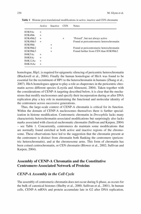
230 M. Glynn et al.
Table 1 Histone post-translational modifications in active, inactive and CEN chromatin
Active Inactive CEN Notes
H3K9Ac +H3K4Me +H3K4Me2 + + “Poised”, but not always activeH3K4Me3 + Found at pericentromeric heterochromatinH3K9Me +H3K9Me2 + Found at pericentromeric heterochromatinH3K9Me3 + Found further from CEN than H3K9Me2H4K5Ac +H4K8Ac +H4K12Ac +H4K16Ac +
homologue, Hip1, is required for epigenetic silencing of pericentric heterochromatin(Blackwell et al., 2004). Finally the human homologue of HirA was found to beessential for the recruitment of HP1 to the heterochromatin in humans (Zhang et al.,2007). HirA homologues appear to play a role as chaperones in the pericentric chro-matin across different species (Loyola and Almouzni, 2004). Taken together withthe considerations of CENP-A targeting described below, it is clear that the mecha-nisms that modify nucleosomes and specify their incorporation during or after DNAreplication play a key role in maintaining the functional and molecular identity ofthe centromere across successive generations.
Thus, the large-scale context of CENP-A chromatin is critical for its function.Within the domain of CENP-A nucleosomes themselves there is further special-ization in histone modification. Centromeric chromatin in Drosophila lacks manycharacteristic heterochromatin-associated modifications but surprisingly also lacksmarks associated with classical euchromatic chromatin (Sullivan and Karpen, 2004)– see Table 1. Concurrently, centromeres do maintain some modifications thatare normally found enriched at both active and inactive regions of the chromo-some. These observations have led to the suggestion that the chromatin present atthe centromere is distinct from chromatin both flanking the centromere (pericen-tric heterochromatin), and at the chromosome arms. This form of chromatin hasbeen coined centrochromatin, or CEN chromatin (Blower et al., 2002; Sullivan andKarpen, 2004).
Assembly of CENP-A Chromatin and the ConstitutiveCentromere-Associated Network of Proteins
CENP-A Assembly in the Cell Cycle
The assembly of centromeric chromatin does not occur during S-phase, as occurs forthe bulk of canonical histones (Shelby et al., 2000; Sullivan et al., 2001). In humancells, CENP-A mRNA and protein accumulate late in G2 after DNA replication,
Centromeres: Assembling and Propagating Epigenetic Function 231
and CENP-A deposition can take place in the absence of DNA replication (Shelbyet al., 2000, 1997). Precisely when does CENP-A assemble? Use of a novel covalentprotein labeling approach has shown directly that CENP-A synthesized in one cellcycle does not assemble into centromeres until passage through mitosis and entry into the G1 phase of the subsequent cell cycle (Jansen et al., 2007). During Drosophiladevelopment, CENP-ACID assembly occurs during anaphase of mitosis in the syn-cytial embryonic nuclear divisions, which cycle between S- and M-phases withoutG1 or G2 phases (Schuh et al., 2007). S. pombe CENP-A can assemble in bothS-phase and G2 (Takayama et al., 2008). There appear to be no absolute restrictionson the timing of CENP-A assembly, suggesting that the process is integrated withother cell cycle and chromosomal replication pathways.
Using a FRAP technique, loading of CENP-A was observed exclusively in G1 inHEp2 human cells expressing GFP-CENP-A (Hemmerich et al., 2008). Fluorescentcentromeres bleached at late mitosis/early G1 showed recovery after 30 min with aslow but steady increase in fluorescence over the following 2 h. The total numberof fluorescent centromeres were also monitored during FRAP at the same stage ofthe cell cycle. The number of fluorescent centromeres in a cell decreased accord-ingly following the bleaching of a region containing 10 centromeres. This numberincreased to pre-bleach numbers after 1 h, thus indicating that all bleached cen-tromeres had acquired new GFP–CENP-A molecules (Hemmerich et al., 2008). Themaximum FRAP recovery of CENP-A was <50%, suggesting centromere incorpo-ration without exchange of already loaded molecules. Using sister telophase cells ascontrols for one another, the rate of exchange between loaded centromeric CENP-A in G1 was assayed by FRAP. The complete set of centromeres in one daughtercell were bleached, resulting in recovery to ~40% pre-bleach fluorescence after 2 h.An area containing 5–10 centromeres was then photobleached for a second time toask if molecules could exchange at this later time in G1. Little or no recovery wasobserved at the double-bleached centromeres, indicating that centromeric CENP-Adid not exchange, while FRAP still occurred in the second daughter cell, demon-strating that CENP-A incorporation was still active at that time (Hemmerich et al.,2008).
Heritability and Dynamics of Centromeric Chromatin Proteins
A key question for understanding the basis of epigenetic inheritance is how molec-ular information is passed from mother to daughter cells in a locus-specific fashion.One possibility is that the epigenetic information can be assembled in each cellcycle on the basis of tissue-specific DNA binding proteins. Developmental biologyhas provided a broad mechanistic view of how stable epigenetic states, manifestas programs of gene expression, are coupled to histone modifications. For exam-ple, tissue specific gene silencing can be regulated during development through thebinding of large protein complexes, such as the Polycomb Repressive Complex 2(PRC2), in proximity to target genes. The binding of these complexes induces gene
232 M. Glynn et al.
repression by causing changes to the local chromatin structure, mainly affectinghistone modifications. The majority of Polycomb target genes are marked by his-tone modifications. A bivalent modification system, which involves both repressiveH3K27me3 modifications and H3K4me3 activating modifications in embryonicstem cells, is predicted to provide the potential for genes cycling between activeand inactive states during development (Barski et al., 2007; Bernstein et al., 2006;Mikkelsen et al., 2007; Pan et al., 2007; Zhao et al., 2007). Perhaps a similarmechanism exists for the maintenance of the centromeric locus? Alternatively,molecules present at the locus can be inherited locally, directly maintaining epi-genetic marks at defined chromosomal sites. Evidence for the latter is seen in theslow rate of turnover of histones in general and, specifically, of CENP-A at cen-tromeres (Regnier et al., 2005; Shelby et al., 2000). Is parental CENP-A “inherited”in equal abundance by both daughter centromeres or is CENP-A segregated exclu-sively at one daughter centromere while the sister centromere was replenished with“new” CENP-A? Direct observation of a cadre of CENP-A molecules fluorescentlylabeled in G1 suggests strongly that pre-existing CENP-A is equally distributed todaughter centromeres following passage through each mitosis (Jansen et al., 2007).Therefore, despite the synthesis of new CENP-A during the cell cycle, CENP-A already loaded into centromeric chromatin by late G1 is redistributed equallyto daughter centromeres, with each daughter receiving a “half-dose” of CENP-A.This results in the surprising conclusion that cells execute mitosis with a “halfdose” of CENP-A at the centromere, and a newly synthesized “half dose” whichis not yet assembled into the centromeric chromatin, heightening the relevance ofthe question of precisely how CENP-A chromatin is configured following DNAreplication.
Targeting CENP-A to the Centromere
A role for histone chaperones is indicated for assembly of CENP-A. HumanRbAp46, a histone chaperone (Verreault et al., 1996), is highly similar to S. pombeMis16, a gene originally identified in a screen for chromosome missegregation(Hayashi et al., 2004). The RbAp46/RbAp48 complex is essential for CENP-Alocalization to centromeres (Hayashi et al., 2004). Components of the human Mis18protein complex have also been shown to be essential for the structure and func-tion of the centromere/kinetochore. The hMis18α/hMis18β/Mis18 Binding Protein1 (Mis18BP1) complex accumulates specifically at the centromere in late telophase-G1 (Fujita et al., 2007), co-incident with CENP-A assembly into the centromere.RNAi depletion of hMis18α resulted in loss of centromeric CENP-A signal, whilethe total cellular level of CENP-A remained constant (Fujita et al., 2007). The Mis18protein complex was also shown to interact directly with the histone H3–H4 chaper-one RbAp46/48, but does not directly interact with CENP-A, however, RbAp48 haspreviously been observed to interact directly with CENP-ACID in vitro and facilitate
Centromeres: Assembling and Propagating Epigenetic Function 233
the assembly of centromeric chromatin (Furuyama et al., 2006). As RbAp46 bindsto histone acetyltransferase catalytic subunit (Hat1), the effect of suppressing his-tone deactylase on CENP-A assembly in the absence of hMis18α was analyzed.results indicate that histone deactylase inhibitors do indeed enhance the recruit-ment of newly synthesized CENP-A to the centromere (Fujita et al., 2007). Theseresults suggest that general nucleosome modification and assembly machinery isused in centromeric chromatin assembly by adaptation through specific proteinfactors.
Genetic analysis in fission yeast has revealed a number of proteins necessary forproper assembly and maintenance of CENP-A (Cnp1) chromatin. Mis6, Mis16-18and Ams2 have all been identified as essential for the assembly of CENP-ACnp1 tocentromere, with defects in of any one resulting in loss of CENP-ACnp1 signal at thecentromere (Chen et al., 2003; Hayashi et al., 2004; Takahashi et al., 2000). Hrp1and Sim4 are also required for CENP-ACnp1 recruitment to the centromere (Pidouxet al., 2003; Walfridsson et al., 2005). Recent characterization of the S. pombe Scm3(Scm3Sp), the sim1 gene product, has provided valuable insight into CENP-A target-ing. Scm3Sp localizes to centromeres of S. pombe and is present in central domainchromatin. However, during mitosis it is absent from the central core, returning dur-ing late anaphase (Pidoux et al., 2009; Williams et al., 2009). CENP-ACnp1 doesnot target to centromeres in scm3 mutants, while Scm3Sp maintains its centromericlocalization in cnp1-1 mutants (Pidoux et al., 2009). However, centromeric target-ing of Scm3Sp is dependent on the constitutive centromeric Mis6-Sim4 complex andalso on Mis16-Mis18 as Scm3Sp mis-localized in mis16 and mis18 mutants (Pidouxet al., 2009; Williams et al., 2009). This places Scm3Sp upstream to CENP-ACnp1
loading, but under control of Mis16-Mis18.In S. cerevisiae, Scm3 is required for targeting of both Ndc10 and CENP-ACse4
at the centromere (Collins et al., 2005; Ortiz et al., 1999). Ndc10 and Scm3Sc arealso co-dependent, as Ndc10 is required for the centromere association of Scm3Sc,while Scm3Sc is also required for Ndc10 localization (Mizuguchi et al., 2007). Incontrast to other CENP-ACse4 interacting partners, SCM3 is essential and in theabsence of Scm3Sc, CENP-ACse4 fails to localize to centromeric DNA. It has beenproposed that Scm3Sc is a core constituent of unconventional, CENP-ACse4 nucle-osomes, replacing H2A-H2B, as Scm3Sc in vitro displaces H2A-H2B dimers fromCENP-ACse4 histone octamers while in vivo the core centromere chromatin appearsto lack both H2A and H2B (Mizuguchi et al., 2007).
A Drosophila genome-wide RNAi screen for mis-localization of CENP-ACID
identified both CENP-C and CAL1, as essential factors for the correct assembly ofnewly synthesized CENP-ACID at the centromere (Erhardt et al., 2008). CAL1 hadbeen previously identified in a screen for mitotic defects (Goshima et al., 2007).Thus, studies in a number of organisms have revealed a variety of components thatare required for the assembly and/or maintenance of the centromere-specific nucleo-some. Understanding how the pathways defined by these proteins are organized andregulated, which ones serve conserved functions and, mechanistically, what thosefunctions are remains a challenge of current research.
234 M. Glynn et al.
Assembly of the Constitutive Centromere-Associated Network,CCAN
In addition to CENP-A, there is a large set of proteins that are uniquely local-ized to centromeres throughout the cell cycle (Cheeseman and Desai, 2008b).These proteins comprise a distinctive chromatin-associated complex that mediatethe functions of centromeric chromatin and may play a role in the heritability of thecentromere. Biochemical and in vivo studies involving CENP-A and other molecu-lar “starting points” have elucidated this large group of chromosome proteins. Theiridentification and functional characterization provides not only a key advance inunderstanding a complex genetic locus, but also a picture of the state of the artof chromosome biology and the challenges faced by researchers bringing genetic,molecular and cellular approaches to bear on a network of protein complexes whosediscrete functions are, for the most part, yet to be identified.
In vertebrates, a set of centromere proteins co-localize with CENP-A throughoutthe cell cycle and co-purify with CENP-A nucleosomes using monoclonal antibod-ies or affinity-based molecular probes (Foltz et al., 2006; Obuse et al., 2004; Okadaet al., 2006). A recent review of this set of proteins has suggested the unifyingnomenclature “constitutive centromere-associated network” (CCAN) (Cheesemanand Desai, 2008b). This network is comprised, in addition to CENP-A, of CENP-Cand 14 interacting proteins (CENP-H, CENP-I, CENP-K–U, CENP-W). CENP-Cis an essential component of the inner kinetochore and is presently the only proteinin this network besides CENP-A that has been identified in all eukaryotes (Heegeret al., 2005; Meluh and Koshland, 1995; Moore and Roth, 2001; Saitoh et al., 1992).
Initial biochemical studies isolated CENP-A chromatin and interacting pro-teins – the “CEN-complex” (Obuse et al., 2004). Known centromere proteins,CENP-A, CENP-B, CENP-C, CENP-H and CENP-I/hMis6 were specificallyfound in the CEN-complex as were a series of known proteins not previouslyreported as centromere proteins, such as uvDDB-1, XAP8, hSNF2H, FACTp180,FACTp80/SSRP1, PcG proteins (BMI-1, RNF1, RNF2, PHP2 and HPC3), KNL5,and racGAP. Only uvDDB-1 and BMI-1 were confirmed to localize to centromeres,however, no functional studies were carried out (Obuse et al., 2004). Multipletandem affinity purifications have since been used to define the most proximalcomplex directly recruited by CENP-A-containing centromeric nucleosomes, thenucleosome associated complex (CENP-A NAC), and a set of seven CENP-A dis-tal proteins (CAD) recruited to centromeric nucleosomes through their interactionwith the CENP-A NAC (Foltz et al., 2006). These studies establish a degree ofstructural hierarchy in the centromere. Found to be uniquely associated with theCENP-A nucleosomes using this approach were CENP-U (also named CENP-50)(Minoshima et al., 2005) and a set of three proteins CENP-M, CENP-N andCENP-T, which had been previously identified (Obuse et al., 2004) but not estab-lished as centromere components. Also associated with CENP-A nucleosomes wereCENP-B, human fetal liver expressing gene 1 (hFLEG1), nucleophosmin- 1 and theFACT complex members.
Centromeres: Assembling and Propagating Epigenetic Function 235
Expressing CENP-M, CENP-N and CENP-T putative centromeric proteins asfluorescent fusions confirmed their localization to the centromere, while subsequentaffinity purifications using a localization and affinity purification (LAP) tag toidentify the binding partners of CENP-M, CENP-N or CENP-U, in which eachprotein was co-purified with each of the others led to the conclusion that theCENP-A NAC was comprised of previously identified centromere componentsCENP-H (Sugata et al., 2000) and CENP-C (Saitoh et al., 1992), in addition toCENP-M, N, T and U(50) (Foltz et al., 2006). Suppression of CENP-M, CENP-Nor CENP-T by siRNA disrupted recruitment of the CENP-A NAC. Reduction ofCENP-M or CENP-N resulted in their reciprocal loss from centromeres as well asloss of CENP-H. Reduction of CENP-T eliminated CENP-M at centromeres, thuspresumably also CENP-N and CENP-H. Eliminating detectable CENP-U did notaffect centromeric recruitment of CENP-A, CENP-B, Hec1, CENP-E, CENP-F,Mis12 or Aurora B. However, it was shown to eliminate a tagged form of bothCENP-O and CENP-P from interphase and mitotic centromeres, suggesting thatthese components are interdependent for localization and/or maintenance at cen-tromeres. Disruption on CENP-M, -N and -T also resulted in mitotic defects, withthe depletion of CENP-T producing the most severe phenotype. The defects weremainly failures in chromosome congression that yielded an increased proportionof cells in prometaphase or pseudometaphase, suggesting the NAC components arerequired for mitotic progression.
Further tandem affinity purifications involving the NAC components yielded a setof proteins associated with each, establishing a CENP-A distal (CAD) componentof the centromere. As CENP-B, hFLEG1, nucleophosmin-1 and the FACT complexmembers were absent in these purifications they were assumed to be not tightlyassociated with the NAC components. CENP-I, CENP-K, CENP-L, CENP-O,CENP-P and CENP-Q were also purified with each of the CENP-M, CENP-N andCENP-U-containing complexes. CENP-R and CENP-S were associated with theCENP-M and CENP-U complexes. CENP-K, CENP-L and CENP-Q have beenshown to be constitutively associated with centromeres (Okada et al., 2006).
Affinity purifications from both human and chicken (DT40) cells using celllines expressing epitope-tagged CENP-H and CENP-I identified other constitutiveCENPs (Okada et al., 2006). Centromere proteins CENP-K to R, CENP-U werefound to constitutively localize to centromeres as components of a complex withCENP-H and CENP-I (Okada et al., 2006). Interestingly the purifications in thisscreen did not isolate CENP-M, CENP-L, CENP-S or CENP-T in human cells.The interactions detectable between various CENPs are dependent on the level ofstringency used in the purifications. Some interactions detected using a single purifi-cation step were not detected following a more stringent tandem affinity purificationor LAP. For example, CENP-H and CENP-I were not present in significant amountsin a tandem affinity purification of human LAP–CENP-O, but were present in aone-step immunoprecipitation. In contrast, both methods isolated a large amountof CENP-O, CENP-P, CENP-Q, CENP-R and CENP-U (Okada et al., 2006). Thissuggests that some protein-protein interactions are stronger than others, that somecomplexes are more stable than others or that there are distinct sub-complexes
236 M. Glynn et al.
within the centromere. It is also possible that the centromere protein interactionsremaining undetectable in a more stringent purification are either less stable or of amore transient nature.
Biochemical and in vivo studies in vertebrates assessing interdependencies,mitotic defects and lethality have divided the CENPs into subclasses of a constitu-tive centromere associated network (CCAN) (Cheeseman and Desai, 2008b). Thereare three distinct protein classes within the set of CENP-H–I-associated proteins,as was suggested by the phenotypic analysis and the purifications: an essentialCENP-H class (CENP-H, -I, -K and -L), and an essential CENP-M class (CENP-M)and a non-essential CENP-O class (CENP-O, -P, -Q and –U) (Okada et al., 2006).Depletion of components of the first subclass of proteins, comprising CENP-H,CENP-I and CENP-K, causes cell-cycle arrest, kinetochore-assembly defects andsevere chromosome missegregation (Foltz et al., 2006; Fukagawa et al., 2001; Izutaet al., 2006; Liu et al., 2003; Mikami et al., 2005; Nishihashi et al., 2002; Okadaet al., 2006). The CENP-O class forms a stable complex, which is required inchicken cells for proper kinetochore function (Hori et al., 2008). Kinetochore local-ization of CENP-O, -P, -Q and -U was shown to be interdependent, yet all proteinscorrectly localized in CENP-R deficient cells. All proteins were also required forrecovery from spindle damage, with phosphorylation of CENP-U essential for therecovery (Hori et al., 2008). In human cells, depletion of individual members ofthe CENP-O subclass of proteins causes defects in chromosome segregation thatare severe enough to cause cell lethality (Foltz et al., 2006; McAinsh et al., 2003;Okada et al., 2006).
Members of the CENP-H-I complex may contribute to the efficient incorporationof newly synthesized CENP-A into centromeres in DT40 cells. The percentage ofcells with CENP-A–GFP foci was observed to be significantly lower in CENP-H-,CENP-I-, CENP-K- and CENP-M-deficient DT40 cells, however, endogenousCENP-A levels were unaltered in CENP-H–I complex mutant cells by western blotanalysis. CENP-A–GFP was also incorporated into non-centromeric chromatinin CENP-M and CENP-K mutants (Okada et al., 2006). In contrast with this,CENP-I has previously been thought to not be required for CENP-A targetingin DT40 cells (Nishihashi et al., 2002). These data suggest that centromereproteins may play a role in the recruitment and/or deposition of newly synthesizedCENP-A at the centromere. As the localization of CENP-H and CENP-I alsorequires CENP-A, the loading of newly synthesized CENP-A is either dependenton preexisting CENP-A nucleosomes, with the CENP-H–I complex acting asan intermediate, or preexisting centromeric protein complexes. It is unknown iswhether the CENPs (H-I complex) that play a role in “new” CENP-A depositionare the CENPs that are already present at the centromere and thus recruit newlysynthesized CENP-A to a “centromere” defined by their presence, or whetherthey are also newly synthesized and are deposited at the centromere coincidentwith CENP-A deposition. As the CENP H-I subclasses of centromere proteinshave a degree of hierarchical dependence, it is possible that the formation ofsub-complexes of “new” CENPs is necessary prior to their deposition at thecentromere.
Centromeres: Assembling and Propagating Epigenetic Function 237
Of the CCAN components, only CENP-S and CENP-T together with the recentlyidentified CENP-W contain histone fold domains, similar to CENP-A. CENP-Wwas identified as a CENP-T interacting factor. Under stringent conditions, CENP-Tcan be separated from the rest of the CCAN and is distinct from the CENP-H-containing complex as purifications of affinity-tagged CENP-T do not containCENP-H or CENP-O complex proteins (Hori et al., 2008). However, in addition tosome histone proteins, a novel 11 kDa protein was identified and named CENP-Wdue to its co-purification with CENP-T and its constitutive localization to cen-tromeres (Hori et al., 2008). The CENP-T/CENP-W complex directly associateswith nucleosomal DNA and with canonical histone H3, but not with CENP-A, incentromeric regions similarly to CENP-C (Hori et al., 2008). The DNA bindingactivities of the complex requires their histone-fold domain. Some studies werecarried out to assign hierarchical importance to the complex in order to elucidateits role in the assembly of the centromere and the mitotic assembly of the kineto-chore. Phenotypic analysis of CENP-T/W and CENP-C deficient DT40 cell linesindicates that CENP-T/W and CENP-C function in separate pathways in the assem-bly of the kinetochore, while in CENP-W-deficient cells, kinetochore localizationof other CCAN proteins, with the exception of CENP-C, was abolished suggest-ing CENP-W functions upstream of other CCAN proteins in centromere assembly.Kinetochore localization of CENP-T complex occurs upstream of the CENP-H com-plex as CENP-T and CENP-W were still visible on mitotic chromosomes in cellsdepleted of CENP-H, thus the T/W complex functions also upstream of the CENP-Hcomplex.
What these studies make clear is that the complex of proteins required to form acentromere and to confer its ability to nucleate kinetochore assembly and functionis indeed complex. Teasing apart the associations and functions of these proteins,how functions are distributed within the network and how this complex is repli-cated will require an integration of biochemical and cellular methods, coupled withtraditional and in vitro genetic approaches. Just as clearly, identifying a consistentset of functional assays for centromere behavior is necessary to systematically inte-grate findings from numerous organisms and experimental perspectives. In additionto the chromatin structure and replication issues, the key functional output of thecentromere is through the kinetochore in mitosis and meiosis.
Kinetochore Function, Assembly and Signaling in the SpindleAssembly Checkpoint
Ultimately, the function of the centromere is to nucleate the assembly of the mitotickinetochore to provide the cell a means of connecting chromosomes to spindle fibersand regulating this association. Chromosome missegregation often leads to aneu-ploidy or polyploidy, which has been recognized as a common characteristic ofcancer cells and possible cause of tumorigenesis (Weaver and Cleveland, 2005). Toavoid loss of genomic information during mitosis, each pair of sister chromatids
238 M. Glynn et al.
connects to spindle microtubule through their kinetochores – macromolecular struc-tures assembled onto the centromeres at mitosis (Liu et al., 2006), that monitor theattachment state and activate signaling pathways to prevent anaphase onset beforeall chromosomes have achieved merotelic (attachement to opposite poles of thespindle) attachment (Cheeseman and Desai, 2008a). Defects in chromosome bi-orientation activate the spindle assembly checkpoint (SAC) which is required toblock sister chromatid separation until all chromosomes are properly attached to themitotic spindle. The SAC prevents cells entering anaphase by inhibiting the ubiq-uitination of cyclin B and securin by the anaphase-promoting complex/cyclosome(APC/C) ubiquitin ligase. The checkpoint proteins, including BubR1, Bub1, Bub3,Mad1, Mad2 and CENP-E, target the essential APC/C activator Cdc20. A sin-gle unattached kinetochore can generate a robust SAC activity thus providing thenecessary time to correct chromosome attachment problems.
Kinetochore Structure and Function
The major function of kinetochore protein complex in mitosis is: (1) kinetochorespecification, (2) kinetochore assembly, (3) microtubule binding and (4) monitor-ing and attachment regulation of kinetochore-microtubule structure (Cheesemanand Desai, 2008a; Logarinho and Bousbaa, 2008). In higher eukaryotes, kineto-chore proteins with MT-binding function can be dissected into: proteins that formthe core attachment site (Ndc80/HEC1) and are responsible for attachment chromo-some to spindle microtubules; proteins that control microtubule dynamics (CLASP,APC/EB1, MAP125) and promote kinetochore motility (dynein and CENP-E) togenerate or to transduce the forces that are required for chromosome segregation(Logarinho and Bousbaa, 2008). The SAC is tightly integrated with these proteins,allowing kinetochore activity and function to directly modulate the generation ofdiffusible anaphase inhibitors. Thus, the irreversible transition from metaphase toanaphase is delayed until every kinetochore is attach to the mitotic spindle and canbe faithfully segregated into a daughter cell.
Kinetochore assembly, structure and function is dynamically regulated during thecell cycle (Cheeseman and Desai, 2008a). Kinetochores have a trilaminar morphol-ogy that consist of: the inner kinetochore that forms the interface with chromatin;the outer kinetochore, a 50–60 nm thick region, which forms the interaction sur-face for spindle microtubules; and the central domain, the region in between outerand inner kinetochore. Kinetochore assembly is nucleated by centromeric chro-matin and its associated protein complexes – CCAN, NAC and CAD, starts withCENP-A chromatin, which forms a foundation for kinetochore structure in yeast,C. elegans, D. melanogaster and vertebrates. In vertebrates, the network of con-stitutively associated centromere chromatin proteins plays an essential role whileadditional proteins specifically required for mitotic events are recruited to kineto-chores starting from late G2 phase, through all stages of mitosis until the onset ofanaphase (Cheeseman and Desai, 2008a; Logarinho and Bousbaa, 2008).
Centromeres: Assembling and Propagating Epigenetic Function 239
The Core Microtubule-Attachment Site – KMN Network
The core microtubule-attachment site is compromised by the components of KNL1-Mis12-Ndc80 complex (KMN) network that is conserved widely in the eukaryotickingdom (Cheeseman and Desai, 2008a). By association with the Mis12 complexKNL1 generates a binding site for the Ndc80 complex, which allows both KNL1and Ndc80 bind to microtubules. The CCAN plays an important role in localizingthe Ndc80 complex, and the amount of CCAN and KMN complexes at kinetochoresdoes not change significantly from late prophase until late anaphase, irrespectivelyof microtubule-attachment status (Cheeseman and Desai, 2008a), in contrast to SACcomponents.
Members of the CCAN that have been shown to play a role in kinetochore assem-bly include CENP-C and CENP-H/I/K (Cheeseman and Desai, 2008a). CENP-C isupstream of most other kinetochore proteins, including the Mis12 complex, whereasCENP-H, -I, and -K are not required for the mitotic localization of CENP-A andCENP-C, but contribute to the localization of outer kinetochore proteins, includingcomponents of the mitotic checkpoint (SAC), the Ndc80 complex and the coiled-coil-domain-containing protein CENP-F (Cheeseman and Desai, 2008a; Liu et al.,2006, 2003). Moreover, CENP-H has been reported to associate with Ndc80 duringmitosis and both proteins are under control of Mis12 and CCAN (Cheeseman andDesai, 2008a).
Although Ndc80 plays a key role in generating the robust chromosome-microtubule attachment, Ndc80-depleted cells still exhibit weak kinetochore-microtubule interactions. Other candidates that have microtubule-binding activitiesthat could participate in forming these attachments in Ndc80-depleted cells arethe motor proteins dynein and CENP-E (Cheeseman and Desai, 2008a). Depletionof CENP-E reduces number of kinetochore-microtubule attachment, but it doesnot cause dramatic chromosome misalignment or segregation defects, although asubset remains at the poles and is missegregated (Cheeseman and Desai, 2008a;Schaar et al., 1997). Taken together, the data indicate that microtubule attachmentby kinetochores occurs through multiple molecular linkages and that their coordi-nation is required for robust, regulated interaction of chromosomes with spindlemicrotubules.
Controlling Dynamics of Kinetochore-Microtubule Attachmentand Chromosome Movement
Kinetochores bind dynamically to microtubules, allowing them to undergo bothpolymerization and depolymerization and influencing these reactions significantlyto coordinate chromosome movement. The major microtubule-associated proteinsthat are responsible for polymerization of microtubules at kinetochores are cytoplas-mic linker protein (CLIP)-associating protein (CLASP), MAP215 and CLIP170.MAP215 is essential for spindle assembly and chromosome segregation in all
240 M. Glynn et al.
eukaryotes. EB1 protein binds and stabilizes the microtubule lattice. A comple-mentary class of microtubule destabilizing proteins, microtubule depolymerases, arecomprised by kinesin-13 (MCAK) and kinesin-8 family members (Cheeseman andDesai, 2008a).
To maintain chromosome attachment on dynamic microtubules kinesins func-tion as couplers to depolymerizing microtubules and in vertebrate cells this functionis accomplished by CENP-E and Dam1 complex in yeast (Cheeseman and Desai,2008a; Schaar et al., 1997; McAinsh et al., 2003). The Dam1 complex consists ofnine or more subunits, including Spc34p, all of which are essential and oligomer-ize to form a ring around the microtubule attached to kinetochore (McAinshet al., 2003). These two proteins are the example of linkage the length changesof microtubules with motility and chromosome movement.
A balance of forces toward spindle poles and the spindle equator is accom-plished, in part, by the complementary activities of two motor proteins that arelocalized to kinetochores – CENP-E and dynein, plus and minus end directedmotors, respectively. During early stages of kinetochore attachment, dynein translo-cates laterally associated kinetochores toward the vicinity of spindle poles. CENP-Etranslocates along the kinetochore fiber of an already bi-oriented chromosome tomove a mono-oriented chromosome towards the spindle equator. Dynein and theassociated proteins LIS1 and ROD-ZW10-Zwilch (RZZ) complex also contribute apoleward force at end-on attached kinetochores that allows chromosome alignmentand segregation (Cheeseman and Desai, 2008a; Schaar et al., 1997; Yang et al.,2007; Musacchio and Salmon, 2007a). Integrating these mechanochemical motoractivities with microtubule dynamics to accomplish proper chromosome alignmentis an intricate task that coordinates a number of diverse molecular mechanisms.
Spindle Assembly Checkpoint (SAC): Maintaining Fidelityof Chromosome Segregation
Kinetochores that are unattached to microtubules recruit additional proteins to facil-itate microtubule interactions and signal cell-cycle arrest (Cheeseman and Desai,2008a). This surveillance strategy is accomplished by the spindle assembly check-point (SAC), also known as mitotic checkpoint (Musacchio and Salmon, 2007a).SAC delays chromosome segregation until the last chromosome has correctlyattached to the spindle. However, it is still not known in detail how this unattachedchromosome can generate a checkpoint signal and inhibit the anaphase promotingcomplex-cyclosome (APC/C) (Musacchio and Salmon, 2007a; Medema, 2009).
Core components of the SAC proteins involve the evolutionarily conservedMAD (mitotic-arrested deficient) Mad1, Mad2, Mad3 (BubR1 in humans) and BUB(budding uninhibited by benzimidazole) Bub1 and Bub3 proteins (Musacchio andSalmon, 2007a; Logarinho and Bousbaa, 2008). Additional proteins of the SACinvolve the kinases multipolar spindle-1 (Mps1) and Aurora B (Ipl1 in S. cerevisiae),proteins that regulate SAC activity in higher eukaryotes like CENP-E, dynein,
Centromeres: Assembling and Propagating Epigenetic Function 241
dynactin, CLIP170, LIS1, and the RZZ (ROD-ZW10-Zwilch) complex (Musacchioand Salmon, 2007a; Logarinho and Bousbaa, 2008).
The SAC targets Cdc20, a co-factor of the APC/C ubiquitin ligase, that isrequired for the ubiquitinylation of several key cell cycle regulators, includingcyclin B and securin. The degradation of these substrates is required for anaphaseonset. The SAC negatively regulates the ability of Cdc20 to activate the APC/C andthus inhibits the downstream proteasome-dependent degradation of key mitotic sub-strates until all chromosome have become bi-orientated between separated spindlepoles on the metaphase plate (Musacchio and Salmon, 2007a).
Kinetochore localization of SAC proteins is hierarchical, meaning that recruit-ment of some proteins depends on prior recruitment of others (Logarinho andBousbaa, 2008; Vigneron et al., 2004). In this cascade, kinetochore localizationof Aurora B/INCENP is the first regulatory step that stimulates the formation ofa kinetochore complex, consisting of Mps1, Bub1, and CENP-E and probably alsoBub3 and BubR1 (Vigneron et al., 2004). This complex will subsequently promotethe recruitment to the kinetochore Mad1/Mad2, Cdc20 and APC/C. Checkpointcomponents Mad1 and Mad2 represent the downstream proteins of the checkpointsignaling pathway due to the fact that they do not regulate the kinetochore localiza-tion of either Cdc20 or APC/C. The latter two do not depend on each other in orderto be localized at these chromosome structures (Vigneron et al., 2004).
During prometaphase Cdc20 and all SAC proteins localize to unattached kine-tochores dynamically (Musacchio and Salmon, 2007a; Howell et al., 2000). Thatprovides a catalytic platform that accelerates the production of the mitotic check-point complex (MCC), which consist of three SAC proteins Mad2, BubR1/Mad3,Bub3 in association with Cdc20 itself (Musacchio and Salmon, 2007a). TheMCC binds and potently inhibits the APC/C. Its formation is inhibited by micro-tubule attachment and chromosome bi-orientation (Musacchio and Salmon, 2007a).Coupled with dissociation of the complex, the kinetochore-dependent formation ofthe MCC links its concentration to the state of chromosome attachment in the cell,producing a robust monitoring system that ensures that anaphase does not occur inthe presence of unattached chromosomes.
The SAC pathway will be active as long as there are unattached kinetochores orimproper attachment unable to produce enough tension between sister chromatids(Logarinho and Bousbaa, 2008). Once the last chromosome, during the unperturbedmitosis, achieves proper bipolar attachment to the spindle, the checkpoint signalingis silenced, Cdc20 release triggers APC/C activation and cyclin B and securin startto be degraded, which promotes anaphase onset and subsequent exit from mitosis(Logarinho and Bousbaa, 2008; Musacchio and Salmon, 2007a).
In addition to kinetochore-microtubule attachment, the SAC can be also inacti-vated by tension, since the attachment is normally destabilized at low kinetochoretension and stabilized by high tension between sister chromatids. This tension-basedstrategy allows discriminating incorrect attachments, such as syntelic and merotelicattachment to the kinetochores (Musacchio and Salmon, 2007a). Dissecting the pre-cise contributions of attachment versus tension per se remains a critical but dauntingtask.
242 M. Glynn et al.
One of the challenges in dissecting centromere kinetochore function is the factthat centromere proteins may play more than one role in the entire structure. Theperfect example of dual role in function is BubR1 kinase protein. BubR1 servesas a checkpoint in mitotic checkpoint complex (MCC) and has been shown tobe essential for proper kinetochore microtubule attachment through an associa-tion with CENP-E as well (Logarinho and Bousbaa, 2008; Musacchio and Salmon,2007a; Yao et al., 2000). One of the possible explanations for these distinct func-tions of BubR1 could be multiple conformations of the protein. It has been shownthat BubR1 is differentially phosphorylated depending on the status of microtubuleattachment and tension (Huang et al., 2008).
In addition to its monitoring function of the microtubule attachment condition,BubR1 plays a role together with Mad2 component with relaying the checkpointsignal from kinetochore to APC/C, thus providing the actual inhibition signal andpreventing untimely anaphase onset (Musacchio and Salmon, 2007a; Kulukianet al., 2009). BubR1 can function as a pseudo-substrate inhibitor of the APC/C,forming independently of unattached kinetochores, in G2 phase, when functionalkinetochores have not yet assembled (Medema, 2009; Kulukian et al., 2009). It hasbeen shown that APC/C inhibitor can form in the absence of kinetochores – it justhappens more slowly (Kulukian et al., 2009). During prometaphase, after assem-bly of kinetochores, Mad2/Mad1 complex is recruited and remains stably bounduntil kinetochore attaches to the spindle. The Mad2/Mad1 complex promotes for-mation of a Mad2/Cdc20 intermediate that can transfer Cdc20 into a complex withBubR1 and therefore block APC/C activity, much more efficiently than in G2 phase(Kulukian et al., 2009). These interrelated yet distinct molecular activities under-score the challenge in precisely identifying the mechanistic roles of the individualelements of the centromere/kinetochore complex.
Summary
Centromeres are unique chromosomal loci, contributing a well-defined genetic func-tion but with a very ambiguous relationship to the DNA that they are associatedwith. Understanding how CENP-A is replenished at centromeres following DNAreplication is a critical question and one that echoes similar questions at other lociwhere distinctive nucleosomes are assembled and modified. The answer is prov-ing to be complex, requiring the dissection of chromatin contextual contributionsthrough heterochromatin, specific nucleosome assembly pathways and the role ofthe CCAN, which appears to both require CENP-A for assembly and to influenceassembly of CENP-A. Many of the activities of CENP-A chromatin and the CCANremain to be elucidated and it is feasible that this platform for kinetochore assem-bly has direct influence on the function of the kinetochore and the SAC in mitosisand meiosis. Understanding how the kinetochore generates and regulates chromo-some movement is essential for the development of novel chemotherapeutic agentsthat work through the spindle and the SAC. The centromere thus provides a keyexperimental tool for unraveling epigenetic inheritance as well as for designing newstrategies to promote human health.
Centromeres: Assembling and Propagating Epigenetic Function 243
References
Allshire, R. C. and Karpen, G. H. (2008) Epigenetic regulation of centromeric chromatin: old dogs,new tricks? Nat Rev Genet, 9(12), 923–937.
Alonso, A., Fritz, B., Hasson, D., Abrusan, G., Cheung, F., Yoda, K., Radlwimmer, B., Ladurner,A., and Warburton, P. (2007) Co-localization of CENP-C and CENP-H to discontinuousdomains of CENP-A chromatin at human neocentromeres. Genome Biol, 8, R148.
Barski, A., Cuddapah, S., Cui, K., Roh, T. Y., Schones, D. E., Wang, Z., Wei, G., Chepelev, I., andZhao, K. (2007) High-resolution profiling of histone methylations in the human genome. Cell,129, 823–837.
Bernstein, B. E., Mikkelsen, T. S., Xie, X., Kamal, M., Huebert, D. J., Cuff, J., Fry, B., Meissner,A., Wernig, M., and Plath, K. (2006) A bivalent chromatin structure marks key developmentalgenes in embryonic stem cells. Cell, 125, 315–326.
Black, B. E., Brock, M. A., Bédard, S., Woods, V. L., and Cleveland, D. W. (2007a) An epigeneticmark generated by the incorporation of CENP-A into centromeric nucleosomes. Proc Nat AcadSci, 104, 5008.
Black, B. E., Foltz, D. R., Chakravarthy, S., Luger, K., Woods, V. L., and Cleveland, D.W. (2004) Structural determinants for generating centromeric chromatin. Nature, 430,578–582.
Black, B. E., Jansen, L. E. T., Maddox, P. S., Foltz, D. R., Desai, A. B., Shah, J. V., and Cleveland,D. W. (2007b) Centromere identity maintained by nucleosomes assembled with histone H3containing the CENP-A targeting domain. Mol Cell, 25, 309–322.
Blackwell, C., Martin, K. A., Greenall, A., Pidoux, A., Allshire, R. C., and Whitehall, S. K. (2004)The Schizosaccharomyces pombe HIRA-like protein Hip1 is required for the periodic expres-sion of histone genes and contributes to the function of complex centromeres. Mol Cell Biol,24, 4309–4320.
Blower, M. D., Sullivan, B. A., and Karpen, G. H. (2002) Conserved organization of centromericchromatin in flies and humans. Dev Cell, 2, 319–330.
Bouck, D. and Bloom, K. (2005) The role of centromere-binding factor 3 (CBF3) in spindlestability, cytokinesis, and kinetochore attachment. Biochem Cell Biol, 83, 696.
Cheeseman, I. and Desai, A. (2008a) Molecular architecture of the kinetochore–microtubuleinterface. Nat Rev Mol Cell Biol, 9, 33–46.
Cheeseman, I. M. and Desai, A. (2008b) Molecular architecture of the kinetochore–microtubuleinterface. Nat Rev Mol Cell Biol, 9, 33–46.
Chen, E. S., Saitoh, S., Yanagida, M., and Takahashi, K. (2003) A cell cycle-regulated GATA factorpromotes centromeric localization of CENP-A in fission yeast. Mol Cell, 11, 175–187.
Chen, Y., Baker, R. E., Keith, K. C., Harris, K., Stoler, S., and Fitzgerald-Hayes, M. (2000) The Nterminus of the centromere H3-like protein Cse4p performs an essential function distinct fromthat of the histone fold domain. Mol Cell Biol, 20, 7037–7048.
Clarke, L. (1998) Centromeres: proteins, protein complexes, and repeated domains at centromeresof simple eukaryotes. Curr Opin Genet Dev, 8, 212–218.
Cleveland, D. W., Mao, Y., and Sullivan, K. F. (2003) Centromeres and kinetochores fromepigenetics to mitotic checkpoint signaling. Cell, 112, 407–421.
Collins, K. A., Castillo, A. R., Tatsutani, S. Y., and Biggins, S. (2005) De novo kinetochoreassembly requires the centromeric histone H3 variant. Mol Biol Cell, 16, 5649–5660.
Conde E. Silva, N., Black, B. E., Sivolob, A., Filipski, J., Cleveland, D. W., and Prunell, A.(2007) CENP-A-containing nucleosomes: easier disassembly versus exclusive centromericlocalization. J Mol Biol, 370, 555–573.
Dalal, Y., Wang, H., Lindsay, S., and Henikoff, S. (2007). Tetrameric structure of centromericnucleosomes in interphase Drosophila cells. PLoS Biol, 5, 1798–1809.
Earnshaw, W. C. (1987) Molecular cloning of cDNA for CENP-B, the major human centromereautoantigen. J Cell Biol, 104, 817–829.
Earnshaw, W. C. and Migeon, B. R. (1985) Three related centromere proteins are absent from theinactive centromere of a stable isodicentric chromosome. Chromosoma, 92, 290–296.
244 M. Glynn et al.
Earnshaw, W. C. and Rothfield, N. (1985) Identification of a family of human centromere proteinsusing autoimmune sera from patients with scleroderma. Chromosoma, 91, 313–321.
Ekwall, K. (2007) Epigenetic control of centromere behaviour. Annu Rev Genet, 41, 63–81.Ekwall, K., Javerzat, J. P., Lorentz, A., Schmidt, H., Cranston, G., and Allshire, R. (1995) The
chromodomain protein Swi6: a key component at fission yeast centromeres. Science, 269, 1429.Ekwall, K., Olsson, T., Turner, B. M., Cranston, G., and Allshire, R. C. (1997) Transient inhibition
of histone deacetylation alters the structural and functional imprint at fission yeast centromeres.Cell, 91, 1021–1032.
Erhardt, S., Mellone, B. G., Betts, C. M., Zhang, W., Karpen, G. H., and Straight, A. F. (2008)Genome-wide analysis reveals a cell cycle-dependent mechanism controlling centromerepropagation. J Cell Biol, 183, 805.
Fischle, W., Tseng, B. S., Dormann, H. L., Ueberheide, B. M., Garcia, B. A., Shabanowitz, J.,Hunt, D. F., Funabiki, H., and Allis, C. D. (2005) Regulation of HP1–chromatin binding byhistone H3 methylation and phosphorylation. Nature, 438, 1116–1122.
Fitzgerald-Hayes, M., Clarke, L., and Carbon, J. (1982) Nucleotide sequence comparisons andfunctional analysis of yeast centromere DNAs. Cell, 29, 235.
Folco, H. D., Pidoux, A. L., Urano, T., and Allshire, R. C. (2008) Heterochromatin and RNAi arerequired to establish CENP-A chromatin at centromeres. Science, 319, 94.
Foltz, D. R., Jansen, L. E., Black, B. E., Bailey, A. O., Yates, J. R., and Cleveland, D. W. (2006)The human CENP-A centromeric nucleosome-associated complex. Nat Cell Biol, 8, 458–469.
Fujita, Y., Hayashi, T., Kiyomitsu, T., Toyoda, Y., Kokubu, A., Obuse, C., and Yanagida, M. (2007)Priming of centromere for CENP-A recruitment by human hMis18a, hMis18ß, and M18BP1.Dev Cell, 12, 17–30.
Fukagawa, T., Mikami, Y., Nishihashi, A., Regnier, V., Haraguchi, T., Hiraoka, Y., Sugata, N.,Todokoro, K., Brown, W., and Ikemura, T. (2001) CENP-H, a constitutive centromere com-ponent, is required for centromere targeting of CENP-C in vertebrate cells. EMBO J, 20,4603–4617.
Furuyama, T., Dalal, Y., and Henikoff, S. (2006) Chaperone-mediated assembly of centromericchromatin in vitro. Proc Nat Acad Sci, 103, 6172–6177.
Gieni, R. S., Chan, G. K. T., and Hendzel, M. J. (2008) Epigenetics regulate centromere formationand kinetochore function. J Cell Biochem, 104, 1949–1952.
Goshima, G., Wollman, R., Goodwin, S. S., Zhang, N., Scholey, J. M., Vale, R. D., and Stuurman,N. (2007) Genes required for mitotic spindle assembly in Drosophila S2 cells. Science,316, 417.
Gregan, J., Riedel, C. G., Pidoux, A. L., Katou, Y., Rumpf, C., Schleiffer, A., Kearsey, S. E.,Shirahige, K., Allshire, R. C., and Nasmyth, K. (2007) The kinetochore proteins Pcs1 and Mde4and heterochromatin are required to prevent merotelic orientation. Curr Biol, 17, 1190–1200.
Grewal, S. I. S. and Elgin, S. C. R. (2007) Transcription and RNA interference in the formation ofheterochromatin. Nature, 447, 399–406.
Hall, I. M., Shankaranarayana, G. D., Noma, K., Ayoub, N., Cohen, A., and Grewal, S. I. S. (2002)Establishment and maintenance of a heterochromatin domain. Science, 297, 2232–2237.
Hayashi, T., Fujita, Y., Iwasaki, O., Adachi, Y., Takahashi, K., and Yanagida, M. (2004) Mis16 andMis18 are required for CENP-A loading and histone deacetylation at centromeres. Cell, 118,715–729.
Heeger, S., Leismann, O., Schittenhelm, R., Schraidt, O., Heidmann, S., and Lehner, C. F. (2005)Genetic interactions of separase regulatory subunits reveal the diverged Drosophila Cenp-Chomolog. Genes Dev, 19, 2041–2053.
Heit, R., Underhill, D. A., Chan, G., and Hendzel, M. J. (2006) Epigenetic regulation of centromereformation and kinetochore function. Biochem Cell Biol, 84, 605–618.
Hemmerich, P., Weidtkamp-Peters, S., Hoischen, C., Schmiedeberg, L., Erliandri, I., andDiekmann, S. (2008) Dynamics of inner kinetochore assembly and maintenance in living cells.J Cell Biol, 180, 1101.
Henikoff, S. and Ahmad, K. (2005) Assembly of variant histones into chromatin. Annu Rev CellDev Biol, 21, 133–153.
Centromeres: Assembling and Propagating Epigenetic Function 245
Henikoff, S., Ahmad, K., and Malik, H. S. (2001) The centromere paradox: stable inheritance withrapidly evolving DNA. Science, 293, 1098–1102.
Henikoff, S., Ahmad, K., Platero, J. S., and van Steensel, B. (2000) Heterochromatic deposition ofcentromeric histone H3-like proteins.. Proc Natl Acad Sci USA 97, 716–721.
Henikoff, S. and Dalal, Y. (2005) Centromeric chromatin: what makes it unique? Curr Opin GenetDev, 15, 177–184.
Hori, T., Okada, M., Maenaka, K., and Fukagawa, T. (2008) CENP-O class proteins form a stablecomplex and are required for proper kinetochore function. Mol Biol Cell, 19, 843.
Howell, B., Hoffman, D., Fang, G., Murray, A., and Salmon, E. (2000) Visualization of Mad2dynamics at kinetochores, along spindle fibers, and at spindle poles in living cells. J Cell Biol,150, 1233–1250.
Huang, H., Hittle, J., Zappacosta, F., Annan, R., Hershko, A., and Yen, T. (2008) Phosphorylationsites in BubR1 that regulate kinetochore attachment, tension, and mitotic exit. J Cell Biol, 183,667–680.
Hudson, D. F., Fowler, K. J., Earle, E., Saffery, R., Kalitsis, P., Trowell, H., Hill, J., Wreford, N. G.,de Kretser, D. M., and Cancilla, M. R. (1998) Centromere protein B null mice are mitoticallyand meiotically normal but have lower body and testis weights. J Cell Biol, 141, 309–319.
Izuta, H., Ikeno, M., Suzuki, N., Tomonaga, T., Nozaki, N., Obuse, C., Kisu, Y., Goshima,N., Nomura, F., and Nomura, N. (2006) Comprehensive analysis of the ICEN (InterphaseCentromere Complex) components enriched in the CENP-A chromatin of human cells. GenesCells, 11, 673.
Jansen, L. E. T., Black, B. E., Foltz, D. R., and Cleveland, D. W. (2007) Propagation of centromericchromatin requires exit from mitosis. J Cell Biol, 176, 795–805.
Keith, K. C., Baker, R. E., Chen, Y., Harris, K., Stoler, S., and Fitzgerald-Hayes, M. (1999)Analysis of primary structural determinants that distinguish the centromere-specific functionof histone variant Cse4p from histone H3. Mol Cell Biol, 19, 6130–6139.
Kulukian, A., Han, J., and Cleveland, D. (2009) Unattached kinetochores catalyze production ofan anaphase inhibitor that requires a Mad2 template to prime Cdc20 for BubR1 binding. DevCell, 16, 105–117.
Kunitoku, N., Sasayama, T., Marumoto, T., Zhang, D., Honda, S., Kobayashi, O., Hatakeyama, K.,Ushio, Y., Saya, H., and Hirota, T. (2003) CENP-A phosphorylation by Aurora-A in prophaseis required for enrichment of Aurora-B at inner centromeres and for kinetochore function. DevCell, 5, 853–864.
Liu, S., Hittle, J., Jablonski, S., Campbell, M., Yoda, K., and Yen, T. (2003) Human CENP-I spec-ifies localization of CENP-F, Mad1 and Mad2 to kinetochores and is essential for mitosis. NatCell Biol, 5, 341–345.
Liu, S., Rattner, J., Jablonski, S., and Yen, T. (2006) Mapping the assembly pathways that specifyformation of the trilaminar kinetochore plates in human cells. J Cell Biol, 175, 41–53.
Lo, A. W. I., Craig, J. M., Saffery, R., Kalitsis, P., Irvine, D. V., Earle, E., Magliano, D. J., andChoo, K. H. A. (2001) A 330 kb CENP-A binding domain and altered replication timing at ahuman neocentromere. EMBO J, 20, 2087–2096.
Logarinho, E. and Bousbaa, H. (2008) Kinetochore-microtubule interactions” in check” by Bub1,Bub3 and BubR1: The dual task of attaching and signaling. Cell Cycle, 7(12), 1763–1768.
Loyola, A. and Almouzni, G. (2004) Histone chaperones, a supporting role in the limelight. BBA-Gene Struct Exp, 1677, 3–11.
Maison, C., Bailly, D., Peters, A., Quivy, J. P., Roche, D., Taddei, A., Lachner, M., Jenuwein,T., and Almouzni, G. (2002) Higher-order structure in pericentric heterochromatin involves adistinct pattern of histone modification and an RNA component. Nat Genet, 30, 329–334.
Marshall, O. J., Chueh, A. C., Wong, L. H., and Choo, K. H. A. (2008) Neocentromeres: newinsights into centromere structure, disease development, and karyotype evolution. Am J HumanGenet, 82, 261–282.
Masumoto, H., Nakano, M., and Ohzeki, J. (2004) The role of CENP-B and a-satellite DNA:de novo assembly and epigenetic maintenance of human centromeres. Chromosome Res, 12,543–556.
246 M. Glynn et al.
McAinsh, A., Tytell, J., and Sorger, P. (2003) Structure, function and regulation of budding yeastkinetochores. Annu Rev Cell Dev Biol, 19, 519–539.
Medema, R. (2009) Relaying the checkpoint signal from kinetochore to APC/C. Dev Cell, 16, 6–8.Mellone, B. G., Ball, L., Suka, N., Grunstein, M. R., Partridge, J. F., and Allshire, R. C. (2003)
Centromere silencing and function in fission yeast is governed by the amino terminus of histoneH3. Curr Biol, 13, 1748–1757.
Meluh, P. B. and Koshland, D. (1995) Evidence that the MIF2 gene of Saccharomyces cerevisiaeencodes a centromere protein with homology to the mammalian centromere protein CENP-C.Mol Biol Cell, 6, 793–807.
Meluh, P. B., Yang, P., Glowczewski, L., Koshland, D., and Smith, M. M. (1998) Cse4p is acomponent of the core centromere of Saccharomyces cerevisiae. Cell, 94, 607–614.
Meraldi, P., McAinsh, A., Rheinbay, E., and Sorger, P. (2006) Phylogenetic and structural analysisof centromeric DNA and kinetochore proteins. Genome Biol, 7, R23.
Mikami, Y., Hori, T., Kimura, H., and Fukagawa, T. (2005) The functional region of CENP-Hinteracts with the Nuf2 complex that localizes to centromere during mitosis. Supplementalmaterial for this article may be found at http://mcb. asm. org. Mol Cell Biol 25, 1958–1970.
Mikkelsen, T. S., Ku, M., Jaffe, D. B., Issac, B., Lieberman, E., Giannoukos, G., Alvarez, P.,Brockman, W., Kim, T. K., and Koche, R. P. (2007) Genome-wide maps of chromatin state inpluripotent and lineage-committed cells. Nature, 448, 553–560.
Minoshima, Y., Hori, T., Okada, M., Kimura, H., Haraguchi, T., Hiraoka, Y., Bao, Y. C.,Kawashima, T., Kitamura, T., and Fukagawa, T. (2005) The constitutive centromere componentCENP-50 is required for recovery from spindle damage. Mol Cell Biol, 25, 10315–10328.
Mizuguchi, G., Xiao, H., Wisniewski, J., Smith, M. M., and Wu, C. (2007) Nonhistone Scm3and histones CenH3-H4 assemble the core of centromere-specific nucleosomes. Cell, 129,1153–1164.
Moore, L. L. and Roth, M. B. (2001) HCP-4, a CENP-C-like protein in Caenorhabditis elegans, isrequired for resolution of sister centromeres. J Cell Biol, 153, 1199–1208.
Musacchio, A. and Salmon, E. (2007a) The spindle-assembly checkpoint in space and time. NatRev Mol Cell Biol, 8, 379–393.
Musacchio, A. and Salmon, E. D. (2007b) The spindle-assembly checkpoint in space and time. NatRev Mol Cell Biol, 8, 379–393.
Nakano, M., Okamoto, Y., Ohzeki, J., and Masumoto, H. (2003) Epigenetic assembly of cen-tromeric chromatin at ectopic a-satellite sites on human chromosomes. J Cell Sci, 116,4021–4034.
Nakayama, J., Rice, J. C., Strahl, B. D., Allis, C. D., and Grewal, S. I. S. (2001) Role of histone H3lysine 9 methylation in epigenetic control of heterochromatin assembly. Science, 292, 110–113.
Nishihashi, A., Haraguchi, T., Hiraoka, Y., Ikemura, T., Regnier, V., Dodson, H., Earnshaw, W. C.,and Fukagawa, T. (2002) CENP-I is essential for centromere function in vertebrate cells. DevCell, 2, 463–476.
Obuse, C., Iwasaki, O., Kiyomitsu, T., Goshima, G., Toyoda, Y., and Yanagida, M. (2004) A con-served Mis12 centromere complex is linked to heterochromatic HP1 and outer kinetochoreprotein Zwint-1. Nat Cell Biol, 6, 1135–1141.
Oegema, K., Desai, A., Rybina, S., Kirkham, M., and Hyman, A. A. (2001) Functional analysis ofkinetochore assembly in Caenorhabditis elegans. J Cell Biol, 153, 1209–1226.
Ohzeki, J., Nakano, M., Okada, T., and Masumoto, H. (2002) CENP-B Box is required for de novocentromere chromatin assembly on human alphoid DNA. J Cell Biol, 159, 765–775.
Okada, M., Cheeseman, I. M., Hori, T., Okawa, K., McLeod, I. X., Yates, J. R. III, Desai, A., andFukagawa, T. (2006) The CENP-HI complex is required for the efficient incorporation of newlysynthesized CENP-A into centromeres. Nat Cell Biol, 8, 446–457.
Okada, T., Ohzeki, J., Nakano, M., Yoda, K., Brinkley, W. R., Larionov, V., and Masumoto, H.(2007) CENP-B controls centromere formation depending on the chromatin context. Cell, 131,1287–1300.
Centromeres: Assembling and Propagating Epigenetic Function 247
Ortiz, J., Stemmann, O., Rank, S., and Lechner, J. (1999) A putative protein complex consistingof Ctf19, Mcm21, and Okp1 represents a missing link in the budding yeast kinetochore. GenesDev, 13, 1140–1155.
Pal-Bhadra, M., Leibovitch, B. A., Gandhi, S. G., Rao, M., Bhadra, U., Birchler, J. A., and Elgin,S. C. R. (2004) Heterochromatic silencing and HP1 localization in Drosophila are dependenton the RNAi machinery. Science, 303, 669–672.
Palmer, D. K. (1987) A 17-kD centromere protein (CENP-A) copurifies with nucleosome coreparticles and with histones. J Cell Biol, 104, 805–815.
Palmer, D. K. and Margolis, R. L. (1985) Kinetochore components recognized by humanautoantibodies are present on mononucleosomes. Mol Cell Biol, 5, 173–186.
Palmer, D. K., O’Day, K., Trong, H. L., Charbonneau, H., and Margolis, R. L. (1991) Purificationof the centromere-specific protein CENP-A and demonstration that it is a distinctive histone.Proc Nat Acad Sci, 88, 3734–3738.
Pan, G., Tian, S., Nie, J., Yang, C., Ruotti, V., Wei, H., Jonsdottir, G. A., Stewart, R., and Thomson,J. A. (2007) Whole-genome analysis of histone H3 lysine 4 and lysine 27 methylation in humanembryonic stem cells. Cell Stem Cell, 1, 299–312.
Peters, A., O’Carroll, D., Scherthan, H., Mechtler, K., Sauer, S., Schöfer, C., Weipoltshammer, K.,Pagani, M., Lachner, M., and Kohlmaier, A. (2001) Loss of the Suv39h histone methyltrans-ferases impairs mammalian heterochromatin and genome stability. Cell, 107, 323–337.
Pidoux, A. L., Choi, E. S., Abbott, J. K. R., Liu, X., Kagansky, A., Castillo, A. G., Hamilton, G.L., Richardson, W., Rappsilber, J., and He, X. (2009) Fission yeast Scm3: a CENP-A receptorrequired for integrity of subkinetochore chromatin. Mol Cell, 33, 299–311.
Pidoux, A. L., Richardson, W., and Allshire, R. C. (2003) Sim4: a novel fission yeast kinetochoreprotein required for centromeric silencing and chromosome segregation. J Cell Biol, 161, 295.
Regnier, V., Vagnarelli, P., Fukagawa, T., Zerjal, T., Burns, E., Trouche, D., Earnshaw, W., andBrown, W. (2005) CENP-A is required for accurate chromosome segregation and sustainedkinetochore association of BubR1. Mol Cell Biol, 25, 3967–3981.
Saffery, R., Irvine, D. V., Griffiths, B., Kalitsis, P., Wordeman, L., and Choo, K. H. A. (2000)Human centromeres and neocentromeres show identical distribution patterns of >20 function-ally important kinetochore-associated proteins. Human Mol Genet, 9(2), 175–185.
Saitoh, H., Tomkiel, J., Cooke, C. A., Ratrie, H., Maurer, M., Rothfield, N. F., and Earnshaw, W. C.(1992) CENP-C, an autoantigen in scleroderma, is a component of the human inner kinetochoreplate. Cell, 70, 115–125.
Schaar, B., Chan, G., Maddox, P., Salmon, E., and Yen, T. (1997) CENP-E function at kinetochoresis essential for chromosome alignment. J Cell Biol, 139, 1373–1382.
Schueler, M. G., Higgins, A. W., Rudd, M. K., Gustashaw, K., and Willard, H. F. (2001) Genomicand genetic definition of a functional human centromere. Science, 294, 109–115.
Schuh, M., Lehner, C. F., and Heidmann, S. (2007) Incorporation of Drosophila CID/CENP-A andCENP-C into centromeres during early embryonic anaphase. Curr Biol, 17, 237–243.
Sharp, J. A., Franco, A. A., Osley, M. A., and Kaufman, P. D. (2002) Chromatin assembly factor Iand Hir proteins contribute to building functional kinetochores in S. cerevisiae. Genes Dev, 16,85–100.
Shelby, R. D., Monier, K., and Sullivan, K. F. (2000) Chromatin assembly at kinetochores isuncoupled from DNA replication. J Cell Biol, 151, 1113–1118.
Shelby, R. D., Vafa, O., and Sullivan, K. F. (1997) Assembly of CENP-A into centromericchromatin requires a cooperative array of nucleosomal DNA contact sites. J Cell Biol, 136,501–513.
Stoler, S., Keith, K. C., Curnick, K. E., and Fitzgerald-Hayes, M. (1995) A mutation in CSE4,an essential gene encoding a novel chromatin-associated protein in yeast, causes chromosomenondisjunction and cell cycle arrest at mitosis. Genes Dev, 9, 573–586.
Sugata, N., Li, S., Earnshaw, W. C., Yen, T. J., Yoda, K., Masumoto, H., Munekata, E., Warburton,P. E., and Todokoro, K. (2000) Human CENP-H multimers colocalize with CENP-A andCENP-C at active centromere-kinetochore complexes. Human Mol Genet, 9(19), 2919–2926.
248 M. Glynn et al.
Sullivan, B. A., Blower, M. D., and Karpen, G. H. (2001) Determining centromere identity: cyclicalstories and forking paths. Nat Rev Genet, 2, 584–596.
Sullivan, B. A. and Karpen, G. H. (2004) Centromeric chromatin exhibits a histone modificationpattern that is distinct from both euchromatin and heterochromatin. Nat Struct Mol Biol, 11,1076–1083.
Sullivan, B. A. and Schwartz, S. (1995) Identification of centromeric antigens in dicentricRobertsonian translocations: CENP-C and CENP-E are necessary components of functionalcentromeres. Human Mol Genet, 4, 2189–2197.
Sullivan, B. A. and Willard, H. F. (1998) Stable dicentric X chromosomes with two functionalcentromeres. Nat Genet, 20, 227–228.
Sullivan, K. F. (1994) Human CENP-A contains a histone H3 related histone fold domain that isrequired for targeting to the centromere. J Cell Biol, 127, 581–592.
Sun, X., Wahlstrom, J., and Karpen, G. (1997) Molecular structure of a functional Drosophilacentromere. Cell, 91, 1007–1019.
Takahashi, K., Chen, E. S., and Yanagida, M. (2000) Requirement of Mis6 centromere connectorfor localizing a CENP-A-like protein in fission yeast. Science, 288, 2215–2219.
Takayama, Y., Sato, H., Saitoh, S., Ogiyama, Y., Masuda, F., and Takahashi, K. (2008) Biphasicincorporation of centromeric histone CENP-A in fission yeast. Mol Biol Cell, 19, 682.
Talbert, P. B., Bryson, T. D., and Henikoff, S. (2004) Adaptive evolution of centromere proteins inplants and animals. J Biol, 3, 18.
Tan, E. M., Rodnan, G. P., Garcia, I., Moroi, Y., Fritzler, M. J., and Peebles, C. (1980) Diversity ofantinuclear antibodies in progressive systemic sclerosis. Arthritis Rheum, 23, 617–625.
Vermaak, D., Hayden, H. S., and Henikoff, S. (2002) Centromere targeting element within thehistone fold domain of Cid. Mol Cell Biol, 22, 7553–7561.
Verreault, A., Kaufman, P. D., Kobayashi, R., and Stillman, B. (1996) Nucleosome assembly by acomplex of CAF-1 and acetylated histones H3/H4. Cell, 87, 95–104.
Vigneron, S., Prieto, S., Bernis, C., Labbe, J., Castro, A., and Lorca, T. (2004) Kinetochorelocalization of spindle checkpoint proteins: who controls whom? Mol Biol Cell, 15, 4584–4596.
Volpe, T. A., Kidner, C., Hall, I. M., Teng, G., Grewal, S. I. S., and Martienssen, R. A.(2002) Regulation of heterochromatic silencing and histone H3 lysine-9 methylation by RNAi.Science, 297, 1833–1837.
Walfridsson, J., Bjerling, P., Thalen, M., Yoo, E. J., Park, S. D., and Ekwall, K. (2005) The CHDremodeling factor Hrp1 stimulates CENP-A loading to centromeres. Nucleic Acids Res, 33,2868.
Warburton, P. E. (2004) Chromosomal dynamics of human neocentromere formation. ChromosomeRes, 12, 617–626.
Weaver, B. and Cleveland, D. (2005) Decoding the links between mitosis, cancer, and chemother-apy: The mitotic checkpoint, adaptation, and cell death. Cancer Cell, 8, 7–12.
Williams, J. S., Hayashi, T., Yanagida, M., and Russell, P. (2009) Fission yeast Scm3 mediatesstable assembly of Cnp1/CENP-A into centromeric chromatin. Mol Cell, 33, 287–298.
Yang, Z., Tulu, U., Wadsworth, P., and Rieder, C. (2007) Kinetochore dynein is required forchromosome motion and congression independent of the spindle checkpoint. Curr Biol, 17,973–980.
Yao, X., Abrieu, A., Zheng, Y., Sullivan, K. F., and Cleveland, D. W. (2000) CENP-E forms a linkbetween attachment of spindle microtubules to kinetochores and the mitotic checkpoint. NatCell Biol, 2, 484–491.
Yeh, E., Haase, J., Paliulis, L. V., Joglekar, A., Bond, L., Bouck, D., Salmon, E. D., andBloom, K. S. (2008) Pericentric chromatin is organized into an intramolecular loop in mitosis.Curr Biol, 18, 81–90.
Yoda, K., Ando, S., Morishita, S., Houmura, K., Hashimoto, K., Takeyasu, K., and Okazaki, T.(2000) Human centromere protein A (CENP-A) can replace histone H3 in nucleosomereconstitution in vitro. Proc Natl Acad Sci USA, 97, 7266–7271.
Centromeres: Assembling and Propagating Epigenetic Function 249
Zeitlin, S. G., Shelby, R. D., and Sullivan, K. F. (2001) CENP-A is phosphorylated by Aurora Bkinase and plays an unexpected role in completion of cytokinesis The online version of thisarticle contains supplemental material. J Cell Biol, 155, 1147–1158.
Zhang, R., Chen, W., and Adams, P. D. (2007) Molecular dissection of formation of senescence-associated heterochromatin foci?. Mol Cell Biol, 27, 2343–2358.
Zhao, X. D., Han, X., Chew, J. L., Liu, J., Chiu, K. P., Choo, A., Orlov, Y. L., Sung, W. K.,Shahab, A., and Kuznetsov, V. A. (2007) Whole-genome mapping of histone H3 Lys4 and 27trimethylations reveals distinct genomic compartments in human embryonic stem cells. CellStem Cell, 1, 286–298.
Appendix B
Authors Publications
PRENDERGAST, L., VAN VUUREN, C., KACZMARCZYK, A., DOERING, V., HELLWIG,
D., QUINN, N., HOISCHEN, C., DIEKMANN, S. & SULLIVAN, K. F. (2011)
Premitotic assembly of human CENPs -T and -W switches centromeric chromatin to a
mitotic state. PLoS Biol, 9, e1001082.
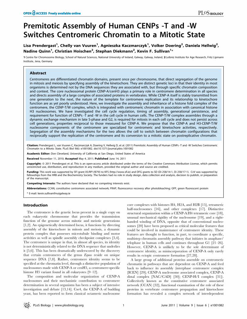
Premitotic Assembly of Human CENPs -T and -WSwitches Centromeric Chromatin to a Mitotic StateLisa Prendergast1, Chelly van Vuuren1, Agnieszka Kaczmarczyk1, Volker Doering2, Daniela Hellwig2,
Nadine Quinn1, Christian Hoischen2, Stephan Diekmann2, Kevin F. Sullivan1*
1 Centre for Chromosome Biology, School of Natural Sciences, National University of Ireland, Galway, Galway, Ireland, 2 Leibniz Institute for Age Research, Fritz Lipmann
Institute, Jena, Germany
Abstract
Centromeres are differentiated chromatin domains, present once per chromosome, that direct segregation of the genomein mitosis and meiosis by specifying assembly of the kinetochore. They are distinct genetic loci in that their identity in mostorganisms is determined not by the DNA sequences they are associated with, but through specific chromatin compositionand context. The core nucleosomal protein CENP-A/cenH3 plays a primary role in centromere determination in all speciesand directs assembly of a large complex of associated proteins in vertebrates. While CENP-A itself is stably transmitted fromone generation to the next, the nature of the template for centromere replication and its relationship to kinetochorefunction are as yet poorly understood. Here, we investigate the assembly and inheritance of a histone fold complex of thecentromere, the CENP-T/W complex, which is integrated with centromeric chromatin in association with canonical histoneH3 nucleosomes. We have investigated the cell cycle regulation, timing of assembly, generational persistence, andrequirement for function of CENPs -T and -W in the cell cycle in human cells. The CENP-T/W complex assembles through adynamic exchange mechanism in late S-phase and G2, is required for mitosis in each cell cycle and does not persist acrosscell generations, properties reciprocal to those measured for CENP-A. We propose that the CENP-A and H3-CENP-T/Wnucleosome components of the centromere are specialized for centromeric and kinetochore activities, respectively.Segregation of the assembly mechanisms for the two allows the cell to switch between chromatin configurations thatreciprocally support the replication of the centromere and its conversion to a mitotic state on postreplicative chromatin.
Citation: Prendergast L, van Vuuren C, Kaczmarczyk A, Doering V, Hellwig D, et al. (2011) Premitotic Assembly of Human CENPs -T and -W Switches CentromericChromatin to a Mitotic State. PLoS Biol 9(6): e1001082. doi:10.1371/journal.pbio.1001082
Academic Editor: Don Cleveland, University of California at San Diego, United States of America
Received November 11, 2010; Accepted May 4, 2011; Published June 14, 2011
Copyright: � 2011 Prendergast et al. This is an open-access article distributed under the terms of the Creative Commons Attribution License, which permitsunrestricted use, distribution, and reproduction in any medium, provided the original author and source are credited.
Funding: This work was supported by SFI grant 05/RP1/B793 to KFS (http://www.sfi.ie) and DFG grants to SD (Di 258/14-1, Di 258/17-1). CvV was supported byfellowships from the HRB and the Biochemistry Society. The funders had no role in study design, data collection and analysis, decision to publish, or preparationof the manuscript.
Competing Interests: The authors have declared that no competing interests exist.
Abbreviations: CCAN, constitutive centromere associated network; FRAP, fluorescence recovery after photobleaching; GFP, green fluorescent protein
* E-mail: [email protected]
Introduction
The centromere is the genetic locus present in a single copy on
each eukaryotic chromosome that provides the transmission
function of the genome across mitotic and meiotic generations
[1,2]. An epigenetically determined locus, it functions by directing
assembly of the kinetochore in mitosis and meiosis, a dynamic
protein complex that possesses microtubule binding and motor
activities as well as spindle assembly checkpoint complexes [3,4].
The centromere is unique in that, in almost all species, its identity
is not deterministically related to the DNA sequence that underlies
it [5,6]. This has been dramatically underscored by the discovery
that certain centromeres of the genus Equus reside on unique
sequence DNA [7,8]. Rather, centromere identity seems to be
specified at the chromatin level, through a distinctive population of
nucleosomes made with CENP-A or cenH3, a centromere-specific
histone H3 variant found in all eukaryotes [9–12].
The composition and molecular organization of CENP-A
nucleosomes and their mechanistic contribution to centromere
determination in several organisms has been a subject of intensive
investigation and debate [13,14]. Cse4, the CENP-A of budding
yeast, has been reported to form classical octameric nucleosome
core complexes with histones H4, H2A, and H2B [15], tetrameric
half-nucleosomes [16], and other complexes [17]. Distinctive
structural organization within a CENP-A:H4 tetrameric core [18],
unusual mechanical rigidity of the nucleosome [19], and a right-
handed winding of DNA, opposite that of conventional nucleo-
somes [20] have been proposed as critical molecular features that
could be involved in maintenance of centromere identity. These
features are thought to function, in part, to coordinate a specific,
multistep chromatin assembly pathway that initiates in anaphase/
telophase in human cells and continues throughout G1 [21–26].
However, CENP-A is unlikely to be the sole determinant of
centromere identity, as misincorporation of CENP-A only rarely
results in ectopic centromere formation [27,28].
A large group of additional proteins assemble on centromeric
chromatin in pathways that are dependent on CENP-A and feed
back to influence its assembly (interphase centromere complex
[ICEN] [29]; CENP-A nucleosome associated complex, CENP-A
distal complex [NAC/CAD] [30]; CENP-H/I complex [31]).
Collectively known as the constitutive centromere associated
network (CCAN) [32], functional examination of the role of these
proteins in vertebrate centromere propagation and kinetochore
formation has revealed a complex network of interdependent
PLoS Biology | www.plosbiology.org 1 June 2011 | Volume 9 | Issue 6 | e1001082
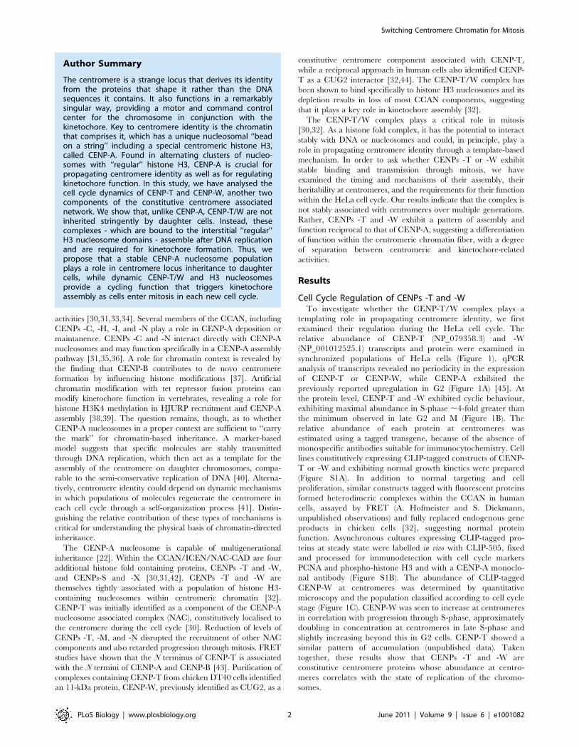
activities [30,31,33,34]. Several members of the CCAN, including
CENPs -C, -H, -I, and -N play a role in CENP-A deposition or
maintanence. CENPs -C and -N interact directly with CENP-A
nucleosomes and may function specifically in a CENP-A assembly
pathway [31,35,36]. A role for chromatin context is revealed by
the finding that CENP-B contributes to de novo centromere
formation by influencing histone modifications [37]. Artificial
chromatin modification with tet repressor fusion proteins can
modify kinetochore function in vertebrates, revealing a role for
histone H3K4 methylation in HJURP recruitment and CENP-A
assembly [38,39]. The question remains, though, as to whether
CENP-A nucleosomes in a proper context are sufficient to ‘‘carry
the mark’’ for chromatin-based inheritance. A marker-based
model suggests that specific molecules are stably transmitted
through DNA replication, which then act as a template for the
assembly of the centromere on daughter chromosomes, compa-
rable to the semi-conservative replication of DNA [40]. Alterna-
tively, centromere identity could depend on dynamic mechanisms
in which populations of molecules regenerate the centromere in
each cell cycle through a self-organization process [41]. Distin-
guishing the relative contribution of these types of mechanisms is
critical for understanding the physical basis of chromatin-directed
inheritance.
The CENP-A nucleosome is capable of multigenerational
inheritance [22]. Within the CCAN/ICEN/NAC-CAD are four
additional histone fold containing proteins, CENPs -T and -W,
and CENPs-S and -X [30,31,42]. CENPs -T and -W are
themselves tightly associated with a population of histone H3-
containing nucleosomes within centromeric chromatin [32].
CENP-T was initially identified as a component of the CENP-A
nucleosome associated complex (NAC), constitutively localised to
the centromere during the cell cycle [30]. Reduction of levels of
CENPs -T, -M, and -N disrupted the recruitment of other NAC
components and also retarded progression through mitosis. FRET
studies have shown that the N terminus of CENP-T is associated
with the N termini of CENP-A and CENP-B [43]. Purification of
complexes containing CENP-T from chicken DT40 cells identified
an 11-kDa protein, CENP-W, previously identified as CUG2, as a
constitutive centromere component associated with CENP-T,
while a reciprocal approach in human cells also identified CENP-
T as a CUG2 interactor [32,44]. The CENP-T/W complex has
been shown to bind specifically to histone H3 nucleosomes and its
depletion results in loss of most CCAN components, suggesting
that it plays a key role in kinetochore assembly [32].
The CENP-T/W complex plays a critical role in mitosis
[30,32]. As a histone fold complex, it has the potential to interact
stably with DNA or nucleosomes and could, in principle, play a
role in propagating centromere identity through a template-based
mechanism. In order to ask whether CENPs -T or -W exhibit
stable binding and transmission through mitosis, we have
examined the timing and mechanisms of their assembly, their
heritability at centromeres, and the requirements for their function
within the HeLa cell cycle. Our results indicate that the complex is
not stably associated with centromeres over multiple generations.
Rather, CENPs -T and -W exhibit a pattern of assembly and
function reciprocal to that of CENP-A, suggesting a differentiation
of function within the centromeric chromatin fiber, with a degree
of separation between centromeric and kinetochore-related
activities.
Results
Cell Cycle Regulation of CENPs -T and -WTo investigate whether the CENP-T/W complex plays a
templating role in propagating centromere identity, we first
examined their regulation during the HeLa cell cycle. The
relative abundance of CENP-T (NP_079358.3) and -W
(NP_001012525.1) transcripts and protein were examined in
synchronized populations of HeLa cells (Figure 1). qPCR
analysis of transcripts revealed no periodicity in the expression
of CENP-T or CENP-W, while CENP-A exhibited the
previously reported upregulation in G2 (Figure 1A) [45]. At
the protein level, CENP-T and -W exhibited cyclic behaviour,
exhibiting maximal abundance in S-phase ,4-fold greater than
the minimum observed in late G2 and M (Figure 1B). The
relative abundance of each protein at centromeres was
estimated using a tagged transgene, because of the absence of
monospecific antibodies suitable for immunocytochemistry. Cell
lines constitutively expressing CLIP-tagged constructs of CENP-
T or -W and exhibiting normal growth kinetics were prepared
(Figure S1A). In addition to normal targeting and cell
proliferation, similar constructs tagged with fluorescent proteins
formed heterodimeric complexes within the CCAN in human
cells, assayed by FRET (A. Hofmeister and S. Diekmann,
unpublished observations) and fully replaced endogenous gene
products in chicken cells [32], suggesting normal protein
function. Asynchronous cultures expressing CLIP-tagged pro-
teins at steady state were labelled in vivo with CLIP-505, fixed
and processed for immunodetection with cell cycle markers
PCNA and phospho-histone H3 and with a CENP-A monoclo-
nal antibody (Figure S1B). The abundance of CLIP-tagged
CENP-W at centromeres was determined by quantitative
microscopy and the population classified according to cell cycle
stage (Figure 1C). CENP-W was seen to increase at centromeres
in correlation with progression through S-phase, approximately
doubling in concentration at centromeres in late S-phase and
slightly increasing beyond this in G2 cells. CENP-T showed a
similar pattern of accumulation (unpublished data). Taken
together, these results show that CENPs -T and -W are
constitutive centromere proteins whose abundance at centro-
meres correlates with the state of replication of the chromo-
somes.
Author Summary
The centromere is a strange locus that derives its identityfrom the proteins that shape it rather than the DNAsequences it contains. It also functions in a remarkablysingular way, providing a motor and command controlcenter for the chromosome in conjunction with thekinetochore. Key to centromere identity is the chromatinthat comprises it, which has a unique nucleosomal ‘‘beadon a string’’ including a special centromeric histone H3,called CENP-A. Found in alternating clusters of nucleo-somes with ‘‘regular’’ histone H3, CENP-A is crucial forpropagating centromere identity as well as for regulatingkinetochore function. In this study, we have analysed thecell cycle dynamics of CENP-T and CENP-W, another twocomponents of the constitutive centromere associatednetwork. We show that, unlike CENP-A, CENP-T/W are notinherited stringently by daughter cells. Instead, thesecomplexes - which are bound to the interstitial ‘‘regular’’H3 nucleosome domains - assemble after DNA replicationand are required for kinetochore formation. Thus, wepropose that a stable CENP-A nucleosome populationplays a role in centromere locus inheritance to daughtercells, while dynamic CENP-T/W and H3 nucleosomesprovide a cycling function that triggers kinetochoreassembly as cells enter mitosis in each new cell cycle.
Switching Centromere Chromatin for Mitosis
PLoS Biology | www.plosbiology.org 2 June 2011 | Volume 9 | Issue 6 | e1001082
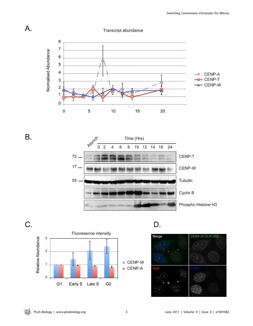
Switching Centromere Chromatin for Mitosis
PLoS Biology | www.plosbiology.org 3 June 2011 | Volume 9 | Issue 6 | e1001082
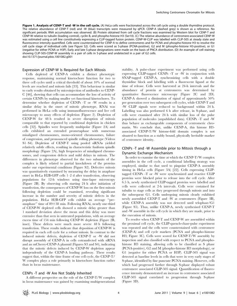
Expression of CENP-W Is Required for Each MitosisCells depleted of CENP-A exhibit a distinct phenotypic
response, maintaining normal kinetochore function for two to
three cell cycles until a critical threshold of about 10% of normal
levels are reached and mitosis fails [33]. This behaviour is similar
to early results obtained by microinjection of antibodies to CENP-
C [46], showing that cells can accommodate the loss of activity of
certain CENPs by making smaller yet functional kinetochores. To
determine whether depletion of CENPs -T or -W results in a
similar delay in the onset of mitotic phenotype, RNAi was
performed in HeLa cells using immunofluorescence and live cell
microscopy to assay effects of depletion (Figure 2). Depletion of
CENP-W for 48 h resulted in severe disruption of mitosis
comparable to that reported by conditional depletion in chicken
cells [32]. Spindles were frequently multipolar (Figure 2A) and
cells exhibited an extended prometaphase with numerous
misaligned chromosomes, mono-oriented chromosomes, failure
of congression, and pronounced spindle rolling phenotype (Videos
S1–S4). Depletion of CENP-T using pooled siRNAs yielded
relatively subtle effects, resulting in characteristic fusiform spindle
morphology (Figure 2A), high frequencies of misaligned chromo-
somes, and congression defects and mild delays in mitosis. The
differences in phenotype observed for the two subunits of the
complex is likely related to partial knockdown of the proteins
under our experimental conditions (Figure S2). Defective mitosis
was quantitatively examined by measuring the delay in anaphase
onset in HeLa H2B-GFP cells 1–2 d after transfection, observing
populations for 12-h windows using time-lapse microscopy
(Figure 2B). By observing cells starting 24 h after initiation of
transfection, the consequences of CENP-W loss on the first mitosis
following depletion could be examined, revealing significant
increase in the number and severity of mitotic defects in the
population. HeLa H2B-GFP cells exhibit an average ‘‘pre-
anaphase’’ time of 89638 min. Following RNAi, nearly one-third
of CENP-W depleted cells showed a mitotic delay greater than
1 standard deviation above the mean and this delay was more
extensive than that seen in untreated populations, with an average
excess time of 154 min following CENP-W depletion (Figure 2B,
inset). These defects were much more severe 48 h following
transfection. These results indicate that deposition of CENP-W is
required in each cell cycle for a robust mitosis. In contrast to the
induced mitotic defects, depletion of CENP-T or -W did not
disrupt assembly of CENP-A in cells cotransfected with siRNA
and an mCherry-CENP-A plasmid (Figures S3 and S4), indicating
that the mitotic defects induced by CENP-W depletion appear
independently of the CENP-A loading pathway. These results
suggest that, within the time frame of one cell cycle, the CENP-T/
W complex plays a role primarily in kinetochore function rather
than in locus maintenance.
CENPs -T and -W Are Not Stably InheritedA different perspective on the role of the CENP-T/W complex
in locus maintenance was gained by examining multigenerational
stability. A pulse-chase experiment was performed using cells
expressing CLIP-tagged CENPs -T or -W in conjunction with
SNAP-tagged CENP-A, synchronizing cells with a double
thymidine block and labelling with a fluorescent ligand at the
time of release. Cells were harvested at 24-h intervals and the
abundance of protein at centromeres was determined by
quantitative fluorescence microscopy (Figure 2C and 2D).
CENP-A showed a diminution of signal of approximately 50%
per generation over two subsequent cell cycles, while CENP-T and
-W CLIP signals were reduced to background within 24 h.
Labelling was also performed 6 h after release, in S-phase, and
cells were examined after 24 h with similar loss of the pulse
population of molecules (unpublished data). CENPs -T and -W
thus behave as exchangeable components of centromeric chro-
matin. We conclude that, unlike CENP-A, this histone H3-
associated CENP-T/W histone-fold domain complex is not
situated to function as a stably bound, physically heritable marker
of centromere identity.
CENPs -T and -W Assemble prior to Mitosis through aDynamic Exchange Mechanism
In order to examine the time at which the CENP-T/W complex
assembles in the cell cycle, a conditional labelling strategy was
employed, similar to that used to pinpoint CENP-A assembly
timing in HeLa cells (Figure 3A) [22]. Cells expressing CLIP-
tagged CENPs -T or -W were synchronized and reactive CLIP
proteins were blocked prior to release into the cell cycle. After
6.5 h, newly synthesized CLIP-tagged proteins were labelled and
cells were collected at 2-h intervals. Cells were costained for
tubulin to stage cells as they progressed through mitosis and into
the subsequent G1. Cells undergoing mitosis clearly exhibited
newly assembled CENP-T and -W at centromeres (Figure 3B),
while CENP-A assembly was not detected until telophase/G1
(Figure S5). Thus, unlike CENP-A, newly synthesised CENP-T
and -W assemble in the cell cycle in which they are made, prior to
the execution of mitosis.
To resolve when CENP-T and CENP-W are assembled within
the proximal cell cycle, the CLIP-quench-chase-pulse experiment
was repeated and the cells were counterstained with centromere
(CENP-A) and cell cycle markers (PCNA and phospho-histone
H3; Figure 3C). Cells were scored for CENP-T/W assembly by
inspection and also classified with respect to PCNA and phospho-
histone H3 staining, allowing cells to be classified as S phase
(PCNA-positive), G2 and M (phospho-histone H3 morphology), or
G1 (negative for either PCNA or H3P). CLIP-505 signal was
detected at baseline levels in cells that were in very early stages of
S-phase, identified by fine punctate PCNA staining. However, cells
which had progressed further through S-phase displayed robust
centromere associated CLIP-505 signal. Quantification of fluores-
cence intensity demonstrated an increase in centromere associated
CLIP-505 signal correlated to progression through S-phase
(Figure 3D).
Figure 1. Analysis of CENP-T and -W in the cell cycle. (A) HeLa cells were fractionated across the cell cycle using a double thymidine protocol.The relative abundance of CENP-T (red) and -W (blue) transcripts were measured by qPCR. CENP-A (dashed grey) is shown as a reference. Nosignificant periodic RNA accumulation was observed. (B) Protein obtained from cell cycle fractions was examined by Western blot for CENP-T andCENP-W relative to tubulin (loading control), cyclin B, and phospho-histone H3 (Ser10). (C) The relative abundance of centromere-associated CENP-Wwas estimated using a cell line constitutively expressing a CLIP-tagged fusion protein. CENP-W-CLIP was labelled with CLIP-505 at steady state andfluorescence intensity quantified. Cells were counterstained for CENP-A to define centromeres and for PCNA and phospho-histone H3 to resolve thecell cycle stage of individual cells (see Figure S2). Cells were scored as S-phase (PCNA-positive), G2 and M (phospho-histone H3-positive), or G1(negative for either PCNA or H3P). Early and late S-phase designations were made on the basis of PNCA distribution. (D) An example of cell stainingshowing CLIP-505-CENP-W assembly in a pair of cells in S-phase and undetected in a pair of G2 cells.doi:10.1371/journal.pbio.1001082.g001
Switching Centromere Chromatin for Mitosis
PLoS Biology | www.plosbiology.org 4 June 2011 | Volume 9 | Issue 6 | e1001082
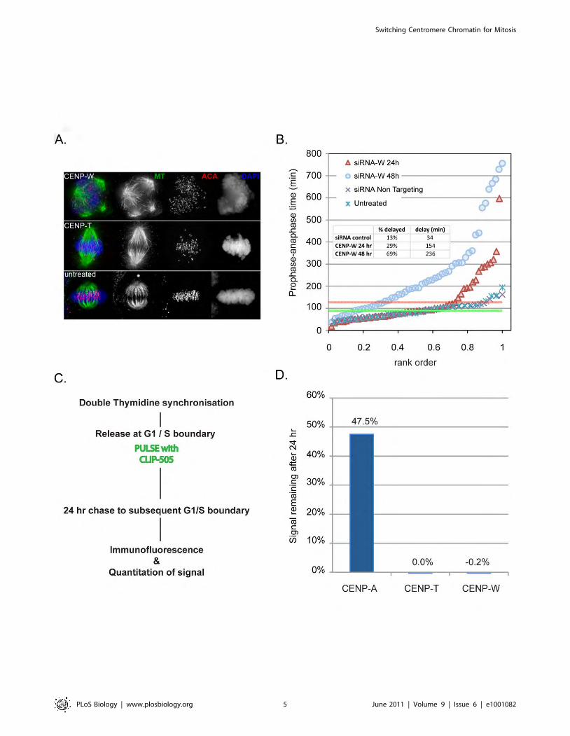
Switching Centromere Chromatin for Mitosis
PLoS Biology | www.plosbiology.org 5 June 2011 | Volume 9 | Issue 6 | e1001082
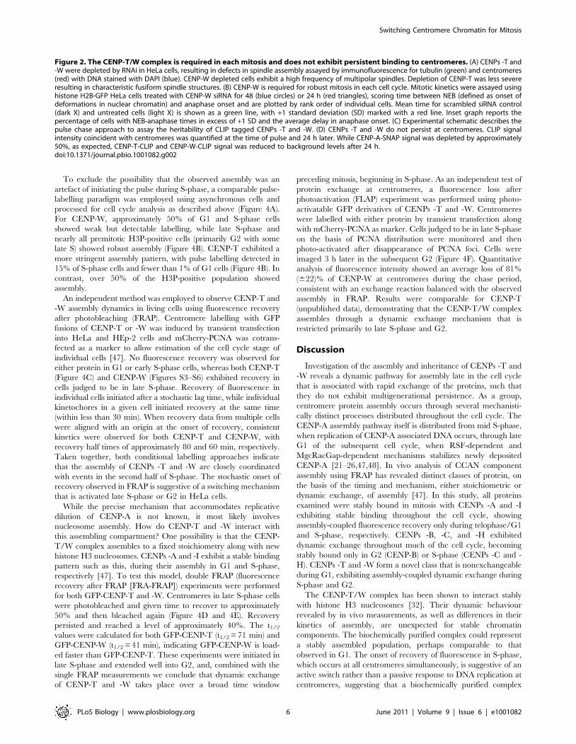
To exclude the possibility that the observed assembly was an
artefact of initiating the pulse during S-phase, a comparable pulse-
labelling paradigm was employed using asynchronous cells and
processed for cell cycle analysis as described above (Figure 4A).
For CENP-W, approximately 50% of G1 and S-phase cells
showed weak but detectable labelling, while late S-phase and
nearly all premitotic H3P-positive cells (primarily G2 with some
late S) showed robust assembly (Figure 4B). CENP-T exhibited a
more stringent assembly pattern, with pulse labelling detected in
15% of S-phase cells and fewer than 1% of G1 cells (Figure 4B). In
contrast, over 50% of the H3P-positive population showed
assembly.
An independent method was employed to observe CENP-T and
-W assembly dynamics in living cells using fluorescence recovery
after photobleaching (FRAP). Centromere labelling with GFP
fusions of CENP-T or -W was induced by transient transfection
into HeLa and HEp-2 cells and mCherry-PCNA was cotrans-
fected as a marker to allow estimation of the cell cycle stage of
individual cells [47]. No fluorescence recovery was observed for
either protein in G1 or early S-phase cells, whereas both CENP-T
(Figure 4C) and CENP-W (Figures S3–S6) exhibited recovery in
cells judged to be in late S-phase. Recovery of fluorescence in
individual cells initiated after a stochastic lag time, while individual
kinetochores in a given cell initiated recovery at the same time
(within less than 30 min). When recovery data from multiple cells
were aligned with an origin at the onset of recovery, consistent
kinetics were observed for both CENP-T and CENP-W, with
recovery half times of approximately 80 and 60 min, respectively.
Taken together, both conditional labelling approaches indicate
that the assembly of CENPs -T and -W are closely coordinated
with events in the second half of S-phase. The stochastic onset of
recovery observed in FRAP is suggestive of a switching mechanism
that is activated late S-phase or G2 in HeLa cells.
While the precise mechanism that accommodates replicative
dilution of CENP-A is not known, it most likely involves
nucleosome assembly. How do CENP-T and -W interact with
this assembling compartment? One possibility is that the CENP-
T/W complex assembles to a fixed stoichiometry along with new
histone H3 nucleosomes. CENPs -A and -I exhibit a stable binding
pattern such as this, during their assembly in G1 and S-phase,
respectively [47]. To test this model, double FRAP (fluorescence
recovery after FRAP [FRA-FRAP]) experiments were performed
for both GFP-CENP-T and -W. Centromeres in late S-phase cells
were photobleached and given time to recover to approximately
50% and then bleached again (Figure 4D and 4E). Recovery
persisted and reached a level of approximately 40%. The t1/2
values were calculated for both GFP-CENP-T (t1/2 = 71 min) and
GFP-CENP-W (t1/2 = 41 min), indicating GFP-CENP-W is load-
ed faster than GFP-CENP-T. These experiments were initiated in
late S-phase and extended well into G2, and, combined with the
single FRAP measurements we conclude that dynamic exchange
of CENP-T and -W takes place over a broad time window
preceding mitosis, beginning in S-phase. As an independent test of
protein exchange at centromeres, a fluorescence loss after
photoactivation (FLAP) experiment was performed using photo-
activatable GFP derivatives of CENPs -T and -W. Centromeres
were labelled with either protein by transient transfection along
with mCherry-PCNA as marker. Cells judged to be in late S-phase
on the basis of PCNA distribution were monitored and then
photo-activated after disappearance of PCNA foci. Cells were
imaged 3 h later in the subsequent G2 (Figure 4F). Quantitative
analysis of fluorescence intensity showed an average loss of 81%
(622)% of CENP-W at centromeres during the chase period,
consistent with an exchange reaction balanced with the observed
assembly in FRAP. Results were comparable for CENP-T
(unpublished data), demonstrating that the CENP-T/W complex
assembles through a dynamic exchange mechanism that is
restricted primarily to late S-phase and G2.
Discussion
Investigation of the assembly and inheritance of CENPs -T and
-W reveals a dynamic pathway for assembly late in the cell cycle
that is associated with rapid exchange of the proteins, such that
they do not exhibit multigenerational persistence. As a group,
centromere protein assembly occurs through several mechanisti-
cally distinct processes distributed throughout the cell cycle. The
CENP-A assembly pathway itself is distributed from mid S-phase,
when replication of CENP-A associated DNA occurs, through late
G1 of the subsequent cell cycle, when RSF-dependent and
MgcRacGap-dependent mechanisms stabilizes newly deposited
CENP-A [21–26,47,48]. In vivo analysis of CCAN component
assembly using FRAP has revealed distinct classes of protein, on
the basis of the timing and mechanism, either stoichiometric or
dynamic exchange, of assembly [47]. In this study, all proteins
examined were stably bound in mitosis with CENPs -A and -I
exhibiting stable binding throughout the cell cycle, showing
assembly-coupled fluorescence recovery only during telophase/G1
and S-phase, respectively. CENPs -B, -C, and -H exhibited
dynamic exchange throughout much of the cell cycle, becoming
stably bound only in G2 (CENP-B) or S-phase (CENPs -C and -
H). CENPs -T and -W form a novel class that is nonexchangeable
during G1, exhibiting assembly-coupled dynamic exchange during
S-phase and G2.
The CENP-T/W complex has been shown to interact stably
with histone H3 nucleosomes [32]. Their dynamic behaviour
revealed by in vivo measurements, as well as differences in their
kinetics of assembly, are unexpected for stable chromatin
components. The biochemically purified complex could represent
a stably assembled population, perhaps comparable to that
observed in G1. The onset of recovery of fluorescence in S-phase,
which occurs at all centromeres simultaneously, is suggestive of an
active switch rather than a passive response to DNA replication at
centromeres, suggesting that a biochemically purified complex
Figure 2. The CENP-T/W complex is required in each mitosis and does not exhibit persistent binding to centromeres. (A) CENPs -T and-W were depleted by RNAi in HeLa cells, resulting in defects in spindle assembly assayed by immunofluorescence for tubulin (green) and centromeres(red) with DNA stained with DAPI (blue). CENP-W depleted cells exhibit a high frequency of multipolar spindles. Depletion of CENP-T was less severeresulting in characteristic fusiform spindle structures. (B) CENP-W is required for robust mitosis in each cell cycle. Mitotic kinetics were assayed usinghistone H2B-GFP HeLa cells treated with CENP-W siRNA for 48 (blue circles) or 24 h (red triangles), scoring time between NEB (defined as onset ofdeformations in nuclear chromatin) and anaphase onset and are plotted by rank order of individual cells. Mean time for scrambled siRNA control(dark X) and untreated cells (light X) is shown as a green line, with +1 standard deviation (SD) marked with a red line. Inset graph reports thepercentage of cells with NEB-anaphase times in excess of +1 SD and the average delay in anaphase onset. (C) Experimental schematic describes thepulse chase approach to assay the heritability of CLIP tagged CENPs -T and -W. (D) CENPs -T and -W do not persist at centromeres. CLIP signalintensity coincident with centromeres was quantified at the time of pulse and 24 h later. While CENP-A-SNAP signal was depleted by approximately50%, as expected, CENP-T-CLIP and CENP-W-CLIP signal was reduced to background levels after 24 h.doi:10.1371/journal.pbio.1001082.g002
Switching Centromere Chromatin for Mitosis
PLoS Biology | www.plosbiology.org 6 June 2011 | Volume 9 | Issue 6 | e1001082

Switching Centromere Chromatin for Mitosis
PLoS Biology | www.plosbiology.org 7 June 2011 | Volume 9 | Issue 6 | e1001082
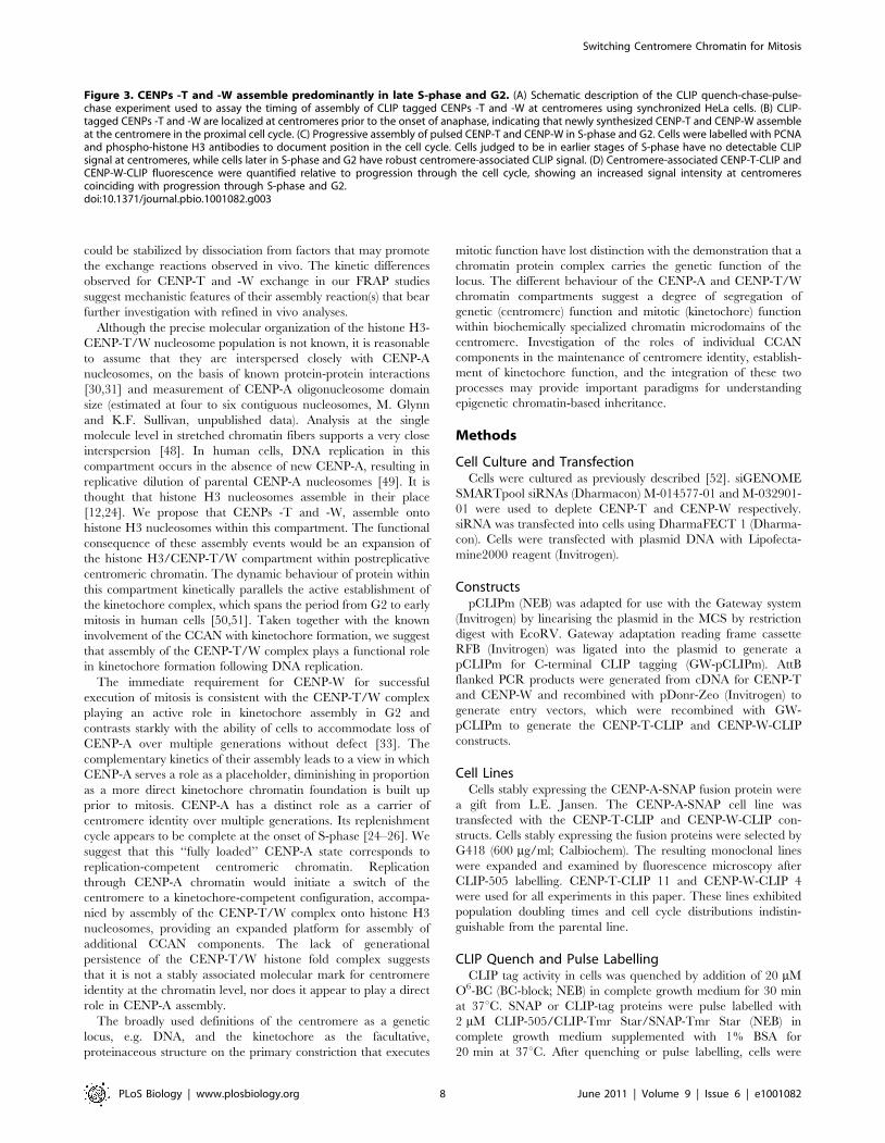
could be stabilized by dissociation from factors that may promote
the exchange reactions observed in vivo. The kinetic differences
observed for CENP-T and -W exchange in our FRAP studies
suggest mechanistic features of their assembly reaction(s) that bear
further investigation with refined in vivo analyses.
Although the precise molecular organization of the histone H3-
CENP-T/W nucleosome population is not known, it is reasonable
to assume that they are interspersed closely with CENP-A
nucleosomes, on the basis of known protein-protein interactions
[30,31] and measurement of CENP-A oligonucleosome domain
size (estimated at four to six contiguous nucleosomes, M. Glynn
and K.F. Sullivan, unpublished data). Analysis at the single
molecule level in stretched chromatin fibers supports a very close
interspersion [48]. In human cells, DNA replication in this
compartment occurs in the absence of new CENP-A, resulting in
replicative dilution of parental CENP-A nucleosomes [49]. It is
thought that histone H3 nucleosomes assemble in their place
[12,24]. We propose that CENPs -T and -W, assemble onto
histone H3 nucleosomes within this compartment. The functional
consequence of these assembly events would be an expansion of
the histone H3/CENP-T/W compartment within postreplicative
centromeric chromatin. The dynamic behaviour of protein within
this compartment kinetically parallels the active establishment of
the kinetochore complex, which spans the period from G2 to early
mitosis in human cells [50,51]. Taken together with the known
involvement of the CCAN with kinetochore formation, we suggest
that assembly of the CENP-T/W complex plays a functional role
in kinetochore formation following DNA replication.
The immediate requirement for CENP-W for successful
execution of mitosis is consistent with the CENP-T/W complex
playing an active role in kinetochore assembly in G2 and
contrasts starkly with the ability of cells to accommodate loss of
CENP-A over multiple generations without defect [33]. The
complementary kinetics of their assembly leads to a view in which
CENP-A serves a role as a placeholder, diminishing in proportion
as a more direct kinetochore chromatin foundation is built up
prior to mitosis. CENP-A has a distinct role as a carrier of
centromere identity over multiple generations. Its replenishment
cycle appears to be complete at the onset of S-phase [24–26]. We
suggest that this ‘‘fully loaded’’ CENP-A state corresponds to
replication-competent centromeric chromatin. Replication
through CENP-A chromatin would initiate a switch of the
centromere to a kinetochore-competent configuration, accompa-
nied by assembly of the CENP-T/W complex onto histone H3
nucleosomes, providing an expanded platform for assembly of
additional CCAN components. The lack of generational
persistence of the CENP-T/W histone fold complex suggests
that it is not a stably associated molecular mark for centromere
identity at the chromatin level, nor does it appear to play a direct
role in CENP-A assembly.
The broadly used definitions of the centromere as a genetic
locus, e.g. DNA, and the kinetochore as the facultative,
proteinaceous structure on the primary constriction that executes
mitotic function have lost distinction with the demonstration that a
chromatin protein complex carries the genetic function of the
locus. The different behaviour of the CENP-A and CENP-T/W
chromatin compartments suggest a degree of segregation of
genetic (centromere) function and mitotic (kinetochore) function
within biochemically specialized chromatin microdomains of the
centromere. Investigation of the roles of individual CCAN
components in the maintenance of centromere identity, establish-
ment of kinetochore function, and the integration of these two
processes may provide important paradigms for understanding
epigenetic chromatin-based inheritance.
Methods
Cell Culture and TransfectionCells were cultured as previously described [52]. siGENOME
SMARTpool siRNAs (Dharmacon) M-014577-01 and M-032901-
01 were used to deplete CENP-T and CENP-W respectively.
siRNA was transfected into cells using DharmaFECT 1 (Dharma-
con). Cells were transfected with plasmid DNA with Lipofecta-
mine2000 reagent (Invitrogen).
ConstructspCLIPm (NEB) was adapted for use with the Gateway system
(Invitrogen) by linearising the plasmid in the MCS by restriction
digest with EcoRV. Gateway adaptation reading frame cassette
RFB (Invitrogen) was ligated into the plasmid to generate a
pCLIPm for C-terminal CLIP tagging (GW-pCLIPm). AttB
flanked PCR products were generated from cDNA for CENP-T
and CENP-W and recombined with pDonr-Zeo (Invitrogen) to
generate entry vectors, which were recombined with GW-
pCLIPm to generate the CENP-T-CLIP and CENP-W-CLIP
constructs.
Cell LinesCells stably expressing the CENP-A-SNAP fusion protein were
a gift from L.E. Jansen. The CENP-A-SNAP cell line was
transfected with the CENP-T-CLIP and CENP-W-CLIP con-
structs. Cells stably expressing the fusion proteins were selected by
G418 (600 mg/ml; Calbiochem). The resulting monoclonal lines
were expanded and examined by fluorescence microscopy after
CLIP-505 labelling. CENP-T-CLIP 11 and CENP-W-CLIP 4
were used for all experiments in this paper. These lines exhibited
population doubling times and cell cycle distributions indistin-
guishable from the parental line.
CLIP Quench and Pulse LabellingCLIP tag activity in cells was quenched by addition of 20 mM
O6-BC (BC-block; NEB) in complete growth medium for 30 min
at 37uC. SNAP or CLIP-tag proteins were pulse labelled with
2 mM CLIP-505/CLIP-Tmr Star/SNAP-Tmr Star (NEB) in
complete growth medium supplemented with 1% BSA for
20 min at 37uC. After quenching or pulse labelling, cells were
Figure 3. CENPs -T and -W assemble predominantly in late S-phase and G2. (A) Schematic description of the CLIP quench-chase-pulse-chase experiment used to assay the timing of assembly of CLIP tagged CENPs -T and -W at centromeres using synchronized HeLa cells. (B) CLIP-tagged CENPs -T and -W are localized at centromeres prior to the onset of anaphase, indicating that newly synthesized CENP-T and CENP-W assembleat the centromere in the proximal cell cycle. (C) Progressive assembly of pulsed CENP-T and CENP-W in S-phase and G2. Cells were labelled with PCNAand phospho-histone H3 antibodies to document position in the cell cycle. Cells judged to be in earlier stages of S-phase have no detectable CLIPsignal at centromeres, while cells later in S-phase and G2 have robust centromere-associated CLIP signal. (D) Centromere-associated CENP-T-CLIP andCENP-W-CLIP fluorescence were quantified relative to progression through the cell cycle, showing an increased signal intensity at centromerescoinciding with progression through S-phase and G2.doi:10.1371/journal.pbio.1001082.g003
Switching Centromere Chromatin for Mitosis
PLoS Biology | www.plosbiology.org 8 June 2011 | Volume 9 | Issue 6 | e1001082
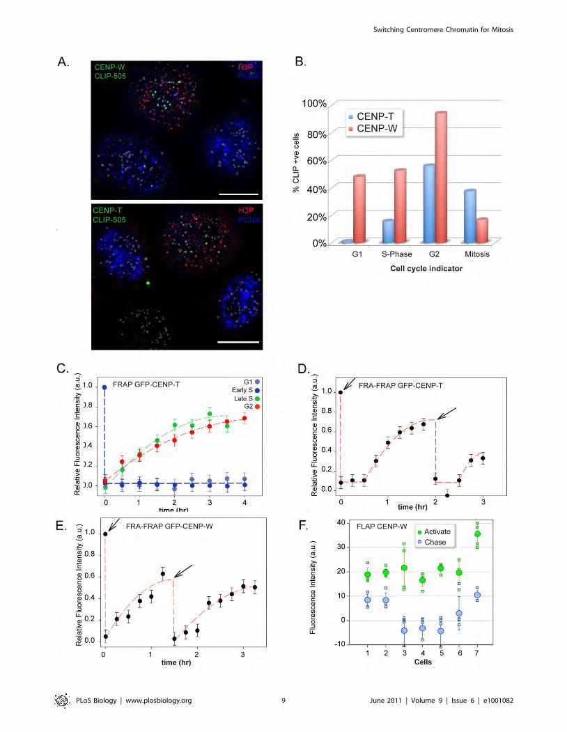
Switching Centromere Chromatin for Mitosis
PLoS Biology | www.plosbiology.org 9 June 2011 | Volume 9 | Issue 6 | e1001082
washed twice with prewarmed PBS and once with complete
DMEM. Following washes cells were reincubated in complete
medium to allow excess labelling compound to diffuse from cells.
After 30 min, cells were washed again twice in PBS followed either
by reincubation in complete DMEM, or fixation.
Cell SynchronizationHeLa cells were treated with 5 mM thymidine in complete
DMEM for 16 h, washed twice in PBS, once in complete DMEM,
and released in complete DMEM 9 h followed by addition of
thymidine to a final concentration of 5 mM for 16 h, after which
cells were released again into complete DMEM.
ImmunofluorescenceCells were grown and CLIP labelled on glass coverslips followed by
fixation in MeOH or 4% PFA and processed for immunofluores-
cence. ACA (1:500), Anti-CENP-A (a gift from K. Yoda, Nagoya
University, Nagoya, Japan) was used at a dilution of 1:200, human
anti-PCNA serum was used at 1:100, anti-a-tubulin (Sigma) was used
at 1:500, and anti-phospho histone H3 (Abcam) was used at 1:300.
Donkey fluorescently conjugated secondary antibodies (anti-
mouse-FITC (1:50) anti-mouse Cy5 (1:200), anti-rabbit TRITC
(1:100), and anti-human AMCA (1:100) were obtained from
Jackson Immunoresearch Laboratories. Samples were mounted in
SloFADE (Invitrogen).
MicroscopyImages were captured using a DeltaVision Core system (Applied
Precision) controlling an interline charge-coupled device camera
(Coolsnap HQ2; Roper) mounted on an inverted microscope (IX-
71; Olympus). For each sample, images were collected at 26binning using a 1006oil objective at 0.2 mm z sections. All images
were deconvolved and maximum intensity projected using Soft-
WoRx (Applied Precision). For quantification, unscaled DeltaVi-
sion images were used. Centromere signal intensity was deter-
mined using ImagePro 6.3. A mask was created using CENP-A or
ACA signal to define centromeres. The ‘‘centromere’’ mask was
applied to the CLIP-505 channel and the mean fluorescence
intensity measured. A background mask was created from three
regions within the nucleus not containing centromeres and applied
to the CLIP-505 images for measurement. Background values
were then subtracted from mean signal.
ImmunoblotsWhole cell extracts equivalent to 50,000 cells were separated by
SDS-PAGE, transferred to PVDF membranes (Millipore), and
processed for immunodetection as described previously [49].
Antibodies: anti-CENP-T (Bethyl), anti-CENP-W (C6ORF173
antibody Abcam, ab75827), anti-tubulin monoclonal (DN1A
Sigma), anti-H3P Ser10 (Millipore), cyclin-B (Upstate).
Supporting Information
Figure S1 A CENP-A-SNAP cell line (gift from LarsJansen) was stably transfected with plasmids CENP-T-
pCLIPm and CENP-W-pCLIPm. Clones stably expressing
CENP-T-CLIP and CENP-W-CLIP were selected and labelled
using SNAP-TMR-Star to label CENP-A-SNAP and CLIP-505 to
label CENP-T and CENP-W CLIP.
(EPS)
Figure S2 Quantitative reverse transcript (RT)-PCRwas used to measure the levels of depletion of bothCENP-T and CENP-W transcripts following a 48-h siRNAtreatment. CENP-W was depleted to approximately 48% while
CENP-T transcripts were reduced to approximately 36%.
Corresponding Western blots shown below demonstrate compa-
rable levels of protein depletion.
(EPS)
Figure S3 Analysis of CENP-A assembly following CENP-W depletion. (A) Experimental schematic describes the approach
to assay assembly of CENP-A following depletion of CENP-W by
siRNA. HeLa cells expressing H2B-GFP were cotransfected with
mCherry-CENP-A and siRNA directed against CENP-W. After
48 h, cells were fixed for immunofluorescence or treated with an
Aurora B inhibitor (ZM44739, JS Research Chemicals) to relieve
mitotic delay associated with CENP-W depletion. Cells were
examined for assembly of mCherry-CENP-A to centromeres. (B)
Immunofluorescence in H2B-GFP HeLa cells using centromere
marker (ACA) and tubulin following depletion of CENP-W.
mCherry-CENP-A is observed at centromeres in early G1 cells
displaying a tubulin staining of the midbody, while some transfected
metaphase cells, indicated by presence of mCherry signal, with
uncongressed chromosomes, have not assembled CENP-A at
centromeres diffuse mCherry signal. (C) Cells were also treated
with an Aurora B inhibitor, following 48-h CENP-W depletion.
Cells judged to have entered G1 as a result of Aurora B inhibition
exhibited abnormal nuclear morphology and centromeres that in
most cases had failed to segregate. Transfected cells positive for
mCherry-CENP-A had assembled CENP-A at centromeres.
(EPS)
Figure S4 Analysis of CENP-A assembly followingCENP-T depletion. In an experiment analogous to that shown
in Figure S5, immunofluorescence in H2B-GFP HeLa cells using
centromere marker (ACA) and tubulin illustrates cell cycle stage in
exemplary cells following depletion of CENP-T. mCherry-CENP-
A is observed at centromeres in early G1 cells displaying a tubulin
staining of the midbody, while some transfected metaphase cells,
indicated by presence of mCherry signal, with monopolar
chromosomes, have not assembled CENP-A at centromeres.
(EPS)
Figure S5 A cell line expressing SNAP-tagged CENP-A(gift of Lars Jansen) was used as a control for timedassembly in SNAP experiments. Cells were synchronized
using a double thymidine arrest and quenched using BG-quench
upon release for arrest. Cell were chased for 7 h to allow synthesis
of ‘‘new’’ SNAP-CENP-A, and pulsed with a fluorescent substrate,
SNAP-505. Assembly of SNAP-CENP-A to centromeres was not
observed until cells had progressed through mitosis into the
Figure 4. CENP-T and -W assembly occur through a dynamic exchange mechanism in S-phase and G2. (A) Asynchronous HeLa cellsexpressing CENP-T and CENP-W CLIP were used to assay timing of assembly in an unperturbed cell population. Cells were cell cycle staged bycounterstaining with PCNA and phospho-histone H3. (B) Cells were classified on the basis of CLIP signal at centromeres and cell cycle stage. (C) FRAPof GFP derivatives of CENP-T only occurs during late S-phase and G2 indicating loading takes place during this period. (D) Centromere-associatedGFP-CENP-T was photobleached and allowed to recover to approximately 40%. Following a second bleach event, recovery continued reachingapproximately 40%, indicating an exchange-based dynamic loading process during this time period. (E) Double FRAP of GFP-CENP-W, as for CENP-T.(F) Fluorescence loss after photoactivation. Photoactivatable GFP-CENP-W was activated in G2 cells. Following a chase period of 3 h, the majority ofthe fluorescent signal had dissociated from the centromere.doi:10.1371/journal.pbio.1001082.g004
Switching Centromere Chromatin for Mitosis
PLoS Biology | www.plosbiology.org 10 June 2011 | Volume 9 | Issue 6 | e1001082
subsequent G1, as previously reported. Insets detail SNAP-505
and ACA staining in interphase and mitotic cells.
(EPS)
Figure S6 FRAP of GFP derivatives of CENP-W onlyoccurs during late S-phase and continues during G2,indicating loading takes place during this time. Experi-
ment analogous to that shown for CENP-T in Figure 4.
(EPS)
Video S1 CENP-W knockdown induces profound mitot-ic delays and induces chromosome/spindle rolling incells. HeLa-H2B-GFP cells were transfected with pooled siRNAs
against CENP-W and imaged for 12 h beginning 48-h transfection
at a rate of 20 frames/hour. Numerous cells are observed entering
mitosis and spending extended time in a prometaphase-like state
with uncongressed chromosomes. Many cells exhibit a pro-
nounced ‘‘rolling’’ phenotype (see upper right quadrant) that can
initiate very soon after the onset of mitosis. See Figure 2B for a
quantitative interpretation of these data.
(MOV)
Video S2 4-Dimensional imaging of rolling spindles:projection. HeLa-H2B-GFP cells transfected with pooled
siRNAs against CENP-W were imaged by 3-dimensional time-
lapse microscopy beginning 48 h after transfection, with frames
collected every 5 min. This sequence shows a projected image
superimposed on a transmitted light image of cells. Two rolling
cells are detailed. In one (first), the metaphase plate rotates in a
continuous motion through almost 180u. In another, the
metaphase plate rotates 90u with respect to the optical axis.
(MOV)
Video S3 4-Dimensional imaging of rolling spindles:reconstruction. HeLa-H2B-GFP cells transfected with pooled
siRNAs against CENP-W were imaged by 3-dimensional time-
lapse microscopy beginning 48 h after transfection, with frames
collected every 5 min. In this sequence, the field is rocked to reveal
the 3-dimensional structure of the chromosomes on the spindle. It
is clear that the chromosomes are moving collectively, character-
istic of whole spindle motion within the cells.
(MOV)
Video S4 Tubulin labeling confirms whole spindlemotion in CENP-W depleted cells. HeLa-H2B-GFP cells
were transfected with mCherry-tubulin to directly visualize spindle
motion following transfection with pooled siRNAs against CENP-
W. It is clear that the entire spindle is moving within cells that
exhibit rolling. The spindle poles can be seen to split in some of
these cells, as though the forces associated with spindle movement
are able to disrupt spindle pole integrity. This may account for the
multipolar spindles frequently observed in fixed specimens of
CENP-W siRNA-treated cells.
(MOV)
Acknowledgments
The authors thank Lars Jansen for the generous gift of HeLa SNAP-
CENP-A cells and Kinya Yoda for CENP-A monoclonal antibody. KFS is
a Research Professor of Science Foundation Ireland.
Author Contributions
The author(s) have made the following declarations about their
contributions: Conceived and designed the experiments: LP CvV SD
KFS. Performed the experiments: LP CvV AK NQ VD DH CH KFS.
Analyzed the data: LP CvV AK NQ VD DH SD KFS. Contributed
reagents/materials/analysis tools: LP CvV AK NQ DH CH KFS. Wrote
the paper: LP SD KFS.
References
1. Cleveland DW, Mao Y, Sullivan KF (2003) Centromeres and kinetochores: from
epigenetics to mitotic checkpoint signaling. Cell 112: 407–421.
2. Przewloka MR, Glover DM (2009) The kinetochore and the centromere: a
working long distance relationship. Annu Rev Genet 43: 439–465.
3. Santaguida S, Musacchio A (2009) The life and miracles of kinetochores.
EMBO J 28: 2511–2531.
4. Cheeseman IM, Desai A (2008) Molecular architecture of the kinetochore-
microtubule interface. Nat Rev Mol Cell Biol 9: 33–46.
5. Williams BC, Murphy TD, Goldberg ML, Karpen GH (1998) Neocentromere
activity of structurally acentric mini-chromosomes in Drosophila. Nat Genet 18:
30–37.
6. Barry AE, Howman EV, Cancilla MR, Saffery R, Choo KH (1999) Sequence
analysis of an 80 kb human neocentromere. Hum Mol Genet 8: 217–227.
7. Wade CM, Giulotto E, Sigurdsson S, Zoli M, Gnerre S, et al. (2009) Genome
sequence, comparative analysis, and population genetics of the domestic horse.
Science 326: 865–867.
8. Piras FM, Nergadze SG, Magnani E, Bertoni L, Attolini C, et al. (2010)
Uncoupling of satellite DNA and centromeric function in the genus Equus. PLoS
Genet 6: e1000845. doi:10.1371/journal.pgen.1000845.
9. Palmer DK, O’Day K, Trong HL, Charbonneau H, Margolis RL (1991)
Purification of the centromere-specific protein CENP-A and demonstration that
it is a distinctive histone. Proc Natl Acad Sci U S A 88: 3734–3738.
10. Sullivan KF, Hechenberger M, Masri K (1994) Human CENP-A contains a
histone H3 related histone fold domain that is required for targeting to the
centromere. J Cell Biol 127: 581–592.
11. Stoler S, Keith KC, Curnick KE, Fitzgerald-Hayes M (1995) A mutation in
CSE4, an essential gene encoding a novel chromatin-associated protein in yeast,
causes chromosome nondisjunction and cell cycle arrest at mitosis. Genes Dev 9:
573–586.
12. Allshire RC, Karpen GH (2008) Epigenetic regulation of centromeric
chromatin: old dogs, new tricks? Nat Rev Genet 9: 923–937.
13. Dalal Y, Bui M (2010) Down the rabbit hole of centromere assembly and
dynamics. Curr Opin Cell Biol 22: 392–402.
14. Black BE, Cleveland DW (2011) Epigenetic centromere propagation and the
nature of CENP-A nucleosomes. Cell 144: 471–479.
15. Camahort R, Shivaraju M, Mattingly M, Li B, Nakanishi S, et al. (2009) Cse4 is
part of an octameric nucleosome in budding yeast. Mol Cell 35: 794–805.
16. Dalal Y, Wang H, Lindsay S, Henikoff S (2007) Tetrameric structure ofcentromeric nucleosomes in interphase Drosophila cells. PLoS Biol 5: e218.
doi:10.1371/journal.pbio.0050218.
17. Mizuguchi G, Xiao H, Wisniewski J, Smith MM, Wu C (2007) NonhistoneScm3 and histones CenH3-H4 assemble the core of centromere-specific
nucleosomes. Cell 129: 1153–1164.
18. Sekulic N, Bassett EA, Rogers DJ, Black BE (2010) The structure of (CENP-A-H4)(2) reveals physical features that mark centromeres. Nature 467: 347–351.
19. Black BE, Foltz DR, Chakravarthy S, Luger K, Woods VL, Jr., et al. (2004)
Structural determinants for generating centromeric chromatin. Nature 430: 578–582.20. Furuyama T, Henikoff S (2009) Centromeric nucleosomes induce positive DNA
supercoils. Cell 138: 104–113.
21. Fujita Y, Hayashi T, Kiyomitsu T, Toyoda Y, Kokubu A, et al. (2007) Primingof centromere for CENP-A recruitment by human hMis18alpha, hMis18beta,
and M18BP1. Dev Cell 12: 17–30.
22. Jansen LE, Black BE, Foltz DR, Cleveland DW (2007) Propagation ofcentromeric chromatin requires exit from mitosis. J Cell Biol 176: 795–805.
23. Perpelescu M, Nozaki N, Obuse C, Yang H, Yoda K (2009) Active
establishment of centromeric CENP-A chromatin by RSF complex. J Cell Biol185: 397–407.
24. Dunleavy EM, Roche D, Tagami H, Lacoste N, Ray-Gallet D, et al. (2009)
HJURP is a cell-cycle-dependent maintenance and deposition factor of CENP-A
at centromeres. Cell 137: 485–497.25. Foltz DR, Jansen LE, Bailey AO, Yates JR. 3rd, Bassett EA, et al. (2009)
Centromere-specific assembly of CENP-a nucleosomes is mediated by HJURP.
Cell 137: 472–484.26. Lagana A, Dorn JF, De Rop V, Ladouceur AM, Maddox AS, et al. (2010) A
small GTPase molecular switch regulates epigenetic centromere maintenance by
stabilizing newly incorporated CENP-A. Nat Cell Biol 12: 1186–1193.27. Heun P, Erhardt S, Blower MD, Weiss S, Skora AD, et al. (2006) Mislocalization
of the Drosophila centromere-specific histone CID promotes formation of
functional ectopic kinetochores. Dev Cell 10: 303–315.28. Van Hooser AA, Ouspenski II, Gregson HC, Starr DA, Yen TJ, et al. (2001)
Specification of kinetochore-forming chromatin by the histone H3 variant
CENP-A. J Cell Sci 114: 3529–3542.29. Izuta H, Ikeno M, Suzuki N, Tomonaga T, Nozaki N, et al. (2006) Comprehensive
analysis of the ICEN (Interphase Centromere Complex) components enriched in
the CENP-A chromatin of human cells. Genes Cells 11: 673–684.
Switching Centromere Chromatin for Mitosis
PLoS Biology | www.plosbiology.org 11 June 2011 | Volume 9 | Issue 6 | e1001082
30. Foltz DR, Jansen LE, Black BE, Bailey AO, Yates JR, 3rd, et al. (2006) The
human CENP-A centromeric nucleosome-associated complex. Nat Cell Biol 8:458–469.
31. Okada M, Cheeseman IM, Hori T, Okawa K, McLeod IX, et al. (2006) The
CENP-H-I complex is required for the efficient incorporation of newlysynthesized CENP-A into centromeres. Nat Cell Biol 8: 446–457.
32. Hori T, Amano M, Suzuki A, Backer CB, Welburn JP, et al. (2008) CCANmakes multiple contacts with centromeric DNA to provide distinct pathways to
the outer kinetochore. Cell 135: 1039–1052.
33. Liu ST, Rattner JB, Jablonski SA, Yen TJ (2006) Mapping the assemblypathways that specify formation of the trilaminar kinetochore plates in human
cells. J Cell Biol 175: 41–53.34. Okada M, Okawa K, Isobe T, Fukagawa T (2009) CENP-H-containing complex
facilitates centromere deposition of CENP-A in cooperation with FACT andCHD1. Mol Biol Cell 20: 3986–3995.
35. Carroll CW, Milks KJ, Straight AF (2010) Dual recognition of CENP-A
nucleosomes is required for centromere assembly. J Cell Biol 189: 1143–1155.36. Carroll CW, Silva MC, Godek KM, Jansen LE, Straight AF (2009) Centromere
assembly requires the direct recognition of CENP-A nucleosomes by CENP-N.Nat Cell Biol 11: 896–902.
37. Okada T, Ohzeki J, Nakano M, Yoda K, Brinkley WR, et al. (2007) CENP-B
controls centromere formation depending on the chromatin context. Cell 131:1287–1300.
38. Bergmann JH, Rodriguez MG, Martins NM, Kimura H, Kelly DA, et al. (2011)Epigenetic engineering shows H3K4me2 is required for HJURP targeting and
CENP-A assembly on a synthetic human kinetochore. Embo J 30: 328–340.39. Nakano M, Cardinale S, Noskov VN, Gassmann R, Vagnarelli P, et al. (2008)
Inactivation of a human kinetochore by specific targeting of chromatin
modifiers. Dev Cell 14: 507–522.40. Meselson M, Stahl FW (1958) The replication of DNA in Escherichia coli. Proc
Natl Acad Sci U S A 44: 671–682.41. Misteli T (2001) The concept of self-organization in cellular architecture. J Cell
Biol 155: 181–185.
42. Amano M, Suzuki A, Hori T, Backer C, Okawa K, et al. (2009) The CENP-S
complex is essential for the stable assembly of outer kinetochore structure. J Cell
Biol 186: 173–182.
43. Hellwig D, Munch S, Orthaus S, Hoischen C, Hemmerich P, et al. (2008) Live-
cell imaging reveals sustained centromere binding of CENP-T via CENP-A and
CENP-B. J Biophotonics 1: 245–254.
44. Kim H, Lee M, Lee S, Park B, Koh W, et al. (2009) Cancer-upregulated gene 2
(CUG2), a new component of centromere complex, is required for kinetochore
function. Mol Cells 27: 697–701.
45. Shelby RD, Vafa O, Sullivan KF (1997) Assembly of CENP-A into centromeric
chromatin requires a cooperative array of nucleosomal DNA contact sites. J Cell
Biol 136: 501–513.
46. Tomkiel J, Cooke CA, Saitoh H, Bernat RL, Earnshaw WC (1994) CENP-C is
required for maintaining proper kinetochore size and for a timely transition to
anaphase. J Cell Biol 125: 531–545.
47. Hemmerich P, Weidtkamp-Peters S, Hoischen C, Schmiedeberg L, Erliandri I,
et al. (2008) Dynamics of inner kinetochore assembly and maintenance in living
cells. J Cell Biol 180: 1101–1114.
48. Ribeiro SA, Vagnarelli P, Dong Y, Hori T, McEwen BF, et al. (2010) A super-
resolution map of the vertebrate kinetochore. Proc Natl Acad Sci U S A 107:
10484–10489.
49. Shelby RD, Monier K, Sullivan KF (2000) Chromatin assembly at kinetochores
is uncoupled from DNA replication. J Cell Biol 151: 1113–1118.
50. Brenner S, Pepper D, Berns MW, Tan E, Brinkley BR (1981) Kinetochore
structure, duplication, and distribution in mammalian cells: analysis by human
autoantibodies from scleroderma patients. J Cell Biol 91: 95–102.
51. Bernat RL, Delannoy MR, Rothfield NF, Earnshaw WC (1991) Disruption of
centromere assembly during interphase inhibits kinetochore morphogenesis and
function in mitosis. Cell 66: 1229–1238.
52. Monier K, Mouradian S, Sullivan KF (2007) DNA methylation promotes
Aurora-B-driven phosphorylation of histone H3 in chromosomal subdomains.
J Cell Sci 120: 101–114.
Switching Centromere Chromatin for Mitosis
PLoS Biology | www.plosbiology.org 12 June 2011 | Volume 9 | Issue 6 | e1001082