Embed Size (px)
Citation preview

Title Studies on the Biosynthesis of Pyocyanine. (IV) : On the Effectof Methionine and Other Promoting Factor in Peptone
Author(s) Kurachi, Mamoru
Citation Bulletin of the Institute for Chemical Research, KyotoUniversity (1959), 37(1): 48-58
Issue Date 1959-03-25
URL http://hdl.handle.net/2433/75684
Right
Type Departmental Bulletin Paper
Textversion publisher
Kyoto University

Studies on the Biosynthesis of Pyocyanine. (1V)
On the Effect of Methionine and Other Promoting
Factor in Peptone
Mamoru KURACHI*
(Katagiri Laboratory)
Received January 8, 1959
In the present paper, methionine was proved to be one of the effective components of peptone, and was discussed concerning the mechanism of its effect on pyocyanine forma-
tion. On the other hand, from the fact that there exists the bacterial strain to be indif- ferent to methionine, another effective factor in peptone was pointed out, and some informations were presented on the characteristics of this effective constituent.
INTRODUCTION
It has been reported in the preceding paper" that one of the effective con-stituents of peptone for pyocyanine formation was Fe, and that besides Fe other promoting factor should be considered to exist in peptone. In general, with the medium prepared from residual part of peptone treated by active charcoal or with the synthetic medium without peptone, pyocyanine formation cannot so satisfactorily be expected as with peptone medium, even if Fe ion may be sup-plemented. As was already reported, acid-hydrolysate of peptone could effectively be used for pyocyanine formation as well as peptone itself'', so that the effective constituent other than Fe would be anticipated to be some amino acids or other acid-stable substance. From the paper partition chromatography of acid-hydro-lysate of peptone, it was concluded that one of the effective components was methionine. On the other hand, it has, in some cases, been observed that peptone hydro-lysate was very much inferior on pyocyanine formation to peptone itself, especial-ly when H2SO4 was used for its hydrolysis. This difference was found to be ascribable to bacterial mutation. On the other hand, effect of methionine on pigmentation was also found to be different according to the kind of bacterial strains : methionine must, in general, be one of the effective constituents of peptone, but it was quite useless for pyocyanine formation with some strains of the bacteria.
Therefore, it may be suggested that there exists another factor in peptone. But at any rate, methionine can be regarded as an essential metabolite in pyo-cyanine synthesis or bacterial growth system, since it is produced together with other amino acids in bacterial cells, and some strains reveal both growth and pyocyanine formation in the synthetic medium without methionine and other amino acids. In the present paper, methionine is shown to be one of the effective
*P- 1thf=
( 48 )

Studies on the Biosynthesis of Pyocyanine. (IV)
components of peptone and the mechanism of its action on pyocyanine synthesis is discussed.
Furthermore, it is pointed out that besides methionine other stimulating factor may exist in peptone, and some informations are presented about the characteristics of this substance.
MATERIALS AND METHODS
Bacterial Strain
Six strains of Pseudomonas aeruginosa were used, which had been isolated in this laboratory.
Two strains of them were observed to respond hardly to methionine, although a remarkable pigmentation was revealed in peptone medium.
Determination of Pyocyanine
Pyocyanine was ordinarily estimated by extraction treatment of cultured solution, but when Beckman photometer was used, especially in the measure-ment at 690 mp, the extraction treatment might be omitted, as the direct method reported previously was applicable.3' For the separation of bacterial cells, ZnSO4
was found to be available instead of calcium phosphate gel, which was a modi-fied method of Somogyi" for precipitation of protein. Since the cultured solution is usually alkaline, the bacterial cells and other protein substances were pre-
cipitated and clear filtrate could be obtained simply by the addition of ZnSO4, and yet NH4OH was added if necessary. In the very aged cultured solution, the determination of pyocyanine at 520 mp with acid solution would not be success-ful, since wine-red pigment possibly derived from green fluorescent pigment is occasionally recognized.
Determination of Glycerol
In order to know the relation between the fluctuation of glycerol and the degree of bacterial growth, the determination of glycerol was carried out accord-ing to the periodate method of Varis et al."
Determination of Methionine
Methionine was estimated by the following procedure : the reaction product of methionine with ninhydrin showed an absorption maximum at 560 mp, and there-fore, from the optical density standard curve for methionine determination was established (Figs. la and lb). Five ml of cultured solution was treated with calcium
phosphate gel to make the solution clear, and this solution was extracted with chloroform in order to remove pyocyanine formed. The colorless solution thus obtained was aspirated to evaporate chloroform, adjusted to pH 7.2, 2% ninhydrin solution was added and final volume was adjusted to 20 ml after being allowed to stand for 30 minutes at 40° so as to react completely with methionine. The measurement was carried out at 560 mp by using Beckman spectrophotometer, model DU.
( 49 )

Mamoru KURACHI
0.3 1 0.5
0.4 o.a•
0.3 -
C0.l 0.1
4. 50D 600 700 80( Wave lcn,A (m/00.25 0.5l,0
Conan. of methionine (mM)
Fig. la. Absorption spectrum of the Fig. lb. Standard curve expressing the relation reaction product of methionine with between the amount of methionine and the value
ninhydrin. Methionine concentra- of optical density of its reaction product with nin- tion is approximately 10-3M.hydrin. Optical density is of value at 560 mtz.
Even when the solution was composed of amino acids more than two kinds, the
quantitative determination would easily be accomplished by dividing the total amount of amino acid shown with the solution into the ratio of the amino acid
of each spot appeared on paper chromatograms.
Estimation of Bacterial Growth
Bacterial growth was expressed as the cell number or the turbidity of cell suspension in comparison with the standard curve reported in the preceding
paper". When bacterial growth is estimated by its turbidity, the dispersion of cell suspension must be uniform. However film of cells which obstracts the uni-
formity is usually formed by the present bacteria. Therefore the cultured
solution followed by heating treatment was shaken so intensively in test tube
with rubber stopper so as to make cell dispersion uniform, before the measure-
ment of the turbidity. On the other hand, one should be careful for a decrease
in turbidity after several days of incubaton owing to the autolysis of bacterial
cells (see Fig. 2).
1.2
3.0Dacterial ce119'- 1.0v '
CC-0.8
0F, 2.0PyOCyanineO.ti ,~ lo ":0.4~
1.0 clrcOrol A - 02 v.
1 2 3 4 5 Days
Fig. 2. Growth curve of the bacteria. Data show the result with the synthetic medium containing 0.5% glutamic acid and 0.1% NH4NO3 as source of nitrogen.
This objection may be avoided by keeping the cell suspension through heat-
ing treatment during the maximum state of the growth prior to the estimation.
EXPERIMENTAL AND DISCUSSION
Adsorption Treatment of Peptone
As mentioned before, the effective constituents of peptone were adsorbed
(50)

Studies on the Biosynthesis of Pyocyanine. (IV)
onto charcoal, so that the following procedure was chosen for their isolation
to 1 liter of 10%; peptone solution adjusted to pH 4 to 5, 50g of active charcoal
were added, allowed to stand for several hours at 45' and the charged adsorbent
was eluted with ammoniacal 60V acetone, after being washed with worm water
.intil no more ninhydrin reaction was observed in the washed solution.
Ammonia and acetone were evaporated in order to concentrate the eluted solution which was once filtered during the concentration, and further concent-
rated into syrup.
Paper Chromatography
On one-dimensional paper chromatogram of the eluate thus obtained, there appeared one clear and one faint small spots. The syrup-like substance was
hydrolyzed with 6N HC1 at 100' for 16 hours and the hydrolysate was repeatedly
distilled with the excess of water to remove HC1, and then calamel-like sub-
stance was eliminated by treating with a small amount of charcoal.
The concentrated hydrolysate was developed by two-dimensional paper chro-
matography with the following solvent systems : ( 1) phenol-ethanol-water (2 : 1 : 1)
and (2) n-butanol-acetic acid-water (4: 1 : 2). For two-dimensional paper chro-
--•n-Butanol-aceticacid-water (4 :1 : 2)
Fig. 3. Paper chromatography of acid-hydrolysate of peptone treated by charcoal adsorption.
a, The eluate before hydrolysis, with the solvent system : n-butanol-acetic acid- water (4 : 1: 2).
b, The hydrolysate of the eluate with the solvent system : phenol-alcohol-water (2 : 1:1).
c , The hydrolysate, with the same solvent system as in a. d, The two-dimensional chromatograms of the hydrolysate, with the solvent sy-
stems : phenol-alcohol-water (2 : 1 1), and n-butanol-acetic-water (4 : 1 : 2).
( 51 )

Mamoru KURACHI
matography of amino acid the present solvent system (1) was found to be more
excellent than the others hitherto presented for the separation of amino acid. Fig. 3 represents the chromatograms of acid-hydrolysate of pepton treated
by charcoal adsorption. Six distinct ninhydrin positive spots were observed to
be corresponding in Rf value to proline, methionine, phenylalanine, threonine,
glycine and gultamic acid. Very faint spot of amino acids similar to aspartic .acid and tyrosine was also detected. In order to examine whether the effective
constituent of peptone will be relating to amino acid, the effect of each fraction
on pyocyanine formation has been tested by eluting the spots corresponding to
Fig. 3, c.
Table 1. Effect of each fraction of peptone hydrolysate.
FractionsNo. 1No. 2 No. 3No. 4No. 5Control Pyocyanine 0.0050 0.0072 0.00920.00380.00420.0036 formed (%)
Basal medium : 2.5% glycerol, 0.2 % urea, 0.05 % MgSO47H20, 0.025 % K2HPO4 and 0.0005% Fe2(SO4)3 ; pH 7.4.
As seen in Table 1, fraction No. 2 revealed the most remarkable effect on
pigmentation and it was ascertained to coincide with methionine from the nature of the chromatography.
Effect of Methionine
The effect of methionine along with the other amino acid was tested on
pyocyanine formation with synthetic medium which contained corresponding amount of various amino acids constituting lg of casein6 (Table 2). As might
have been expected, the remarkable effect was observed with methionine on
pyocyanine formation, although any noticeable effect on bacterial growth or carbon (glycerol or glucose) assimilation was never pointed out (Tables 3 and 4). It was noted therefore that the effect of methionine on pyocyanine formation is inde-
pendent of the bacterial growth and that the mixture of each amino acid other than methionine does not reveal any significant effect on pigmentatim as well as
Table 2. Amino acid component of casein.
DL-Alanine0.037 (%)L-Lysine0.086 (%) L-Arginine0.041L-Methionine0.033
L-Aspartic acid0.078L-Phenylalanine0.054 L-Cystine0.005i Hydroxy-L-proline 0.117
L-Glutamic acid0.233DL-Serine0.054 Glycine0.021DL-Threonine0.048
L-Histidine0.034L-Tryptophan0.014 L-Isoleucine0.066L-Tyrosine0.033 L-Leucine0.105DL-Valine0.070
bacterial growth, while peptone shows a remarkable- effect in both systems.
Ester et al.') have already reported the effect of amino acid such as DL-alanine
( 52 )

Studies on the Biosynthesis of Pyocyanine. (IV)
Table 3. Effect of methionine and other amino acids on pyocyanine formation.
Pyocyanine formed Glycerol remained Bacterial cell number (mg%)(%) (X 108/m1)
MediumStrainsStrainsStrains
B16 Ba Bk Bo B1 B16 Ba Bk Bo B1 B16 Ba Biz B. B1
A 4.4 7.3 2.5 2.5 5.0 1.51 1.49 1.48 1.42 1.14 11.5 11.0 9.8 9.0 10.0
A M 12.0 12.5 3.5 3.8 10.0 1.43 1.45 1.48 1.38 1.36 10.8 9.8 8.8 11.5 10.5
Basal medium is the same as in Table 1. A : among all the amino acids shown in Table 2, mentionine is omitted.
AM : all the amino acids including methionine are contained in the corresponding concentrations shown in Table 2.
Table 4. Effect of menthionine formation.
*Pyocyanine Glycerol **Glucose remained *Bact . No. formedremained (/oo)(%)(X 108/ml)
(mg%) MediumStrains StrainsStrainsStrains
B16 Ba B1 B16 Ba B1 B16 Ba B. B1 B16 Ba B1
U 4.0 5.2 4.4 1.17 1.00 1.13 0.97 - 0.80 - 10.6 11.0 9.6
UM 11.0 11.4 6.2 1.02 1.07 1.11 0.72 - 0.82 - 9.8 10.6 -
G 4.8 8.0 5.4 1.08 1.14 1.20 0.97 0.60 0.80 0.80 10.0 10.8 10.2
GM 11.2 12.0 8.0 1.00 1.25 1.19 0.84 0.90 0.87 0.90 11.0 11.8 11.2
U : medium composed of 2.5% glycerol, 0.2% urea, 0.05% MgSO47H2O, 0.025% K2HPO4 and 0.0005% Fe2(SO)3. UM : U-1-0.05% methionine. G : urea in medium U was replaced
by 0.5% glutamic acid and 0.1% NH4NO3. GM : G-1-0.05% methionine. *Data with glycerol medium.
**Glycerol in media was replaced by 2 .5% glucose, and CaCO3 was added ;-, no measure- ment.
or L-leucine, but it was not supported by the present author concerning the for-
mation of pyocyanine. However, as regards the bacterial growth, the effect of
amino acid could not necessarily be denied, because the lag period was found top
be shortened by the administration of amino acids such as alanine especially
alanine, glutamic and aspartic acids. These effects were also observed with the
Table 5. Effect of the concentration of methionine on pyocyanine formation.
methionine added (mg%)100.0 50.0 25.0 10.0 5.0 2.5 1.0 0.5 control
PyocyanineB1 12.6 12.4 10.6 11.8 8.2 6.3 4.2 5.5 4.4 found (mg%) B16 14.2 9.2 14.0 10.6 8.8 5.2 4.7 4.8 4.8
B1, B16 : kinds of strains. Basal medium is the same as in Table 1.
members of trycarboxylic acid cycle such as a-ketoglutarate, fumarate or malate,.
when one of them was added to the synthetic medium"'. As shown in Table 5,
pyocyanine formation was increased when an increasing amount of methionine was
administered, but a far smaller amount of methionine did not cause a notice-
able acceleration. In order to eliminate the effect on bacterial growth, methio-
C53)
Mamoru KURACHI
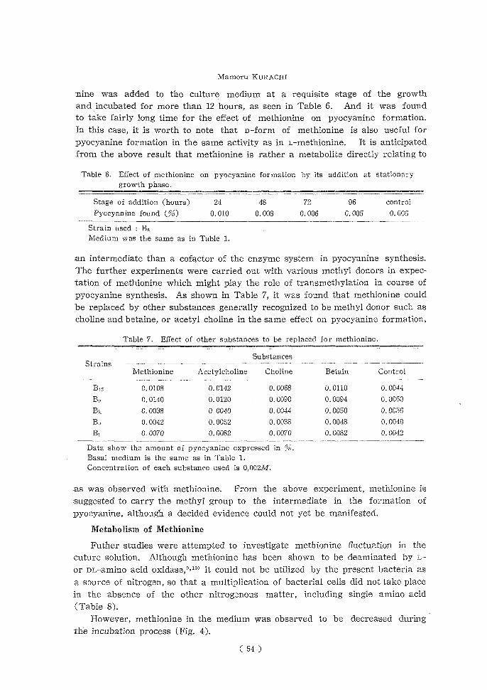
nine was added to the culture medium at a requisite stage of the growth
and incubated for more than 12 hours, as seen in Table 6. And it was found to take fairly long time for the effect of methionine on pyocyanine formation.
In this case, it is worth to note that D-form of methionine is also useful for
pyocyanine formation in the same activity as in L-methionine. It is anticipated from the above result that methionine is rather a metabolite directly relating to
Table 6. Effect of methionine on pyocyanine formation by its addition at stationary growth phase.
Stage of addition (hours)24487296 control Pyocyanine found (%)0.010 0.008 0.006 0.005 0.006
Strain used : B,L Medium was the same as in Table 1.
an intermediate than a cofactor of the enzyme system in pyocyaninesynthesis.
The further experiments were carried out with various methyl donors in expec-tation of methionine which might play the role of transmethylation in course of
pyocyanine synthesis. As shown in Table 7, it was found that methionine could be replaced by other substances generally recognized to be methyl donor such as
choline and betaine, or acetyl choline in the same effect on pyocyanine formation,
Table 7. Effect of other substances to be replaced for methionine.
Substances Strains
MethionineAcetylcholine Choline Betain Control
B160.01080.0142 0.0068 0.0110 0.0044
0.01400.0120 0.0090 0.0094 0.0060 Bz;0.00380 0040 0.0044 0.0050 0.0036 B.,0.00420.0052 0.0038 0.0048 0.0040
B10.00700.0082 0.0070 0.0082 0.0042
Data show the amount of pyocyanine expressed in %. Basal medium is the same as in Table 1.
Concentration of each substance used is 0,002M.
as was observed with methionine. From the above experiment, methionine is
suggested to carry the methyl group to the intermediate in the formation of
pyocyanine, although a decided evidence could not yet be manifested.
Metabolism of Methionine
Futher studies were attempted to investigate methionine fluctuation in the cuture solution. Although methionine has been shown to be deaminated by L-
or DL-amino acid oxidase,9"10' it could not be utilized by the present bacteria as
a source of nitrogen, so that a multiplication of bacterial cells did not take place
in the absence of the other nitrogenous matter, including single amino acid
(Table 8). However, methionine in the medium was observed to be decreased during
the incubation process (Fig. 4).
( 54 )

Studies on the Biosynthesis of Pyocyanine. (IV)
Table 8. Assimirability of amino acids as source of nitrogen.
GrowthPyocyanineGrowth Pyocyanine formationformation
DL-Alanine++ L-Lysinetrace — L-Arginine++ L-Methionine—--
L-Aspartic acid +— L-Phenylalanine—— L-Cystine—— Oxy-L-proline——
L-Glutamic acid +— L-Serine+— Glycine++ DL-Threoninetrace —
L-Histidine -I- + DL-Tyrosinetrace — L-Isoleucine +— L-Tryptophan+—
L-Lencinetrace — DL-Valinetrace —
Basal medium : 2% glycerol, 0.05 % MgSO47H2O, 0.025% K2HP05 and 0.0005% Fe2(SO4)3, pH7.4. Each amino acid was tested at a concentration of 0.1 % as sole source of
nitrogen.
On the other hand, not only bacterial growth but also pyocyanine formation was observed in the synthetic medium containting methionine, even in the absence
of sulfate which was an essential element for both the pigmentation and the bacterial growth, as had been reported previously.1,2' It is suggested from the
above result that sulfate would serve as a source of methionine in course of
pyocyanine formation, and otherwise that methionine may be utilized for the synthesis of other sulfur compound. Methionine has been recognized to play the
0.05
0.04 •
o 0.03 c , 0.02
0.01
------------.4 1y 2 3 4 5
Days
Fig. 4. Consumption of methionine in the cultural solution. To the same medium as shown in Table 1, 0.05% methionine was added.
role of methyl transfer in animal body, and the demethylation product may be
expected to be homocysteine, judging from the fact that methionin is synthesized
by methylation of homocysteine in the presence of choline or, betaine."-l4'
On the other hand, it has been shown that from mthionine, homoserine was
derived when it was incubated with liver preparation, and that cystathionine which was derived from methionine as an intermediate in the formation of homo-
serine and cysteine, was formed by the combination of homocysteine with serine.1" In order to detect homocysteine or homoserine in an incubation mixture ow-
ing to translnethylation reaction, the following experiment was carried out : the
( 55 )

Mamoru KURACHI
bacterial cells grown on the synthetic medium prepared with urea as a sole
source of nitrogen, were harvested by centrifugation after being incubated for
36 hours when pyocyanine was appreciablly formed and the cells were washed
with 0.01 M phosphate buffer of pH 7.2. One g of the wet cells was suspended in
100 ml of 0.01 M phosphate buffer of pH 7.4, containing 0.05 g of MgSO47H~O and
0.1 g of Na-succinate. To 50 ml of this suspension, methionine was added to at-
tain 0.002 M concentration and incubated at 37° for 16 hours with control experi-
ment without methionine. The reaction mixtures were treated with calcium
phosphate gel to make the solution clear, concentrated and then mounted on
paper chromatography. The result is shown in Fig. 5. The paper chromatograms
Maniamn<
H.rmste(ne Q .Homaurlee
Fig. 5. Paper chromatography of a reaction product of methionine by resting cells compared with known amino acids (left), using the
solvent system: n-butanol-acetic acid-water (5 : 1 : 2).
of the reaction mixture showed two faint spots besides the spot of methionine,
whereas with the control experiment any spot did never appear, suggesting no
autolysis of the bacterial cells. Among the three spots, the one was found to be
corresponding rather to homocysteine than to homoserine, although the other spot
was not yet identified. On the other hand, in the experiment with the growing
state of the bacteria, these spots were hardly recognized. The reason of this
phenomenon may be explained to be ascribable to the further metabolism of methionine in growing system. The experiment with the cell-free preparation of
the bacteria which were harvested after the cultivation of 24 hours has also suc-
ceeded in obtaining the result similar to that of the experiment with resting cells
mentioned above.
Although vitamin Bu was observed to be hardly effective in the metabolism
of methionine, the effect of methionine on pigmentation is assumed to play the
role of methyl carrier in course of pyocyanine biosynthesis. As regards the trans-
methylation, a noticeable fact has been found : in paper chromatographic study
of the methionine metabolism, the spot corresponding to homocysteine was more
markedly detectede by the administration of anthranilic acid which might be as-
signed a role of methyl acceptor. The result of further studies on this problem
will be presented later on.
Other Effective Component of Peptone
As was already illustrated, the strain unresponsive to methionine which re-
vealed the remarkable pigmentation in peptone medium, was recognized among
( 56 )

Studies on the Biosynthesis of Pyocyanine. (IV)
the strains used in the present experiment. For the reason why no effect of
methionine was observed with this strain, the following assumption will be pro-
posed. Methionine would possibly be offered for the bacterial growth before the synthesis of pyocyanine, and therefore the requirement of methionine
could be divided according to the kinds of bacterial strain, depending on their
capacity for the synthesis of methionine. Similarly, the other stimulating factor
for pigmentation which exists in peptone, may also be required by the bacteria
according to their capacity for its synthesis in the synthetic medium without pep-
tone. In other words, methionine may be no more than the effective substance
which invests the bacteria with one of the conditions necessary for pyocyanine
formation. It was found that in residual part of phosphotungstic acid-precipita-
tion of peptone solution, the promoting effect on pyocyanine formation with major kinds of strain was remained, and that the same effect as with peptone itself
was still observed to be maintained in the experiment with the methionine-un-
responsive strain, after the deamination treatment of peptone by means of nitrous
acid.
It is, on the other hand, interesting to point out that the effective substance similar to peptone is found in cow's milk serum or yeast extract. These effectve
substances are supposed to serve rather as a stimulating facter than as a pre-
cursor of pyocyanine. Although the effective substance recognized in peptone can
not yet be obtained in pure state owing to its less solubility in organic solvent,
it is presumed to be an aromatic compound, according to the phosphomolybdate
test10) : when phosphomolybdic acid is added to the acidified solution of this ef-
fective substance and is made alkaline, it is immediately colored to blue as is recognizable with ordinary benzene compound.
00 - 0 02_ jr\ O220----------------------230 240 250. 200 275 200 200 --Wavo length (In )
Fig. 6. Paper chromatography of the constituent of Fig. 7. Absorption curve of peptone being regarded as a stimulating factor for the component of peptone
pyocyanine formation.expected to be a stimulat- Solvent is the same as in Fig. 5.ing factor for pyocyanine
a, With the sample obtained by the adsorption formation, obtained by the treatment.adsorption treatment and b, Detected in the cultured solution of the bacteria paper partition chromato-
grown on peptone medium.graphy.
On the other hand, the following result has been offered as a further evidence :
the aqueous eluate of one of the fluorescent spots (Fig. 6, a) obtained by paper
chromatography according to the same procedure as was described before, re-
vealed an increasing effect on pyocyanine formation. Futhermore, the photometric
( 57 )

Mamoru KURACHI
experiment has shown that its absorption maximum exists at 260 mp, as seen in Fig. 7. It is of interest to note that another fluorescent spot has appeared on
paper chromatograms from the butanol extract of the cultured solution of the bacteria grown on peptone medium, while it was not observed with synthetic
medium without peptone (Fig. 6, b).
SUMMARRY
1. It has been shown that one effective constituent of peptone was identified
with methionine, and that the other amino acid components did not reveal any
effect on pyocyanine formation.
2. It was assumed that methionine would play the role of methyl carrier in
course of pyocyanine synthesis, since the same effect was observed with known
methyl donor such as choline and betain, or acetylcholine, and the product de-
rived from methionine was expected to be identical with homocysteine.
3. From the characteristics of the bacterial strain unresponsive to methio- nine, the other stimulating factor was suggested to exist in peptone and some
informations on this substance were presented.
The author is deeply grateful to Prof. H. Katagiri for his generous directions
throughout this work.
REFERENCES
(1) M. Kurachi, This Bulletine, 26, 163 (1958). (2) H. Katagiri, T. Shibutani and M. Kurachi, This Bulletino, 25, 71 (1951).
(3) M. Kurachi, This Bulletin, 26, 174 (1958). (4) M. Somogyi, J. Biol. Chem., 86, 155 (1930).
(5) L. Varies, et.al., J. Biol. Chem., 133, 491 (1940). (6) B. F. Steele, J. Gen. Microbiol., 2, 633 (1951).
(7) F. Esther, J. Biol. Chem., 177., 533 (1949). (8) M. Kurachi, Unpublished data.
C9) M. Blanchard, F. Green, V. Nocito and S. Ratner. J. Biol. Chem., 155, 421 (1944), 161, 583 (1945).
(10) A. B. Binder and H. A. Krebs. J. Biol. Chem., 46, 210 (1950) (11) N. H. Horowitz, J. Biol. Chem., 171, 255 (1947)
(12) V. du Vigneaud, Proc. Am. Physiol. Soc., 92, 127 (1948). (13) H. Borsook and J. W. Dubnoff, J. Biol. Chem., 169, 247 (1947).
(14) W. Dubnoff, Arch. Biochem., 24, 251 (1949). (15) J. H. Teas, N. H. Horowitz and M. Fling, J. Biol. Chem., 172, 651 (1948).
(16) M. Kurachi, Unpublished data.
( 58 )
![Lipid Biosynthesis in Eukaryotic Cells - pub.epsilon.slu.sepub.epsilon.slu.se/1173/1/Avhandling_nr_078[1].2006_Tryckfil.pdf · Lipid Biosynthesis in Eukaryotic Cells Studies on Enzyme](https://img.pdfslide.net/doc/110x75/5cbc44c388c9937f418c630e/lipid-biosynthesis-in-eukaryotic-cells-pub-12006tryckfilpdf-lipid-biosynthesis.jpg)