Embed Size (px)
Citation preview

TitleTobacco streak virus (strain dahlia) suppresses post-transcriptional gene silencing of flavone synthase II in blackdahlia cultivars and causes a drastic flower color change.
Author(s) Deguchi, Ayumi; Tatsuzawa, Fumi; Hosokawa, Munetaka;Doi, Motoaki; Ohno, Sho
Citation Planta (2015), 242(3): 663-675
Issue Date 2015-07-18
URL http://hdl.handle.net/2433/201879
Right
The final publication is available at Springer viahttp://dx.doi.org/10.1007/s00425-015-2365-6.; The full-textfile will be made open to the public on 18 July 2016 inaccordance with publisher's 'Terms and Conditions for Self-Archiving'.; This is not the published version. Please cite onlythe published version. この論文は出版社版でありません。引用の際には出版社版をご確認ご利用ください。
Type Journal Article
Textversion author
Kyoto University

1
Tobacco streak virus (strain dahlia) suppresses post-transcriptional gene silencing of flavone synthase II in
black dahlia cultivars and causes a drastic flower color change
Ayumi Deguchi1, Fumi Tatsuzawa2, Munetaka Hosokawa1, Motoaki Doi1 and Sho Ohno1*
1 Graduate School of Agriculture, Kyoto University, Sakyo-ku, Kyoto 606-8502, Japan
2 Faculty of Agriculture, Iwate University, Morioka 020-8550, Japan
*Corresponding author: Sho Ohno
Laboratory of Vegetable and Ornamental Horticulture, Graduate School of Agriculture, Kyoto University, Sakyo-
ku, Kyoto 606-8502, Japan
Telephone: +81-75-753-6048, Fax: +81-75-753-6068
E-mail: [email protected]
Main Conclusion
Tobacco streak virus suppressed post-transcriptional gene silencing and caused a flower color change in black
dahlias, which supported the role of cyanidin-based anthocyanins for black flower appearance.
Author contribution
MH and SO conceived the research. MD and SO designed the research. AD conducted the research. FT contributed
pigment analysis. AD and SO wrote the manuscript. All authors read and approved the manuscript.

2
Abstract
Black flower color of dahlia (Dahlia variabilis) has been attributed, in part, to the high accumulation of cyanidin-
based anthocyanins that occurs when flavone synthesis is reduced because of post-transcriptional silencing (PTGS)
of flavone synthase II (DvFNS). There are also purple-flowering plants that have emerged from a black cultivar
‘Kokucho’. We report that the purple color is not caused by a mutation, as previously thought, but by infection with
tobacco streak virus (TSVdahlia), which suppresses the PTGS of DvFNS. When TSVdahlia was eliminated from the
purple-flowering ‘Kokucho’ by leaf-primordia-free shoot apical meristem culture, the resulting flowers were black.
TSVdahlia-infected purple flowers had lower numbers of siRNAs to DvFNS than black flowers, suggesting that
TSVdahlia has a silencing suppressor. The graft inoculation of other black cultivars with TSVdahlia altered their flower
color drastically except for ‘Fidalgo Blacky’, a very deep black cultivar with the highest amount of cyanidin-based
anthocyanins. The flowers of all six TSVdahlia-infected cultivars accumulated increased amounts of flavones and
reduced amounts of cyanidin-based anthocyanins. ‘Fidalgo Blacky’ remained black despite the change in pigment
accumulation, and the amounts of cyanidin-based anthocyanins in its TSVdahlia-infected plants were still higher than
those of other cultivars. We propose that black flower color in dahlia is controlled by two different mechanisms that
increase the amount of cyanidin-based anthocyanins: DvFNS PTGS-dependent and independent mechanisms. If
both mechanisms occur simultaneously, the flower color will be blacker than if only a single mechanism is active.
Key words
Cyanidin, Dahlia variabilis, FNS II, PTGS, Silencing suppressor

3
Abbreviations
ANS Anthocyanidin synthase
AV-2 Asparagus virus 2
bHLH Basic helix-loop-helix
CHI Chalcone isomerase
CHS Chalcone synthase
CMV Cucumber mosaic virus
CP Coat protein
CSVd Chrysanthemum stunt viroid
DFR Dihydroflavonol 4-reductase
DMV Dahlia mosaic virus
F3H Flavanone 3-hydroxylase
F3′H Flavonoid 3′-hydroxylase
FNS II Flavone synthase II
HPLC High performance liquid chromatography
INSV Impatiens necrotic spot virus
LP-free SAM Leaf-primordia-free shoot apical meristem
MP Movement protein
PTGS Post-transcriptional gene silencing
PSTVd Potato spindle tuber viroid
RT-PCR Reverse transcription-PCR
siRNA Small interfering RNA
TSV Tobacco streak virus
TSWV Tomato spotted wilt virus

4
Introduction
Flower color is one of the most important traits for ornamental plants. There are various flower colors (e.g., red,
purple, pink, orange, yellow, blue, and white) as well as variegated and bicolor flowers. Among flower colors, black
is considered to be attractive because it is rare and present in few genera. Black flower color is found in wild pansy
(Viola tricolor) (Clausen 1930) and wild Lisianthius nigrescens (Markham et al. 2004). Recently, some black flower
cultivars of petunia (Petunia hybrida), hollyhock (Althaea rosea), and dahlia (Dahlia variabilis) have begun to be
produced commercially. While black flowers are increasingly popular, the mechanism producing this color is
unclear.
To elucidate this mechanism, we have studied dahlia that has a wide range of flower colors and
comparatively large numbers of the black cultivars. Moreover, the pigments, enzymes and genetic factors related to
flower colors have been largely elucidated in this species. The pigments accumulating in dahlia petals belong to
three major flavonoid compounds: anthocyanins (pelargonidin-based and cyanidin-based), flavones (apigenin-based
and luteolin-based), and chalcones (isoliquiritigenin-based and butein-based) (Nordström and Swain 1953; 1956;
1958; Harborne et al. 1990). These contribute to red ~ purple, ivory, and yellow colors, respectively. Generally,
anthocyanidins, the aglycone of anthocyanins are synthesized by chalcone synthase (CHS), chalcone isomerase
(CHI), flavanone 3-hydroxylase (F3H), dihydroflavonol 4-reductase (DFR), and anthocyanidin synthase (ANS)
(Grotewold 2006; Tanaka et al. 2008). Anthocyanins result from the addition of sugar moieties. Flavones are
synthesized by flavone synthase (FNS I and FNS II) from flavanones, the products of CHI (Martens and Mithöfer
2005). Chalcones are synthesized by CHS, chalcone reductase (CHR) (Davies et al. 1998), and chalcone 3-
hydroxylase (CH3H) (Schlangen et al. 2010a). In the pathway leading to anthocyanins and flavones, cyanidin and
luteolin are synthesized via flavonoid 3′-hydroxylase (F3′H) activity as counterparts to pelargonidin and apigenin,
respectively (Ayabe and Akashi 2006; Tanaka and Brugliera 2013). In dahlia, these enzymes have been studied
(Fischer et al. 1988; Wimmer et al. 1998; Halbwirth et al. 2008; Thill et al. 2012) and the genes encoding the
enzymes have been isolated (Schlangen et al. 2010b; Ohno et al. 2011a; Ohno et al. 2011b; Deguchi et al. 2013). In
addition, a bHLH transcription factor, DvIVS, regulates anthocyanin levels by regulating the expression of DvCHS1,
DvF3H, DvDFR, and DvANS (Ohno et al. 2011a; Ohno et al. 2013).
In a previous study (Deguchi et al. 2013), the black dahlia cultivars showed lower petal lightness (L*)
and chroma (C*) values than the other color cultivars. This appeared to be a phenotypic indicator for the black
appearance. Black dahlias indeed accumulate high amounts of total anthocyanins (Thill et al. 2012; Deguchi et al.
2013), however, total anthocyanins are lower in black cultivars than in some purple cultivars, suggesting that the
black appearance cannot be explained solely by total anthocyanin levels. Black dahlias exhibit two other distinct
features of pigment accumulation. One is high accumulation of cyanidin-based anthocyanins. In cultivars with
similar levels of total anthocyanins, the L* and C* values were inversely proportional to the cyanidin-based
anthocyanin levels, suggesting that the cyanidin-based anthocyanins contribute more strongly to the black
appearance than pelargonidin-based ones (Deguchi et al. 2013). Therefore, we considered that the high accumulation
of cyanidin-based anthocyanins was also related to the black appearance in addition to the higher accumulation of
total anthocyanins. The other feature of black dahlias is low accumulation of flavones. Interestingly, a comparison
between the black cultivar ‘Kokucho’ and its purple-flowering plant showed that the suppression of flavone

5
synthesis is caused by post-transcriptional gene silencing (PTGS) of FNS II (DvFNS), leading to the high
accumulation of both total anthocyanins and cyanidin-based anthocyanins. This may be due to the abolishment of
competition between anthocyanidin and flavone synthesis.
The purple-flowering ‘Kokucho’, which had been previously used as a putative mutant (Deguchi et al.
2013) sometimes produces mosaic-colored flowers (black parts in purple petals) (Supplementary Fig. S1), raising
the question of whether it is truly a mutant. Some reports have shown flower color changes to be induced by plant
virus infection. In tulip (Tulipa gesneriana) and petunia, the alteration of anthocyanin pigmentation patterns are
attributed to infection with certain plant viruses (Dekker et al. 1993; Lesnaw and Ghabrial 2000; Teycheney and
Tepfer 2001). Thus, we assumed that purple-flowering ‘Kokucho’ is infected by some virus. In the present study,
we examined virus infection and our data indicated that the flower color change from black to purple in ‘Kokucho’
was induced by a tobacco streak virus (TSV) infection. The inoculation of other black cultivars with the virus
provided us further insights into black flower coloring.
Materials and Methods
Plant materials
Eighteen tuberous roots of the black dahlia cultivar ‘Kokucho’ were grown in the experimental field at Kyoto
University (Kyoto, Japan). Sets of five plants of nearly the same height, each with either normal black flowers (B1–
B5) or color changed purple flowers (P1–P5), were selected for the subsequent experiments. Along with ‘Kokucho’,
three black cultivars (‘Fidalgo Blacky’, ‘Ms. Noir’, and ‘Black Cat’), two black–white bicolor cultivars (‘Kazusa-
shiranami’ and ‘Kageboushi’), and one pink cultivar (‘Jyunn-ai’) were also grown and the petals were collected for
analysis.
Detection of viruses and viroids
The detection of viruses and viroids was performed with reverse transcription-PCR (RT-PCR). The total RNA of
leaves was extracted using Sepasol- RNA I Super G (Nacalai Tesque, Kyoto, Japan) followed by purification using
High-Salt Solution for Precipitation (Takara Bio Inc., Ohtsu, Japan). The viruses and viroids tested were as follows;
cucumber mosaic virus (CMV), dahlia mosaic virus (DMV), impatiens necrotic spot virus (INSV), TSV, tomato
spotted wilt virus (TSWV), chrysanthemum stunt viroid (CSVd), and potato spindle tuber viroid (PSTVd), which
have been reported to be infectable to dahlias. The primer set used for the detection of each virus or viroid is given
in Supplementary Table S1. Total RNA was reverse-transcribed with ReverTra Ace (Toyobo Co. Ltd., Osaka, Japan)
using a specific reverse primer for each virus or viroid (Supplementary Table S1) or with ReverTra Ace qPCR RT
Master Mix (Toyobo). The plasmids containing the full or partial sequence for coat protein (CP) of CMV, DMV,
TSV or TSWV and the full sequence of CSVd were used in RT-PCR as positive controls, and water was used as a
negative control.
Elimination of viruses and viroids by leaf-primordia-free shoot apical meristem culture

6
‘Kokucho’ purple plants (P3, P4, and P5) were used for leaf-primordia-free shoot apical meristem (LP-free SAM)
culture (Hosokawa et al. 2004) to eliminate viruses and viroids. Regenerated shoots from these three plants were
transplanted onto modified MS medium (Murashige and Skoog 1962), and five rooted plants were transplanted in
the experimental field. The flowers of these five lines were used for the color and pigment accumulation analyses.
Sequence analysis of TSV RNA
The reverse primers for RNA2 and RNA3 of TSV were designed around the 3′ ends with reference to the existing
TSV sequences in the BLAST database. The primer sets used to amplify the full-length 2b protein gene on RNA2
and the movement protein (MP) and CP genes on RNA3 were also designed (Supplementary Table S2). Total RNA
extracted from the leaves of TSV-infected ‘Kokucho’ was subjected to reverse transcription using RNA2 and RNA3
full reverse primers. Then, the 2b protein, CP, and MP genes were amplified using RT-PCR. The amplified PCR
products were cloned into pTAC-1 vector (BioDynamics Laboratory Inc., Tokyo, Japan) according to the
manufacturer’s instructions. Eight plasmids for each gene were sequenced using a BigDye Terminator v3.1 Cycle
Sequencing Kit and a 3100 Genetic Analyzer (Applied Biosystems). Because the TSV sequence isolated from
‘Kokucho’ was not identical to known TSV sequences, we refer to it here as TSVdahlia.
Measurements of plant growth
‘Kokucho’ line 4, which remained infected by TSVdahlia even after LP-free SAM culture, and line 5, from which
TSVdahlia had been eliminated, were used to compare plant growth characteristics. Nine plants propagated from each
line were grown in 24 cm in diameter pots in a glasshouse from July to December 2014. The height, node number,
inflorescence diameter, leaf color, and tuber weight of these plants were measured.
In vitro graft inoculation of virus
In vitro grown TSVdahlia-infected ‘Kokucho’ line 4 was used as a root stock and black, black–white bicolor, and pink
cultivars were used as a scion. The shoots of these cultivars not infected with TSVdahlia were in vitro grafted onto
the ‘Kokucho’ line 4 root stock. About one month later, TSVdahlia infection was confirmed in petals using RT-PCR
and the rooted shoots of TSVdahlia-infected plants were transplanted in the field. The plants without grafts (originals)
and those grafted onto TSVdahlia-uninfected ‘Kokucho’ line 5 (graft controls) were also grown in the same
experimental field. Flowers were used for the analyses of color, pigment accumulation, and transcript abundance.
Color analysis
To evaluate flower color, the color components of the CIE L*a*b* coordinate system, namely lightness (L*) and
chroma [C*, calculated as C* = (a*2 + b*2)1/2], were measured using a hand spectrophotometer (NR-3000, Nippon
Denshoku Industries Co., Ltd., Tokyo, Japan). Three areas of the adaxial petal surface were subjected to color
measurement. In the case of bicolor cultivars, colored parts (petal bases) were measured. Scores from three expanded,
completely colored petals taken from three different flowers were averaged.
Quantification of pigments

7
Pigments were quantified with high performance liquid chromatography (HPLC). Fresh petals (100 mg) used in the
color analysis were homogenized with a mortar and a pestle under liquid nitrogen. Added 1 mL of extraction solution
(5% hydrochloric acid in 50% methanol), the mixture was stored overnight at 4°C in darkness. The mixture was
then centrifuged at 4°C at 15,000 rpm for 15 min, and the supernatant was collected and diluted 50 times with the
same solvent. For hydrolysis, 1.2 mL of the diluted solution was boiled at 95°C for 2 h and 20 µl of the hydrolyzed
solution was injected into the HPLC apparatus. The analysis was performed using HPLC system (Hitachi L-7100,
L-7200, L-7420, L-7500: Hitachi Systems, Ltd., Tokyo, Japan) with a C18 column (Nihon Waters K. K., Tokyo,
Japan), maintained at 40°C. The detection wavelength was 350 nm for flavones and 520 nm for anthocyanidins.
Eluent preparation and HPLC analysis proceeded according to Ohno et al. (2011b). For the standard curves, 1 mg
of pelargonidin chloride (Sigma-Aldrich Corporation, St. Louis, USA), cyanidin chloride (Wako Pure Chemical
Industries, Ltd., Osaka, Japan), apigenin (Wako), and luteolin (LKT Laboratories Inc., St. Paul, MN, USA) were
dissolved in 1 mL of 5% hydrochloric acid in 50% methanol, and one-half dilution series were made. Twenty micro
liters of each dilution was injected, and the relationship between peak areas and pigment concentrations was
calculated. Calculations for pelargonidin and cyanidin were converted from those for each chloride. The assay was
performed with three petals from three different flowers.
Real-time RT-PCR
Quantitation of DvFNS, DvF3′H, DvIVS, DvCHS1, DvCHS2, DvCHI, DvF3H, DvDFR, and DvANS transcripts was
performed by real-time RT-PCR. Total RNA was reverse transcribed with ReverTra Ace (Toyobo) using oligo dT
primer, and real-time RT-PCR was performed with SYBR Premix EX TaqII (Takara) using the LightCycler 480
system (Roche Diagnostics K.K., Tokyo, Japan). The real-time RT-PCR was performed as follows: 95°C for 5 min,
followed by 45 cycles at 95°C for 10 sec and 60°C for 30 sec. Single-target product amplification was checked
using a melting curve. To measure the highest levels of transcript abundance, half-colored, unexpanded petals were
used for analysis of DvFNS and DvF3′H and completely colored, unexpanded petals were used for other genes.
Completely colored, unexpanded petals of bicolor cultivars were separated into bases and tips. Three or two
biological replications were performed and DvActin was used as an internal standard. Primers for DvActin and
DvF′3H are given in Supplementary Table S3; primers for other genes are described in Ohno et al. (2011b) and
Deguchi et al. (2013).
Detection of siRNAs
The detection of small interfering RNA (siRNA) was performed according to Ohno et al. (2011b). Total RNA was
extracted from TSVdahlia-uninfected and TSVdahlia-infected petals of black, black–white bicolor, and pink cultivars.
Total RNA of the red–white bicolor cultivar ‘Yuino’ wherein the PTGS of DvCHS occurs (Ohno et al. 2011b), was
used as a positive control for the detection of DvCHS siRNA. A digoxigenin-labeled RNA probe was synthesized
using T7 RNA polymerase (Roche, Basel, Switzerland) by in vitro transcription of a 3′-digested pTAC-1 vector
(BioDynamics Laboratory) carrying the exon 2 region of DvFNS and the full length of DvCHS2 in the antisense
orientation.

8
Mapping of small RNAs
Small RNAs were extracted from 200 mg of half-colored fresh petals of TSVdahlia-infected ‘Kokucho’ (line 4) using
a MirVana miRNA Isolation Kit (Applied Biosystems) and sequenced with the Illumina HiSeq System (Illumina
Inc., San Diego, CA, USA) in a single-end read. Small RNAs (18–32 nt) were mapped onto the DvFNS genomic
sequence of ‘Kokucho’ (AB769841) as a reference sequence, with no mismatches allowed.
Result
Virus detection and elimination from purple-flowering ‘Kokucho’
Purple ‘Kokucho’ produces entirely purple flowers from autumn to winter, but black sectors are sometimes observed
in summer (Supplementary Fig. S1). The presence of the mosaic flower color prompted us to investigate whether
virus infection could explain the color change. Five of 18 ‘Kokucho’ plants grown from tuberous roots produced
color-changed purple flowers, while the other 13 plants produced normal black flowers. These five purple-flowering
plants (P1–P5) and five black-flowering plants (B1–B5) of almost the same plant size were used for virus and viroid
detection. RT-PCR analysis revealed that TSVdahlia was present in all purple flowered plants, but not in black
flowered plants. (Fig. 1). CMV and DMV were also detected in some purple and black plants but the infection did
not correlate with flower color. TSWV, CMV, and CSVd were not detected in any plants. Moreover, the bands of
expected lengths for INSV or PSTVd were not detected (data not shown). These findings suggest that infection with
TSVdahlia correlated with flower color in ‘Kokucho’, but CMV, DMV, INSV, TSWV, CSVd, and PTSVd were
irrelevant to its flower color change.
To eliminate viruses from purple ‘Kokucho’, LP-free SAM cultures (Hosokawa et al. 2004) were prepared.
This method is superior to ordinary apical meristem culture for eliminating viruses. Five regenerated lines (1–5)
were obtained and grown in the experimental field. One of the lines (line 4) remained to produce purple flowers like
its mother plant (P3). However, the other lines (1–3, 5) completely recovered the black flower color (Table 1). As a
result of RT-PCR analysis, TSVdahlia was detected by RT-PCR only in line 4, whereas CMV was detected in all lines
(Fig. 2). These results indicate that elimination of TSVdahlia succeeded in four lines (1–3, 5) but failed in line 4 and
that purple flower color corresponded to TSVdahlia infection. The detection of CMV in all lines may be due to the
failure of elimination or re-infection during growth because two mother plants (P3 and P4) were infected and a
mother plant (P5) was not infected. The elimination of TSVdahlia completely correlated with the recovery of black
flower color, suggesting that purple ‘Kokucho’ is not a mutant and that purple flower color is caused by TSVdahlia
infection.
The comparison of plant growth between TSVdahlia-infected line 4 and TSVdahlia-uninfected line 5, which
were regenerated from the same mother plant (P3), showed no significant differences (P > 0.05) in plant height,
node number, inflorescence diameter, tuberous root weight, and leaf color (Supplementary Fig. S2, Table S4).
Therefore, infection with the virus did not affect aspects of plant growth, aside from flower color.
The sequence of TSV isolated from ‘Kokucho’

9
TSV, belonging to the genus Ilarvirus and the family Bromoviridae, can infect a wide variety of plant species,
including dahlia. The virus isolated from purple ‘Kokucho’ shares high homology with registered TSV, but the
absence of identical sequences in the database implied that TSVdahlia is a new strain of TSV. For TSVdahlia isolated
from ‘Kokucho’, the full-length sequences of genes encoding 2b protein (LC030106), MP (LC030107), and CP
(LC030108) were 618, 873, and 717 bp long and encoded 205, 290, and 238 putative amino acid residues,
respectively. The sequences of genes for 2b, MP, and CP share 89%, 88%, and 92% identity, respectively, with
reported TSV sequences [strain WC: U75538 (Scott et al. 1998), X00435 (Cornelissen et al. 1984)]. They share
91%, 95%, and 96% identity, respectively, with those isolated from crownbeard (Verbesina encelioides; JX463338,
JX463339) (Sharman and Thomas 2013); 89%, 92%, and 90% identity, respectively, with soybean (Glycine max;
FJ403376, FJ403377); and 87%, 91%, and 90% identity, respectively, with Cucurbita pepo (KM504247,
KM504248) (Padmanabhan et al. 2014). The sequence of CP also shares 98% identity with that isolated from
Xanthium occidentale (JX463349) (Sharman and Thomas 2013) and fava bean (Vicia faba, AM933669).
Mapping of small RNAs on the DvFNS genomic sequence in TSVdahlia-infected purple ‘Kokucho’
Purple-flowering ‘Kokucho’ line 4, which remained infected by TSVdahlia accumulated flavones, while color-
recovered black-flowering line 5 accumulated low amounts of flavones (Fig. 3a). DvFNS transcript abundance was
higher in petals of line 4 than in petals of line 5 (Fig. 3b), as shown in Deguchi et al. (2013) in which purple-
flowering ‘Kokucho’ was described as a mutant.
In original black ‘Kokucho’, siRNA of DvFNS had been detected and abundant small RNAs were mapped
onto the DvFNS gene, suggesting that DvFNS is post-transcriptionally silenced (Deguchi et al. 2013). Therefore,
the obvious increase in DvFNS transcript abundance in purple ‘Kokucho’ may result from PTGS suppression by
virus infection. To confirm this, we mapped small RNAs extracted from the petals of TSVdahlia-infected purple
‘Kokucho’ (line 4) onto the DvFNS genomic sequence. Of the 24,846,705 reads, which were 18–32 nt long, 2,126
reads were successfully mapped (Fig. 4). The mapped small RNAs of 21 nt accounted for only 1,779 reads and were
mostly mapped onto exon 2. The number of mapped reads in purple ‘Kokucho’ was much fewer than in black
‘Kokucho’, wherein 11,574 of 14,895,667 reads were mapped onto DvFNS genome (Deguchi et al. 2013). Moreover,
the 21 nt antisense small RNA, which had the highest number with 4,192 reads in black ‘Kokucho’, was not mapped
at all in purple ‘Kokucho’. These results indicated that the PTGS of DvFNS was suppressed in color-changed purple
‘Kokucho’ by TSVdahlia infection.
Changes in flower color, pigment accumulation, and transcript abundance in black and black–white bicolor
cultivars with TSVdahlia inoculation
To verify whether TSVdahlia changes flower color in other cultivars, we performed graft inoculations. Four black
cultivars (‘Fidalgo Blacky’, ‘Ms. Noir’, ‘Black Cat’, and TSVdahlia-uninfected ‘Kokucho’), two black–white bicolor
cultivars (‘Kazusa-shiranami’ and ‘Kageboushi’), and one pink cultivar (‘Jyunn-ai’) were in vitro grafted onto
purple-flowering ‘Kokucho’ (line 4) because the mechanical inoculation hardly succeeded in infecting these
cultivars with TSVdahlia. The transmission and infection with TSVdahlia was confirmed using RT-PCR (Supplementary
Fig. S3). TSVdahlia-infected plants of each black and bicolor cultivar produced flowers differing in color from the

10
originals. The altered color was purple in most cultivars, but it remained black only in ‘Fidalgo Blacky’, despite a
slight bluish color change (Fig. 5). All TSVdahlia-infected plants of the black and bicolor cultivars showed higher
petal lightness (L*) values than plants of the original cultivars, and they also showed higher petal chroma (C*)
values except for ‘Kageboushi’, whereas graft control showed the original color (Fig. 6, Supplementary Table S5).
‘Fidalgo Blacky’, which has the blackest appearance, with low L* and the lowest C* values, showed the smallest
increase in L* score as a result of the virus infection.
These TSVdahlia-infected plants accumulated flavones in their color-changed petals and accumulated
lower amounts of anthocyanins (anthocyanidins) than their TSVdahlia-uninfected counterparts (originals and graft
controls) (Fig. 7). Moreover, in these cultivars, the reducing ratio of cyanidin was higher than that of pelargonidin
(Fig. 7). In contrast, the pink cultivar ‘Jyunn-ai’, which originally accumulated flavones, exhibited no changes in
flower color or pigment accumulation (Fig. 5, 6, 7; Supplementary Table S5). ‘Kazusa-shiranami’ original cultivar
was reported to accumulate flavone in its petals (Ohno et al. 2011b), but it actually accumulated low flavones similar
to the other black cultivars in this study.
According to the results of real-time RT-PCR, DvFNS transcript abundance was markedly increased in
TSVdahlia-infected plants of black and bicolor cultivars (Fig. 8). The transcript abundance in TSVdahlia-infected plants
were about 7–100 times higher than those of TSVdahlia-uninfected plants. In contrast, the genes involved in
anthocyanin biosynthesis, DvIVS, DvCHS1, DvCHS2, DvCHI, DvF3H, DvDFR, DvANS, and DvF3′H, which are
also involved in the synthesis of cyanidin, showed no marked changes in the transcript abundance upon virus
infection in all black and bicolor cultivars (Supplementary Fig. S4). In these cultivars, siRNA of DvFNS was
detected in TSVdahlia-uninfected plants, while it was not detected in TSVdahlia-infected plants (Fig. 9). These results
suggest that the suppression of DvFNS PTGS by TSVdahlia alters the accumulation of pigments and flower color in
these black and black–white bicolor cultivars, as demonstrated in ‘Kokucho’ line 4.
Interestingly, in the cultivars ‘Kazusa-shiranami’ and ‘Kageboushi’, the white tips of the bicolor petals
disappeared upon virus infection, and the flower color turned entirely purple (Fig. 5e, f, l, m). The accumulation of
pigments was similar in petal tips and petal bases, both anthocyanidins and flavones were accumulated (data not
shown). The white tips of bicolor petals are known to result from simultaneous PTGS of DvCHS1 and DvCHS2
(Ohno et al. 2011b). In color-changed petal tips of ‘Kazusa-shiranami’ and ‘Kageboushi’, DvCHS2 transcript
abundance increased compared with the same parts of TSVdahlia-uninfected plants and in ‘Kageboushi’, DvCHS1
transcript abundance also showed the same tendency (Supplementary Fig. S4). Moreover, the siRNA of DvCHS was
detected in TSVdahlia-uninfected plants but was not detected in TSVdahlia-infected plants (Fig. 9). These results
suggest that the PTGS of DvCHS1 and DvCHS2, in addition to DvFNS, was suppressed by TSVdahlia infection in
petal tips of bicolor cultivars, causing the color change from white to purple. Furthermore, in ‘Jyunn-ai’, the siRNA
of either DvFNS or DvCHS was not detected in TSVdahlia-uninfected plants (Fig. 9) and no changes of transcript
abundance occurred with TSVdahlia infection (Fig. 8, Supplementary Fig. S4). This suggests that the PTGS of these
genes does not occur in ‘Jyunn-ai’; hence, no change in flower color was observed upon infection with TSVdahlia.

11
Discussion
Flower color change is induced by TSVdahlia through the suppression of endogenous PTGS
The flower color of purple ‘Kokucho’ is sometimes partially recovered to black in summer (Supplementary Fig. S1),
suggesting that purple ‘Kokucho’ is not a mutant. There have been reports of flower color changes caused by virus
infection, called color-breaking. In tulip, this is caused by potyviruses, such as tulip-breaking virus and tulip tip-
breaking virus (Dekker et al. 1993; Lesnaw and Ghabrial 2000). Bicolor daffodils (Narcissus pseudonarcissus)
result from potexvirus narcissus mosaic virus (Hunter et al. 2011) and RedStar-type petunia cultivars result from
cucumovirus CMV, potyviruses potato virus Y and tobacco etch virus (Teycheney and Tepfer 2001). Furthermore,
some reports showed that high temperature enhances plant resistance to viruses by activating the silencing of virus
RNA (Szittya et al. 2003; Chellappan et al. 2005; Ghoshal and Sanfaçon 2014). Therefore, we speculated that a
similar phenomenon which enhanced host resistance against TSVdahlia may occur in mosaic black petals of purple-
flowering ‘Kokucho’. TSVdahlia was detected in purple-flowering plants of this cultivar, whereas it was not detected
in original black-flowering plants (Fig. 1). Purple flower color was recovered to black after elimination of the virus
using LP-free SAM culture, whereas ‘Kokucho’ line 4, which remained infected with TSVdahlia, retained purple
flowers (Fig. 2, Table 1). Among the viruses and viroids we examined, only TSVdahlia completely corresponded to
purple flower color of ‘Kokucho’. Although we could not completely rule out the possibility that an unknown virus
caused the color change, our results suggested the flower color change observed in ‘Kokucho’ was not caused
because of a mutation but by TSVdahlia infection. The fact that flower color was recovered from purple to black in
all lines wherein TSVdahlia had been eliminated (lines 1–3, 5), strongly supports our conclusion that TSVdahlia caused
the flower color change.
Purple ‘Kokucho’ accumulated more flavones but lower amount of particularly cyanidin-based
anthocyanins than the original black ‘Kokucho’ (Fig. 3a). These differences caused the observed flower color change.
The transcript abundance of DvFNS was higher in purple ‘Kokucho’ than in the black original (Fig. 3b). Similar
phenomena were observed in color-changed TSVdahlia-infected plants of all black and black–white bicolor cultivars
(Fig. 7, 8). A previous study suggested that the low transcript abundance of DvFNS in most black dahlias was
induced by the PTGS of DvFNS (Deguchi et al. 2013). Fewer small RNAs mapped onto the DvFNS genomic
sequence in TSVdahlia-infected purple ‘Kokucho’ than in original cultivar (Fig. 4, Deguchi et al. 2013), suggesting
the suppression of DvFNS PTGS in TSVdahlia-infected ‘Kokucho’. In addition, the lower levels of DvFNS siRNA
were detected in color-changed TSVdahlia-infected plants than TSVdahlia-uninfected plants (Fig. 9), strongly
suggesting that the suppression of DvFNS PTGS in these cultivars was caused by the virus. Some existing studies
report that color changes in plant tissues can result from the viral suppression of endogenous PTGS (Teycheney and
Tepfer 2001; Senda et al. 2004). Thus, the suppression of DvFNS PTGS may alter pigment accumulation, resulting
in flower color change from black to purple in most black dahlia cultivars. In black–white bicolor cultivars (‘Kazusa-
shiranami’ and ‘Kageboushi’), TSVdahlia infection changed flower color not only from black to purple at the petal
base but also from white to purple at the petal tip (Fig. 5e, f, l, m). The white tips of bicolor petals are due to
simultaneous PTGS of DvCHS1 and DvCHS2 (Ohno et al. 2011b). Our analysis of transcript abundance and siRNA
detection (Fig. 9, Supplementary Fig. S4) suggested that the suppression of DvCHS PTGS also occurred in petal
tips in these TSVdahlia-infected plants, inducing the accumulation of anthocyanins and resulting in flower color

12
change from white to purple. Furthermore, the pink cultivar ‘Jyunn-ai’, which lacks PTGS showed no flower color
change when infected by TSVdahlia (Fig. 5g, n). Therefore, these results indicate that flower color changes in dahlia
cultivars may be induced through the suppression of endogenous PTGS by TSVdahlia infection.
The result from this study suggests that TSVdahlia possesses a silencing suppressor. Some plant viruses are
known to have silencing suppressors to prevent silencing of viral genes and also to suppress endogenous PTGS in
the host. While there are no current reports of a silencing suppressor in TSV, the 2b protein on RNA 2 of asparagus-
virus 2 (AV-2), which belongs to the same genus (Ilarvirus), is known to act as a silencing suppressor (Shimura et
al. 2013). Therefore, it is possible that the 2b protein (LC030106) acts as the silencing suppressor in TSVdahlia,
although its amino-acid sequence shares only 41% identity with that of AV-2. The 2b protein of CMV acts as a
silencing suppressor (Brigneti et al. 1998), but it failed to suppress DvFNS PTGS and did not change flower color
in ‘Kokucho’ (Fig. 1, 2). TSWV, which has a silencing suppressor NSs protein (Takeda et al. 2002), also did not
change flower color in either black or bicolor dahlias. This may be because these viruses poorly invade reproductive
organs in dahlia; in fact, neither virus could be detected in dahlia petals (data not shown). In contrast, TSVdahlia was
clearly detected in dahlia petals (Supplementary Fig. S3). The ability of TSV to invade reproductive organs is
indicated by its high seed transmission ratio [2.6%–30.6% in soybean (Ghanekar and Schwenk 1974); 6.8%–48%
in Parthenium hysterophorus (Sharman et al. 2009); and 0%–35% in strawberry (Johnson et al. 1984)] and its low
elimination ratio by ordinary apical meristem culture (Naka et al. 2007) or LP-free SAM culture in dahlia
(Hosokawa 2008). Owing to its ability to invade floral organs, TSVdahlia may be able to suppress PTGS efficiently
in dahlia petals and induce flower color change.
Detail consideration of the mechanism for black flower color
Upon TSVdahlia infection, flowers of black and black–white bicolor cultivars tended to change to a purplish color,
with higher L* and C* values (Fig. 5, 6; Supplementary Table S5). Flowers of infected plants never appeared black,
except for ‘Fidalgo Blacky’. TSVdahlia-infected plants accumulated flavones through increased DvFNS transcript
abundance, thereby reducing the amounts of anthocyanins, independently of the transcript abundance of the genes
related to anthocyanin synthesis (Fig. 7, Supplementary Fig. S4). Increased flavone accumulation resulted from the
suppression of DvFNS PTGS by TSVdahlia, and the resultant change in anthocyanin accumulation may be explained
by substrate competition between flavone synthesis and anthocyanidin synthesis, as discussed previously (Thill et
al. 2012; Deguchi et al. 2013). Thus, these results demonstrate that endogenous PTGS of DvFNS is a general
characteristic of black dahlias and essential for black flower coloring in most black cultivars.
‘Fidalgo Blacky’ showed little change and retained the black appearance despite flavone accumulation
and anthocyanin reduction (Fig. 5, 6, Supplementary Table S5). It proved the following two points: The first is that
the existence of flavone itself has little effect on black flower appearance. Flavones are nearly colorless pigments
but influences the color of anthocyanins, a phenomenon called co-pigmentation (Asen et al. 1972). The flowers of
TSVdahlia-infected ‘Fidalgo Blacky’ remained black even though flavones accumulated and abaxial petals turned
from reddish to bluish due to co-pigmentation (data not shown). This suggests that flavone itself and its co-
pigmentation effects do not prevent black flower coloring. The second point is that black flower coloring can occur
without PTGS of DvFNS. The fact that TSVdahlia-infected ‘Fidalgo Blacky’ remained black suggesting that the PTGS

13
of DvFNS and subsequent low flavone accumulation and increased total and cyanidin-based anthocyanins are not
absolutely necessary for black flower coloring.
The question remains of how TSVdahlia-infected ‘Fidalgo Blacky’ is able to maintain black flower coloring.
L* and C* scores were markedly lower for TSVdahlia-infected ‘Fidalgo Blacky’ than for other TSVdahlia-infected
cultivars, despite nearly identical amounts of total anthocyanins (Fig 6, 7; Supplementary Table S5). The only
substantial difference among these TSVdahlia-infected plants was in the ratio between cyanidin-based and
pelargonidin-based anthocyanins, and ‘Fidalgo Blacky’ had the highest levels of cyanidin-based anthocyanins. We
previously proposed that cyanidin-based anthocyanins contribute much more to lowering L* (described as the
“darkness unit”) than equivalent levels of pelargonidin-based anthocyanins (Deguchi et al. 2013). In Matthiola
incana, ‘Vintage Burgundy’ accumulates only cyanidin-based anthocyanins and has lower petal L* scores than
‘Vintage Red’ which accumulates nearly equal levels of pelargonidin-based anthocyanins (Tatsuzawa et al. 2012),
supporting the higher contribution to lowering L* of cyanidin-based anthocyanins. Cyanidin-based anthocyanins
may also contribute more to lowering C*. Therefore, it may be that TSVdahlia-infected ‘Fidalgo Blacky’ retained
sufficient levels of cyanidin-based anthocyanins to maintain the black flower appearance. These findings suggest
that the high accumulation of cyanidin-based anthocyanins is the proximate causal factor in black flower appearance
in dahlias.
High accumulation of cyanidin-based anthocyanins can be induced via two mechanisms: a DvFNS PTGS-
dependent mechanism and a DvFNS PTGS-independent mechanism. In the latter one, high ability of cyanidin
synthesis may induce the high accumulation of cyanidin-based anthocyanins independently of DvFNS PTGS, as
indicated by TSVdahlia-infected ‘Fidalgo Blacky’ in this study. Because the black color expressed by this mechanism
is immune to be changed by TSVdahlia infection, it may be stable and useful for breeding. We must note that both
mechanisms may be premised on the high potential for anthocyanin synthesis conferred by the Type 1 DvIVS
promoter (Ohno et al. 2013), which was present in all black and black–white bicolor cultivars used in this study
(data not shown). The flower appearance of most black dahlias we examined was induced only by the former
mechanism, but that of ‘Fidalgo Blacky’ was induced by both the mechanisms and its appearance was the most
black because of having the highest accumulation of cyanidin-based anthocyanins. Thus, the combination of these
two mechanisms, involving DvFNS PTGS and high ability of cyanidin synthesis, can produce blacker flower
appearance than with a single mechanism.
Acknowledgements
This work was supported by JSPS KAKENHI Grant-in-Aid for JSPS Fellows Grant Number 14J01993.
Supplementary data
Table S1. Primer sets for virus and viroid detection

14
Table S2. Primers for sequence analysis of TSVdahlia
Table S3. Primers designed for real-time PCR
Table S4. Leaf color described with CIE L*a*b*C* coordinates.
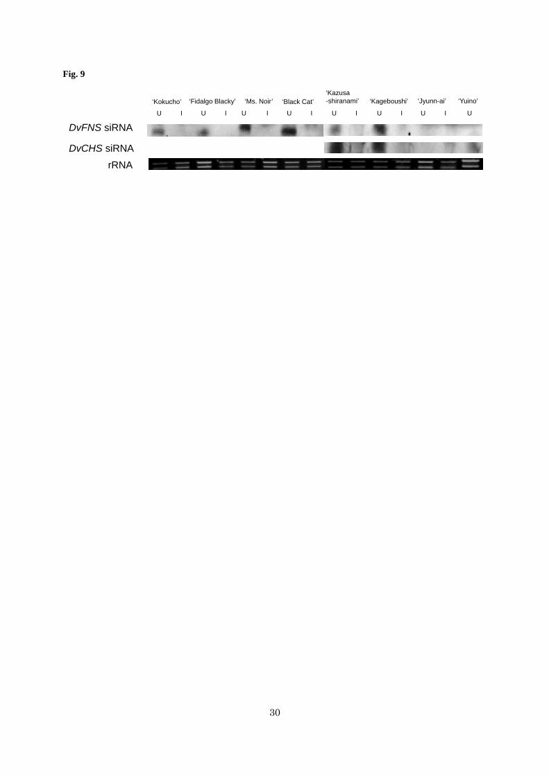
Table S5. Petal color described with CIE L*a*b*C* coordinates.
Figure S1. Purple ‘Kokucho’ flower with partially black petals occurring in summer
Figure S2. Comparison of growth between TSVdahlia-infected ‘Kokucho’ (line 4) and TSVdahlia-uninfected
‘Kokucho’ (line 5)
Figure S3. Detection of TSVdahlia in in vitro grafted plants
Figure S4. Relative transcript abundance of the genes involved in anthocyanidin synthesis in petals of TSVdahlia-
uninfected plants (original and graft control) and TSVdahlia-infected plants

15
Figure legends
Fig. 1
Virus and viroid detection in purple-flowering and original black-flowering ‘Kokucho’.
The cDNA from the leaves of five purple-flowering plants (P1–P5) and five black-flowering plants (B1–B5) was
used for PCR. Primers used to detect each virus or viroid are listed in Supplementary Table S1. Plasmids containing
the sequence of each virus or viroid were used as positive controls (PC) and water was used as a negative control
(NC). TSVdahlia, tobacco streak virus in dahlia; CMV, cucumber mosaic virus; DMV, dahlia mosaic virus; TSWV,
tomato spotted wilt virus; CSVd, chrysanthemum stunt viroid.
Fig. 2
Virus and viroid detection from lines regenerated from leaf-primordia-free shoot apical meristem (LP-free SAM)
cultures of purple-flowering ‘Kokucho’.
Primers used to detect each virus and viroid are listed in Supplementary Table S1. Leaf cDNA from each line was
used. Line 1 was regenerated from purple ‘Kokucho’ P5, line 2 and line 3 were regenerated from P4, line 4 and line
5 were regenerated from P3. Plasmids containing the sequence of each virus and viroid were used as positive
controls (PC). TSVdahlia, tobacco streak virus in dahlia; CMV, cucumber mosaic virus; DMV, dahlia mosaic virus;
TSWV, tomato spotted wilt virus; CSVd, chrysanthemum stunt viroid.
Fig. 3
Flavonoid aglycone contents (a) and relative transcript abundance of DvFNS (b) in petals of ‘Kokucho’ line 4
(purple) and line 5 (black), derived from leaf-primordia-free shoot apical meristem cultures.
All data represent averages of two biological replications. Completely colored, expanded petals were analyzed. The
transcript abundance of DvFNS in line 4 is relative to that in line 5. DvActin was used as the internal standard.
Fig. 4
Mapping of small RNAs of TSVdahlia-infected purple ‘Kokucho’ onto the DvFNS genomic sequence.
Small RNAs (18–32 nt long) with no mismatches to ‘Kokucho’ DvFNS genomic DNA (AB769841) were mapped
onto either the sense strand (above the X-axis) or the antisense strand (below the X-axis). The numbers of matched
reads are calculated per million total reads. The total number of matched reads after calculation is about 86 in
TSVdahlia-infected ‘Kokucho’ and 777 in TSVdahlia-uninfected ‘Kokucho’ (Deguchi et al. 2013). The specific 21 nt
antisense small RNA mapped in greatest number to position 2,307–2,327 of the DvFNS genomic sequence in
TSVdahlia-uninfected one (Deguchi et al. 2013) was not mapped at all in TSVdahlia-infected ‘Kokucho’.
Fig. 5
Flowers of TSVdahlia-uninfected original cultivars (a–g) and TSVdahlia-infected plants (h–n).
a and h, ‘Kokucho’; b and i, ‘Fidalgo Blacky’; c and j, ‘Ms. Noir’; d and k, ‘Black Cat’; e and l, ‘Kazusa-shiranami’;
f and m, ‘Kageboushi’; g and n, ‘Jyunn-ai’.

16
Fig. 6
Distributions of L* (lightness) and C* (chroma) values of TSVdahlia-uninfected (original and graft control) plants
and TSVdahlia-infected plants. (a) ‘Kokucho’; (b) ‘Fidalgo Blacky’; (c) ‘Ms. Noir’; (d) ‘Black Cat’; (e) ‘Kazusa-
shiranami’; (f) ‘Kageboushi’; (g) ‘Jyunn-ai’.
Each data point represents the average of 27 total values: 3 locations × 3 petals × 3 independent flowers. Bars
represent SE. The raw data are shown in Supplementary Table S5.
Fig. 7
Flavonoid aglycone contents of petals of TSVdahlia-uninfected (original and graft control) plants and TSVdahlia-
infected plants.
All data represent the average ± SE of each flavonoid (three flowers; the values for ‘Ms. Noir’ graft control and
‘Jyunn-ai’ original each represent two flowers). Completely colored, expanded petals were used. Although values
are presented as masses (mg), the same trends apply to molar masses because the molecular weights of pelargonidin,
cyanidin, apigenin, and luteolin are nearly equal, 271, 287, 270, and 286 g mol-1, respectively.
Fig. 8
Relative transcript abundance of DvFNS in the petals of TSVdahlia-uninfected (original and graft control) plants and
TSVdahlia-infected plants.
RNA was extracted from half-colored, unexpanded petals. All data points represent averages ± SE of three biological
replications (the values for ‘Kazusa-shiranami’ original, TSVdahlia-infected 1, 2 ,‘Jyunn-ai’ original and grafted
control represent two flowers). DvActin was used as the internal standard. Calculations were conducted using a
standard curve prepared from a diluted series of ‘Fidalgo Blacky’ TSVdahlia-infected 2 cDNA.
Fig. 9
RNA gel blot hybridization for DvFNS siRNA and DvCHS siRNA detection.
U, TSVdahlia-uninfected plant; I, TSVdahlia-infected plant. Ribosomal RNA on the electrophoresis gel is shown in the
lower photograph.
References
Asen S, Stewart RN, Norris KH (1972) Co-pigmentation of anthocyanins in plant tissues and its effect on color.
Phytochemistry 11:1139-1144
Ayabe S, Akashi T (2006) Cytochrome P450s in flavonoid metabolism. Phytochem Rev 5:271-282
Brigneti G, Voinnet O, Li WX, Ji LH, Ding SW, Baulcombe DC (1998) Viral pathogenicity determinants are
suppressors of transgene silencing in Nicotiana benthamiana. EMBO J 17:6739-6746

17
Chellappan P, Vanitharani R, Ogbe F, Fauquet CM (2005) Effect of temperature on geminivirus-induced RNA
silencing in plants. Plant Physiol 138:1828-1841
Clausen J (1930) Inheritance of variegation and of black flower colour in Viola tricolor L. Hereditas 13:342-356
Cornelissen BJ, Janssen H, Zuidema D, Bol JF (1984) Complete nucleotide sequence of tobacco streak virus RNA
3. Nucleic Acids Res 12:2427-2437
Davies KM, Bloor SJ, Spiller GB, Deroles SC (1998) Production of yellow colour in flowers: Redirection of
flavonoid biosynthesis in Petunia. Plant J 13:259-266
Deguchi A, Ohno S, Hosokawa M, Tatsuzawa F, Doi M (2013) Endogenous post-transcriptional gene silencing of
flavone synthase resulting in high accumulation of anthocyanins in black dahlia cultivars. Planta 237:1325-1335
Dekker EL, Derks AF, Asjes CJ, Lemmers ME, Bol JF, Langeveld SA (1993) Characterization of potyviruses
from tulip and lily which cause flower-breaking. J Gen Virol 74:881-887
Duan CG, Fang YY, Zhou BJ, Zhao JH, Hou WN, Zhu H, Ding SW, Guo HS (2012) Suppression of Arabidopsis
ARGONAUTE1-mediated slicing, transgene-induced RNA silencing, and DNA methylation by distinct domains
of the Cucumber mosaic virus 2b protein. Plant Cell 24:259-274
Fischer D, Stich K, Britsch L, Grisebach H (1988) Purification and characterization of (+)dihydroflavonol (3-
hydroxyflavanone) 4-reductase from flowers of Dahlia variabilis. Arch Biochem Biophys 264:40-47
Ghanekar A, Schwenk F (1974) Seed transmission and distribution of tobacco streak virus in six cultivars of
soybeans. Phytopathology 64:112-114
Ghoshal B, Sanfaçon H (2014) Temperature-dependent symptom recovery in Nicotiana benthamiana plants
infected with tomato ringspot virus is associated with reduced translation of viral RNA2 and requires
ARGONAUTE 1. Virology 456:188-97
Grotewold E (2006) The genetics and biochemistry of floral pigments. Annu Rev Plant Biol 57:761-780
Halbwirth H, Muster G, Stich K (2008) Unraveling the biochemical base of dahlia flower coloration. Nat Prod
Commun 3:1259-1266
Harborne JB, Greenham J, Eagles J (1990) Malonylated chalcone glycosides in Dahlia. Phytochemistry 29:2899-
2900

18
Hosokawa M, Otake A, Ohishi K, Ueda E, Hayashi T, Yazawa S (2004) Elimination of chrysanthemum stunt
viroid from an infected chrysanthemum cultivar by shoot regeneration from a leaf primordium-free shoot apical
meristem dome attached to a root tip. Plant Cell Rep 22:859-63
Hosokawa M (2008) Leaf Primordia-Free Shoot Apical Meristem Culture: A New Method for Production of
Viroid-Free Plants. J Jpn Soc Hortic Sci 77:341-349
Hunter DA, Fletcher JD, Davies KM, Zhang H (2011) Colour break in reverse bicolour daffodils is associated
with the presence of Narcissus mosaic virus. Virol J 8:412
Johnson H, Converse R, Amorao A, Espejo J, Frazier N (1984) Seed transmission of tobacco streak virus in
strawberry. Plant Dis 68:390-392
Lesnaw JA, Ghabrial SA (2000) Tulip breaking: Past, present, and future. Plant Dis 84:1052-1060
Markham K, Bloor SJ, Nicholson R, Rivera R, Shemluck M, Kevan PG, Michener C (2004) Black flower
coloration in wild Lisianthius nigrescens: its chemistry and ecological consequences. Zeitschrift fur
Naturforschung C 59:625-630
Martens S, Mithöfer A (2005) Flavones and flavone synthases. Phytochemistry 66:2399-2407
Murashige T, Skoog F (1962) A revised medium for rapid growth and bio assays with tobacco tissue cultures.
Physiol Plantarum 15:473-497
Naka T, Fujii Y, Hosokawa M, Nakajima A, Asao H, Okada K, Maeda S (2007) Effect of apical meristem culture
propagation of Dahlia (Dahlia×cultorum) on the growth and viruliferous level. Bull Nara Agr Exp Sta 38:17-22
Nordström C, Swain T (1953) The flavonoid glycosides of Dahlia variabilis. Part I. General introduction.
Cyanidin, apigenin, and luteolin glycosides from the variety “Dandy.”. J Chem Soc :2764-2773
Nordström C, Swain T (1956) The flavonoid glycosides of Dahlia variabilis. II. Glycosides of yellow varieties
“Pius IX” and “Coton”. Arch Biochem Biophys 60:329-344
Nordström C, Swain T (1958) The flavonoid glycosides of Dahlia variabilis. III. Glycosides from white varieties.
Arch Biochem Biophys 73:220-223
Ohno S, Deguchi A, Hosokawa M, Tatsuzawa F, Doi M (2013) A basic helix-loop-helix transcription factor
DvIVS determines flower color intensity in cyanic dahlia cultivars. Planta 238:331-343

19
Ohno S, Hosokawa M, Hoshino A, Kitamura Y, Morita Y, Park KI, Nakashima A, Deguchi A, Tatsuzawa F, Doi
M, Iida S, Yazawa S (2011a) A bHLH transcription factor, DvIVS, is involved in regulation of anthocyanin
synthesis in dahlia (Dahlia variabilis). J Exp Bot 62:5105-5116
Ohno S, Hosokawa M, Kojima M, Kitamura Y, Hoshino A, Tatsuzawa F, Doi M, Yazawa S (2011b)
Simultaneous post-transcriptional gene silencing of two different chalcone synthase genes resulting in pure white
flowers in the octoploid dahlia. Planta 234:945-958
Padmanabhan C, Gao S, Li R, Zhang S, Fei Z, Ling KS (2014) Complete genome sequence of an emerging
genotype of tobacco streak virus in the United States. Genome Announc 2:e01138-14.
doi:10.1128/genomeA.01138-14.
Schlangen K, Miosic S, Thill J, Halbwirth H (2010a) Cloning, functional expression, and characterization of a
chalcone 3-hydroxylase from Cosmos sulphureus. J Exp Bot 61:3451-3459
Schlangen K, Miosic S, Halbwirth H (2010b) Allelic variants from Dahlia variabilis encode flavonoid 3′-
hydroxylases with functional differences in chalcone 3-hydroxylase activity. Arch Biochem Biophys 494:40–45
Scott S, Zimmerman M, Ge X (1998) The sequence of RNA 1 and RNA 2 of tobacco streak virus: additional
evidence for the inclusion of alfalfa mosaic virus in the genus Ilarvirus. Arch Virol 143:1187-1198
Senda M, Masuta C, Ohnishi S, Goto K, Kasai A, Sano T, Hong JS, MacFarlane S (2004) Patterning of virus-
infected glycine max seed coat is associated with suppression of endogenous silencing of chalcone synthase
genes. Plant Cell 16:807-818
Sharman M, Thomas J (2013) Genetic diversity of subgroup 1 ilarviruses from eastern Australia. Arch Virol
158:1637-1647
Sharman M, Persley DM, Thomas JE (2009) Distribution in Australia and seed transmission of Tobacco streak
virus in Parthenium hysterophorus. Plant Dis 93:708-712
Shimura H, Masuta C, Yoshida N, Sueda K, Suzuki M (2013) The 2b protein of Asparagus virus 2 functions as an
RNA silencing suppressor against systemic silencing to prove functional synteny with related cucumoviruses.
Virology 442:180-188
Szittya G, Silhavy D, Molnár A, Havelda Z, Lovas Á, Lakatos L, Bánfalvi Z, Burgyán J (2003) Low temperature
inhibits RNA silencing‐mediated defence by the control of siRNA generation. EMBO J 22:633-640
Takeda A, Sugiyama K, Nagano H, Mori M, Kaido M, Mise K, Tsuda S, Okuno T (2002) Identification of a novel
RNA silencing suppressor, NSs protein of Tomato spotted wilt virus. FEBS Lett 532:75-79

20
Tanaka Y, Brugliera F (2013) Flower colour and cytochromes P450. Phil Trans R Soc B 368:1612
Tanaka Y, Sasaki N, Ohmiya A (2008) Biosynthesis of plant pigments: anthocyanins, betalains and carotenoids.
Plant J 54:733-749
Tatsuzawa F, Saito N, Toki K, Shinoda K, Honda T (2012) Flower colors and their anthocyanins in matthiola
incana cultivars (Brassicaceae). J Jpn Soc Hortic Sci 81:91-100
Teycheney PY, Tepfer M (2001) Virus-specific spatial differences in the interference with silencing of the chs-A
gene in non-transgenic petunia. J Gen Virol 82:1239-1243
Thill J, Miosic S, Ahmed R, Schlangen K, Muster G, Stich K, Halbwirth H (2012) 'Le Rouge et le Noir': a decline
in flavone formation correlates with the rare color of black dahlia (Dahlia variabilis hort.) flowers. BMC Plant
Biol 12:225
Wimmer G, Halbwirth H, Wurst F, Forkmann G, Stich K (1998) Enzymatic hydroxylation of 6'-deoxychalcones
with protein preparations from petals of Dahlia variabilis. Phytochemistry 47:1013-1016

21
Tables and Figures
Table 1. Petal color described with lightness (L *) and chroma (C *).
L *(Lightness) 14.8 ± 0.3 18.3 ± 6.1 15.3 15.0 15.0 18.4 14.8C *(Chroma) 88.2 ± 0.8 117.3 ± 1.2 89.7 87.6 93.5 121.2 83.7Flower color
Black 'Kokucho'(Original) Purple 'Kokucho'
Individuals regenerated by leaf primordia-free shoot apicalmeristem (LP-free SAM) culture from purple 'Kokucho'1 2 3 4 5
Data of black and purple 'Kokucho' represent the average value ± SE of five plants each (B1-B5, P1-P5). Data of LP-free SAMcultured lines represent the average of two biological replications of each line (1-5).
BlackBlack Purple Black Black Black Purple

22
Fig. 1
TSVdahlia
CMV
DMV
TSWV
CSVd
PC NC
BlackPurple
P1 P2 P3 P4 P5 B1 B2 B3 B4 B5

23
Fig. 2
PC 1 2 3 4 5
Regenerated ‘Kokucho’ lines
TSVdahlia
CMV
DMV
TSWV
CSVd

24
Fig. 3
0.0
1.0
2.0
3.0
4.0
5.0
6.0
7.0
8.0
line 4 line 5
Rel
ativ
e tra
nscr
ipt a
bund
ance
of D
vFN
S
LP-free SAM cultured line
0.0
0.5
1.0
1.5
2.0
2.5
3.0
line 4 line 5
Am
ount
of e
ach
flavo
noid
agl
ycon
e co
unta
ined
in 1
00 m
g fre
sh p
etal
(m
g)
LP-free SAM cultured line
PelargonidinCyanidinApigeninLuteolin
(a) (b)

25
Fig. 4
0
2
4
6
-6
-4
-2
0
Num
bero
fmat
ched
read
spe
r milli
onre
ads
Exon 1 Exon 2Intron
100 bp

26
Fig. 5
a b c d e f g
h i j k l m n

27
Fig. 6
0
10
20
30
105 115 125 135
L*
C*
‘Black Cat’
TSVdahlia-infectedgraft controloriginal
0
10
20
30
90 100 110 120
L*C*
‘Fidalgo Blacky’
TSVdahlia-infectedgraft controloriginal
50
60
70
80
30 40 50 60
L*
C*
‘Jyunn-ai’
TSVdahlia-infectedgraft controloriginal
0
10
20
30
100 110 120 130
L*
C*
‘Kokucho’
TSVdahlia-infectedgraft controloriginal
0
10
20
30
100 110 120 130
L*
C*
‘Ms. Noir’
TSVdahlia-infectedgraft controloriginal
10
20
30
40
95 105 115 125
L*
C*
‘Kageboushi’
TSVdahlia-infectedoriginal
0
10
20
30
100 110 120 130
L*
C*
‘Kazusa-shiranami’
TSVdahlia-infectedgraft controloriginal
(a) (b) (c)
(d) (e) (f)
(g)

28
Fig. 7
0.0
0.5
1.0
1.5
2.0
Am
ount
of e
ach
flavo
noid
agl
ycon
e co
unta
ined
in 1
00 m
g fre
sh p
etal
(m
g)
LuteolinApigeninCyanidinPeralgonidin
'Kokucho' 'Fidalgo Blacky' 'Ms. Noir' 'Black Cat' 'Kazusa-shiranami' 'Kageboushi' 'Jyunn-ai'

29
Fig. 8
0.0
0.5
1.0
1.5
2.0
2.5
3.0
DvF
NS
/ D
vAct
in
DvFNS originalgraft controlTSVdahlia-infected 1TSVdahlia-infected 2
30
Fig. 9
DvFNS siRNA
rRNADvCHS siRNA
‘Fidalgo Blacky’‘Kokucho’ ‘Ms. Noir’ ‘Black Cat’‘Kazusa-shiranami’ ‘Kageboushi’ ‘Jyunn-ai’
U I U I U I U I U I U I U I
‘Yuino’
U

31
Supplementary Tables and Figures
Tobacco streak virus (strain dahlia) suppresses post-transcriptional gene silencing of flavone synthase II in
black dahlia cultivars and causes a drastic flower color change
Ayumi Deguchi1, Fumi Tatsuzawa2, Munetaka Hosokawa1, Motoaki Doi1 and Sho Ohno1*
1 Graduate School of Agriculture, Kyoto University, Sakyo-ku, Kyoto 606-8502, Japan
2 Faculty of Agriculture, Iwate University, Morioka 020-8550, Japan
*Corresponding author: Sho Ohno
Laboratory of Vegetable and Ornamental Horticulture, Graduate School of Agriculture, Kyoto University, Sakyo-
ku, Kyoto 606-8502, Japan
Telephone: +81-75-753-6048, Fax: +81-75-753-6068
E-mail: [email protected]

32
Supplementary Fig. S1
Purple ‘Kokucho’ flowers with partially black petals occurring in summer.
Arrows indicate black mosaic regions in purple petals.
Supplementary Fig S2.
Comparison of growth parameters between TSVdahlia-infected ‘Kokucho’ (line 4) and TSVdahlia-uninfected ‘Kokucho’
(line 5). (a) plant height at four time points; (b) node number per plant at three time points; (c) diameter of the first-
opened inflorescence; (d) weight of tuberous roots per plant measured on December 29, 2014. The rooted cuttings
of both lines were transplanted to 24 cm pots on July 11, 2014. Nine plants from each line were grown in a
greenhouse. All data represent the average ± SE. No significant differences (P > 0.05) were observed for any
parameter, according to t-test.
Supplementary Fig. S3
Detection of TSVdahlia in in vitro grafted plants.
cDNA from half-colored, unexpanded petals from each plant was used. O, original cultivars without grafting; GC,
graft controls (plants grafted onto TSVdahlia-uninfected ‘Kokucho’ line 5); TSV1 and TSV2, plants grafted onto
TSVdahlia-infected ‘Kokucho’ line 4 independently. Plasmid containing the full sequence of TSV CP was used as a
positive control (PC) and water served as a negative control (NC). Primers used are listed in Table S1.
Supplementary Fig. S4
Relative transcript abundance of the genes involved in anthocyanidin synthesis in petals of TSVdahlia-uninfected
(original and graft control) plants and TSVdahlia-infected plants. (a) DvIVS, (b) DvCHS1, (c) DvCHS2, (d) DvCHI,
(e) DvF3H, (f) DvDFR, (g) DvANS, and (h) DvF3′H
RNA was extracted from half-colored, unexpanded petals for DvF3′H and from completely colored, unexpanded
petals for the other genes. Petal bases of ‘Kazusa-shiranami’ and ‘Kageboushi’ corresponded to the colored parts of
TSVdahlia-uninfected petals and the tips corresponded to the uncolored parts. All data points represent the average of
two or three biological replications, excluding some abnormal scores. Standard error bars are applied to the data
with three biological replications. DvActin was used as the internal standard. Calculation was conducted using a
standard curve prepared from a serial dilution of ‘Kokucho’ TSVdahlia-infected 2 cDNA and ‘Fidalgo Blacky’
TSVdahlia-infected 2 cDNA.

33
Supplementary Table S1. Primer sets for virus and viroid detection
Virus / Viroid sense (5'-3') antisense (5'-3')TSVdahlia CCCATAATACCGTGAACACT CCTGTTACTCCATCAACCATCMV TATTACCCTAAAGCCACCAA GTTAGCTTGGACTCCAGATGDMV AAAAAGAGGCTACCATACCC ACTTCCTGCTAGGACACTCATSWV GTCAGGGGACAATAACTG CTGCTTCTCACTGTTTCCINSV AAATCAATAGTAGCATTAAA CTTCCTCAAGAATAGGCAATCSVd CAACTGAAGCTTCAACGCCTT AGGATTACTCCTGTCTCGCAPSTVd TAAACTCGTGGTTCCTGTGG GCCCCGAAGCAAAGAAAGAT

34
Name Orientation Sequense (5'-3')TSV-RNA2 Full-R2 antisense GCATCTCCATTTGGAGGCTSV-RNA3-Full-R2 antisense GCATCTCCTATAAAGGAGGCATCAGTAGTSV-2b-F1 sense GTTTGTCGAAGAACCGCTSV-2b-R1 antisense AAAGCGGCATCTCTCTSV-CP-F1 sense GCGAAGGCGTCGTTGAGGTTTSV-CP-R1 antisense GATTTCGGGAATCCCCTCGACTSV-MP-F sense ATGGCGTTAGTACCAACGATGTSV-MP-R antisense TCAGGCTGAAAGCAGGTTCC
Supplementary Table S2. Primers for sequence analysis of TSVdahlia

35
Supplementary Table S3. Primers designed for real-time PCRGene Orientation Sequense (5'-3')
DvActin antisense GCTGACAGGATGAGCAAGsense TCCACATCTGTTGGAAGG
DvF3'H antisense CATAACTGCCTTACTATTGTACsense GTAGTTATACGCAATATGCTC

36
L* a* b* C*line 4 (TSVdahlia-infected) 43.32 ± 0.46 6.11 ± 0.28 13.69 ± 0.51 15.03 ± 0.45line 5 (TSVdahlia-uninfected) 44.49 ± 0.48 5.34 ± 0.46 15.48 ± 0.71 16.50 ± 0.51
Supplementary Table S4. Leaf color described with CIE L*a*b*C* coordinates.
n = 9. All values represent the average ± SE of three point in the uppermost leaf at 1st flower opening.
37
original graft control
TSV
dahlia -infected 1 TSV
dahlia -infected 2 original
graft control TS
Vdahlia -infected 1 TS
Vdahlia -infected 2
‘Kokucho’
15.38 ± 0.3815.18 ± 0.17
20.22 ± 0.8220.38 ± 0.48
107.32 ± 3.23106.63 ± 1.53
122.57 ± 0.73125.44 ± 4.18
‘Fidalgo Blacky’
15.99 ± 0.1315.79 ± 0.45
16.79 ± 0.3717.72 ± 0.47
92.26 ± 1.6599.98 ± 1.42
113.55 ± 3.97114.26 ± 1.54
‘Ms. N
oir’15.35 ± 0.60
15.88 ± 0.6720.32 ± 0.92
21.10 ± 0.38105.90 ± 3.10
102.26 ± 3.24121.03 ± 1.56
130.38 ± 0.72‘B
lack Cat’
16.45 ± 0.4418.86 ± 1.17
20.74 ± 0.4924.38 ± 1.51
113.89 ± 1.75114.26 ± 1.55
122.86 ± 0.62119.69 ± 4.13
‘Kazusa-shiranam
i’17.40 ± 0.04
16.27 ± 0.1621.31 ± 0.61
20.07 ± 0.23115.01 ± 1.34
111.70 ± 1.04121.72 ± 1.99
121.54 ± 2.85‘K
ageboushi’19.73 ± 0.91
-32.38 ± 1.13
-118.07 ± 1.56
-97.59 ± 1.27
-
‘Jyunn-ai’65.34 ± 1.34
64.22 ± 1.8664.53 ± 1.89
62.22 ± 4.0144.96 ± 1.55
49.93 ± 3.8646.36 ± 4.42
53.82 ± 6.50
original graft control
TSV
dahlia -infected 1 TSV
dahlia -infected 2 original
graft control TS
Vdahlia -infected 1 TS
Vdahlia -infected 2
‘Kokucho’
107.13 ± 3.20106.49 ± 1.53
122.17 ± 0.73124.84 ± 4.16
6.35 ± 0.535.39 ± 0.08
9.23 ± 2.0312.06 ± 1.26
‘Fidalgo Blacky’
92.17 ± 1.6499.87 ± 1.40
113.52 ± 3.98114.21 ± 1.57
3.95 ± 0.404.61 ± 0.35
2.86 ± 0.252.43 ± 1.28
‘Ms. N
oir’105.77 ± 3.09
102.08 ± 3.26120.53 ± 1.55
129.73 ± 0.715.28 ± 0.43
6.11 ± 0.1911.02 ± 0.39
12.85 ± 1.08‘B
lack Cat’
113.64 ± 1.70113.78 ± 1.58
122.38 ± 0.67118.92 ± 4.35
7.55 ± 0.7110.36 ± 0.89
10.81 ± 0.6812.88 ± 2.12
‘Kazusa-shiranam
i’114.69 ± 1.29
111.43 ± 1.01121.08 ± 2.04
121.16 ± 2.838.52 ± 0.80
7.79 ± 0.3911.94 ± 1.92
9.34 ± 1.15‘K
ageboushi’117.49 ± 1.49
-94.94 ± 1.05
-11.38 ± 1.59
-22.54 ± 1.06
-
‘Jyunn-ai’43.00 ± 1.82
48.35 ± 3.8844.50 ± 4.56
52.31 ± 6.53-13.01 ± 0.66
-12.41 ± 0.70-12.83 ± 0.15
-12.53 ± 0.78
All data represent the average±SE
of three independent flowers. C
* = (a*2+
b*2) 1/2
Supplem
entary Table S5. P
etal color described with C
IE L*a*b*C
* coordinates
Cutivar
L*C
*
Cutivar
a*b*

38
Supplementary Fig. S1

39
Supplementary Fig. S2
0
20
40
60
80
100
120
140
160
11 Aug. 11 Sep. 6 Oct. 1st floweropening
Pla
nt h
eigh
t (cm
)
Measurement date
line 4 (TSVdahlia-infected)line 5 (TSVdahlia-uninfected)
02468
101214161820
11 Aug. 11 Sep. 6 Oct.
Nod
e nu
mbe
r per
pla
nt
Measurement date
line 4 (TSVdahlia-infected)line 5 (TSVdahlia-uninfected)
0
2
4
6
8
10
12
line 4 line 5
Dia
met
er o
f the
firs
t ope
nd
inflo
resc
ence
(cm
)
0
20
40
60
80
100
120
line 4 line 5
Wei
ght o
f tub
erou
s ro
ots
per p
lant
(g)
(a) (b)
(c) (d)

40
Supplementary Fig. S3
O GC TSV1 TSV2NCPC
‘Kokucho’
‘Fidalgo Blacky’
‘Ms. Noir’
‘Black Cat’
‘Kazusa-shiranami’
‘Kageboushi’
‘Jyunn-ai’

41
Supplementary Fig. S4
0.01.02.03.04.05.06.0
DvI
VS
/DvA
ctin
DvIVS originalgraft controlTSVdahlia-infected 1TSVdahlia-iinfected 2
0.01.02.03.04.05.06.0
DvC
HS
1 / D
vAct
in
DvCHS1originalgraft controlTSVdahlia-infected 1TSVdahlia-iinfected 2
0.04.08.0
12.016.020.024.028.0
DvC
HS
2 / D
vAct
in
DvCHS2 originalgraft controlTSVdahlia-infected 1TSVdahlia-iinfected 2
0.01.02.03.04.05.06.07.08.09.0
DvC
HI /
DvA
ctin
DvCHIoriginalgraft controlTSVdahlia-infected 1TSVdahlia-iinfected 2
0.00.10.20.30.40.50.60.70.80.91.0
DvD
FR /
DvA
ctin
DvDFR originalgraft controlTSVdahlia-infected 1TSVdahlia-iinfected 2
0.01.02.03.04.05.06.0
DvA
NS
/ D
vAct
in
DvANS originalgraft controlTSVdahlia-infected 1TSVdahlia-iinfected 2
0.00.20.40.60.81.01.21.41.6
DvF
3H /
DvA
ctin
DvF3H originalgraft controlTSVdahlia-infected 1TSVdahlia-iinfected 2
(a) (b)
(c) (d)
(e) (f)
(g)
0.000.020.040.060.080.100.120.14
DvF
3'H
/ D
vAct
in
DvF3'H originalgraft controlTSVdahlia-infected 1TSVdahlia-iinfected 2
(h)