Embed Size (px)
Citation preview
Can Respir J Vol 12 No 1 January/February 2005 13
Toll-like receptors 4 and 2 expression in the bronchialmucosa of patients with cystic fibrosis
Hans-Peter Hauber MD1*, Meri K Tulic PhD1*, Anne Tsicopoulos MD2, Benoit Wallaert MD2,
Ron Olivenstein MD1, Patrick Daigneault MD1, Qutayba Hamid MD PhD1
1Meakins-Christie Laboratories, McGill University, Montreal, Quebec; 2U416 Institut National de la Santé et de la Recherche Médicale, Institut Pasteur de Lille and Centre de Soins Mucoviscidose Adulte, Hôpital Calmette, Lille, France
*HPH and MKT contributed equally to this workCorrespondence: Dr Qutayba Hamid, Meakins-Christie Laboratories, McGill University, 3626 St-Urbain Street, Montreal, Quebec H2X 2P2.
Telephone 514-398-3864, fax 514-398-7483, e-mail [email protected]
H-P Hauber, MK Tulic, A Tsicopoulos, et al. Toll-likereceptors 4 and 2 expression in the bronchial mucosa ofpatients with cystic fibrosis. Can Respir J 2005;12(1):13-18.
BACKGROUND: Cystic fibrosis (CF) is a lung disease character-ized by chronic infection with Gram-negative bacteria Pseudomonas
aeruginosa and Gram-positive bacteria Staphylococcus aureus.Recently, toll-like receptor (TLR) 4 has been shown to be responsi-ble for the lipopolysaccharide (LPS)-mediated immune response.While TLR2 mediates responses driven by bacterial lipoproteins andpeptidoglycans from Gram-positive bacteria, LPS derived fromP aeruginosa may stimulate the immune response in the airways ofpatients with CF via activation of TLR4.OBJECTIVES: To investigate TLR4 and TLR2 expression in thebronchial mucosa of patients with CF and normal control subjects.PATIENTS AND METHODS: Endoscopic bronchial biopsies fromseven patients with CF and six healthy control subjects wereobtained. TLR4 and TLR2 expression was assessed using immmuno-cytochemistry. Real-time polymerase chain reaction was used todetect TLR4 messenger RNA in blood cells from patients with CFand to compare TLR4 expression in CF bronchial epithelial cellswith non-CF bronchial epithelial cells.RESULTS: In patients with CF, the number of TLR4-positive cells wassignificantly increased in their submucosa (P<0.05) but significantlyreduced in their epithelium compared with control subjects (P<0.05).The majority of TLR4-positive cells were neutrophils. Patients with CF(n=4) and control subjects (n=4) had a similar percentage of TLR4-expressing neutrophils and monocytes/lymphocytes in peripheralblood. CF cells (IB-3) had significantly decreased basal TLR4 messen-ger RNA expression compared with non-CF cells (Calu-3) (P<0.05).Although there was a trend toward reduced TLR2 expression in theairway epithelium of patients with CF (P=0.07), there was no signifi-cant difference in TLR2 expression in the submucosa of patients withCF compared with that of control subjects.CONCLUSIONS: Both TLR4 and TLR2 expression in the bronchialepithelium of patients with CF were significantly reduced comparedwith healthy control subjects. In contrast, the number of TLR4-positiveneutrophils in the submucosa of patients with CF was higher than incontrol subjects. This may suggest that the loss of epithelial TLRexpression may contribute to the impaired defense against LPS.
Key Words: Cystic fibrosis; Immunocytochemistry; Neutrophils;
TLR2; TLR4
L’expression des récepteurs ressemblant à unpéage 4 et 2 dans la muqueuse bronchique depatients atteints de fibrose kystique
HISTORIQUE : La fibrose kystique (FK) est une maladie pulmonaire car-actérisée par une infection chronique à la bactérie gram-négativePseudomonas aeruginosa et à la bactérie gram-positive Staphylococcus aureus.Récemment, il a été démontré que le récepteur ressemblant à un péage(TLR) 4 est responsable de la réponse immunitaire à médiation glucidolipi-doprotéique (LPS). Tandis que les TLR2 assurent la médiation des répons-es poussées par les lipoprotéines bactériennes et les peptidoglycanes desbactéries gram-positives, des complexes LPS dérivés de P aeruginosa peu-vent stimuler la réponse immunitaire dans les voies respiratoires despatients atteints de FK par l’activation du TLR4.OBJECTIFS : Étudier l’expression du TLR4 et du TLR2 dans la muqueusebronchique de patients atteints de FK et de sujets témoins en santé.PATIENTS ET MÉTHODOLOGIE : Des biopsies bronchiques parendoscopie ont été obtenues auprès de sept patients atteints de FK et de sixsujets témoins en santé. L’expression du TLR4 et du TLR2 a été évaluée aumoyen d’une immunocytochimie. Une réaction en chaîne de la polyméraseen temps réel a permis de déceler l’ARN messager du TLR4 dans les glob-ules des patients atteints de FK et de comparer l’expression du TLR4 dansles cellules épithéliales bronchiques de FK à celle des cellules épithélialesbronchiques sans FK.RÉSULTATS : Chez les patients atteints de FK, le nombre de cellulesTLR4-positives était considérablement plus élevé dans la sous-muqueuse(P<0,05) mais considérablement réduit dans l’épithélium par rapport àcelui des sujets témoins (P<0,05). La majorité des cellules TLR4-positivesétaient des neutrophiles. Des patients atteints de FK (n=4) et des sujetstémoins (n=4) présentaient un pourcentage similaire de neutrophiles expri-mant des TLR4 et de monocytes et lymphocytes dans le sang périphérique.Les cellules de FK (IB-3) présentaient une expression d’ARN messager duTLR4 de base beaucoup moins élevée que les cellules sans FK (Calu-3)(P<0,05). Bien qu’on ait remarqué une tendance vers une expressionréduite du TLR2 dans l’épithélium des voies respiratoires des patientsatteints de FK (P=0,07), on n’a décelé aucune différence marquée de l’ex-pression du TLR2 dans la sous-muqueuse des patients atteints de FK parrapport à celle des sujets témoins.CONCLUSIONS : L’expression tant du TLR4 que du TLR2 dans l’ép-ithélium bronchique des patients atteints de FK était beaucoup plus faibleque celle des sujets témoins en santé. Par contre, le nombre de neutrophilesTLR4-positifs dans la sous-muqueuse des patients atteints de FK était plusélevé que celui des sujets témoins. Ce phénomène pourrait indiquer que laperte de l’expression du TLR épithélial contribue à l’altération de ladéfense contre le complexe LPS.
©2005 Pulsus Group Inc. All rights reserved
ORIGINAL ARTICLE
Hauber.qxd 2/4/2005 1:36 PM Page 13
Cystic fibrosis (CF) is an autosomal recessive disordercaused by mutations in the cystic fibrosis transmembrane
conductance regulator (CFTR) gene encoding a chloridechannel located in the airway epithelial cells (1,2). Althoughpathological changes are observed in the pancreas, liver andreproductive tract, the most profound changes occur in thelungs. In the lungs, CF is characterized by mucus overproduc-tion and neutrophil-dominated airway inflammation, whichare driven by intense chronic infection with the Gram-negativebacteria Pseudomonas aeruginosa (1,2). These pathologicalchanges destroy the airways, impair gas exchange and are themain cause of mortality among patients with CF. Anotherimportant pathogen in CF is the Gram-positive Staphylococcusaureus, which often appears early in the course of CF lung dis-ease (1). Infection with S aureus is also associated withincreased neutrophilia and macrophages in bronchoalveolarlavage, as well as with significant elevation of interleukin-8(3). Currently, the susceptibility of CF airways to S aureus andP aeruginosa is not fully understood.
Recently discovered toll-like receptors (TLRs) are a familyof evolutionary conserved receptors that mediate the initialphase of innate immunity via recognition of pathogen-associatedmolecular patterns. To date, TLRs 1 to 10 have been identified(4,5). TLR2 has been shown to recognize bacterial lipoproteinsand peptidoglycan from Gram-positive bacteria, while TLR4 isa receptor for Gram-negative bacterial lipopolysaccharide(LPS) (6-8). In humans, mutations in the TLR4 gene are asso-ciated with LPS hyporesponsiveness (9). Because the airwaysof patients with CF are chronically infected with Gram-positive and -negative bacteria, we postulate that TLR2 andTLR4 may play an important role in mediating host-defenseresponses in these patients.
The expression and role of TLR2 and TLR4 in patients withCF have not been evaluated. Although a recent study (10) hasdemonstrated TLR2 and TLR4 expression in CF epithelium, thein vitro experiments were performed using CF epithelial celllines. The expression and regulation of TLRs in vivo in thebronchial mucosa and peripheral blood of patients with CF com-pared with those of healthy individuals is currently unknown.Furthermore, the phenotype of the cells that express thesereceptors in patients with CF remains to be elucidated. Here, wedescribe, for the first time, a difference in TLR4 and TLR2expression among patients with CF compared with control sub-jects, and propose some potential insights into the differentialexpression and regulation of these receptors in patients with CF.
PATIENTS AND METHODSSubjectsThis study was approved by the Montreal Chest Institute
(Montreal, Quebec) and the Ethics Committe of the Calmette
Hospital (Lille, France). Endoscopic bronchial biopsies were taken
from seven patients with CF and six healthy control subjects.
Three to four mucosal biopsies from each patient were taken from
the subcarinae of the lower lobes or middle lobe of the lung. In
addition, blood was taken from a different set of patients with CF
(n=4) and control subjects (n=4). Table 1 summarizes the demo-
graphics of the two groups. Forced vital capacity and forced expi-
ratory volume in 1 s were significantly lower in patients with CF
than in control subjects (P<0.05), reflecting the reduced lung
function in patients with CF (Table 1). Sputum cultures showed
S aureus in one CF patient and P aeruginosa in six patients with CF.
Age and sex distribution between the two groups were similar.
Tissue and blood sample preparationThree to four mucosal biopsies were obtained from the subcarinae of
the lower lobes or middle lobe of each patient. All biopsy specimens
were fixed with 4% paraformaldehyde, blocked in optimal cutting
temperature medium and snap frozen in liquid nitrogen-cooled
isopentane. Sections (5 µm to 10 µm thick) were mounted on poly-
L-lysine (0.1%)-coated glass slides and baked overnight at 37°C.
Peripheral blood was diluted (1:1) with sterile phosphate buffer
solution (PBS) and centrifugated over Ficoll Paque (Amersham
Biosciences, Sweden). The interphase, containing mononuclear
cells, was removed and washed twice with PBS to obtain peripheral
blood mononuclear cells (PBMs). To isolate polymorphonuclear
cells (PMNs), the phase containing PMNs and red blood cells was
mixed with 10% dextran (Sigma-Aldrich, USA). After a 30 min
incubation at room temperature, the supernatant was removed, and
the remaining interphase was diluted with PBS (1:1) and centrifu-
gated at 1600 rpm for 10 min. The supernatant was discarded and
the cell pellet was resuspended in hypotonic NaCl solution (0.2%)
to lyse the remaining red blood cells. Hypertonic NaCl solution
(1.6%) was then added to restore normotonic osmolarity. After a
second centrifugation, the supernatant was discarded and the cell
pellet containing PMNs was resuspended in PBS. Routinely, neu-
trophils made up more than 90% of PMNs. PMN cytospins were
prepared (100,000 cells/slide), and the slides were fixed in an
acetone:methanol solution (60:40).
ImmunocytochemistryTLR2 and TLR4 immunoreactivity was assessed by alkaline phos-
phatase antialkaline phosphatase immunocytochemistry as previ-
ously described (11), using polyclonal primary antibodies against
TLR2 (goat antihuman TLR2, 1:200; Santa Cruz Biotechnology,
USA) or against TLR4 (rabbit antihuman TLR4, 1:10; Santa Cruz
Biotechnology). Briefly, after applying blocking solution (DAKO
Protein Block; DAKO, Denmark) to prevent nonspecific binding,
the sections were incubated with primary antibody overnight at 4°C.
The next day, the slides were incubated with the secondary antibody
for 30 min (rabbit antigoat-immunoglobulin biotin for TLR2 or
swine antirabbit-immunoglobulin biotin for TLR4), washed and ter-
tiary antibody (Streptavidin-AP; Dako, Denmark) was added for a
further 30 min. Immunostaining was developed with Fast Red
(Sigma Chemicals, Canada). All slides were counterstained with
Gill II hematoxylin (Dako, Denmark). Positive cells appeared red
under bright field illumination. For negative controls, the primary
antibody was replaced by an isotype-matched control antibody.
Double alkaline phosphatase antialkaline phosphatase immuno-
cytochemistry was used to examine whether neutrophils or
macrophages expressed TLR4 in the bronchial mucosa. Briefly,
slides were incubated overnight at 4°C with a mixture of primary
Hauber et al
Can Respir J Vol 12 No 1 January/February 200514
TABLE 1Clinical characteristics of the total study population
Patients with CF Control subjects(n=11)* (n=10)† P
Mean age (years) 34.6±5.1 33.8±4.6 NS
Sex (male:female) 5:6 7:3
Forced vital capacity (L) 2.5±0.2 5.2±0.2 <0.01
Forced vital capacity (%) 70.9±5.5 106.3±5.4 <0.01
FEV1 (L) 1.7±0.1 4.2±0.2 <0.01
FEV1 (%) 56.9±6.6 104.2±5.5 <0.01
*Seven patients with biopsy and four with blood; †Six patients with biopsy andfour with blood. Results represented as mean ± SEM. CF Cystic fibrosis;FEV1 Forced expiratory volume in 1 s; NS Not significant
Hauber.qxd 2/9/2005 3:49 PM Page 14
monoclonal antibody directed against elastase (1:100) or CD68(1:50) (mouse antihuman, DAKO) with polyclonal antibodydirected against TLR4 (1:10). Secondary and tertiary antibodieswere applied at different incubation periods, each for 30 min at roomtemperature. Immunostaining was developed with Fast Red (SigmaChemicals), and all slides were counterstained with Gill II hema-toxylin (Dako). Elastase- or CD68-positive cells appeared red, andelastase/TLR4 or CD68/TLR4 double positive cells appeared as red-brown immunostaining. Immunocytochemistry was also performedon isolated neutrophils and PBMs from the peripheral blood smearsof patients with CF and control subjects to examine TLR4 expression.
QuantificationAll cells in individual biopsy specimens were counted by twoobservers, who were blinded to the conditions, using an Olympuslight microscope (Carson Group Inc, Canada) at ×200 magnifica-tion with an eyepiece graticule of 0.202 mm2. At least two sectionswere counted for each subject, and the counts were averaged. Ineach section, the number of positive cells was averaged from six toeight random, nonoverlapping grids and expressed as the meannumber of positive cells per field. Individual cells in the submucosawere identified by the presence of a nucleus. Only cells that stainedred and whose nucleus could be identified were counted as positivecells. The columnar and basal epithelial cells were identified by thepresence of dark nuclei sitting in the intact epithelium above thebasement membrane. An intact epithelium was an epithelium thatdid not have epithelial desquamation or loss of epithelial cells (ie,the cell had basal epithelial cells attached at the basement mem-brane). TLR immunoreactivity in the epithelium was semiquanti-fied by measuring the length of the epithelium that stained positivefor TLR4 or TLR2, and was expressed as a percentage of the totallength of the intact epithelium in a given biopsy section. TLR4immunoreactivity in PMN and PBM cytospin preparations wasdetermined by counting the number of TLR4-positive cells out of atotal of 400 cells counted in random, nonoverlapping fields, andwas expressed as the percentage of total cells.
Cell cultureCFTR-deficient bronchial epithelial cells (IB-3) were cultured inlight harvesting complex-9 medium (Medicorp, Canada). Mucus-producing bronchial epithelial cells expressing CFTR (Calu-3) werecultured in Eagle’s minimum essential medium (Invitrogen, USA)supplemented with 10% fetal calf serum. Cell passages 3 to 5 wereused for experiments. Total RNA was extracted from epithelial cells(80% to 90% confluent) using the Qiagen RNeasy kit (Qiagen,USA) and TLR4 messenger RNA (mRNA) was measured usingreverse transcription polymerase chain reaction (PCR).
Reverse transcription and real-time PCR for detection ofTLR4 mRNARNA from IB-3 and Calu-3 cells was extracted using an RNeasy MiniKit (Qiagen). RNA from PBM and PMN lysates (n=4) was extractedwith TRIzol (Invitrogen, USA) and chloroform (greater than 99%,Sigma-Aldrich), and precipitated with isopropanol (greater than99%, Sigma-Aldrich). The resultant mRNA was reverse transcribedinto complemetary DNA. Real-time PCR, using a LightCycler system(Roche Diagnostics, USA) and QuantiTect SYBR Green PCR Kit(Qiagen), was performed to detect TLR4 mRNA expression. TLR4primers used in the present study were designed based on the previ-ously published TLR4 mRNA sequence (12). ALAS-1 was used as ahousekeeping gene for the quantification of TLR4 expression in IB-3and Calu-3 cells. All PCR experiments were carried out in triplicate.
Statistical analysisThe difference in the number of TLR2 or TLR4 immunoreactivecells and the difference in the percentage of epithelium positivefor TLR2/TLR4 between CF patients and normal control subjectswas compared using ANOVA with Dunnett post hoc analysis formultiple comparisons (Systat, version 10; SyStat Inc, USA). Tocompare TLR4 expression in IB-3 and Calu-3 cells, ratios of TLR4expression to ALAS-1 expression were calculated. The means ofthree experiments were calculated to compare TLR4 expression inIB-3 and Calu-3 cells. All results are expressed as mean ± SEM.P<0.05 was considered statistically significant.
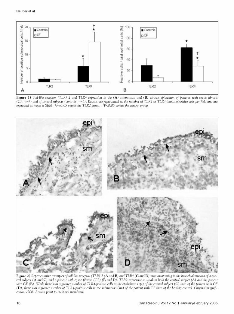
RESULTSTLR2 and TLR4 expression in bronchial mucosaTLR2- and TLR4-positive cells were expressed in the submucosa(Figure 1A) and airway epithelium (Figure 1B) of both CF andcontrol subjects. TLR4 expression was significantly higher thanTLR2 expression in the submucosa and epithelium of CFpatients and control subjects (Figure 1) (P<0.05). Figure 2 is arepresentative example of TLR2 and TLR4 expression in a con-trol subject and a patient with CF. TLR2 expression in theepithelium and the submucosa was low in control subjects andpatients with CF (Figure 2A and B). TLR4 immunoreactivitywas low in the airway epithelium, but strong in the submucosa ofpatients with CF (Figure 2D) compared with control subjects(Figure 2C). The number of TLR4-positive cells in CF submu-cosa (14.67±3.61 cells/field) was almost threefold higher than inthe submucosa of control subjects (5.80±2.84 cells/field)(P<0.05) (Figure 1A). The number of TLR2-positive cells in CFsubmucosa (0.86±0.26 cells/field) was similar to that observed inthe submucosa of control subjects (1.20±0.37 cells/field)(Figure 1A). In the airway epithelium, TLR4 expression was sig-nificantly lower in patients with CF (28.57±10.10%) than incontrol subjects (62.50±7.22%) (P<0.05) (Figure 1B).Although not significant, there was a trend toward increasedTLR2 expression in the airway epithelium of control subjectscompared with patients with CF (7.14±4.61% versus30.00±12.25%, respectively; P=0.07) (Figure 1B).
Source of TLR4 in CF submucosaAs expected, the number of elastase-positive neutrophils in CFmucosa (9.1±2.4 cells/field) was significantly higher than in controlsubjects (4.9±1.1 cells/field) (P<0.05). There was no significant dif-ference in the number of CD68-positive monocytes/macrophagesbetween CF patients and healthy control subjects(3.6±0.9 cells/field versus 4.5±1.3 cells/field, respectively). Thenumber of TLR4-positive, elastase-positive cells was significantlyhigher in CF patients than in control subjects (6.1±1.3 cells/fieldversus 3.3±0.9 cells/field) (P<0.05). In contrast, there was no sig-nificant difference in the number of TLR4-positive, CD68-positivecells between the two groups (CF: 1.9±0.6 cells/field versus controlsubjects: 2.8±0.9 cells/field) (Figure 3).
In patients with CF, 64±6% of all TLR4 immunoreactive cellswere elastase-positive neutrophils and 32±12% were CD68-positive monocytes/macrophages; in the control group, only50±4% of all TLR4-positive cells were elastase-positive neutrophilsand 45±12% were CD68-positive monocytes/macrophages. Thepercentage of TLR4-positive cells that were also elastase-positivewas significantly higher in CF patients than in control subjects(P<0.05). No significant difference was found in the percentagesof TLR4-positive, CD68-positive cells between the groups.Figure 4A shows an example of TLR4-positive, elastase-positiveimmunopositive cells in the bronchial mucosa of a patient with
TLR4 and TLR2 expression in CF bronchial mucosa
Can Respir J Vol 12 No 1 January/February 2005 15
Hauber.qxd 2/4/2005 1:36 PM Page 15
Hauber et al
Can Respir J Vol 12 No 1 January/February 200516
Figure 1) Toll-like receptor (TLR) 2 and TLR4 expression in the (A) submucosa and (B) airway epithelium of patients with cystic fibrosis (CF; n=7) and of control subjects (controls; n=6). Results are represented as the number of TLR2 or TLR4 immunopositive cells per field and areexpressed as mean ± SEM. *P<0.05 versus the TLR2 group.; †P<0.05 versus the control group
Figure 2) Representative examples of toll-like receptor (TLR) 2 (A and B) and TLR4 (C and D) immunostaining in the bronchial mucosa of a con-trol subject (A and C) and a patient with cystic fibrosis (CF) (B and D). TLR2 expression is weak in both the control subject (A) and the patientwith CF (B). While there was a greater number of TLR4-positive cells in the epithelium (epi) of the control subject (C) than of the patient with CF(D), there was a greater number of TLR4-positive cells in the submucosa (sm) of the patient with CF than of the healthy control. Original magnifi-cation ×200. Arrows point to the basal membrane
Hauber.qxd 2/4/2005 1:36 PM Page 16
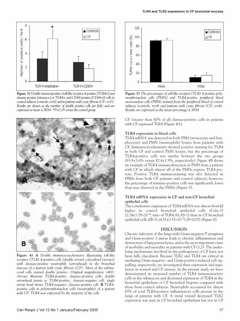
CF. Greater than 60% of all elastase-positive cells in patientswith CF expressed TLR4 (Figure 4A).
TLR4 expression in blood cellsTLR4 mRNA was detected in both PBM (monocytes and lym-phocytes) and PMN (neutrophils) lysates from patients withCF. Immunocytochemistry showed positive staining for TLR4in both CF and control PMN lysates, but the percentage ofTLR4-positive cells was similar between the two groups(93.9±3.6% versus 92.4±1.9%, respectively). Figure 4B showsan example of TLR4 immunodetection in PMN from a patientwith CF in which almost all of the PMNs express TLR4 pro-tein. Positive TLR4 immunostaining was also detected inPBMs from both CF patients and control subjects; however,the percentage of immuno-positive cells was significantly lowerthan was observed in the PMNs (Figure 5).
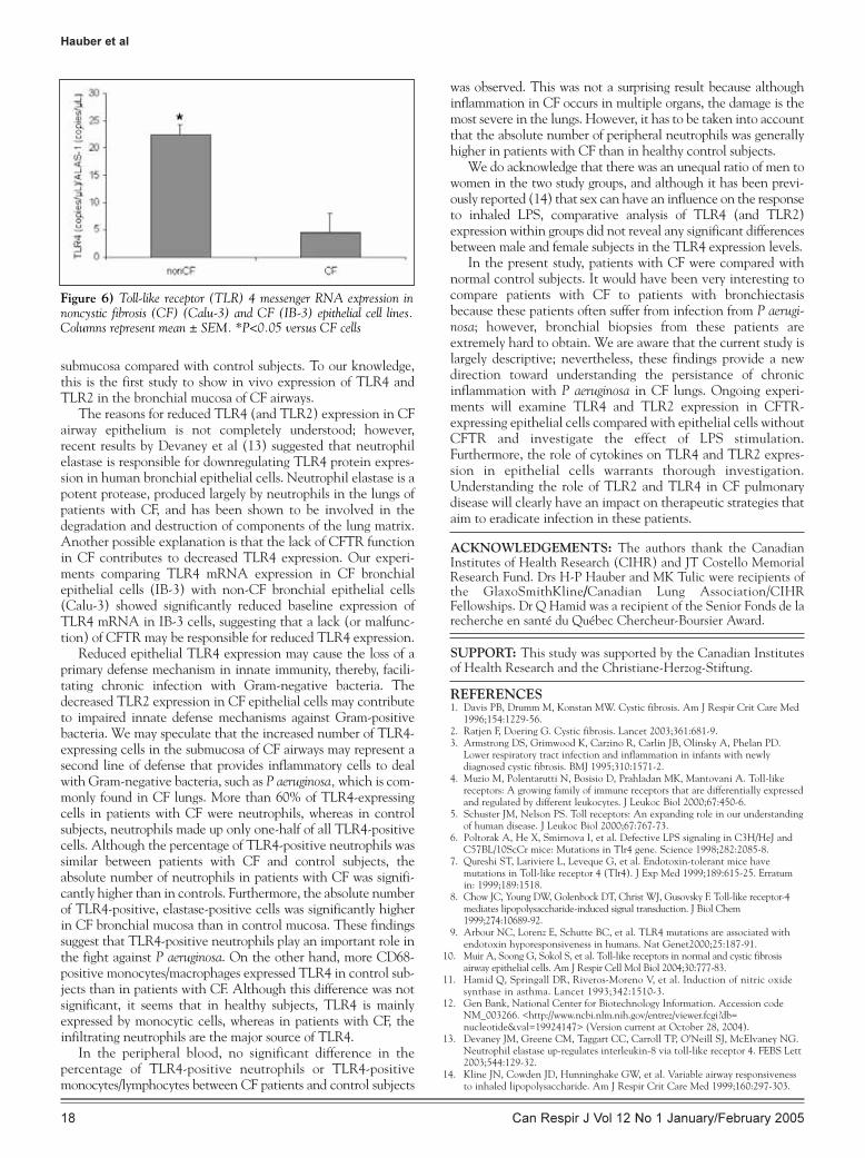
TLR4 mRNA expression in CF and non-CF bronchialepithelial cellsThe constitutive expression of TLR4 mRNA was almost fivefoldhigher in control bronchial epithelial cells (Calu-3)(2.24±1.79×10⁻⁴; ratio of TLR4:ALAS-1) than in CF bronchialepithelial cells (IB-3) (4.51±3.53×10⁻⁵) (P<0.05) (Figure 6).
DISCUSSIONChronic infection of the lungs with Gram-negative P aeruginosaand Gram-positive S aureus leads to chronic inflammation anddestruction of lung parenchyma, and is the most important causeof morbidity and mortality in patients with CF (1,2). The under-lying mechanisms involved in the pathogenesis of CF have notbeen fully elucidated. Because TLR2 and TLR4 are critical inmediating Gram-negative- and Gram-positive-induced cell sig-nalling, respectively, we investigated their expression and regu-lation in normal and CF airways. In the present study, we havedemonstrated an increased number of TLR4 immunoreactivecells in the submucosa and decreased expression of TLR4 in thebronchial epithelium of CF bronchial biopsies compared withthose from control subjects. Neutrophils accounted for almost65% of total TLR4-positive inflammatory cells present in thelungs of patients with CF. A trend toward decreased TLR2expression was seen in CF bronchial epithelium but not in CF
TLR4 and TLR2 expression in CF bronchial mucosa
Can Respir J Vol 12 No 1 January/February 2005 17
Figure 3) Double immunopositive (toll-like receptor 4-positive [TLR4+] andelastase-positive [elastase+] or TLR4+ and CD68-positive [CD68+]) cells incontrol subjects (controls; n=6) and in patients with cystic fibrosis (CF; n=7).Results are shown as the number of double positive cells per field, and areexpressed as mean ± SEM. *P<0.05 versus the control group
Figure 4) A Double immunocytochemistry illustrating toll-likereceptor (TLR) 4-positive cells (double arrow) colocalized (arrows)with elastase-positive neutrophils (arrowhead) in the bronchialmucosa of a patient with cystic fibrosis (CF). Most of the submu-cosal cells stained double positive. Original magnification ×400. Arrows illustrate TLR4-positive, elastase-positive cells; doublearrowhead points to TLR4-positive, elastase-negative cell; singlearrow head shows TLR4-negative, elastase-positive cell. B TLR4-positive cells in polymorphonuclear cells (neutrophils) of a patientwith CF. TLR4 was expressed by the majority of the cells
Figure 5) The percentages of toll-like receptor (TLR) 4-positive poly-morphonuclear cells (PMN) and TLR4-positive peripheral bloodmononuclear cells (PBM) isolated from the peripheral blood of controlsubjects (controls; n=4) and patients with cystic fibrosis (CF; n=4).Results are expressed as the mean percentage ± SEM
Hauber.qxd 2/9/2005 3:49 PM Page 17
submucosa compared with control subjects. To our knowledge,this is the first study to show in vivo expression of TLR4 andTLR2 in the bronchial mucosa of CF airways.
The reasons for reduced TLR4 (and TLR2) expression in CFairway epithelium is not completely understood; however,recent results by Devaney et al (13) suggested that neutrophilelastase is responsible for downregulating TLR4 protein expres-sion in human bronchial epithelial cells. Neutrophil elastase is apotent protease, produced largely by neutrophils in the lungs ofpatients with CF, and has been shown to be involved in thedegradation and destruction of components of the lung matrix.Another possible explanation is that the lack of CFTR functionin CF contributes to decreased TLR4 expression. Our experi-ments comparing TLR4 mRNA expression in CF bronchialepithelial cells (IB-3) with non-CF bronchial epithelial cells(Calu-3) showed significantly reduced baseline expression ofTLR4 mRNA in IB-3 cells, suggesting that a lack (or malfunc-tion) of CFTR may be responsible for reduced TLR4 expression.
Reduced epithelial TLR4 expression may cause the loss of aprimary defense mechanism in innate immunity, thereby, facili-tating chronic infection with Gram-negative bacteria. Thedecreased TLR2 expression in CF epithelial cells may contributeto impaired innate defense mechanisms against Gram-positivebacteria. We may speculate that the increased number of TLR4-expressing cells in the submucosa of CF airways may represent asecond line of defense that provides inflammatory cells to dealwith Gram-negative bacteria, such as P aeruginosa, which is com-monly found in CF lungs. More than 60% of TLR4-expressingcells in patients with CF were neutrophils, whereas in controlsubjects, neutrophils made up only one-half of all TLR4-positivecells. Although the percentage of TLR4-positive neutrophils wassimilar between patients with CF and control subjects, theabsolute number of neutrophils in patients with CF was signifi-cantly higher than in controls. Furthermore, the absolute numberof TLR4-positive, elastase-positive cells was significantly higherin CF bronchial mucosa than in control mucosa. These findingssuggest that TLR4-positive neutrophils play an important role inthe fight against P aeruginosa. On the other hand, more CD68-positive monocytes/macrophages expressed TLR4 in control sub-jects than in patients with CF. Although this difference was notsignificant, it seems that in healthy subjects, TLR4 is mainlyexpressed by monocytic cells, whereas in patients with CF, theinfiltrating neutrophils are the major source of TLR4.
In the peripheral blood, no significant difference in the percentage of TLR4-positive neutrophils or TLR4-positivemonocytes/lymphocytes between CF patients and control subjects
was observed. This was not a surprising result because althoughinflammation in CF occurs in multiple organs, the damage is themost severe in the lungs. However, it has to be taken into accountthat the absolute number of peripheral neutrophils was generallyhigher in patients with CF than in healthy control subjects.
We do acknowledge that there was an unequal ratio of men towomen in the two study groups, and although it has been previ-ously reported (14) that sex can have an influence on the responseto inhaled LPS, comparative analysis of TLR4 (and TLR2)expression within groups did not reveal any significant differencesbetween male and female subjects in the TLR4 expression levels.
In the present study, patients with CF were compared withnormal control subjects. It would have been very interesting tocompare patients with CF to patients with bronchiectasisbecause these patients often suffer from infection from P aerugi-nosa; however, bronchial biopsies from these patients areextremely hard to obtain. We are aware that the current study islargely descriptive; nevertheless, these findings provide a newdirection toward understanding the persistance of chronicinflammation with P aeruginosa in CF lungs. Ongoing experi-ments will examine TLR4 and TLR2 expression in CFTR-expressing epithelial cells compared with epithelial cells withoutCFTR and investigate the effect of LPS stimulation.Furthermore, the role of cytokines on TLR4 and TLR2 expres-sion in epithelial cells warrants thorough investigation.Understanding the role of TLR2 and TLR4 in CF pulmonarydisease will clearly have an impact on therapeutic strategies thataim to eradicate infection in these patients.
ACKNOWLEDGEMENTS: The authors thank the CanadianInstitutes of Health Research (CIHR) and JT Costello MemorialResearch Fund. Drs H-P Hauber and MK Tulic were recipients ofthe GlaxoSmithKline/Canadian Lung Association/CIHRFellowships. Dr Q Hamid was a recipient of the Senior Fonds de larecherche en santé du Québec Chercheur-Boursier Award.
SUPPORT: This study was supported by the Canadian Institutesof Health Research and the Christiane-Herzog-Stiftung.
Hauber et al
Can Respir J Vol 12 No 1 January/February 200518
REFERENCES1. Davis PB, Drumm M, Konstan MW. Cystic fibrosis. Am J Respir Crit Care Med
1996;154:1229-56.2. Ratjen F, Doering G. Cystic fibrosis. Lancet 2003;361:681-9.3. Armstrong DS, Grimwood K, Carzino R, Carlin JB, Olinsky A, Phelan PD.
Lower respiratory tract infection and inflammation in infants with newlydiagnosed cystic fibrosis. BMJ 1995;310:1571-2.
4. Muzio M, Polentarutti N, Bosisio D, Prahladan MK, Mantovani A. Toll-likereceptors: A growing family of immune receptors that are differentially expressedand regulated by different leukocytes. J Leukoc Biol 2000;67:450-6.
5. Schuster JM, Nelson PS. Toll receptors: An expanding role in our understandingof human disease. J Leukoc Biol 2000;67:767-73.
6. Poltorak A, He X, Smirnova I, et al. Defective LPS signaling in C3H/HeJ andC57BL/10ScCr mice: Mutations in Tlr4 gene. Science 1998;282:2085-8.
7. Qureshi ST, Lariviere L, Leveque G, et al. Endotoxin-tolerant mice havemutations in Toll-like receptor 4 (Tlr4). J Exp Med 1999;189:615-25. Erratumin: 1999;189:1518.
8. Chow JC, Young DW, Golenbock DT, Christ WJ, Gusovsky F. Toll-like receptor-4mediates lipopolysaccharide-induced signal transduction. J Biol Chem1999;274:10689-92.
9. Arbour NC, Lorenz E, Schutte BC, et al. TLR4 mutations are associated withendotoxin hyporesponsiveness in humans. Nat Genet2000;25:187-91.
10. Muir A, Soong G, Sokol S, et al. Toll-like receptors in normal and cystic fibrosisairway epithelial cells. Am J Respir Cell Mol Biol 2004;30:777-83.
11. Hamid Q, Springall DR, Riveros-Moreno V, et al. Induction of nitric oxidesynthase in asthma. Lancet 1993;342:1510-3.
12. Gen Bank, National Center for Biotechnology Information. Accession codeNM_003266. <http://www.ncbi.nlm.nih.gov/entrez/viewer.fcgi?db=nucleotide&val=19924147> (Version current at October 28, 2004).
13. Devaney JM, Greene CM, Taggart CC, Carroll TP, O’Neill SJ, McElvaney NG.Neutrophil elastase up-regulates interleukin-8 via toll-like receptor 4. FEBS Lett2003;544:129-32.
14. Kline JN, Cowden JD, Hunninghake GW, et al. Variable airway responsivenessto inhaled lipopolysaccharide. Am J Respir Crit Care Med 1999;160:297-303.
Figure 6) Toll-like receptor (TLR) 4 messenger RNA expression innoncystic fibrosis (CF) (Calu-3) and CF (IB-3) epithelial cell lines.Columns represent mean ± SEM. *P<0.05 versus CF cells
Hauber.qxd 2/4/2005 1:36 PM Page 18

Submit your manuscripts athttp://www.hindawi.com
Stem CellsInternational
Hindawi Publishing Corporationhttp://www.hindawi.com Volume 2014
Hindawi Publishing Corporationhttp://www.hindawi.com Volume 2014
MEDIATORSINFLAMMATION
of
Hindawi Publishing Corporationhttp://www.hindawi.com Volume 2014
Behavioural Neurology
EndocrinologyInternational Journal of
Hindawi Publishing Corporationhttp://www.hindawi.com Volume 2014
Hindawi Publishing Corporationhttp://www.hindawi.com Volume 2014
Disease Markers
Hindawi Publishing Corporationhttp://www.hindawi.com Volume 2014
BioMed Research International
OncologyJournal of
Hindawi Publishing Corporationhttp://www.hindawi.com Volume 2014
Hindawi Publishing Corporationhttp://www.hindawi.com Volume 2014
Oxidative Medicine and Cellular Longevity
Hindawi Publishing Corporationhttp://www.hindawi.com Volume 2014
PPAR Research
The Scientific World JournalHindawi Publishing Corporation http://www.hindawi.com Volume 2014
Immunology ResearchHindawi Publishing Corporationhttp://www.hindawi.com Volume 2014
Journal of
ObesityJournal of
Hindawi Publishing Corporationhttp://www.hindawi.com Volume 2014
Hindawi Publishing Corporationhttp://www.hindawi.com Volume 2014
Computational and Mathematical Methods in Medicine
OphthalmologyJournal of
Hindawi Publishing Corporationhttp://www.hindawi.com Volume 2014
Diabetes ResearchJournal of
Hindawi Publishing Corporationhttp://www.hindawi.com Volume 2014
Hindawi Publishing Corporationhttp://www.hindawi.com Volume 2014
Research and TreatmentAIDS
Hindawi Publishing Corporationhttp://www.hindawi.com Volume 2014
Gastroenterology Research and Practice
Hindawi Publishing Corporationhttp://www.hindawi.com Volume 2014
Parkinson’s Disease
Evidence-Based Complementary and Alternative Medicine
Volume 2014Hindawi Publishing Corporationhttp://www.hindawi.com