Embed Size (px)
Citation preview
RESEARCH ARTICLE
Tomatidine reduces Chikungunya virus
progeny release by controlling viral protein
expression
Berit Troost-KindID1, Martijn J. van Hemert2, Denise van de Pol1, Heidi van der Ende-
Metselaar1, Andres Merits3, Malte BorggreweID4, Izabela A. Rodenhuis-ZybertID
1, Jolanda
M. SmitID1*
1 Department of Medical Microbiology and Infection Prevention, University of Groningen; University Medical
Center Groningen; Groningen, the Netherlands, 2 Department of Medical Microbiology, Molecular Virology
Laboratory, Leiden University Medical Center, Leiden, the Netherlands, 3 Institute of Technology, University
of Tartu, Tartu, Estonia, 4 Department of Biomedical Sciences of Cells & Systems, Section Molecular
Neurobiology, University of Groningen; University Medical Center Groningen; Groningen, the Netherlands
Abstract
Tomatidine, a natural steroidal alkaloid from unripe green tomatoes has been shown to
exhibit many health benefits. We recently provided in vitro evidence that tomatidine reduces
the infectivity of Dengue virus (DENV) and Chikungunya virus (CHIKV), two medically
important arthropod-borne human infections for which no treatment options are available.
We observed a potent antiviral effect with EC50 values of 0.82 μM for DENV-2 and 1.3 μM
for CHIKV-LR. In this study, we investigated how tomatidine controls CHIKV infectivity.
Using mass spectrometry, we identified that tomatidine induces the expression of p62,
CD98, metallothionein and thioredoxin-related transmembrane protein 2 in Huh7 cells. The
hits p62 and CD98 were validated, yet subsequent analysis revealed that they are not
responsible for the observed antiviral effect. In parallel, we sought to identify at which step of
the virus replication cycle tomatidine controls virus infectivity. A strong antiviral effect was
seen when in vitro transcribed CHIKV RNA was transfected into Huh7 cells treated with
tomatidine, thereby excluding a role for tomatidine during CHIKV cell entry. Subsequent
determination of the number of intracellular viral RNA copies and viral protein expression
levels during natural infection revealed that tomatidine reduces the RNA copy number and
viral protein expression levels in infected cells. Once cells are infected, tomatidine is not
able to interfere with active RNA replication yet it can reduce viral protein expression. Collec-
tively, the results delineate that tomatidine controls viral protein expression to exert its antivi-
ral activity. Lastly, sequential passaging of CHIKV in presence of tomatidine did not lead to
viral resistance. Collectively, these results further emphasize the potential of tomatidine as
an antiviral treatment towards CHIKV infection.
PLOS NEGLECTED TROPICAL DISEASES
PLOS Neglected Tropical Diseases | https://doi.org/10.1371/journal.pntd.0009916 November 11, 2021 1 / 23
a1111111111
a1111111111
a1111111111
a1111111111
a1111111111
OPEN ACCESS
Citation: Troost-Kind B, van Hemert MJ, van de Pol
D, van der Ende-Metselaar H, Merits A, Borggrewe
M, et al. (2021) Tomatidine reduces Chikungunya
virus progeny release by controlling viral protein
expression. PLoS Negl Trop Dis 15(11): e0009916.
https://doi.org/10.1371/journal.pntd.0009916
Editor: Alain Kohl, University of Glasgow, UNITED
KINGDOM
Received: June 18, 2021
Accepted: October 16, 2021
Published: November 11, 2021
Copyright: © 2021 Troost-Kind et al. This is an
open access article distributed under the terms of
the Creative Commons Attribution License, which
permits unrestricted use, distribution, and
reproduction in any medium, provided the original
author and source are credited.
Data Availability Statement: All relevant data are
within the manuscript and its Supporting
Information files.
Funding: B.T was supported by the Graduate
School of Medical Sciences of the University of
Groningen and the de Cock Grant from the
Hadders Stichting. I.A.R.Z and J.M.S. were
supported by the University Medical Center
Groningen. The funders had no role in study
design, data collection and analysis, decision to
publish, or preparation of the manuscript.
Author summary
Chikungunya fever is a debilitating disease caused by the mosquito-borne Chikungunya
virus. Over the past two decades the geographical spread of the virus and its mosquito vec-
tor has drastically increased thereby causing millions of infections. To date there is no
antiviral drug and no vaccine available to treat/prevent Chikungunya virus infection. We
recently showed that the natural steroidal alkaloid tomatidine has potent antiviral activity
towards Chikungunya virus at submicromolar concentrations. In this study we dissected
how tomatidine reduces the production of Chikungunya virus particles. We show that
tomatidine lowers viral protein expression and we hypothesize that the effect of tomati-
dine on viral protein translation hampers the production of progeny viral RNA copies /
number of infected cells thereby leading to a reduced production of secreted virus parti-
cles. Also, we show that Chikungunya virus does not readily become resistant to tomati-
dine. Collectively, we deciphered the mechanism by which tomatidine exerts antiviral
activity to Chikungunya virus and our results strengthen the potential of tomatidine as an
antiviral treatment strategy towards Chikungunya virus.
Introduction
Chikungunya virus (CHIKV) is a re-emerging arbovirus transmitted to humans via the mos-
quito vectors Aedes aegypti and Aedes albopictus [1]. Until 2004 the virus was limited to Africa
and Asia; however, following the recurrence of CHIKV in Kenya in 2005, CHIKV has globally
expanded from the tropical zone to America and Europe causing millions of infections [2,3].
CHIKV typically causes an acute self-limiting febrile disease including symptoms such as
fever, rash, nausea, fatigue, headache and myalgia [4]. In 30–40% of the cases, the disease pro-
gresses to a chronic form involving debilitating muscle and join pains which can persist for
months to years after the initial infection [5].
Currently, there are no licensed antiviral treatments or vaccines available to combat
CHIKV infection. Treatment is limited to administration of pain killers, antipyretics and non-
steroidal anti-inflammatory drugs to alleviate the symptoms [6]. Antiviral therapies aim to
reduce viral replication by directly targeting a viral protein (direct-acting antivirals) or by
modulating host factors that are crucial for replication (host-directed antivirals) [7]. Previous
in vitro CHIKV research identified both types of antivirals [7]. Direct-acting antivirals were
shown to target structural (capsid, envelope E1 and E2, 6k) and non-structural proteins (nsP1,
nsP2, nsP3 or nsP4) of CHIKV. Host-directed antivirals were found to target various host fac-
tors including kinases (e.g. CND3514 [8]), furin proteases (e.g. decanoyl-RVKR-chloromethyl
[9]), HSP-90 (e.g. ganetespib [10]) endosomal acidification (e.g. chloroquine [11]), inosine
monophosphate dehydrogenase (e.g. ribavirin [12,13]) and interferon (e.g. recombinant IFN-
α [14]). Thus far, two antivirals have been tested in clinical trials: the virus cell entry inhibitor
chloroquine and the inosine monophosphate dehydrogenase inhibitor ribavirin; however,
without success [15,16]. This shows the need to follow-up more and identify new antiviral
compounds.
Recently, we identified tomatidine, a natural steroidal alkaloid extracted from the stems
and leaves of green tomatoes, as a potent inhibitor of different CHIKV and Dengue virus
(DENV) strains in vitro [17,18]. Tomatidine was shown to interfere with CHIKV production
in a variety of cell lines including, African green monkey Vero cells, human skin fibroblast
HFF-1 cells, human osteosarcoma U2OS cells and in human hepatic Huh-7 cells thereby
excluding a cell type specific effect [17]. Tomatidine is the aglycone metabolite of tomatine,
PLOS NEGLECTED TROPICAL DISEASES Chikungunya virus protein expression is reduced by tomatidine
PLOS Neglected Tropical Diseases | https://doi.org/10.1371/journal.pntd.0009916 November 11, 2021 2 / 23
Competing interests: The authors have declared
that no competing interests exist.
which naturally protects the tomato plant from pathogens such as fungi, viruses, bacteria and
insects [19,20]. Tomatidine has been shown to exert various health benefits including an anti-
inflammatory, anti-metastatic, anti-atherosclerotic, anti-osteoporosis and antimicrobial activ-
ity [20–27]. Moreover, tomatidine was shown to protect from age-related muscle atrophy and
ischemic neuronal injury [28,29]. To identify the mechanism of protection from muscle atro-
phy, an microarray analysis on mouse skeletal muscle cells was performed and subsequent
gene set enrichment analysis revealed 7 gene sets significantly induced and 25 gene sets signifi-
cantly suppressed in the presence of tomatidine [28]. Among the suppressed gene set was the
transcription factor ATF4, which was identified as a key mediator of age-related muscle atro-
phy. We tested the importance of ATF4 in the antiviral effect of tomatidine towards DENV
and revealed that although it might be involved it is not the main factor responsible for the
antiviral effect [18]. Furthermore, in the context of its anti-atherosclerotic effect, tomatidine
was shown to inhibit acyl-CoA:cholesterol acyl-transferase-1 and 2, which are enzymes
involved in the formation of foam cells, a characteristic feature of atherosclerotic lesions [24].
A study on the anti-inflammatory properties of tomatidine demonstrated that it has the ability
to inhibit the NF-kB and JNK pathway [21]. Thus, exposure of cells to tomatidine modulates
various pathways within the cell.
Here, we analyzed the mechanism underlying the antiviral activity of tomatidine towards
CHIKV in Huh7 cells from the host cell as well as the virus perspective. In our experimental
set-up, tomatidine treatment had no major consequences on the host cell proteome and the
validated proteins are not responsible for the observed antiviral effect. Subsequent analysis of
the steps in the viral replication cycle suggests that tomatidine controls the expression of viral
proteins thereby reducing virus progeny secretion. Furthermore, sequential passaging of
CHIKV under tomatidine selection pressure revealed that viral resistance is not quickly
obtained. In conclusion, these results further strengthen the potential of tomatidine as an anti-
viral compound towards CHIKV.
Methods
Chemicals
Tomatidine hydrochloride and ivermectin were purchased from Sigma Aldrich (St. Louis,
Missouri, USA) and dissolved in absolute ethanol to a stock solution of 5 mM. Aliquots were
stored at -20˚C and were used for a maximum of 3 months. The final concentration of ethanol
was below 0.1% in all experiments.
Cell culture
The human hepatic cell line Huh7 (JCRB0403) was a kind gift from Tonya Colpitts (University
of South Carolina). Cells were maintained in Dulbecco’s minimal essential medium (DMEM)
Glutamax (Gibco, The Netherlands) supplemented with 10% FBS (Lonza, Basel, Switzerland)
and 100 U/mL penicillin and 100 mg/mL streptomycin (PAA Laboratories, Pasching, Austria).
The renal African Green monkey cell line Vero-WHO, was maintained in DMEM supple-
mented with 5% FBS, 100 U/mL penicillin and 100 mg/mL streptomycin. All cells were cul-
tured at a temperature of 37˚C with 5% CO2.
Mass spectrometry
Huh7 cells were lysed using the RIPA lysis buffer system (Santa Cruz Biotechnology, Dallas,
Texas, USA) and proteins were extracted according to the manufacturer’s instructions. Sam-
ples were loaded on a 4–12% pre-cast NuPAGE gel (Invitrogen, Carlsbad, California, USA),
PLOS NEGLECTED TROPICAL DISEASES Chikungunya virus protein expression is reduced by tomatidine
PLOS Neglected Tropical Diseases | https://doi.org/10.1371/journal.pntd.0009916 November 11, 2021 3 / 23
and briefly ran into the gel, without separation. The gel was stained with Coomassie dye R-250
(Thermo Fisher Scientific, Waltham, Massachusetts, USA) followed by destaining with ultra-
pure water. The protein blob was excised as a single band and destained with 70% 50 mM
NH4HCO3 and 30% acetonitrile. Reduction was performed using 10 mM DTT dissolved in 50
mM NH4HCO3 for 30 min at 55˚C. Samples were alkylated using 55 mM iodoacetamide in 50
mM NH4HCO3 for 30 min at room temperature. Subsequently, samples were washed for 10
min with 50 mM NH4HCO3 and for 30 min with 100% acetonitrile. The gel pieces were dried
for 15 min at 55˚C. Tryptic digestion was performed with sequencing-grade modified trypsin
(Promega, Madison, Wisconsin, USA, 10 ng/ml in 50 mM NH4HCO3) with overnight incuba-
tion at 37˚C. Peptides were extracted using 5% formic acid followed by a second elution with
5% formic acid in 75% acetonitrile. Samples were dried in a SpeedVac centrifuge and dissolved
in 5% formic acid for injection on an Ultimate 3000 nanoLC system (Dionex, Sunnyvale, Cali-
fornia, USA) interfaced online with a Q Exactive Plus mass spectrometer (Thermo Fisher Sci-
entific). Samples were loaded onto a 5 mm × 300 μm i.d. C18 PepMAP100 trapping column
(Dionex) in 2% acetonitrile in water with 0.1% formic acid at 20 μL/min. Peptides were sepa-
rated on a 50 cm × 75 μm i.d. C18 PepMAP100 nanocolumn (Dionex). The following mobile
phase gradient was delivered at the flow rate of 300 nL/min: 2%-50% of solvent B in 90 min;
50%–90% B in 1 min; 90% B during 13 min, and back to 2% B in 1 min (held for 15 min). Sol-
vent A was 100 water with 0.1% formic acid, and solvent B was acetonitrile with 0.1% formic
acid. Peptides were infused into the mass spectrometer using a stainless-steel emitter at a spray
voltage was 1.8 kV with no sheath and auxiliary gas flow; the ion transfer tube temperature
was 275˚C. The mass spectrometer was operated in data-dependent mode (DDA) with MS/
MS fragmentation of the top 12 precursor ions in each cycle. A cycle consisted of a survey scan
of m/z 400–1800 at 70000 resolution and MS/MS scans at 17500 resolution. Singly charged
ions were excluded from MS/MS experiments and precursor ions were dynamically excluded
for 30 s. Proteins with no intensity in at least one sample were filtered. EdgeR R-package
(v3.24.3) was used for normalization (trimmed mean of M values; TMM) and differential pro-
tein expression analysis (glmQLFTest) [30]. Proteins were regarded differentially expressed
with a false discovery rate (FDR) lower than 0.1. The mass spectrometry proteomics data have
been deposited to the ProteomeXchange consortium via the PRIDE partner repository with
the dataset identifier PXD028693.
Virus stocks and titration
The La Reunion CHIKV isolate (LR2006 OPY1, East/Central/South African genotype was res-
cued from the corresponding infectious cDNA clone and produced on Vero-WHO cells as
described by Troost et al [17]. The infectious virus titer was determined via plaque assay on
Vero-WHO cells as number of plaque forming units (PFU) as described before [17]. The intra-
cellular viral genome copy number was determined via Q-RT-PCR. The primers are described
in Troost et al and bind within the CHIKV E1 region [17]. Cellular RNA was isolated using
the RNeasy kit (Qiagen, Hilden, Germany) according to the manufacturer’s protocol. The
cDNA production and Q-RT-PCR were performed as described previously [17,31].
Infection experiments
Huh7 cells were infected with CHIKV at the multiplicity of infection (MOI) 1 in virus resis-
tance studies or MOI 10 in mode-of-action experiments. In mode-of action experiments, a
high MOI was used to increase the sensitivity of the experiment. Tomatidine (10 μM) or iver-
mectin (7 μM) treatment was added at the time of infection unless otherwise indicated. As a
vehicle control the equivalent volume of the EtOH solvent was used. The virus inoculum was
PLOS NEGLECTED TROPICAL DISEASES Chikungunya virus protein expression is reduced by tomatidine
PLOS Neglected Tropical Diseases | https://doi.org/10.1371/journal.pntd.0009916 November 11, 2021 4 / 23
removed 2 hours post-infection (hpi), cells were washed three times with plain medium and
fresh compound-containing medium was added to the cells unless indicated otherwise.
Protein extraction and western blot
Huh7 cells were lysed via the RIPA lysis buffer system (Santa Cruz Biotechnology) and protein
was extracted according to the manufacturer’s instructions. The protein concentration was
determined using the Bradford assay (Expedeon, Swavesey, UK). Sample preparation and
western blot analysis were performed as described before [18]. The primary antibodies were
diluted as follows: GAPDH (Abcam, Cambridge, UK) 1:10,000, vinculin (Sigma-Aldrich)
1:500, CD98 (Sino Biological, Peking, China) and p62 (Progen, Heidelberg, Germany) 1:500,
thioredoxin-related transmembrane protein 2 (Thermo Fisher Scientific) 1:125, and nsP2
(Abgenex, Bhubaneswar, Odisha, India), E1 (kindly provided by Gorben Pijman, Wageningen
University) and capsid (Native antigen company, Kidlington, UK) 1:1,000.
Dilutions of the antibodies were prepared in TBS-Tween with 0.1% of sodium azide and
either 5% BSA or 5% milk. Secondary antibodies conjugated with HRP anti-mouse or anti-
rabbit (Thermo Fisher Scientific) were used according to the manufacturer’s recommenda-
tions. Pierce ECL western blotting substrate (Thermo Fisher Scientific) or Super Signal West
FEMTO (Thermo Fisher Scientific) was used for detection by chemiluminescence using LAS-
4000 mini camera system (GE Healthcare, Chicago, Illinois, USA). Analysis was performed
with the Image QuantTL software (GE Healthcare). The band intensity of each protein was
normalized to that of the loading control (GAPDH or vinculin) and expressed as the fold-
change over EtOH-treated cells.
Transfection of siRNAs
The siRNAs p62 #1(ID: J-010230-05-0005), P62 #4(ID: J-010230-08-0002), CD98 (SLC3A2,
ID: L-003542-00-0005) or non-targeting siRNA (siScramble, ID: D-001810-10) were pur-
chased from Dharmacon (Horizon, Lafayette, Colorado, USA). Reverse transfection of Huh7
cells was performed using Lipofectamine RNAiMAX (Invitrogen) with a final concentration
of 20 nM for the siRNAs according to the manufacturer’s protocol. Cells were seeded in a
12-well plate at a density of 7�104 cells per well. At 48 h post-transfection, cells were infected
with CHIKV at MOI 10 and treated with 10 μM tomatidine or the equivalent volume of EtOH.
Supernatants were collected at 9 hpi and used to titrate the produced virus using plaque assay.
Cells were harvested and subjected to WB analysis to determine the expression of p62 and
CD98 proteins.
Electroporation study
In vitro transcribed RNA was produced from the infectious clone CHIK-LR [32]. The RNA
was electroporated into Huh7 cells (260 V and 950 μF) via the Gene Pulser Xcell system (Bio-
Rad, Hercules, California, USA). Cells were counted and seeded into a 12-well plate containing
medium supplemented with 10 μM tomatidine or the equivalent volume of EtOH at a density
of 0.3�106 cells per well. Supernatants were collected 16 h post-electroporation and the infec-
tious virus titer was determined by plaque assay.
Replication study
The trans-replicase system of CHIKV consisted of two plasmids one encoding for the
CHIKV-LR replicase and the other for the truncated CHIKV RNA template containing a
sequence encoding for a tomato fluorescent marker protein under the control of the CHIKV
PLOS NEGLECTED TROPICAL DISEASES Chikungunya virus protein expression is reduced by tomatidine
PLOS Neglected Tropical Diseases | https://doi.org/10.1371/journal.pntd.0009916 November 11, 2021 5 / 23
subgenomic promotor. As a control, a plasmid expressing an inactive form of the CHIKV rep-
licase was used. Inactivity was due to a mutation in the catalytic Gly-Asp-Asp (GDD) motif of
nsP4 to Gly-Ala-Ala (GAA) [33]. Huh7 cells were seeded in a 24 well-plate at a density of
0.6x10⁵ cells/well. At 24 h post-seeding, cells were co-transfected with 0.25 μg of both plasmids
using Lipofectamine LTX reagent (Invitrogen) according to manufacturer’s protocol. At 1 h
post-transfection, 10 μM tomatidine, 7 μM ivermectin or the equivalent volume of EtOH was
added to the cells. Cells were collected at 24 hpi, fixed and analyzed for the expression of the
tomato fluorescent protein using flow cytometry. The expression of the marker indicates active
replication in cells transfected with of this trans-replicase system.
In-gel hybridization
Huh7 cells were infected with CHIKV at MOI 10, as described above. Cells were harvested at 6
hpi and cellular RNA was isolated using the RNeasy kit (Qiagen). Denaturating formaldehyde
gel electrophoresis was performed as previously described [34]. RNA was detected via 32P-
labelled oligonucleotides via in-gel hybridization. Hereto, the 32P-labelled probe CHIKV Hyb4
(5’TGTGGGTTCGGAGAATCGTGGAAGAGTT-30) and as control the 18S ribosomal RNA
probe (50-ATGCCCCCGGCCGTCCCTCT-30) were used, as described previously [34]. For
detection of the 32P-labelled RNA, dried gels were exposed to a Phosphor Imaginer screen and
then scanned via a Tryphoon 9410 imager (GE Healthcare).
Time-trace and time-chase experiments
In the time-trace experiments, Huh7 cells were infected with CHIKV at MOI 10 and treated
with 10 μM tomatidine, as described above. Cellular RNA was extracted at 2, 4, 6 or 8 hpi. For
the time-chase experiments, tomatidine was added at 6 hpi and cellular RNA was isolated at 8
and 10 hpi. Intracellular RNA was isolated with the RNeasy kit (Qiagen) and Q-RT-PCR was
performed to determine the intracellular RNA copies [17].
Resistance study
We used two commonly used approaches to evaluate the possible development of virus resis-
tance to tomatidine. In the first approach, the virus was initially infected at MOI 1 and then
continuously passaged using a 1:100 dilution of the virus-containing supernatant on Huh7
cells in the presence or absence of tomatidine. With each passage, the tomatidine concentra-
tion was increased from 2 μM to 4, 6, 7, 8, 9 and 10 μM. After reaching 10 μM, the remaining
passages (passage 8 to 15) were performed in the presence of 10 μM tomatidine. During infec-
tion, the virus inoculum was not removed. The infected cells were incubated until a similar
cytopathic effect (CPE) in the non-treated (NT) or tomatidine-treated wells was observed and
then further passaged on a fresh monolayer of Huh7 cells. For each treatment group, four bio-
logical replicates were performed (A-D). In the second approach, the first virus passage was
performed at MOI 1 on Huh7 cells treated with 10 μM tomatidine, EtOH or plain medium.
After 24 h, the virus-containing supernatant was removed and used to infect a fresh monolayer
of Huh7 cells. For virus passages two to seven, 45 μL (corresponds to a 1:10 dilution) of super-
natant was used to re-infect the NT and EtOH control and 450 μL of supernatant was used to
re-infect the tomatidine samples. A high volume of virus inoculum was added for tomatidine-
treated samples to correct for the antiviral effect. During each passage, the virus inoculum was
removed at 2 hpi, cells were washed three times and medium containing the corresponding
compound was added. The virus was passaged for a total of 7 times and three biological repli-
cates were performed for each treatment group (A-C). The CHIKV passage (p) 15 and CHIKV
p7 viruses produced in the respective studies were titrated by plaque assay. Each produced
PLOS NEGLECTED TROPICAL DISEASES Chikungunya virus protein expression is reduced by tomatidine
PLOS Neglected Tropical Diseases | https://doi.org/10.1371/journal.pntd.0009916 November 11, 2021 6 / 23
virus-stock was then used to infect Huh7 cells at MOI 1 and treated with 10 μM tomatidine or
the equivalent volume of EtOH. Supernatants were collected at 9 hpi and plaque assay was
used to determine with infectious virus titer.
ATPLite
The cytotoxic effect of ivermectin on Huh7 cells was evaluated with use of the ATPLite Lumi-
nescence Detection assay system. Huh7 cells were seeded in a white polystyrene 96-well plate
at a density of 0.8�103 cells per well. At 24 h post-seeding, cells were treated with increasing
concentrations of ivermectin ranging from 2 to 40 μM and incubated for 24 h. Subsequently,
ATPLite assay was performed and luminescence was detected with a microplate reader (Bio-
tek, Sinergy, HT, Vermont, USA) [17]. Cytotoxicity was expressed as indicated below and val-
ues were plotted as percentage compared to a NT control.
% Cytotoxicity ¼ðAbs sampleÞ � ðAbs blankÞ
ðAbs negative controlÞ � ðAbs blankÞ� 100
Statistical analysis
The ivermectin concentration causing 50% cytotoxicity is defined as CC50 and was deter-
mined by a dose-response curve fitted by non-linear regression analysis (sigmoidal model).
Data was analyzed using GraphPad Prism (La Jolla, CA, USA) and presented as ±SEM. The
tests used for statistical analysis are described in each figure legend. A p value� 0.05 was con-
sidered significant (�p� 0.05, ��p� 0,01, ���p�0.001, ����p�0.0001).
Results
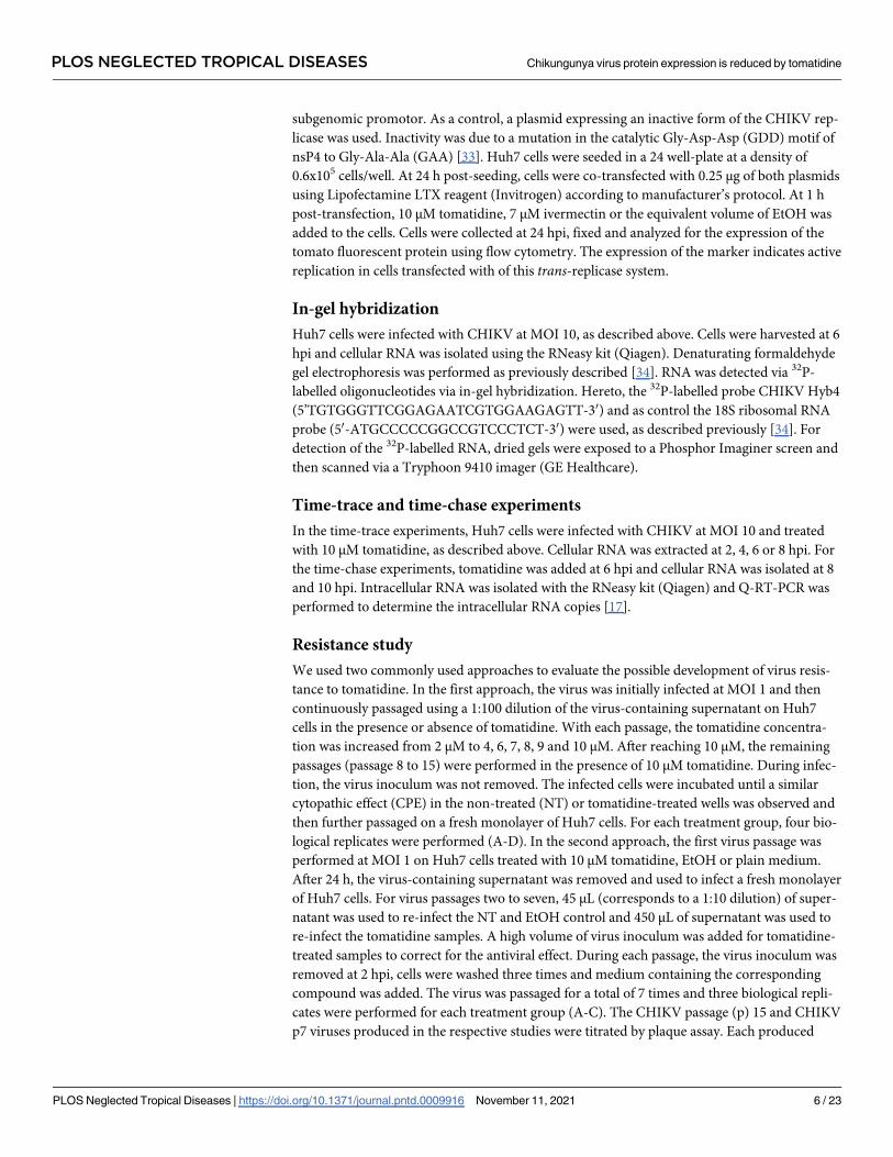
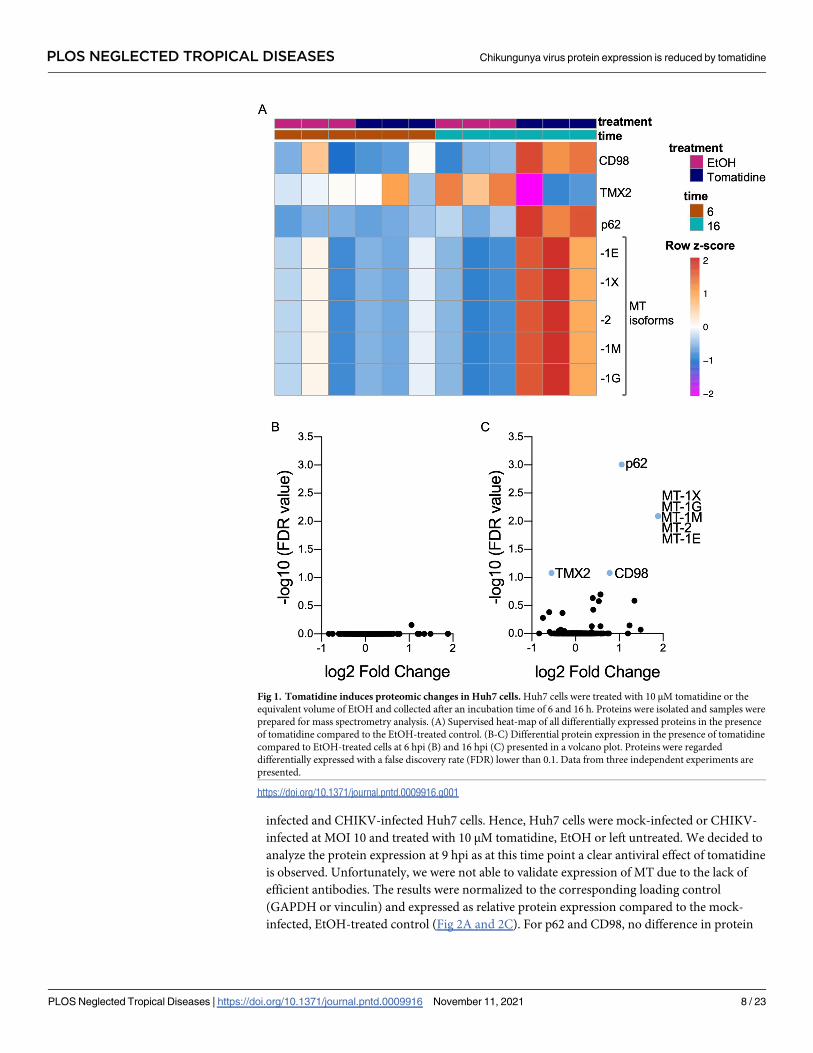
Tomatidine induces a limited number of proteomic changes in Huh7 cells
Tomatidine may exert its antiviral activity towards CHIKV by modulating one or multiple cel-
lular factors required for CHIKV replication. To investigate this hypothesis, we first evaluated
the effect of tomatidine on the host proteome by mass spectrometry. Briefly, human hepatic
Huh7 cells were treated with the highest non-toxic yet antiviral dose of tomatidine (10 μM) or
the equivalent volume of EtOH as solvent control [17]. Human hepatic cells were chosen as
CHIKV is known to infect hepatocytes during natural infection and we previously showed
potent antiviral activity of tomatidine in this cell line [17]. At 6 or 16 h post-treatment, the cel-
lular proteins were isolated and prepared for mass spectrometry. These time-points were cho-
sen to measure early and late proteomic changes upon tomatidine treatment in relation to the
CHIKV replication cycle. In Huh7 cells, one CHIKV replication cycle lasts approximately 8 h.
Our previous study showed a potent antiviral effect up to roughly three replication cycles [17].
Initial review of the data revealed that 1215 proteins were identified during the mass spectrom-
etry analysis. Subsequent analysis revealed that the treatment groups segregated at 16 hpi but
not 6 hpi (S1A and S1B Fig). In line with this, no significant proteomic changes were observed
in tomatidine-treated cells at 6 h when compared to the EtOH control (Fig 1A and 1B),
Table 1). At 16 h of tomatidine treatment, four cellular proteins were differentially expressed
compared to the EtOH control (Fig 1A and 1C). Of these, CD98, p62 and metallothionein
(MT) were significantly upregulated, whereas thioredoxin-related transmembrane protein 2
(TMX2) was significantly downregulated (Fig 1, Table 1). The strongest effect was observed
for MT (3.68-fold), followed by p62 (2.07-fold), CD98 (1.72-fold), and lastly TMX2
(0.68-fold).
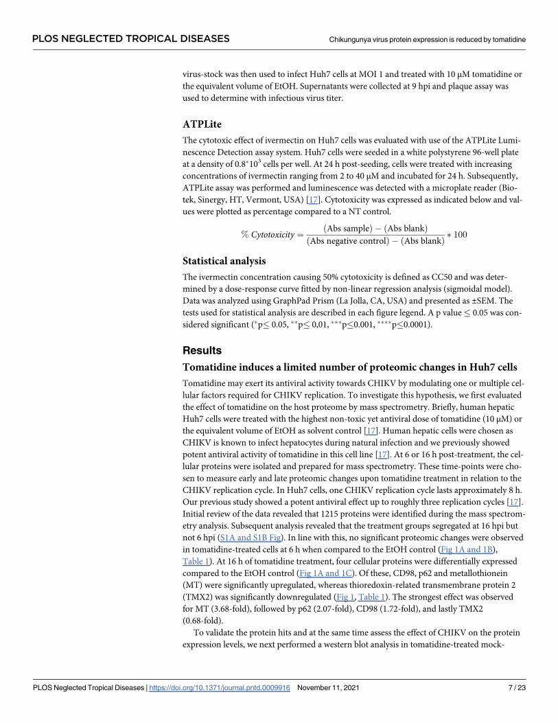
To validate the protein hits and at the same time assess the effect of CHIKV on the protein
expression levels, we next performed a western blot analysis in tomatidine-treated mock-
PLOS NEGLECTED TROPICAL DISEASES Chikungunya virus protein expression is reduced by tomatidine
PLOS Neglected Tropical Diseases | https://doi.org/10.1371/journal.pntd.0009916 November 11, 2021 7 / 23
infected and CHIKV-infected Huh7 cells. Hence, Huh7 cells were mock-infected or CHIKV-
infected at MOI 10 and treated with 10 μM tomatidine, EtOH or left untreated. We decided to
analyze the protein expression at 9 hpi as at this time point a clear antiviral effect of tomatidine
is observed. Unfortunately, we were not able to validate expression of MT due to the lack of
efficient antibodies. The results were normalized to the corresponding loading control
(GAPDH or vinculin) and expressed as relative protein expression compared to the mock-
infected, EtOH-treated control (Fig 2A and 2C). For p62 and CD98, no difference in protein
Fig 1. Tomatidine induces proteomic changes in Huh7 cells. Huh7 cells were treated with 10 μM tomatidine or the
equivalent volume of EtOH and collected after an incubation time of 6 and 16 h. Proteins were isolated and samples were
prepared for mass spectrometry analysis. (A) Supervised heat-map of all differentially expressed proteins in the presence
of tomatidine compared to the EtOH-treated control. (B-C) Differential protein expression in the presence of tomatidine
compared to EtOH-treated cells at 6 hpi (B) and 16 hpi (C) presented in a volcano plot. Proteins were regarded
differentially expressed with a false discovery rate (FDR) lower than 0.1. Data from three independent experiments are
presented.
https://doi.org/10.1371/journal.pntd.0009916.g001
PLOS NEGLECTED TROPICAL DISEASES Chikungunya virus protein expression is reduced by tomatidine
PLOS Neglected Tropical Diseases | https://doi.org/10.1371/journal.pntd.0009916 November 11, 2021 8 / 23
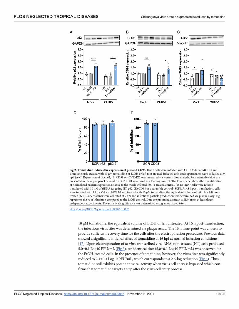
expression was observed between the NT and EtOH-treated samples, demonstrating that the
solvent does not induce changes in the expression of these proteins (Fig 2A and 2B). Tomati-
dine treatment increased p62 expression by 1.7-fold in mock-infected cells and 1.8-fold in
CHIKV-infected samples when compared to the solvent control. For CD98, a 1.6-fold increase
in the mock-infected samples and a 1.4-fold increase in the CHIKV-infected samples was
observed upon tomatidine treatment (Fig 2B). For TMX2, a significant difference between the
NT and EtOH-treated samples was seen during mock-infection. However, the presence of
tomatidine did not induce any significant change in the protein expression of TMX2 (Fig 2C).
Altogether, we validated p62 and CD98, which are both upregulated upon tomatidine treat-
ment irrespective of whether the cells are infected with CHIKV or not.
CD98 and p62 are not involved in the anti-Chikungunya virus activity of
tomatidine
We next evaluated whether the upregulation of CD98 and p62 plays a role in the antiviral
mechanism of tomatidine. Hence, we performed a siRNA knockdown of CD98 and p62 in
Huh7 cells using reverse transfection. A non-coding scramble siRNA (SCR) was used as con-
trol. At 48 h post-transfection, Huh7 cells were infected with CHIKV at MOI 10 and treated
with 10 μM tomatidine, EtOH or left untreated. At 9 hpi, expression of CD98 and p62 and the
production of progeny virus was determined. The expression of CD98 and p62 was normalized
to the loading control and displayed as relative expression compared the mock-infected NT
SCR control. Upon transfection with the siRNA, the average CD98 protein level was signifi-
cantly reduced by 3.2-fold when compared to the SCR control (S2A Fig). For cells transfected
with p62 siRNAs #1 and #4, the average p62 level was reduced by 3.3-fold and 3.5-fold com-
pared to the SCR control, respectively (S2B Fig). All original western blot scans of S2 Fig are
depicted in S3 Fig. Despite the efficient knockdown of CD98 and p62, tomatidine was still
found highly antiviral towards CHIKV (Fig 2D and 2E). For cells with knock-down of CD98
and p62 expression, the inhibition of infectious particle production was 91.68% and 86.37%
(siRNA #1) or 90.82% (siRNA #4) respectively. This was similar to the inhibitory effect
observed in cells treated with the corresponding SCR controls (Fig 2D and 2E). Thus, p62 and
CD98 are not involved in the antiviral potency of tomatidine towards CHIKV in Huh7 cells.
Tomatidine does not affect Chikungunya virus cell entry
Next, we focused on elucidating the steps of the virus replication cycle that are inhibited by
tomatidine. The time-of-addition experiment presented in our previous study suggests that
tomatidine acts after virus attachment and entry into the host cell as the most pronounced
antiviral effect was seen when the compound was added at post-entry conditions [17]. To vali-
date this finding, we here performed a virus cell entry-bypass assay. Hereto, in vitro tran-
scribed RNA was directly electroporated into cells which were subsequently treated with
Table 1. Fold changes in protein expression.
Protein hit 6 h Toma vs EtOH 16 h Toma vs EtOH
CD98 0.95 1.72�
TMX-2 1.04 0.68�
p62 1.02 2.07�
MT 0.93 3.68�
�FDR <0.1 # Orange: upregulation; blue: downregulation
https://doi.org/10.1371/journal.pntd.0009916.t001
PLOS NEGLECTED TROPICAL DISEASES Chikungunya virus protein expression is reduced by tomatidine
PLOS Neglected Tropical Diseases | https://doi.org/10.1371/journal.pntd.0009916 November 11, 2021 9 / 23
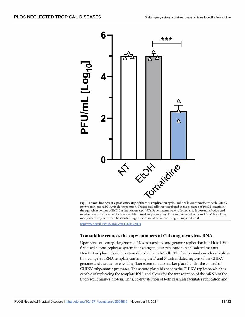
10 μM tomatidine, the equivalent volume of EtOH or left untreated. At 16 h post-transfection,
the infectious virus titer was determined via plaque assay. The 16 h time-point was chosen to
provide sufficient recovery time for the cells after the electroporation procedure. Previous data
showed a significant antiviral effect of tomatidine at 16 hpi at normal infection conditions
[17]. Upon electroporation of in vitro transcribed viral RNA, non-treated (NT) cells produced
5.0±0.1 Log10 PFU/mL (Fig 3). An identical titer (5.0±0.1 Log10 PFU/mL) was observed for
the EtOH-treated cells. In the presence of tomatidine, however, the virus titer was significantly
reduced to 2.4±0.3 Log10 PFU/mL, which corresponds to a 2.6-log reduction (Fig 3). Thus,
tomatidine still exhibits potent antiviral activity when virus cell entry is bypassed which con-
firms that tomatidine targets a step after the virus cell entry process.
Fig 2. Tomatidine induces the expression of p62 and CD98. Huh7 cells were infected with CHIKV-LR at MOI 10 and
simultaneously treated with 10 μM tomatidine or EtOH or left non-treated. Infected cells and supernatants were collected at 9
hpi. (A-C) Expression of (A) p62, (B) CD98 or (C) TMX2 was measured via western blot analysis. Representative blots are
presented in the upper panel. Vinculin or GAPDH were used as a loading control. The lower panel shows the quantification
of normalized protein expression relative to the mock-infected EtOH-treated control. (D-E) Huh7 cells were reverse-
transfected with 10 nM of siRNA targeting (D) p62, (E) CD98 or a scramble control (SCR). At 48 h post-transfection, cells
were infected with CHIKV-LR at MOI 10 and treated with 10 μM tomatidine, the equivalent volume of EtOH or left non-
treated (NT). Supernatants were collected at 9 hpi and infectious particle production was determined via plaque assay. Fig
represents the % of inhibition compared to the EtOH control. Data are presented as mean ± SEM from at least three
independent experiments. The statistical significance was determined using an unpaired t-test.
https://doi.org/10.1371/journal.pntd.0009916.g002
PLOS NEGLECTED TROPICAL DISEASES Chikungunya virus protein expression is reduced by tomatidine
PLOS Neglected Tropical Diseases | https://doi.org/10.1371/journal.pntd.0009916 November 11, 2021 10 / 23
Tomatidine reduces the copy numbers of Chikungunya virus RNA
Upon virus cell entry, the genomic RNA is translated and genome replication is initiated. We
first used a trans-replicase system to investigate RNA replication in an isolated manner.
Hereto, two plasmids were co-transfected into Huh7 cells. The first plasmid encodes a replica-
tion competent RNA template containing the 5’ and 3’ untranslated regions of the CHIKV
genome and a sequence encoding fluorescent tomato marker placed under the control of
CHIKV subgenomic promoter. The second plasmid encodes the CHIKV replicase, which is
capable of replicating the template RNA and allows for the transcription of the mRNA of the
fluorescent marker protein. Thus, co-transfection of both plasmids facilitates replication and
Fig 3. Tomatidine acts at a post-entry step of the virus replication cycle. Huh7 cells were transfected with CHIKV
in vitro transcribed RNA via electroporation. Transfected cells were incubated in the presence of 10 μM tomatidine,
the equivalent volume of EtOH or left non-treated (NT). Supernatants were collected at 16 h post-transfection and
infectious virus particle production was determined via plaque assay. Data are presented as mean ± SEM from three
independent experiments. The statistical significance was determined using an unpaired t-test.
https://doi.org/10.1371/journal.pntd.0009916.g003
PLOS NEGLECTED TROPICAL DISEASES Chikungunya virus protein expression is reduced by tomatidine
PLOS Neglected Tropical Diseases | https://doi.org/10.1371/journal.pntd.0009916 November 11, 2021 11 / 23
the expression of the fluorescent tomato marker is indicative of RNA replication. As a control,
a non-functional replicase plasmid (GAA) was used. Moreover, ivermectin, a previously
reported antiviral compound described to inhibit CHIKV replication, was included as a posi-
tive control [35]. At 1 h post-transfection, 10 μM tomatidine, the equivalent volume of EtOH
or 7 μM ivermectin were added to the cells. The selected ivermectin concentration was shown
to be non-toxic in Huh7 cells via an ATPLite assay (S4A Fig). At 24 h post-transfection, the
expression of the fluorescent tomato marker was analyzed via flow cytometry. As expected, co-
transfection of the template-expressing plasmid with the non-functional replicase plasmid
(GAA) did not yield any fluorescent signal (S4B Fig). In contrast, co-transfection with the
functional replicase plasmid led to 7.55±0.75% and 8.18±0.45% fluorescent tomato-expressing
cells, in NT and EtOH-treated samples, respectively (S4B Fig). Treatment with the positive
control ivermectin reduced the percentage of tomato-expressing cells to 2.19±0.5%, which cor-
responds to a reduction of 73% compared to the EtOH control. Tomatidine treatment reduced
the percentage of marker protein expressing cells to 4.4±0.69%, which represents a reduction
by 46% compared to the EtOH control. Hence, tomatidine inhibits the RNA replication in a
CHIKV trans-replicase system, albeit to a lesser extent than ivermectin. In this assay, we also
determined the mean fluorescent intensity (MFI) of the tomato marker within the marker-
positive cell population and expressed the data as MFI relative to the EtOH control. Ivermectin
treatment significantly reduced the MFI to 0.4±0.06 (S4C Fig). For tomatidine, however, no
effect on the MFI was observed (1.0±0.06), indicating that tomatidine reduces the initiation of
replication but once replication is established within the cell, tomatidine can no longer inter-
fere with the process. Hereafter, we aimed to investigate the effect of tomatidine on RNA repli-
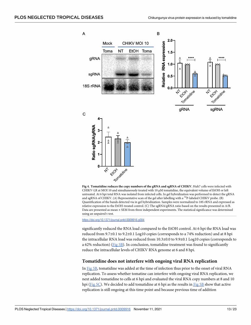
cation under CHIKV infection conditions. First, we visualized the positive-sense genomic
RNA (gRNA) and subgenomic RNA (sgRNA) via in-gel hybridization. The sgRNA is pro-
duced later during the infection process and contains the 3’ ORF encoding the structural pro-
teins needed to produce progeny virions [2]. Huh7 cells were infected with CHIKV at MOI 10
and treated with 10 μM tomatidine, the equivalent volume of EtOH or left non-treated. At 6
hpi, the total RNA was isolated from infected cells and in gel-hybridization was performed. As
loading control, the 18S ribosomal RNA (rRNA) was used (Fig 4A). Results were normalized
to the loading control and expressed as relative RNA level compared to the EtOH control (Fig
4B). Upon CHIKV infection, both gRNA and sgRNA was detected and no difference was seen
in RNA levels between the NT and EtOH samples (Fig 4A and 4B). Importantly, a clear reduc-
tion in gRNA (1.7-fold, corresponding to 40%) and sgRNA (2-fold, corresponds to 50%) band
intensity was observed for the tomatidine-treated samples when compared to the EtOH con-
trol (Fig 4A and 4B). To assess whether tomatidine interferes with the specific expression of
one of the two viral RNAs, we next determined the sgRNA to gRNA ratio (Fig 4C). No statisti-
cal differences were observed in the presence or absence of tomatidine, indicating that tomati-
dine interferes with the overall expression of gRNA and sgRNA rather than with the synthesis
of gRNA or sgRNA, in particular.
To gain a better understanding of the dynamics and timing of the inhibitory effect of toma-
tidine, we next infected Huh7 cells in the presence or absence of tomatidine with CHIKV at
MOI 10 and determined the number of intracellular viral RNA copies at 1, 2, 4, 6 and 8 hpi
(Fig 5A). For the mock-infected samples, the detected viral RNA copies fell below the thresh-
old of detection (N.D.) (Fig 5B). For the CHIKV-infected samples, the amount of viral RNA
increased over time and reached 10.3±0.1 Log10 RNA copies at 8 hpi (Fig 5B). At 1 and 2 hpi,
the RNA load remained constant (~7 Log10 copies) for all experimental conditions, which
suggests that these numbers reflect RNA released from incoming virus particles (Fig 5B). At 4
hpi, the RNA load increased to 8.0±0.2 Log10 copies in the EtOH sample and 7.8±0.1 Log10
copies in the tomatidine-treated samples (Fig 5B). At 6 hpi and 8 hpi, tomatidine treatment
PLOS NEGLECTED TROPICAL DISEASES Chikungunya virus protein expression is reduced by tomatidine
PLOS Neglected Tropical Diseases | https://doi.org/10.1371/journal.pntd.0009916 November 11, 2021 12 / 23
significantly reduced the RNA load compared to the EtOH control. At 6 hpi the RNA load was
reduced from 9.7±0.1 to 9.2±0.1 Log10 copies (corresponds to a 74% reduction) and at 8 hpi
the intracellular RNA load was reduced from 10.3±0.0 to 9.9±0.1 Log10 copies (corresponds to
a 62% reduction) (Fig 5B). In conclusion, tomatidine treatment was found to significantly
reduce the intracellular levels of CHIKV RNA genome at 6 and 8 hpi.
Tomatidine does not interfere with ongoing viral RNA replication
In Fig 5B, tomatidine was added at the time of infection thus prior to the onset of viral RNA
replication. To assess whether tomatine can interfere with ongoing viral RNA replication, we
next added tomatidine to cells at 6 hpi and evaluated the viral RNA copy numbers at 8 and 10
hpi (Fig 5C). We decided to add tomatidine at 6 hpi as the results in Fig 5B show that active
replication is still ongoing at this time point and because previous time of addition
Fig 4. Tomatidine reduces the copy numbers of the gRNA and sgRNA of CHIKV. Huh7 cells were infected with
CHIKV-LR at MOI 10 and simultaneously treated with 10 μM tomatidine, the equivalent volume of EtOH or left
untreated. At 6 hpi total RNA was isolated from infected cells. In gel hybridization was performed to detect the gRNA
and sgRNA of CHIKV. (A) Representative scan of the gel after labelling with a 32P-labeled CHIKV probe. (B)
Quantification of the bands detected via in gel hybridization. Samples were normalized to 18S rRNA and expressed as
relative expression to the EtOH-treated control. (C) The sgRNA/gRNA ratio based on the results presented in A/B.
Data are presented as mean ± SEM from three independent experiments. The statistical significance was determined
using an unpaired t-test.
https://doi.org/10.1371/journal.pntd.0009916.g004
PLOS NEGLECTED TROPICAL DISEASES Chikungunya virus protein expression is reduced by tomatidine
PLOS Neglected Tropical Diseases | https://doi.org/10.1371/journal.pntd.0009916 November 11, 2021 13 / 23
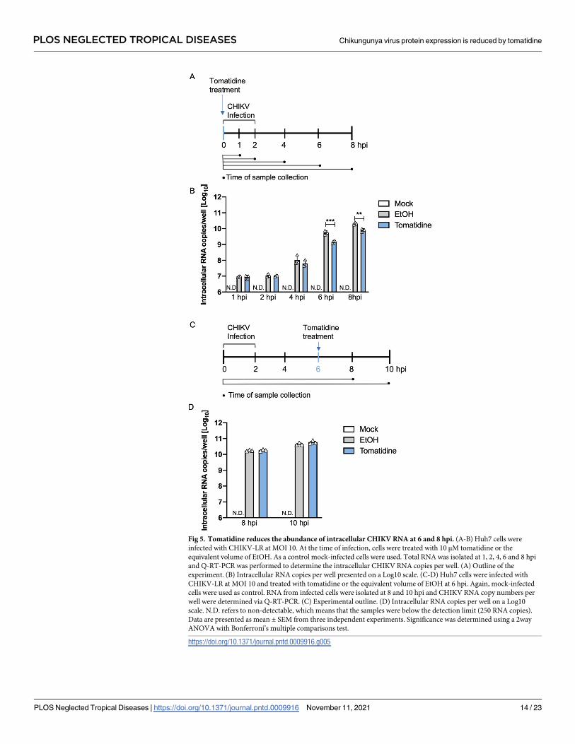
Fig 5. Tomatidine reduces the abundance of intracellular CHIKV RNA at 6 and 8 hpi. (A-B) Huh7 cells were
infected with CHIKV-LR at MOI 10. At the time of infection, cells were treated with 10 μM tomatidine or the
equivalent volume of EtOH. As a control mock-infected cells were used. Total RNA was isolated at 1, 2, 4, 6 and 8 hpi
and Q-RT-PCR was performed to determine the intracellular CHIKV RNA copies per well. (A) Outline of the
experiment. (B) Intracellular RNA copies per well presented on a Log10 scale. (C-D) Huh7 cells were infected with
CHIKV-LR at MOI 10 and treated with tomatidine or the equivalent volume of EtOH at 6 hpi. Again, mock-infected
cells were used as control. RNA from infected cells were isolated at 8 and 10 hpi and CHIKV RNA copy numbers per
well were determined via Q-RT-PCR. (C) Experimental outline. (D) Intracellular RNA copies per well on a Log10
scale. N.D. refers to non-detectable, which means that the samples were below the detection limit (250 RNA copies).
Data are presented as mean ± SEM from three independent experiments. Significance was determined using a 2way
ANOVA with Bonferroni’s multiple comparisons test.
https://doi.org/10.1371/journal.pntd.0009916.g005
PLOS NEGLECTED TROPICAL DISEASES Chikungunya virus protein expression is reduced by tomatidine
PLOS Neglected Tropical Diseases | https://doi.org/10.1371/journal.pntd.0009916 November 11, 2021 14 / 23

experiments [17] revealed that the presence of tomatidine between 6 and 9 hpi significantly
reduced virus progeny production. As before, for the mock-infected samples no viral RNA was
detectable (N.D.) (Fig 5D). In the absence of tomatidine, an intracellular viral RNA copy num-
ber of 10.3±0.0 (8 hpi) and 10.6±0.0 (10 hpi) Log10 was obtained. Comparable results were
obtained in the presence of tomatidine (Fig 5D). Hence, when added during active RNA repli-
cation, tomatidine is not able to interfere with viral RNA replication, indicating that tomati-
dine cannot interfere with ongoing RNA replication.
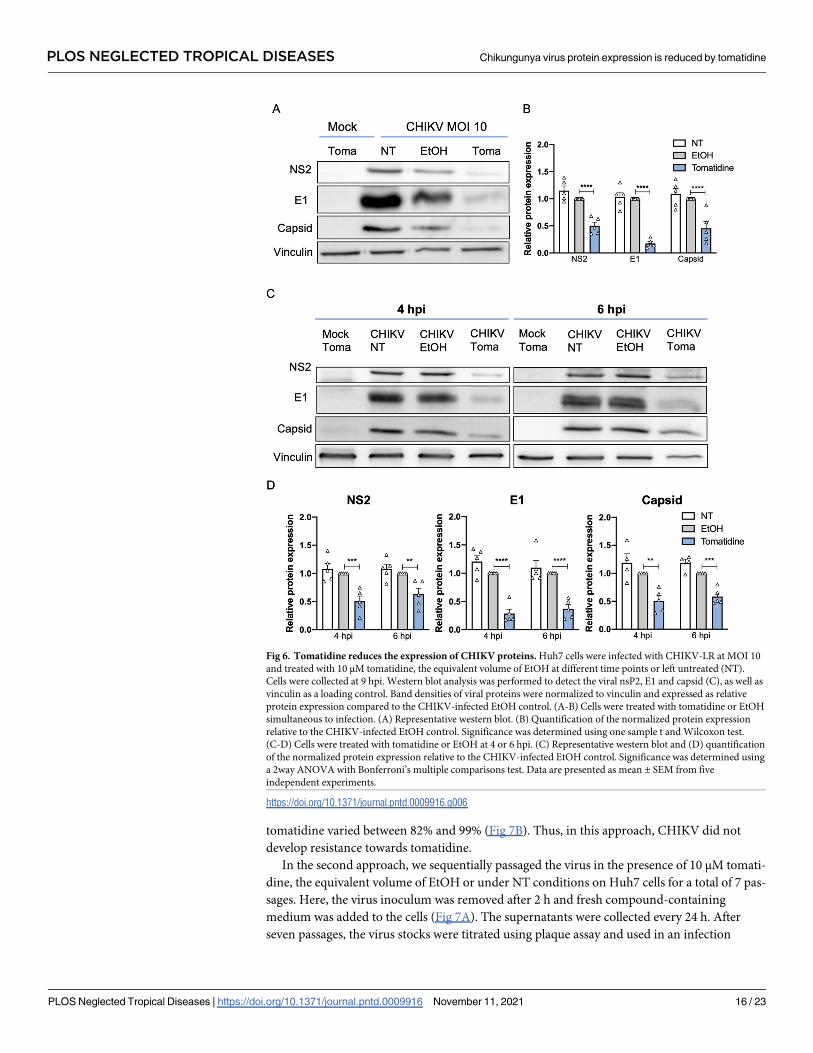
Tomatidine reduces the expression of Chikungunya virus proteins
Next, to test whether the reduction in CHIKV particle production upon tomatidine treatment
is attributed to an effect on viral protein expression, we evaluated the expression levels of the
CHIKV nsP2, E1 and capsid (C) proteins at 9 hpi upon tomatidine treatment at different time-
points (0, 4 and 6 hpi) using western blot analysis. In these experiments, the protein expression
levels were determined at 9 hpi to allow for sufficient protein production for detection. Vincu-
lin was used as a loading control (Fig 6A and 6C). The observed nsP2, E1 and C band densities
on the western blot were normalized to that of vinculin and expressed as fold-change to the
EtOH control (Fig 6B and 6D). Comparable viral protein expression was observed between the
NT and EtOH-treated samples for all detected proteins at all time points, indicating that EtOH
does not affect the viral protein expression levels (Fig 6A and 6D). When tomatidine was
added simultaneous to infection, the nsP2, E1 and C expression was reduced to 0.5±0.1 (50%),
0.18±0.1 (18%) and 0.46±0.1 (46%) respectively, when compared to the EtOH control (Fig 6A
and 6B). A comparable pattern was seen when tomatidine was added at 4 hpi (Fig 6C and 6D).
Here, tomatidine reduced the protein expression to 0.5±0.1 (50%) for nsP2, 0.3±0.1 (30%) for
E1 and 0.5±0.2 (50%) for C. Importantly, the addition of tomatidine at 6 hpi still had an effect
on viral protein expression (Fig 6C and 6D), Under these conditions, nsP2 was reduced to 0.6
±0.2 (60%), E1 was reduced to 0.4± 0.2 (40%) and C was reduced to 0.6±0.1 (60%) compared
to the corresponding EtOH control. The results demonstrate that tomatidine significantly
reduces the expression of the CHIKV proteins during infection when added at the time of
infection but also when added during active CHIKV replication (i.e. at 4 and 6 hpi).
Chikungunya virus does not readily develop resistance towards tomatidine
Resistance to antiviral compounds is often described. This is particularly true for RNA viruses,
since the RNA-dependent RNA polymerase lacks proof-reading activity and has a mutation
rate of 10−6–10−4 substitutions per nucleotide per cell infection [36]. To test resistance devel-
opment of CHIKV towards tomatidine, we applied two frequently used sequential passaging
approaches presented in Fig 7A. In the first approach, CHIKV was passaged in the presence of
increasing tomatidine concentrations on Huh7 cells for 15 passages. More specifically, with
each passage (p), a higher tomatidine concentration was added starting from 2 to 4, 6, 7, 8, 9
and lastly 10 μM (Fig 7A). Passage 8 till p15 was performed in presence of 10 μM tomatidine.
As a control, CHIKV was passaged 15 times in the absence of tomatidine (NT). In this set-up,
the virus inoculum was not removed. Cell culture supernatants were collected once the cyto-
pathic effect (CPE) between the NT and tomatidine-treated samples was similar. The obtained
virus stocks were titrated and then used in an infection experiment (MOI 1) in Huh7 cells
treated with tomatidine or the EtOH control. For the p15 CHIKV of NT stocks, two of the rep-
licates (A and B) were lost in the passaging process (N.D., Fig 7A). The remaining p15 NT
stocks, the p15 tomatidine-treated virus stocks and the parental CHIKV control were highly
sensitive to tomatidine treatment (Figs 7B and S5A). For all samples, the inhibitory effect of
PLOS NEGLECTED TROPICAL DISEASES Chikungunya virus protein expression is reduced by tomatidine
PLOS Neglected Tropical Diseases | https://doi.org/10.1371/journal.pntd.0009916 November 11, 2021 15 / 23
tomatidine varied between 82% and 99% (Fig 7B). Thus, in this approach, CHIKV did not
develop resistance towards tomatidine.
In the second approach, we sequentially passaged the virus in the presence of 10 μM tomati-
dine, the equivalent volume of EtOH or under NT conditions on Huh7 cells for a total of 7 pas-
sages. Here, the virus inoculum was removed after 2 h and fresh compound-containing
medium was added to the cells (Fig 7A). The supernatants were collected every 24 h. After
seven passages, the virus stocks were titrated using plaque assay and used in an infection
Fig 6. Tomatidine reduces the expression of CHIKV proteins. Huh7 cells were infected with CHIKV-LR at MOI 10
and treated with 10 μM tomatidine, the equivalent volume of EtOH at different time points or left untreated (NT).
Cells were collected at 9 hpi. Western blot analysis was performed to detect the viral nsP2, E1 and capsid (C), as well as
vinculin as a loading control. Band densities of viral proteins were normalized to vinculin and expressed as relative
protein expression compared to the CHIKV-infected EtOH control. (A-B) Cells were treated with tomatidine or EtOH
simultaneous to infection. (A) Representative western blot. (B) Quantification of the normalized protein expression
relative to the CHIKV-infected EtOH control. Significance was determined using one sample t and Wilcoxon test.
(C-D) Cells were treated with tomatidine or EtOH at 4 or 6 hpi. (C) Representative western blot and (D) quantification
of the normalized protein expression relative to the CHIKV-infected EtOH control. Significance was determined using
a 2way ANOVA with Bonferroni’s multiple comparisons test. Data are presented as mean ± SEM from five
independent experiments.
https://doi.org/10.1371/journal.pntd.0009916.g006
PLOS NEGLECTED TROPICAL DISEASES Chikungunya virus protein expression is reduced by tomatidine
PLOS Neglected Tropical Diseases | https://doi.org/10.1371/journal.pntd.0009916 November 11, 2021 16 / 23
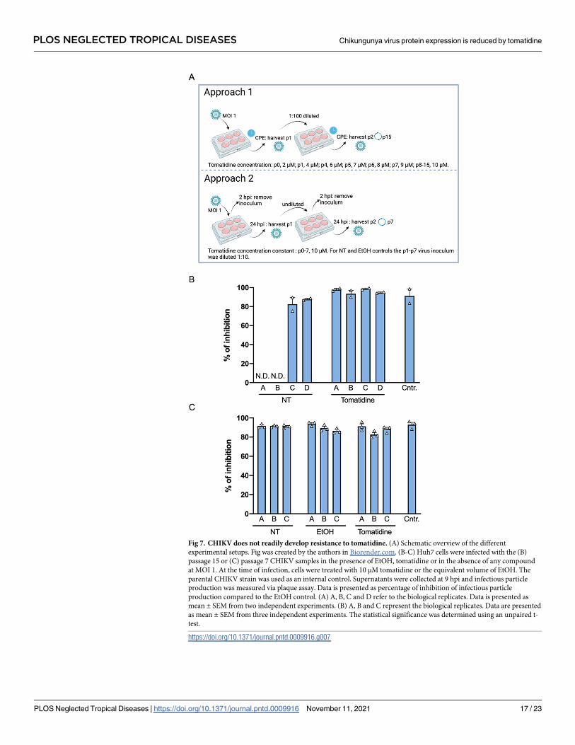
Fig 7. CHIKV does not readily develop resistance to tomatidine. (A) Schematic overview of the different
experimental setups. Fig was created by the authors in Biorender.com. (B-C) Huh7 cells were infected with the (B)
passage 15 or (C) passage 7 CHIKV samples in the presence of EtOH, tomatidine or in the absence of any compound
at MOI 1. At the time of infection, cells were treated with 10 μM tomatidine or the equivalent volume of EtOH. The
parental CHIKV strain was used as an internal control. Supernatants were collected at 9 hpi and infectious particle
production was measured via plaque assay. Data is presented as percentage of inhibition of infectious particle
production compared to the EtOH control. (A) A, B, C and D refer to the biological replicates. Data is presented as
mean ± SEM from two independent experiments. (B) A, B and C represent the biological replicates. Data are presented
as mean ± SEM from three independent experiments. The statistical significance was determined using an unpaired t-
test.
https://doi.org/10.1371/journal.pntd.0009916.g007
PLOS NEGLECTED TROPICAL DISEASES Chikungunya virus protein expression is reduced by tomatidine
PLOS Neglected Tropical Diseases | https://doi.org/10.1371/journal.pntd.0009916 November 11, 2021 17 / 23

experiment at MOI 1. Irrespective of the treatment condition, the p7 virus stocks remained
highly sensitive towards tomatidine treatment (Figs 7C and S5B) as the inhibitory effect of
tomatidine varied between 82% and 94% (Fig 7C). Hence, the sequential passaging of CHIKV
in the presence of 10 μM tomatidine did not lead to the development of resistance. Altogether,
we did not observe resistance development of CHIKV towards tomatidine highlighting the
potential of tomatidine as an antiviral treatment option for CHIKV infection.
Discussion
In this study we investigated the mechanism by which tomatidine interferes with CHIKV
infectivity. Initial mass spectrometry analysis in Huh7 cells revealed that tomatidine signifi-
cantly controls the expression of four cellular proteins. CD98 and p62 were validated yet subse-
quent siRNA knockdown experiments revealed that these proteins are not responsible for the
observed antiviral effect of tomatidine towards CHIKV. In parallel, the effect of tomatidine on
the CHIKV replication cycle was studied and the results are summarized in Fig 8. Potent anti-
viral activity was seen when in vitro transcribed CHIKV RNA was transfected into Huh7 cells
treated with tomatidine. Furthermore, tomatidine treatment was found to significantly inhibit
the number of CHIKV intracellular RNA copies at 6 and 8 hpi. The levels of gRNA and
sgRNA were equally reduced. Also, a clear inhibition of viral nsP2, E1 and the C protein
expression was observed at 9 hpi when tomatidine was added simultaneous to infection but
also when added 4 and 6 hpi. Intriguingly, however, no effect on the RNA copy number was
seen when tomatidine was added during active viral replication, a finding that was supported
Fig 8. Effect of tomatidine on the CHIKV replication cycle. After binding of the virus to cell surface receptors (1), the virus
is internalized and fuses from within early endosomes to release the nucleocapsid (2). The viral RNA is translated to produce
P1234 (3), a polyprotein which upon processing facilitates genome replication (4). Thereafter, replication of a subgenomic
RNA occurs which is translated into the structural proteins C, p62-E1. The C together with progeny genomic RNA form a
nucleocapsid (5). The p62-E1 structural proteins are processed during transit via the secretory pathway and are finally
expressed at the cell surface where virus assembly (5) and budding (6) occurs. Based on the obtained results (box) we
hypothesize that tomatidine actively interferes with protein translation which reduces viral genome replication thereby
limiting the chance to productively infect a cell. The effect of tomatidine on protein translation culminates in a reduced
secretion of progeny virions. Fig was created by the authors in Biorender.com.
https://doi.org/10.1371/journal.pntd.0009916.g008
PLOS NEGLECTED TROPICAL DISEASES Chikungunya virus protein expression is reduced by tomatidine
PLOS Neglected Tropical Diseases | https://doi.org/10.1371/journal.pntd.0009916 November 11, 2021 18 / 23
by data obtained using the CHIKV trans-replicase system. Lastly, sequential passaging of
CHIKV on Huh7 cells in the presence of tomatidine did not induce viral resistance towards
tomatidine.
Previous time-of-addition experiments [17] and the in vitro RNA transfection experiments
presented in this study exclude an inhibiting role of tomatidine during virus cell entry [17].
Indeed, the strongest antiviral effect is seen when the compound is added at post-infection
conditions [17]. Furthermore, the addition of tomatidine to cells was shown to reduce the abil-
ity of the virus to productively infect cells [17]. Subsequent experiments revealed that tomati-
dine treatment reduces the intracellular viral RNA levels yet tomatidine cannot interfere with
ongoing RNA replication. Lastly, tomatidine was shown to reduce viral protein expression lev-
els when added both early and late in infection. This suggests that tomatidine interferes with
RNA translation or facilitates enhanced degradation of viral proteins. Taken together, and
based on the above findings, we hypothesize that tomatidine controls viral protein expression
early in infection thereby hampering efficient RNA replication and thus the chance to produc-
tively infect a cell. Cells that are infected may also have reduced protein expression levels as
tomatidine was still able to interfere with viral protein expression levels when added at 6 hpi.
Thus, we propose that the effect of tomatidine on viral protein expression level early and late
in infection culminates in reduced secretion of progeny virus particles.
Cellular proteins play crucial roles in the replicative cycle of viruses. Furthermore, the high
barrier of CHIKV resistance to tomatidine also suggests that tomatidine exert its antiviral
activity through the modulation of cellular proteins or other host factors. By use of mass spec-
trometry, we aimed to unravel whether host proteins are responsible for the observed antiviral
effect of tomatidine. Notably, we only found a limited number of proteomic changes in cells
treated with tomatidine. This was unexpected given the numerous pathways that have been
reported to be modulated by tomatidine [20–27]. It might be possible that we missed less pro-
nounced proteomic changes as the unlabeled mass spectrometry approach we used picks-up
major changes in the host cell proteome [37]. Importantly, however, the validated individual
protein hits were not responsible for the observed antiviral effect of tomatidine towards
CHIKV. Whether or not simultaneous downregulation of both proteins or other as yet
unknown cellular proteins may affect the antiviral activity of tomatidine towards CHIKV
remains to be elucidated.
Alternatively, the inhibitory activity may not be related to protein expression but rather to the
location or functionality of a protein. With regard to protein functionality, metabolomic studies
may help to identify cellular process that may be inhibited or modulated by tomatidine. One
example could be the host cellular lipid metabolism, which has been reported to play a key role in
alpha- and flavivirus infectivity, translation, replication and morphogenesis [38,39]. During repli-
cation, the virus induces vesicle spherules consisting of host-derived cellular membranes where
replication and translation of the viral RNA occurs [2]. Since it has been reported that tomatidine
ameliorates atherosclerosis-associated hyperlipidemia in vivo it may be possible that tomatidine
modulates the lipid metabolism to exert its antiviral activity [24]. Future label-based mass spec-
trometry analysis combined with immunofluorescence analysis are needed to detect not only the
less profound changes in the protein expression but also their cellular localization. This, together
with metabolomic and lipidomic studies will facilitate to unveil the importance of cellular host
proteins and pathways in the antiviral activity of tomatidine.
RNA viruses are prone to mutations due to the lack of proof-reading activity and therefore
can quickly develop resistance towards an antiviral treatment [40]. As an example, CHIKV
resistance towards arbidol occurs by one single amino acid substitution in the viral E2 protein
and therefore the virus has a low genetic barrier to resistance [41]. In this study, we used two
commonly used approaches to test if tomatidine treatment induces resistance. The results
PLOS NEGLECTED TROPICAL DISEASES Chikungunya virus protein expression is reduced by tomatidine
PLOS Neglected Tropical Diseases | https://doi.org/10.1371/journal.pntd.0009916 November 11, 2021 19 / 23
show that passaging of CHIKV under selection pressure of tomatidine does not readily lead to
viral resistance, which is an essential characteristic of an antiviral compound and strengthens
the potential use of tomatidine as an antiviral treatment towards CHIKV infection. Treatment
should most likely start early after disease onset as its major function would be to reduce the
viral load thereby alleviating disease symptoms.
In conclusion, we delineated that tomatidine interferes with CHIKV infectivity by modulat-
ing the expression levels of viral proteins. The exact viral and/or host factors involved in this
inhibitory effect still need to be elucidated. Future studies on host cell proteomics, lipidomics,
metabolomics will help to elucidate the exact mechanism by which tomatidine controls the
expression of viral proteins. The ability of tomatidine to reduce CHIKV particle production
and the absence of tomatidine resistance warrants further in vivo investigation of its pharma-
cological properties and antiviral efficacy to CHIKV.
Supporting information
S1 Fig. Principal component analysis of tomatidine an EtOH-treated cells. (A) Principle
component analysis at 6 hpi and (B) 16 hpi.
(TIF)
S2 Fig. Knockdown efficiency of siRNAs targeting p62 and CD98. Huh7 cells were reverse-
transfected with 10 nM of siRNA targeting (A) CD98 or (B) p62 or a scramble control (SCR). At
48 h post-transfection, cells were infected with CHIKV-LR at MOI 10 and treated with 10 μM
tomatidine, the equivalent volume of EtOH or left non-treated. Protein was collected at 9 hpi and
CD98, p62 or GAPDH as a loading control were detected via western blot. Samples were analyzed
side-by-side on the same gel yet for clarity the blots were split. (A) The left panel shows the repre-
sentative blots for CD98 and GAPDH. The right panel displays the band quantification normal-
ized to GAPDH and expressed as relative protein expression compared to the mock-infected,
non-treated (NT) control. (B) The left panel shows the representative blots for p62 and GAPDH.
The samples were loaded on distinct gels yet the proteins bands were detected at the same time
and at identical conditions. The right panel displays the band quantification normalized to
GAPDH and expressed as relative protein expression compared to the mock-infected, non-
treated (NT) control. For p62, two different siRNAs (siRNA #1 and #4) were used. Data are pre-
sented as mean ± SEM from three independent experiments. The statistical significance was
determined using one-way ANOVA Dunnett’s multiple comparisons test.
(TIF)
S3 Fig. Original western blot scans of siRNA knockdown targeting CD98 and p62. (A-C)
Original western blot scans of CD98 knockdown including n = 1 (A), n = 2 (B) and n = 3 (C).
(D-F) Western blot scans of p62 knockdown including n = 1 (D), n = 2 (E) and n = 3 (F).
(PDF)
S4 Fig. Tomatidine inhibits CHIKV replication in a trans-replicase system. (A) Dose-
response curve of the ATP level in Huh7 cells assessed by ATPLite assay in the presence of increas-
ing ivermectin concentrations at 24 h treatment period. (B-C) Huh7 cells were transfected with
CHIKV-LR trans-replicase system consisting of a template plasmid including a tomato marker
and a replicase plasmid or its nonfunctional mutant (GAA). At 1 h post-transfection, cells were
treated with 10 μM tomatidine, the equivalent volume of EtOH or ivermectin (7 μM or 0.8 log10)
or left non-treated (NT). At 24 h post-transfection cells were collected, fixed and analyzed via flow
cytometry. (B) Percentage of transfected cells detected via the fluorescent tomato marker and (C)
MFI relative to the CHIKV-infected EtOH control. Data are presented as mean ± SEM from four
PLOS NEGLECTED TROPICAL DISEASES Chikungunya virus protein expression is reduced by tomatidine
PLOS Neglected Tropical Diseases | https://doi.org/10.1371/journal.pntd.0009916 November 11, 2021 20 / 23
independent experiments. The statistical significance was determined using an unpaired t-test.
(TIF)
S5 Fig. CHIKV does not readily develop resistance to tomatidine. (A-B) Huh7 cells were
infected with the (A) passage 15 or (B) passage 7 CHIKV samples in the presence of EtOH,
tomatidine or in the absence of any compound at MOI 1. At the time of infection, cells were
treated with 10 μM tomatidine or the equivalent volume of EtOH. The parental CHIKV strain
was used as an internal control. Supernatants were collected at 9 hpi and infectious particle
production was measured via plaque assay. Data is presented as Log10 PFU per mL. (A) A, B, C
and D refer to the biological replicates. Data is presented as mean ± SEM from two indepen-
dent experiments. (B) A, B and C represent the biological replicates. Data are presented as
mean ± SEM from three independent experiments. The statistical significance was determined
using an unpaired t-test.
(TIF)
Author Contributions
Conceptualization: Berit Troost-Kind, Martijn J. van Hemert, Izabela A. Rodenhuis-Zybert,
Jolanda M. Smit.
Data curation: Berit Troost-Kind, Denise van de Pol, Heidi van der Ende-Metselaar.
Formal analysis: Berit Troost-Kind, Denise van de Pol, Heidi van der Ende-Metselaar, Malte
Borggrewe.
Funding acquisition: Berit Troost-Kind, Izabela A. Rodenhuis-Zybert, Jolanda M. Smit.
Investigation: Berit Troost-Kind, Denise van de Pol, Heidi van der Ende-Metselaar, Andres
Merits, Jolanda M. Smit.
Methodology: Berit Troost-Kind, Martijn J. van Hemert, Denise van de Pol, Heidi van der
Ende-Metselaar, Andres Merits, Malte Borggrewe, Izabela A. Rodenhuis-Zybert, Jolanda
M. Smit.
Project administration: Berit Troost-Kind.
Resources: Martijn J. van Hemert, Andres Merits, Jolanda M. Smit.
Software: Jolanda M. Smit.
Supervision: Martijn J. van Hemert, Izabela A. Rodenhuis-Zybert, Jolanda M. Smit.
Validation: Berit Troost-Kind, Denise van de Pol, Heidi van der Ende-Metselaar, Malte Borg-
grewe, Jolanda M. Smit.
Visualization: Berit Troost-Kind, Malte Borggrewe, Izabela A. Rodenhuis-Zybert.
Writing – original draft: Berit Troost-Kind, Jolanda M. Smit.
Writing – review & editing: Berit Troost-Kind, Martijn J. van Hemert, Andres Merits, Izabela
A. Rodenhuis-Zybert, Jolanda M. Smit.
References1. Fros JJ, Pijlman GP. Alphavirus Infection: Host Cell Shut-Off and Inhibition of Antiviral Responses.
Viruses. 2016; 8: 166. https://doi.org/10.3390/v8060166 PMID: 27294951
2. Silva LA, Dermody TS. Chikungunya virus: Epidemiology, replication, disease mechanisms, and pro-
spective intervention strategies. J Clin Invest. 2017; 127: 737–749. https://doi.org/10.1172/JCI84417
PMID: 28248203
PLOS NEGLECTED TROPICAL DISEASES Chikungunya virus protein expression is reduced by tomatidine
PLOS Neglected Tropical Diseases | https://doi.org/10.1371/journal.pntd.0009916 November 11, 2021 21 / 23
3. Gao S, Song S, Zhang L. Recent Progress in Vaccine Development Against Chikungunya Virus. Front
Microbiol. 2019; 10: 2881. https://doi.org/10.3389/fmicb.2019.02881 PMID: 31921059
4. Galan-Huerta KA, Martınez-Landeros E, Delgado-Gallegos JL, Caballero-Sosa S, Malo-Garcıa IR, Fer-
nandez-Salas I, et al. Molecular and Clinical Characterization of Chikungunya Virus Infections in South-
east Mexico. Viruses. 2018; 10: 248. https://doi.org/10.3390/v10050248 PMID: 29747416
5. Schwartz O, Albert ML. Biology and pathogenesis of chikungunya virus. Nat Rev Microbiol. 2010; 8:
491–500. https://doi.org/10.1038/nrmicro2368 PMID: 20551973
6. Abdelnabi R, Neyts J, Delang L. Towards antivirals against chikungunya virus. Antiviral Res. 2015; 121:
59–68. https://doi.org/10.1016/j.antiviral.2015.06.017 PMID: 26119058
7. Subudhi BB, Chattopadhyay S, Mishra P, Kumar A. Current strategies for inhibition of Chikungunya
infection. Viruses. MDPI AG; 2018. p. 235. https://doi.org/10.3390/v10050235 PMID: 29751486
8. Cruz DJM, Bonotto RM, Gomes RGB, da Silva CT, Taniguchi JB, No JH, et al. Identification of Novel
Compounds Inhibiting Chikungunya Virus-Induced Cell Death by High Throughput Screening of a
Kinase Inhibitor Library. PLoS Negl Trop Dis. 2013; 7: 2471. https://doi.org/10.1371/journal.pntd.
0002471 PMID: 24205414
9. Ozden S, Lucas-Hourani M, Ceccaldi PE, Basak A, Valentine M, Benjannet S, et al. Inhibition of Chikun-
gunya virus infection in cultured human muscle cells by furin inhibitors: Impairment of the maturation of
the E2 surface glycoprotein. J Biol Chem. 2008; 283: 21899–21908. https://doi.org/10.1074/jbc.
M802444200 PMID: 18559340
10. Jhaveri K, Modi S. Ganetespib: Research and clinical development. Onco Targets Ther. 2015; 8: 1849–
1858. https://doi.org/10.2147/OTT.S65804 PMID: 26244021
11. Khan M, Santhosh SR, Tiwari M, Lakshmana Rao P V., Parida M. Assessment of in vitro prophylactic
and therapeutic efficacy of chloroquine against Chikungunya virus in Vero cells. J Med Virol. 2010; 82:
817–824. https://doi.org/10.1002/jmv.21663 PMID: 20336760
12. Leyssen P, De Clercq E, Neyts J. The anti-yellow fever virus activity of ribavirin is independent of error-
prone replication. Mol Pharmacol. 2006; 69: 1461–1467. https://doi.org/10.1124/mol.105.020057
PMID: 16421290
13. Paeshuyse J, Dallmeier K, Neyts J. Ribavirin for the treatment of chronic hepatitis C virus infection: A
review of the proposed mechanisms of action. Curr Opin Virol. 2011; 1: 590–598. https://doi.org/10.
1016/j.coviro.2011.10.030 PMID: 22440916
14. Briolant S, Garin D, Scaramozzino N, Jouan A, Crance JM. In vitro inhibition of Chikungunya and Sem-
liki Forest viruses replication by antiviral compounds: Synergistic effect of interferon-α and ribavirin
combination. Antiviral Res. 2004; 61: 111–117. https://doi.org/10.1016/j.antiviral.2003.09.005 PMID:
14670584
15. Lamballerie X De, Boisson V, Reynier J-C, Enault S, Charrel RN, Flahault A, et al. On Chikungunya
Acute Infection and Chloroquine Treatment. Vector-Borne Zoonotic Dis. 2008; 8: 837–840. https://doi.
org/10.1089/vbz.2008.0049 PMID: 18620511
16. Ravichandran R, Manian M. Ribavirin therapy for Chikungunya arthritis. J Infect Dev Ctries. 2008; 2:
140. https://doi.org/10.3855/jidc.286 PMID: 19738340
17. Troost B, Mulder LM, Diosa-Toro M, van de Pol D, Rodenhuis-Zybert IA, Smit JM. Tomatidine, a natural
steroidal alkaloid shows antiviral activity towards chikungunya virus in vitro. Sci Rep. 2020; 10: 1–12.
https://doi.org/10.1038/s41598-019-56847-4 PMID: 31913322
18. Diosa-Toro M, Troost B, van de Pol D, Heberle AM, Urcuqui-Inchima S, Thedieck K, et al. Tomatidine, a
novel antiviral compound towards dengue virus. Antiviral Res. 2019; 161: 90–99. https://doi.org/10.
1016/j.antiviral.2018.11.011 PMID: 30468746
19. Friedman M. Tomato glycoalkaloids: Role in the plant and in the diet. J Agric Food Chem. 2002; 50:
5751–5780. https://doi.org/10.1021/jf020560c PMID: 12358437
20. Hsieh M-H, Yang J-S, Lin R-C, Hsieh Y-H, Yang S-F, Chang H-R, et al. Tomatidine Represses Invasion
and Migration of Human Osteosarcoma U2OS and HOS Cells by Suppression of Presenilin 1 and c-
Raf–MEK–ERK Pathway. Molecules. 2020; 25: 326. https://doi.org/10.3390/molecules25020326
PMID: 31941156
21. Chiu FL, Lin JK. Tomatidine inhibits iNOS and COX-2 through suppression of NF-κB and JNK pathways
in LPS-stimulated mouse macrophages. FEBS Lett. 2008; 582: 2407–2412. https://doi.org/10.1016/j.
febslet.2008.05.049 PMID: 18544347
22. Kuo CY, Huang WC, Liou CJ, Chen LC, Shen JJ, Kuo ML. Tomatidine Attenuates Airway Hyperrespon-
siveness and Inflammation by Suppressing Th2 Cytokines in a Mouse Model of Asthma. Mediators
Inflamm. 2017; 2017: 1–9. https://doi.org/10.1155/2017/5261803 PMID: 29386751
PLOS NEGLECTED TROPICAL DISEASES Chikungunya virus protein expression is reduced by tomatidine
PLOS Neglected Tropical Diseases | https://doi.org/10.1371/journal.pntd.0009916 November 11, 2021 22 / 23
23. Yan K-H, Lee L-M, Yan S-H, Huang H-C, Li C-C, Lin H-T, et al. Tomatidine inhibits invasion of human
lung adenocarcinoma cell A549 by reducing matrix metalloproteinases expression. Chem Biol Interact.
2013; 203: 580–587. https://doi.org/10.1016/j.cbi.2013.03.016 PMID: 23566884
24. Fujiwara Y, Kiyota N, Tsurushima K, Yoshitomi M, Horlad H, Ikeda T, et al. Tomatidine, a tomato sapo-
genol, ameliorates hyperlipidemia and atherosclerosis in ApoE-deficient mice by inhibiting acyl-CoA:
cholesterol acyl-transferase (ACAT). J Agric Food Chem. 2012; 60: 2472–2479. https://doi.org/10.
1021/jf204197r PMID: 22224814
25. Yu T, Wu Q, You X, Zhou H, Xu S, He W, et al. Tomatidine Alleviates Osteoporosis by Downregulation
of p53. Med Sci Monit. 2020; 26: e923996. https://doi.org/10.12659/MSM.923996 PMID: 32300098
26. Dorsaz S, Snaka T, Favre-Godal Q, Maudens P, Boulens N, Furrer P, et al. Identification and mode of
action of a plant natural product targeting human fungal pathogens. Antimicrob Agents Chemother.
2017; 61: e00829–17. https://doi.org/10.1128/AAC.00829-17 PMID: 28674054
27. Boulet ML, Isabelle C, Guay I, Brouillette E, Langlois JP, Jacques PE, et al. Tomatidine is a lead antibi-
otic molecule that targets staphylococcus aureus ATP Synthase subunit C. Antimicrob Agents Che-
mother. 2018; 62: e02197–17. https://doi.org/10.1128/AAC.02197-17 PMID: 29610201
28. Ebert SM, Dyle MC, Bullard SA, Dierdorff JM, Murry DJ, Fox DK, et al. Identification and small molecule
inhibition of an activating transcription factor 4 (ATF4)-dependent pathway to age-related skeletal mus-
cle weakness and atrophy. J Biol Chem. 2015; 290: 25497–25511. https://doi.org/10.1074/jbc.M115.
681445 PMID: 26338703
29. Ahsan A, Zheng Y, Ma S, Liu M, Cao M, Li Y, et al. Tomatidine protects against ischemic neuronal injury
by improving lysosomal function. Eur J Pharmacol. 2020; 882: 173280. https://doi.org/10.1016/j.ejphar.
2020.173280 PMID: 32580039
30. Robinson MD, McCarthy DJ, Smyth GK. edgeR: A Bioconductor package for differential expression
analysis of digital gene expression data. Bioinformatics. 2009; 26: 139–140. https://doi.org/10.1093/
bioinformatics/btp616 PMID: 19910308
31. Van Duijl-Richter MKS, Blijleven JS, van Oijen AM, Smit JM. Chikungunya virus fusion properties eluci-
dated by single-particle and bulk approaches. J Gen Virol. 2015; 96: 2122–2132. https://doi.org/10.
1099/vir.0.000144 PMID: 25872739
32. Tssetsarkin K, Higgs S, McGee CE, De Lamballerie X, Charrel RN, Vanlandingham DL. Infectious
clones of Chikungunya virus (la reunion isolate) for vector compentence studies. Vector-Borne Zoonotic
Dis. 2006; 6: 325–337. https://doi.org/10.1089/vbz.2006.6.325 PMID: 17187566
33. Utt A, Quirin T, Saul S, Hellstrom K, Ahola T, Merits A. Versatile Trans-Replication Systems for Chikun-
gunya Virus Allow Functional Analysis and Tagging of Every Replicase Protein. Menendez-Arias L, edi-
tor. PLoS One. 2016; 11: e0151616. https://doi.org/10.1371/journal.pone.0151616 PMID: 26963103
34. Scholte FEM, Tas A, Martina BEE, Cordioli P, Narayanan K, Makino S, et al. Characterization of Syn-
thetic Chikungunya Viruses Based on the Consensus Sequence of Recent E1-226V Isolates. Poh LNF,
editor. PLoS One. 2013; 8: e71047. https://doi.org/10.1371/journal.pone.0071047 PMID: 23936484
35. Varghese FS, Kaukinen P, Glasker S, Bespalov M, Hanski L, Wennerberg K, et al. Discovery of berber-
ine, abamectin and ivermectin as antivirals against chikungunya and other alphaviruses. Antiviral Res.
2016; 126: 117–124. https://doi.org/10.1016/j.antiviral.2015.12.012 PMID: 26752081
36. Sanjuan R, Domingo-Calap P. Mechanisms of viral mutation. Cellular and Molecular Life Sciences. Bir-
khauser Verlag AG; 2016. pp. 4433–4448. https://doi.org/10.1007/s00018-016-2299-6 PMID:
27392606
37. Megger DA, Pott LL, Ahrens M, Padden J, Bracht T, Kuhlmann K, et al. Comparison of label-free and
label-based strategies for proteome analysis of hepatoma cell lines. Biochim Biophys Acta—Proteins
Proteomics. 2014; 1844: 967–976. https://doi.org/10.1016/j.bbapap.2013.07.017 PMID: 23954498
38. Ng CG, Coppens I, Govindarajan D, Pisciotta J, Shulaev V, Griffin DE. Effect of host cell lipid metabo-
lism on alphavirus replication, virion morphogenesis, and infectivity. Proc Natl Acad Sci U S A. 2008;
105: 16326–16331. https://doi.org/10.1073/pnas.0808720105 PMID: 18845681
39. Wichit S, Hamel R, Bernard E, Talignani L, Diop F, Ferraris P, et al. Imipramine Inhibits Chikungunya
Virus Replication in Human Skin Fibroblasts through Interference with Intracellular Cholesterol Traffick-
ing. Sci Rep. 2017; 7: 1–12. https://doi.org/10.1038/s41598-016-0028-x PMID: 28127051
40. Irwin KK, Renzette N, Kowalik TF, Jensen JD. Antiviral drug resistance as an adaptive process. Virus
Evol. 2016; 2: vew014. https://doi.org/10.1093/ve/vew014 PMID: 28694997
41. Delogu I, Pastorino B, Baronti C, Nougairède A, Bonnet E, de Lamballerie X. In vitro antiviral activity of
arbidol against Chikungunya virus and characteristics of a selected resistant mutant. Antiviral Res.
2011; 90: 99–107. https://doi.org/10.1016/j.antiviral.2011.03.182 PMID: 21440006
PLOS NEGLECTED TROPICAL DISEASES Chikungunya virus protein expression is reduced by tomatidine
PLOS Neglected Tropical Diseases | https://doi.org/10.1371/journal.pntd.0009916 November 11, 2021 23 / 23