Embed Size (px)
Citation preview
Copyright � 2009 by the Genetics Society of AmericaDOI: 10.1534/genetics.108.098707
Tools for Fungal Proteomics: Multifunctional Neurospora Vectors forGene Replacement, Protein Expression and Protein Purification
Shinji Honda and Eric U. Selker1
Institute of Molecular Biology, University of Oregon, Eugene, Oregon 97403-1229
Manuscript received November 13, 2008Accepted for publication January 16, 2009
ABSTRACT
The completion of genome-sequencing projects for a number of fungi set the stage for detailed inves-tigations of proteins. We report the generation of versatile expression vectors for detection and isolation ofproteins and protein complexes in the filamentous fungus Neurospora crassa. The vectors, which can beadapted for other fungi, contain C- or N-terminal FLAG, HA, Myc, GFP, or HAT–FLAG epitope tags witha flexible poly-glycine linker and include sequences for targeting to the his-3 locus in Neurospora. Tointroduce mutations at native loci, we also made a series of knock-in vectors containing epitope tags followedby the selectable marker hph (resulting in hygromycin resistance) flanked by two loxP sites. We adapted theCre/loxP system for Neurospora, allowing the selectable marker hph to be excised by introduction of Crerecombinase into a strain containing a knock-in cassette. Additionally, a protein purification methodwas developed on the basis of the HAT–FLAG tandem affinity tag system, which was used to purifyHETEROCHROMATIN PROTEIN 1 (HP1) and associated proteins from Neurospora. As expected onthe basis of yeast two-hybrid and co-immunoprecipitation (Co-IP) experiments, the Neurospora DNAmethyltransferase DIM-2 was found in a complex with HP1. Features of the new vectors allowed forverification of an interaction between HP1 and DIM-2 in vivo by Co-IP assays on proteins expressed eitherfrom their native loci or from the his-3 locus.
THE filamentous ascomycete Neurospora crassa hasserved as a model eukaryote for more than 70 years
(Davis 2000; Davis and Perkins 2002). Fifty years agoBeadle and Tatum were awarded a Nobel Prize for their‘‘one-gene, one-enzyme’’ research project with Neuros-pora and numerous subsequent studies with theorganism have provided important insights into metab-olism, gene regulation, chromosome behavior, DNArepair, DNA methylation, genome defense, photobiol-ogy, circadian rhythms, differentiation, development,and other biological phenomena of relevance to highereukaryotes (Dunlap et al. 2007). Simultaneously, Neuros-pora researchers developed a variety of biochemical andgenetic tools.Recently, sequencingoftheN.crassagenome(Galagan et al. 2003) greatly stimulated the field, in partby immediately revealing �10,000 predicted genes(Borkovich etal.2004).Bioinformaticsdataareaccessiblevia public databases, and microarrays covering all thepredicted genes are available from the Fungal GeneticsStock Center and used for transcriptional profiling(Kasuga et al. 2005; Tian et al. 2007; Kasuga and Glass
2008) and for microarray-based genetic mapping (Lewis
et al. 2007). Moreover, a systematic gene knockoutproject todisruptall thepredictedgenes iswellunderway(Colot et al. 2006). Although genetic resources forNeurospora are plentiful and invaluable, proteomicstechnology for this and other filamentous fungi are lessdeveloped. We have sought to improve this situation.
Proteins obviously play critical roles in diverse bi-ological processes through interactions with other pro-teins, DNA, RNA, and small molecules both inside andoutside cells. Antibodies are extremely valuable for thedetection and isolation of proteins in vivo, but genera-tion of antibodies specific for a protein of interest is notalways successful and can be costly. An alternativeapproach is to tag proteins of interest with epitopesthat can be detected with commercially available anti-bodies ( Jarvik and Telmer 1998; Brizzard 2008).Although this obviates the need to raise antibodies,epitope tags fused to either the C terminus or the Nterminus may influence protein folding, mask neigh-boring domains or signal sequences, inhibit proteinprocessing and modification, and interfere with properprotein function (Booher and Kaiser 2008; Sung et al.2008). Similarly, the use of multiple epitopes canincrease sensitivity, but bulky epitopes can also interferewith protein function. A flexible polypeptide linker,such as a 10-glycine chain (flexible due to its absence ofside groups) placed between an epitope and a taggedprotein, can minimize such problems and improve theaccessibility of an epitope to antibodies (Borjigin and
Supporting information is available online at http://www.genetics.org/cgi/content/full/genetics.108.098707/DC1.
Sequence data from this article have been deposited with the EMBL/GenBank Data Libraries under accession nos. FJ456996–FJ457013.
1Corresponding author: Institute of Molecular Biology, University ofOregon, 1370 Franklin Blvd., Eugene, OR 97403-1229.E-mail: [email protected]
Genetics 182: 11–23 (May 2009)
Nathans 1994; Sabourin et al. 2007). We and otherspreviously constructed first-generation expression vec-tors for Neurospora with epitope tags such as FLAG,hemagglutinin (HA), Myc, green fluorescent protein(GFP), and red fluorescent protein, but their cloningsites and other features were not always consistent(Freitag et al. 2004b; Freitag and Selker 2005; He
et al. 2005; Dementhon et al. 2006; Kawabata andInoue 2007). Thus it was laborious to generate differenttagged constructs, as is generally necessary to obtainfully functional and detectable epitope-tagged proteinsbecause of the peculiarities of specific epitopes placed atC or N termini (Booher and Kaiser 2008; Sung et al.2008). Here we report standardized, advanced multi-purpose vectors that simplify simultaneous constructionof a family of epitope-tagged expression constructs.
We also developed materials and methods to identifyprotein complexes. Identification of proteins interact-ing with a given target protein is an excellent initialstrategy for characterizing protein complexes (Pandey
and Mann 2000; Gavin et al. 2002; Cravatt et al. 2007).Yeast two-hybrid assays have been widely used, includingin Neurospora (Li and Borkovich 2006; Honda andSelker 2008), for identification of interacting proteins.The utility of this approach is limited, however, becauseit is time-consuming, is normally limited to detection ofpairwise interactions, and does not generally allow forpost-translational modifications that may create bindingsites for partner proteins. In the past decade, massspectrometry (MS) has become the method of choicefor characterization of protein complexes (Aebersold
and Mann 2003). Typically, epitope-tagged proteins areaffinity-purified from cellular extracts, and associatedproteins are then identified by MS (Chang 2006). Afeature of this method is that it can identify all the com-ponents associated, directly or indirectly, with epitope-tagged proteins of interest under natural conditions. Toreduce background from nonspecific binding proteins,it is common to employ a two-step purification using atandem affinity purification (TAP) tag, which consists oftwo different high-affinity tags (Rigaut et al. 1999).Each TAP tag has advantages and disadvantages (Chang
2006). A Myc-6xHis TAP tag purification system hasbeen successfully used in Neurospora (He et al. 2002,2005), but the FLAG-6xHis system is more economicalthan any other available affinity purification systems andhas been demonstrated to work well in mammals andDrosophila (Yang et al. 2006; Ooi et al. 2007). Unfor-tunately, the high charge of the 6xHis tag can influencefolding of 6xHis-tagged proteins. A natural 19-amino-acidpoly-histidine affinity tag (HAT) derived from chickenlactate dehydrogenase is preferable to the 6xHis epitopebecause the overall charge of the HAT epitope is lower(Chaga et al. 1999).
After identifying potential interacting proteins, re-searchers can use different tagged epitopes to verifytheir interaction by co-immunoprecipitation (Co-IP)
from extracts. In Neurospora, it is common to targetepitope-tagged genes to the his-3 locus by replacing ahis-3 mutation with wild-type sequences (Margolin et al.1997; Lee et al. 2003), while in yeast the high efficiencyof homologous recombination makes it easy to expressepitope-tagged proteins from their native loci (Longtine
et al. 1998), facilitating coexpression of multiple con-structs. Most eukaryotes, including Neurospora, have astrong nonhomologous end-joining (NHEJ) system thatresults in ectopic insertions of exogenous DNA, and thiscomplicates strategies based on homologous recombi-nation. This problem has recently been overcome inNeurospora, however, by utilization of mutants (e.g.,mus-51, mus-52, or mus-53) that are defective in NHEJand therefore are only capable of homologous recom-bination (Ninomiya et al. 2004; Ishibashi et al. 2006).The current Neurospora gene knock-out project usesSaccharomyces cerevisiae for assembly of constructs, takingadvantage of the fact that yeast needs only 30–40nucleotides of sequence homology for recombination(Oldenburg et al. 1997). Following their example(Colot et al. 2006), we used this approach to generateknock-in constructs for Neurospora, incorporatingfeatures of the Cre/loxP site-specific recombinationsystem (Yu and Bradley 2001). The bacteriophageCre recombinase specifically recognizes two loxP sitesand leads to reciprocal recombination between them,resulting in excision, inversion, and translocation (Van
Duyne 2001). This system functions in various organ-isms, including some fungi (Krappmann et al. 2005;Forment et al. 2006), and has been exploited for variouspurposes, including excision of selectable markers thatare no longer required or desirable.
Here we report the development of convenient entryand expression vectors to build N- or C-terminal fusionproteins with FLAG, HA, Myc, GFP, or HAT–FLAG TAPtags and poly-glycine flexible linkers for expression atnative loci or at his-3. To demonstrate the utility of thesetools, we purified HETEROCHROMATIN PROTEIN 1(HP1)-associated proteins using the HAT–FLAG purifi-cation system and identified DIM-2 among the interactingproteins by MS analysis. The DIM-2–HP1 interaction wasthen verified in vivo by Co-IP using the epitope-taggingsystem. We also demonstrate that the Cre/loxP systemworks efficiently in Neurospora and allows for ‘‘recycling’’of the popular hygromycin-resistance selectable marker,hph. Key plasmids constructed in this study will be madeavailable through the Fungal Genetics Stock Center.
MATERIALS AND METHODS
Neurospora strains and isolation of genomic DNA: N.crassa strains used in this study are listed in supportinginformation, Table S1 and were grown, maintained, andcrossed according to standard published procedures (Davis
2000). For isolation of genomic DNA, Neurospora strains weregrown with shaking in Vogel’s minimal medium with 1.5%
12 S. Honda and E. U. Selker
sucrose and required supplements at 32� for 2 days; genomicDNA was isolated and used for PCR and Southern hybrid-izations as described previously (Freitag et al. 2004a).
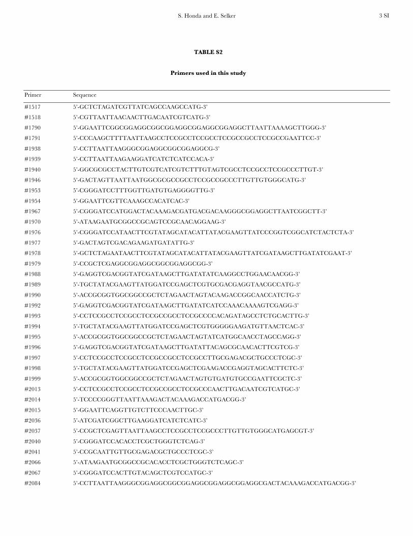
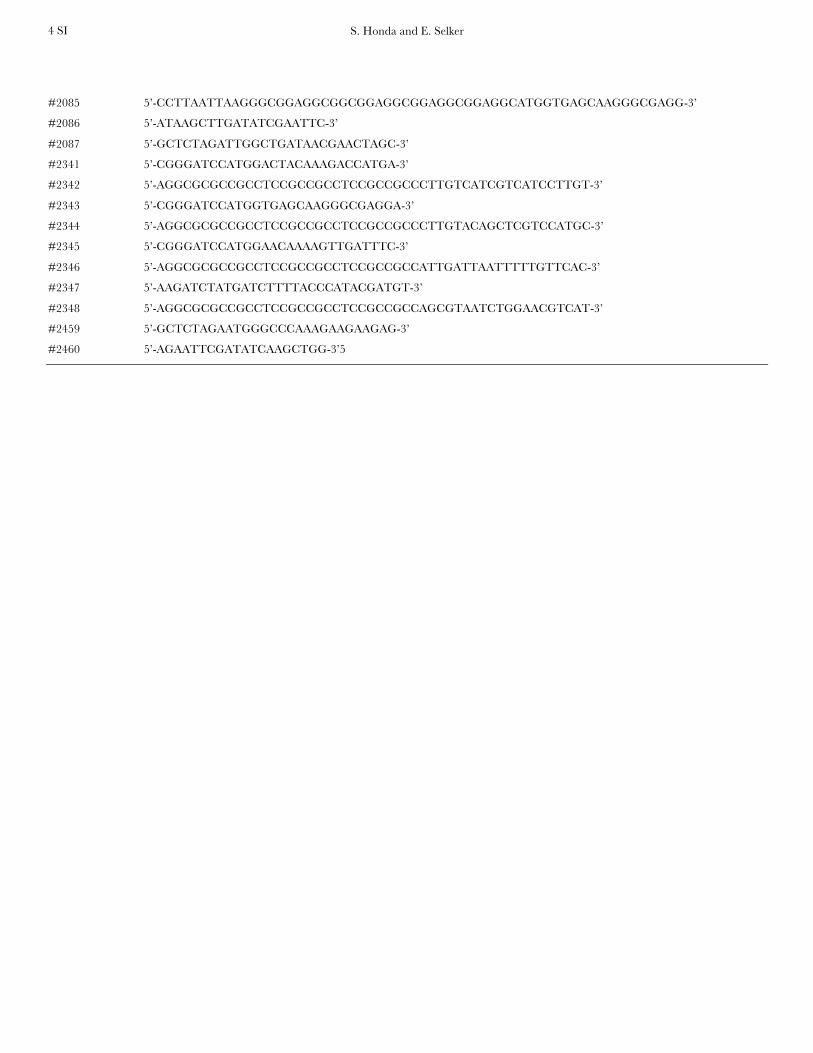
Construction of yeast shuttle vector containing a 10xGlylinker: All primers and synthetic oligonucleotides used in thisstudy are listed in Table S2. Synthetic oligonucleotides #1790and #1791, which contain a 10xGly linker sequence, an EcoRIsite on one end, and a PacI site followed by a HindIII site on theother end, were incubated at room temperature for 30 minafter boiling for 5 min. The annealed oligonucleotides weredigested with EcoRI and HindIII and inserted into EcoRI 1HindIII-digested pRS416 yeast shuttle vector (NEB), yieldingpRS416-10xGly.
Construction of his-3-targeting vectors containing C-terminal3xFLAG or HAT–FLAG tags: We amplified a fragmentcontaining the 3xFLAG sequence by PCR using the p3xFLAGCMV14 vector (Sigma) as the template with forward primer#2014 containing a PacI site and reverse primer #2015containing an EcoRI site. The PCR products were digestedwith PacI and EcoRI and inserted into PacI 1 EcoRI-digestedpMF270, which is a his-3 targeting vector containing a C-terminal 3xHA-epitope tag. In this way, we replaced the 3xHAtag with the 3xFLAG tag, yielding pCCGTC-3xFLAG. In somecases, fewer than 10 glycines were included in the poly-glycinelinker to render constructions practical. To construct a vectorcontaining a HATT5xGlyTFLAG tag, a fragment containing aHAT tag was amplified by PCR from the vector pHAT10(Clontech) with forward primer #2036, which contains a PvuIsite, and reverse primer #2037, which contains a 5xGly linkerand a PacI site followed by a XhoI site. The PCR fragments weredigested with XhoI and PvuI and inserted into pRS416-10xGlydigested with XhoI and PacI, which produce ends compatiblewith PvuI, yielding pRS416-10xGly-HAT-5xGly. A fragmentcontaining a HATT5xGlyTFLAG tag was generated by PCRusing pRS416-10xGly-HAT-5xGly as template with forwardprimer #1939, which contains a 1xFLAG sequence followedby a PacI site, and reverse primer #1940, which contains an AscIsite. The PCR products were digested with PacI and AscI andinserted into PacI 1 AscI-digested pMF270, yielding pCCGTC-HATTFLAG.
Construction of his-3-targeting vectors containing a 10xGlylinker followed by C-terminal 3xFLAG, GFP, or HAT–FLAGtags: We amplified a fragment containing the 3xFLAG tag byPCR using the p3xFLAG CMV14 vector (Sigma) as thetemplate with forward primer #2084, which contains a 10xGlylinker plus a PacI site, and reverse primer #2015, whichcontains an EcoRI site. The PCR products were digested withPacI and EcoRI and inserted into PacI 1 EcoRI-digestedpMF270 to replace the 3xHA tag with the 10xGlyT3xFLAGtag, yielding pCCGTC-GlyT3xFLAG. pCCGTC-GlyTGFP,which is a his-3 targeting vector containing a 10xGly linkerfollowed by a GFP tag at its C terminus, was similarly generatedusing forward primer #2085, reverse primer #2086, andpMF272 (Freitag et al. 2004b) as template. A fragmentcontaining a 10xGlyTHATT5xGlyTFLAG tag was amplifiedby PCR with forward primer #1938 containing a PacI site,reverse primer #1940 containing an AscI site, and pRS416-10xGly-HAT-5xGly as the template. The PCR products weredigested with PacI and AscI and inserted into PacI 1 AscI-digested pMF270, yielding pCCGTC-GlyTHATTFLAG.
Construction of his-3-targeting vectors containing N-terminal3xFLAG, GFP, 3xMyc, 3xHA, or FLAG–HAT tags followedby a poly-glycine linker: We amplified a fragment containingthe Neurospora ccg-1 promoter (Pccg-1) (McNally and Free
1988) by PCR from pMF270 with forward primer #1954, whichcontains an EcoRI site, and reverse primer #1953, whichcontains a BamHI site. The PCR products were digested withEcoRI and BamHI and inserted into pBM61 digested with
the corresponding restriction enzymes (Margolin et al.1997), yielding pCCG-N. To obtain a fragment containingthe FLAG tag followed by a HAT tag, we amplified the HAT tagsequence by PCR from the pCCGTC-HATTFLAG vector withforward primer #1967, which contains a BamHI site followedby a start codon (ATG), a single FLAG sequence, and a 3xGlylinker, and reverse primer #1946, which contains a 5xGly tailsequence followed by AscI, PacI, and SpeI sites. The PCRproducts were digested with BamHI and SpeI and inserted intoBamHI 1 SpeI-digested pCCG-N, yielding pCCGTN-FLAGTHAT. A fragment containing the 3xFLAG tag was amplified byPCR from pCCGTC-FLAG with forward primer #2341, whichcontains a BamHI site followed by a start codon (ATG), andreverse primer #2342, which contains an 8xGly linker followedby an AscI site. The PCR products were digested with BamHIand AscI and inserted into pCCGTN-FLAGTHAT digestedwith the corresponding restriction enzymes to replace itsFLAGTHAT tag with the 3xFLAG tag, yielding pCCGTN-3xFLAG. pCCGTN-GFP and pCCGTN-3xMyc, which are his-3targeting vectors with N-terminal GFP or 3xMyc tags followedby 8xGly linkers, were similarly generated using forwardprimer #2343, reverse primer #2344, and pMF272 as templateor forward primer #2345, reverse primer #2346, and pMF276as template. To generate pN-3xHA, a fragment containing a3xHA tag was amplified by PCR from pMF270 with forwardprimer #2347, which contains a BglII site, and reverse primer#2348 and then introduced into SmaI 1 SpeI-digested pBM61.
Construction of vectors to target dim-2-3xFLAG or hpo-gfpto the his-3 locus: A 500-bp segment of dim-2, including thepromoter region, was amplified by PCR with forward primer#1970, which contains a NotI site and the reverse primer #2087,which contains an XbaI site. The PCR product and the vectorpCCGTC-3xFLAG were digested with NotI and XbaI andligated to replace the Pccg-1 with the dim-2 promoter. Theplasmid containing the dim-2 promoter was then digested withXbaI and PacI and ligated with a fragment containing thecoding region of the dim-2 gene, which had been amplified byPCR from Neurospora genomic DNA with forward primer#1517, which contains an XbaI site, reverse primer #1518,which contains a PacI site, and digested with XbaI and PacI.For the HP1 construct, a 500-bp promoter region of hpo wasamplified by PCR with primers #2066 and #2067. The codingregion of hpo was cut out from pMF308 (Freitag et al. 2004a)by digestion with BamHI and XbaI. The two fragments wereinserted into pCCGTC-GFP. The constructs for GFP-taggedHP1 and FLAG-tagged DIM-2 were linearized and inserted atthe his-3 locus of the hpo strain N3395 and the dim-2 nullmutant N3396, respectively, by the gene replacement methodpreviously described (Margolin et al. 1997).
Construction of knock-in vectors containing the 10xGlylinker followed by a 3xFLAG, 3xHA, GFP, or 13xMyc tag atthe C terminus and the hph gene flanked by loxP sequences:We amplified a fragment containing the bacterial hph gene,which confers hygromycin resistance (hygR) in Neurospora,from pCSN44 (Staben et al. 1989) by PCR with forward primer#1977, which contains a SpeI site, and reverse primer #1976,which contains a loxP sequence followed by a BamHI site. ThePCR products were digested with SpeI and BamHI and in-serted into SpeI 1 BamHI-digested vector pZErO-2 (Clontech),yielding pZErO-hphTloxP. To insert epitope tags and a 10xGlylinker for construction of knock-in modules, we first gener-ated constructs containing the hpo gene with a 10xGly linkerfollowed by epitope tags and then used them as templates forPCR. A fragment of the hpo coding region with its promoterwas amplified by PCR from genomic DNA of Neurospora N623with forward primer #2040, which contains a BamHI site, andreverse primer #2041, which contains a MfeI site. The PCRproducts were digested with BamHI and MfeI and inserted into
Biochemical and Genetic Tools for Neurospora 13
pRS416-10xGly between the BamHI and EcoRI sites (endscompatible with those generated by MfeI), yielding pRS416-HP1T10xGly. A fragment containing the hpo gene with a10xGly tail was cut out from pRS416-HP1T10xGly by digestionof NotI and PacI and inserted into pCCGTC-3xFLAG, pMF270,pMF272, or pMF276 between the NotI and PacI sites, yieldingpHP1T10xGlyT3xFLAG, pHP1T10xGlyT3xHA, pHP1T10xGlyTGFP, or pHP1T10xGlyT13xMyc, respectively. Finally, we usedforward primer #1979, containing a XhoI site, and reverseprimer #1978, which contains the loxP sequence followed byan XbaI site, to amplify the 10xGly and epitope tags frompHP1T10xGlyT3xFLAG, pHP1T10xGlyT3xHA, pHP1T10xGlyTGFP, or pHP1T10xGlyT13xMyc. The PCR products then weredigested with XhoI and XbaI and inserted into pZErO-hphTloxPbetween the XhoI and SpeI sites (compatible overhang withXbaI), yielding p3xFLAGThphTloxP, p3xHAThphTloxP, pGFPThphTloxP, or p13xMycThphTloxP, respectively.
Engineering of epitope-tagged proteins for expression ofgenes at their native loci: We modified a gene knock-outprocedure (Colot et al. 2006) to serve as a convenient knock-in strategy for Neurospora genes. We used recombination-mediated plasmid construction in S. cerevisiae (Oldenburg
et al. 1997) to make knock-in cassettes. The system worked well:58 of 66 transformants (87.9%) from 12 different constructswere found to have generated the intended cassettes. Yeaststrains were grown and maintained according to the YeastProtocol Handbook (Clontech), and the S. cerevisiae strainPJ69-4A was used as the host strain ( James et al. 1996). For theDIM-2-FLAG knock-in cassette, pRS416 was linearized bydigestion with BamHI and EcoRI, and a 3xFLAG knock-inmodule (3xFLAGTloxPThphTloxP) was isolated fromp3xFLAGThphTloxP by digestion with KpnI and XhoI. A 1-kbfragment including the 39-end of the dim-2 coding region,without the stop codon, was amplified by PCR with forwardprimer #1988, which contains 29 nt of homology at one end ofthe linearized pRS416, and reverse primer #2013, whichcontains 29 nt of homology at the 59-end of the 3xFLAGknock-in module. A 500-bp fragment of the 39 dim-2 flankingregion was amplified with forward primer #1989, whichcontains 29 nt of homology with the 39-end of the 3xFLAGknock-in module, and reverse primer #1990, which contains29 nt of homology at the other end of the linearized pRS416.The yeast strain PJ64-4A was cotransformed with linearizedpRS416, the 3xFLAG knock-in module, and the two PCRproducts for assembly in yeast by using its endogenoushomologous recombination system (Oldenburg et al.1997). Plasmids were extracted from pooled transformantsand introduced into Escherichia coli DH5a cells by electro-poration. The correct cassette was cut out by digestion withXhoI and XbaI and transformed into the Dmus-52 Neurosporastrain (N2930) by electroporation. The subsequent steps wereperformed as described (Colot et al. 2006). The HP1–GFPknock-in cassette was similarly generated using primers:#1996, #1997, #1998, and #1999.
Construction of a strain with cre recombinase driven by thePccg-1 at the his-3 locus: The cre recombinase gene of bacterio-phage P1 was amplified by PCR from plasmid HZ-73 (gift ofH. Zong) with forward primer #2459, which contains an XbaIsite, and reverse primer #2460, which contains an EcoRI site.The product was then digested with XbaI and EcoRI andinserted into pCCGTC-3xFLAG between the XbaI and EcoRIsites, yielding pCCGTCre. This plasmid was then linearizedby digestion with DraI and inserted at the his-3 locus of astrain (N3322) containing an hpo-gfpTloxPThph1TloxP cas-sette by our gene replacement method (Margolin et al.1997). Ten transformants were confirmed to have correctintegration events by Southern blotting and were used forfurther analysis.
Visualization of HP1–GFP by fluorescence microscopy: Asuspension of conidia was spotted on a slide glass andimmediately covered by a glass coverslip. Pictures were takenusing an Axioplan2 fluorescence microscope and an AxioCamHRm digital camera (Carl Zeiss, Thornwood, NY).
Construction of a strain containing hpo-HAT-FLAG at thehis-3 locus: A fragment containing the hpo gene with a 10xGlytail was cut out from pRS416-HP1T10xGly by digestion withNotI and PacI and inserted into pCCGTC-HATTFLAG be-tween the NotI and PacI sites, yielding pHP1THATTFLAG.The HP1–HAT–FLAG construct was linearized by digestionwith SapI and NheI and inserted at the his-3 locus of hpo strainN2556 by gene replacement. Correct integration and properDNA methylation were confirmed for the transformants bySouthern blotting, and a homokaryotic strain (N3278) wasselected for subsequent study.
Purification of HP1 complex(es) using a HAT–FLAG tag:Neurospora strain N3278 was grown at 32� for 7 days in two250-ml flasks, each containing 50 ml of Vogel’s medium with1.5% sucrose and 1.5% agar. The conidia were suspended in100 ml of fresh medium and filtered through cheesecloth. Thesuspended conidia were inoculated into two 2.8-liter flasks,each containing 1000 ml of medium and grown for 20 hr at 32�with shaking at 150 rpm. Cells were harvested by filtration andwashed twice with 500 ml water. The collected tissue (�50 g)was pressed between paper towels to remove extra water andthen quickly frozen in liquid nitrogen. The frozen tissue wasground with a mortar and pestle to a fine powder andsuspended in 500 ml of ice-cold nuclear extraction bufferwithout EDTA [15 mm HEPES, pH 7.5, 300 mm NaCl, 5 mm
MgCl2, 20 mm ZnCl2, 5% glycerol, 1 mm PMSF (Sigma), 1 mg/mlleupeptin (Roche), 1 mg/ml pepstatin (Roche), and 1 mg/mlE-64 (Roche)] and centrifuged at 10,000 3 g for 20 min at 4�.One milliliter of 10% NP 40 (final concentration 0.02%) and15 ml of elution buffer (20 mm Tris–HCl, pH 7.4, 150 mm
NaCl, 0.01% NP40, and 150 mm imidazole) was added to thesupernatant to give a final concentration of 4.5 mm ofimidazole, and it was then centrifuged again at 10,000 3 gfor 20 min at 4�. The supernatant was transferred carefully intotwo 250-ml centrifuge bottles and incubated with a 5-mlvolume TALON Metal Affinity Resin (Clonetech) with rota-tion at 4�. The resin was loaded into a column and washed in200 ml of wash buffer (20 mm Tris–HCl, pH 7.4, 150 mm NaCl,0.01% NP40, 6 mm imidazole; �2 ml/min). The HAT-taggedHP1 protein and associated proteins were eluted in 9 mlelution buffer five times and collected in a 50-ml tubecontaining 100 ml of 0.5 m EDTA. Proteinase inhibitors (finalconcentrations: 1 mm PMSF, 1 mg/ml leupeptin, 1 mg/mlpepstatin, and 1 mg/ml E-64) and 0.2 ml of 10% NP40 (finalconcentration: 0.05% NP40) were then added to the eluate.For the second purification, based on the FLAG tag, the eluatewas incubated overnight at 4� with 500 ml anti-FLAG M2affinity gel (A2220, Sigma) with rotation on a 3D-rotator.Immune complexes were collected by centrifugation andwashed three times, with rotation at 10-min intervals, in 50 mlof ice-cold TBSE (20 mm Tris–HCl, pH 7.4, 150 mm NaCl, 1 mm
EDTA, and 0.05% NP40) containing 1 mm PMSF. The affinitygel was transferred to a 1.5-ml centrifuge tube and incubatedin 500 ml of ice-cold TBSE containing 0.5 mg/ml FLAGpeptide (H2N-DYKDDDDK-OH, custom order, New EnglandPeptide), 1 mm PMSF, 1 mg/ml leupeptin, 1 mg/ml pepstatin,and 1 mg/ml E-64 for 6 hr with rotation at 4�. The gel wassedimented by centrifugation, and the supernatant was trans-ferred into a fresh 1.5-ml centrifuge tube. The gel was brieflywashed in 500 ml of ice-cold TBSE and spun down, and thesupernatant was combined with the first supernatant. Thepurified complexes were concentrated with a Microcon YM-10(Millipore), separated by SDS–PAGE and visualized by Coo-
14 S. Honda and E. U. Selker
massie Blue. Each stained band was cut out and subjected tomass spectrometry.
Co-IP assay: N. crassa strains were grown at 32� with shakingin 20 3 150-mm glass test tubes containing 7 ml Vogel’sminimal medium with 1.5% sucrose and histidine. After 16 hr,tissue was harvested by filtration, washed in PBS [10 mm
phosphate, pH 7.4, 137 mm NaCl, and 2.7 mm KCl] andsuspended in 1 ml of ice-cold lysis buffer [50 mm HEPES,pH 7.5, 150 mm NaCl, 10% glycerol, 0.02% NP40, 1 mm EDTA,1 mg/ml leupeptin, 1 mg/ml pepstatin A, and 1 mm PMSF].Extracts were sonicated (Branson Sonifier 450) three times at10-min intervals for 20 sec with a duty cycle 80 and output set to2. After centrifugation at 12,000 rpm for 10 min in a micro-fuge, aliquots of the supernatants were incubated with 10 ml ofanti-FLAG M2 affinity gel. Immune complexes were washedtwice in lysis buffer and suspended in SDS-sample buffer.Samples were separated by SDS–PAGE and transferred toPVDF membranes in 48 mm Tris, 39 mm glycine (pH 9.2)containing 20% methanol at 150 mA for 2 hr. Membranes wereblocked in 10 mm Tris, pH 7.5, 150 mm NaCl, and 0.05%Tween 20 (TBS-Tween) containing 3% skim milk powder for30 min and incubated with anti-FLAG M2 antibody in TBS-
Tween containing 3% skim milk and anti-GFP antibody(ab290, Abcam) in TBS-Tween containing 3% BSA for 2 hrat room temperature. The tagged proteins were detectedusing horseradish-peroxidase-conjugated secondary antibod-ies and SuperSignal West Femto chemiluminescent substrate(Pierce) as instructed by the manufacturer.
RESULTS
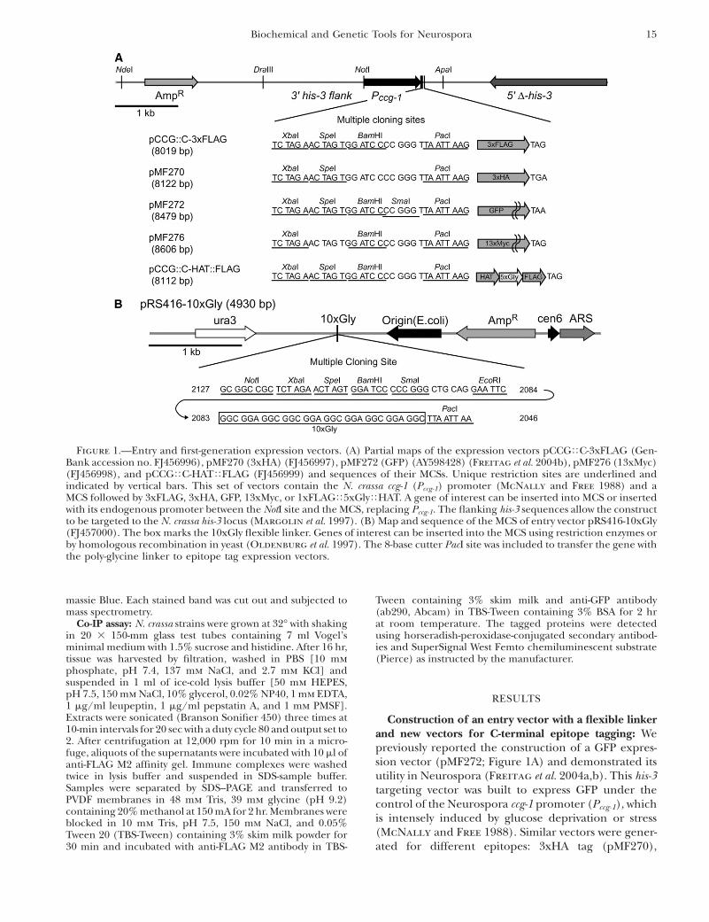
Construction of an entry vector with a flexible linkerand new vectors for C-terminal epitope tagging: Wepreviously reported the construction of a GFP expres-sion vector (pMF272; Figure 1A) and demonstrated itsutility in Neurospora (Freitag et al. 2004a,b). This his-3targeting vector was built to express GFP under thecontrol of the Neurospora ccg-1 promoter (Pccg-1), whichis intensely induced by glucose deprivation or stress(McNally and Free 1988). Similar vectors were gener-ated for different epitopes: 3xHA tag (pMF270),
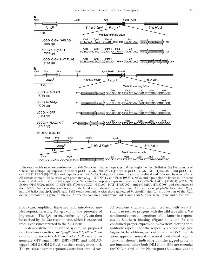
Figure 1.—Entry and first-generation expression vectors. (A) Partial maps of the expression vectors pCCGTC-3xFLAG (Gen-Bank accession no. FJ456996), pMF270 (3xHA) (FJ456997), pMF272 (GFP) (AY598428) (Freitag et al. 2004b), pMF276 (13xMyc)(FJ456998), and pCCGTC-HATTFLAG (FJ456999) and sequences of their MCSs. Unique restriction sites are underlined andindicated by vertical bars. This set of vectors contain the N. crassa ccg-1 (Pccg-1) promoter (McNally and Free 1988) and aMCS followed by 3xFLAG, 3xHA, GFP, 13xMyc, or 1xFLAGT5xGlyTHAT. A gene of interest can be inserted into MCS or insertedwith its endogenous promoter between the NotI site and the MCS, replacing Pccg-1. The flanking his-3 sequences allow the constructto be targeted to the N. crassa his-3 locus (Margolin et al. 1997). (B) Map and sequence of the MCS of entry vector pRS416-10xGly(FJ457000). The box marks the 10xGly flexible linker. Genes of interest can be inserted into the MCS using restriction enzymes orby homologous recombination in yeast (Oldenburg et al. 1997). The 8-base cutter PacI site was included to transfer the gene withthe poly-glycine linker to epitope tag expression vectors.
Biochemical and Genetic Tools for Neurospora 15
13xMyc tag (pMF276), 3xFLAG tag (pCCGTC-3xFLAG), and a HAT–FLAG tandem tag with a 5xGlylinker (pCCGTC-HATTFLAG), as illustrated in Figure1A. All vectors contain the same multiple cloning sites(MCS) followed by epitope tag sequences and an in-frame stop codon; thus, a gene of interest amplified witha primer pair can be introduced simultaneously intoseveral of these expression vectors. The Pccg-1 is partic-ularly useful for visualization of GFP fusion protein inNeurospora because of its strength (Freitag et al.2004b), and this promoter is helpful in detectingproteins that are nondetectable when expressed underthe control of their endogenous promoters. Overex-pression can cause artifactual effects, however (Rigaut
et al. 1999). We therefore constructed the plasmids sothat Pccg-1 can be replaced with a gene of interest with itsendogenous promoter using a NotI site and the MCS,which flank the Pccg-1.
We previously noted that C-terminal GFP fusions withNeurospora genes are not always successful. It is notuncommon to find undetectable expression of the GFPfusion proteins even though they were overexpressed bythe Pccg-1 (Freitag et al. 2004b). One of a number ofreasons for this is that a GFP tag can influence folding ofthe tagged protein, resulting in instability of the GFPfusion proteins. Flexible protein linkers can be used toimprove the folding and function of epitope-taggedproteins (Sabourin et al. 2007). We therefore con-structed an entry vector containing a flexible poly-glycinelinker (10xGly). We inserted a flexible poly-glycine linkerinto the yeast shuttle vector pRS416 to produce pRS416-10xGly (Figure 1B). After subcloning into the entryvector, a gene of interest with the poly-glycine linkercan be easily transferred into a series of our constructedepitope-tagging expression vectors, which contain thesame MCS and PacI site. Genes can be introduced intopRS416-10xGly by a conventional subcloning system byusing restriction enzyme sites in the MCS or by using ayeast homologous recombination system (Oldenburg
et al. 1997).New vectors for N- and C-terminal epitope tagging
with flexible linkers: The entry vector system describedabove allows one to take advantage of previouslygenerated vectors such as pMF272 but requires twocloning steps. To speed building constructs for express-ing epitope-tagged proteins with a flexible poly-glycinelinker, we generated new vectors containing a poly-glycinelinker and the alternative epitopes 3xFLAG (pCCGTC-GlyT3xFLAG) and GFP (pCCGTC-GlyTGFP) or aHAT–FLAG combination separated by a flexible linker(pCCGTC-GlyTHATTFLAG) (Figure 2A). These flexi-ble linker vectors sport the features of the first-generationvectors (Figures 1A and 2A), allowing the genes insertedinto the previously generated vectors to be directly trans-ferred into the new expression vectors.
In some cases, C-terminal epitope tags interfere withprotein function, e.g., by interfering with targeting sites
or post-translational modification adjacent to the Cterminus, even when a flexible linker is included(Booher and Kaiser 2008). To deal with such situa-tions, we generated a series of new expression vectorsfor N-terminal epitope tagging with 3xFLAG (pCCGTN-3xFLAG), 3xMyc (pCCGTN-3xMyc), GFP (pCCGTN-GFP), and 3xHA (pN-3xHA) followed by an eight-Glychain. We also built an N-terminal TAP tag vector:1xFLAGT3xGlyTHATT5xGly (pCCGTN-FLAGTHAT)(Figure 2B). These plasmids are all suitable for targetingto his-3 and all except pN-3xHA contain Pccg-1. Thispromoter can be replaced with the native promoter of agene of interest by using an EcoRI or ApaI site and theBamHI site. In the case of pN-3xHA, Pccg-1 or a nativepromoter can be inserted between the EcoRI or ApaI siteand the BglII site (which generates ends compatible withthose of BamHI; the 3xHA sequence in pN-3xHA con-tains a BamHI site). All the vectors contain an identicalMCS in the same frame so that a gene of interest can beeasily inserted into several of these expression vectors atsame time.
Expression of epitope-tagged proteins from genes attheir native chromosomal location: The describedvectors possess a common backbone suitable for target-ing to Neurospora his-3, similar to the commonly usedvector pBM61 and its derivatives (Margolin et al. 1997).We also developed expression vectors for gene replace-ment by taking advantage of mutants deficient for NHEJ(Dmus-51 or Dmus-52) (Ninomiya et al. 2004). Utilizingthe yeast homologous recombination system (Oldenburg
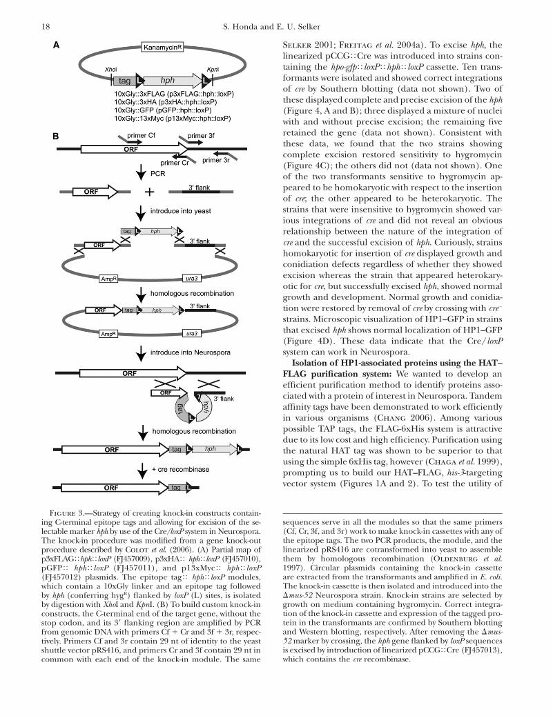
et al. 1997), we first generated a knock-in modulecontaining an epitope tag with a flexible poly-glycinelinker followed by the selectable marker hph, flanked byloxP sites (Figure 3A). The idea was to utilize the Cre/loxP system to remove the hph gene after it was no longerneeded, allowing the valuable marker to be ‘‘recycled.’’Vectors with four epitope tags (3xFLAG, 3xHA, GFP,and 13xMyc) were made to allow the modules to betransferred from any of them (p3xFLAGThphTloxP,p3xHAThphTloxP, pGFPThphTloxP, or p13xMycThphTloxP) by digestion with XhoI and KpnI. All the modulescontain identical sequences of the 59- and 39-ends,allowing constructions of four epitope tag knock-incassettes with common primers designed to work withthe yeast homologous recombination system. For expres-sion of bacteriophage Cre recombinase in Neurospora,we generated the his-3-targeting plasmid pCCGTCre,which allows cre to be expressed under the control of Pccg-1
at the his-3 locus. The scheme is illustrated in Figure 3and described in detail in materials and methods. Inbrief, fragments immediately before and after the stopcodon of a target gene are amplified with four primerscontaining sequences allowing for assembly in the yeastshuttle vector pRS416. The two PCR products, thelinearized pRS416, and the isolated knock-in moduleare assembled in yeast using the endogenous recombi-nation system. The circular knock-in cassettes are isolated
16 S. Honda and E. U. Selker
from yeast, amplified, linearized, and introduced intoNeurospora, selecting for growth in the presence ofhygromycin. The hph marker, conferring hygR, can thenbe excised by the Cre recombinase, which is expressedfrom a construct targeted to the his-3 locus.
To demonstrate the described system, we preparedtwo knock-in cassettes, an hpo-gfpTloxPThphTloxP cas-sette and a dim-2-3xFLAGTloxPThphTloxP cassette, togenerate GFP-tagged HP1 (HP1–GFP) and 3xFLAG-tagged DIM-2 (DIM-2-FLAG) at their endogenous loci.The two cassettes were separately introduced into Dmus-
52 recipient strains and then crossed with mus-521
strains to recover progeny with the wild-type allele. Weconfirmed correct integrations of the knock-in sequen-ces by Southern blotting (Figure 4, A and B) andconfirmed proper expression by Western blotting withantibodies specific for the respective epitope tags (seeFigure 6). In addition, we confirmed that DNA methyl-ation appeared normal in several methylated regions(data not shown), indicating that the tagged proteinsare functional since both DIM-2 and HP1 are essentialfor DNA methylation in Neurospora (Kouzminova and
Figure 2.—Advanced expression vectors with N- or C-terminal epitope tags and a poly-glycine flexible linker. (A) Partial maps ofC-terminal epitope tag expression vectors pCCGTC-GlyT3xFLAG (FJ457001), pCCGTC-GlyTGFP (FJ457002), and pCCGTC-GlyTHATTFLAG (FJ457003) and sequences of their MCSs. Unique restriction sites are underlined and indicated by vertical bars.All vectors contain the N. crassa ccg-1 promoter (Pccg-1) (McNally and Free 1988), a MCS, and a poly-glycine linker in the sameframe and direction. (B) Partial maps of the N-terminal epitope-tag expression vectors pCCGTN-3xFLAG (FJ457004), pCCGTN-3xMyc (FJ457005), pCCGTN-GFP (FJ457006), pCCGTN-FLAGTHAT (FJ457007), and pN-3xHA (FJ457008) and sequences oftheir MCS. Unique restriction sites are underlined and indicated by vertical bars. All vectors except pN-3xHA contain Pccg-1,and pN-3xHA has ApaI, EcoRI, and BglII (ends compatible with those generated by BamHI) sites for introduction of the Pccg-1
or the promoter of a gene of interest. All vectors contain a poly-glycine linker and a MCS in the same frame and direction.
Biochemical and Genetic Tools for Neurospora 17
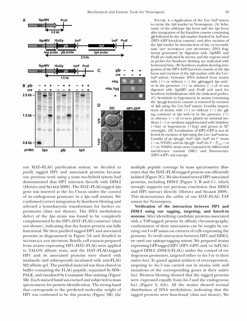
Selker 2001; Freitag et al. 2004a). To excise hph, thelinearized pCCGTCre was introduced into strains con-taining the hpo-gfpTloxPThphTloxP cassette. Ten trans-formants were isolated and showed correct integrationsof cre by Southern blotting (data not shown). Two ofthese displayed complete and precise excision of the hph(Figure 4, A and B); three displayed a mixture of nucleiwith and without precise excision; the remaining fiveretained the gene (data not shown). Consistent withthese data, we found that the two strains showingcomplete excision restored sensitivity to hygromycin(Figure 4C); the others did not (data not shown). Oneof the two transformants sensitive to hygromycin ap-peared to be homokaryotic with respect to the insertionof cre; the other appeared to be heterokaryotic. Thestrains that were insensitive to hygromycin showed var-ious integrations of cre and did not reveal an obviousrelationship between the nature of the integration ofcre and the successful excision of hph. Curiously, strainshomokaryotic for insertion of cre displayed growth andconidiation defects regardless of whether they showedexcision whereas the strain that appeared heterokary-otic for cre, but successfully excised hph, showed normalgrowth and development. Normal growth and conidia-tion were restored by removal of cre by crossing with cre�
strains. Microscopic visualization of HP1–GFP in strainsthat excised hph shows normal localization of HP1–GFP(Figure 4D). These data indicate that the Cre/loxPsystem can work in Neurospora.
Isolation of HP1-associated proteins using the HAT–FLAG purification system: We wanted to develop anefficient purification method to identify proteins asso-ciated with a protein of interest in Neurospora. Tandemaffinity tags have been demonstrated to work efficientlyin various organisms (Chang 2006). Among variouspossible TAP tags, the FLAG-6xHis system is attractivedue to its low cost and high efficiency. Purification usingthe natural HAT tag was shown to be superior to thatusing the simple 6xHis tag, however (Chaga et al. 1999),prompting us to build our HAT–FLAG, his-3-targetingvector system (Figures 1A and 2). To test the utility of
Figure 3.—Strategy of creating knock-in constructs contain-ing C-terminal epitope tags and allowing for excision of the se-lectable marker hph by use of the Cre/loxP system in Neurospora.The knock-in procedure was modified from a gene knock-outprocedure described by Colot et al. (2006). (A) Partial map ofp3xFLAGThphTloxP (FJ457009), p3xHAT hphTloxP (FJ457010),pGFPT hphTloxP (FJ457011), and p13xMycT hphTloxP(FJ457012) plasmids. The epitope tagT hphTloxP modules,which contain a 10xGly linker and an epitope tag followedby hph (conferring hygR) flanked by loxP (L) sites, is isolatedby digestion with XhoI and KpnI. (B) To build custom knock-inconstructs, the C-terminal end of the target gene, without thestop codon, and its 39 flanking region are amplified by PCRfrom genomic DNA with primers Cf 1 Cr and 3f 1 3r, respec-tively. Primers Cf and 3r contain 29 nt of identity to the yeastshuttle vector pRS416, and primers Cr and 3f contain 29 nt incommon with each end of the knock-in module. The same
sequences serve in all the modules so that the same primers(Cf, Cr, 3f, and 3r) work to make knock-in cassettes with any ofthe epitope tags. The two PCR products, the module, and thelinearized pRS416 are cotransformed into yeast to assemblethem by homologous recombination (Oldenburg et al.1997). Circular plasmids containing the knock-in cassetteare extracted from the transformants and amplified in E. coli.The knock-in cassette is then isolated and introduced into theDmus-52 Neurospora strain. Knock-in strains are selected bygrowth on medium containing hygromycin. Correct integra-tion of the knock-in cassette and expression of the tagged pro-tein in the transformants are confirmed by Southern blottingand Western blotting, respectively. After removing the Dmus-52 marker by crossing, the hph gene flanked by loxP sequencesis excised by introduction of linearized pCCGTCre (FJ457013),which contains the cre recombinase.
18 S. Honda and E. U. Selker
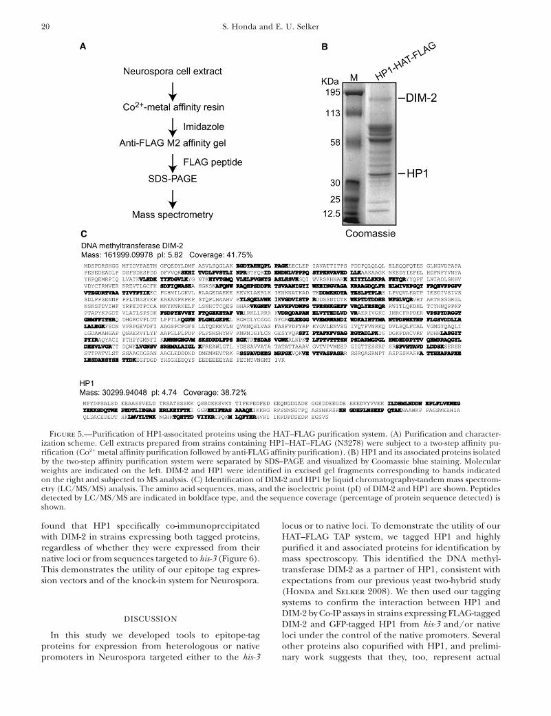
our HAT–FLAG purification system, we decided topurify tagged HP1 and associated proteins becauseour previous work using a yeast two-hybrid system haddemonstrated that HP1 interacts directly with DIM-2(Honda and Selker 2008). The HAT–FLAG-tagged hpogene was inserted at the his-3 locus under the controlof its endogenous promoter in a hpo null mutant. Weconfirmed correct integration by Southern blotting andselected a homokaryotic transformant for further ex-periments (data not shown). The DNA methylationdefect of the hpo strain was found to be completelycomplemented by the HP1–HAT–FLAG construct (datanot shown), indicating that the fusion protein was fullyfunctional. We then purified tagged HP1 and associatedproteins as diagrammed in Figure 5A and detailed inmaterials and methods. Briefly, cell extracts preparedfrom strains expressing HP1–HAT–FLAG were appliedto TALON affinity resin, and the HAT–FLAG-taggedHP1 and its associated proteins were eluted withimidazole and subsequently incubated with anti-FLAGM2 affinity gel. The purified material was then eluted inbuffer containing the FLAG peptide, separated by SDS–PAGE, and visualized by Coomassie blue staining (Figure5B). Each stained band was excised and subjected to massspectrometry for protein identification. The strong bandthat corresponds to the predicted molecular weight ofHP1 was confirmed to be this protein (Figure 5B); the
multiple peptide coverage by mass spectrometry illus-trates that the HAT–FLAG-tagged protein was efficientlyisolated (Figure 5C). We also found several HP1-associatedproteins, including DIM-2 (Figure 5, B and C), whichstrongly supports our previous conclusion that DIM-2and HP1 interact directly (Honda and Selker 2008).This demonstrates the utility of our HAT–FLAG TAPsystem for Neurospora.
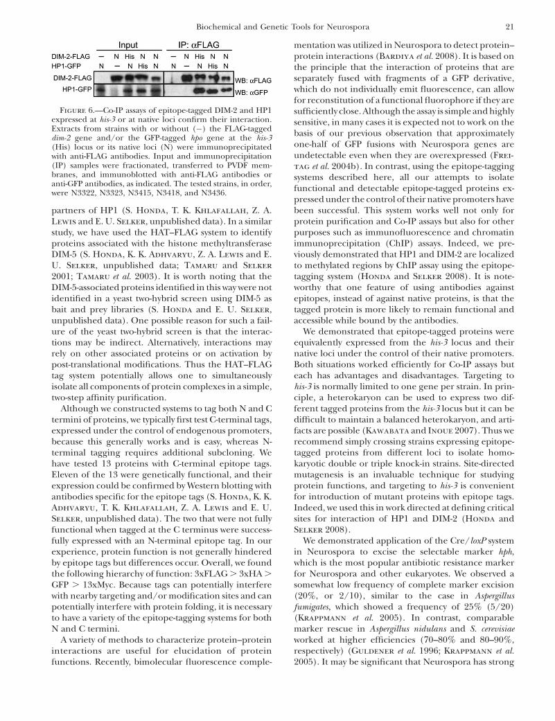
Verification of the interaction between HP1 andDIM-2 using our tagging, targeting, and knock-insystems: After identifying candidate proteins associatedwith a TAP-tagged protein by affinity chromatography,confirmation of their association can be sought by car-rying out Co-IP assays on extracts of cells expressing theproteins. To verify interaction between HP1 and DIM-2,we used our epitope-tagging system. We prepared strainsexpressing GFP-tagged HP1 (HP1–GFP) and/or 3xFLAG-tagged DIM-2 (DIM-2–FLAG) under the control of en-dogenous promoters, targeted either to his-3 or to theirnative loci. To guard against artifacts of overexpression,targeting to his-3 was carried out in strains with nullmutations of the corresponding genes at their nativeloci. Western blotting showed that the tagged proteinswere expressed equally from his-3 and the endogenousloci (Figure 6, left). All the strains showed normaldistribution of DNA methylation, indicating that thetagged proteins were functional (data not shown). We
Figure 4.—Application of the Cre/loxP systemto excise the hph marker in Neurospora. (A) Sche-matic of the wild-type hpo locus and the hpo locusafter integration of the knock-in cassette containinggfp followed by the hph marker flanked by loxP sites(HP1–GFP knock-in cassette) and after excision ofthe hph marker by introduction of the cre recombi-nase (see materials and methods). DNA frag-ments generated by digestion with NgoMIV andPvuII are indicated by arrows, and the regions usedas probes for Southern blotting are indicated withhorizontal bars. (B) Southern analysis showing inte-gration of the HP1–GFP knock-in cassette at the hpolocus and excision of the hph marker with the Cre/loxP system. Genomic DNA isolated from strainswith (1) or without (�) the gfp-tagged hpo and/or in the presence (1) or absence (�) of cre wasdigested with NgoMIV and PvuII and used forSouthern hybridizations with the indicated probes.(C) Sensitivity to hygromycin in strains containingthe hpo-gfp knock-in cassette is restored by excisionof hph using the Cre/loxP system. Conidia suspen-sions of strains with (1) or without (�) the gfp-tag construct at hpo and/or in the presence (1)or absence (�) of cre were plated on minimal me-dium (�) or medium supplemented with histidine(1his) or hygromycin (1hyg) and grown at 32�overnight. (D) Localization of HP1–GFP is not af-fected by excision of hph using the Cre/loxP system.Conidia of an hpo-gfpTloxPThphTloxP; his-3� strain(�cre, N3322) and an hpo-gfpTloxP; his-31TPccg-1Tcre(1cre, N3684) strain were examined by differentialinterference contrast (DIC) and fluorescence(HP1–GFP) microscopy.
Biochemical and Genetic Tools for Neurospora 19
found that HP1 specifically co-immunoprecipitatedwith DIM-2 in strains expressing both tagged proteins,regardless of whether they were expressed from theirnative loci or from sequences targeted to his-3 (Figure 6).This demonstrates the utility of our epitope tag expres-sion vectors and of the knock-in system for Neurospora.
DISCUSSION
In this study we developed tools to epitope-tagproteins for expression from heterologous or nativepromoters in Neurospora targeted either to the his-3
locus or to native loci. To demonstrate the utility of ourHAT–FLAG TAP system, we tagged HP1 and highlypurified it and associated proteins for identification bymass spectroscopy. This identified the DNA methyl-transferase DIM-2 as a partner of HP1, consistent withexpectations from our previous yeast two-hybrid study(Honda and Selker 2008). We then used our taggingsystems to confirm the interaction between HP1 andDIM-2 by Co-IP assays in strains expressing FLAG-taggedDIM-2 and GFP-tagged HP1 from his-3 and/or nativeloci under the control of the native promoters. Severalother proteins also copurified with HP1, and prelimi-nary work suggests that they, too, represent actual
Figure 5.—Purification of HP1-associtated proteins using the HAT–FLAG purification system. (A) Purification and character-ization scheme. Cell extracts prepared from strains containing HP1–HAT–FLAG (N3278) were subject to a two-step affinity pu-rification (Co21 metal affinity purification followed by anti-FLAG affinity purification). (B) HP1 and its associated proteins isolatedby the two-step affinity purification system were separated by SDS–PAGE and visualized by Coomassie blue staining. Molecularweights are indicated on the left. DIM-2 and HP1 were identified in excised gel fragments corresponding to bands indicatedon the right and subjected to MS analysis. (C) Identification of DIM-2 and HP1 by liquid chromatography-tandem mass spectrom-etry (LC/MS/MS) analysis. The amino acid sequences, mass, and the isoelectric point (pI) of DIM-2 and HP1 are shown. Peptidesdetected by LC/MS/MS are indicated in boldface type, and the sequence coverage (percentage of protein sequence detected) isshown.
20 S. Honda and E. U. Selker
partners of HP1 (S. Honda, T. K. Khlafallah, Z. A.Lewis and E. U. Selker, unpublished data). In a similarstudy, we have used the HAT–FLAG system to identifyproteins associated with the histone methyltransferaseDIM-5 (S. Honda, K. K. Adhvaryu, Z. A. Lewis and E.U. Selker, unpublished data; Tamaru and Selker
2001; Tamaru et al. 2003). It is worth noting that theDIM-5-associated proteins identified in this way were notidentified in a yeast two-hybrid screen using DIM-5 asbait and prey libraries (S. Honda and E. U. Selker,unpublished data). One possible reason for such a fail-ure of the yeast two-hybrid screen is that the interac-tions may be indirect. Alternatively, interactions mayrely on other associated proteins or on activation bypost-translational modifications. Thus the HAT–FLAGtag system potentially allows one to simultaneouslyisolate all components of protein complexes in a simple,two-step affinity purification.
Although we constructed systems to tag both N and Ctermini of proteins, we typically first test C-terminal tags,expressed under the control of endogenous promoters,because this generally works and is easy, whereas N-terminal tagging requires additional subcloning. Wehave tested 13 proteins with C-terminal epitope tags.Eleven of the 13 were genetically functional, and theirexpression could be confirmed by Western blotting withantibodies specific for the epitope tags (S. Honda, K. K.Adhvaryu, T. K. Khlafallah, Z. A. Lewis and E. U.Selker, unpublished data). The two that were not fullyfunctional when tagged at the C terminus were success-fully expressed with an N-terminal epitope tag. In ourexperience, protein function is not generally hinderedby epitope tags but differences occur. Overall, we foundthe following hierarchy of function: 3xFLAG . 3xHA .
GFP . 13xMyc. Because tags can potentially interferewith nearby targeting and/or modification sites and canpotentially interfere with protein folding, it is necessaryto have a variety of the epitope-tagging systems for bothN and C termini.
A variety of methods to characterize protein–proteininteractions are useful for elucidation of proteinfunctions. Recently, bimolecular fluorescence comple-
mentation was utilized in Neurospora to detect protein–protein interactions (Bardiya et al. 2008). It is based onthe principle that the interaction of proteins that areseparately fused with fragments of a GFP derivative,which do not individually emit fluorescence, can allowfor reconstitution of a functional fluorophore if they aresufficiently close. Although the assay is simple and highlysensitive, in many cases it is expected not to work on thebasis of our previous observation that approximatelyone-half of GFP fusions with Neurospora genes areundetectable even when they are overexpressed (Frei-
tag et al. 2004b). In contrast, using the epitope-taggingsystems described here, all our attempts to isolatefunctional and detectable epitope-tagged proteins ex-pressed under the control of their native promoters havebeen successful. This system works well not only forprotein purification and Co-IP assays but also for otherpurposes such as immunofluorescence and chromatinimmunoprecipitation (ChIP) assays. Indeed, we pre-viously demonstrated that HP1 and DIM-2 are localizedto methylated regions by ChIP assay using the epitope-tagging system (Honda and Selker 2008). It is note-worthy that one feature of using antibodies againstepitopes, instead of against native proteins, is that thetagged protein is more likely to remain functional andaccessible while bound by the antibodies.
We demonstrated that epitope-tagged proteins wereequivalently expressed from the his-3 locus and theirnative loci under the control of their native promoters.Both situations worked efficiently for Co-IP assays buteach has advantages and disadvantages. Targeting tohis-3 is normally limited to one gene per strain. In prin-ciple, a heterokaryon can be used to express two dif-ferent tagged proteins from the his-3 locus but it can bedifficult to maintain a balanced heterokaryon, and arti-facts are possible (Kawabata and Inoue 2007). Thus werecommend simply crossing strains expressing epitope-tagged proteins from different loci to isolate homo-karyotic double or triple knock-in strains. Site-directedmutagenesis is an invaluable technique for studyingprotein functions, and targeting to his-3 is convenientfor introduction of mutant proteins with epitope tags.Indeed, we used this in work directed at defining criticalsites for interaction of HP1 and DIM-2 (Honda andSelker 2008).
We demonstrated application of the Cre/loxP systemin Neurospora to excise the selectable marker hph,which is the most popular antibiotic resistance markerfor Neurospora and other eukaryotes. We observed asomewhat low frequency of complete marker excision(20%, or 2/10), similar to the case in Aspergillusfumigates, which showed a frequency of 25% (5/20)(Krappmann et al. 2005). In contrast, comparablemarker rescue in Aspergillus nidulans and S. cerevisiaeworked at higher efficiencies (70–80% and 80–90%,respectively) (Guldener et al. 1996; Krappmann et al.2005). It may be significant that Neurospora has strong
Figure 6.—Co-IP assays of epitope-tagged DIM-2 and HP1expressed at his-3 or at native loci confirm their interaction.Extracts from strains with or without (�) the FLAG-taggeddim-2 gene and/or the GFP-tagged hpo gene at the his-3(His) locus or its native loci (N) were immunoprecipitatedwith anti-FLAG antibodies. Input and immunoprecipitation(IP) samples were fractionated, transferred to PVDF mem-branes, and immunoblotted with anti-FLAG antibodies oranti-GFP antibodies, as indicated. The tested strains, in order,were N3322, N3323, N3415, N3418, and N3436.
Biochemical and Genetic Tools for Neurospora 21
genomic defense systems (Galagan and Selker 2004),including DNA methylation, which interfere with tran-scriptional elongation (Rountree and Selker 1997);however, we did not detect DNA methylation in theintegrated cre gene (data not shown). The efficiencydifference may be attributable to differences in expres-sion of Cre driven by different promoters. In the A.nidulans and S. cerevisiae systems, the chemically in-ducible promoters tightly regulate the expression ofCre, while, in the Neurospora and A. fumigates systems,expression of Cre was initiated by introduction of the cregene by transformation. Use of tightly inducible pro-moters, such as the quinic acid-2 promoter (Pqa-2) (Giles
et al. 1985), may improve the efficiency in Neurospora.Further improvements to this system could includeapplication of dual selectable marker modules such as‘‘tk-blaster,’’ which combines hph and the fluorodeoxy-uridine (FUDR)-sensitive marker thymidine kinase (tk)(Sachs et al. 1997; Pratt and Aramayo 2002), allowingselection of strains that have excised the cassette. Futurework may optimize the Cre/loxP system for Neurospora.The Cre/loxP system could potentially be used not onlyfor marker excision but also for advanced engineeringof chromosomal rearrangements, such as in buildingconditional knock-outs in Neurospora.
Characterization and identification of protein com-plexes is a basic step in deciphering gene functions. Wehave developed here a combination of genetic andbiochemical approaches in Neurospora, and thesetechniques can be easily applied to other filamentousfungi. We have used these tools for analyzing therelationship between HP1 and DIM-2 to elucidateDNA methylation machinery in Neurospora. Thesetools will facilitate various analyses in clarifying thefundamental processes in Neurospora and other fila-mentous fungi. Indeed, our methods have already beensuccessfully adapted to tag and express fusion proteinsin Fusarium graminearum (L. Connolly and M. Freitag,unpublished results).
We thank Michael Freitag for the construction of pMF270 andpMF276 and Hui Zhong for the generous gift of pHZ76. We also thankMichael Freitag, Zachary Lewis, Keyur Adhvaryu, and Kirsty Jamiesonfor comments on the manuscript. We thank Ragna Sack of theFriedrich Miescher Insitute (Basel, Switzerland) for MS analysis ofHP1-associated proteins. This work was supported by grant GM025690-22 to E.U.S. from the National Institutes of Health.
LITERATURE CITED
Aebersold, R., and M. Mann, 2003 Mass spectrometry-based pro-teomics. Nature 422: 198–207.
Bardiya, N., W. G. Alexander, T. D. Perdue, E. G. Barry, R. L.Metzenberg et al., 2008 Characterization of interactionsbetween and among components of the meiotic silencing byunpaired DNA machinery in Neurospora crassa using bimolecu-lar fluorescence complementation. Genetics 178: 593–596.
Booher, K. R., and P. Kaiser, 2008 A PCR-based strategy to gener-ate yeast strains expressing endogenous levels of amino-terminalepitope-tagged proteins. Biotech. J. 3: 524–529.
Borjigin, J., and J. Nathans, 1994 Insertional mutagenesis as aprobe of rhodopsin’s topography, stability, and activity. J. Biol.Chem. 269: 14715–14722.
Borkovich, K. A., L. A. Alex, O. Yarden, M. Freitag, G. E. Turner
et al., 2004 Lessons from the genome sequence of Neurosporacrassa: tracing the path from genomic blueprint to multicellularorganism. Microbiol. Mol. Biol. Rev. 68: 1–108.
Brizzard, B., 2008 Epitope tagging. BioTechniques 44: 693–695.Chaga, G., D. E. Bochkariov, G. G. Jokhadze, J. Hopp and P.
Nelson, 1999 Natural poly-histidine affinity tag for purificationof recombinant proteins on cobalt(II)-carboxymethylaspartatecrosslinked agarose. J. Chromatogr. A 864: 247–256.
Chang, I. F., 2006 Mass spectrometry-based proteomic analysis ofthe epitope-tag affinity purified protein complexes in eukaryotes.Proteomics 6: 6158–6166.
Colot, H. V., G. Park, G. E. Turner, C. Ringelberg, C. M. Crew
et al., 2006 A high-throughput gene knockout procedure forNeurospora reveals functions for multiple transcription factors.Proc. Natl. Acad. Sci. USA 103: 10352–10357.
Cravatt, B. F., G. M. Simon and J. R. Yates, III, 2007 The biological im-pact of mass-spectrometry-based proteomics. Nature 450: 991–1000.
Davis, R. H., 2000 Neurospora: Contributions of a Model Organism.Oxford University Press, Oxford.
Davis, R. H., and D. D. Perkins, 2002 Timeline: Neurospora: a modelof model microbes. Nat. Rev. Genet. 3: 397–403.
Dementhon, K., G. Iyer and N. L. Glass, 2006 VIB-1 is required forexpression of genes necessary for programmed cell death in Neu-rospora crassa. Eukaryot. Cell 5: 2161–2173.
Dunlap, J. C., K. A. Borkovich, M. R. Henn, G. E. Turner, M. S.Sachs et al., 2007 Enabling a community to dissect an organ-ism: overview of the Neurospora functional genomics project.Adv. Genet. 57: 49–96.
Forment, J. V., D. Ramon and A. P. MacCabe, 2006 Consecutivegene deletions in Aspergillus nidulans: application of the Cre/loxPsystem. Curr. Genet. 50: 217–224.
Freitag, M., and E. U. Selker, 2005 Expression and visualization ofred fluorescent protein (RFP) in Neurospora crassa. Fungal Genet.Newsl. 52: 14–17.
Freitag, M., P. C. Hickey, T. K. Khlafallah, N. D. Read and E. U.Selker, 2004a HP1 is essential for DNA methylation in Neuros-pora. Mol. Cell 13: 427–434.
Freitag, M., P. C. Hickey, N. B. Raju, E. U. Selker and N. D. Read,2004b GFP as a tool to analyze the organization, dynamics andfunction of nuclei and microtubules in Neurospora crassa. FungalGenet. Biol. 41: 897–910.
Galagan, J. E., and E. U. Selker, 2004 RIP: the evolutionary cost ofgenome defense. Trends Genet. 20: 417–423.
Galagan, J. E., S. E. Calvo, K. A. Borkovich, E. U. Selker, N. D.Read et al., 2003 The genome sequence of the filamentous fun-gus Neurospora crassa. Nature 422: 859–868.
Gavin, A. C., M. Bosche, R. Krause, P. Grandi, M. Marzioch et al.,2002 Functional organization of the yeast proteome by system-atic analysis of protein complexes. Nature 415: 141–147.
Giles, N. H., M. E. Case, J. Baum, R. Geever, L. Huiet et al.,1985 Gene organization and regulation in the qa (quinic acid)gene cluster of Neurospora crassa. Microbiol. Rev. 49: 338–358.
Guldener, U., S. Heck, T. Fielder, J. Beinhauer and J. H.Hegemann, 1996 A new efficient gene disruption cassette forrepeated use in budding yeast. Nucleic Acids Res. 24: 2519–2524.
He, Q., P. Cheng, Y. Yang, L. Wang, K. H. Gardner et al.,2002 White collar-1, a DNA binding transcription factor anda light sensor. Science 297: 840–843.
He, Q., P. Cheng and Y. Liu, 2005 The COP9 signalosome regulatesthe Neurospora circadian clock by controlling the stability of theSCFFWD-1 complex. Genes Dev. 19: 1518–1531.
Honda, S., and E. U. Selker, 2008 Direct interaction between DNAmethyltransferase DIM-2 and HP1 is required for DNA methyla-tion in Neurospora. Mol. Cell. Biol. 28: 6044–6055.
Ishibashi, K., K. Suzuki, Y. Ando, C. Takakura and H. Inoue,2006 Nonhomologous chromosomal integration of foreignDNA is completely dependent on MUS-53 (human Lig4 homo-log) in Neurospora. Proc. Natl. Acad. Sci. USA 103: 14871–14876.
James, P., J. Halladay and E. A. Craig, 1996 Genomic libraries anda host strain designed for highly efficient two-hybrid selection inyeast. Genetics 144: 1425–1436.
22 S. Honda and E. U. Selker
Jarvik, J. W., and C. A. Telmer, 1998 Epitope tagging. Annu. Rev.Genet. 32: 601–618.
Kasuga, T., and N. L. Glass, 2008 Dissecting colony developmentof Neurospora crassa using mRNA profiling and comparative ge-nomics approaches. Eukaryot. Cell 7: 1549–1564.
Kasuga, T., J. P. Townsend, C. Tian, L. B. Gilbert, G. Mannhaupt
et al., 2005 Long-oligomer microarray profiling in Neurosporacrassa reveals the transcriptional program underlying biochemi-cal and physiological events of conidial germination. NucleicAcids Res. 33: 6469–6485.
Kawabata, T., and H. Inoue, 2007 Detection of physical interac-tions by immunoprecipitation of FLAG- and HA-tagged proteinsexpressed at the his-3 locus in Neurospora crassa. Fungal Genet.Newsl. 54: 5–8.
Kouzminova, E., and E. U. Selker, 2001 dim-2 encodes a DNAmethyltransferase responsible for all known cytosine methylationin Neurospora. EMBO J. 20: 4309–4323.
Krappmann, S., O. Bayram and G. H. Braus, 2005 Deletion and al-lelic exchange of the Aspergillus fumigatus veA locus via a novelrecyclable marker module. Eukaryot. Cell 4: 1298–1307.
Lee, D. W., J. R. Haag and R. Aramayo, 2003 Construction ofstrains for rapid homokaryon purification after integration ofconstructs at the histidine-3 (his-3) locus of Neurospora crassa.Curr. Genet. 43: 17–23.
Lewis, Z. A., A. L. Shiver, N. Stiffler, M. R. Miller, E. A. Johnson
et al., 2007 High-density detection of restriction-site-associatedDNA markers for rapid mapping of mutated loci in Neurospora.Genetics 177: 1163–1171.
Li, L., and K. A. Borkovich, 2006 GPR-4 is a predicted G-protein-coupled receptor required for carbon source-dependent asexualgrowth and development in Neurospora crassa. Eukaryot. Cell 5:1287–1300.
Longtine, M. S., A. McKenzie, III, D. J. Demarini, N. G. Shah, A.Wach et al., 1998 Additional modules for versatile and econom-ical PCR-based gene deletion and modification in Saccharomycescerevisiae. Yeast 14: 953–961.
Margolin, B. S., M. Freitag and E. U. Selker, 1997 Improved plas-mids for gene targeting at the his-3 locus of Neurospora crassa byelectroporation. Fungal Genet. Newsl. 44: 34–36.
McNally, M. T., and S. J. Free, 1988 Isolation and characterizationof a Neurospora glucose-repressible gene. Curr. Genet. 14: 545–551.
Ninomiya, Y., K. Suzuki, C. Ishii and H. Inoue, 2004 Highly effi-cient gene replacements in Neurospora strains deficient for non-homologous end-joining. Proc. Natl. Acad. Sci. USA 101: 12248–12253.
Oldenburg, K. R., K. T. Vo, S. Michaelis and C. Paddon,1997 Recombination-mediated PCR-directed plasmid construc-tion in vivo in yeast. Nucleic Acids Res. 25: 451–452.
Ooi, S. K., C. Qiu, E. Bernstein, K. Li, D. Jia et al., 2007 DNMT3Lconnects unmethylated lysine 4 of histone H3 to de novo methyl-ation of DNA. Nature 448: 714–717.
Pandey, A., and M. Mann, 2000 Proteomics to study genes and ge-nomes. Nature 405: 837–846.
Pratt, R. J., and R. Aramayo, 2002 Improving the efficiency of genereplacements in Neurospora crassa: a first step towards a large-scalefunctional genomics project. Fungal Genet. Biol. 37: 56–71.
Rigaut, G., A. Shevchenko, B. Rutz, M. Wilm, M. Mann et al.,1999 A generic protein purification method for protein com-plex characterization and proteome exploration. Nat. Biotech-nol. 17: 1030–1032.
Rountree, M. R., and E. U. Selker, 1997 DNA methylation inhibitselongation but not initiation of transcription in Neurospora crassa.Genes Dev. 11: 2383–2395.
Sabourin, M., C. T. Tuzon, T. S. Fisher and V. A. Zakian, 2007 Aflexible protein linker improves the function of epitope-taggedproteins in Saccharomyces cerevisiae. Yeast 24: 39–45.
Sachs, M. S., E. U. Selker, B. Lin, C. J. Roberts, Z. Luo et al.,1997 Expression of herpes virus thymidine kinase in Neurosporacrassa. Nucleic Acids Res. 25: 2389–2395.
Staben, C., B. Jensen, M. Singer, J. Pollock, M. Schechtman et al.,1989 Use of a bacterial hygromycin B resistance gene as a dom-inant selectable marker in Neurospora crassa transformation. Fun-gal Genet. Newsl. 36: 79–81.
Sung, M. K., C. W. Ha and W. K. Huh, 2008 A vector system for ef-ficient and economical switching of C-terminal epitope tags inSaccharomyces cerevisiae. Yeast 25: 301–311.
Tamaru, H., and E. U. Selker, 2001 A histone H3 methyltransferasecontrols DNA methylation in Neurospora crassa. Nature 414: 277–283.
Tamaru, H., X. Zhang, D. McMillen, P. B. Singh, J. Nakayama et al.,2003 Trimethylated lysine 9 of histone H3 is a mark for DNAmethylation in Neurospora crassa. Nat. Genet. 34: 75–79.
Tian, C., T. Kasuga, M. S. Sachs and N. L. Glass, 2007 Transcriptionalprofiling of cross pathway control in Neurospora crassa and compar-ative analysis of the Gcn4 and CPC1 regulons. Eukaryot. Cell 6:1018–1029.
Van Duyne, G. D., 2001 A structural view of Cre-loxP site-specific re-combination. Annu. Rev. Biophys. Biomol. Struct. 30: 87–104.
Yang, P., H. M. Sampson and H. M. Krause, 2006 A modified tan-dem affinity purification strategy identifies cofactors of the Dro-sophila nuclear receptor dHNF4. Proteomics 6: 927–935.
Yu, Y., and A. Bradley, 2001 Engineering chromosomal rearrange-ments in mice. Nat. Rev. Genet. 2: 780–790.
Communicating editor: M. S. Sachs
Biochemical and Genetic Tools for Neurospora 23
Supporting Information http://www.genetics.org/cgi/content/full/genetics.108.098707/DC1
Tools for Fungal Proteomics: Multifunctional Neurospora Vectors for Gene Replacement, Protein Expression and Protein Purification
Shinji Honda and Eric U. Selker
Copyright © 2009 by the Genetics Society of America DOI: 10.1534/genetics.108.098707

S. Honda and E. Selker 2 SI
TABLE S1
Neurospora crassa strains used in this study
Strain Genotype Reference
N623 mat A his-3 FGSC#6103
N1877 mat a his-3; ∆dim-2::hph+ Kouzminova & Selker
(2001)
N2556 mat a his-3; hpoRIP2 Freitag et al. (2004a)
N2930 mat A his-3 ∆mus-52::bar+ Honda & Selker (2008)
N3322 mat A his-3; hpo-gfp::loxP::hph+::loxP Honda & Selker (2008)
N3323 mat a his-3; dim-2-3xFLAG::loxP::hph+::loxP Honda & Selker (2008)
N3324 mat A his-3; dim-2-3xFLAG::loxP::hph+::loxP Honda & Selker (2008)
N3278 mat a +his-3+::Phpo::hpo-HAT-FLAG; hpoRIP2 this study
N3395 mat a his-3; hpoRIP2; dim-2-3xFLAG::loxP::hph+::loxP Honda & Selker (2008)
N3396 mat a his-3; ∆dim-2::hph+; hpo-gfp::loxP::hph+::loxP Honda & Selker (2008)
N3415 mat a his-3+::Pdim-2::dim-2-3xFLAG; ∆dim-2::hph+; hpo-gfp::loxP::hph+::loxP Honda & Selker (2008)
N3418 mat a his-3+::Phpo::hpo-gfp; hpoRIP2; dim-2-3xFLAG::loxP::hph+::loxP Honda & Selker (2008)
N3491 mat a his-3; dim-2-3xFLAG::loxP::hph+::loxP; hpo-gfp::loxP::hph+::loxP this study
N3684
N3686
mat a +his-3+::Pccg-1::cre+; hpo-gfp::loxP
mat a his-3; hpo-gfp::loxP
this study
this study
S. Honda and E. Selker 3 SI
TABLE S2
Primers used in this study
Primer Sequence
#1517 5’-GCTCTAGATCGTTATCAGCCAAGCCATG-3’
#1518 5’-CGTTAATTAACAACTTGACAATCGTCATG-3’
#1790 5’-GGAATTCGGCGGAGGCGGCGGAGGCGGAGGCGGAGGCTTAATTAAAAGCTTGGG-3’
#1791 5’-CCCAAGCTTTTAATTAAGCCTCCGCCTCCGCCTCCGCCGCCTCCGCCGAATTCC-3’
#1938 5’-CCTTAATTAAGGGCGGAGGCGGCGGAGGCG-3’
#1939 5’-CCTTAATTAAGAAGGATCATCTCATCCACA-3’
#1940 5’-GGCGCGCCTACTTGTCGTCATCGTCTTTGTAGTCGCCTCCGCCTCCGCCCTTGT-3’
#1946 5’-GACTAGTTAATTAATGGCGCGCCGCCTCCGCCGCCCTTGTTGTGGGCATG-3’
#1953 5’-CGGGATCCTTTGGTTGATGTGAGGGGTTG-3’
#1954 5’-GGAATTCGTTCAAAGCCACATCAC-3’
#1967 5’-CGGGATCCATGGACTACAAAGACGATGACGACAAGGGCGGAGGCTTAATCGGCTT-3’
#1970 5’-ATAAGAATGCGGCCGCAGTCCGCAACAGGAAG-3’
#1976 5’-CGGGATCCATAACTTCGTATAGCATACATTATACGAAGTTATCCCGGTCGGCATCTACTCTA-3’
#1977 5’-GACTAGTCGACAGAAGATGATATTG-3’
#1978 5’-GCTCTAGAATAACTTCGTATAGCATACATTATACGAAGTTATCGATAAGCTTGATATCGAAT-3’
#1979 5’-CCGCTCGAGGCGGAGGCGGCGGAGGCGG-3’
#1988 5’-GAGGTCGACGGTATCGATAAGCTTGATATATCAAGGCCTGGAACAACGG-3’
#1989 5’-TGCTATACGAAGTTATGGATCCGAGCTCGTGCGACGAGGTAACGCCATG-3’
#1990 5’-ACCGCGGTGGCGGCCGCTCTAGAACTAGTACAAGACCGGCAACCATCTG-3’
#1992 5’-GAGGTCGACGGTATCGATAAGCTTGATATCATCCAAACAAAAGTCGAGG-3’
#1993 5’-CCTCCGCCTCCGCCTCCGCCGCCTCCGCCCCACAGATAGCCTCTGCACTTG-3’
#1994 5’-TGCTATACGAAGTTATGGATCCGAGCTCGTGGGGGAAGATGTTAACTCAC-3’
#1995 5’-ACCGCGGTGGCGGCCGCTCTAGAACTAGTATCATGGCAACCTAGCCAGG-3’
#1996 5’-GAGGTCGACGGTATCGATAAGCTTGATATTACAGCGCAACACTTCGTCG-3’
#1997 5’-CCTCCGCCTCCGCCTCCGCCGCCTCCGCCTTGCGAGACGCTGCCCTCGC-3’
#1998 5’-TGCTATACGAAGTTATGGATCCGAGCTCGAAGACCGAGGTAGCACTTCTC-3’
#1999 5’-ACCGCGGTGGCGGCCGCTCTAGAACTAGTGTGATGTGCCGAATTCGCTC-3’
#2013 5’-CCTCCGCCTCCGCCTCCGCCGCCTCCGCCCAACTTGACAATCGTCATGC-3’
#2014 5’-TCCCCGGGTTAATTAAAGACTACAAAGACCATGACGG-3’
#2015 5’-GGAATTCAGGTTGTCTTCCCAACTTGC-3’
#2036 5’-ATCGATCGGCTTGAAGGATCATCTCATC-3’
#2037 5’-CCGCTCGAGTTAATTAAGCCTCCGCCTCCGCCCTTGTTGTGGGCATGAGCGT-3’
#2040 5’-CGGGATCCACACCTCGCTGGGTCTCAG-3’
#2041 5’-CCGCAATTGTTGCGAGACGCTGCCCTCGC-3’
#2066 5’-ATAAGAATGCGGCCGCACACCTCGCTGGGTCTCAGC-3’
#2067 5’-CGGGATCCACTTGTACAGCTCGTCCATGC-3’
#2084 5’-CCTTAATTAAGGGCGGAGGCGGCGGAGGCGGAGGCGGAGGCGACTACAAAGACCATGACGG-3’
S. Honda and E. Selker 4 SI
#2085 5’-CCTTAATTAAGGGCGGAGGCGGCGGAGGCGGAGGCGGAGGCATGGTGAGCAAGGGCGAGG-3’
#2086 5’-ATAAGCTTGATATCGAATTC-3’
#2087 5’-GCTCTAGATTGGCTGATAACGAACTAGC-3’
#2341 5’-CGGGATCCATGGACTACAAAGACCATGA-3’
#2342 5’-AGGCGCGCCGCCTCCGCCGCCTCCGCCGCCCTTGTCATCGTCATCCTTGT-3’
#2343 5’-CGGGATCCATGGTGAGCAAGGGCGAGGA-3’
#2344 5’-AGGCGCGCCGCCTCCGCCGCCTCCGCCGCCCTTGTACAGCTCGTCCATGC-3’
#2345 5’-CGGGATCCATGGAACAAAAGTTGATTTC-3’
#2346 5’-AGGCGCGCCGCCTCCGCCGCCTCCGCCGCCATTGATTAATTTTTGTTCAC-3’
#2347 5’-AAGATCTATGATCTTTTACCCATACGATGT-3’
#2348 5’-AGGCGCGCCGCCTCCGCCGCCTCCGCCGCCAGCGTAATCTGGAACGTCAT-3’
#2459 5’-GCTCTAGAATGGGCCCAAAGAAGAAGAG-3’
#2460 5’-AGAATTCGATATCAAGCTGG-3’5