Embed Size (px)
Citation preview
8/6/2019 Tooth Morph
http://slidepdf.com/reader/full/tooth-morph 1/84
Chapter 12Tooth Morphology
Chapter 12Tooth Morphology
Copyright 2003, Elsevier Science (USA). All rights reserved. No part of this product may be reproduced or transmitted in any
form or by any means, electronic or mechanical, including input into or storage in any
information system, without permission in writing from the publisher.
PowerPoint® presentation slides may be displayed and may be reproduced in print
form for instructional purposes only, provided a proper copyright notice appears on
the last page of each print-out.
Produced in the United States of America
ISBN 0-7216-9770-4
8/6/2019 Tooth Morph
http://slidepdf.com/reader/full/tooth-morph 2/84
8/6/2019 Tooth Morph
http://slidepdf.com/reader/full/tooth-morph 3/84
Copyright 2003, Elsevier Science (USA). All rights reserved.
Cli nical Uses for Tooth MorphologyCli nical Uses for Tooth Morphology
Mounting dental radiographs.
Assisting in charting a mouth with missing teethand teeth that have drifted.
Selecting temporary crowns from a box with avariety of shapes.
Forming matrix bands before application.
Mounting dental radiographs.
Assisting in charting a mouth with missing teethand teeth that have drifted.
Selecting temporary crowns from a box with avariety of shapes.
Forming matrix bands before application.

8/6/2019 Tooth Morph
http://slidepdf.com/reader/full/tooth-morph 4/84
Copyright 2003, Elsevier Science (USA). All rights reserved.
F ig. 12-1 Stai nless steel crowns are available i n a variety of sizes for eachtooth.F ig. 12-1 Stai nless steel crowns are available i n a variety of sizes for eachtooth.
Fig. 12-1Fig. 12-1
8/6/2019 Tooth Morph
http://slidepdf.com/reader/full/tooth-morph 5/84
Copyright 2003, Elsevier Science (USA). All rights reserved.
Anterior Permanent Dentition Anterior Permanent Dentition
There are 12 anterior teeth in the permanent dentition,six in each dental arch.
The permanent anterior teeth include the central incisors,
lateral incisors, and canines. The central incisors are closest to the midline, the lateral
incisors are the second teeth from the midline, and thecanines are the third teeth from the midline.
All anterior teeth are succedaneous teeth, replacingprimary teeth of the same type.

8/6/2019 Tooth Morph
http://slidepdf.com/reader/full/tooth-morph 6/84
Copyright 2003, Elsevier Science (USA). All rights reserved.
F ig. 12-2 Attractive teeth are important for a nice smile.F ig. 12-2 Attractive teeth are important for a nice smile.
Fig. 12-2Fig. 12-2
8/6/2019 Tooth Morph
http://slidepdf.com/reader/full/tooth-morph 7/84
8/6/2019 Tooth Morph
http://slidepdf.com/reader/full/tooth-morph 8/84
Copyright 2003, Elsevier Science (USA). All rights reserved.
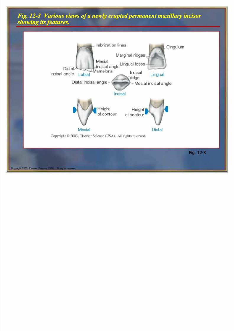
F ig. 12-3 Various views of a newly erupted permanent maxillary i ncisor showi n g its features.F ig. 12-3 Various views of a newly erupted permanent maxillary i ncisor showi n g its features.
Fig. 12-3Fig. 12-3
8/6/2019 Tooth Morph
http://slidepdf.com/reader/full/tooth-morph 9/84
Copyright 2003, Elsevier Science (USA). All rights reserved.
Maxillary Central Incisors Maxillary Central Incisors
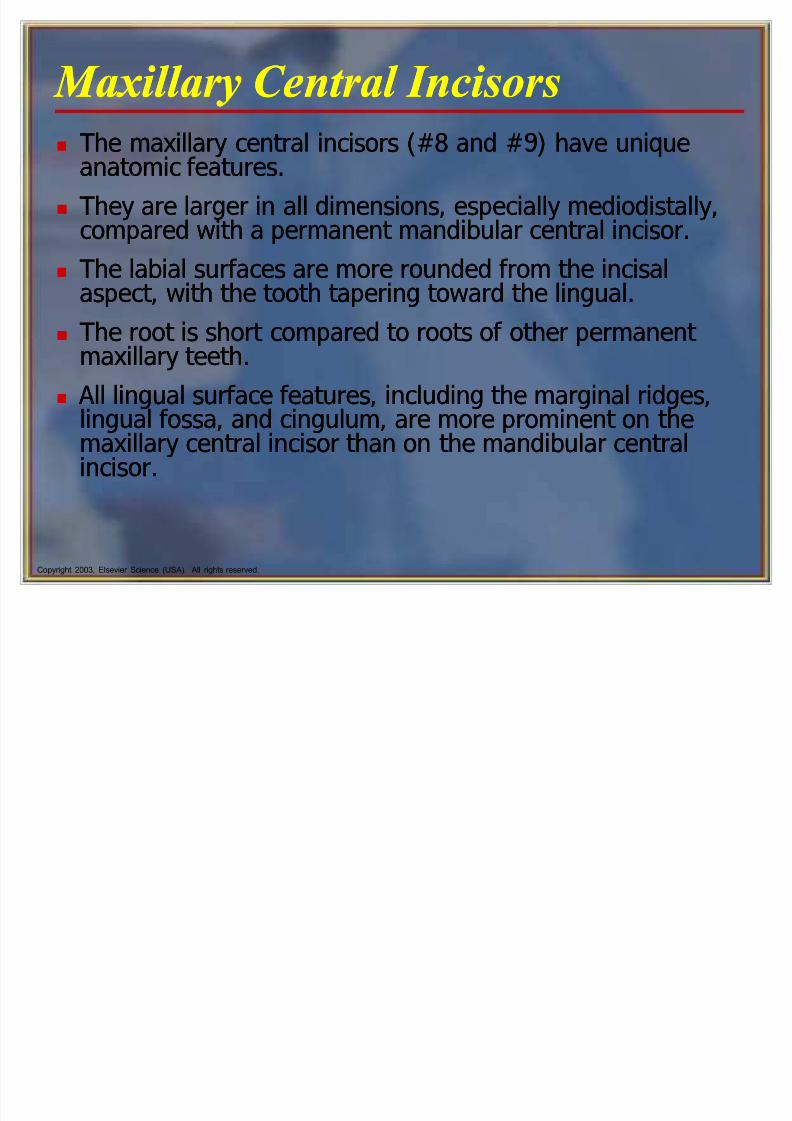
The maxillary central incisors (#8 and #9) have uniqueanatomic features.
They are larger in all dimensions, especially mediodistally,compared with a permanent mandibular central incisor.
The labial surfaces are more rounded from the incisalaspect, with the tooth tapering toward the lingual.
The root is short compared to roots of other permanent maxillary teeth.
All lingual surface features, including the marginal ridges,lingual fossa, and cingulum, are more prominent on themaxillary central incisor than on the mandibular centralincisor.
8/6/2019 Tooth Morph
http://slidepdf.com/reader/full/tooth-morph 10/84
Copyright 2003, Elsevier Science (USA). All rights reserved.
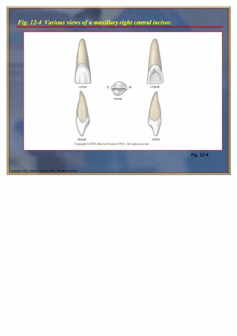
F ig. 12-4 Various views of a maxillary right central i ncisor.F ig. 12-4 Various views of a maxillary right central i ncisor.
Fig. 12-4Fig. 12-4

8/6/2019 Tooth Morph
http://slidepdf.com/reader/full/tooth-morph 11/84
Copyright 2003, Elsevier Science (USA). All rights reserved.
Maxillary Central Incisors cont¶d Maxillary Central Incisors cont¶d
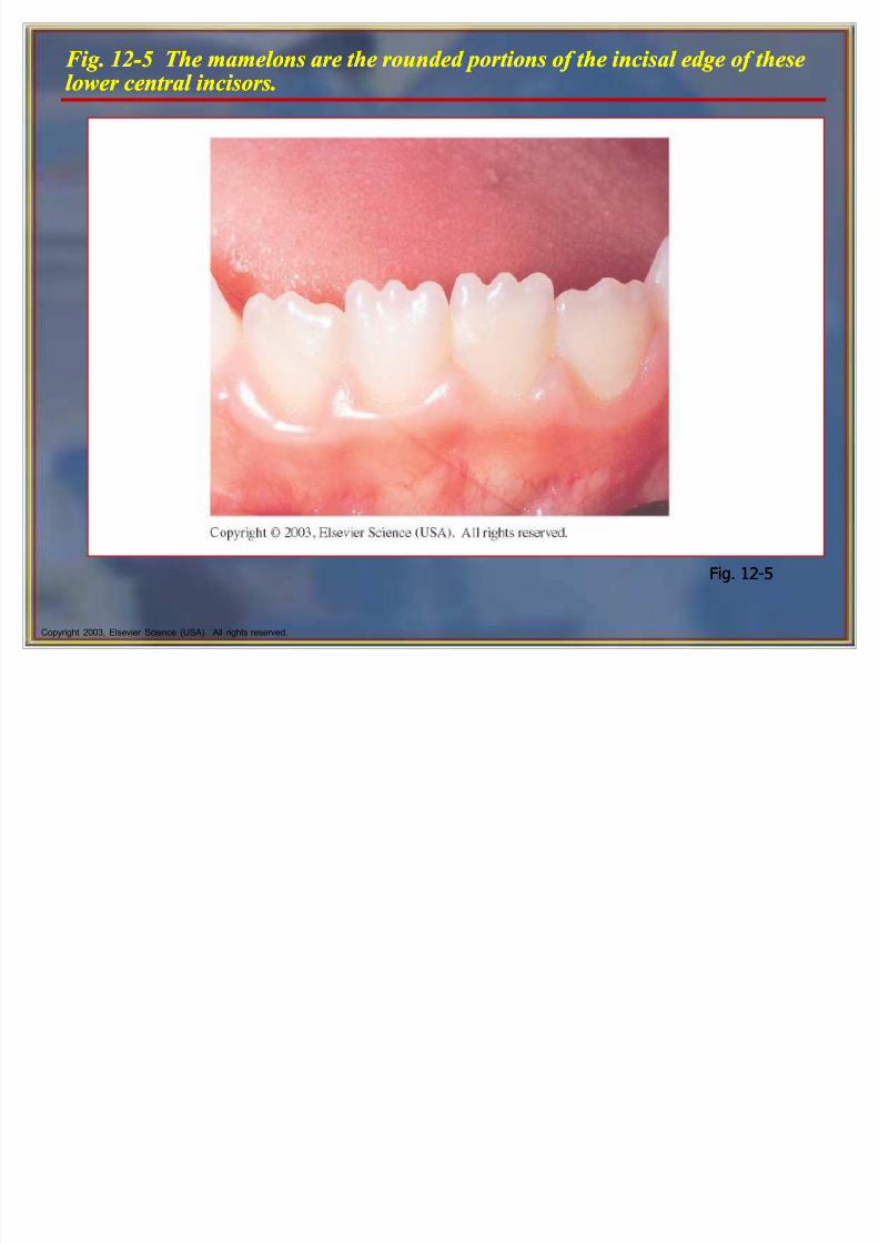
The incisal edges of these teeth are formed at the labioincisal line angle and do not exist until anedge has been created by wear.
The incisal edge is also known as the incisal
surface or incisal plane. When newly erupted, the central and lateral
incisors have three mamelons, or roundedenamel extensions on the incisal ridge, or edge.
The mamelons usually undergo attrition shortlyafter eruption.
8/6/2019 Tooth Morph
http://slidepdf.com/reader/full/tooth-morph 12/84
Copyright 2003, Elsevier Science (USA). All rights reserved.
F ig. 12-5 The mamelons are the rounded portions of the i ncisal edge of theselower central i ncisors.F ig. 12-5 The mamelons are the rounded portions of the i ncisal edge of theselower central i ncisors.
Fig. 12-5Fig. 12-5
8/6/2019 Tooth Morph
http://slidepdf.com/reader/full/tooth-morph 13/84
Copyright 2003, Elsevier Science (USA). All rights reserved.
Maxillary Lateral Incisors Maxillary Lateral Incisors
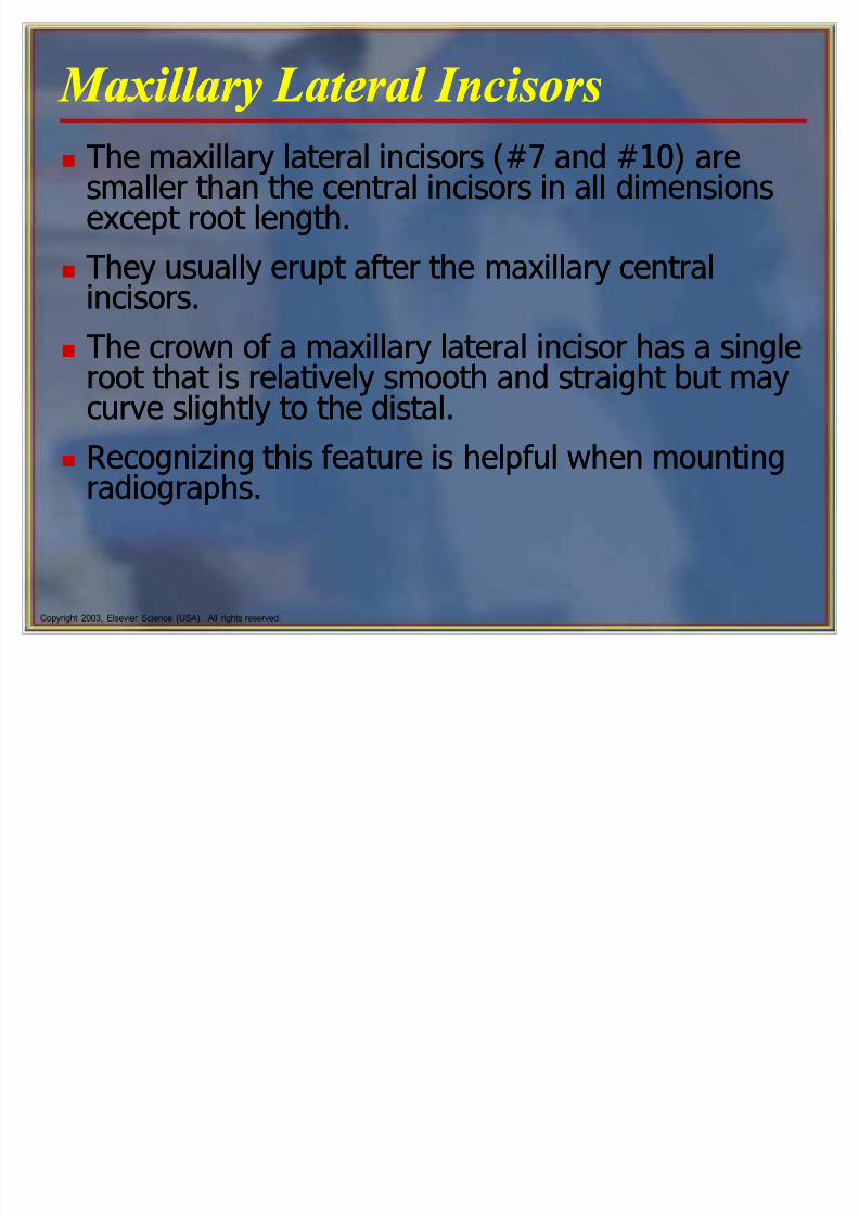
The maxillary lateral incisors (#7 and #10) aresmaller than the central incisors in all dimensionsexcept root length.
They usually erupt after the maxillary central
incisors. The crown of a maxillary lateral incisor has a single
root that is relatively smooth and straight but maycurve slightly to the distal.
Recognizing this feature is helpful when mountingradiographs.
8/6/2019 Tooth Morph
http://slidepdf.com/reader/full/tooth-morph 14/84
Copyright 2003, Elsevier Science (USA). All rights reserved.
Maxillary Lateral Incisors cont¶d Maxillary Lateral Incisors cont¶d
The lateral incisors vary in form more than anyother tooth in the mouth, except the third molars,and frequently are congenitally missing.
Because of the variations in form, the permanent
maxillary lateral incisors present challenges duringpreventive, restorative, and orthodonticprocedures.
Often, unattractive open contacts (spaces between
teeth) called diastemas occur in this area becauseof the variations in tooth size and position in thearch.

8/6/2019 Tooth Morph
http://slidepdf.com/reader/full/tooth-morph 15/84
8/6/2019 Tooth Morph
http://slidepdf.com/reader/full/tooth-morph 16/84
Copyright 2003, Elsevier Science (USA). All rights reserved.
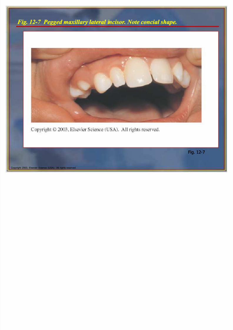
F ig. 12-7 Pegged maxillary lateral i ncisor. Note concial shape.F ig. 12-7 Pegged maxillary lateral i ncisor. Note concial shape.
Fig. 12-7Fig. 12-7
8/6/2019 Tooth Morph
http://slidepdf.com/reader/full/tooth-morph 17/84
Copyright 2003, Elsevier Science (USA). All rights reserved.
Mandibular Permanent Incisors Mandibular Permanent Incisors
The permanent mandibular incisors are thesmallest teeth of the permanent dentition and themost symmetric.
The central and lateral incisors of the mandibular
arch resemble each other. Generally, the lateral incisor is larger than the
central incisor, in contrast to the teeth in themaxillary arch.
Supragingival tooth deposits, such as plaque,calculus, and stain, tend to collect in the lingualconcavity of the mandibular incisors.

8/6/2019 Tooth Morph
http://slidepdf.com/reader/full/tooth-morph 18/84
Copyright 2003, Elsevier Science (USA). All rights reserved.
F ig. 12-9 Various views of a mandibular right lateral i ncisor.F ig. 12-9 Various views of a mandibular right lateral i ncisor.
Fig. 12-9Fig. 12-9

8/6/2019 Tooth Morph
http://slidepdf.com/reader/full/tooth-morph 19/84
8/6/2019 Tooth Morph
http://slidepdf.com/reader/full/tooth-morph 20/84

8/6/2019 Tooth Morph
http://slidepdf.com/reader/full/tooth-morph 21/84
8/6/2019 Tooth Morph
http://slidepdf.com/reader/full/tooth-morph 22/84
Copyright 2003, Elsevier Science (USA). All rights reserved.
Permanent Cani nes Permanent Cani nes
The permanent canines are the four anterior teethlocated at the corners of each quadrant for eachdental arch.
Their name is derived from the Latin word for dog
(canus) because these teeth resemble dogs teeth.
Patients often complain of the normal slightlydeeper yellow color of their canines compared
with their incisor teeth. The permanent canines are the longest teeth in
the dentition.
8/6/2019 Tooth Morph
http://slidepdf.com/reader/full/tooth-morph 23/84
Copyright 2003, Elsevier Science (USA). All rights reserved.
Permanent Cani nes cont¶d Permanent Cani nes cont¶d
The permanent canines are the longest teeth inthe dentition.
The root is usually one times the length of the
crown. This large root is externally manifested by the
bony vertical ridge called the canine eminence.
Patients commonly call the canines their eyeteeth.
The permanent canines are the longest teeth inthe dentition.
The root is usually one times the length of the
crown. This large root is externally manifested by the
bony vertical ridge called the canine eminence.
Patients commonly call the canines their eyeteeth.
8/6/2019 Tooth Morph
http://slidepdf.com/reader/full/tooth-morph 24/84
Copyright 2003, Elsevier Science (USA). All rights reserved.
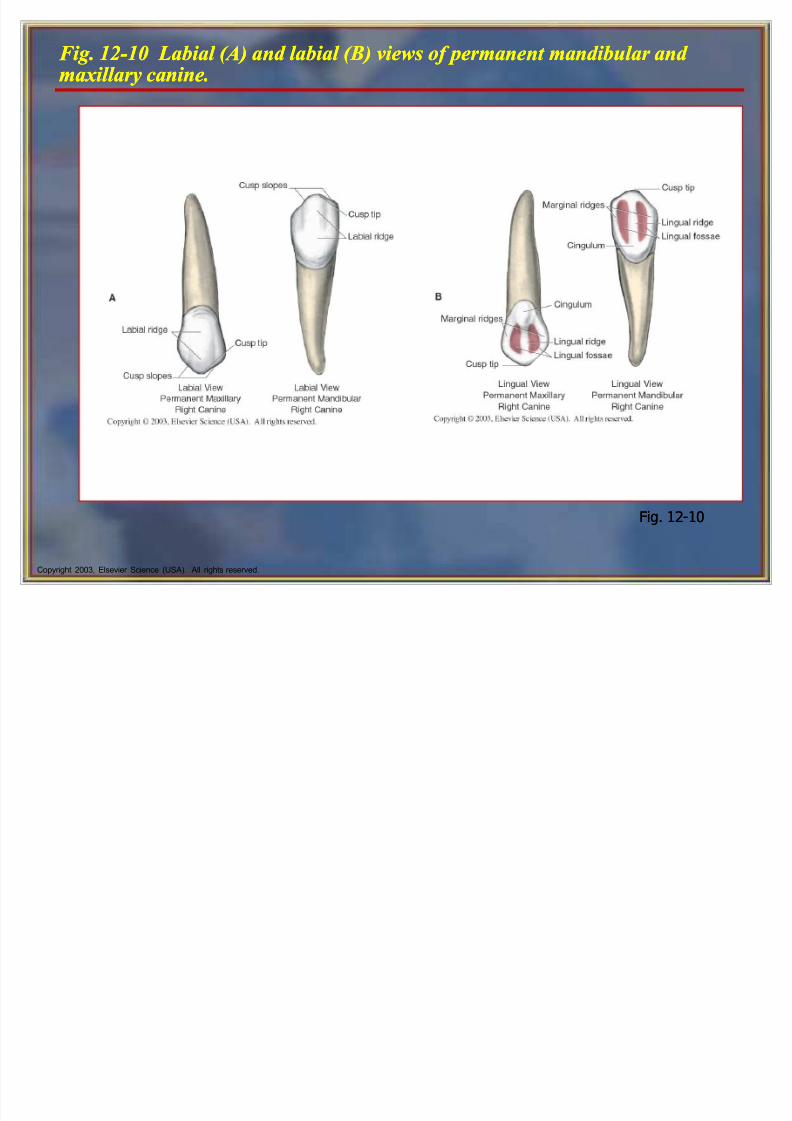
F ig. 12-10 Labial ( A) and labial (B ) views of permanent mandibular and maxillary cani ne.F ig. 12-10 Labial ( A) and labial (B ) views of permanent mandibular and maxillary cani ne.
Fig. 12-10Fig. 12-10
8/6/2019 Tooth Morph
http://slidepdf.com/reader/full/tooth-morph 25/84
Copyright 2003, Elsevier Science (USA). All rights reserved.
Maxillary Cani nes Maxillary Cani nes
The maxillary canines (#6 and #11) usually erupt after the mandibular canines, after the maxillaryincisors, and possibly after the maxillary premolars.
The cusp tip is sharper on a maxillary canine.
The mesial cusp slope is usually shorter than thedistal cusp slope in both the maxillary and themandibular canines when they first erupt.
The length of these cusp slopes and the cusp tipcan change with attrition.
8/6/2019 Tooth Morph
http://slidepdf.com/reader/full/tooth-morph 26/84
Copyright 2003, Elsevier Science (USA). All rights reserved.
Cli nical Considerations with MaxillaryCani nesCli nical Considerations with MaxillaryCani nes
The maxillary canines may erupt labially orlingually in relation to the surrounding teeth.
The maxillary canines may also fail to erupt fully
and may remain impacted.
This occurs because the permanent maxillarycanines erupt after the maxillary incisors andpossibly after the premolars and their arch spacehas closed.
8/6/2019 Tooth Morph
http://slidepdf.com/reader/full/tooth-morph 27/84
Copyright 2003, Elsevier Science (USA). All rights reserved.
Mandibular Cani nes Mandibular Cani nes
The mandibular canines (#22 and #27) usually erupt before the maxillary canines and after most of theincisors have erupted.
A mandibular canine closely resembles a maxillarycanine.
Although the entire tooth is usually as long, amandibular canine is narrower labiolingually andmesiodistally than a maxillary canine.
The lingual surface of the crown of the mandibularcanines is smoother than that of the maxillary caninesand has a less developed cingulum and two marginalridges.
8/6/2019 Tooth Morph
http://slidepdf.com/reader/full/tooth-morph 28/84
Copyright 2003, Elsevier Science (USA). All rights reserved.
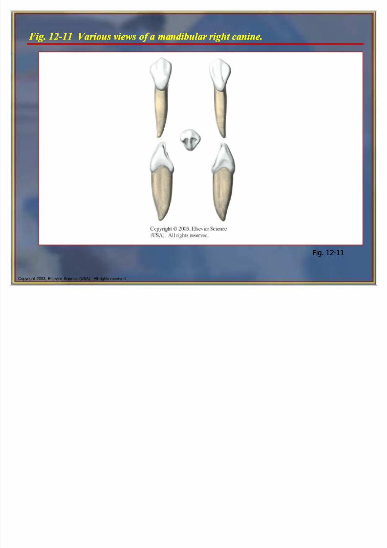
F ig. 12-11 Various views of a mandibular right cani ne.F ig. 12-11 Various views of a mandibular right cani ne.
Fig. 12-11Fig. 12-11
8/6/2019 Tooth Morph
http://slidepdf.com/reader/full/tooth-morph 29/84
Copyright 2003, Elsevier Science (USA). All rights reserved.
Posterior Permanent Dentition Posterior Permanent Dentition
The permanent posterior teeth include thepremolars and molars.
The crown of each posterior tooth has an occlusalsurface, bordered distally and mesially by marginal
ridges. The occlusal surfaces have two or more cusps.
Imagine each cusp as a mountain with slopingareas, or cusp ridges, extending from the top of
the mountain; between the ridges are slopingareas called inclined cuspal planes.
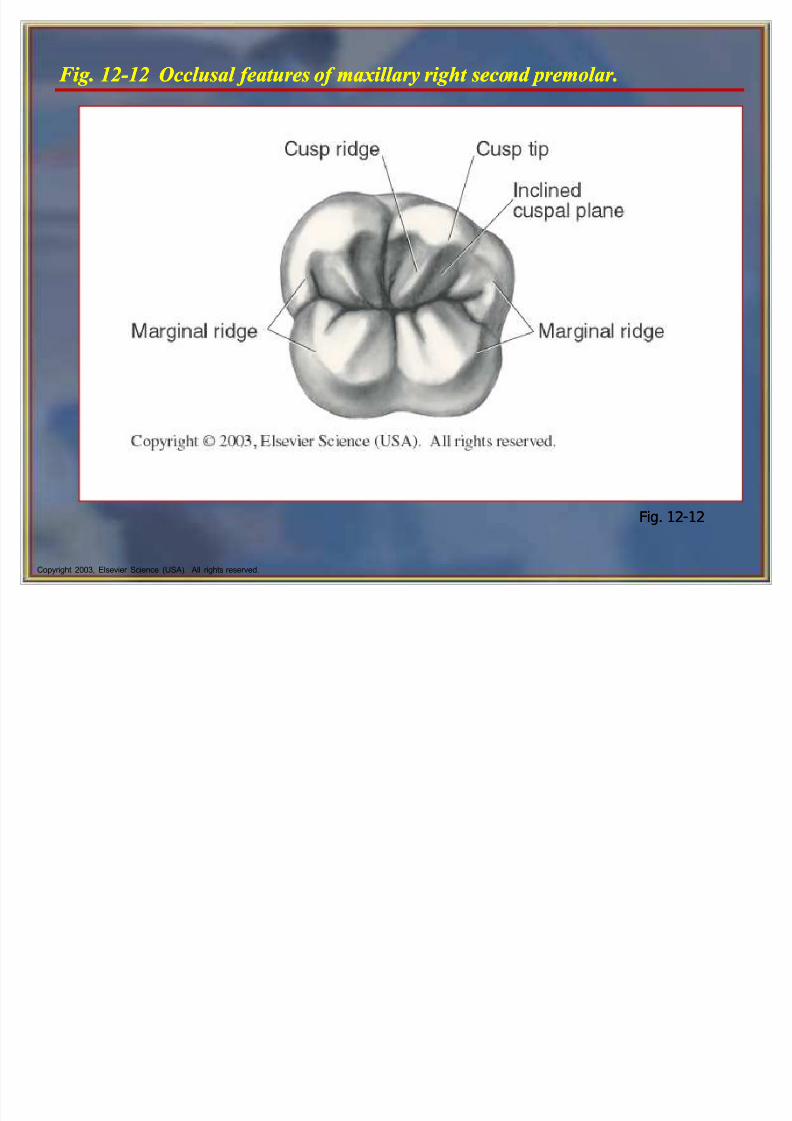
8/6/2019 Tooth Morph
http://slidepdf.com/reader/full/tooth-morph 30/84
Copyright 2003, Elsevier Science (USA). All rights reserved.
F ig. 12-12 Occlusal features of maxillary right second premolar.F ig. 12-12 Occlusal features of maxillary right second premolar.
Fig. 12-12Fig. 12-12
8/6/2019 Tooth Morph
http://slidepdf.com/reader/full/tooth-morph 31/84
8/6/2019 Tooth Morph
http://slidepdf.com/reader/full/tooth-morph 32/84
Copyright 2003, Elsevier Science (USA). All rights reserved.
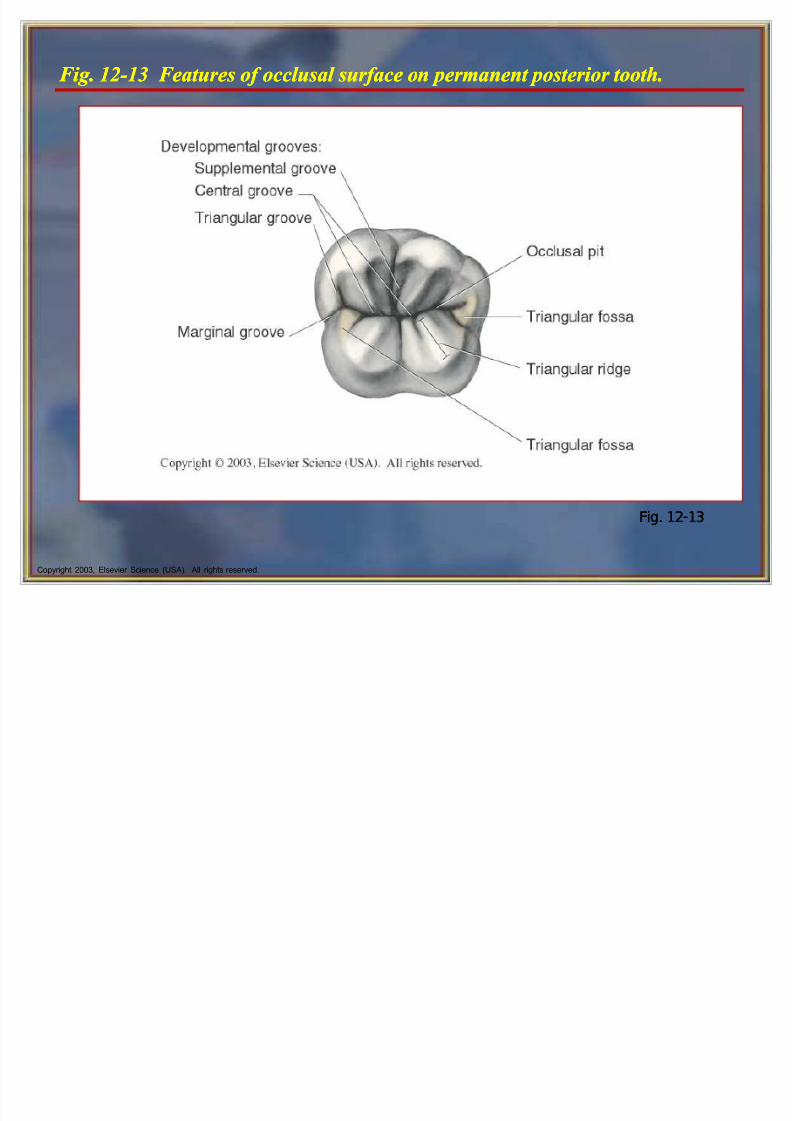
F ig. 12-13 F eatures of occlusal surface on permanent posterior tooth.F ig. 12-13 F eatures of occlusal surface on permanent posterior tooth.
Fig. 12-13Fig. 12-13
8/6/2019 Tooth Morph
http://slidepdf.com/reader/full/tooth-morph 33/84
8/6/2019 Tooth Morph
http://slidepdf.com/reader/full/tooth-morph 34/84

8/6/2019 Tooth Morph
http://slidepdf.com/reader/full/tooth-morph 35/84
Copyright 2003, Elsevier Science (USA). All rights reserved.
Maxillary F irst Premolars Maxillary F irst Premolars
A maxillary first premolar (#5 and #12) is largerthan a maxillary second premolar.
Each maxillary first premolar has two cusps (buccaland lingual) and two roots (facial and lingual).
Both maxillary premolars erupt earlier than themandibular premolars.
A maxillary first premolar (#5 and #12) is largerthan a maxillary second premolar.
Each maxillary first premolar has two cusps (buccaland lingual) and two roots (facial and lingual).
Both maxillary premolars erupt earlier than themandibular premolars.

8/6/2019 Tooth Morph
http://slidepdf.com/reader/full/tooth-morph 36/84
Copyright 2003, Elsevier Science (USA). All rights reserved.
Maxillary F irst Premolars cont¶d Maxillary F irst Premolars cont¶d
The maxillary first premolar has a bifurcated root (two roots, one buccal and one lingual).
Some first premolars have roots that are joined
together, or fused. The roots are shorter in length and resemble the
roots of the molars.
The maxillary first premolar has a bifurcated root (two roots, one buccal and one lingual).
Some first premolars have roots that are joined
together, or fused. The roots are shorter in length and resemble the
roots of the molars.
8/6/2019 Tooth Morph
http://slidepdf.com/reader/full/tooth-morph 37/84
Copyright 2003, Elsevier Science (USA). All rights reserved.
Cli nical Considerations with F irst Maxillary PremolarsCli nical Considerations with F irst Maxillary
Premolars
The maxillary first premolar is frequently extracted aspart of orthodontic treatment when the patients teethare crowded and more room is needed.
The orthodontist closes the space, and the patientsfacial appearance is not changed.
The slight depression in the shape of the root makesthe first maxillary premolar more susceptible toperiodontal disease.
Therefore, when it is necessary to remove one of thepremolars, the choice for removal is the first maxillarypremolar.

8/6/2019 Tooth Morph
http://slidepdf.com/reader/full/tooth-morph 38/84
Copyright 2003, Elsevier Science (USA). All rights reserved.
F ig. 12-14 Various views of a maxillary right first premolar.F ig. 12-14 Various views of a maxillary right first premolar.
Fig. 12-14Fig. 12-14
8/6/2019 Tooth Morph
http://slidepdf.com/reader/full/tooth-morph 39/84
Copyright 2003, Elsevier Science (USA). All rights reserved.
Maxillary Second Premolars Maxillary Second Premolars
Each maxillary second premolar (#4 and #13)has two cusps (buccal and lingual) and oneroot.
Each maxillary second premolar (#4 and #13)has two cusps (buccal and lingual) and oneroot.

8/6/2019 Tooth Morph
http://slidepdf.com/reader/full/tooth-morph 40/84
Copyright 2003, Elsevier Science (USA). All rights reserved.
Differences Between Second and F irst Premolars Differences Between Second and F irst Premolars
The cusps are more equal in length on the second premolar. The lingual cusp is slightly shorter but not as short as the
cusp on the maxillary first premolar.
The mesiobuccal cusp slope is shorter than the distobuccalcusp slope on the second premolar.
The cusps of the secondary premolar are not as sharp asthose of the maxillary first premolar.
The second premolar has only one root and one root canal.
The second premolar has a slight depression on the mesialroot.
The second premolar is wider buccolingually thanmesiodistally.
8/6/2019 Tooth Morph
http://slidepdf.com/reader/full/tooth-morph 41/84
Copyright 2003, Elsevier Science (USA). All rights reserved.
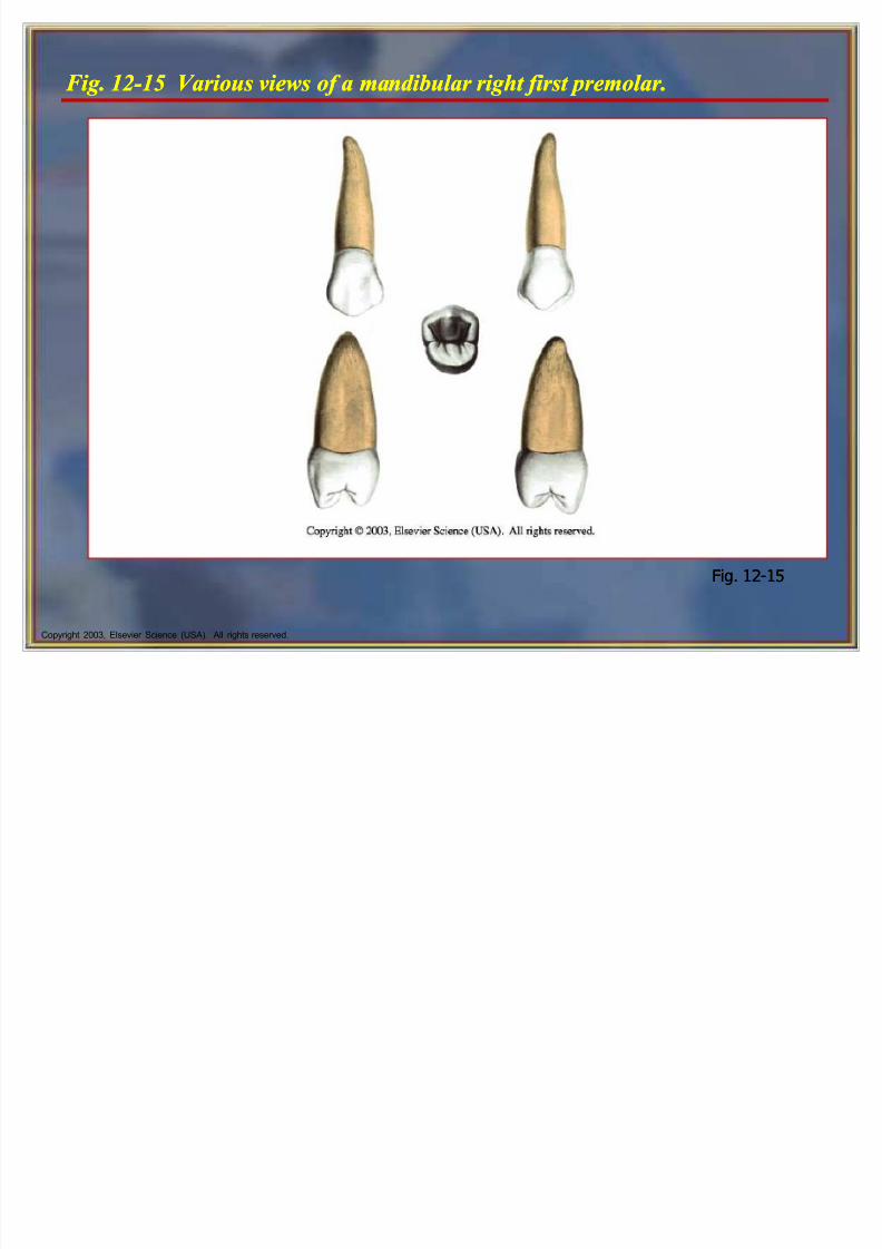
Mandibular F irst Premolars Mandibular F irst Premolars
The mandibular first premolars (#21 and #28)have a long and well-formed buccal cusp and asmall, nonfunctioning lingual cusp.
The lingual cusp may be no larger than thecingulum on some maxillary canines.
The mandibular first premolars are smaller andshorter than the mandibular second premolars.
The mandibular first premolars (#21 and #28)have a long and well-formed buccal cusp and asmall, nonfunctioning lingual cusp.
The lingual cusp may be no larger than thecingulum on some maxillary canines.
The mandibular first premolars are smaller andshorter than the mandibular second premolars.
8/6/2019 Tooth Morph
http://slidepdf.com/reader/full/tooth-morph 42/84
Copyright 2003, Elsevier Science (USA). All rights reserved.
F ig. 12-15 Various views of a mandibular right first premolar.F ig. 12-15 Various views of a mandibular right first premolar.
Fig. 12-15Fig. 12-15
8/6/2019 Tooth Morph
http://slidepdf.com/reader/full/tooth-morph 43/84
Copyright 2003, Elsevier Science (USA). All rights reserved.
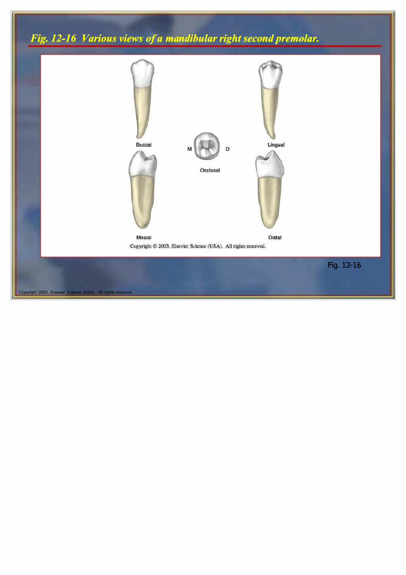
Mandibular Second Premolars Mandibular Second Premolars
The permanent mandibular second premolars(#20 and #29) erupt distal to the mandibular first premolars.
They are the succedaneous replacements for the
primary mandibular second molars. There are two forms of the mandibular second
premolar:
Three-cusp type, or tricuspidate form
Two-cusp type, or bicuspidate form
8/6/2019 Tooth Morph
http://slidepdf.com/reader/full/tooth-morph 44/84
Copyright 2003, Elsevier Science (USA). All rights reserved.
F ig. 12-16 Various views of a mandibular right second premolar.F ig. 12-16 Various views of a mandibular right second premolar.
Fig. 12-16Fig. 12-16
8/6/2019 Tooth Morph
http://slidepdf.com/reader/full/tooth-morph 45/84
Copyright 2003, Elsevier Science (USA). All rights reserved.
Permanent Molars Permanent Molars
The name molar comes from the Latin word for grinding.
There are three types of molars: the first molar,
second molar, and third molar. The first and second molars are also called the 6-
year and 12-year molars because of theapproximate eruption ages.
The name molar comes from the Latin word for grinding.
There are three types of molars: the first molar,
second molar, and third molar. The first and second molars are also called the 6-
year and 12-year molars because of theapproximate eruption ages.

8/6/2019 Tooth Morph
http://slidepdf.com/reader/full/tooth-morph 46/84
Copyright 2003, Elsevier Science (USA). All rights reserved.
Permanent Molars cont¶d Permanent Molars cont¶d
There are 12 molars, three in each quadrant, inthe permanent dentition.
The molar crowns have four or five short, blunt cusps, and each molar has two or three rootsthat help to support the larger crown.
There are 12 molars, three in each quadrant, inthe permanent dentition.
The molar crowns have four or five short, blunt cusps, and each molar has two or three rootsthat help to support the larger crown.
8/6/2019 Tooth Morph
http://slidepdf.com/reader/full/tooth-morph 47/84
Copyright 2003, Elsevier Science (USA). All rights reserved.
Maxillary Molars Maxillary Molars
They are usually the first permanent teeth to erupt into the maxillary arch.
Each maxillary molar usually has four major cusps,with two cusps on the buccal portion of theocclusal table and two on the lingual.
Each maxillary molar has three well-separated andwell-developed roots.
A tooth with three roots is said to be trifurcated,which means divided into thirds.
8/6/2019 Tooth Morph
http://slidepdf.com/reader/full/tooth-morph 48/84
Copyright 2003, Elsevier Science (USA). All rights reserved.
Maxillary F irst Molars Maxillary F irst Molars
The maxillary first molars (#3 and #14) are the first permanent teeth to erupt into the maxillary arch.
They erupt distal to the primary maxillary second molarsand thus are nonsuccedaneous.
The maxillary first molar is the largest tooth in the maxillaryarch and also has the largest crown in the permanent dentition.
This molar is composed of five developmental lobes, twobuccal and three lingual.
The fifth cusp is called the cusp of Carabelli.
8/6/2019 Tooth Morph
http://slidepdf.com/reader/full/tooth-morph 49/84
Copyright 2003, Elsevier Science (USA). All rights reserved.
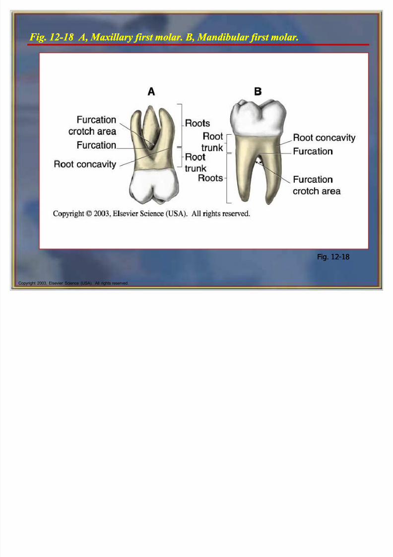
F ig. 12-18 A, Maxillary first molar. B , Mandibular first molar.F ig. 12-18 A, Maxillary first molar. B , Mandibular first molar.
Fig. 12-18Fig. 12-18
8/6/2019 Tooth Morph
http://slidepdf.com/reader/full/tooth-morph 50/84
Copyright 2003, Elsevier Science (USA). All rights reserved.
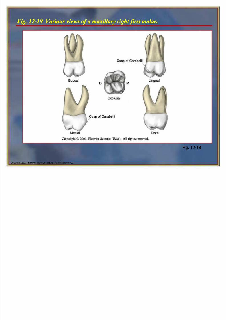
F ig. 12-19 Various views of a maxillary right first molar.F ig. 12-19 Various views of a maxillary right first molar.
Fig. 12-19Fig. 12-19
8/6/2019 Tooth Morph
http://slidepdf.com/reader/full/tooth-morph 51/84
Copyright 2003, Elsevier Science (USA). All rights reserved.
Maxillary Second Molars Maxillary Second Molars
The crown is somewhat shorter than that of the first molar,and the maxillary second molar usually has four cusps.
No fifth cusp is present.
There are three roots.
The roots of the secondary molars are smaller than roots of the first molars. The lingual root is still the largest andlongest.
The buccal groove is located farther distally on the buccalsurface of the second than the first maxillary molar.
The mesiobuccal cusp of the second maxillary molar islonger and has a less sharp cusp tip than the distobuccalcusp.

8/6/2019 Tooth Morph
http://slidepdf.com/reader/full/tooth-morph 52/84
Copyright 2003, Elsevier Science (USA). All rights reserved.
F ig. 12-20 Various views of a maxillary right second molar.F ig. 12-20 Various views of a maxillary right second molar.
Fig. 12-20Fig. 12-20
8/6/2019 Tooth Morph
http://slidepdf.com/reader/full/tooth-morph 53/84
Copyright 2003, Elsevier Science (USA). All rights reserved.
Maxillary Third Molars Maxillary Third Molars
The maxillary third molars (#1 and #16) differconsiderably in size and contour.
The crown is smaller, and the roots usuallyare shorter.
The roots of this tooth tend to fuse, and theresult is a single, tapered root.
People sometimes refer to this tooth as the
wisdom tooth because it erupts last.
The maxillary third molars (#1 and #16) differconsiderably in size and contour.
The crown is smaller, and the roots usuallyare shorter.
The roots of this tooth tend to fuse, and theresult is a single, tapered root.
People sometimes refer to this tooth as the
wisdom tooth because it erupts last.
8/6/2019 Tooth Morph
http://slidepdf.com/reader/full/tooth-morph 54/84
Fig 12 21 Buccal views of maxillary right third molars A First molar B SecondFig 12 21 Buccal views of maxillary right third molars A First molar B Second
8/6/2019 Tooth Morph
http://slidepdf.com/reader/full/tooth-morph 55/84
Copyright 2003, Elsevier Science (USA). All rights reserved.
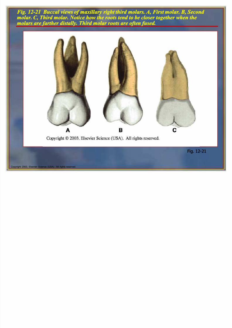
F ig. 12-21 Buccal views of maxillary right third molars. A, F irst molar. B , Second molar. C , Third molar. Notice how the roots tend to be closer together when themolars are farther distally. Third molar roots are often fused.
F ig. 12-21 Buccal views of maxillary right third molars. A, F irst molar. B , Second molar. C , Third molar. Notice how the roots tend to be closer together when themolars are farther distally. Third molar roots are often fused.
Fig. 12-21Fig. 12-21
8/6/2019 Tooth Morph
http://slidepdf.com/reader/full/tooth-morph 56/84
8/6/2019 Tooth Morph
http://slidepdf.com/reader/full/tooth-morph 57/84
Copyright 2003, Elsevier Science (USA). All rights reserved.
Mandibular Molars cont¶d Mandibular Molars cont¶d
Each mandibular molar has two well-developedroots: one mesial and one distal.
A tooth with two roots is referred to as bifurcated,
which means divided into two. A bifurcation is the area at which the two roots
divide.
Each mandibular molar has two well-developedroots: one mesial and one distal.
A tooth with two roots is referred to as bifurcated,
which means divided into two. A bifurcation is the area at which the two roots
divide.

8/6/2019 Tooth Morph
http://slidepdf.com/reader/full/tooth-morph 58/84
Copyright 2003, Elsevier Science (USA). All rights reserved.
Mandibular F irst Molars Mandibular F irst Molars
The permanent mandibular first molars (#19 and#30) erupt between 6 and 7 years of age.
These teeth are usually the first permanent teethto erupt in the oral cavity.
The two roots, mesial and distal, of a mandibularfirst molar are larger and more divergent thanthose of a second molar.
When this molar has three roots, the mesial root
has both buccal and lingual branches.
8/6/2019 Tooth Morph
http://slidepdf.com/reader/full/tooth-morph 59/84
Copyright 2003, Elsevier Science (USA). All rights reserved.
Mandibular Second Molars Mandibular Second Molars
The mandibular second molars (#18 and #31)erupt between 11 and 12 years of age.
These teeth erupt distal to the permanent first
molars and thus are nonsuccedaneous. The crown of the mandibular second molar is
slightly smaller than the first molar in all directions.
The crown has four well-developed cusps.
8/6/2019 Tooth Morph
http://slidepdf.com/reader/full/tooth-morph 60/84
Copyright 2003, Elsevier Science (USA). All rights reserved.
Mandibular Third Molars Mandibular Third Molars
The mandibular third molars (#17 and #32) aresimilar to the maxillary third molars in that they arevary greatly in shape.
There is no typical mandibular third molar.
This molar is usually smaller in all dimensions thanthe second molar.
The third molar usually consists of fourdevelopmental lobes.
A mandibular third molar usually has two roots that are fused, irregularly curved, and shorter than thoseof a mandibular second molar.
Clinical Considerations withClinical Considerations with
8/6/2019 Tooth Morph
http://slidepdf.com/reader/full/tooth-morph 61/84
Copyright 2003, Elsevier Science (USA). All rights reserved.
Cli nical Considerations with Mandibular MolarsCli nical Considerations with
Mandibular Molars
The mandibular molars can present difficulty inpositioning the oral evacuator because of thelingual inclination of the crowns.
Patients often have problems with their oralhygiene because of the lingual inclination of themolar teeth, and they may miss cleaning thelingual gingiva with the toothbrush.
The mandibular molars can present difficulty inpositioning the oral evacuator because of thelingual inclination of the crowns.
Patients often have problems with their oralhygiene because of the lingual inclination of themolar teeth, and they may miss cleaning thelingual gingiva with the toothbrush.
8/6/2019 Tooth Morph
http://slidepdf.com/reader/full/tooth-morph 62/84
Copyright 2003, Elsevier Science (USA). All rights reserved.
The Primary DentitionThe Primary Dentition
There are 20 primary teeth, 10 per dental arch.
These teeth include incisors, canines, and molars.
The primary teeth are numbered in the Universal ToothNumbering System by using the capital letters A through T.
The primary teeth are smaller overall and have whiterenamel than the permanent teeth.
The crown of any primary tooth is short in relation to itstotal length.
The crowns are narrower at the CEJ.

8/6/2019 Tooth Morph
http://slidepdf.com/reader/full/tooth-morph 63/84
Copyright 2003, Elsevier Science (USA). All rights reserved.
The Primary Dentition cont¶d The Primary Dentition cont¶d
The pulp chambers and pulp horns in primaryteeth are relatively large in proportion to those of the permanent teeth.
There is a thick layer of dentin between the pulp
chambers and the enamel, especially in theprimary mandibular second molar.
The enamel layer is relatively thin.
The pulp chambers and pulp horns in primaryteeth are relatively large in proportion to those of the permanent teeth.
There is a thick layer of dentin between the pulp
chambers and the enamel, especially in theprimary mandibular second molar.
The enamel layer is relatively thin.
Clinical Considerations with PrimaryClinical Considerations with Primary
8/6/2019 Tooth Morph
http://slidepdf.com/reader/full/tooth-morph 64/84
Copyright 2003, Elsevier Science (USA). All rights reserved.
Cli nical Considerations with PrimaryTeethCli nical Considerations with PrimaryTeeth
Often, parents do not understand the importanceof the primary teeth.
Primary teeth hold the eruption space for thepermanent teeth that will replace the primary
teeth. Because the enamel and dentin is thinner in
primary teeth, decay can travel quickly throughthe enamel to the pulp, possibly causing loss of the tooth.
Early dental health education and dental care areessential for keeping the primary dentition.
8/6/2019 Tooth Morph
http://slidepdf.com/reader/full/tooth-morph 65/84
Copyright 2003, Elsevier Science (USA). All rights reserved.
Primary Maxillary Incisors Primary Maxillary Incisors
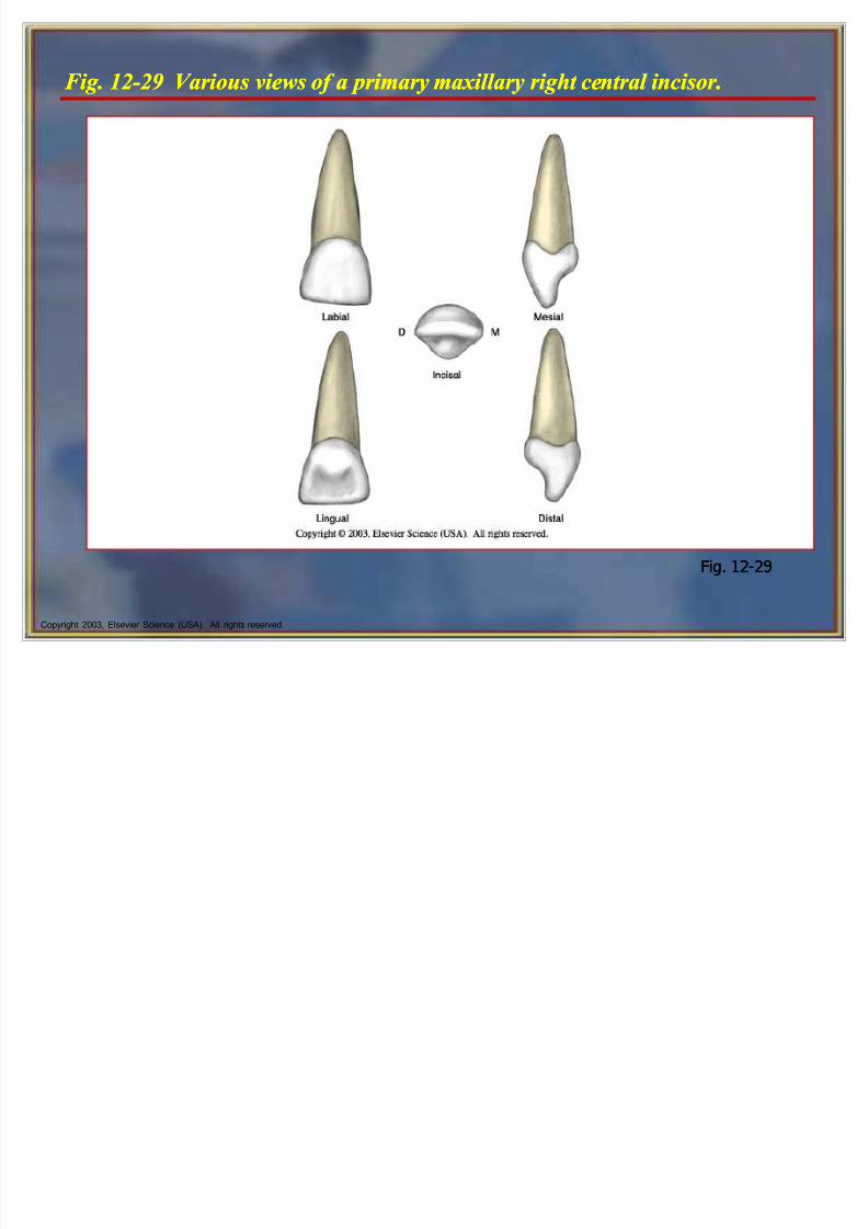
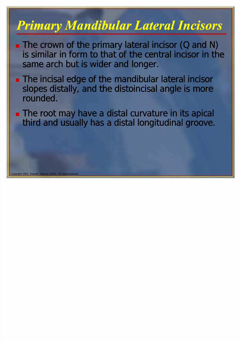
The crown of the primary maxillary central incisor(E and F) is wider mesiodistally thanincisocervically.
It is the only tooth of either dentition with this
crown dimension. The primary maxillary incisors have no mamelons.
The cingulum and marginal ridges are moreprominent than on the permanent successor, and
the lingual fossa is deeper.
8/6/2019 Tooth Morph
http://slidepdf.com/reader/full/tooth-morph 66/84
Copyright 2003, Elsevier Science (USA). All rights reserved.
F ig. 12-29 Various views of a primary maxillary right central i ncisor.F ig. 12-29 Various views of a primary maxillary right central i ncisor.
Fig. 12-29Fig. 12-29
8/6/2019 Tooth Morph
http://slidepdf.com/reader/full/tooth-morph 67/84
Copyright 2003, Elsevier Science (USA). All rights reserved.
Primary Maxillary Lateral Incisors Primary Maxillary Lateral Incisors
The crown of the primary maxillary lateral incisor(D and G) is similar to that of the central incisorbut is much smaller in all dimensions.
The incisal angles on the lateral incisor are alsomore rounded than on the central incisor.
The lateral root is longer in proportion to its crown,and its apex is sharper.

8/6/2019 Tooth Morph
http://slidepdf.com/reader/full/tooth-morph 68/84
Copyright 2003, Elsevier Science (USA). All rights reserved.
F ig. 12-30 Various views of a primary maxillary right lateral i ncisor.F ig. 12-30 Various views of a primary maxillary right lateral i ncisor.
Fig. 12-30Fig. 12-30
8/6/2019 Tooth Morph
http://slidepdf.com/reader/full/tooth-morph 69/84
8/6/2019 Tooth Morph
http://slidepdf.com/reader/full/tooth-morph 70/84
8/6/2019 Tooth Morph
http://slidepdf.com/reader/full/tooth-morph 71/84
8/6/2019 Tooth Morph
http://slidepdf.com/reader/full/tooth-morph 72/84
Copyright 2003, Elsevier Science (USA). All rights reserved.
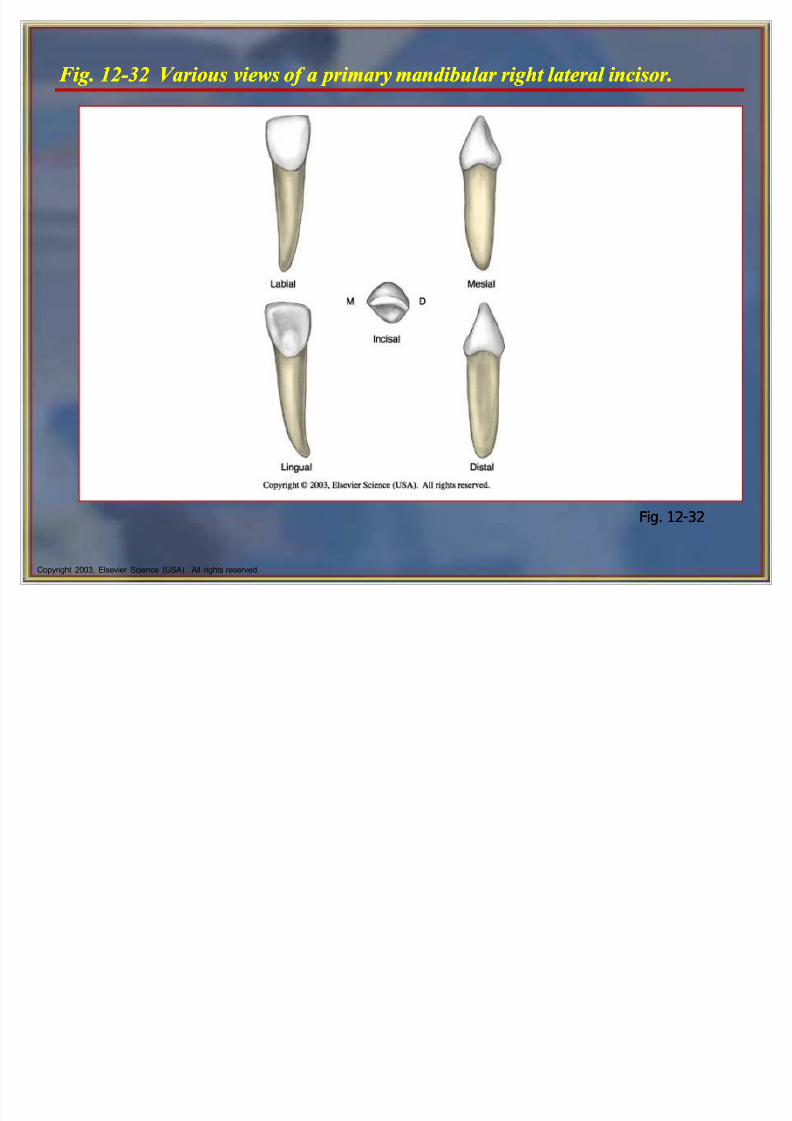
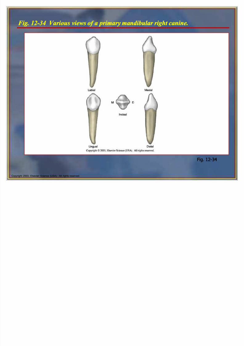
F ig. 12-32 Various views of a primary mandibular right lateral i ncisor.F ig. 12-32 Various views of a primary mandibular right lateral i ncisor.
Fig. 12-32Fig. 12-32
8/6/2019 Tooth Morph
http://slidepdf.com/reader/full/tooth-morph 73/84
Copyright 2003, Elsevier Science (USA). All rights reserved.
Primary Cani nes Primary Cani nes
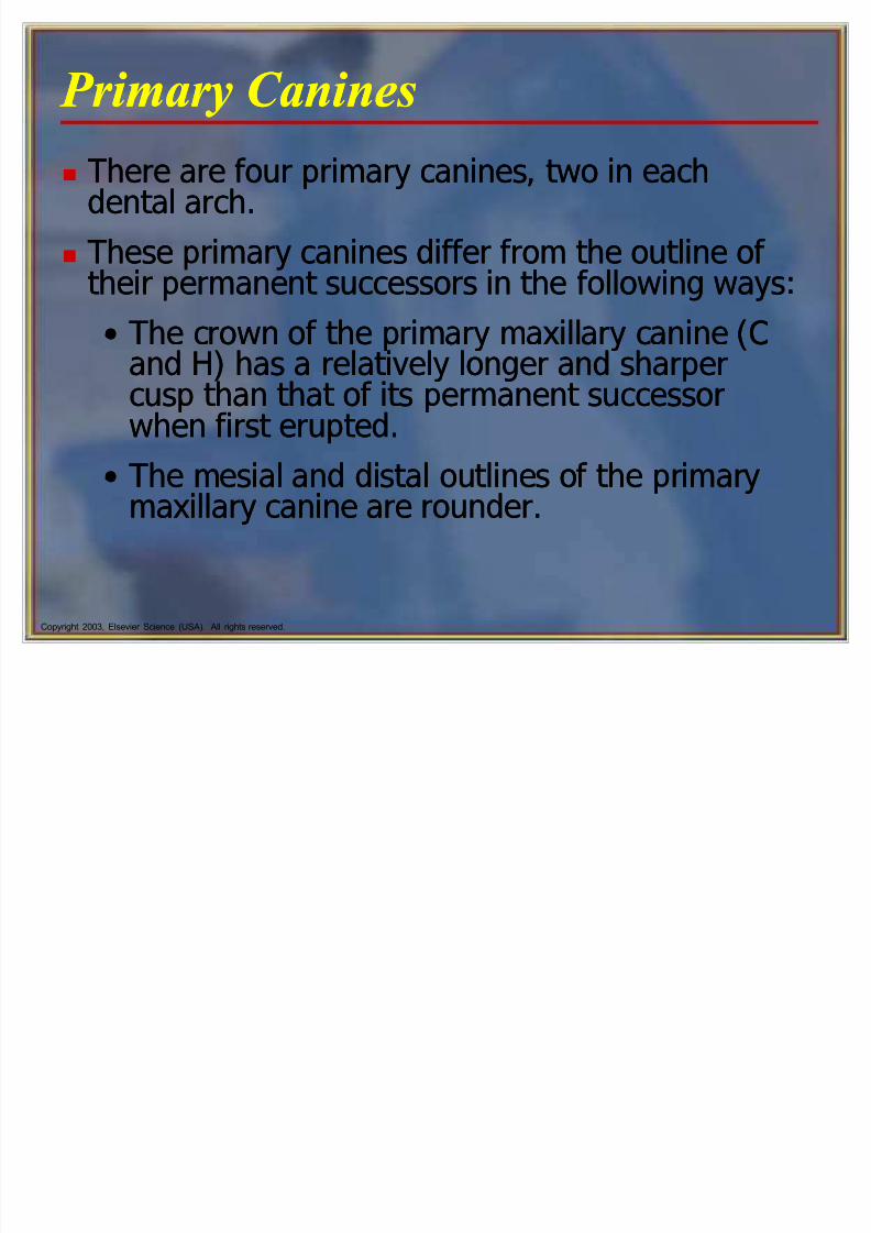
There are four primary canines, two in eachdental arch.
These primary canines differ from the outline of their permanent successors in the following ways:
The crown of the primary maxillary canine (Cand H) has a relatively longer and sharpercusp than that of its permanent successorwhen first erupted.
The mesial and distal outlines of the primarymaxillary canine are rounder.
8/6/2019 Tooth Morph
http://slidepdf.com/reader/full/tooth-morph 74/84
8/6/2019 Tooth Morph
http://slidepdf.com/reader/full/tooth-morph 75/84
Copyright 2003, Elsevier Science (USA). All rights reserved.
Primary Mandibular Cani nes Primary Mandibular Cani nes
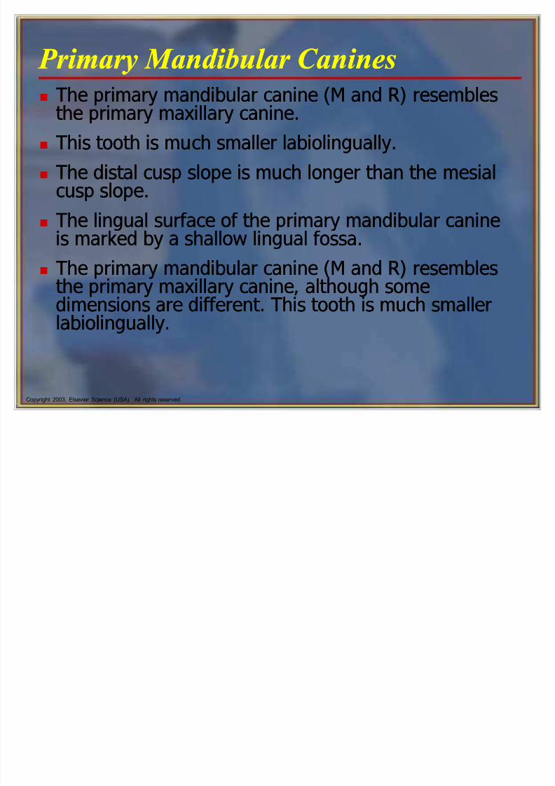
The primary mandibular canine (M and R) resemblesthe primary maxillary canine.
This tooth is much smaller labiolingually.
The distal cusp slope is much longer than the mesialcusp slope.
The lingual surface of the primary mandibular canineis marked by a shallow lingual fossa.
The primary mandibular canine (M and R) resemblesthe primary maxillary canine, although somedimensions are different. This tooth is much smallerlabiolingually.
8/6/2019 Tooth Morph
http://slidepdf.com/reader/full/tooth-morph 76/84
8/6/2019 Tooth Morph
http://slidepdf.com/reader/full/tooth-morph 77/84
Copyright 2003, Elsevier Science (USA). All rights reserved.
Primary Molars Primary Molars
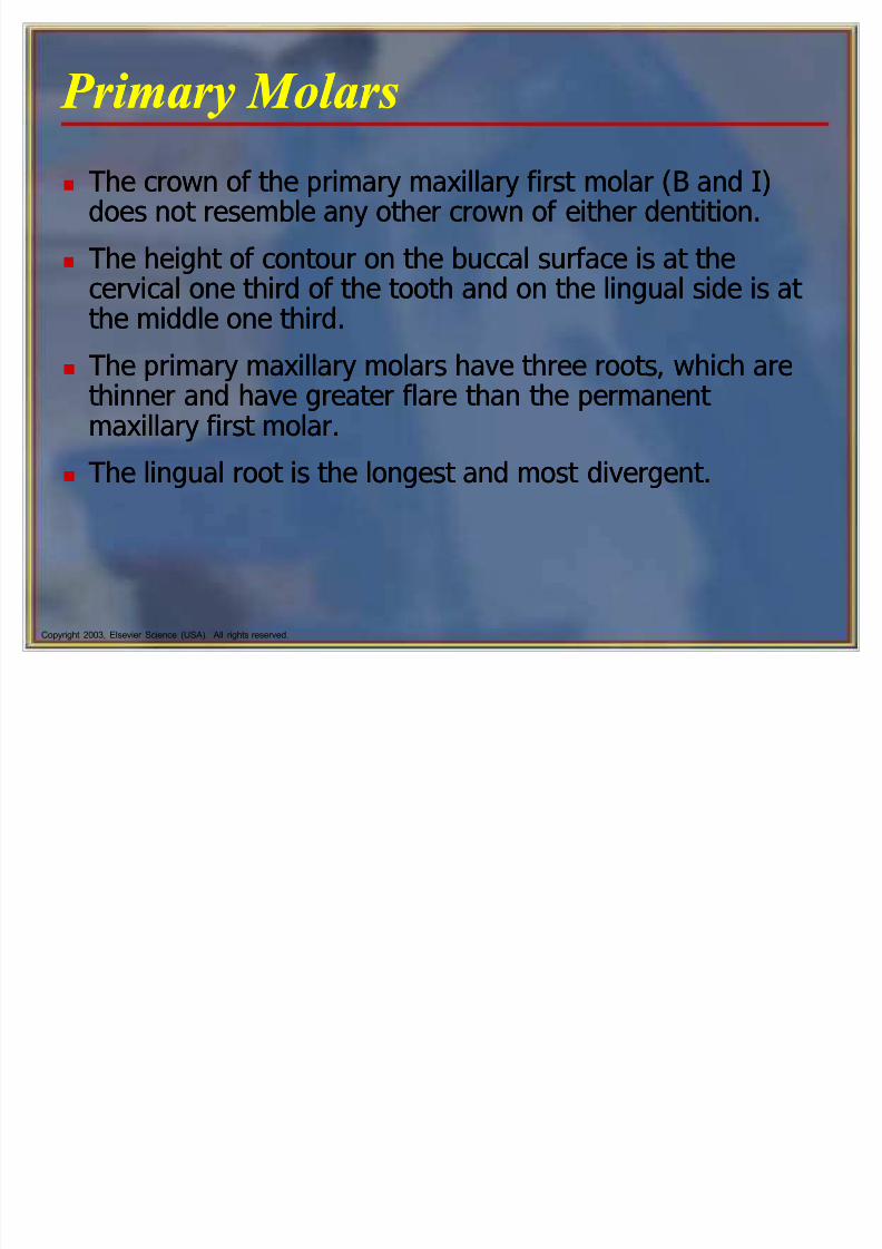
The crown of the primary maxillary first molar (B and I)does not resemble any other crown of either dentition.
The height of contour on the buccal surface is at thecervical one third of the tooth and on the lingual side is at
the middle one third. The primary maxillary molars have three roots, which are
thinner and have greater flare than the permanent maxillary first molar.
The lingual root is the longest and most divergent.
8/6/2019 Tooth Morph
http://slidepdf.com/reader/full/tooth-morph 78/84
Copyright 2003, Elsevier Science (USA). All rights reserved.
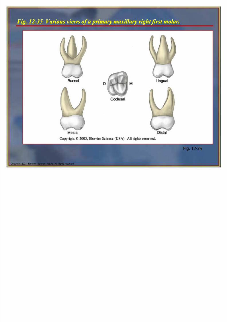
F ig. 12-35 Various views of a primary maxillary right first molar.F ig. 12-35 Various views of a primary maxillary right first molar.
Fig. 12-35Fig. 12-35
8/6/2019 Tooth Morph
http://slidepdf.com/reader/full/tooth-morph 79/84
Copyright 2003, Elsevier Science (USA). All rights reserved.
Primary Maxillary Second Molars Primary Maxillary Second Molars
The primary maxillary second molar (A and J) islarger than the primary maxillary first molar.
This tooth most closely resembles the form of the
permanent maxillary first molar but is smaller in alldimensions.
The second molar usually has a cusp of Carabelli,the minor fifth cusp.
The primary maxillary second molar (A and J) islarger than the primary maxillary first molar.
This tooth most closely resembles the form of the
permanent maxillary first molar but is smaller in alldimensions.
The second molar usually has a cusp of Carabelli,the minor fifth cusp.
8/6/2019 Tooth Morph
http://slidepdf.com/reader/full/tooth-morph 80/84
Copyright 2003, Elsevier Science (USA). All rights reserved.
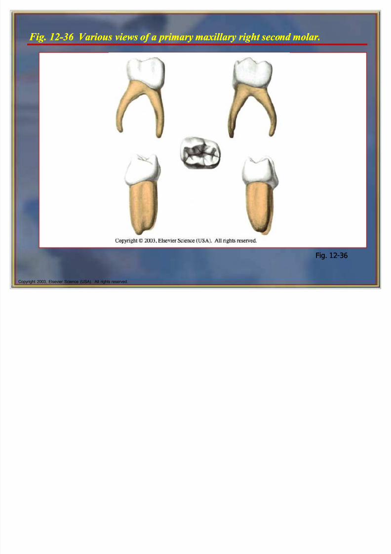
F ig. 12-36 Various views of a primary maxillary right second molar.F ig. 12-36 Various views of a primary maxillary right second molar.
Fig. 12-36Fig. 12-36
8/6/2019 Tooth Morph
http://slidepdf.com/reader/full/tooth-morph 81/84
8/6/2019 Tooth Morph
http://slidepdf.com/reader/full/tooth-morph 82/84
8/6/2019 Tooth Morph
http://slidepdf.com/reader/full/tooth-morph 83/84
8/6/2019 Tooth Morph
http://slidepdf.com/reader/full/tooth-morph 84/84
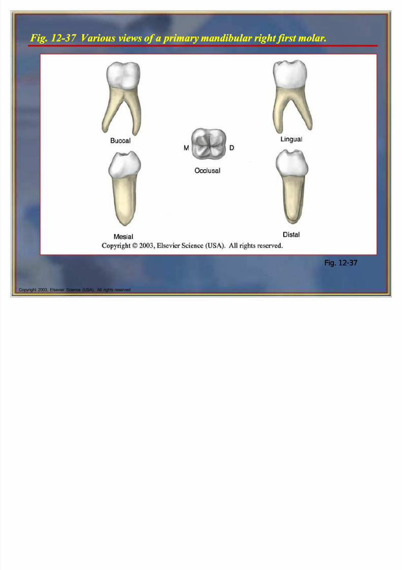
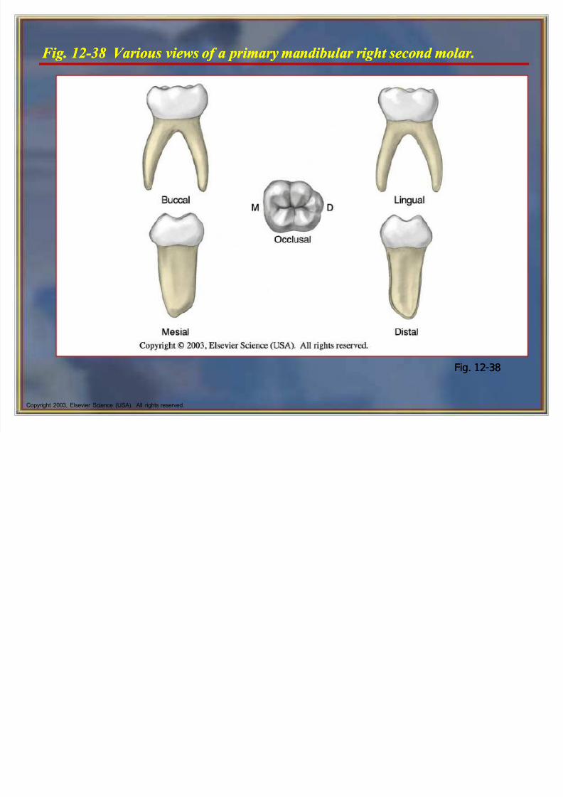
F ig. 12-38 Various views of a primary mandibular right second molar.F ig. 12-38 Various views of a primary mandibular right second molar.
Fig. 12-38Fig. 12-38