Embed Size (px)
Citation preview

94 cliniciansbrief.com • January 2014
Peer reviewedTop 5 Cardiology
Arrhythmias in Dogs & Cats
Ashley Jones, DVMAmara Estrada, DVM, DACVIM (Cardiology) University of Florida
The term cardiac arrhythmia encompasses all cardiacrhythms other than a normal sinus rhythm, including both bradyarrhythmias (ie, abnormally slow heart rate)
and tachyarrhythmias (ie, abnormally fast heart rate). Cardiacarrhythmias can be benign or life threatening. The followingreviews five of the most common cardiac arrhythmias identifiedin dogs and cats and outlines recommended treatment protocols.
TOP 5
TOP 5 Arrhythmias Seen in Dogs & Cats
1. Sinus arrhythmia
2. Atrial fibrillation
3. Ventricular tachycardia
4. Premature beats
5. Atrioventricular block
Sinus ArrhythmiaSinus arrhythmia results from variations in vagal toneassociated with respiration; the overall heart rate isnormal, but the rate rises on inspiration and slows onexpiration. This can be common in dogs, which nor-
mally have a high resting vagal tone. Brachycephalic breedsappear predisposed to sinus arrhythmia because of increasedbreathing effort.
In cats, sinus arrhythmia is less common, as the feline heart rateis more sympathetically driven, especially when a cat is at theclinic. If sinus arrhythmia is identified in a cat, the patient mayhave excessive vagal tone from pathology in the intracranial,intraocular, respiratory, GI, or (less common) urogenital systems.
The ECG characteristics of sinus arrhythmia are a normal heartrate and a regularly irregular sinus rhythm, with more than 10%variation in R–R intervals (Figure 1A).
An atropine response test can help differentiate true conductiondisturbance (eg, sick sinus syndrome) from vagally mediatedsinus arrhythmia. Patients with high vagal tone should have acomplete response to atropine and a resultant sinus tachycardia(heart rate >160 bpm; Figure 1B), whereas patients with sicksinus syndrome may not respond or may respond only partiallyto atropine (Figures 1C and 1D). The authors typically adminis-ter 0.04 mg/kg of IM atropine, wait 20 to 30 minutes, and thenrecheck the heart rate via ECG or auscultation.
1

January 2014 • clinician’s brief 95
(A) Six-lead ECG illustrating sinus arrhythmiain a healthy dog. The 4 beats in the middleare at a faster rate (100 bpm) than the sinus
beats (~60 bpm; paper speed, 50 mm/sec; sensitivity, 5 mm/mV).
(B) Six-lead ECG from the same dog as in Figure 1Aillustrating complete response to an atropine test.Sinus tachycardia has developed following atropineadministration, and the heart rate is now ~214 bpm(paper speed, 50 mm/sec; sensitivity 10 mm/mV).
(C) Six-lead ECG from a dog with bradyarrhythmia.There is sinus bradycardia along with second-degreeatrioventricular (AV) block (blocked P waves,arrows). The P waves are conducted very slowlythrough the AV node with first-degree AV block(circled).
(D) Six-lead ECG (paper speed, 50 mm/sec; sensitivity,5 mm/mV) from the dog in Figure 1B illustratingpartial response to an atropine test. The heart rate is increased from baseline but only to 70 bpm, andthere is persistent 2:1 second degree AV block(blocked P waves, arrows) as well as first-degree AV block (circled).
MORE
AV = atrioventricular
1
A
B
C
D

96 cliniciansbrief.com • January 2014
Atrial FibrillationAtrial fibrillation is caused by multiple reentrant cir-cuits in the atria that can fire at rates up to 500 to600 bpm. The atrioventricular (AV) node acts as agatekeeper and determines how many of these
impulses are propagated through to the ventricles. Giant-breeddogs (eg, Irish wolfhound) can develop lone atrial fibrillation,wherein they do not have underlying structural heart diseasebased on ECG assessment. More commonly in dogs and almostexclusively in cats, atrial fibrillation is associated with significantheart disease and atrial enlargement because a critical mass isrequired to support the 5 or 6 reentry circuits necessary foratrial fibrillation propagation. The atria in giant-breed dogs(and humans and horses) can potentially support atrial fibrilla-tion, whereas cats and most other dogs must have significantatrial dilation as a result of cardiac disease to achieve the criticalmass.
The ECG characteristics of atrial fibrillation are lack of P wavesand irregularly irregular supraventricular rhythm, with or with-out tachycardia (heart rate >160 bpm; Figure 2A).
Top 5
2In dogs, the goal of treatment is typically to slow the ventricularresponse rate to a target heart rate of approximately 140 to 160bpm as assessed by ECG (Figure 2B). This is best accomplishedwith a combination of extended-release diltiazem preparationand digoxin.1
Although some prefer use of a β-blocker, β-blockers and cal-cium-channel blockers can have negative inotropic effects andthus should be used cautiously and slowly up-titrated when thereis concurrent heart failure or systolic dysfunction. In cases oflone atrial fibrillation, medical or electrical cardioversion (ie,conversion back to normal sinus rhythm) can be attempted;however, this requires general anesthesia, and survival orimproved quality of life has not been definitively demonstratedin humans or dogs. Many patients with lone atrial fibrillationhave fairly slow ventricular response rates and do not requiremedical therapy. Cats with atrial fibrillation are generally treatedwith either a calcium-channel blocker or a β-blocker, using cau-tion in the face of congestive heart failure or systolic dysfunction.
(A) Three-lead ECG from a Doberman pinscher with dilated cardiomyopathy. There is uncontrolled atrial fibrillation. The fast heartrate, lack of discernible P waves, and irregularity are all characteristic of atrial fibrillation. In this patient, the QRS complexes are widebecause of left ventricular enlargement. Because of heart orientation in deep-chested animals (eg, Doberman pinschers), left ventricular
enlargement often creates a widened QRS complex (instead of a tall R wave). Heart rate ranges from 230–300 bpm (paper speed, 50 mm/sec;sensitivity, 10 mm/mV).
(B) Three-lead ECG in a golden retriever with dilated cardiomyopathy. Atrial fibrillation in this dog is being treated with digoxin and diltiazem. Pwaves are still not discernible and the rate is still irregular but at a much slower ventricular response rate because of blockade of the AV node. Theaverage heart rate is now 100–140 bpm (paper speed, 50 mm/sec; sensitivity, 10 mm/mV).
2
A
B

January 2014 • clinician’s brief 97
Ventricular TachycardiaVentricular tachycardia (VT) is a potentially life-threatening arrhythmia that can result in suddendeath. VT can occur in patients with primary cardiac disease such as arrhythmogenic right ven-
tricular cardiomyopathy (common in boxers) or dilated car-diomyopathy (common in Doberman pinschers). Ventriculararrhythmias can also be hereditary and can occur in patientswith a structurally normal heart.
The ECG characteristics of VT include wide and bizarre QRScomplexes without any associated P waves in addition to tachy-cardia (heart rate, >180 bpm; Figure 3A).
Although no treatment has been proven to prevent sudden deathin patients with VT, antiarrhythmic therapy is used especially ifthe patient is a breed predisposed to sudden death, the patient issymptomatic, and/or malignant characteristics are present onVT (eg, rapid heart rate [>180 bpm]) or R-on-T or polymorphicQRS complexes are present (Figure 3B). The most common
3
(A) Six-lead ECG in a boxer witharrhythmogenic right ventricularcardiomyopathy. There is mono-
morphic VT at an extremely fast rate of ~350bpm (paper speed, 50 mm/sec; sensitivity, 10mm/mV).
(B) Three-lead ECG from a Dobermanpinscher with rapid polymorphic VT. Theheart rate ranges from 260–300 bpm. Notethe abrupt changes in morphology of theventricular complexes throughout the ECG(arrows; paper speed, 50 mm/sec; sensitivity,5 mm/mV).
(C) Six-lead ECG from a dog with vehiculartrauma showing an initial accelerated idio-ventricular rhythm followed by normal sinusrhythm. Initial 7 beats are wide and bizarre inmorphology (typical for ventricular ectopy),but the rate is slow (~125 bpm). When theidioventricular rhythm breaks spontaneously,normal sinus beats are seen (arrows) at a rate of ~90 bpm (paper speed, 50 mm/sec;sensitivity, 5 mm/mV).
3
A
B
MORE
AV = atrioventricular, VT = ventricular tachycardiaC

medications used for ventricular arrhythmias include IV lido-caine (acutely administered), sotalol, and PO mexiletine(Table). Sotalol and mexiletine can be compounded into a liquidformulation for titration of doses, if necessary. In cases of signifi-cant systolic dysfunction, mexiletine can be used at 4 to 8 mg/kgPO q8h. This same dose can be added to sotalol when ventricu-lar arrhythmias persist despite administration of maximal dosesof sotalol. Other medication combinations (amiodarone, fle-cainide, propafenone, procainamide) may be needed for patientsthat are refractory to conventional therapy. Implantable defibril-lators are occasionally required to treat high-risk patients.
Drug Dose Arrhythmia
Aminophylline–theophylline PO: 10 mg/kg q12h (extended release) (D, dog; C, cat) Sick sinus syndrome, second- and third-IV: 10 mg/kg (D) degree AV block
Atenolol PO: 0.2–2 mg/kg q12–24h (D); 6.25–12.5 mg Supraventricular tachycardia or VT (rate q12–24h (C) control or conversion), atrial fibrillation
(rate control)
Atropine SC, IM, IV: 0.02–0.04 mg/kg (D, C) Sinus bradycardia, sick sinus syndrome, first- and second-degree AV block
Digoxin PO: 0.005 mg/kg q12h (D) Atrial fibrillation (rate control)
Diltiazem IV bolus: 0.1–0.4 mg/kg over 5 min (D, C) Supraventricular tachycardia (rate control CRI: 0.05–0.15 mg/kg/h (D, C) or conversion), atrial fibrillation (rate PO: 1–2 mg/kg q8h (D, C); 2–4 mg/kg q12h control)(D); 30 mg q12–24h (C)
Esmolol IV bolus: 0.2–0.5 mg/kg over 1 min, Supraventricular tachycardia or repeat q5min (D, C) VT conversion
Glycopyrrolate SC, IM, IV: 0.005–0.01 mg/kg (D, C) Sinus bradycardia, sick sinus syndrome, first- and second-degree AV block
Isoproterenol CRI IV: 0.04–0.09 µg/kg/min, to effect (D, C) Second- and third-degree AV block
Lidocaine IV bolus (maximum of three): 2 mg/kg Supraventricular tachycardia conversion, over 30 sec (D); 0.25–0.5 mg/kg over 30 sec (C) vagally mediated atrial fibrillation CRI: 25–80 µg/kg/min (D) conversion, VT conversion
Mexiletine PO: 4–8 mg/kg PO q8h (D) Sinus rhythm maintenance
Procainamide IV bolus: 5–15 mg/kg over 1 min (D); Supraventricular tachycardia conversion1–2 mg/kg over 5 min (C) CRI: 20–50 µg/kg/min (D)
Sotalol PO: 1–3 mg/kg q12h (D, C) Supraventricular tachycardia or VT conversion; sinus rhythm maintenance
Terbutaline PO: 0.2 mg/kg q8–12h (D); 0.625 mg Sinus bradycardia, sick sinus syndromeq8–12h (C)IV: 0.01 mg/kg IV (D, C)
AV = atrioventricular, C = cat, D = dog, VT = ventricular tachycardia
Drugs Used to Treat Arrhythmias in Dogs & CatsTable
98 cliniciansbrief.com • January 2014
Top 5
An accelerated idioventricular rhythm should not be confusedwith VT. Accelerated idioventricular rhythm can also bereferred to as slow VT because of the similar appearance onECG, but the heart rate is generally less than 180 bpm (Figure3C, previous page). Accelerated idioventricular rhythms areoften seen in patients with systemic disease (eg, gastric dilata-tion–volvulus, pancreatitis, hemangio sarcoma, immune-medi-ated hemolytic anemia) rather than primary cardiac disease.Antiarrhythmic therapy is often unnecessary, as this type ofarrhythmia is not typically hemodynamically compromisingand is often self-limiting with treatment of the primary disease.
January 2014 • clinician’s brief 99
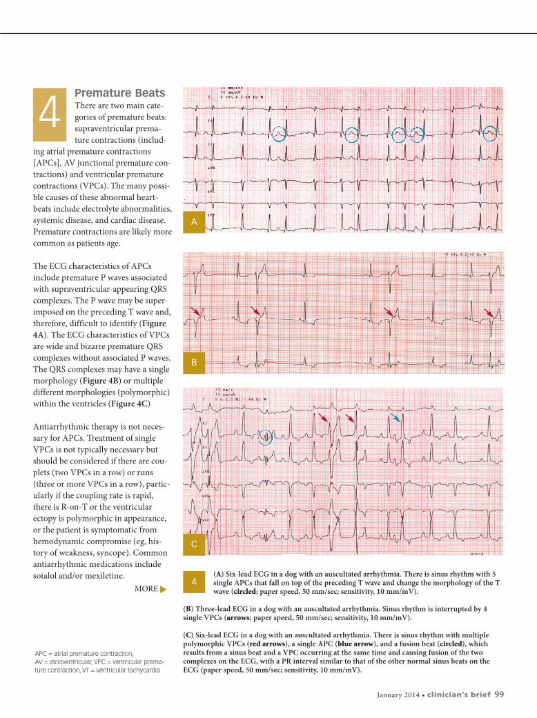
Premature Beats There are two main cate-gories of premature beats:supraventricular prema-ture contractions (includ-
ing atrial premature contractions[APCs], AV junctional premature con-tractions) and ventricular prematurecontractions (VPCs). The many possi-ble causes of these abnormal heart-beats include electrolyte abnormalities,systemic disease, and cardiac disease.Premature contractions are likely morecommon as patients age.
The ECG characteristics of APCsinclude premature P waves associatedwith supraventricular-appearing QRScomplexes. The P wave may be super-imposed on the preceding T wave and,therefore, difficult to identify (Figure4A). The ECG characteristics of VPCsare wide and bizarre premature QRScomplexes without associated P waves.The QRS complexes may have a singlemorphology (Figure 4B) or multipledifferent morphologies (polymorphic)within the ventricles (Figure 4C)
Antiarrhythmic therapy is not neces-sary for APCs. Treatment of singleVPCs is not typically necessary butshould be considered if there are cou-plets (two VPCs in a row) or runs(three or more VPCs in a row), partic-ularly if the coupling rate is rapid,there is R-on-T or the ventricularectopy is polymorphic in appearance,or the patient is symptomatic fromhemodynamic compromise (eg, his-tory of weakness, syncope). Commonantiarrhythmic medications includesotalol and/or mexiletine. (A) Six-lead ECG in a dog with an auscultated arrhythmia. There is sinus rhythm with 5
single APCs that fall on top of the preceding T wave and change the morphology of the Twave (circled; paper speed, 50 mm/sec; sensitivity, 10 mm/mV).
(B) Three-lead ECG in a dog with an auscultated arrhythmia. Sinus rhythm is interrupted by 4single VPCs (arrows; paper speed, 50 mm/sec; sensitivity, 10 mm/mV).
(C) Six-lead ECG in a dog with an auscultated arrhythmia. There is sinus rhythm with multiplepolymorphic VPCs (red arrows), a single APC (blue arrow), and a fusion beat (circled), whichresults from a sinus beat and a VPC occurring at the same time and causing fusion of the twocomplexes on the ECG, with a PR interval similar to that of the other normal sinus beats on theECG (paper speed, 50 mm/sec; sensitivity, 10 mm/mV).
4
4
A
B
C
MORE
APC = atrial premature contraction, AV = atrioventricular, VPC = ventricular prema-ture contraction, VT = ventricular tachycardia

Atrioventricular BlockFirst-degree AV block is characterized by aprolonged PR interval, with all P waveshaving an associated QRS complex. Sec-ond-degree AV block is characterized by
some P waves being blocked, while others are con-ducted with an associated QRS complex. There arethree types of second-degree AV block:� Mobitz type I (Wenckebach): The PR interval pro-
gressively lengthens before the blocked P wave (Figure 5A).
� Mobitz type II: The PR interval is constant beforethe blocked P wave (Figure 5B).
� High grade: No two P waves are consecutivelyconducted (2:1 conduction ratio or higher);therefore, it cannot determine whether the PRinterval is lengthening before blocked P waves(Figure 5C).
Third-degree AV block, or complete AV block, ischaracterized by complete dissociation between theatria and ventricles, with an atrial rate independent ofa junctional or ventricular escape rhythm (Figure 5D).
Typically first-degree and Mobitz type I second-degreeAV block are vagally mediated and will respond com-pletely with sinus tachycardia to an atropine responsetest as described earlier. The higher-grade AV blocksare usually pathologic in origin, with the most com-mon cause being idiopathic fibrosis of the cardiac conduction system. Although an atropine response test is still recommended, these often fail to respond.In such cases, especially in symptomatic patients, apacemaker is indicated for definitive therapy. Somepatients’ ventricular escape rates will respond tran-siently to theophylline (sympathomimetic) or pro-pantheline (antimuscarinic). � cb
See Aids & Resources, back page, for references & suggested reading.
100 cliniciansbrief.com • January 2014
Top 5
(A) Three-lead ECG in adog with an auscultatedarrhythmia. There is
sinus rhythm with episodes ofMobitz type I second-degree AVblock. Note prolongation of thePR interval before the last P wavethat is blocked (circled) (paperspeed, 50 mm/sec; sensitivity, 10mm/mV).
(B) Three-lead ECG in a dog withan auscultated arrhythmia. Thereis sinus rhythm with episodes ofMobitz type II second-degree AV block. Note that there is nochange in the PR interval beforethe blocked P waves (circled)(paper speed, 50 mm/sec;sensitivity, 10 mm/mV).
(C) Six-lead ECG in a dog with an auscultated bradyarrhythmia.There is high-grade second-degree AV block with 4:1 con-duction, meaning that only one of every four P waves isconducted (circled) (paper speed, 50 mm/sec; sensitivity, 5 mm/mV).
(D) Three-lead ECG in a dogwith an auscultated brady-arrhythmia. Note that there is nocorrelation between the P wavesand the ventricular complexes, as no constant PR interval ispresent. This is a third-degree AV block with ventricular escaperhythm (paper speed, 50 mm/sec;sensitivity, 10 mm/mV).
5
5
A
B
C
D
AV = atrioventricular