Embed Size (px)
Citation preview
Toward Classification of BRCA1 Missense Variants Using aBiophysical Approach□S
Received for publication, November 26, 2009, and in revised form, April 8, 2010 Published, JBC Papers in Press, April 8, 2010, DOI 10.1074/jbc.M109.088922
Pamela J. E. Rowling, Rebecca Cook, and Laura S. Itzhaki1
From the Medical Research Council (MRC) Cancer Cell Unit, Hutchison/MRC Research Centre, Hills Road,Cambridge CB2 0XZ, United Kingdom
Carriers of germ linemutations in breast cancer susceptibilitygene BRCA1 have an increased risk of developing breast andovarian cancers; missense mutations have, however, been diffi-cult to assess for disease association. Here we have used a bio-physical approach to classify these variants. We established anassay for measuring the thermodynamic stability of the BRCA1BRCT domains and investigated the effects of 36 missensemutations. The mutations show a range of effects. Some do notchange the stability, whereas others destabilize the protein by asmuch as 6 kcal mol�1; one-third of the mutants could not beexpressed in soluble form in Escherichia coli, and we concludethat these destabilize the protein by an even greater amount.Wetested several computer algorithms for their ability to predictthe mutant effects and found that by grouping them into twoclasses (destabilizing by less than or more than 2.2 kcal mol�1),the algorithms could predict the stability changes. Importantly,with the exceptionof the fewmutants located in the binding site,none showed a significant reduction in affinity for phosphory-lated substrate. These results indicate that despite very largelosses in stability, the integrity of the structure is not compro-mised by the mutations. Thus, the majority of mutations causeloss of function by reducing the proportion of BRCA1moleculesthat are in the folded state and increasing the proportion ofmolecules that are unfolded. Consequently, small molecule sta-bilization of the structure could be a generally applicable pre-ventative therapeutic strategy for rescuing many BRCA1mutations.
Carriers of germ line mutations in BRCA1 have an increasedlifetime risk of developing breast and ovarian cancers, andmutations in the BRCA1 gene account for 80% of all familialbreast and ovarian cancer cases (1, 2). BRCA1 encodes a largeprotein of 1863 residues with only two small structural motifscharacterized to date (3). At the C terminus is a repeat of twoBRCT domains. BRCT domains are found in proteins involvedin DNA repair andmaintenance of genomic stability, andmorerecently, the BRCT repeat has been recognized as a phos-phopeptide-binding domain (4, 5). At theN terminus is a RINGfinger domain,which, by binding toBARD1 (another RINGandBRCT domain-containing protein), gives the BRCA1 protein aubiquitin ligase activity (6, 7). BRCA1 interactswithmany other
proteins, and based on these associations, it has been impli-cated in a variety of functions, which include DNA damageresponse, maintenance of genomic stability, and transcriptionregulation (8, 9).There are a number of databases that collate information on
mutations in proteins: for example, the Single Nucleotide Poly-morphism database (dbSNP) and the Breast Cancer Informa-tion database (BIC).2 Although it is easy to classify variants thatresult in large truncations as being deleterious to function (andtherefore disease-associated), missense mutations typicallyremain unclassified. Thus, the BIC database currently contains108 missense mutations in the BRCT domains of BRCA1, butonly 7%of themhave been classified. Thesemissensemutationsmay be either polymorphisms or mutations predisposing thecarrier to cancer progression.It is important to understand the molecular basis of BRCA1
inactivation, but the functions of BRCA1 that are critical fortumor suppression are still not fully characterized, and there istherefore no single functional assay available that encompassesthe breadth of BRCA1 function. Moreover, it has been pre-dicted for proteins in general that the vast majority of disease-associatedmissensemutations cause loss of function in an indi-rect way by destabilizing the three-dimensional structure,rather than directly by disrupting a binding site or active site (aneffect that would be restricted to a comparatively small numberof residues (10)). It would therefore be useful to have an assayfor BRCA1 that measures the structural stability of the proteinso that the effects of mutations can be determined and diseaserisk thereby assessed. We have focused, to begin with, on theBRCT repeat of BRCA1. The reasons are: first, a number ofstudies have indicated that the BRCT domains are critical fortumor suppression; second, many mutations in BRCA1 arelocated in the BRCT domains; third, the structure of the BRCTrepeat is known, and therefore, we can relate our experimentalresults to the location of the mutations in the structure; finally,we can look at whether computer algorithms, which require astructure, are able to predict these effects.Here we establish a reliable assay for measuring the thermo-
dynamic stability of the BRCA1 BRCT domain structure. Weanalyzed the effects on stability and onphosphopeptide bindingof 36 missense mutations selected from the BIC database.Mutationswere chosen so as to include different types of aminoacid substitutions and at sites having a range of different localstructural environments.We found that the effects of themuta-Author’s Choice—Final version full access.
□S The on-line version of this article (available at http://www.jbc.org) containssupplemental text, Fig. S1, and Table S1.
1 To whom correspondence should be addressed. Tel.: 44-1223-763344; Fax:44-1223-763241; E-mail: [email protected].
2 The abbreviations used are: BIC, Breast Cancer Information database;GdmCl, guanidinium hydrochloride.
THE JOURNAL OF BIOLOGICAL CHEMISTRY VOL. 285, NO. 26, pp. 20080 –20087, June 25, 2010Author’s Choice © 2010 by The American Society for Biochemistry and Molecular Biology, Inc. Printed in the U.S.A.
20080 JOURNAL OF BIOLOGICAL CHEMISTRY VOLUME 285 • NUMBER 26 • JUNE 25, 2010
by guest on Novem
ber 25, 2018http://w
ww
.jbc.org/D
ownloaded from
tions were varied, ranging from slightly stabilizing to destabi-lizing by up to 6 kcal mol�1. There was also a subset of mutantproteins that could not be expressed in a soluble form in Esch-erichia coli, and we conclude that these mutants are destabi-lized to an even great extent. By extrapolating our measure-ments of stability,made at a lower temperature, to physiologicalconditions, we conclude that the majority of the mutations willresult in a significant amount of unfolding of the protein in thecell. Despite the large changes in stability, all of the solublyexpressed mutants (with the exception of those located in thebinding site) were able to bind a phosphorylated peptide withnear wild-type affinities (at the lower temperature), indicatingthat the mutations do not induce a misfolded conformation.The finding that the native structure is preserved on mutationleads us to propose that using small molecules to stabilize thestructure would be a therapeutic approach that could beapplied as a preventive strategy to rescue a large number of themutants. Finally, we compared our experimental findings withthe stability changes calculated using various computer algo-rithms. We found that by using only two broad categories(destabilizing by less than or more than 2.2 kcal mol�1), thealgorithms were able to predict the stability changes onmutation.
EXPERIMENTAL PROCEDURES
Protein Expression and Purification—The BRCA1 BRCTregion encoding amino acids 1646–1863 of the full-length pro-tein was cloned into amodified pRSET(A) plasmid (Invitrogen)in which the His tag has been replaced with a glutathioneS-transferase tag. The mutations were introduced using theQuikChange site-directed mutagenesis kit (Stratagene) andconfirmed by sequencing.The BRCA1 BRCT construct was expressed in E. coli
C41(DE3) (11) and grown in 2TY containing 50 �g/ml ampicil-lin at 37 °C to an optical density of�0.6 before overnight induc-tion of expressionwith 0.1mM isopropyl-�-D-thiogalactoside at25 °C. Themutant proteinswere expressed in an identicalman-ner except that the temperature after induction was lowered to20 °C to aid soluble expression. The protein was purified byaffinity chromatography followed by anion exchange aftercleavage of the glutathione S-transferase tag. The purified pro-tein was supplementedwith 1mMdithiothreitol and frozen andstored at�80 °C. The proteinswere judged�95%pure by SDS-PAGE and mass spectrometry. Protein concentration wasdetermined spectrophotometrically using a theoretical extinc-tion coefficient of 37,770 cm�1M�1 (12). The oligomeric statuswas determined by analytical size-exclusion chromatographyon an S200 HR10/30 (GE Healthcare). All of the solublyexpressed BRCA1 BRCT variants eluted with a single symmet-rical, monomeric peak at a volume expected for the molecularweight of the protein.EquilibriumDenaturation—Aliquots of guanidinium hydro-
chloride (GdmCl) were prepared by dispensing the appropriatevolumes of concentrated stock solutions ofGdmCl in buffer (50mMHepes buffer, pH 8.5, 1 mM dithiothreitol) and buffer aloneusing a HamiltonMicrolab M. Protein stock in buffer was thenadded to a final concentration of 0.5 �M. The samples wereincubated at 10 °C for 2 h prior to measurement. Fluorescence
was recorded on a PerkinElmer Life Sciences LS55 lumines-cence spectrophotometer. The excitation wavelength was 280nm, and excitation and emission slit widths were 2.5 nm. Fluo-rescencewasmeasured between 315 and 390nmat a scan speedof 1 nm/s. The temperature in the cell was maintained at 10 °Cusing a water bath and was monitored using an external Edalethermocouple.The denaturation curves obtained by plotting the fluores-
cence at 1 nm intervals between 320 and 380 nm were fittedglobally. The following equation assumes a three-statemodel inwhich the fluorescence intensity of the folded state (F) andunfolded state (UN), FF and FUN, respectively, have a lineardependence on denaturant concentration, but the fluorescenceintensity of the intermediate (I), FI, does not.
F �
FF � exp�mI-F
[GdmCl] � D50I-F
RT � � �FI � FUN exp�mUN-I
[GdmCl] � D50UN-I
RT ��1 � exp�mI-F
[GdmCl] � D50I-F
RT � � �1 � exp�mUN-I
[GdmCl] � D50UN-I
RT ��(Eq. 1)
where m is a constant that is proportional to the increase insolvent-accessible surface area between the two states involvedin the transition,D50I-F andmI-F are themidpoint andm value,respectively, for the transition between the folded state and theintermediate, and D50UN-I and mUN-I are the midpoint and mvalue, respectively, for the transition between I and theunfolded state, T is the absolute temperature, and R is the uni-versal gas constant. For each protein (wild type or mutant), thedata at the different wavelengths were globally fitted to thisequation using GraphPad Prism 4.0 with shared m values andmidpoints and no constraints on the other parameters. Thedata were then refitted using the average values of m deter-mined for wild type and all mutants; these were calculated to be4.5� 0.09 kcal mol�1 M�1 for the transition between the foldedstate and the intermediate and 1.3 � 0.04 kcal mol�1 M�1 forthe transition between the intermediate and the unfolded state.Extrapolation of the Free Energy of Unfolding to Physiological
Temperature—To determine the free energy of unfolding at37 °C by extrapolation ofmeasurementsmade at lower temper-atures, each of the two unfolding transitions needs to be con-sidered separately as each will have its own change in heatcapacity on unfolding (�Cp), melting temperature (Tm), andchange in the enthalpy on unfolding (�H). For each transition,the plot of the free energy of unfolding versus temperature wasthen fitted to Equation 2
�GT � �HTm�1 �T
Tm� � �Cp� �T � Tm� � Tln� T
Tm��
(Eq. 2)
The thermodynamic stabilities of the mutants at 37 °C werethen determined by subtracting the values for the change onmutation in the free energy of unfolding between the folded andintermediate states and between the intermediate and unfoldedstates (determined at the lower temperature of 10 °C) from therespective free energy change of each transition for wild type at37 °C. It has been shown that the change in the free energy of
Classification of BRCA1 BRCT Variants
JUNE 25, 2010 • VOLUME 285 • NUMBER 26 JOURNAL OF BIOLOGICAL CHEMISTRY 20081
by guest on Novem
ber 25, 2018http://w
ww
.jbc.org/D
ownloaded from
unfolding on mutation does not change significantly with tem-perature (13).The fraction of molecules that are in the folded state (ff) at
37 °C was then calculated using the following equation
ff �1
1 � exp���GI-F
RT � � exp���GI-F
RT �exp���GUN-I
RT � (Eq. 3)
where �GI-F is the free energy change for the first unfoldingtransition between F and I, and �GUN-I is the free energychange for the secondunfolding transition between I andU,T isthe absolute temperature, and G is the universal gas constant.The fractions of molecules in the intermediate and denaturedstates can also be obtained using similar equations.Fluorescence Anisotropy—Fluorescence anisotropymeasure-
ments were recorded at 25 °C on a PerkinElmer Life SciencesLS55 luminescence spectrophotometer equipped with a Ham-ilton Microlab titrator controlled by laboratory software. Exci-tation and emission wavelengths were 480 and 530 nm, respec-tively, and excitation and emission slit widths were 10 nm. Thefluorescently labeled phosphopeptide VNKpSYFND-fluores-cein (Pepceuticals Ltd.) was used at a concentration of 20 nM inanisotropy buffer (50 mM Tris-HCl, pH 7.5, 0.15 M NaCl, 2 mM
dithiothreitol, 1 mM EDTA, 0.01% (w/v) Igepal CA-630). Wild-type or mutant BRCA1 BRCT was used as titrant in anisotropybuffer at a concentration of between 10 and 315 �M. After eachaddition of protein, the solutionwas stirred for 30 s, and 30 s later,the fluorescence and fluorescence polarization values wererecorded. The data were fitted to a single-site bindingmodel
robs � ro �rAB[B]0
Kd � �B]0(Eq. 4)
where ro is the anisotropy value for the free fluorescein-labeledphosphopeptide, rAB is the change in anisotropy on complexformation, and [B]0 is the concentration of BRCA1 BRCT.
RESULTS
E. coli Expression of the BRCA1 BRCT Variants—The con-struct used in this study corresponds to residue 1646 to residue1863 (C terminus of the protein) of BRCA1 and comprises thetwo BRCT domains (referred to subsequently as BRCA1BRCT). We expressed wild-type and mutant BRCA1 BRCT inE. coli. 13 of the 36 mutants were found to be expressed exclu-sively in the inclusion body fraction, even when the growth andexpression conditionswere varied by, for example, lowering thetemperature or the concentration of isopropyl-�-D-thiogalac-toside used for induction of protein expression. We alsoattempted to refold the inclusion bodies by a number of differ-ent methods, but we were not able to produce correctly folded,monomeric proteins. These mutants were D1692Y, A1708E,S1715C/S1715N/S1715R (i.e.mutants in which Ser at position1715 was changed to Cys or Asn or Arg), L1764P, I1766S,L1780P, V1833M, W1837G/W1837R, and S1841N. The fol-lowing observations lead us to conclude that inclusion bodyformation arises from low stability. First, all of the proteins,including wild type, showed some inclusion body expression(for the wild type, �80% of the protein was in the soluble
fraction); when we measured the stabilities of the solublefractions of the mutants, there was a correlation between theextent of inclusion body formation and reduced stability.Second, a few of the mutants expressed exclusively as inclu-sion bodies on initial attempts, but we were subsequentlyable to obtain some soluble protein by lowering the induc-tion temperature. These mutants were more unstable thanthe solubly expressing mutants. We classed the 13 mutantsas highly unstable, and no further work was carried out onthem.Comparison of Thermodynamic Stability of Wild-type and
Mutant BRCA1 BRCT—BRCA1 BRCT has 5 tryptophan resi-dues (2 in the first BRCT repeat and 3 in the second BRCTrepeat), and their fluorescence was used tomonitor the unfold-ing of the protein. As shown previously by Ekblad et al. (14), theBRCTdomains unfold via an intermediate species that is aggre-gation-prone. A range of conditionswas tested tominimize thiseffect. We found that there was no aggregation when the tem-perature was lowered to 10 °C and using the buffer 50 mM
Hepes, pH 8.5. Although the conditions are different fromthose used by Ekblad et al. (14), the unfolding profiles obtainedunder the two conditions appear similar (Fig. 1A). Unfoldingoccurs in two stages, with a large increase in fluorescence asso-ciated with the transition from the folded state to an interme-diate, partly folded state and a smaller decrease in fluorescencebetween the intermediate and the denatured state. The freeenergy of unfolding in the absence of guanidinium chloride wascalculated to be 10.56 � 0.27 kcal mol�1 at 10 °C (see “Experi-mental Procedures” for data analysis).All of the mutant proteins displayed denaturation profiles
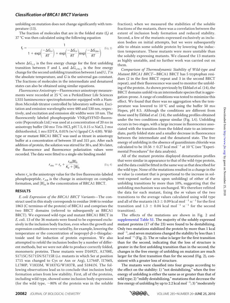
that were similar in appearance to that of the wild-type protein,and the data could be fitted in the sameway as that described forthe wild type. None of themutations resulted in a change in them value (a constant that is proportional to the increase in sol-vent-exposed surface area upon unfolding) of either of theunfolding transitions by more than 10%, suggesting that theunfolding mechanism was unchanged. We therefore refittedthe data for each mutant, fixing the m values of the twotransitions to the average values calculated using wild typeand all of the mutants (4.5 � 0.09 kcal mol�1 M�1 for the firsttransition and 1.3 � 0.04 kcal mol�1 M�1 for the secondtransition).The effects of the mutations are shown in Fig. 2 and
supplemental Table S1. The majority of the solubly expressedmutant proteins (17 of the 23) were found to be destabilizing.Only two mutations stabilized the protein by more than 1 kcalmol�1, and sevenmutations changed the stability by less than 1kcal mol�1 (Fig. 2). Them value is larger for the first transitionthan for the second, indicating that the loss of structure isgreater in the first unfolding transition than in the second; thechanges in the free energy of unfolding on mutation are muchlarger for the first transition than for the second (Fig. 2), con-sistent with a greater loss of structure.The mutants were classified into four groups according to
the effect on the stability: 1) “not destabilizing,” when the freeenergy of unfolding is either the same as or greater than that ofwild type; 2) “mildly destabilizing,” mutations that decrease thefree energy of unfolding by up to 2.2 kcal mol�1; 3) “moderately
Classification of BRCA1 BRCT Variants
20082 JOURNAL OF BIOLOGICAL CHEMISTRY VOLUME 285 • NUMBER 26 • JUNE 25, 2010
by guest on Novem
ber 25, 2018http://w
ww
.jbc.org/D
ownloaded from
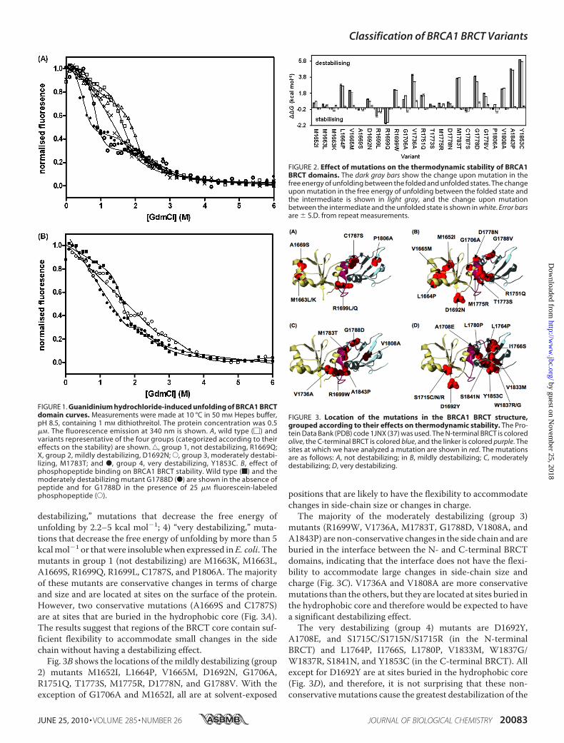
destabilizing,” mutations that decrease the free energy ofunfolding by 2.2–5 kcal mol�1; 4) “very destabilizing,” muta-tions that decrease the free energy of unfolding by more than 5kcalmol�1 or thatwere insolublewhen expressed inE. coli. Themutants in group 1 (not destabilizing) are M1663K, M1663L,A1669S, R1699Q, R1699L, C1787S, and P1806A. The majorityof these mutants are conservative changes in terms of chargeand size and are located at sites on the surface of the protein.However, two conservative mutations (A1669S and C1787S)are at sites that are buried in the hydrophobic core (Fig. 3A).The results suggest that regions of the BRCT core contain suf-ficient flexibility to accommodate small changes in the sidechain without having a destabilizing effect.Fig. 3B shows the locations of the mildly destabilizing (group
2) mutants M1652I, L1664P, V1665M, D1692N, G1706A,R1751Q, T1773S, M1775R, D1778N, and G1788V. With theexception of G1706A and M1652I, all are at solvent-exposed
positions that are likely to have the flexibility to accommodatechanges in side-chain size or changes in charge.The majority of the moderately destabilizing (group 3)
mutants (R1699W, V1736A, M1783T, G1788D, V1808A, andA1843P) are non-conservative changes in the side chain and areburied in the interface between the N- and C-terminal BRCTdomains, indicating that the interface does not have the flexi-bility to accommodate large changes in side-chain size andcharge (Fig. 3C). V1736A and V1808A are more conservativemutations than the others, but they are located at sites buried inthe hydrophobic core and therefore would be expected to havea significant destabilizing effect.The very destabilizing (group 4) mutants are D1692Y,
A1708E, and S1715C/S1715N/S1715R (in the N-terminalBRCT) and L1764P, I1766S, L1780P, V1833M, W1837G/W1837R, S1841N, and Y1853C (in the C-terminal BRCT). Allexcept for D1692Y are at sites buried in the hydrophobic core(Fig. 3D), and therefore, it is not surprising that these non-conservativemutations cause the greatest destabilization of the
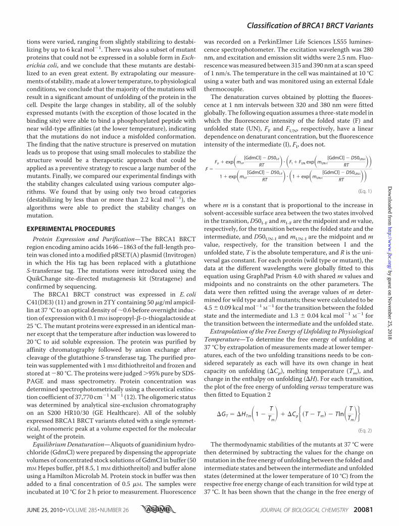
FIGURE 1. Guanidinium hydrochloride-induced unfolding of BRCA1 BRCTdomain curves. Measurements were made at 10 °C in 50 mM Hepes buffer,pH 8.5, containing 1 mM dithiothreitol. The protein concentration was 0.5�M. The fluorescence emission at 340 nm is shown. A, wild type (�) andvariants representative of the four groups (categorized according to theireffects on the stability) are shown. ‚, group 1, not destabilizing, R1669Q;X, group 2, mildly destabilizing, D1692N; E, group 3, moderately destabi-lizing, M1783T; and F, group 4, very destabilizing, Y1853C. B, effect ofphosphopeptide binding on BRCA1 BRCT stability. Wild type (f) and themoderately destabilizing mutant G1788D (F) are shown in the absence ofpeptide and for G1788D in the presence of 25 �M fluorescein-labeledphosphopeptide (E).
FIGURE 2. Effect of mutations on the thermodynamic stability of BRCA1BRCT domains. The dark gray bars show the change upon mutation in thefree energy of unfolding between the folded and unfolded states. The changeupon mutation in the free energy of unfolding between the folded state andthe intermediate is shown in light gray, and the change upon mutationbetween the intermediate and the unfolded state is shown in white. Error barsare � S.D. from repeat measurements.
FIGURE 3. Location of the mutations in the BRCA1 BRCT structure,grouped according to their effects on thermodynamic stability. The Pro-tein Data Bank (PDB) code 1JNX (37) was used. The N-terminal BRCT is coloredolive, the C-terminal BRCT is colored blue, and the linker is colored purple. Thesites at which we have analyzed a mutation are shown in red. The mutationsare as follows: A, not destabilizing; in B, mildly destabilizing; C, moderatelydestabilizing; D, very destabilizing.
Classification of BRCA1 BRCT Variants
JUNE 25, 2010 • VOLUME 285 • NUMBER 26 JOURNAL OF BIOLOGICAL CHEMISTRY 20083
by guest on Novem
ber 25, 2018http://w
ww
.jbc.org/D
ownloaded from
structure. Two variants, A1708E and L1780P, are buried in theinterface of the BRCT domains. The equivalent positions to theC-terminal sites Val-1833, Trp-1837, Ser-1841, and Tyr-1853in the N-terminal BRCT domain are Trp-1718, Ser-1722, Val-1713, and Phe-1734, and they should be likewise important forstability; indeed mutations at all of these sites have beenrecorded in the BIC database. Ser-1715 is a buried residue in�-strand 4, the same structural element in which Val-1833 islocated in the C-terminal BRCT domain. All of the three muta-tions in the BIC data base at Ser-1715 resulted in expression ininclusion bodies, even the relatively conservative mutantS1715C, suggesting that Ser-1715 is crucial for the packing ofthe hydrophobic core. Therefore, it might be expected thatmutation of the analogous position in the C-terminal BRCTdomain, Arg-1835, would also be detrimental; however, cur-rently there are no reported variants in BIC at this position.Mutant BRCA1 BRCTDomains Bind to Phosphopeptide with
Near Wild-type Affinity—Tandem BRCT domains have beenshown to bind phosphopeptides at a site located between thetwo repeats. The BRCT domains of BRCA1 have been found tobind the phosphorylated proteins BACH1 and CtIP (15, 16).Rodriguez et al. (17) showed for tandem BRCT domains fromseveral proteins that there was selectivity in phosphopeptidesubstrate binding. The phosphopeptide used here, VNKpSYFND,was the one that was optimized for binding to BRCA1 BRCT inthe Rodriguez study; it was labeled at its C terminus with fluo-rescein tomonitor binding by fluorescence anisotropy. The dis-sociation constant, Kd, for the binding of wild-type BRCA1BRCT to the phosphopeptide was 112 � 6 nM. A competitionexperiment using unlabeled peptide gave a similar value for theKd, indicating that the binding affinity was not affected signifi-cantly by the presence of the fluorescein label.We characterized one binding-site residue in our study, Arg-
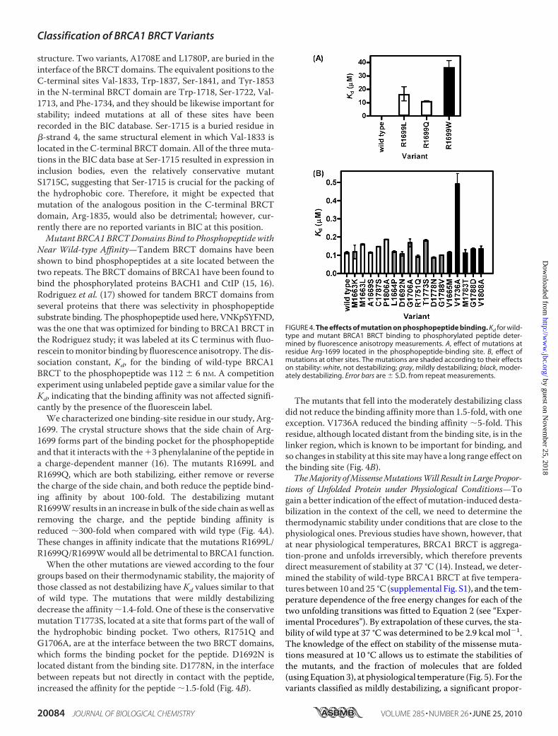
1699. The crystal structure shows that the side chain of Arg-1699 forms part of the binding pocket for the phosphopeptideand that it interacts with the3 phenylalanine of the peptide ina charge-dependent manner (16). The mutants R1699L andR1699Q, which are both stabilizing, either remove or reversethe charge of the side chain, and both reduce the peptide bind-ing affinity by about 100-fold. The destabilizing mutantR1699Wresults in an increase in bulk of the side chain aswell asremoving the charge, and the peptide binding affinity isreduced �300-fold when compared with wild type (Fig. 4A).These changes in affinity indicate that the mutations R1699L/R1699Q/R1699Wwould all be detrimental to BRCA1 function.When the other mutations are viewed according to the four
groups based on their thermodynamic stability, the majority ofthose classed as not destabilizing have Kd values similar to thatof wild type. The mutations that were mildly destabilizingdecrease the affinity�1.4-fold. One of these is the conservativemutation T1773S, located at a site that forms part of the wall ofthe hydrophobic binding pocket. Two others, R1751Q andG1706A, are at the interface between the two BRCT domains,which forms the binding pocket for the peptide. D1692N islocated distant from the binding site. D1778N, in the interfacebetween repeats but not directly in contact with the peptide,increased the affinity for the peptide �1.5-fold (Fig. 4B).
The mutants that fell into the moderately destabilizing classdid not reduce the binding affinitymore than 1.5-fold, with oneexception. V1736A reduced the binding affinity �5-fold. Thisresidue, although located distant from the binding site, is in thelinker region, which is known to be important for binding, andso changes in stability at this sitemay have a long range effect onthe binding site (Fig. 4B).TheMajorityofMissenseMutationsWillResult inLargePropor-
tions of Unfolded Protein under Physiological Conditions—Togain a better indication of the effect ofmutation-induced desta-bilization in the context of the cell, we need to determine thethermodynamic stability under conditions that are close to thephysiological ones. Previous studies have shown, however, thatat near physiological temperatures, BRCA1 BRCT is aggrega-tion-prone and unfolds irreversibly, which therefore preventsdirect measurement of stability at 37 °C (14). Instead, we deter-mined the stability of wild-type BRCA1 BRCT at five tempera-tures between 10 and 25 °C (supplemental Fig. S1), and the tem-perature dependence of the free energy changes for each of thetwo unfolding transitions was fitted to Equation 2 (see “Exper-imental Procedures”). By extrapolation of these curves, the sta-bility of wild type at 37 °C was determined to be 2.9 kcal mol�1.The knowledge of the effect on stability of the missense muta-tions measured at 10 °C allows us to estimate the stabilities ofthe mutants, and the fraction of molecules that are folded(using Equation 3), at physiological temperature (Fig. 5). For thevariants classified as mildly destabilizing, a significant propor-
FIGURE 4. The effects of mutation on phosphopeptide binding. Kd for wild-type and mutant BRCA1 BRCT binding to phosphorylated peptide deter-mined by fluorescence anisotropy measurements. A, effect of mutations atresidue Arg-1699 located in the phosphopeptide-binding site. B, effect ofmutations at other sites. The mutations are shaded according to their effectson stability: white, not destabilizing; gray, mildly destabilizing; black, moder-ately destabilizing. Error bars are � S.D. from repeat measurements.
Classification of BRCA1 BRCT Variants
20084 JOURNAL OF BIOLOGICAL CHEMISTRY VOLUME 285 • NUMBER 26 • JUNE 25, 2010
by guest on Novem
ber 25, 2018http://w
ww
.jbc.org/D
ownloaded from
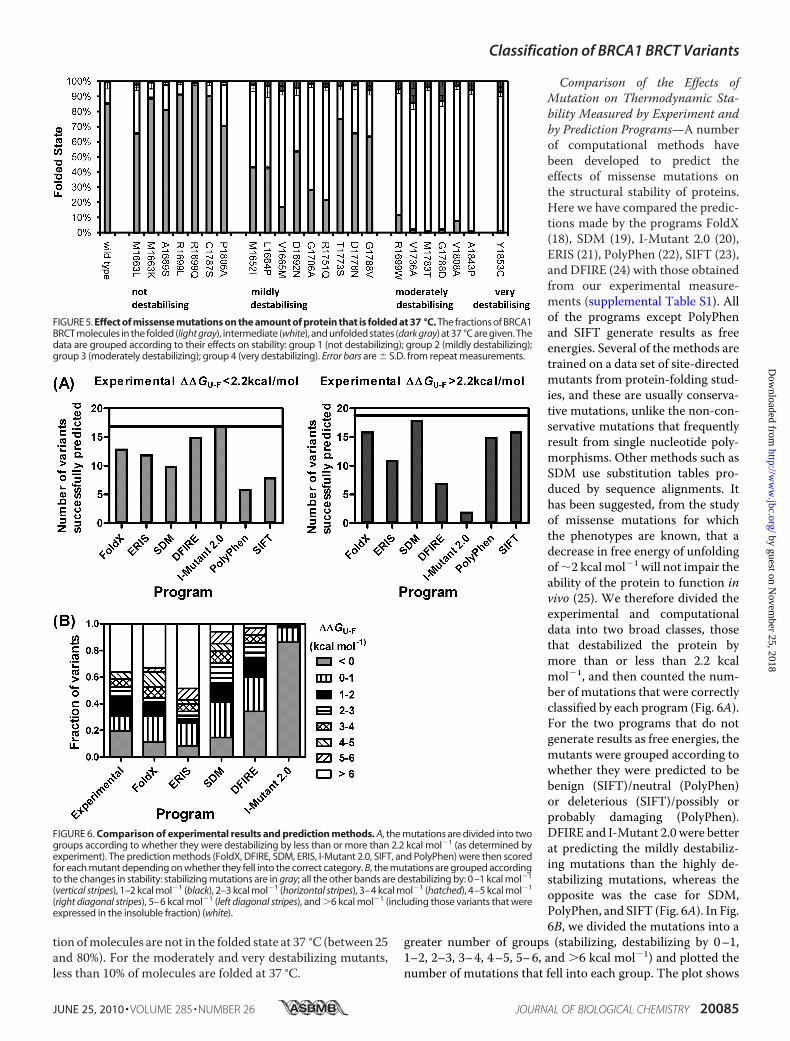
tion ofmolecules are not in the folded state at 37 °C (between 25and 80%). For the moderately and very destabilizing mutants,less than 10% of molecules are folded at 37 °C.
Comparison of the Effects ofMutation on Thermodynamic Sta-bility Measured by Experiment andby Prediction Programs—A numberof computational methods havebeen developed to predict theeffects of missense mutations onthe structural stability of proteins.Here we have compared the predic-tions made by the programs FoldX(18), SDM (19), I-Mutant 2.0 (20),ERIS (21), PolyPhen (22), SIFT (23),and DFIRE (24) with those obtainedfrom our experimental measure-ments (supplemental Table S1). Allof the programs except PolyPhenand SIFT generate results as freeenergies. Several of the methods aretrained on a data set of site-directedmutants from protein-folding stud-ies, and these are usually conserva-tive mutations, unlike the non-con-servative mutations that frequentlyresult from single nucleotide poly-morphisms. Other methods such asSDM use substitution tables pro-duced by sequence alignments. Ithas been suggested, from the studyof missense mutations for whichthe phenotypes are known, that adecrease in free energy of unfoldingof�2 kcal mol�1 will not impair theability of the protein to function invivo (25). We therefore divided theexperimental and computationaldata into two broad classes, thosethat destabilized the protein bymore than or less than 2.2 kcalmol�1, and then counted the num-ber of mutations that were correctlyclassified by each program (Fig. 6A).For the two programs that do notgenerate results as free energies, themutants were grouped according towhether they were predicted to bebenign (SIFT)/neutral (PolyPhen)or deleterious (SIFT)/possibly orprobably damaging (PolyPhen).DFIRE and I-Mutant 2.0 were betterat predicting the mildly destabiliz-ing mutations than the highly de-stabilizing mutations, whereas theopposite was the case for SDM,PolyPhen, and SIFT (Fig. 6A). In Fig.6B, we divided the mutations into a
greater number of groups (stabilizing, destabilizing by 0–1,1–2, 2–3, 3–4, 4–5, 5–6, and �6 kcal mol�1) and plotted thenumber of mutations that fell into each group. The plot shows
FIGURE 5. Effect of missense mutations on the amount of protein that is folded at 37 °C. The fractions of BRCA1BRCT molecules in the folded (light gray), intermediate (white), and unfolded states (dark gray) at 37 °C are given. Thedata are grouped according to their effects on stability: group 1 (not destabilizing); group 2 (mildly destabilizing);group 3 (moderately destabilizing); group 4 (very destabilizing). Error bars are � S.D. from repeat measurements.
FIGURE 6. Comparison of experimental results and prediction methods. A, the mutations are divided into twogroups according to whether they were destabilizing by less than or more than 2.2 kcal mol�1 (as determined byexperiment). The prediction methods (FoldX, DFIRE, SDM, ERIS, I-Mutant 2.0, SIFT, and PolyPhen) were then scoredfor each mutant depending on whether they fell into the correct category. B, the mutations are grouped accordingto the changes in stability: stabilizing mutations are in gray; all the other bands are destabilizing by: 0–1 kcal mol�1
(vertical stripes), 1–2 kcal mol�1 (black), 2–3 kcal mol�1 (horizontal stripes), 3–4 kcal mol�1 (hatched), 4–5 kcal mol�1
(right diagonal stripes), 5–6 kcal mol�1 (left diagonal stripes), and �6 kcal mol�1 (including those variants that wereexpressed in the insoluble fraction) (white).
Classification of BRCA1 BRCT Variants
JUNE 25, 2010 • VOLUME 285 • NUMBER 26 JOURNAL OF BIOLOGICAL CHEMISTRY 20085
by guest on Novem
ber 25, 2018http://w
ww
.jbc.org/D
ownloaded from
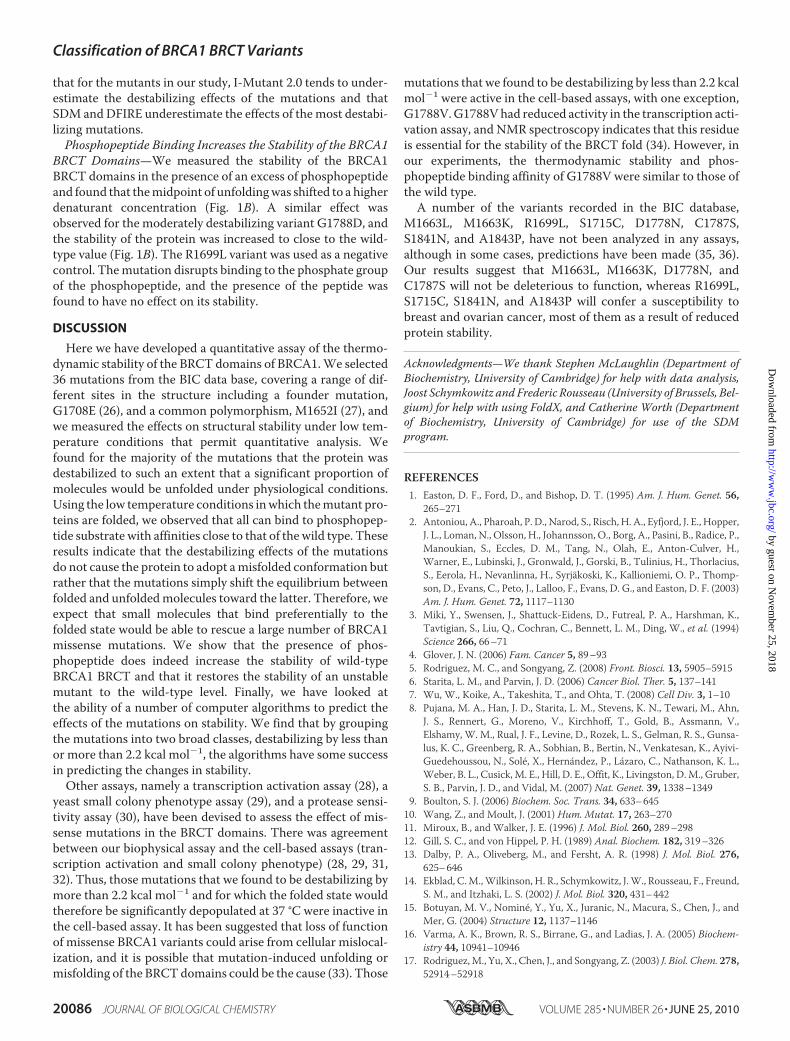
that for the mutants in our study, I-Mutant 2.0 tends to under-estimate the destabilizing effects of the mutations and thatSDM andDFIRE underestimate the effects of themost destabi-lizing mutations.Phosphopeptide Binding Increases the Stability of the BRCA1
BRCT Domains—We measured the stability of the BRCA1BRCT domains in the presence of an excess of phosphopeptideand found that themidpoint of unfoldingwas shifted to a higherdenaturant concentration (Fig. 1B). A similar effect wasobserved for the moderately destabilizing variant G1788D, andthe stability of the protein was increased to close to the wild-type value (Fig. 1B). The R1699L variant was used as a negativecontrol. Themutation disrupts binding to the phosphate groupof the phosphopeptide, and the presence of the peptide wasfound to have no effect on its stability.
DISCUSSION
Here we have developed a quantitative assay of the thermo-dynamic stability of the BRCT domains of BRCA1.We selected36 mutations from the BIC data base, covering a range of dif-ferent sites in the structure including a founder mutation,G1708E (26), and a common polymorphism, M1652I (27), andwe measured the effects on structural stability under low tem-perature conditions that permit quantitative analysis. Wefound for the majority of the mutations that the protein wasdestabilized to such an extent that a significant proportion ofmolecules would be unfolded under physiological conditions.Using the low temperature conditions inwhich themutant pro-teins are folded, we observed that all can bind to phosphopep-tide substrate with affinities close to that of the wild type. Theseresults indicate that the destabilizing effects of the mutationsdo not cause the protein to adopt amisfolded conformation butrather that the mutations simply shift the equilibrium betweenfolded and unfoldedmolecules toward the latter. Therefore, weexpect that small molecules that bind preferentially to thefolded state would be able to rescue a large number of BRCA1missense mutations. We show that the presence of phos-phopeptide does indeed increase the stability of wild-typeBRCA1 BRCT and that it restores the stability of an unstablemutant to the wild-type level. Finally, we have looked atthe ability of a number of computer algorithms to predict theeffects of the mutations on stability. We find that by groupingthe mutations into two broad classes, destabilizing by less thanor more than 2.2 kcal mol�1, the algorithms have some successin predicting the changes in stability.Other assays, namely a transcription activation assay (28), a
yeast small colony phenotype assay (29), and a protease sensi-tivity assay (30), have been devised to assess the effect of mis-sense mutations in the BRCT domains. There was agreementbetween our biophysical assay and the cell-based assays (tran-scription activation and small colony phenotype) (28, 29, 31,32). Thus, those mutations that we found to be destabilizing bymore than 2.2 kcal mol�1 and for which the folded state wouldtherefore be significantly depopulated at 37 °C were inactive inthe cell-based assay. It has been suggested that loss of functionof missense BRCA1 variants could arise from cellular mislocal-ization, and it is possible that mutation-induced unfolding ormisfolding of the BRCTdomains could be the cause (33). Those
mutations that we found to be destabilizing by less than 2.2 kcalmol�1 were active in the cell-based assays, with one exception,G1788V.G1788Vhad reduced activity in the transcription acti-vation assay, and NMR spectroscopy indicates that this residueis essential for the stability of the BRCT fold (34). However, inour experiments, the thermodynamic stability and phos-phopeptide binding affinity of G1788V were similar to those ofthe wild type.A number of the variants recorded in the BIC database,
M1663L, M1663K, R1699L, S1715C, D1778N, C1787S,S1841N, and A1843P, have not been analyzed in any assays,although in some cases, predictions have been made (35, 36).Our results suggest that M1663L, M1663K, D1778N, andC1787S will not be deleterious to function, whereas R1699L,S1715C, S1841N, and A1843P will confer a susceptibility tobreast and ovarian cancer, most of them as a result of reducedprotein stability.
Acknowledgments—We thank Stephen McLaughlin (Department ofBiochemistry, University of Cambridge) for help with data analysis,Joost Schymkowitz and Frederic Rousseau (University of Brussels, Bel-gium) for help with using FoldX, and Catherine Worth (Departmentof Biochemistry, University of Cambridge) for use of the SDMprogram.
REFERENCES1. Easton, D. F., Ford, D., and Bishop, D. T. (1995) Am. J. Hum. Genet. 56,
265–2712. Antoniou, A., Pharoah, P. D., Narod, S., Risch, H. A., Eyfjord, J. E., Hopper,
J. L., Loman, N., Olsson, H., Johannsson, O., Borg, A., Pasini, B., Radice, P.,Manoukian, S., Eccles, D. M., Tang, N., Olah, E., Anton-Culver, H.,Warner, E., Lubinski, J., Gronwald, J., Gorski, B., Tulinius, H., Thorlacius,S., Eerola, H., Nevanlinna, H., Syrjakoski, K., Kallioniemi, O. P., Thomp-son, D., Evans, C., Peto, J., Lalloo, F., Evans, D. G., and Easton, D. F. (2003)Am. J. Hum. Genet. 72, 1117–1130
3. Miki, Y., Swensen, J., Shattuck-Eidens, D., Futreal, P. A., Harshman, K.,Tavtigian, S., Liu, Q., Cochran, C., Bennett, L. M., Ding, W., et al. (1994)Science 266, 66–71
4. Glover, J. N. (2006) Fam. Cancer 5, 89–935. Rodriguez, M. C., and Songyang, Z. (2008) Front. Biosci. 13, 5905–59156. Starita, L. M., and Parvin, J. D. (2006) Cancer Biol. Ther. 5, 137–1417. Wu, W., Koike, A., Takeshita, T., and Ohta, T. (2008) Cell Div. 3, 1–108. Pujana, M. A., Han, J. D., Starita, L. M., Stevens, K. N., Tewari, M., Ahn,
J. S., Rennert, G., Moreno, V., Kirchhoff, T., Gold, B., Assmann, V.,Elshamy, W. M., Rual, J. F., Levine, D., Rozek, L. S., Gelman, R. S., Gunsa-lus, K. C., Greenberg, R. A., Sobhian, B., Bertin, N., Venkatesan, K., Ayivi-Guedehoussou, N., Sole, X., Hernandez, P., Lazaro, C., Nathanson, K. L.,Weber, B. L., Cusick, M. E., Hill, D. E., Offit, K., Livingston, D.M., Gruber,S. B., Parvin, J. D., and Vidal, M. (2007) Nat. Genet. 39, 1338–1349
9. Boulton, S. J. (2006) Biochem. Soc. Trans. 34, 633–64510. Wang, Z., and Moult, J. (2001) Hum. Mutat. 17, 263–27011. Miroux, B., and Walker, J. E. (1996) J. Mol. Biol. 260, 289–29812. Gill, S. C., and von Hippel, P. H. (1989) Anal. Biochem. 182, 319–32613. Dalby, P. A., Oliveberg, M., and Fersht, A. R. (1998) J. Mol. Biol. 276,
625–64614. Ekblad, C.M.,Wilkinson, H. R., Schymkowitz, J.W., Rousseau, F., Freund,
S. M., and Itzhaki, L. S. (2002) J. Mol. Biol. 320, 431–44215. Botuyan, M. V., Nomine, Y., Yu, X., Juranic, N., Macura, S., Chen, J., and
Mer, G. (2004) Structure 12, 1137–114616. Varma, A. K., Brown, R. S., Birrane, G., and Ladias, J. A. (2005) Biochem-
istry 44, 10941–1094617. Rodriguez,M., Yu, X., Chen, J., and Songyang, Z. (2003) J. Biol. Chem. 278,
52914–52918
Classification of BRCA1 BRCT Variants
20086 JOURNAL OF BIOLOGICAL CHEMISTRY VOLUME 285 • NUMBER 26 • JUNE 25, 2010
by guest on Novem
ber 25, 2018http://w
ww
.jbc.org/D
ownloaded from
18. Guerois, R., Nielsen, J. E., and Serrano, L. (2002) J.Mol. Biol. 320, 369–38719. Worth, C. L., Bickerton, G. R., Schreyer, A., Forman, J. R., Cheng, T. M.,
Lee, S., Gong, S., Burke, D. F., and Blundell, T. L. (2007) J. Bioinform.Comput. Biol. 5, 1297–1318
20. Capriotti, E., Fariselli, P., and Casadio, R. (2005) Nucleic Acids Res. 33,W306–310
21. Yin, S., Ding, F., and Dokholyan, N. V. (2007) Nat. Methods 4, 466–46722. Ramensky, V., Bork, P., and Sunyaev, S. (2002) Nucleic Acids Res. 30,
3894–390023. Ng, P. C., and Henikoff, S. (2003) Nucleic Acids Res. 31, 3812–381424. Zhou, H., and Zhou, Y. (2002) Proteins 49, 483–49225. Yue, P., Li, Z., and Moult, J. (2005) J. Mol. Biol. 353, 459–47326. Ferla, R., Calo, V., Cascio, S., Rinaldi, G., Badalamenti, G., Carreca, I.,
Surmacz, E., Colucci, G., Bazan, V., and Russo, A. (2007) Ann. Oncol. 18,Suppl. 6, vi93–vi98
27. Deffenbaugh, A. M., Frank, T. S., Hoffman, M., Cannon-Albright, L., andNeuhausen, S. L. (2002) Genet. Test 6, 119–121
28. Hayes, F., Cayanan, C., Barilla, D., andMonteiro, A. N. (2000) Cancer Res.60, 2411–2418
29. Coyne, R. S., McDonald, H. B., Edgemon, K., and Brody, L. C. (2004)Cancer Biol. Ther. 3, 453–457
30. Williams, R. S., Chasman, D. I., Hau, D. D., Hui, B., Lau, A. Y., and Glover,
J. N. (2003) J. Biol. Chem. 278, 53007–5301631. Carvalho, M. A., Marsillac, S. M., Karchin, R., Manoukian, S., Grist, S.,
Swaby, R. F., Urmenyi, T. P., Rondinelli, E., Silva, R., Gayol, L., Baumbach,L., Sutphen, R., Pickard-Brzosowicz, J. L., Nathanson, K. L., Sali, A., Gold-gar, D., Couch, F. J., Radice, P., andMonteiro, A. N. (2007)Cancer Res. 67,1494–1501
32. Vallon-Christersson, J., Cayanan, C., Haraldsson, K., Loman, N., Bergth-orsson, J. T., Brøndum-Nielsen, K., Gerdes, A. M., Møller, P., Kristoffers-son, U., Olsson, H., Borg, A., andMonteiro, A. N. (2001)Hum.Mol. Genet.10, 353–360
33. Rodriguez, J. A., Au, W. W., and Henderson, B. R. (2004) Exp. Cell Res.293, 14–21
34. Gaiser, O. J., Ball, L. J., Schmieder, P., Leitner, D., Strauss, H., Wahl, M.,Kuhne, R., Oschkinat, H., and Heinemann, U. (2004) Biochemistry 43,15983–15995
35. Karchin, R., Monteiro, A. N., Tavtigian, S. V., Carvalho, M. A., and Sali, A.(2007) PLoS Comput. Biol. 3, e26
36. Abkevich, V., Zharkikh, A., Deffenbaugh, A.M., Frank, D., Chen, Y., Shat-tuck, D., Skolnick, M. H., Gutin, A., and Tavtigian, S. V. (2004) J. Med.Genet. 41, 492–507
37. Williams, R. S., Green, R., and Glover, J. N. (2001) Nat. Struct. Biol. 8,838–842
Classification of BRCA1 BRCT Variants
JUNE 25, 2010 • VOLUME 285 • NUMBER 26 JOURNAL OF BIOLOGICAL CHEMISTRY 20087
by guest on Novem
ber 25, 2018http://w
ww
.jbc.org/D
ownloaded from
Pamela J. E. Rowling, Rebecca Cook and Laura S. ItzhakiToward Classification of BRCA1 Missense Variants Using a Biophysical Approach
doi: 10.1074/jbc.M109.088922 originally published online April 8, 20102010, 285:20080-20087.J. Biol. Chem.
10.1074/jbc.M109.088922Access the most updated version of this article at doi:
Alerts:
When a correction for this article is posted•
When this article is cited•
to choose from all of JBC's e-mail alertsClick here
Supplemental material:
http://www.jbc.org/content/suppl/2010/04/08/M109.088922.DC1
http://www.jbc.org/content/285/26/20080.full.html#ref-list-1
This article cites 37 references, 6 of which can be accessed free at
by guest on Novem
ber 25, 2018http://w
ww
.jbc.org/D
ownloaded from