Embed Size (px)
Citation preview
Toward the Biochemical Assessment of Myocardial Fibrosis in HypeHensive
Patients Javier Diez, MD, PhD, Concepcibn Laviades, MD, PhD, lgnacio Monreal, DSc, MD,
Maria J. Gil, DSc, Angel Panizo, MD, and Javier Pardo, MD, PhD
The serum concentrations of amino-terminal procol- lagen type III and carboxy-terminal procollagen type l-derived peptides, which have been proposed as useful markers of the tissue synthesis of collagen types Ill and type I, respectively, were abnormally increased in patients with essential hypertension and became normal after angiotensin-converting enzyme (ACE) inhibition. An association was found between baseline serum concentrations of these peptides and left ventricular hypetirophy, diastolic dysfunction, and ventricular arrhythmias in hyper- tensive patients. On the other hand, increased se-
rum concentration of the carboxy-terminal procolla- gen type l-derived peptide was found in spon- taneously hypertensive rats compared with normo- tensive Wistar-Kyoto control mts. An association was found between the serum concentmtion of this peptide and the extent of myocardial fibrosis and the hydroxyproline concentmtion in the left ventricle of spontaneously hypertensive rats. It is proposed that procollagen-derived peptides in serum may be markers of exaggerated collagen tissue synthesis involved in hypertensive myocardial fibrosis.
(Am J Cardiol 1995; 76: 14D-17D)
A significant increase in fibrillar collagen content has been observed in the cardiac ventricles of both animals1-7 and hu-
mans8-l2 with arterial hypertension. This myocar- dial fibrosis is accomplished by alterations in colla- gen synthesis and degradation and by fibroblast proliferation.r3J4 Hemodynamic and nonhemody- anmic factors may participate in the development of myocardial fibrosis that occurs in hyperten- sion.15,16 ,As shown experimentally3J7 and clini- cally,r8 a rise in collagen content adversely raises myocardial stiffness and promotes abnormalities of cardiac function. In addition, it has been demon- strated that ventricular arrhythmias in hyperten- sive patients are related to the degree of myocar- dial’ fibrosis.r9 Although some antihypertensive agents, such as minoxidil, hydralazine, or hydrochlo- rothiazide, have failed to show any significant effect on myocardial fibrosis, others, such as angio- tensin-converting enzyme (ACE) inhibitors, cal- cium antagonists, or aldosterone antagonists, have demonstrated profound effects on either preven- tion or regression of myocardial fibrosis.20
Although cardiac biopsies are reliable for mea- suring myocardial fibrosis,10J1J8,21 it seems neces-
From the Vascular Pathophysiolagy Unit, Department of Internal Medicine, Center far Biomedical Research, School of Medicine, University of Navarra (J.D.); Division of Nephrology, San Jorge General Hospital, Huesca (C.L.); Department of Clinical Biochemis- try, University Clinic, School of Medicine, University of Navarra (I.M., M.J.G.); and Department of Pathology, University Clinic, School of Medicine, University of Navarra, Pamplona, Spain (A.P., J.P.).
Address for reprints: Javier Diez, MD, PhD, Unidad de Fisiopato- logia Vascular, Facultad de Medicina, C/ lrunlarreo s/n, 31080 Pomplona, Spain.
sat-y to develop noninvasive methods that indicate the presence of myocardial fibrosis in hypertension (i.e., biochemi,cal markers of collagen synthesis). This article is based’on the proposal of a biochemi- cal method ‘to assess indirectly the myocardial synthesis and deposition of fibrillar collagen.
A BIOCHEMICAL APPROACH TO THE ASSESSMENT OF THE CARDIAC SYNTHESIS OF FIBRUAR COLLAGEN
Collagen types III and I are synthesized as procollagens with a small amino-terminal and a larger carboxy-terminal propeptide. Once secreted into the extracellular space, the propeptides are removed by specific endopeptidases, thus allowing integration of the rigid collagen triple helix into the growing fibril. ** The procollagen type III amino- terminal peptide (PIIIP) formed during this pro- cess is released into the blood. The serum concen- tration of PIIIP has been proposed as a useful marker of collagen type III synthesis.23 This is supported by a diversity of clinical observations demonstrating that high serum levels of the pep- tides reflect ongoing tissue fibrosis.2428
The procollagen type I carboxy-terminal pep- tide (PIP) is cleaved off procollagen type I during the synthesis of the fibril forming collagen type I. In contrast to PIIIP, PIP is completely removed from its procollagen precursor during the extracellular processing of the collagen type I,** thus offering the theoretical advantage of directly reflecting fibrogen- esis. This has been confirmed in studies conducted in patients with different clinical conditions.29-31
14D THE AMERICAN JOURNAL OF CARDIOLOGY VOLUME 76 NOVEMBER 2, 1995
Therefore, we investigated the serum concentra- tions of PIP and PIIIP in different clinical and experimental studies to assess the intensity of the fibrogenic process in patients with essential hyper- tension and in rats with spontaneous hypertension. In addition, the relations between serum concentra- tions of the 2 peptides and several parameters of left ventricular anatomy, structure, biochemistry, and function were analyzed to delineate the value of these peptides as potential markers of ventricu- lar fibrosis in hypertension.
STUDIES ON SERUM PEPTIDES OF COLLAGEN SYNTHESIS IN ARTERIAL HYPERTENSION
Clinical studies: In a previous study32 we deter- mined, by specific radioimmunoassay, the serum concentrations of PIIIP in 24 patients with essen- tial hypertension who had never been treated and in 30 normotensive control subjects. None of the subjects exhibited abnormalities suggestive of con- ditions associated with elevated serum PIIIP con- centrations (chronic liver disease, pulmonary fibro- sis, rheumatoid arthritis, extensive wounds, acute myocardial infarction).
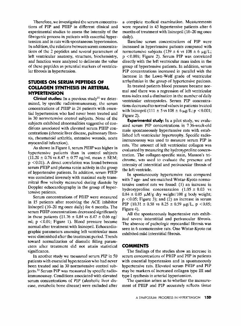
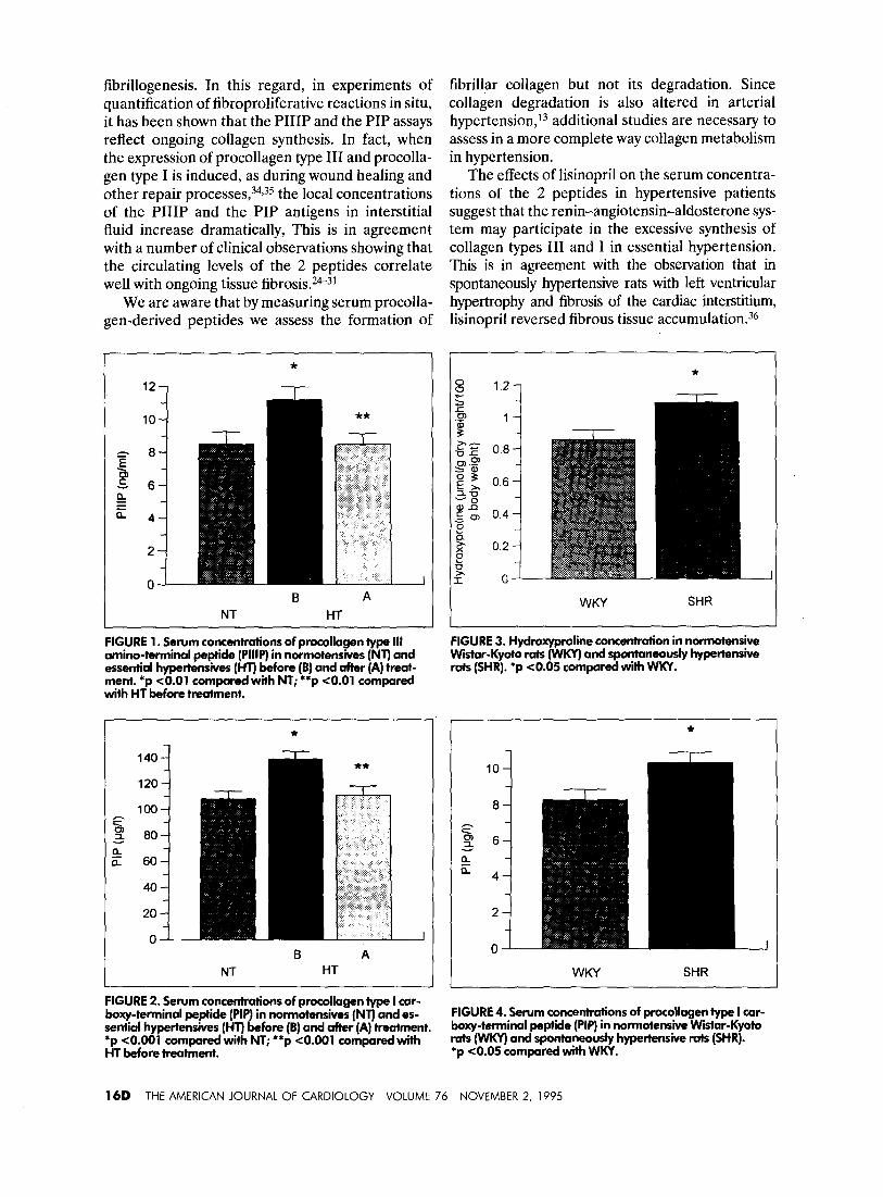
As shown in Figure 1, serum PIIIP was higher in hypertensive patients than in control subjects (11.20 f 0.76 vs 8.47 +- 0.77 rig/ml, mean rl: SEM, p < 0.01). A direct correlation was found between serum PIIIP and plasma renin activity in the group of hypertensive patients. In addition, serum PIIIP was correlated inversely with maximal early trans- mitral flow velocity measured during diastole by Doppler echocardiography in the group of hyper- tensive patients,
Serum concentrations of PIIIP were measured in 15 patients after receiving the ACE inhibitor lisinopril (10-20 mg once daily) for 6 months. The serum PIIIP concentrations decreased significantly in these patients (11.76 +- 0.84 vs 8.47 -+ 0.66 ng/ ml; p <O.Ol; Figure 1). Blood pressure became normal,after treatment with lisinopril. Echocardio- graphic parameters assessing left ventricular mass were diminished after the treatment period. Trends toward normalization of diastolic filling param- eters after treatment did not attain statistical significance.
In another study we measured serum PIP in 50 patients with essential hypertension who had never been treated and in 30 normotensive control sub- jects.33 Serum PIP was measured by specific radio- immunoassay. Conditions associated with elevated serum concentrations of PIP (alcoholic liver dis- ease, metabolic bone disease) were excluded after
a complete medical examination. Measurements were repeated in 43 hypertensive patients after 6 months of treatment with lisinopril (lO-20 mg once daily).
Baseline serum concentrations of PIP were increased in hypertensive patients compared with normotensive subjects (139 + 6 vs 108 +- 6 kg/L; p <O.OOl; Figure 2). Serum PIP was correlated directly with the left ventricular mass index in the group of hypertensive patients. In addition, serum PIP concentrations increased in parallel with the increase in the Lown-Wolf grade of ventricular arrhythmias in the group of hypertensive patients.
In treated patients blood pressure became nor- mal and there was a regression of left ventricular mass index and a diminution in the number of daily ventricular extrasystoles. Serum PIP concentra- tions decreased to normal values in patients treated with lisinopril(ll1 + 5 vs 108 5 6 kg/L; p < 0.001; Figure 2).
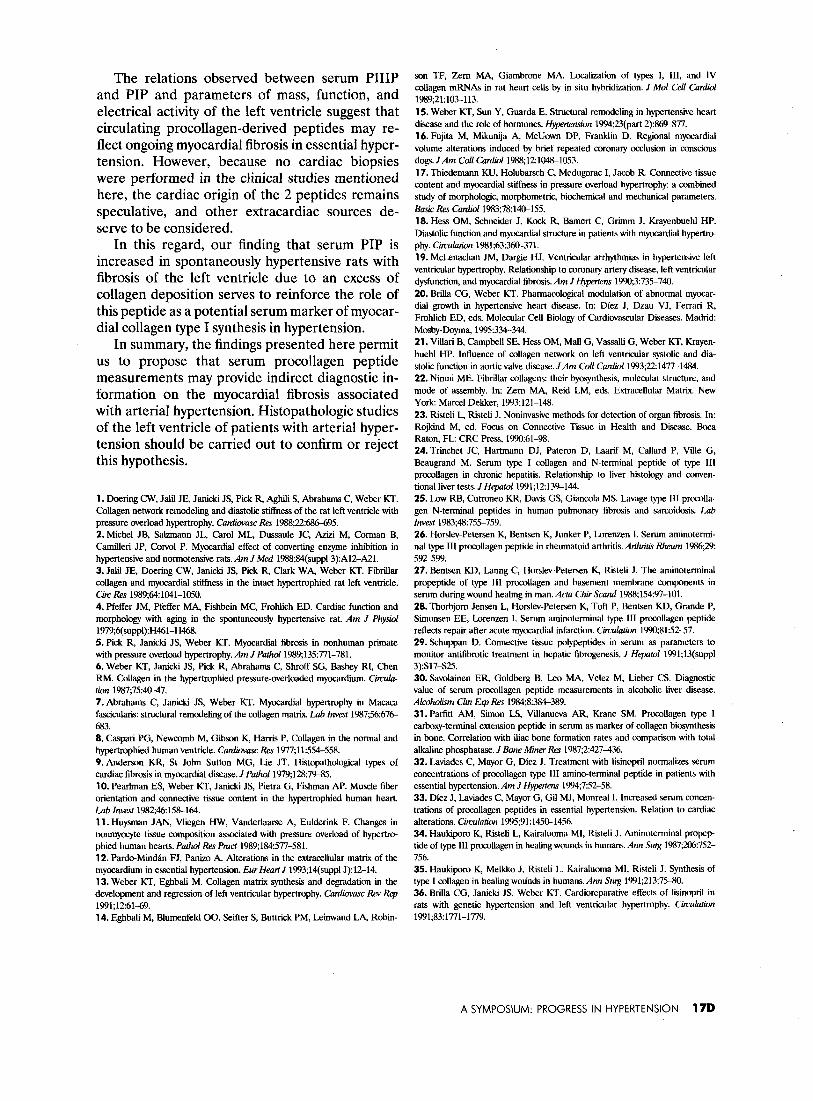
Experimental study: In a pilot study, we evalu- ated serum PIP concentrations in 7 36-week-old male spontaneously hypertensive rats with estab- lished left ventricular hypertrophy. Specific radio- immunoassay was used to measure serum PIP in rats. The amount of left ventricular collagen was evaluated by measuring the hydroxyproline concen- tration. The collagen-specific stain, Masson’s tri- chrome, was used to evaluate the presence and intensity of interstitial and perivascular fibrosis of the left ventricle.
In spontaneously hypertensive rats compared with 7 age- and sex-matched Wistar-Kyoto normo- tensive control rats we found: (1) an increase in hydroxyproline concentration (1.05 2 0.03 vs 0.84 + 0.05 m/g dry weight/100 g body weight; p ~0.05; Figure 3); and (2) an increase in serum PIP (10.31 + 0.58 vs 8.25 -t 0.59 &L, p < 0.05; Figure 4).
All the spontaneously hypertensive rats exhib- ited severe interstitial and perivascular fibrosis. The absence of pathologic myocardial fibrosis was seen in 6 normotensive rats. One Wistar-Kyoto rat exhibited mild interstitial fibrosis.
COMMENTS The findings of the studies show an increase in
serum concentrations of PIIIP and PIP in patients with essential hypertension and in spontaneously hypertensive rats. Elevated serum PIIIP and PIP may be markers of increased collagen type III and type I synthesis in arterial hypertension.
The question arises as to whether the measure- ment of PIIIP and PIP accurately reflects tissue
A SYMPOSIUM: PROGRESS IN HYPERTENSION 15D
fibrillogenesis. In this regard, in experiments of quantification of fibroproliferative reactions in situ, it has been shown that the PIIIP and the PIP assays reflect ongoing collagen synthesis. In fact, when the expression of procollagen type III and procolla- gen type I is induced, as during wound healing and other repair processes, 34,35 the local concentrations of the PIIIP and the PIP antigens in interstitial fluid increase dramatically, This is in agreement with a number of clinical observations showing that the circulating levels of the 2 peptides correlate well with ongoing tissue fibrosis.243i
We are aware that by measuring serum procolla- gen-derived peptides we assess the formation of
B A NT HT I
FIGURE 1. Serum concentrations of procollagen type III amino-terminal peptide (PIIIP) in normotensives (NT) and essential hypertensives (HT) Before (B) and after (A) treat- ment. *p co.01 compared with NT; **p <O.Ol compared with HT Before treatment.
*
s loo 75 3 80 % a 60
40
B A NT HT
FIGURE 2. Serum concentrations of procollagen type I car- boxy-terminal peptide (PIP) in normotensives (NT) and es- sential hypertensives (HT) Before (B) and after (A) treatment. *p ~0.001 compared with NT; **p ~0.001 compared with HT Before treatment.
fibrillar collagen but not its degradation. Since collagen degradation is also altered in arterial hypertension, l3 additional studies are necessary to assess in a more complete way collagen metabolism in hypertension.
The effects of lisinopril on the serum concentra- tions of the 2 peptides in hypertensive patients suggest that the rerun-angiotensin-aldosterone sys- tem may participate in the excessive synthesis of collagen types III and I in essential hypertension. This is in agreement with the observation that in spontaneously hypertensive rats with left ventricular hypertrophy and fibrosis of the cardiac interstitium, lisinopril reversed fibrous tissue accumulation.36
WKY SHR
FIGURE 3. Hydroxyproline concentration in normotensive Wistar-Kyoto rats (WKV) and spontaneously hypertensive rats (SHR). *p co.05 compared with WKY.
*
8
f a .3 6 a E 4
WKY SHR
FIGURE 4. Serum concentrations of procollagen type I car- boxy-terminal peptide (PIP) in normotensive Wistar-Kyoto rats (WKY) and spontaneously hypertensive rats (SHR). *p ~0.05 compared with WKY.
160 THE AMERICAN JOURNAL OF CARDIOLOGY VOLUME 76 NOVEMBER 2, 1995
The relations observed between serum PIIIP and PIP and parameters of mass, function, and electrical activity of the left ventricle suggest that circulating procollagen-derived peptides may re- flect ongoing myocardial fibrosis in essential hyper- tension. However, because no cardiac biopsies were performed in the clinical studies mentioned here, the cardiac origin of the 2 peptides remains speculative, and other extracardiac sources de- serve to be considered.
In this regard, our finding that serum PIP is increased in spontaneously hypertensive rats with fibrosis of the left ventricle due to an excess of collagen deposition serves to reinforce the role of this peptide as a potential serum marker of myocar- dial collagen type I synthesis in hypertension.
In summary, the findings presented here permit us to propose that serum procollagen peptide measurements may provide indirect diagnostic in- formation on the myocardial fibrosis associated with arterial hypertension. Histopathologic studies of the left ventricle of patients with arterial hyper- tension should be carried out to confirm or reject this hypothesis.
1. Doering CW, Jalil JE, Jar&i JS, Pick R, Aghili S, Abrahams C, W&r KT. CoUagen network remodeling and diastolic stiffness of the rat left ventricle with pressure overload hypertrophy. Cntiwc Res 1988;22:686X% 2. Michel JB, Salzmann JL, Carol ML, Dussaole JC, Aziii M, Corman B, CamiUeri JP, Corvol P. Myocardial effect of converting enzyme inhibition in hypertensive and normotensive rats. Am J Med 1988$4(suppl3):A12-AZ. 3. Jalil JE, Doering CW, Janicki JS, Pick R, Clark WA, Weber KT. Fibrillar collagen and myocardiil sttiess in the intact hypertrophied rat left ventricle. Circ Res 1989;64:1041-1050. 4. Pfeffer JM, Pfeffer MA Fishbein MC, Frohlich ED. Cardiac function and morphology with aging in the spontaneously hypertensive rat. Am J Physid 1979;6(suppl):H46-H468. 5. Pick R, Jan& JS, Weber KT. Myocardii fibrosis in nonhuman primate with pressure overload hypertrophy. Am J Path& 1989;135:771-781. 6. Weber KT, Janicki JS, Pick R, Abrahams C, Shroff SG, Bashey RI, Chen RM. Collagen in the hypertrophied pressure-overloaded myocardium. Circula- tion 1987;75:4&47. 7.Abrahams C, Janicki JS, Weber KT. Myocardial hypertrophy in Macaca fascicularis: structural remodeling of the collagen matrix. La6 Invest 1987;56:676- 683. 8. Caspari PG, Newcomb M, Gibson K, Harris P. Collagen in the normal and hypertrophied human ventricle. Cwdimwc Res 1977;11:554-558. 9. Anderson KR, St John Sutton MG, Lie JT. Hitopathological types of cardiac fibrosis in myocardial disease. I Path& 1979$28:7%35. 10. Pearlman ES, Weber KT, Janicki JS, Pietra G, Fishman AP. Muscle fiber orientation and connective tissue content in the hypertrophied human heart. Lab Inve.~t 1982;46:158-164. 11. Huysman JAN, Vliegen HW, Vaoderlaarse A, Eulderink F. Changes in nonmyocyte tissue composition associated with pressure overload of hype&o- phied human hearts. Path01 Res Pmcf 3989;1843577-581. 12. Pard~Miid&n FJ, Panizo A, Alterations in the extracellular matrix of the myocardium in essential hypertension. Ew Heart J 1993;14(suppl J):12-14. 13. Weber KT, EghbaIi M. Collagen matrix synthesis and degradation in the development and regression of left ventricular hypertrophy. Cardiovasc Rev Rep 1991;12:6169. 14. EghbaIi M, Blumenfeld 00, Seifter S, Buttrick PM, Leiiwand LA, Robm-
son TF, Zem MA, Giambrone MA. Localization of types I, III, and IV collagen mRNAs in rat heart cells by in situ hybridization. J Mel Cell Cardid 1989,21:103-113. 15. Weber KT, Sun Y, Guarda E. Structural remodeling in hypertensive heart disease and the role of hormones. Hypprtension 1994,23(part 2):86=77. 16. Fujita M, Mikunija A, McUown DP, Franklin D. Regional myocardial volume alterations induced by brief repeated coronary occlusion in conscious dogs. JAm Co[l Cur&l 1988;12:104&1053. 17. Thiedemann KU, Holubarsch C, Medugorac I, Jacob R. Comxctive tissue content and myocardial stifTnes.s in pressure overload hypertmphy: a combined study of morphologic, morphometric, biochemical and mechanical parameters. Basic Res Can&l 1983;78:14&155. 18. Hess OM, Schneider J, Kock R, Bamert C, Grimm J, Krayenbuehl HP. Diastolic function and myocardial structure in patients with myocardial hypertm phy. Circulntin 1981;63:360-371. 19. McLenachan JM, Dargie HJ. Ventricular arrhythmias in hypertensive left ventricular hypertrophy. Relationship to coronary artery dii, left ventricular dysfunction, and myocardial fibrosis. Am J Hpoteens 1990;3:735-740. 20. BriUa CG, Weber KT. Pharmacological modulation of abnormal myocar- dial growth in hypertensive heart disease. In: Diez J, Dzau VJ, Ferrari R, FrohIich ED, eds. Molecular Cell Biology of Cardiovascular Diseases. Madrid: Mosty-Doyma, 19993~344. 21. Villari B, Campbell SE, Hess OM, MaU G, VassaUi G, Weber KT, Krayen- buehl HP. Influence of collagen network on left ventricular systolic and dia- stolic function in aortic valve disease. JAm Cd Car&l 1993;22:1477-1484. 22. Nimni ME. Fibrillar collagens: their byosynthesis, molecular structure, and mode of assembly. In: Zem MA, Reid LM, eds. Extracellular Matrix. New York: Marcel Dekker, 1993121-148. 23. Risteli 1 Riiteli J. Noninvasive methods for detection of organ fibrosis. In: Rojkind M, ed. Focus on Connective Tissue in Health and Disease. Boca Raton, FL: CRC Press, 1990~61-98. 24.Trinchet JC, Hartmann DJ, Pateron D, Laarif M, Callard P, ViUe G, Beaugrand M. Serum type I collagen and N-terminal peptide of type III procollagen in chronic hepatitis. Relationship to liver histology and conven- tional liver tests. J Hepatol1991;12:13%144. 25. Low RB, Cutroneo KR, Davis GS, Giancola MS. Lavage type III procolla- gen N-terminal peptides in human pulmonary fibrosis and sarcoidosis. Lab Invest 198%48x755-759. 26. Ho&v-Petersen K, Bentsen K, Junker P, Lorenzen 1. Serum aminotermi- nal type III procollagen peptide in rheumatoid arthritis. Arthritis Rheum 19%,29: 592-599. 27. Bcntsen KD, Lanng C, Ho&v-Petersen K, Risteli J. The aminoterminal propeptide of type III procollagen and basement membrane components in serum during wound healing in man. Acta Chi Stand 1988;154:97-101. 28. Thorbjom Jensen L, Ho&v-Petersen K, Toft P, Bentsen KD, Grande P, Simonsen EE, Lorenzen I. Serum aminoterminal type 111 procollagen peptide reflects repair after acute mywardial infarction. Circulatior~ 1990;81:52-57. 29. Schuppan D. Connective tissue polypeptides in serum as parameters to monitor antifibrotic treatment in hepatic fibrogenesis. J Hepat 1991;13(suppl 3):S17S25. 30. Savolainen ER, Goldberg B, Leo MA, Velez M, Lieber CS. Diagnostic value of serum promllagen peptide measurements in alcoholic liver disease. Alcoholism Clin ELI Res 1984;8:384389. 31. Parfitt AM, Simon IS, Villanueva AR, Krane SM. Procollagen type I carboxy-terminal extension peptide in serum as marker of collagen biosynthesis in bone. Correlation with iliac bone formation rates and comparison with total alkaline phosphatase. J Bone Miner Rex 1987;2:427436. 32. Laviades C, Mayor G, Diez J. Treatment with lisinopril normalizes scram concentrations of procollagen type III amino-terminal peptide in patients with essential hypertension. Am J Hjpetem 1994;7:52-58. 33. Diez J, Laviades C, Mayor G, Gil MJ, Monreal 1. Increased serum concen- trations of procollagen peptides in essential hypertension. Relation to cardiac alterations. Circulation 1995;91:1450-1456. 34. Haukiporo K, Risteli L, Kairaluoma Ml, Risteli J. Aminoterminal propep tide of type I11 procollagen in healing wounds in humans. Ann Sug 1987;m:752- 756. 35. Haukiporo K, Melkko J, Risteli L, Kairaluoma MI, Risteli J. Synthesis of type I collagen in healing wodnds in humans. Ann Swg 1991;213:75-80. 36. Brilla CG, Janicki JS, Weber KT. Cardioreparative effects of lisinopril in rats with genetic hypertension and left ventricular hypertrophy. Cimularion 1991;83:1771-1779.
A SYMPOSIUM: PROGRESS IN HYPERTENSION 17D