Embed Size (px)
Citation preview

Interface Focus (2012) 2, 433–450
on July 26, 2018http://rsfs.royalsocietypublishing.org/Downloaded from
doi:10.1098/rsfs.2011.0122Published online 15 February 2012
REVIEW
*Author for c
One contribuTuring centen
Received 13 DAccepted 23 J
Towards an integrated experimental–theoretical approach for assessing themechanistic basis of hair and feather
morphogenesisK. J. Painter1,*, G. S. Hunt1, K. L. Wells2, J. A. Johansson2
and D. J. Headon2
1Department of Mathematics/Maxwell Institute for Mathematical Sciences,Heriot-Watt University, Edinburgh EH14 4AS, UK
2The Roslin Institute, University of Edinburgh, Easter Bush EH25 9RG, UK
In his seminal 1952 paper, ‘The Chemical Basis of Morphogenesis’, Alan Turing lays down amilestone in the application of theoretical approaches to understand complex biological pro-cesses. His deceptively simple demonstration that a system of reacting and diffusing chemicalscould, under certain conditions, generate spatial patterning out of homogeneity provided anelegant solution to the problem of how one of nature’s most intricate events occurs: the emer-gence of structure and form in the developing embryo. The molecular revolution that hastaken place during the six decades following this landmark publication has now placed thisgeneration of theoreticians and biologists in an excellent position to rigorously test thetheory and, encouragingly, a number of systems have emerged that appear to conform tosome of Turing’s fundamental ideas. In this paper, we describe the history and more recentintegration between experiment and theory in one of the key models for understandingpattern formation: the emergence of feathers and hair in the skins of birds and mammals.
Keywords: morphogenesis; Turing patterns; reaction–diffusion; activator–inhibitor; feather buds and hair follicles; skin patterning
1. INTRODUCTION
Pattern formation is a repetitive theme in nature, occur-ring at disparate spatial scales (figure 1a–c). At the scaleof landscapes, patchy or striated vegetation can arise insemi-arid climates [1], competition and predationwithin ecosystems can structure populations into par-ticular niches, while insects, fishes, birds and mammalshave an inbuilt capacity to swarm, school, flock or herd[2]. At the scale of cells, colonies of bacteria constructhighly intricate patterns in the presence of suitable foodsources [3–6] and populations of cellular slime mould,such as Dictyostelium undergo a carefully orchestratedsingle to multi-cellular transition that ensures theirlongevity following starvation [7]. A common feature tothese examples is that the pattern arises through a pro-cess of self-organization: the global order emergesthrough intrinsic local interactions, rather than someexternal control.
orrespondence ([email protected]).
tion of 13 to a Theme Issue ‘Computability and theary’.
ecember 2011anuary 2012 433
One of the most astonishing examples of self-organiz-ation is the transformation from a single fertilized cell toa complex organism through embryonic development,an event that occurs again and again despite its depen-dence on the accurately timed coordination of amultitude of events, spanning spatial scales from themolecular to the full embryo. Within the wider sphereof developmental biology, morphogenesis, or the emer-gence of structure and form, has particularly capturedthe attention of theoretical scientists, with two funda-mental classes of model proposed to explain thisphenomenon: pre-patterning mechanisms and sym-metry-breaking processes. Simplistically, these twoclasses can be distinguished according to the initialstate of the system. In the former, structure and formare generated through a process of new patterns build-ing on previous patterns, a classic example being thesegmentation of the fruitfly Drosophila: here, thestriped pattern of gene expression marking the futuresegments can be traced back to a maternally inheritedgradient in the newly fertilized egg [8]. Symmetrybreaking mechanisms, on the other hand, require nosuch specific initial condition: the physical and chemical
This journal is q 2012 The Royal Society

(a) (b) (c)
(d) (e) ( f )
(g) (h) (i) ( j)
Figure 1. Biological pattern formation. (a–c) Self-organization at various spatial scales: (a) aggregation of Dictyostelium discoi-deum; (b) flocking; (c) patchy vegetation in Nigeria. (d– f ) Pigmentation patterns: (d) swallowtail butterfly; (e) zebra;( f ) sailfish tang. (g– j) Feathers, hairs and scales: (g) peacock; (h) Vladimir the cat; (i) three-banded armadillo; ( j) centralbearded dragon. Image information: (d– j) K.J.P.; (a) http://en.wikipedia.org/wiki/File:Dictyostelium_Aggregation.JPG,released into public domain, accessed 21/01/2012; (b) http://en.wikipedia.org/wiki/File:Fugle,_%C3%B8rns%C3%B8_073.jpg, released into public domain by C. Rasmussen, accessed 21 January 2012; (c) http://en.wikipedia.org/wiki/File:Gapped_Bush_Niger_Nicolas_Barbier.jpg, released into public domain by Nicolas Barbier, accessed 21 January 2012.
434 Review. Models of hair and feather patterning K. J. Painter et al.
on July 26, 2018http://rsfs.royalsocietypublishing.org/Downloaded from
interactions of the systems conspire to amplify inherentnoise into regular structure. Effectively, the symmetryof the uniform steady state is broken to create orderedheterogeneity from homogeneity.
Turing’s seminal paper [9] provides an elegant exampleof a symmetry breaking mechanism. At the heart of themodel lies the fundamentally counterintuitive idea thatadding diffusion to a system of reacting molecules couldbreak the symmetry of a spatially homogeneous systemto generate a regular spatial pattern. In a swoop, Turingprovided a mechanism for pattern formation using onlya standard biochemical toolkit of chemical reaction andmolecular diffusion. Unsurprisingly, this elegant ideahas been enthusiastically seized upon in the theoreticalmodels for pattern formation, not only applied to numer-ous processes of developmental biology, but also scatteredto fields including ecology [10] and economics [11]. Turingalso coined the term morphogen to describe these elusivechemical drivers of pattern formation, although it shouldbe noted that the concept of morphogens predatedTuring, and the term is more generally recognized to
Interface Focus (2012)
denote a graded signalling molecule with the capacity todifferentiate a field of cells into patterns based on its mul-tiple threshold concentrations, rather than a specificcomponent in Turing’s mechanism.
In development, earlier references to Turing’s mech-anism were typically confined to a broader discussion ofits merit in classic examples of patterning, such as bris-tle formation in Drosophila [12] and hair folliclepatterning [13]. More formal applications of the ideasstarted appearing in the 1970s, using numerical andmathematical analyses to demonstrate its predictivecapacity in patterning processes such as buddinghydra [14,15], Drosophila segmentation [16] and pig-mentation markings [17,18], and the study ofreaction–diffusion patterning rapidly established itselfas its own field. Yet, despite this interest, Turing’sideas suffered from a lack of bona fide examples at themolecular level, particularly in the wake of the biotech-nology revolution of the 1980s. Furthermore, whilereactions such as the Belousov–Zhabotinsky reactionhad been found and shown to be capable of generating
1.95
10
5
0
10
5
0
10
5
0
10
5
0
501.21.011.000.99
1.011.000.99
1.00.8
1.11.00.9 0.4 0.6 0.8 0.4 0.6 0.8 0.4 0.6 0.8
50 50
105 105
105(ii)
diffusion diffusion
diffusion
selfactivation
selfactivation
diffusion
diffusion diffusion
diffusion diffusion
inhibition
inhibition
activation
activation
(i) (iii) (iv) (v) (vi)1050 1050 1050 1050
105 1050 1050 1050 1050
2.00 2.05 1.95 2.00 2.05
3.95 4.00 4.053.95 4.00 4.05
2 4 6 2 4 6 8
4 6 8
2 4 6 8
u
v
u
v
(a)
(b)
(ii)(i)
(iii) (iv) (v) (vi)
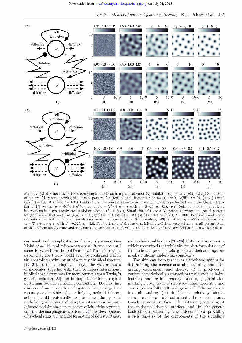
Figure 2. (a(i)) Schematic of the underlying interactions in a pure activator (u)–inhibitor (v) system. (a(ii)–a(vi)) Simulationof a pure AI system showing the spatial pattern for (top) u and (bottom) v at (a(ii)) t ¼ 0, (a(iii)) t ¼ 20, (a(iv)) t ¼ 40(a(v)) t ¼ 100, at (a(vi)) t ¼ 1000. Peaks of u and v concentration lie in phase. Simulations performed using the Gierer–Mein-hardt [15] system, ut ¼ dr2u þ u2=v � au and vt ¼ r2v þ u2 � v with d ¼ 0.025, a ¼ 0.5. (b(i)) Schematic of the underlyinginteractions in a cross activator–inhibitor system. (b(ii)–b(vi)) Simulation of a cross AI system showing the spatial patternfor (top) u and (bottom) v at (b(ii)) t ¼ 0, (b(iii)) t ¼ 10, (b(iv)) t ¼ 20, (b(v)) t ¼ 50, at (b(vi)) t ¼ 1000. Peaks of u and v con-centration lie out of phase. Simulations were performed using Schnakenberg [43] kinetics, ut ¼ dr2u þ u2v � u andvt ¼ r2v þ a � u2v, with d ¼ 0.025, a ¼ 1.0. For both sets of simulations, initial conditions were set at a small perturbationof the uniform steady state and zero-flux conditions were employed at the boundaries of a square field of dimensions 10 � 10.
Review. Models of hair and feather patterning K. J. Painter et al. 435
on July 26, 2018http://rsfs.royalsocietypublishing.org/Downloaded from
sustained and complicated oscillatory dynamics (seeMaini et al. [19] and references therein), it was not untilsome 40 years from the publication of Turing’s originalpaper that the theory could even be confirmed withinthe controlled environment of a purely chemical reaction[19–21]. In the developing embryo, the vast numbersof molecules, together with their countless interactions,implied that nature was far more tortuous than Turing’sgraceful solution [22] and its importance for biologicalpatterning became somewhat contentious. Despite this,evidence from a number of systems has emerged inrecent years in which the underlying molecular inter-actions could potentially conform to the generalunderlying principles, including the interactions betweenleftyand nodal in the determination of left–right asymme-try [23], the morphogenesis of teeth [24], the developmentof tracheal rings [25] and the formation of skin structures,
Interface Focus (2012)
such as hairs and feathers [26–28]. Notably, it is now morewidely recognized that while the simplest formulations ofthe model can provide useful guidance, their assumptionsmask significant underlying complexity.
The skin can be regarded as a textbook system fordetermining the mechanisms of patterning and inte-grating experiment and theory: (i) it produces avariety of periodically arranged patterns such as hairs,feathers and scales, sensory bristles, pigmentationmarkings, etc.; (ii) it is relatively large, accessible andcan be successfully cultured, greatly facilitating exper-imental studies; (iii) it has a relatively simplestructure and can, at least initially, be construed as atwo-dimensional surface with patterning occurring atthe epidermal–dermal interface; and (iv) the geneticbasis of skin patterning is well documented, providinga rich tapestry of the components of the signalling
436 Review. Models of hair and feather patterning K. J. Painter et al.
on July 26, 2018http://rsfs.royalsocietypublishing.org/Downloaded from
pathways and their many interactions. Turing’s ideashave frequently recurred in explanations of skin pat-terning, particularly in the context of pigmentationmarkings [29–35] and, the subject of the presentpaper, the periodic arrangement of hairs and feathers.
In this paper, we will provide a brief history of modelsbased on self-organizing principles in morphogenesis,paying particular attention to the reaction–diffusionmodel of Turing. We do not use this review to cover the-ories related to gradients and positional information,noting that a number of recent reviews consider thisarea in detail [8,36–38]. We will proceed to explore theuse of these models to explain the patterning of hairsand feathers in developing mammalian and avian skin,illustrating how integrating theory into the experimentalcycle can be used to corroborate hypotheses and generatepredictions. We will conclude with a brief discussion andargue that, for this system, we are now in a position tomove away from the more simplistic modelling that hasdominated the field to date, and towards a model foundedon the molecular mechanisms that operate at the coreof patterning.
2. MODELS FOR SELF-ORGANIZATIONIN MORPHOGENESIS: AN OVERVIEW
A fundamental goal for biological modelling is illumi-nating and informing our understanding of a givensystem. While concepts related to gradients and pos-itional information date to Driesch at the end of thenineteenth century [39], relatively few explanationshave been made into the origins of ‘pattern’ at thetime of Turing’s contribution. A notable exception isWigglesworth’s [40] exploration into the factors control-ling the size and spacing of bristles in the abdomen ofRhodnius prolixus, proposing a ‘competitive model’based on the underlying hypothesis that all epidermalcells start off with the same potential to specialize,although when one cell does, the surrounding cells areinhibited from the same course of action.
Pattern formation was therefore an immature field andTuring was alive to the number of liberties he was takingduring the model’s formulation, stressing early in theintroduction that ‘This model will be a simplificationand an idealization, and consequently a falsification’:modelling processes where mechanical interactionsbetween cells were considered negligible; concentratinganalysis on the relatively simple scenarios of just two orthree morphogens; exploring patterning from a quasi-homogeneous initial state rather than a prior pattern;idealizing the tissue to a ‘ring’ of cells, and so on. Despitethe huge advance in our biological understanding in thepast 60 years, as well as the increased computationalpower, theoretical modellers still face the same trickyconundrum at the outset: how much detail is necessary?A biological model of some developing process, suchas figure 3d, is the accumulated effort of numerousexperiments, incorporating multiple components andinteractions occurring at scales ranging between the sub-cellular and the full extent of the developing organism.Yet, these models exclude some unknown fraction ofthe full system’s complexity and any attempt to write
Interface Focus (2012)
down a mathematical model can only ever be viewed asa cartoon of a caricature.
One method would be to apply ‘bottom-up’ think-ing: formulate a mechanistic and parametrizedmathematical model for the system, complete as far asthe biological understanding permits. The validity ofa hypothesis can then be examined in rigour, predic-tions can be made, and failure to reconcile experimentand theory could point the way to missing interactionsand components. Against this, there may simply not beenough information to begin with, or the rapid turnoverin the molecular level understanding of embryonic pat-terning raises the risk of a model becoming redundantbefore completion. Consequently, a critical initial jud-gement is whether a given system is sufficiently wellunderstood to adopt this tactic. Certain developmentalsystems, where a substantial portion of the molecularbiology is known, are now amenable to this approach.Early morphogenesis of the fruitfly Drosophila wouldprovide one example [34,36], where the existence of asaturated genetic screening ensures that a completeset of jigsaw pieces is available and they only requireto be pieced together appropriately.
For less well-understood systems, a general approachwould be to adopt a ‘top-down’ method: channelEinstein’s maxim that ‘a model should be as simple aspossible, but no simpler’ and take a reductionist view.This would involve schematizing proposed biologicalinteractions wherever possible and only adding extracomponents as demanded; for example, to test a specifichypothesis arising from a particular experiment. Classictheoretical ideas for pattern formation, such as Turing’sown work and ideas of positional information and pre-patterning [39,41], may not always carry the finesse togenerate a pin-sharp prediction in a particular systembut can still provide an important conceptual frameworkwithin which experimental work can be targeted.
In §1, we briefly touched upon fundamental distinc-tions between pre-patterning and symmetry-breakingmodels of morphogenesis. In this section, we willexpand our discussion of the latter, paying particularattention to the reaction–diffusion-based model pro-posed by Turing.
2.1. Turing and other chemical-based models
The pioneering model of Turing [9] functions solelythrough the molecular processes of reaction and passivediffusion: there is no need to take mechanical forcesarising from cell migration, tissue movements, etc., intoaccount, although these processes undoubtedly play asignificant role in many processes during embryonicpatterning. The experimental confirmation of Turingpatterns within controlled chemical reactions, such aschloride–iodide–malonic acid [20,21] and thiourea–iodate–sulphite [42] reactions, proves the sufficiency ofthese minimal requirements, even though the nature ofthe chemical components renders such reactions whollyunrealistic within biological tissues.
While more commonly presented as a system com-posed of just two morphogens (see §3), the standardTuring model [9,34] comprised multiple (m � 2) partialdifferential equations, describing the diffusion and

Review. Models of hair and feather patterning K. J. Painter et al. 437
on July 26, 2018http://rsfs.royalsocietypublishing.org/Downloaded from
reaction between m morphogens within some boundedregion of space, V [ <l ; l � 3:
@cðx; tÞ@t
¼ Dr2cðx; tÞzfflfflfflfflfflfflffl}|fflfflfflfflfflfflffl{
diffusion
þ f ðcðx; tÞ; x; tÞzfflfflfflfflfflfflfflfflfflfflffl}|fflfflfflfflfflfflfflfflfflfflffl{
reaction
: ð2:1Þ
In the above, cðx; tÞ ¼ ðc1; c2; � � � cmÞT is the vectordenoting the concentration of each morphogen at pos-ition x [ V and time t, D is the m � m diagonal matrixdescribing their diffusion coefficients, and f ðcðx; tÞ; x; tÞdescribes the reactions–interactions between themorphogens. For applications, the above system is aug-mented with suitable initial and boundary conditions:for the latter, a typical assumption is to assume nogain/loss (zero-flux) across the boundary of V.
Textbook linear stability analysis for equations (2.1)yields a set of diffusion-driven instability conditions: aset of requirements for which a spatially uniformsteady-state morphogen distribution, stable in theabsence of diffusive terms, is driven unstable throughtheir addition [31,34]. Under these conditions, a non-uniform distribution emerges, which is typically in theform of a stationary and periodic pattern of high- andlow-morphogen regions separated by a characteristicspatial wavelength (see figure 2a,b for examples of pat-tern evolution), although patterns can also undergocontinual evolution in time. The implicit assumptionin many applications is that this pattern provides theblueprint for cell differentiation and tissue organization,for example, by acting as a positional information cue.
In its most modest and familiar form, and our buildingblock for the present paper, Turing’s model requires justtwo reacting and diffusing components to operate, albeitunder a set of precise constraints. A classical and intuitiveexplanation is the short-range activation, long-range inhi-bition concept popularized by Gierer and Meinhardt[15,44] (and, independently, by Segel & Jackson [45] inan ecological setting). For example, we suppose thatthe two reactants adopt the roles of an ‘activator’ andan ‘inhibitor’, respectively, with the activator activatingboth its own upregulation (self-activation), as well asthat of the inhibitor, and the inhibitor downregulatingthe activator (figure 2a). Crucially, if the activator hasa shorter range—it diffuses less rapidly than the inhibi-tor—then self-activation can dominate locally, whilefaster inhibitor diffusion leads to a long-range suppressionand the emergence of spatial structure.
More generally, two component systems capable ofpatterning through Turing’s mechanism comprise onemore slowly diffusing reactant with ‘self-activating’(such as autocatalysis) properties and inter-componentinteractions structured into one of two forms—pure orcross activator–inhibitor systems—according to theloop structure [46] (cf. figure 2a,b). The simplicity ofthese ideas has provided a useful framework for examin-ing whether a given system could form a pattern: in §3,we show how searching for processes of activation andinhibition has shaped experimental research into themechanisms underlying aspects of skin morphogenesis.At the same time, care should be taken before equatingbehaviour of the much broader reaction–diffusionmodel (2.1) with the two-component activator–
Interface Focus (2012)
inhibitor systems. For example, a constraint such asthe distinct ranges of diffusion required in the latterrelaxes when moving to the greater than two com-ponents permitted by the general model (2.1) [47,48].
Nearly, all applications and analyses of Turing’smechanism set up the model as a coupled system of con-tinuous partial differential equations, as in system (2.1).In the context of embryonic development, continuousformulations are an abstraction of the in vivo tissueenvironment and biochemistry, where distinct elementsof a signalling pathway can occur inside, outside or atthe cell membrane, and any diffusion-like transportwill be contingent on interactions between moleculesand the extracellular matrix or the transfer of moleculesbetween cells, etc. In fact, Turing first proposed adiscrete cell reaction–diffusion mechanism before pro-ceeding to the ‘continuous’ idealization. Specifically,Turing formulated a system of ordinary differentialequations for the dynamical change of each morphogenin each cell in a hypothetical ring, proposing transfer ofmorphogens between adjacent cells based on their localconcentration difference. An extension of Turing’s ideasto more generalized discrete cellular networks wasundertaken by Othmer & Scriven [49].
Other discrete cell models of pattern formation havealso been proposed, relying on more explicit descriptionsfor the signalling interactions between cells. In juxtacrinesignalling, the membrane to membrane binding betweena ligand on one cell and its associated receptor on aneighbour triggers an internal signal transduction path-way which could, for example, subsequently feed backto modulate the ligand and/or receptor expression atthe cell surface [8]. For appropriate interactions, modelsof such signalling also show symmetry-breaking phenom-ena, with a quasi-homogeneous population taking onalternating expression levels [50,51]. Molecular diffusionis not required and the resulting patterns appear to besomewhat distinct, with the alternating salt andpepper pattern that arises typically separated by just afew cells rather than the broader wavelengths that canpotentially be generated by Turing’s model.
2.2. Mechanical-based models
It is appropriate to note that Turing’s proposed modelwas for the chemical basis of morphogenesis: the mech-anism generates only a biochemical template thatmorphogenesis slavishly follows and the visible mani-festation of form would arise through the subsequentdifferentiation of cells into various subtypes, with theirdistinct mechanical and growth characteristics. Othermodels for morphogenesis explicitly incorporate thedynamics of cells, such as their growth, movement andadhesive properties, together with their interactionswith the environment. In certain instances, their inter-play can also conspire to break the symmetry andcreate spatial pattern from uniformity, a simple instancebeing the aggregation of a population through cell–celladhesion. For multiple populations, this adhesion cangenerate more complicated spatial patterning througha process of cell-sorting, commonly attributed to aprocess of differential adhesion [52], in which the distinctadhesive properties of the various populations drive

(a)
(b) (d)
Edar Eda
Bmps
Ctgf
Wnts
Dkks
hair follicle
b–catenin
(c)
Figure 3. Developmental patterning of hairs and feathers. (a) Spatio-temporal sequence of feather development in the chickenembryo (stages shown range from a third to midway through the incubation period). Feather buds initially develop along twolines either side of the midline and subsequently spread into lateral regions. Note that significant growth occurs during the pat-terning process. (b) Naked neck chickens, conspicuous by their absence of neck feathering. (c) Mid to late gestation of the mouseembryo, stained for a marker of developing follicles (purple foci). (d) Schematic of the molecular network underpinningmouse follicle formation. The dashed region encloses a specific loop with the features of a pure activator–inhibitor system.
438 Review. Models of hair and feather patterning K. J. Painter et al.
on July 26, 2018http://rsfs.royalsocietypublishing.org/Downloaded from
the rearrangement of the populations through energy-minimizing principles akin to the separation of oil andwater. Mathematical models for adhesion, both at thelevel of individual cells and continuous populations, vali-date the inherent capacity of adhesion to cluster and sortcell populations [53,54].
Chemotaxis, and related forms of directed cell move-ment, offers another mechanism for self-organization.Insight into chemotaxis-induced pattern formation has pri-marily resulted from studies into certain bacteria species,such as Escherichia coli and Salmonella tymphirium[3–6] and the cellular slime mould Dictyosteliumdiscoideum [7,55]. Mathematical models [56–58] revealthat at the heart of self-organization lies a powerfulfeedback mechanism in which the secretion by cells oftheir own chemoattractant (or degradation of their ownchemorepellent) can mobilize a dispersed population intoself-supported aggregations. For Dictyostelium, the samechemotaxis mechanism that aggregates the initial popu-lation also plays a critical role in the subsequentdifferentiation and organization of diverse cell subpopu-lations [7]. A number of lines of enquiry have pointed tochemotaxis playing a role in various processes of embryonicdevelopment, including gastrulation [59] and neural crestmigration [60].
Moving beyond the relative simplicity of chemotaxis,the mechano-chemical theory of pattern formation
Interface Focus (2012)
pioneered by Oster et al. [61] embraces many of theabove concepts (chemical patterning, adhesion andother mechanical interactions) and more (e.g. contractileforces exerted by cells on their surroundings), assumingthat the distinct mechanisms that contribute tomorphogenetic patterning are not so trivially separated.While, unsurprisingly, mathematical descriptions of themechano-chemical framework can quickly become com-plex, they provide enormous scope and have beenapplied to a wide variety of embryonic processes, includ-ing skin patterning, wound healing, skeletal patterningand vasculogenesis (see Murray [31,62] for reviews).
3. MODELLING MORPHOGENESIS: HAIRAND FEATHER PATTERNING AS CASESTUDIES
The skin of birds and mammals typically carries hairor feathers which first form during embryogenesis.This development is achieved through communicationbetween the two tissue layers of the skin: the dermallayer and the overlying epidermis. At the earlieststages of skin patterning, dermal–epidermal signalexchange results in the formation of localized con-densations of epidermal cells, which undergo rapidproliferation to form feather buds or follicle primordia.

Review. Models of hair and feather patterning K. J. Painter et al. 439
on July 26, 2018http://rsfs.royalsocietypublishing.org/Downloaded from
Subsequent morphogenesis then results in the formationof a mature feather or hair [63,64].
Having explored in more general terms some of thecommon self-organizing models, in this section, wemove on to a key case study: hair and feather patterningin the developing skin of mammals and birds. We treadswiftly through the history of the field, providing a briefaccount of the salient biology and the contributions ofmodelling through the decades-long and ongoing pro-cess of integrating experiment and theory. Thissection also acts as the prelude to the more detailed sec-tion that follows, where we demonstrate the specificapplication of reaction–diffusion ideas and its capacityto both recapitulate and predict experiment.
3.1. Biology and modelling of sheep hair folliclepatterning
While recent attention has shifted to the de facto devel-opmental model of mice, much of the earlier work onhair development in mammals was performed forsheep, an animal of economic significance in terms ofwool production. Hair follicles in sheep first appearapproximately 60 days following conception andadvance through various stages from initiation to thefully formed fibre [65]. The very first follicles developat approximately equal distances from their neighbours,with subsequent follicles developing in a series of wavesover the following three weeks. Within-pattern pattern-ing occurs, with later generations of follicle initiationproducing two further follicles either side of a primaryfollicle to form a trio group [65].
Early attempts to explain follicle initiation and theirequidistant arrangement borrowed heavily from analysesof the spatial distribution of sensory bristles in insectssuch as Drosophila and R. prolixus. Claxton [13] demon-strated a favourable comparative analysis betweenfollicle-initiation sites and the predictions based on thecompetitive model of Wigglesworth [40] described earlier.This was expanded upon in a more formal manner byClaxton & Sholl [66], yet the model required additionalassumptions and complexity to generate the timing andarrangement of the characteristic trios.
A specific application of Turing’s mechanism toexplain follicle patterning, again for sheep, was pioneeredby Nagorcka and Mooney in a series of papers beginningin the early 1980s [67–72]. Early work explored the cross-sectional pattern within an individual adult hair fibre,proposing that a reaction–diffusion system operatedinside each developing hair fibre bulb and generated apre-pattern that triggers cell differentiation [67]. Despitea lack of information on the precise molecular com-ponents, comparative simulations supported the ideathat this could pattern the forming fibre. Furtherpapers in the series [68–72] expanded the application ofreaction–diffusion models to consider the skin-widespatial distribution of both primary and secondarywool follicles, as well as the positioning of structuressuch as sebaceous glands in the bulb. Effectively, pre-viously formed follicles were proposed to create a fixed‘organizing’ template around which later follicles wouldform, indicating that a common mechanism could beresponsible for many aspects of hair morphogenesis.
Interface Focus (2012)
Despite this early work, matching the developing woolfollicle pattern to Turing-type simulations in terms oftheir intermediate stages and end result, studies of wooldevelopment have declined in recent years owing, in nosmall part, to the relative impracticality of sheep as anexperimental system. Instead, attention has shiftedto feather bud formation in birds and hair follicle for-mation in mice—a direct consequence of their positionsas ‘model organisms’. Refer to the study of Rogers [73]for a recent review of sheep hair patterning, and a pleafor its rediscovery.
3.2. Biology and modelling of chick featherbud patterning
Formation of the feather bud pattern in the developingchick embryo has been used extensively in theoreticaland experimental studies of pattern formation, with theaccessibility of the embryo itself granting it a dominantplace in studies of development. Feather developmentbegins with the previously undetermined cells in theskin acquiring the cellular fate that leads to the formationof feather buds,which grow rapidlyout from the surface ofthe skin to produce a feather and surrounding follicle. Asfor sheep, buds develop in a specific spatial and temporalsequence [74]: the first buds to form are arranged in regu-larly spaced intervals in two anterior–posterior stripeseither side of the dorsal midline, with further rows ofbuds subsequently added in a wave-like fashion thatspreads into lateral skin regions (figure 3a). A variety ofmodels, including those based on mechano-chemical [75]and reaction–diffusion [69] principles have been pro-posed, capable of capturing the spatial and temporalpatterning of the feather array. Further extensions haveintegrated these two modelling approaches [76–78].
At the time of these studies, little was known regard-ing the genes and molecules responsible for embryonicdevelopment. Yet, a seismic shift was taking place,beginning with the ground-breaking screening whichrevealed the critical genes driving early morphogenesisof the fruitfly Drosophila [79]. Combined with a hostof further technical advances, recent decades haveunleashed a torrent of information on the genes andmolecules that drive pattern formation, not only in Dro-sophila, but also in the other principal model systems ofdevelopment, including the chicken and mouse. At thispoint, we remark that notation varies between specieswhen referring to individual proteins, protein families,genes, etc., and we have preserved this variation tofacilitate comparison with cited studies.
In the context of feather bud morphogenesis, this hasled to one of the first biological systems in whichlines can begin to be drawn between the theoretical con-cepts of Turing’s mechanism and molecular biology,starting in the 1990s. With the underlying principles ofactivator–inhibitor systems in mind, Jung et al. [80]searched for candidate regulators involved in featherbud formation by focusing on signalling pathwaysknown to operate widely during the development ofmany organs and in diverse animal species. Based on thegene-expressionpatterns at the sites of the forming featherbuds, sonic hedgehog (SHH) and fibroblast growthfactor-4 (FGF-4) were identified as candidate activators

440 Review. Models of hair and feather patterning K. J. Painter et al.
on July 26, 2018http://rsfs.royalsocietypublishing.org/Downloaded from
and bone morphogenetic protein-2, 4 (BMP-2,4) aspossible inhibitors, with their initially widespread pro-duction followed by their co-restriction to developingfeather primordia as patterning progresses consistentwith the operation of a pure activator–inhibitor system(cf. figure 2a). Local application of recombinant proteinsfrom beads on the skin also suggested a greater rangeof action of the inhibitors over the activators [80–82].Furthermore, the capacity of disassociated and reconsti-tuted skin to form buds favours a truly self-organizingmechanism acting during skin morphogenesis [81].
With this greater foundation, more recent modelling hasbeen immersed into an integrated experimental/theoreticalframework, with mathematical variables in a model moreformally linked to specific molecular components. Michonet al. [83] have developed an extended reaction–diffusionframework, with BMP7 and BMP2 fulfilling the roles ofactivator and inhibitor and augmented by additionalequations to describe the proliferative and migratory be-haviour of dermal cells, and demonstrated that the modelcan replicate experimental in vivo data. Lin et al. [84] (seealso Baker et al. [85]) have linked experimental data andmodelling for the extracellular signal-regulated kinases-dependent chemotaxis of mesenchymal cells in the for-mation of feather primordia. Results of Mou et al. [86]support a reaction–diffusion-based mechanism underpin-ning patterning, combining experiment and theory todemonstrate how retinoic acid (RA) can regionally tuneparameters in a reaction–diffusion system (see §4).
Moving beyond the formation of a spatially periodicpattern of two-dimensional placodes distributed acrossthe skin, reaction–diffusion ideas have also beenproposed to explain the subsequent within-placode pat-terning which will ultimately lead to a fully formedfeather [28,87]. Harris et al. [28] obtained molecularsupport that SHH and BMP2 operate in an activator–inhibitor type pairing within the feather bud epithelium.Augmented by simulations of a reaction–diffusionmodel, it is suggested that these interactions form acore component of the mechanism that generates the tub-ular pattern of ‘barb ridges’ that determines shape andfunction of the feather. Beyond the structural formationof the feather itself, reaction–diffusion models havefurther been proposed as a mechanism for the pigmenta-tion patterning within a feather [87], although identitiesof potential molecular components are lacking. Similar tosheep wool formation above, this hints that a common setof principles may operate at distinct levels and scales tocontrol multiple aspects of feather patterning.
3.3. Biology and modelling of mouse hairfollicle formation
As the dominating mammalian model, understandingmorphogenesis in the mouse has enormous significancewhen it comes to determining our own development.Advantageously, the mouse offers unique genetic tools,such as the ability to create targeted mutations, whichhave greatly contributed towards piecing together themolecular control of development. As for sheep, mousehair development proceeds through several rounds of fol-licle formation, with the primary hair follicles appearingin an approximately equidistant pattern between 13 and
Interface Focus (2012)
16 days following fertilization and secondary and tertiaryfollicles forming later in the expanding regions surround-ing the primary follicles (figure 3c) [88]. Again, modelsbased on reaction–diffusion ideas are highly adept atreplicating experimental observations and features offollicle patterning [27,89]; for example, see figure 5.
Having defined possible activatory–inhibitory signal-ling pathways in the chicken, together with the geneticidentification of additional pathways dedicated to skindevelopment in the mouse, the logical next step was toexplore whether interactions between these pathwayswere consistent with the theoretical network topologiesof figure 2. Mou et al. [26] paid attention to the signallingpathway composed of the extracellular ligand, Eda, andits receptor, Edar, and its interaction with certain mem-bers of the BMP family. Edar expression is critical forprimary follicle patterning with Edar signalling resultingin both a rapid positive feedback loop that stimulates bothits own expression as well as production of the inhibitors,Bmp4 and Bmp7. Diffusion transports these extracellularsignals laterally where they strongly repress Edar tran-scription and, hence, suppress hair follicle fate. Theseinteractions—a locally operating autocatalytic processcoupled to a long-range inhibitory mechanism—appearto conform to the underlying principles of an activator–inhibitor system.Furthermore, thiswork has also revealeda locally acting BMP-inhibiting process driven by Edarsignalling, mediated through the BMP-binding proteinconnective tissue growth factor (CTGF). Mathematicalmodelling [48] has demonstrated the sufficiency of theEdar–BMP–CTGF interactions in producing a periodicpattern, at least under certain constraints.
A combination of mathematical modelling and exper-imental work by Sick et al. [27] has further enhanced ourunderstanding of follicle patterning, uncovering a secondpotential reaction–diffusion loop that operates bothduring primary and later stages of follicle patterning.Here, a proposed mechanism counters the activatingaction of the Wnt/b-catenin pathway against the inhibit-ing actions of Dkks to localize the developing follicles.Yet, rather than operating in complete independencefrom the above Edar–BMP–CTGF network, these twoloops are proposed to be interlocked, with Edar acti-vating the expression of Wnt10a, Wnt10b and Dkk4,encoding b-catenin pathway ligands and an antagonist,and b-catenin activity subsequently stimulating theexpression of Edar itself [90–92].
Overall, these results are inching us towards a detailedknowledge of the molecular processes underpinning hairpatterning (figure 3d) and hint at a significantly morecomplex activator–inhibitor framework than capturedin the most simplified realizations of Turing’sscheme. It appears that multiple signalling pathwaystake on the distinct roles of activators and inhibitors,acting together to localize the positions of follicles.
4. MODELLING MORPHOGENESIS INPRACTICE: REACTION–DIFFUSIONSYSTEMS APPLIED TO SKINMORPHOGENESIS
The striking qualitative resemblance between thearrangement of feather buds and hair follicles and the

Review. Models of hair and feather patterning K. J. Painter et al. 441
on July 26, 2018http://rsfs.royalsocietypublishing.org/Downloaded from
patterns produced by reaction–diffusion systems (cf.figures 2 and 3) is compelling. While such prima facieevidence alone is by means sufficient for proof—otherplausible mechanisms such as chemotaxis andmechano-chemical models can generate a similar out-come—it has offered a crucial line of enquiry to bothbiologists and modellers. For biologists, does an under-lying network of interactions operate according to theprecepts of Turing’s theory? For modellers, how welldoes the theory hold up when confronted with a morerigorous examination?
Regarding the first, we have already summarized someencouraging data that implicate reaction–diffusion net-works in the patterning process. In mouse skin, at leasttwo network loops appear to conform to the principles[26,27] while in the chicken, although specific pathwayshave not been firmly established, the skin’s capacity toself-organize and the identification of potential activators(such as Eda, Wnts, FGFs) and inhibitors (e.g. BMPs,Dkks) in mouse or chicken skin provides encouragement.In this section, we aim to highlight the manner in whichmodelling can both replicate and predict certain featuresof skin patterning.
4.1. Model formulation
For the purpose of the present review, here we concen-trate on the simplest top-down approach founded onTuring’s ideas: two principal components (an activator,A and inhibitor, I) both undergoing spatial diffusionand reaction. Following a philosophy of schematic mod-elling, these components and their interactions need notbe strictly considered to represent single molecular enti-ties, rather they can loosely schematize the multiplecomponents and their pathways that generate an overallactivatory (or inhibitory) contribution. This frameworkdoes, however, still allow us to simulate a specificexperimental procedure: for example, application ofexogeneous BMP to explanted skin (figure 6) can berecapitulated in silico through a simple additive termto the equation describing the inhibitor component.As we aim to show, this schematic modelling can stillprovide useful insight. Our simple model is given by:
@A@t¼ DAr2Aþ f ðA; I Þ ð4:1Þ
and
@I@t¼ DIr2I þ gðA; I Þ: ð4:2Þ
In the above, DA and DI are the diffusion coefficients ofthe activator and inhibitor, characterizing their respect-ive spatial range of action: most directly this would bemediated via molecular diffusion in the extracellularspace, but it could also be a simplification for otherforms of molecular transport, including direct cell tocell passage of signalling molecules through membranechannels such as gap junctions. The kinetic functions f(A, I ) and g (A, I ) are selected according to the proposednetwork interactions, and can be modified as appropriateto simulate some experimental perturbation. Takingthe interactions between Edar and BMPs in mousehair follicle specification (cf. boxed region in figure 3d),
Interface Focus (2012)
we observe the hallmarks of a pure-type activator–inhibitor system and choose kinetic terms accordingly,exploiting the well-characterized Gierer–Meinhardt [15]type model:
f ðA; I Þ ¼ pAA2
k21 þ A2
� 11þ k2I
zfflfflfflfflfflfflfflfflfflfflfflfflfflfflfflfflfflffl}|fflfflfflfflfflfflfflfflfflfflfflfflfflfflfflfflfflffl{activator�upregulation
þ SA
z}|{basal�upregulation
� dAAzffl}|ffl{
activator�decay
ð4:3Þ
and
gðA;I Þ¼ pIA2
k23þA2
zfflfflfflfflfflffl}|fflfflfflfflfflffl{inhibitor�upregulation
þ SI
z}|{basal�upregulation
� dII :z}|{
inhibitor�decay
ð4:4Þ
The first term in equation (4.3) incorporates boththe autocatalysis of the activator and the inhibitionof autocatalysis by the inhibitor. Second and thirdterms, respectively, represent an independent orbasal level of activator upregulation and decay ofactivity. In the second equation, the first term modelsthe upregulation of inhibitor by the activator whilethe latter terms again describe basal upregulation anddecay. Each of the parameters in the above modelð pA; pI; k1; k2; k3; g; dA; dI;DA;DI; SA; SIÞ holds a biologi-cal interpretation (table 1). Equations (4.1) to (4.4) needto be augmented with suitable initial and boundary con-ditions and our default will be small spatially randomperturbations about the uniform steady-state solutionfor the former, and zero flux (i.e. no loss/gain) for thelatter. Of course, specific applications should take intoconsideration any proposed role for prestructure in pat-tern initiation, such as the initial appearance of nascentchicken feather buds along the midline.
4.2. Parametrization
Linear stability analysis (e.g. see [31]) can be applied toestablish that diffusion-driven instability will occur forequations (4.1) to (4.4), provided that parameter valueslie in a suitable region of parameter space. Such analysestherefore provide a useful checkpoint for determining if agiven mechanism has the potential to generate pattern.To illustrate this, in figure 4 we demonstrate the patternsformed when varying two of the parameters and holdingthe remainder fixed. Notably, patterning is confined tocertain regions of the parameter space, with regions out-side this generating spatially uniform patterns of eitherhigh or low expression (relative to some normalizedlevel). Furthermore, the pattern generated varies over arange, including ‘spots’, ‘stripes’ (or ‘labyrinthine’) and‘holes’. The question as to whether spotted or stripedpatterns form has been subject to some more rigorousinvestigation, with the dominating nonlinearity in thereaction believed to be a major determinant [93–95].
The above clearly reveals the criticality of model par-ameter values in the generation of Turing-type spatialpatterns: while qualitatively similar patterns to thoseobserved experimentally can be obtained, this will

5.0
4.5
3.5
2.5
1.5
0.5
0
back
grou
nd/b
asal
rat
e of
inhi
bito
r up
regu
latio
n
1.0
1.00
1.50
2.00
sensitivity of activator upregulation to inhibitor3.
00
4.00
2.25
2.50
3.50
2.75
B N
3.75
1.75
1.25
3.25
2.0
3.0
4.0
Figure 4. Turing patterns for the reaction–diffusion system (4.1) to (4.4). Parameters fixed according to set 1 in table 1 except SI
and k2 (varied as indicated). For each plot, equations (4.1) to (4.4) were solved until a fixed time and activator concentration wasplotted using a black (high . 3) to white (low ¼ 0) scale. Patterns develop along a central band, with regions left and right gen-erating ubiquitously high and low activator levels, respectively. Notably, we observe a broad spectrum of patterning, rangingbetween spots, fusions, stripes and holes. Points marked B and N refer to prospective body and neck parameter sets (see textfor details). For all simulations, initial conditions were set at a small perturbation of the uniform steady-state and zero-fluxconditions were employed at the boundaries of a square field of dimension 2 � 2.
Table 1. Parameters, definitions and values employed for simulations. (For set 1, we do not assume any specificdimensional units. Set 2 is a representative dimensional set used solely for illustration and not based on specific data(concentration units remain unspecified, denoted by X ). Each parameter marked with an asterisk (*) is scaled by g for thesimulations in figure 7.)
parameter definition set 1 set 2
DA activator diffusion coefficient 0.00025 5 � 1027 mm2 s21 *DI inhibitor diffusion coefficient 0.0125 5 � 1029 mm2 s21 *pA maximum activator upregulation rate 1000 1X s21 *pI maximum inhibitor upregulation rate 100 0.1X s21 *SA basal activator upregulation rate 0 0 *SI basal inhibitor upregulation rate 1 0 *dA activator degradation rate 1 (In 2) h21 *dI inhibitor degradation rate 1 (In 2) h21 *k1 activator auto-upregulation coefficient 10 20Xk2 sensitivity of activator to inhibitor 3 2.5X21
k3 inhibitor upregulation coefficient 10 20X
442 Review. Models of hair and feather patterning K. J. Painter et al.
on July 26, 2018http://rsfs.royalsocietypublishing.org/Downloaded from
require parameters to be appropriately tuned. Further-more, in vivo patterning has clear quantitative features,occurring at the spatial and temporal scales relevant todevelopmental processes: the initially homogeneousmouse skin becomes patterned with the early indicatorsof the primary hair follicle array over approximatelyhalf a day.
Ideally, given a working model of the underlying mol-ecular network for follicle/bud patterning, dimensionalmodel parameters would be determined through targeted
Interface Focus (2012)
experiments in the developing skin and corroborationof a hypothesis could be achieved by exploring whethera mathematical model of the network populated withthese numbers could quantitatively recapitulate theobserved pattern. As remarked earlier, such approachesare now possible in more established systems such asthe formation of morphogen gradients during earlyDrosophila development. However, for hair and featherpatterning, the detail is lacking and, while diffusion coef-ficients [96–98] and half lives [98,99] have been estimated

tissu
e ed
ge
(a) (b) (c)
Figure 5. Comparison between experimental and model patterns. (a) Cultured embryonic mouse skin, stained for a marker ofdeveloping follicles (purple foci). Note the clear lines of foci aligned parallel to the cut tissue edge. (b) Blow-up of the dashedsquare in (a). (c) Simulation of equations (4.1) to (4.4). The right-hand side boundary is set to be ‘lossy’, with components ofthe reaction–diffusion model assumed to flow across the tissue edge boundary, with zero-flux conditions on the remainingthree. Simulations were performed using parameters drawn from set 1 of table 1, except pA ¼ 4000; pI ¼ 400; k1 ¼ 20; k3 ¼ 20and solved on a square field of dimension 3 � 3. For experimental details, see the study of Mou et al. [26].
Review. Models of hair and feather patterning K. J. Painter et al. 443
on July 26, 2018http://rsfs.royalsocietypublishing.org/Downloaded from
for other molecular components in other biological sys-tems, their relevance, say, to mouse skin patterningwould be highly debatable.
For the purposes of the simulations presented inthis section, we will generally confine ourselves tospecifying arbitrary and dimensionless sets, concentrat-ing instead on how the framework can still provideuseful qualitative information in terms of corroboratinghypotheses and generating testable predictions. We willsubsequently proceed to consider briefly the impli-cations of a specified quantitative set, demonstratinghow a tuned set of parameter values would be necessaryto produce a pattern within developmentally relevantspatial scales and time frames.
4.3. Qualitative modelling: demonstration of themodelling workflow
As described earlier, a substantial literature has grownin the decades following Turing’s paper, specificallyapplying the ideas to skin patterning. In recent years,this work has morphed into one more integrated withexperiment, targeting the modelling to test specifichypotheses. Given a model capable of capturing basicfeatures of a process, most studies proceed through atest–predict–refine cycle, in which a hypothesis istested according to the existing data and then used togenerate experimental predictions and model refinement.We use this section to illustrate this process; we generallytake for granted the capacity for reaction-diffusion basedmodels to capture basic features of skin patterning, suchas the spatio-temporal sequence of patterning and refer tothe literature outlined in the previous sections.
4.3.1. Replicating core featuresFigure 5a shows a fragment of cultured embryonic mouseskin, stained to reveal the foci of presumptive follicles.Notably, we observe activated foci (see expanded regionin figure 5b) forming in lines running close to and inparallel to the boundary edge, with those further fromthe edge showing no such global alignment. Under thedefault assumption of zero-flux boundary conditions,equations (4.1) to (4.4) do not typically generate such aprecise alignment, yet intuitively, the experimental set-up would impose a specific boundary condition alongthe line of cut: for example, we could hypothesize that
Interface Focus (2012)
extracellular molecules would flow over the edge andout of the patterning field. We can test this assumptionby modifying the boundary conditions in equations(4.1) to (4.4) to incorporate a ‘lossy’ edge, where com-ponents are assumed to flow across the edge at a rateproportional to their boundary concentration. Simu-lations (figure 5c) demonstrate that this recreates theexperimentally observed pattern, with the edge followingfollicles in both simulations and experiments lying closerto the edge than interior follicles.
4.3.2. Test–predict–refineTo illustrate the test–predict–refine cycle, we review pre-vious work in our group exploring the across-body patternvariation.We note that while the experimental data repro-duced in this paper comes from the set originally publishedearlier [86], modelling results are newly generated for thespecific formulation given by equations (4.1) to (4.4),rather than that employed by Mou et al. [86].
Regional variation is a common feature in patterning:in figure 1, we note the distinct spatial scales of whiskersand fur on the cat and the regionally varying pig-mentation on the sailfish tang; our own bodies show(often unwanted) large variations in hair density fromone skin region to the next. Reaction–diffusion systemscontaining spatially varying parameters [100–102] orspecific boundary conditions [46] can give rise to differentpatterns in distinct portions of a field, suggesting a poten-tial origin. In the study of Mou et al. [86], we tackled thisproblem experimentally and theoretically through astudy of the naked neck chicken (figure 3b). Unsurpris-ingly, naked necks take their name from a lack of neckfeathering (a feature that improves tolerance to hotter cli-mates) and resulting from an increase in BMP signalling(which inhibits feather formation) during development.
Surprisingly, wild-type chicken skin treated with theright dose of exogeneous inhibitor (BMP) recreated thenaked neck phenotype (figure 6a), rather than comple-tely obliterating feathering. This suggests that normalneck skin is more sensitive to the inhibitor than thebody, with this ‘cryptic’ pattern only being revealedby targeted experiments: the feather density ofnormal chickens varies only slightly between neck andbody in embryonic skin, with differential growth furthermasking any variation in the adult.

exogeneousBmp
(a) (b) (c)
(e) (g)( f )
(d )
body
nec
k
body
nec
k
body
nec
k
experiment
experiment refined modelsaturating response
2000
1000
1500
500
Dhr
s3 r
elat
ive
expr
essi
on
0 0.5 1.0
RA (mM conc.)
2.01.5
model
exogenous RA exogenous RA
prediction experiment
increaseSI
decreasek2
reduce BMPsensitivity
Figure 6. Figure illustrating the test–predict–refine cycle for modelling. Experimental data are reproduced from the study of Mouet al. [86] and we refer there for full details. Modelling in (b) and (d) is similar to that produced earlier [86], however performed forequations (4.1) to (4.4). Modelling in parts (e–g) is new. (a) Recreation of a ‘naked-neck’ in wild-type skin through application ofexogenous BMP12. From left to right: control (no exogenous inhibitor); þ20 ng ml–1; þ40 ng ml–1. (b) In silico hypothesis test-ing through simulating equations (4.1) to (4.4) with k2 varying smoothly from neck ðkn
2 ¼ 3:25Þ to body ðkb2 ¼ 2:75Þ to replicate
the hypothesized variation in inhibitor sensitivity. Exogenous inhibitor is increased from left to right: control, SI ¼ 1; SI ¼ 1.5;SI ¼ 2. (c) A model prediction showing the impact of uniformly reducing inhibitor sensitivity. From left to right: control,kn
2 ¼ 3:25; kb2 ¼ 2:75; kn
2 ¼ 0:8� 3:25; kb2 ¼ 0:8� 2:75; kn
2 ¼ 0:6� 3:25; kb2 ¼ 0:6� 2:75. (d) An experimental reproduction
shows the same qualitative behaviour. From left to right: control; þ8 mM DM, þ5 mM SB; þ10 mM DM, þ5 mM SB. DM, Dor-somorphin; SB, SB203580. (e) RA, which is normally only produced by neck skin, sensitizes the skin to the inhibitor. From left toright: control (no exogenous RA); þ0.2 mM RA; þ1.0 mM RA; þ5.0 mM RA. ( f ) A refinement of the model in which k2 saturatesaccording to equation (4.5) and recapitulates the experiment. We set kmax ¼ 4, K ¼ 5 and set varying RA levels for neck (RAn ¼
20 þ RAe) and body (RAb ¼ 10 þ RAe) regions. From left to right: control, RAe ¼ 0 ; RAe ¼ 20; RAe ¼ 100; RAe ¼ 500.(g) Demonstration of saturability of RA signalling in cultured chicken skin. Each data point show the response of the skin to increas-ing concentrations of RA, measured through the relative expression of the RA response gene Dhrs3. Each point shows the meanexpression and error bars, respectively. The RA dose–response curve was generated by quantitative PCR method using FastStartUniversal SYBR Green Master Mix (Roche). The primer sequences used were: for Dhrs3, 50-CTCTGCTGCCACCCAAAC-30 and50-TGGTCTCCTTCAGGCATTTC-3; and for Gapdh, 50-ATCTTTAACCACTGCTCCTTG-30 and 50-CATGCTGAGCC-TATTCACTG-30. The dashed line plots the saturating form of the function given by equation (4.5), with kmax ¼ 2000, K ¼ 0.175.
444 Review. Models of hair and feather patterning K. J. Painter et al.
on July 26, 2018http://rsfs.royalsocietypublishing.org/Downloaded from
To test whether this hypothesis is supported by ourmodel (4.1) to (4.4), we reappraise figure 4. Accordingto our hypothesis, neck and body should occupy twodistinct points in parameter space: specifically, weexpect the neck to occupy a location to the right ofthe body, in the direction of increasing sensitivity tothe inhibitor (for example, positions ‘B’ and ‘N’).Unperturbed, both locations would reveal a similararrangement of activated foci, yet treatment withincreasing levels of exogenous inhibitor would shifteach point upwards (increasing the background levelof inhibitor, SI) and an expected loss of neck patterningat a lower concentration than the body. Performing this
Interface Focus (2012)
process through simulations of equations (4.1) to (4.4)(see also Mou et al. [86]) on a two-dimensional field inwhich k2 steps smoothly between simulated body andneck regions shows exactly this phenomenon, reprodu-cing the experimental observations and validating thehypothesis (figure 6b).
Determining the impact of parameter variation (sen-sitivity analysis) as in figure 4 therefore allows us tointuitively understand experimental perturbations, yetit can also generate testable predictions: our proposedlocations for the body/neck also suggests that decreas-ing sensitivity to inhibitor across the entire patterningfield would correspond to a shift left in figure 4, and

6 h(a)
(b)
8 h 10 h 12 h 14 h 16 h 24 h
0 h
mou
se s
kin
g =
2ha
lflif
e =
0.5
hr
f
g =
1ha
lflif
e =
1hr
g =
0.5
half
life
= 2
hrg
= 0
.25
half
life
= 4
hr
4 h 8 h 12 h 16 h 24 h 48 h
Figure 7. (a) Experimental timecourse of Edar expression on a portion of treated mouse skin of field dimension 1 � 1 mm; dataoriginally published in Mou et al. [26]. Note that each time point has been drawn from a distinct experimental dataset. (b) Simu-lations of equations (4.1) to (4.4) on a square field of dimension 1 � 1 mm showing pattern evolution over 48 h and based on thedimensional parameters drawn from set 2 in table 1. Each frame plots the activator expression, using a white (low)–blue(medium)–black (high) colour scale. Parameters are illustrative and chosen to yield a comparable density to the primary hairfollicle pattern shown in (a). For each row, the parameters marked with an asterisk (*) as in table 1 have been scaled by thesame constant g, to produce the same spot density at each parameter set. We also label the corresponding molecular halflives for each value of g. Initial conditions were set as a small (1%) random perturbation of the uniform steady state, withzero-flux conditions imposed at the domain boundaries.
Review. Models of hair and feather patterning K. J. Painter et al. 445
on July 26, 2018http://rsfs.royalsocietypublishing.org/Downloaded from
the intuitive expectation of fused/labyrinthine fociappearing first on the body and later on the neck; seefigure 6c for a simulation in which this in silico predic-tion is performed. An experimental test (figure 6d)confirms this phenomenon, demonstrating the capacityof equations (4.1) to (4.4) to both validate and predict.
Further work by Mou et al. [86] revealed that higherneck sensitivity is mediated through RA signalling,which inhibits feather formation through sensitizingthe skin to the inhibitor. Effectively, RA acts as atuner that locally modulates the feather pattern andparameter k2 can be reinterpreted as a function ofRA. Exogenous RA increases the sensitivity of theskin to inhibitor, with eventual loss of feather pattern-ing (figure 6e) and, with respect to figure 4, we shiftto the right. Yet, while in silico experiments (assuminga linear relationship between k2 and the regionally vary-ing RA level) do capture feather loss when theexogenous level of RA is increased, the observed conver-gence between neck and body is not captured. Thisfailure to recapitulate a new experimental resultrequires a model refinement: inevitably, any direct/linear correspondence between a single parameter (k2)
Interface Focus (2012)
and a specific molecular entity (RA) is overly simplisticand the response would be (highly) nonlinear, with fac-tors such as saturation coming into operation. To testthis, we considered simple saturation in dependenceon RA for k2 on neck–body regions:
k2ðxÞ ¼kmax[RAðxÞ]K þ [RAðxÞ] ; ð4:5Þ
where [RA (x)] is the varying RA level as we move frombody to neck, and kmax, K constants. Simulations(figure 6f ) show that the above can account for theconvergence in the neck–body regions and furtherexperiments to determine the functional response ofskin to RA reveals a similar saturation (figure 6g).
4.4. Quantitative modelling: patterning inbiological timescales
Above, we have shown how a broad-brush modellingapproach—lumping together multiple components,schematizing pathways into simple interactions, ignor-ing biological scales, etc.—can provide a useful testingground for guiding experimental studies. The schematic
446 Review. Models of hair and feather patterning K. J. Painter et al.
on July 26, 2018http://rsfs.royalsocietypublishing.org/Downloaded from
model (4.1) to (4.4) is clearly capable of replicating andpredicting pattern formation, yet we should remainmindful of its inherent simplifications and the conse-quences of moving towards a more rigorous andquantitative test. For example, by ignoring the spatialand temporal scales of pattern formation, we lose a cru-cial source of information for assessing the predictivecapacity of the model.
Tissues form at the spatial and temporal scales rel-evant to development: for example, the molecularpattern marking locations of primary hair follicles formouse skin appear within about 12 h from an (assumed)unpatterned state, see figure 7a and [26]. While it wouldbe misleading to suggest that in vivo mouse and featherbud patterning develop completely de novo (i.e. froma completely homogeneous field)—mammary glandsappear to act as an organizing centre about whichprimary hair follicles first form in the mouse, whilechicken feather placodes develop initially along themidline before becoming added laterally—dermalcondensations in disassociated and reconstitutedembryonic chicken skin synchronously appear some24 h later, suggesting that completely de novo patterningcan still take place within a day [81,103].
The speed at which a reaction–diffusion model formsa pattern is a relatively unexplored area. Page et al.[104] have performed a general analysis, casting doubton their ability to explain a completely de novo modeof patterning for certain embryonic events andsuggesting that some accelerating factor (e.g. previouspatterning via imposed initial conditions or spatiallyvarying parameters) would be required. Furthermore,incorporating further biological details, such as thedelays imposed by gene expression, can further slowdown the speed of patterning [105,106].
Intuitively, it would seem unlikely that componentsoperating in some reaction–diffusion system with half-lives of around 10 h could form a sufficiently robust pat-tern within the same time frame: sufficient turnover ofthe core components would be expected. In fact, thiscan be illustrated more clearly by plotting the resultsof dimensional reaction–diffusion systems as shownin figure 7b. We solve equations (4.2) to (4.5) on asquare field of 1 � 1 mm over a timescale of 48 h and,for each simulation, scale certain parameters by thesame constant, g, such that a follicle-like pattern is gen-erated with a foci density comparable with the mouseprimary hair follicle pattern. Using the half-lives asguiding parameters (noting that other system parametersare scaled similarly), we observe that for a pattern to begenerated within approximately 12 h, many parametersmust be suitably bounded. While mindful that the mod-elling here is confined to the schematic model, thisillustrates that moving from a qualitative to a quantitat-ive assessment of a model allows us to more rigorouslyexplore the constraints under which particular processesmay be forced to operate.
5. DISCUSSION
Turing’s model for morphogenesis has proved to be arich and powerful tool, providing a mechanism in
Interface Focus (2012)
which pattern can be conjured from uniformity usingonly simple molecular processes. The original paper ispossibly rather obtuse to many experimental scientists:written in an age before visually striking solutions fromcomputer simulations could be produced in minutes,biological reality was sacrificed for analytical tractabil-ity. As remarked on earlier, Turing was not blind tothese limitations, even noting the core feature of sym-metry breaking—seized upon by future generations oftheoretical scientists—should be taken with care:‘Most of an organism, most of the time, is developingfrom one pattern into another, rather than fromhomogeneity into a pattern’ (pp. 71–72).
In this review, we have concentrated on its applica-tion to a classic case of embryonic pattern formation,and one ideally suited for integrating theory andexperiment: the emergence of feathers and hairs indeveloping skin. To date, the majority of modellingin skin morphogenesis has occurred at a top-downlevel: the well-established capacity of a model to gener-ate an underlying pattern has been exploited, with asubsequent adaptation to generate specific features ofpatterning in a given situation. This was highlightedthrough a selection of examples, demonstrating thatsuch analyses provide a reasonable basis for corroborat-ing hypotheses and formulating predictions. Indeed,various studies have emerged [27,83,84,86] that havelinked modelling and experimental investigations ofhair and feather patterning more concretely.
An assumption of many modelling studies has been tosimplify the field within which pattern is formed to a two-dimensional surface fixed in time. In certain scenarios,such as a tissue explanted and cultured in a controlledin vitro environment, these assumptions may provide areasonable approximation of the system, yet in vivoembryonic tissues tend to be three-dimensional, hetero-geneous and evolving structures: for example, thedeveloping chicken skin in figure 3a is a multi-layered(epithelial, mesenchymal) tissue that undergoes signifi-cant expansion and deformation over the course ofpatterning. The impact of field growth, shape, curvatureand specific boundary conditions on patterning inreaction–diffusion systems has been the subject of con-siderable investigation [31,46,105,107–115], revealingtheir potential to determine the robust arrangementand sequence of patterning. For example, simulations ofreaction–diffusion equations on growing fields predictthe insertion of new patterning elements as the fieldgrows in size, a feature recapitulated in certain examplesof biological patterning, including the intercalation ofnew hair follicles in mammals, pigmentation markingsin fishes and additional digits during limb development[26,29,107,111,116].
While studies certainly suggest that a reaction–diffusion model could underpin patterning, we cannottake this as ‘proof’: other models, including thosebased solely on chemotaxis, epidermal–dermal inter-actions, adhesion, etc., are also highly adept atrecapitulating the observed patterning. With a reason-ably well-populated schematic for the molecular-levelnetwork that underpins mouse hair follicle morphogen-esis now in place (figure 3d), we may therefore askwhether the time is ripe for moving towards a

Review. Models of hair and feather patterning K. J. Painter et al. 447
on July 26, 2018http://rsfs.royalsocietypublishing.org/Downloaded from
‘bottom-up’ framework: develop a model based on theunderlying molecular network topology and determin-ing whether this is capable of producing the desiredpattern. Indeed, that the topology of the network,based on current knowledge, is capable of producingsome pattern is not in question: a more detailed inves-tigation of a model consisting solely of the topologicalloop composed from Edar, BMP and Ctgf interactionsalone suggests that pattern formation is possible, atleast under certain constraints [48]. Less clear cut iswhether a model, when populated with parametersdetermined from experimental data, is capable of givingthe right pattern in the right spatial and temporalscales. While this is likely to be both theoretically andexperimentally exhausting, it will only be through suchapproaches that we can truly start to rule whether a par-ticular network alone is sufficient for generating pattern,or whether additional pathways, components andmechanisms must be uncovered.
We acknowledge funding from the Leverhulme Trust (RF-2011-045) and BBSRC.
REFERENCES
1 Klausmeier, C. A. 1999 Regular and irregular patterns insemiarid vegetation. Science 284, 1826–1828. (doi:10.1126/science.284.5421.1826)
2 Couzin, I. D. 2009 Collective cognition in animalgroups. Trends Cogn. Sci. 13, 36–43. (doi:10.1016/j.tics.2008.10.002)
3 Budrene, E. O. & Berg, H. C. 1991 Complex patternsformed by motile cells of Escherichia coli. Nature 349,630–633. (doi:10.1038/349630a0)
4 Budrene, E. O. & Berg, H. C. 1995 Dynamics of for-mation of symmetrical patterns by chemotacticbacteria. Nature 376, 49–53. (doi:10.1038/376049a0)
5 Woodward, D. E., Tyson, R., Myerscough, M. R.,Murray, J. D., Budrene, E. O. & Berg, H. C. 1995Spatio-temporal patterns generated by Salmonella typhi-murium. Biophys. J. 68, 2181–2189. (doi:10.1016/S0006-3495(95)80400-5)
6 Ben-Jacob, E., Cohen, I. & Gutnick, D. L. 1998 Coopera-tive organization of bacterial colonies: from genotype tomorphotype. Annu. Rev. Microbiol. 52, 779–806.(doi:10.1146/annurev.micro.52.1.779)
7 Weijer, C. J. 2009 Collective cellmigration in development.J. Cell Sci. 122, 3215–3223. (doi:10.1242/jcs.036517)
8 Lewis, J. 2008 From signals to patterns: space, time, andmathematics in developmental biology. Science 322,399–403. (doi:10.1126/science.1166154)
9 Turing, A. M. 1952 The chemical basis of morphogenesis.Phil. Trans. R. Soc. Lond. B 237, 37–72. (doi:10.1098/rstb.1952.0012)
10 Cantrell, R. S. & Cosner, C. 2004 Spatial ecology viareaction–diffusion equations. Chichester, UK: Wiley.
11 Brock, W. & Xepapadeas, A. 2010 Pattern formation,spatial externalities and regulation in coupled econ-omic-ecological systems. J. Environ. Econ. Manag. 59,149–164. (doi:10.1016/j.jeem.2009.07.003)
12 Maynard Smith, J. & Sondhi, K. C. 1961 The arrange-ment of bristles in Drosophila. J. Embryol. Exp. Morph.9, 661–672.
13 Claxton, J. H. 1964 The determination of patterns withspecial reference to that of the central primary skin
Interface Focus (2012)
follicles in sheep. J. Theor. Biol. 7, 302–317. (doi:10.1016/0022-5193(64)90074-8)
14 Meinhardt, H. & Gierer, A. 1974 Applications of a theoryof biological pattern formation based on lateral inhi-bition. J. Cell Sci. 15, 321–346.
15 Gierer, A. & Meinhardt, H. 1972 Theory of biologicalpattern formation. Kybernetik 12, 30–39. (doi:10.1007/BF00289234)
16 Kauffman, S. A., Shymko, R. M. & Trabert, K. 1978 Con-trol of sequential compartment formation in Drosophila.Science 199, 259–270. (doi:10.1126/science.413193)
17 Murray, J. D. 1981 On pattern formation mechanisms forlepidopteran wing patterns and mammalian coat mark-ings. Phil. Trans. R. Soc. Lond. B 295, 473–496.(doi:10.1098/rstb.1981.0155)
18 Murray, J. D. 1981 A pre-pattern formation mechanismfor animal coat markings. J. Theor. Biol. 88, 161–199.(doi:10.1016/0022-5193(81)90334-9)
19 Maini, P. K., Painter, K. J. & Chau, H. N. P. 1997Spatial pattern formation in chemical and biological sys-tems. J. Chem. Soc. Faraday Trans. 93, 3601–3610.(doi:10.1039/a702602a)
20 Castets, V., Dulos, E., Boissonade, J. & Dekepper, P. 1990Experimental evidence of a sustained standing Turing-typenonequilibrium chemical pattern. Phys. Rev. Lett. 64,2953–2956. (doi:10.1103/PhysRevLett.64.2953)
21 Lengyel, I. & Epstein, I. R. 1991 Modeling of Turingstructures in the chlorite iodide malonic-acid starch reac-tion system. Science 251, 650–652. (doi:10.1126/science.251.4994.650)
22 Akam, M. 1989 Drosophila development: makingstripes inelegantly. Nature 341, 282–283. (doi:10.1038/341282a0)
23 Nakamura, T., Mine, N., Nakaguchi, E., Mochizuki, A.,Yamamoto, M., Yashiro, K., Meno, C. & Hamada, H.2006 Generation of robust left-right asymmetry in themouse embryo requires a self-enhancement and lateral-inhibition system. Dev. Cell 11, 495–504. (doi:10.1016/j.devcel.2006.08.002)
24 Cho, S. W. et al. 2011 Interactions between SHH, Sostdc1and Wnt signaling and a new feedback loop for spatialpatterning of the teeth. Development 138, 1807–1816.(doi:10.1242/dev.056051)
25 Sala, F. G., Del Moral, P. M., Tiozzo, C., Alam, D. A.,Warburton, D., Grikscheit, T., Vertmaat, J. M. &Bellusci, S. 2011 FGF10 controls the patterning of thetracheal cartilage rings via SHH. Development 138,273–282. (doi:10.1242/dev.051680)
26 Mou, C., Jackson, B., Schneider, P., Overbeek, P. A. &Headon, D. J. 2006 Generation of the primary hair folliclepattern. Proc. Natl Acad. Sci. USA 103, 9075–9080.(doi:10.1073/pnas.0600825103)
27 Sick, S., Reinker, S., Timmer, J. & Schlake, T. 2006WNT and DKK determine hair follicle spacing througha reaction–diffusion mechanism. Science 314,1447–1450. (doi:10.1126/science.1130088)
28 Harris, M. P., Williamson, S., Fallon, J. F., Meinhardt, H. &Prum, R. O. 2005 Molecular evidence for an activator–inhibitor mechanism in development of embryonicfeather branching. Proc. Natl Acad. Sci. USA 102,11 734–11 739. (doi:10.1073/pnas.0500781102)
29 Kondo, S. & Asai, R. 1995 A reaction–diffusion wave onthe skin of the marine angelfish pomacanthus. Nature376, 765–768. (doi:10.1038/376765a0)
30 Asai, R., Taguchi, E., Kume, Y., Saito, M. & Kondo, S.1999 Zebrafish Leopard gene as a component of the puta-tive reaction–diffusion system. Mech. Dev. 89, 87–92.(doi:10.1016/S0925-4773(99)00211-7)

448 Review. Models of hair and feather patterning K. J. Painter et al.
on July 26, 2018http://rsfs.royalsocietypublishing.org/Downloaded from
31 Murray, J. D. 2003 Mathematical biology: II. Spatialmodels and biomedical applications. Heidelberg,Germany: Springer.
32 Nijhout, H. F., Maini, P. K., Madzvamuse, A., Wathen,A. J. & Sekimura, T. 2003 Pigmentation pattern formationin butterflies: experiments and models. C. R. Biol. 326,717–727. (doi:10.1016/j.crvi.2003.08.004)
33 Barrio, R. A., Baker, R. E., Vaughan, B., Tribuzy, K.,de Carvalho, M. R., Bassanezi, R. & Maini, P. K. 2009Modeling the skin pattern of fishes. Phys. Rev. E 79,031 908. (doi:10.1103/PhysRevE.79.031908)
34 Othmer, H. G., Painter, K., Umulis, D. & Xue, C. 2009The intersection of theory and application in elucidat-ing pattern formation in developmental biology. Math.Model. Nat. Phenom. 4, 3–82. (doi:10.1051/mmnp/20094401).
35 Kondo, S. & Miura, T. 2010 Reaction–diffusion model as aframework for understanding biological pattern formation.Science 329, 1616–1620. (doi:10.1126/science.1179047)
36 Jaeger, J. 2009 Modelling the Drosophila embryo. Mol.Biosyst. 5, 1549–1568. (doi:10.1039/b904722k)
37 Grimm, O., Coppey, M. & Wieschaus, E. 2010 Modellingthe bicoid gradient. Development 137, 2253–2264.(doi:10.1242/dev.032409)
38 Wartlick, O., Mumcu, P., Julicher, F. & Gonzalez-Gaitan, M. 2011 Understanding morphogenetic growthcontrol: lessons from flies. Nat. Rev. Mol. Cell Biol. 12,594–604. (doi:10.1038/nrm3169)
39 Wolpert, L. 1996 One hundred years of positional infor-mation. Trends Genet. 12, 359–364. (doi:10.1016/S0168-9525(96)80019-9)
40 Wigglesworth, V. B. 1940 Local and general factorsin the development of ‘pattern’ in Rhodnius prolixus(Hemiptera). J. Exp. Biol. 17, 180–201.
41 Wolpert, L. 1969 Positional information and the spatialpattern of cellular differentiation. J. Theor. Biol. 25,1–47. (doi:10.1016/S0022-5193(69)80016-0)
42 Horvath, J., Szalai, I. & De Kepper, P. 2009 An exper-imental design method leading to chemical Turingpatterns. Science 324, 772–775. (doi:10.1126/science.1169973)
43 Schnakenberg, J. 1979 Simple chemical reaction systemswith limit cycle behaviour. J. Theor. Biol. 81, 389–400.(doi:10.1016/0022-5193(79)90042-0)
44 Meinhardt, H. & Gierer, A. 2000 Pattern formation bylocal self-activation and lateral inhibition. Bioessays 22,753–760. (doi:10.1002/1521-1878(200008)22:8,753::AID-BIES9.3.0.CO;2-Z)
45 Segel, L. A. & Jackson, J. L. 1972 Dissipative structure,an explanation and an ecological example. J. Theor.Biol. 37, 545–559. (doi:10.1016/0022-5193(72)90090-2)
46 Dillon, R., Maini, P. K. & Othmer, H. G. 1994 Pattern for-mation in generalized Turing systems. 1. Steady-statepatterns in systems with mixed boundary-conditions.J. Math. Biol. 32, 345–393. (doi:10.1007/BF00160165)
47 Hunding, A. & Soresnson, P. G. 1988 Size adaptation ofTuring prepatterns. J. Math. Biol. 26, 27–39. (doi:10.1007/BF00280170)
48 Klika, V., Baker, R. E., Headon, D. & Gaffney, E. A. Inpress. The influence of receptor-mediated interactions onreaction–diffusion mechanisms of cellular self-organis-ation. Bull. Math. Biol. (doi:10.1007/s11538-011-9699-4)
49 Othmer, H. G. & Scriven, L. E. 1971 Instability anddynamic pattern in cellular networks. J. Theor. Biol.32, 507–537. (doi:10.1016/0022-5193(71)90154-8)
50 Collier, J. R., Monk, N. A. M., Maini, P. K. & Lewis,J. H. 1996 Pattern formation by lateral inhibition withfeedback: a mathematical model of Delta-Notch
Interface Focus (2012)
intercellular signalling. J. Theor. Biol. 183, 429–446.(doi:10.1006/jtbi.1996.0233)
51 Owen, M. R., Sherratt, J. A. & Wearing, H. J. 2000 Lat-eral induction by juxtacrine signaling is a new mechanismfor pattern formation. Dev. Biol. 217, 54–61. (doi:10.1006/dbio.1999.9536)
52 Steinberg, M. S. 2007 Differential adhesion in morpho-genesis: a modern view. Curr. Opin. Genet. Dev. 17,281–286. (doi:10.1016/j.gde.2007.05.002)
53 Glazier, J. A. & Graner, F. 1993 Simulation of the differ-ential adhesion driven rearrangement of biological cells.Phys. Rev. E 47, 2128–2154. (doi:10.1103/PhysRevE.47.2128)
54 Armstrong, N. J., Painter, K. J. & Sherratt, J. A. 2006A continuum approach to modelling cell–cell adhesion.J. Theor. Biol. 243, 98–113. (doi:10.1016/j.jtbi.2006.05.030)
55 King, J. S. & Insall, R. H. 2009 Chemotaxis: finding theway forward with Dictyostelium. Trends Cell Biol. 19,523–530. (doi:10.1016/j.tcb.2009.07.004)
56 Keller, E. F. & Segel, L. A. 1970 Initiation of slime moldaggregation viewed as an instability. J. Theor. Biol. 26,399–415. (doi:10.1016/0022-5193(70)90092-5)
57 Keller, E. F. & Segel, L. A. 1971 Model for chemotaxis.J. Theor. Biol. 30, 225–234. (doi:10.1016/0022-5193(71)90050-6)
58 Hillen, T. & Painter, K. J. 2009 A user’s guide to PDEmodels for chemotaxis. J. Math. Biol. 58, 183–217.(doi:10.1007/s00285-008-0201-3)
59 Yang, X., Dormann, D., Munsterberg, A. E. & Weijer, C.J. 2002 Cell movement patterns during gastrulation inthe chick are controlled by positive and negativechemotaxis mediated by FGF4 and FGF8. Dev. Cell 3,425–437. (doi:10.1016/S1534-5807(02)00256-3)
60 Svetic, V. et al. 2007 Sdf1a patterns zebrafish melano-phores and links the somite and melanophore patterndefects in choker mutants. Development 134,1011–1022. (doi:10.1242/dev.02789)
61 Oster, G. F., Murray, J. D. & Harris, A. K. 1983 Mechan-ical aspects of mesenchymal morphogenesis. J. Embryol.Exp. Morphol. 78, 83–125.
62 Murray, J. D. 2003 On the mechanochemical theory ofbiological pattern formation with application to vasculo-genesis. Comp. Rend. Biol. 326, 239–252. (doi:10.1016/S1631-0691(03)00065-9)
63 Yu, M., Yue, Z., Wu, P., Wu, D. Y., Mayer, J. A.,Medina, M., Widelitz, R. B., Jiang, T. X. & Chuong,C. M. 2004 The biology of feather follicles. Int. J. Dev.Biol. 48, 181–191. (doi:10.1387/ijdb.15272383)
64 Millar, S. E. 2002 Molecular mechanisms regulating hairfollicle development. J. Invest. Dermatol. 118, 216–225.(doi:10.1046/j.0022-202x.2001.01670.x)
65 Hardy, M. H. & Lyne, A. G. 1956 The pre-natal develop-ment of wool follicles in Merino sheep. Aust. J. Biol. Sci.9, 423–441.
66 Claxton, J. H. & Sholl, C. A. 1973 Model of pattern forma-tion in primary skin follicle population of sheep. J. Theor.Biol. 40, 353–367. (doi:10.1016/0022-5193 (73)90137-9)
67 Nagorcka, B. N. & Mooney, J. R. 1982 The role of a reac-tion diffusion system in the formation of hair fibers.J. Theor. Biol. 98, 575–607. (doi:10.1016/0022-5193(82)90139-4)
68 Nagorcka, B. N. & Mooney, J. R. 1985 The role of areaction diffusion system in the initiation of primaryhair-follicles. J. Theor. Biol. 114, 243–272. (doi:10.1016/S0022-5193(85)80106-5)
69 Nagorcka, B. N. 1986 The role of a reaction–diffusionsystem in the initiation of skin organ primordia. 1. The

Review. Models of hair and feather patterning K. J. Painter et al. 449
on July 26, 2018http://rsfs.royalsocietypublishing.org/Downloaded from
first wave of initiation. J. Theor. Biol. 121, 449–475.(doi:10.1016/S0022-5193(86)80102-3)
70 Mooney, J. R. & Nagorcka, B. N. 1985 Spatial patternsproduced by a reaction diffusion system in primary hairfollicles. J. Theor. Biol. 115, 299–317. (doi:10.1016/S0022-5193(85)80102-8)
71 Nagorcka, B. N. 1995 The reaction-diffusion (RD) theoryof wool (hair) follicle initiation and development.I. Primary follicles. Aust. J. Agric. Res. 46, 333–355.(doi:10.1071/AR9950333)
72 Nagorcka, B. N. 1995 The reaction-diffusion (RD) theoryof wool (hair) follicle initiation and development.II. Original secondary follicles. Aust. J. Agric. Res. 46,357–378. (doi:10.1071/AR9950357)
73 Rogers, G. E. 2006 Biology of the wool follicle: an excur-sion into a unique tissue interaction system waiting to bere-discovered. Exp. Dermatol. 15, 931–949. (doi:10.1111/j.1600-0625.2006.00512.x)
74 Sengel, P. 1976 Morphogenesis of skin. Cambridge, UK:Cambridge University Press.
75 Murray, J. D., Oster, G. F. & Harris, A. K. 1983 A mech-anical model for mesenchymal morphogenesis. J. Math.Biol. 17, 125–129.
76 Nagorcka, B. N., Manoranjan, V. S. & Murray, J. D. 1987Complex spatial patterns from tissue interaction: anillustrative model. J. Theor. Biol. 128, 359–374.(doi:10.1016/S0022-5193(87)80078-4)
77 Shaw, L. J. & Murray, J. D. 1990 Analysis of a modelfor complex skin patterns. SIAM J. Appl. Math. 50,628–648. (doi:10.1137/0150037)
78 Cruywagen, G. C., Maini, P. K. & Murray, J. D. 1992Sequential pattern formation in a model for skin morpho-genesis. IMA J. Math. Appl. Med. Biol. 9, 227–248.(doi:10.1093/imammb/9.4.227)
79 Nusslein-Volhard, C. & Wieschaus, E. 1980 Mutationsaffecting segment number and polarity in Drosophila.Nature 287, 795–801. (doi:10.1038/287795a0)
80 Jung, H. S., Francis-West, P. H., Widelitz, R. B., Jiang,T. X., Ting-Berreth, S., Tickle, C., Wolpert, L. &Chuong, C. M. 1998 Local inhibitory action of BMPsand their relationships with activators in feather for-mation: implications for periodic patterning. Dev. Biol.196, 11–23. (doi:10.1006/dbio.1998.8850)
81 Jiang, T. X., Jung, H. S., Widelitz, R. B. & Chuong, C.M. 1999 Self-organization of periodic patterns by disso-ciated feather mesenchymal cells and the regulation ofsize, number and spacing of primordia. Development126, 4997–5009.
82 Patel, K., Makarenkova, H. & Jung, H. S. 1999 The role oflong range, local and direct signalling molecules duringchick feather bud development involving the BMPs, follis-tatin and the Eph receptor tyrosine kinase Eph-A4. Mech.Dev. 86, 51–62. (doi:10.1016/S0925-4773(99) 00107-0)
83 Michon, F., Forest, L., Collomb, E., Demongeot, J. &Dhouailly, D. 2008 BMP2 and BMP7 play antagonis-tic roles in feather induction. Development 135,2797–2805. (doi:10.1242/dev.018341)
84 Lin, C. M., Jiang, T. X., Baker, R. E., Maini, P. K.,Widelitz, R. B. & Chuong, C. M. 2009 Spots and stripes:pleomorphic patterning of stem cells via p-ERK-depen-dent cell chemotaxis shown by feather morphogenesisand mathematical simulation. Dev. Biol. 334, 369–382.(doi:10.1016/j.ydbio.2009.07.036)
85 Baker, R. E., Schnell, S. & Maini, P. K. 2009 Wavesand patterning in developmental biology: vertebratesegmentation and feather bud formation as case studies.Int. J. Dev. Biol. 53, 783–794. (doi:10.1387/ijdb.072493rb)
Interface Focus (2012)
86 Mou, C. et al. 2011 Cryptic patterning of avian skinconfers a developmental facility for loss of neck feather-ing. PLoS Biol. 9, e1001028. (doi:10.1371/journal.pbio.1001028)
87 Prum, R. O. & Williamson, S. 2002 Reaction–diffusionmodels of within-feather pigmentation patterning.Proc. R. Soc. Lond. B 269, 781–792. (doi:10.1098/rspb.2001.1896)
88 Schmidt-Ullrich, R. & Paus, R. 2005 Molecular principlesof hair follicle induction and morphogenesis. BioEssays27, 247–261. (doi:10.1002/bies.20184)
89 Headon, D. J. & Painter, K. J. 2009 Stippling the skin:generation of anatomical periodicity by reaction–diffu-sion mechanisms. Math. Model. Nat. Phen. 4, 83–102.(doi:10.1051/mmnp/20094402)
90 Bazzi, H., Fantauzzo, K. A., Richardson, G. D., Jahoda,C. A. & Christiano, A. M. 2007 The Wnt inhibitor, Dick-kopf 4, is induced by canonical Wnt signaling duringectodermal appendage morphogenesis. Dev. Biol. 305,498–507. (doi:10.1016/j.ydbio.2007.02.035)
91 Fliniaux, I., Mikkola, M. L., Lefebvre, S. & Thesleff, I.2008 Identification of dkk4 as a target of Eda-A1/Edarpathway reveals an unexpected role of ectodysplasin asinhibitor of Wnt signalling in ectodermal placodes. Dev.Biol. 320, 60–71. (doi:10.1016/j.ydbio.2008.04.023)
92 Zhang, Y. et al. 2009 Reciprocal requirements for EDA/EDAR/NF-kappaB and Wnt/beta-catenin signalingpathways in hair follicle induction. Dev. Cell 17, 49–61.(doi:10.1016/j.devcel.2009.05.011)
93 Lyons, M. J. & Harrison, L. G. 1991 A class of reactiondiffusion mechanisms which preferentially select stripedpatterns. Chem. Phys. Lett. 183, 158–164. (doi:10.1016/0009-2614(91)85117-F)
94 Lyons, M. J. & Harrison, L. G. 1992 Stripe selection: anintrinsic property of some pattern-forming models withnonlinear dynamics. Dev. Dyn. 195, 201–215. (doi:10.1002/aja.1001950306)
95 Ermentrout, B. 1991 Stripes or spots: nonlinear effects inbifurcation of reaction–diffusion equations on the square.Proc. R. Soc. Lond. A 434, 413–417. (doi:10.1098/rspa.1991.0100)
96 Gregor, T., Wieschaus, E. F., McGregor, A. P., Bialek,W. & Tank, D. W. 2007 Stability and nuclear dynamicsof the bicoid morphogen gradient. Cell 130, 141–152.(doi:10.1016/j.cell.2007.05.026)
97 Abu-Arish, A., Porcher, A., Czerwonka, A., Dostatni, N. &Fradin, C. 2010 High mobility of bicoid captured by fluor-escence correlation spectroscopy: implication for the rapidestablishment of its gradient. Biophys. J. 99, L33–L35.(doi:10.1016/j.bpj.2010.05.031)
98 Kicheva, A., Pantazis, P., Bollenbach, T., Kalaidzidis, Y.,Bittig, T., Julicher, F. & Gonzalez-Gaitan, M. 2007 Kin-etics of morphogen gradient formation. Science 315,521–525. (doi:10.1126/science.1135774)
99 Schwanhausser, B., Busse, D., Li, N., Dittmar, G.,Schuchhardt, J., Wolf, J., Chen, W. & Selbach, M. 2011Global quantification of mammalian gene expressioncontrol. Nature 473, 337–442. (doi:10.1038/nature10098)
100 Glimm, T., Zhang, J. Y. & Shen, Y. Q. 2009 Interactionof Turing patterns with an external linear morphogengradient. Nonlinearity 22, 2541–2560. (doi:10.1088/0951-7715/22/10/012)
101 Page, K. M., Maini, P. K. & Monk, N. A. M. 2005Complex pattern formation in reaction–diffusion systemswith spatially varying parameters. Phys. D 202, 95–115.(doi:10.1016/j.physd.2005.01.022)
102 Page, K. M., Maini, P. K. & Monk, N. A. M. 2003Pattern formation in spatially heterogeneous Turing

450 Review. Models of hair and feather patterning K. J. Painter et al.
on July 26, 2018http://rsfs.royalsocietypublishing.org/Downloaded from
reaction–diffusion models. Phys. D 181, 80–101. (doi:10.1016/S0167-2789(03)00068-X)
103 Jiang, T. X., Widelitz, R. B., Shen, W. M., Will, P., Wu,D. Y., Lin, C. M., Jung, H. S. & Chuong, C. M. 2004 Inte-gument pattern formation involves genetic and epigeneticcontrols: feather arrays simulated by digital hormonemodels. Int. J. Dev. Biol. 48, 117–135. (doi:10.1387/ijdb.15272377)
104 Page, K. M., Monk, N. A. & Maini, P. K. 2007 Speed ofreaction diffusion in embryogenesis. Phys. Rev. E 76,011902. (doi:10.1103/PhysRevE.76.011902)
105 Gaffney, E. A. & Monk, N. A. 2006 Gene expressiontime delays and Turing pattern formation systems.Bull. Math. Biol. 68, 99–130. (doi:10.1007/s11538-006-9066-z)
106 Seirin Lee, S., Gaffney, E. A. & Monk, N. A. 2010The influence of gene expression time delays on Gierer–Meinhardt pattern formation systems. Bull. Math. Biol.72, 2139–2160. (doi:10.1007/s11538-010-9532-5)
107 Painter, K. J., Maini, P. K. & Othmer, H. G. 1999 Stripeformation in juvenile pomacanthus explained by a gener-alized Turing mechanism with chemotaxis. Proc. NatlAcad. Sci. USA 96, 5549–5554. (doi:10.1073/pnas.96.10.5549)
108 Crampin, E. J., Hackborn, W. W. & Maini, P. K. 2002Pattern formation in reaction–diffusion models with non-uniform domain growth. Bull. Math. Biol. 64, 747–769.(doi:10.1006/bulm.2002.0295)
109 Crampin, E. J., Gaffney, E. A. & Maini, P. K. 1999Reaction and diffusion on growing domains: scenarios
Interface Focus (2012)
for robust pattern formation. Bull. Math. Biol. 61,1093–1120. (doi:10.1006/bulm.1999.0131)
110 Barreira, R., Elliott, C. M. & Madzvamuse, A. 2011The surface finite element method for pattern forma-tion on evolving biological surfaces. J. Math. Biol. 63,1095–1119. (doi:10.1007/s00285-011-0401-0)
111 Barrio, R. A., Varea, C., Aragon, J. L. & Maini, P. K.1999 A two-dimensional numerical study of spatial pat-tern formation in interacting Turing systems. Bull.Math. Biol. 61, 483–505. (doi:10.1006/bulm.1998.0093)
112 Madzvamuse, A., Gaffney, E. & Maini, P. 2010 Stabilityanalysis of non-autonomous reaction–diffusion systems:the effects of growing domains. J. Math. Biol. 61,133–164. (doi:10.1007/s00285-009-0293-4)
113 Neville, A. A., Matthews, P. C. & Byrne, H. M. 2006Interactions between pattern formation and domaingrowth. Bull. Math. Biol. 68, 1975–2003. (doi:10.1007/s11538-006-9060-5)
114 Arcuri, P. & Murray, J. D. 1986 Pattern sensitivity toboundary and initial conditions in reaction–diffusionmodels. J. Math. Biol. 24, 141–165.
115 Plaza, R. G., Sanchez-Gaduno, F., Padilla, P., Barrio,R. A. & Maini, P. K. 2006 The effect of growth and cur-vature on pattern formation. J. Dyn. Diff. Eqns 16,1093–1121. (doi:10.1007/s10884-004-7834-8)
116 Miura, T., Shiota, K., Morriss-Kay, G. & Maini, P. K.2006 Mixed-mode pattern in doublefoot mutant mouselimb–Turing reaction–diffusion model on a growingdomain during limb development. J. Theor. Biol. 240,562–573. (doi:10.1016/j.jtbi.2005.10.016)


![Turing’s theory of morphogenesis of 1952 and the ...rsfs.royalsocietypublishing.org/content/royfocus/2/4/407.full.pdf · ‘The chemical basis of morphogenesis’, Turing [6] showed](https://img.pdfslide.net/doc/110x75/5b5a4ead7f8b9a302a8b9337/turings-theory-of-morphogenesis-of-1952-and-the-rsfsro-the-chemical.jpg)