Embed Size (px)
Citation preview

PERSPECTIVES
Toxicity and cellular uptake of gold nanoparticles:what we have learned so far?
Alaaldin M. Alkilany • Catherine J. Murphy
Received: 6 November 2009 / Accepted: 20 March 2010 / Published online: 6 April 2010
� Springer Science+Business Media B.V. 2010
Abstract Gold nanoparticles have attracted enor-
mous scientific and technological interest due to their
ease of synthesis, chemical stability, and unique
optical properties. Proof-of-concept studies demon-
strate their biomedical applications in chemical
sensing, biological imaging, drug delivery, and
cancer treatment. Knowledge about their potential
toxicity and health impact is essential before these
nanomaterials can be used in real clinical settings.
Furthermore, the underlying interactions of these
nanomaterials with physiological fluids is a key
feature of understanding their biological impact, and
these interactions can perhaps be exploited to miti-
gate unwanted toxic effects. In this Perspective we
discuss recent results that address the toxicity of gold
nanoparticles both in vitro and in vivo, and we
provide some experimental recommendations for
future research at the interface of nanotechnology
and biological systems.
Keywords Gold nanoparticles �Nanoparticle toxicity � Cellular uptake �Pharmacokinetics � Nanotechnology safety �Environment � Exposure
Introduction
Since the early 1990s, enormous efforts worldwide
have led to the production of many types of
nanomaterials (Alivisatos 1996; Tervonen et al.
2009). The interest in nanomaterials is a result of
the extreme dependence of properties (electronic,
magnetic, optical, mechanical, etc.) on particle size
and shape in the 1–100 nm regime. These interesting
new properties at the nanoscale are the basis of the
nanomaterial various applications. The 1–100 nm
scale is of interest for biological interfaces; for
example, objects less than 12 nm in diameter may
cross the blood–brain barrier (Oberdorster et al. 2004;
Sarin et al. 2008; Sonavane et al. 2008), and objects
of 30 nm or less can be endocytosed by cells (Conner
and Schmid 2003). With these traits in mind it is not
surprising that the biomedical applications of nanom-
aterials have been increasingly studied (Ferrari 2005;
Rosi and Mirkin 2005; Han et al. 2007; Jain et al.
2008; Murphy et al. 2008b).
However, the impact of these nanomaterials on
human and environmental health remains unclear
(Colvin 2003; Maynard et al. 2006; Nel et al. 2006;
Helmus 2007). An increasing number of scientific
reports have appeared in the last decade that highlight
this issue, with the goal of understanding the
interactions between different types of nanoparticles
and cells as functions of size, shape, and surface
chemistry of the nanomaterial (Lewinski et al. 2008).
Unfortunately, no simple conclusions have emerged
A. M. Alkilany � C. J. Murphy (&)
Department of Chemistry, University of Illinois at
Urbana-Champaign, Urbana, IL 61801, USA
e-mail: [email protected]
A. M. Alkilany
e-mail: [email protected]
123
J Nanopart Res (2010) 12:2313–2333
DOI 10.1007/s11051-010-9911-8

from the available studies due to the variability of
parameters such as the physical and chemical prop-
erties of the particle, cell type, dosing parameters, and
the biochemical assays used. Moreover, the majority
of the scientific reports that investigate the cellular
impact of nanomaterials are in vitro, with far less
effort to understand the real situation in vivo (Fischer
and Chan 2007).
The ‘‘nanotoxicity’’ of different nanomaterials has
been a subject of excellent available reviews/per-
spectives (Colvin 2003; Maynard et al. 2006; Nel
et al. 2006; Helmus 2007; Lewinski et al. 2008). In
order to focus this Perspective, we highlight one
chemical type of nanoparticle: gold. Bulk gold is well
known to be ‘‘safe’’ and chemically inert, and gold-
based compounds have been used in the clinic as anti-
inflammatory agents to treat rheumatoid arthritis
(Auranofin� and Tauredon�) (Finkelstein et al.
1976). Furthermore, radioactive gold microparticles
have been effectively used in local radioisotope
cancer therapy (Metz et al. 1982). Nanoscale gold
particles show great potential as photothermal ther-
apy agents and as imaging agents in living systems,
as will be described below. In most of these imaging
and therapeutic applications, the gold particles are
*5 nm or larger. At sizes larger than *5 nm, the
general assumption is that gold is chemically inert
like the bulk. However, the chemical reactivity of
gold particles for diameters less than 3 nm is most
likely different than both organogold complexes
(Turner et al. 2008) and larger gold nanoparticles
(Tsoli et al. 2005). In this paper we review the very
recent research in the area of cytotoxicity and
biological uptake for gold nanoparticles.
Gold plasmonic properties: the basis of their
biomedical applications
Bulk gold is, of course, gold in color. But gold at the
nanoscale can appear red, blue, green, or brown
(Fig. 1). These colors arise as a result from interac-
tion of conduction band electrons in the metallic
nanoparticles with the electric field vector of the
incident light. Depending on the gold nanoparticle’s
size, shape, and surrounding medium, a relatively
narrow range of frequencies of incident light induce
resonant conduction band electron oscillation. This
resonance is called the localized surface plasmon
resonance (LSPR), which occurs in the visible and
near-infrared regime of the spectrum for gold nano-
particles, depending on their shape and size (Kelly
et al. 2003). When the wavelength of light is
optimum to satisfy the LSPR, extinction (sum of
absorption and scattering) is observed from the
nanoparticle. In the case of spherical nanoparticles,
a single ‘‘plasmon’’ band is observed in the visible
region. But, when the nanoparticles have an aniso-
tropic shape such as a rod, two plasmon bands occur
as a result of electron oscillation along the two axes
(Fig. 1). The ‘‘transverse’’ plasmon band of gold
nanorods occurs at *520 nm, corresponding to
electron oscillation along the short axis of the
particle; the ‘‘longitudinal’’ plasmon band at longer
wavelengths is governed by the nanorods’ length/
width ratio (aspect ratio). The wavelength of the
longitudinal band can be tuned by controlling the
dimensions of the gold nanorods (Fig. 1).
The dependence of the plasmon band position on
the gold nanorod dimensions, and the synthetic
ability to control nanorod dimensions, makes it
possible to prepare nanoparticles which absorb in
the biological ‘‘water window’’ of *800–1200 nm.
In this wavelength range, few chromophores absorb,
background fluorescence is low, water does not
absorb, and thus light can penetrate deeper in
biological tissues (Weissleder 2001). These proper-
ties are of clinical significance and contribute to the
popularity of gold nanorods and other anisotropic
shapes for biomedical therapeutic/imaging agents
(Jain et al. 2008; Lal et al. 2008; Murphy et al.
2008b; Skrabalak et al. 2008).
The strong light extinction (absorption and scatter-
ing) of gold nanorods has been employed in various
biomedical imaging applications. For example, strong
optical absorption of gold nanorods (at k = 757 nm)
was used to detect them in mouse tissue (4 cm depth)
using an optoacoustic method (Eghtedari et al. 2007).
In our own work, we took advantage of the strong
elastic light scattering properties of gold nanorods to
measure strain generated by cardiac fibroblast cells in
collagen thin films (Stone et al. 2007).
Furthermore, the dependence of the plasmon band
position on the degree of aggregation of the nano-
particles and on the dielectric constant of the local
environment forms the basis for chemical sensing
with gold nanoparticles. The presence of chemical or
biological analytes can induce aggregation, disaggre-
gation, or change the local refractive index, which
2314 J Nanopart Res (2010) 12:2313–2333
123

accordingly results in change of the plasmon band
position (for a review on the chemical sensing using
gold nanoparticles see Murphy et al. 2008a).
The plasmon by its nature creates an electrical
field around the excited gold nanoparticles that
enhances the Raman scattering cross section of
nearby molecules. This phenomenon is the basis of
surface-enhanced Raman spectroscopy (SERS) and
can lead in, theory, to single molecule detection and
identification (Anker et al. 2008). For example, gold
spheres, 60 nm in diameter, functionalized with
targeting antibodies, were used as SERS substrates
for targeted detection of tumors in living mice (Qian
et al. 2008). Anker et al. (2008) have developed an
implantable SERS sensor (based on silver structures)
to monitor glucose level in a living rat.
The excited electrons in the conduction band lose
their energy in form of heat to the surrounding media;
the heat generation is the basis of the photothermal
therapy (Jain et al. 2008). In these experiments, gold
nanoparticles are designed to absorb light in the water
window of *800–1200 nm by virtue of their shape.
Illumination at their absorbance maximum increases
the temperature of the solution—some reports state
[30 �C (Hirsch et al. 2003). This temperature rise is
enough to kill nearby cells (e.g., cancer cells or
pathogenic bacteria) (Hirsch et al. 2003; Dickerson
et al. 2008; Jain et al. 2008; Norman et al. 2008; von
Maltzahn et al. 2009). The optical properties of gold
nanoparticles and their corresponding applications
are summarized in Fig. 2.
The promise of gold nanoparticles for so many
different biological applications has led to a strong
interest in studying their potential to cause deleteri-
ous effects in biological systems, and how these
effects might be mitigated. For the remainder of the
Perspective, we focus on recent methods and results
that explore the effect of gold nanoparticle exposure
on living systems.
Nanoparticle–physiological media interactions
Ultimately, some applications of gold nanoparticles
will require that the particles be introduced into a
living system (at either the cellular level or at the
organismal level). The bloodstream of an organism,
the cytoplasm of the cell, and even the media in
which cells grow are all complex aqueous mixtures of
Fig. 1 Gold nanorods of
different aspect ratios have
different colors and tunable
ultraviolet–visible–near-
infrared spectra. Scale barsin the transmission electron
micrographs at the top are
100 nm
J Nanopart Res (2010) 12:2313–2333 2315
123

electrolytes, proteins, nutrients, metabolites, etc.
What happens at the molecular level when nanopar-
ticles are introduced into these systems? We expect
that biological media–nanoparticle interactions pre-
cede the next biological steps (distribution, metabo-
lism, elimination, etc.). Thus, understanding the
chemical and physical interaction of nanoparticles
with the biological media is essential to understand-
ing and predicting the subsequent processes.
The cellular growth media (for in vitro studies)
contains serum proteins, essential amino acids, vita-
mins, electrolytes, and other chemicals (antibiotics,
trace metals, etc.). These various components could
interact with nanoparticles and change their physio-
chemical properties such as size and aggregation
state, surface charge, and surface chemistry. The
nanoparticles, especially if made in aqueous solution,
have a surface charge to stabilize them against
aggregation via electrostatic repulsion. The presence
of electrolytes and the high ionic strength of the
biological media can result in nanoparticle aggrega-
tion via electrostatic screening (Vesaratchanon et al.
2007). Aggregation of nanoparticles could influence
their ability to interact with or enter cells, and thus
adds complexity to the system. If the in situ
aggregation state of the nanoparticles is not consid-
ered, difficulties arise in the interpretation of data
about nanoparticle biodistribution or uptake.
Cedervall et al. (2007) demonstrated that many
different plasma proteins adsorb on nanoparticles
spontaneously, and that the surface chemistry of the
nanoparticles in growth media/plasma is not the same
as the originally synthesized materials. Instead, the
nanoparticles adopt the physiochemical properties of
the adsorbed protein shell: a ‘‘protein corona’’ as
demonstrated in Fig. 3 (Cedervall et al. 2007; Lynch
et al. 2007; Lynch and Dawson 2008).
In the context of studying the nanoparticle–
growth media interaction, in our own work we
found that proteins from the growth media adsorb
within 5 min to the surfaces of both cationic and
anionic gold nanorods, and increase their hydrody-
namic radius. More interestingly, protein adsorption
to the surface of the nanorods flips their charge
immediately to similar negative value of the serum
proteins in the original media (Fig. 3) (Alkilany
et al. 2009). Therefore, nanoparticles that had a
positive effective surface charge upon preparation
are no longer cationic in the cellular media. This is
important when considering the molecular effect of
charge on toxicity and cellular uptake, and argues
against the simple picture, still propagated in the
literature, that cationic nanoparticles disrupt the
negatively charged cellular membrane by electro-
static interactions.
Protein adsorption to the nanoparticle surface can
mediate the uptake of the nanomaterial via receptor-
mediated endocytosis (Conner and Schmid 2003).
Therefore, different media with different protein
compositions could result in different toxicity and
uptake results. This is important when comparing
results from different reports addressing the toxicity
and uptake of nanoparticles using different
methodologies.
In a similar scenario, we expect that the nanopar-
ticle properties will change when injected into blood
Fig. 2 Schematic showing
the physical events that
occur as a result of
satisfying the localized
surface plasmon resonance
condition, with the
corresponding applications.
See text for details
2316 J Nanopart Res (2010) 12:2313–2333
123
for in vivo animal studies. Blood contains proteins
and electrolytes that can change the effective surface
charge of the nanoparticles and their aggregation
state. For example, it was shown that positively
charged gold nanorods aggregated upon mixing with
mouse blood for 4 h. However, functionalizing these
rods with poly(ethylene) glycol (PEG), a surface
treatment commonly used to prevent nonspecific
protein adsorption, was found to prevent this aggre-
gation (Eghtedari et al. 2009). The fate of the
nanoparticles in blood and their physical and chem-
ical properties in biological fluids should be consid-
ered in any in vivo investigation (Dobrovolskaia et al.
2008).
Cellular toxicity of a gold nanoparticle solution:
standard methods for in vitro assessment
Over the last decade, many methods to prepare gold
nanoparticles of controlled size and shape have been
developed (Murphy et al. 2005a; Grzelczak et al.
2008; Jain et al. 2008; Skrabalak et al. 2008), and
gold nanorods, in particular, are now commercially
available in a range of sizes and shapes from several
different chemical companies. In contrast to
*20 years ago, it is far more common today for
chemists who make materials to also assess material
biocompatibility. The most common form that bio-
compatibility studies take is the assessment of
toxicity of gold nanoparticles in vitro, meaning in
cell culture, using assays similar to those used in drug
development screening. Viability assays assess the
overall dose-dependent toxicity of nanoparticles on
cultured cells, looking for cell survival and prolifer-
ation after nanoparticle exposure. We cannot empha-
size enough that knowledge of the dose is critical:
many drugs that are beneficial at low doses are toxic
at high doses. In the literature, however, the dosages
of nanoparticles used vary widely across different
research groups, and the number of cells exposed to
Fig. 3 (Upper panel):Cartoon demonstrating the
formation of protein corona
on a gold nanoparticle
surface. Adsorption of
serum proteins onto the
surface of gold
nanoparticles flips their
effective surface charge.
(Lower panel): Effective
surface charge (zeta
potential) of gold nanorods
capped with
cetyltrimethylammonium
bromide, CTAB (whitebars) and poly(acrylic acid),
PAA (black bars). In
aqueous solution, CTAB-
capped gold nanorods have
a positive effective surface
charge and PAA-coated
nanorods are negative.
However, both have the
same negative effective
surface charge after they
mixed with serum proteins
and subsequently purified
J Nanopart Res (2010) 12:2313–2333 2317
123

their nanoparticles at a given concentration is not
always reported.
There are many assays used to measure the cellular
impact of a drug that can also be applied to measure
the impact of nanoparticle exposure on cells. One
common assay is the LDH assay, which is a
colorimetric assay measuring the release of lactate
dehydrogenase (LDH) into the culture media as an
indicator of cellular membrane disruption (Marquis
et al. 2009). A metabolic assay considered the ‘‘gold
standard’’ for cytotoxicity is the MTT assay, which is
a colorimetric assay that measures the enzymatic
activity of cellular mitochondria. If cells properly
metabolize the MTT dye, the cell culture will turn
blue, allowing for simple absorbance measurements
to be used to quantify cellular activity (Marquis et al.
2009).
Beyond these relatively simple measures of cell
health, many standard assays for other indicators are
generally available as commercial kits. These include
ROS assays (monitoring oxidative stress by measur-
ing the level of ROS, reactive oxygen species), and
real-time polymerase chain reaction amplification and
DNA micro-array analysis to examine the expression
levels of genes that are, for example, related to stress
in the cell. For a recent review addressing the
analytical methods to measure nanoparticle toxicity
includ uptake, see Marquis et al. 2009. An important
point to make about these assays is that many of them
rely on colorimetric or fluorescence changes. Since
gold nanoparticles absorb light in the visible region,
their interference with these assays should be consid-
ered (AshaRani et al. 2009). In addition, as noted in
the previous section, gold nanoparticles can adsorb
molecules (such as indicator dyes) from the surround-
ing media (Alkilany et al. 2008) and thus quench their
fluorescence (Willets and Van Duyne 2007); thus
nanomaterial interference with fluorescence-based
assays should also be considered and controlled.
To measure cellular response is one task; to
measure how many nanoparticles are actually taken
up by cells, and where they are localized within the
cell, and what happens to the nanoparticles over time,
is quite another. To qualitatively measure cellular
uptake, gold nanoparticles can be visualized in
microtomed-cell slices after exposure by transmission
electron microscopy (TEM), which takes advantage
of the high electron density of gold nanoparticles.
Dark field optical microscopy can be performed on
living cells to visualize the location of gold nano-
particles (within the diffraction limits of the instru-
ment, typically *200 nm) which takes advantage of
the elastic light scattering properties of the gold
nanoparticles from the plasmon bands (Stone et al.
2007). Fluorescence microscopy can be used with
living cells, if fluorescent dyes are conjugated to the
nanoparticles (but special care should be taken to
minimize quenching by the gold core). These tech-
niques, however, are semiquantitative at best. Quan-
tification of gold nanoparticle uptake by cells is best
performed by a technique that has high specificity
and low limits of detection such as inductively
coupled plasma mass spectrometry (ICP-MS). ICP-
MS has excellent limits of detection (18 parts per
trillion for gold) and can be applied to quantify the
cellular uptake by digesting the cells with strong acid
(Alkilany et al. 2009). While ICP-MS is an excellent
quantitative tool, it is a destructive technique, and
cannot differentiate between nanoparticles adsorbed
to the surface of the cell and internalized into cells.
Treatment of cells with heparin sulfate before ana-
lyzing the cells can be used to desorb surface-
adsorbed nanoparticles, assuming that heparin sulfate
polymer has a higher binding affinity to the cellular
surface to displace surface-bound gold nanoparticles
(Liu et al. 2007). Another approach is to selectively
etch the gold nanoparticles on the surface of the cells,
as was demonstrated by Cho et al. (2009a) using
solutions of I2 and KI. ICP-MS analysis combined
with I2/KI etching was used to quantify the number of
gold nanoparticles both ‘‘on’’ and ‘‘in’’ the cells (Cho
et al. 2009a).
Cellular toxicity of a gold nanoparticle solution:
nanoparticle solution versus supernatant
Pharmaceutical drugs have different functional
groups within their chemical structure that determine
their solubility, stability, pharmacological activity,
and pharmacokinetics properties. Similarly, nanopar-
ticles are multi-component systems that may have
surface capping agents, antifouling molecules, rec-
ognition molecules, etc. The simplest gold nanopar-
ticle solution contains the core material (gold) and
surface-bound stabilizing ligands, and, potential left-
over chemicals from the synthesis. Observed toxicity
from a gold nanoparticle solution could arise from
any of these components, and thus evaluating the
2318 J Nanopart Res (2010) 12:2313–2333
123

contribution of each component is essential to
understand the origin of toxicity (Alkilany et al.
2009). For example, preparing gold nanorods using a
standard seed-mediated approach requires the use of a
cationic surfactant (cetyltrimethylammonium bro-
mide, CTAB) (Sau and Murphy 2004; Murphy
et al. 2005b). This preparation is the main one that
has been commercialized, and users of these mate-
rials need to be conscious of the reagents involved.
CTAB molecules form a bilayer on the surface of the
gold nanorods and direct the nanorod growth in one
direction (Nikoobakht and El-Sayed 2001). Indeed,
the use of the CTAB molecules is essential and thus
the gold nanorods are ‘‘born’’ with bound surfactant,
giving the nanorods a high positive charge (Nik-
oobakht and El-Sayed 2001; Murphy et al. 2005b).
CTAB alone is a quite toxic to cells at sub-
micromolar dose (Alkilany et al. 2009). Free CTAB
molecules in gold nanoparticle solutions can origi-
nate from inadequate purification or desorption of
surfactant from the surface of the nanorods. We
quantitatively confirmed that free CTAB molecules
in gold nanorod solutions are responsible for their
apparent toxicity, and not the rods themselves, by
comparing the toxicity of the ‘‘whole’’ gold nanorod
solution and its supernatant after centrifugation to
remove the nanorods. The toxicity of the supernatant
(which contains no nanorods) found to be similar to
the whole gold nanorod solution even at maximum
purification (Fig. 4). Furthermore, the CTAB level in
the supernatant, as measured by liquid chromatogra-
phy/mass spectrometry, was found to be similar to the
required dose to reduce the viability to the observed
values (Alkilany et al. 2009). These results strongly
highlight the importance of comparing the superna-
tant toxicity with the original nanoparticle solution as
a valuable control experiment to understand the
origin of the nanoparticles toxicity: are the nanopar-
ticles themselves toxic, or are the surrounding
chemicals responsible for apparent toxicity?
Knowledge of the origin of nanoparticle toxicity
allows chemists to design solutions to mitigate the
toxicity. In the case of CTAB-capped nanoparticles,
various approaches have been employed to retard
CTAB desorption and to eliminate the free CTAB
molecules in nanoparticle solutions. For example,
overcoating CTAB-capped gold nanorods with a
polyelectrolyte reduces their toxicity significantly by
retarding the physical desorption of the CTAB
molecules (Hauck et al. 2008; Leonov et al. 2008;
Alkilany et al. 2009). Another approach is to fix a
polymerizable version of the CTAB surfactant via
Fig. 4 ‘‘The supernatant
control’’. A gold nanorod
solution is exposed to cells,
and in this cartoon kills
70% of the cells at a certain
dose. An identical gold
nanorod solution is
centrifuged, and the
colorless supernatant
exposed to cells. The
similar toxicity of both
solutions indicates that the
nanoparticles are not toxic
by themselves, but small
molecules (leftover
reagents, or desorbed
capping agents) are
J Nanopart Res (2010) 12:2313–2333 2319
123

free radical polymerization on the nanoparticles
surface; this was shown to hinder the desorption of
the surfactant molecules (Alkilany and Murphy
2009). An additional approach would be to develop
methods to make the original gold nanorods with a
more biocompatible molecule; however, so far pro-
gress on this front has been limited, and requires a
more thorough understanding of how the nanorods
crystallize and grow. Yet another approach to
enhance the biocompatibility of nanomaterials is to
replace/exchange the surface-bound CTAB mole-
cules with more biocompatible molecules such as
PEG or phospholipids (Takahashi et al. 2006).
Takahashi et al. extracted the CTAB from aqueous
solution of gold nanorods using a chloroform phase
that contained phosphatidylcholine (Takahashi et al.
2006). This surface ligand replacement did not induce
particle aggregation but did enhance the biocompat-
ibility of the gold nanorods compared to the CTAB-
capped nanoparticles (Takahashi et al. 2006). The
above examples demonstrate the ability to manipulate
the toxicity of gold nanoparticles if the origin of the
toxicity is identified (in our case the surfactant
desorption).
Standard biological assays for nanoparticle
toxicity and biodistribution
In vivo assessment
A whole organism is much more complex than a
single cell; therefore more toxicological studies are
required to assess the safety of nanoparticles at the
whole animal level, in vivo. These studies should
include general health indicators such as behavioral
abnormality, weight loss, percent of mortality, and
average life span. Specific tissue-level toxicological
studies include the hepatotoxicity (liver), nephrotox-
icity (kidney), immunogenicity, hematological toxic-
ity (blood), and inflammatory and oxidative
responses due to the nanoparticles. The specific
parameters of these studies have been summarized
elsewhere (Dobrovolskaia and McNeil 2007; Aillon
et al. 2009).
Drug pharmacokinetics is the sum of vital pro-
cesses including drug absorption, distribution, metab-
olism, and elimination. Before any drug obtains
regulatory approval, its pharmacokinetic parameters
should be determined. Similar to pharmaceutical
drugs, studying the pharmacokinetics of nanoparticles
in vivo to assess their absorption, biodistribution,
metabolism, elimination processes is essential (Chen
et al. 2009). The biodistribution of gold nanoparticles
into different tissues can be studied by isolation of the
targeted organ, followed by acid digestion to oxidize
and extract the gold ions, which can be then
quantified by ICP-MS. The same concept can be
employed to study the blood and renal clearance of
gold nanoparticles by analyzing the gold content in
the blood or urine samples as a function of time. After
obtaining the required information about the level of
gold nanoparticles in different compartments (blood
and urine) as function of time, classical pharmaco-
kinetics models can be applied to obtain important
pharmacokinetic parameters such as volume of
distribution (Vd), maximum plasma concentration
(Cmax), blood half time (t1/2), total body clearance
(Cl), etc. (Cho et al. 2009b).
Given this brief overview of the issues and
methods, we now turn to the results of specific
studies in which gold nanoparticles were introduced
into either in vitro or in vivo systems.
Recent results of gold nanoparticles effects
on cells in vitro
In vitro cytotoxicity
Nanoparticles could have many adverse effects at the
cellular level by interacting with vital cell compo-
nents such as the membrane, mitochondria, or
nucleus. Adverse outcomes could include organelle
or DNA damage, oxidative stress, apoptosis (pro-
grammed cell death), mutagenesis, and protein up/
down regulation (Unfried et al. 2007; Aillon et al.
2009; Jia et al. 2009; Pan et al. 2009). Since it is
simpler to perform, most nanotoxicological screening
studies are done in vitro, on cell cultures in plates.
Even though these results may not accurately predict
the in vivo toxicity (Griffith and Swartz 2006), it does
provide a basis for understanding the mechanism of
toxicity and nanoparticle uptake at the cellular level.
Gold nanoparticles have been found to be ‘‘non-
toxic’’ according to many reports. Using a human
leukemia cell line, gold nanospheres of different sizes
(4, 12, and 18 nm in diameter) and capping agents
(citrate, cysteine, glucose, biotin, and cetyltrimethyl-
ammonium bromide) were found to be nontoxic
2320 J Nanopart Res (2010) 12:2313–2333
123

based on the MTT assay (Connor et al. 2005). Similar
results were obtained using gold nanoparticles
(spheres, 3.5 nm in diameter) on immune system
cell lines (Shukla et al. 2005). In this study, gold
nanoparticles entered the cell by (presumably) endo-
cytosis, did not induce any toxicity, and reduced the
level of reactive oxygen species. Villiers et al. studied
the toxicity of citrate-capped gold nanoparticles
(spheres, 10 nm in diameter) on dendritic cells (part
of the human immune system, which process and
present antigens on their surfaces for other cells).
They found that nanoparticles were not cytotoxic, did
not induce activation, and did not change phenotype
of the cells (Villiers et al. 2009).
In contrast to these results, other groups have
found that gold nanoparticles are ‘‘toxic’’. For
example, Goodman et al. found that cationic gold
nanospheres (2 nm in diameter) are toxic (at certain
doses). Interestingly, the same nanoparticles with a
negatively charged surface found to be not toxic at
the same concentration and in the same cell line
(Goodman et al. 2004). This observation was
explained by the ability of the cationic nanoparticles
to interact with the negatively charged cellular
membrane and the resultant membrane disruption
(Goodman et al. 2004). However, neither nanoparti-
cle interaction with the culture media, nor the
supernatant toxicity of the nanoparticle solution was
studied. Pan et al. (2009) found that 1.4-nm gold
nanospheres triggered necrosis, mitochondrial dam-
age, and induced an oxidative stress on all examined
cell line (Table 1). Interestingly, they found no
evidence for cellular damage for 15-nm gold nano-
spheres bearing the same surface group (Pan et al.
2009). This result highlights possible size-dependent
toxicity of gold nanoparticles (Pan et al. 2009). In
particular, gold nanoparticles less than 2 nm in
diameter show evidence of chemical reactivity that
does not occur at larger sizes (Turner et al. 2008).
The conflicting results could arise from the
variability of the used toxicity assays, cell lines,
and nanoparticles chemical/physical properties. For
example, cytotoxicity results can vary with the used
cell line. Citrate-capped gold nanoparticles (13 nm in
diameter) were found to be toxic to a human
carcinoma lung cell line but not to human liver
carcinoma cell line at same dosage (Patra et al. 2007).
Furthermore, the dosing parameters and the exposure
time of gold nanoparticles to the cells in these studies
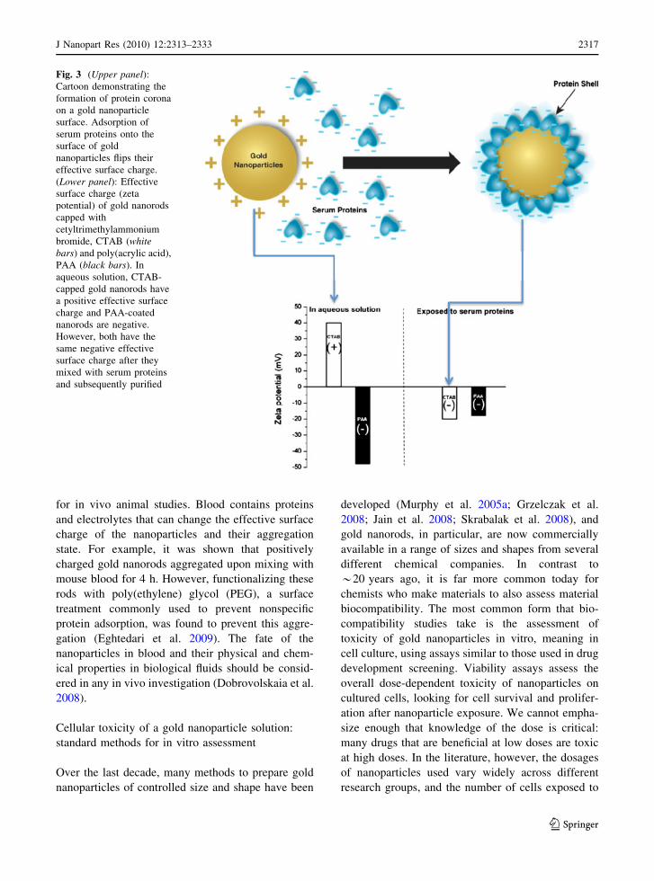
vary, making it difficult to compare. Recent results of
gold nanoparticle toxicity to cells in vitro are
summarized in Table 1.
In vitro three-dimensional (3D) cell culture models
have been used as a bridge between the in vitro two-
dimensional (2D) plated cell culture and the in vivo
models (Griffith and Swartz 2006; Yamada and
Cukierman 2007). In this context, Lee et al. compared
the toxicity of gold nanoparticles in both 2D and 3D
cell culture constructs. They used hydrogel inverted
colloidal crystals as a cell growth substrate and
human hepatocarcinoma cells to construct the 3D cell
culture environment. They found that toxicity of both
citrate (anionic)- and CTAB (cationic) capped gold
nanoparticles were significantly reduced in the 3D
environment compared to 2D (Lee et al. 2009). These
results point out that in vitro studies alone are not
adequate to assess toxicity of nanoparticles.
In vitro cellular uptake
As discussed in the previous sections, there are
various methods to visualize and measure gold
nanoparticle concentration inside cells. Since gold
nanoparticles are electron-dense, it is easy to distin-
guish them from other cellular components using
TEM. Other techniques that could be used for
imaging nanoparticle location are dark field optical
microscopy, fluorescence microscopy, and differen-
tial interference contrast microcopy (Marquis et al.
2009). To quantify the number of nanoparticles per
cell, ICP-MS is an excellent technique to analyze
gold content inside the cells or the remaining portion
in the growth media (Marquis et al. 2009).
Understanding the mechanism of gold nanoparti-
cle uptake by cells is important for intracellular drug
and gene delivery (Rosi et al. 2006; Han et al. 2007).
To internalize macromolecules and particles, cells
utilize phagocytosis, micropinocytosis, and receptor-
mediated endocytosis (RME) pathways including
caveolae-mediated, clathrin-mediated, and caveolae/
clathrin independent endocytosis (Conner and
Schmid 2003). These pathways operate using dif-
ferent receptors, cellular signaling cascades, and
type of particles (Dobrovolskaia and McNeil 2007).
For example, phagocytosis operates for particles
[500 nm, where smaller particles enter via the RME
pathways (Dobrovolskaia and McNeil 2007; Hess
and Tseng 2007).
J Nanopart Res (2010) 12:2313–2333 2321
123
Ta
ble
1S
um
mar
yo
fin
vit
rog
old
nan
op
arti
cle
tox
icit
yre
sult
s
Cel
lli
ne
Nan
op
arti
cle
dim
ensi
on
s(n
m)
Nan
op
arti
cle
shap
e
Nan
op
arti
cle
surf
ace
gro
up
Do
sea;
incu
bat
ion
tim
eC
on
clu
sio
ns
Ref
.
CO
S-1
mam
mal
ian
cell
s,re
d
blo
od
cell
s,E
.co
li2
Sp
her
esQ
uat
ern
ary
amm
on
ium
,
carb
ox
yli
cac
id
0.3
8–
3l
M;
1–
24
hC
atio
nic
nan
op
arti
cles
fou
nd
tob
eto
xic
wh
ere
anio
nic
no
t
Go
od
man
etal
.
20
04
RA
W2
64
.7m
ou
sem
acro
ph
age
3.5
±0
.7S
ph
eres
Ly
sin
e,p
oly
(ly
sin
e)1
0–
10
0lM
;2
4–
72
hN
ano
par
ticl
esar
en
ot
tox
ic
and
no
tim
mu
no
gen
ic
Sh
uk
laet
al.
(20
05
)
K5
62
hu
man
leu
kem
ia4
,1
2,
18
Sp
her
esC
TA
B,
citr
ate,
cyst
ein
e,
glu
cose
,b
ioti
n
0.0
01
–0
.25
lM;
72
hA
lln
ano
par
ticl
esw
ere
no
t
tox
ic
Co
nn
or
etal
.
(20
05
)
MV
3an
dB
LM
(Met
asta
tic
mel
ano
ma)
1.4
Sp
her
ical
clu
ster
Tri
ph
eny
lph
osp
hin
e
mo
no
sulf
on
ate
Up
to0
.4lM
;7
2h
10
0%
cell
dea
that
0.4
lMco
mp
ared
to1
0%
cell
dea
thfo
rci
spla
tin
atsa
me
con
cen
trat
ion
Tso
liet
al.
(20
05
)
HeL
a6
59
11
Ro
ds
CT
AB
,P
EG
0.0
9–
1.4
5lM
;2
4h
Rep
laci
ng
CT
AB
wit
hP
EG
on
the
surf
ace
of
nan
oro
ds
red
uce
dth
eto
xic
ity
Tak
ahas
hi
etal
.(2
00
6)
Hu
man
der
mal
fib
rob
last
13
.1S
ph
eres
Cit
rate
0–
4m
M;
24
–1
44
hN
ano
par
ticl
esd
ecre
ased
cell
pro
life
rati
on
rate
,
adh
esio
n,
and
mo
tili
ty
Per
no
det
etal
.
(20
06
)
(1)
bab
yh
amst
erk
idn
eyce
lls
BH
K2
1
33
Sp
her
esC
TA
Ban
dci
trat
e0
–1
20
nM
;3
6h
for
A5
49
and
72
hfo
rb
oth
Hep
2G
and
BH
K2
1
Nan
op
arti
cles
are
no
tto
xic
toH
ep2
Gan
dB
HK
21
bu
t
toA
54
9ce
llli
ne
Pat
raet
al.
(20
07
)
(2)
Hu
man
liv
erca
rcin
om
a
Hep
2G
(3)
Hu
man
lun
gca
rcin
om
a
cell
sA
54
9
HeL
a1
8S
ph
eres
Cit
rate
0.2
–2
nM
;3
–6
hN
ano
par
ticl
esar
en
ot
tox
ic
and
did
no
tch
ang
eg
ene-
exp
ress
ion
pat
tern
s
Kh
anet
al.
(20
07
)
(1)
Ep
ith
elia
l:H
eLa
0.8
,1
.2,
1.4
,
1.8
,1
5
Sp
her
esT
rip
hen
ylp
ho
sph
ine
mo
no
and
tri-
sulf
on
ate
Up
to5
.6lM
;7
2h
(a)
1.4
nm
:M
ost
tox
icsi
ze;
(b)
0.8
,1
.2,
1.8
:4–
6fo
ld
tox
icit
yco
mp
ared
to
1.4
nm
;(c
)1
5n
m:
com
ple
tely
no
nto
xic
;
(d)
tox
icit
yis
no
tce
ll
lin
ed
epen
den
t
Pan
etal
.
(20
07
)(2
)E
nd
oth
elia
l:S
K-M
el-2
8
(3)
Fib
rob
last
s:L
92
9
(4)
Ph
ago
cyte
s:j7
74
A1
2322 J Nanopart Res (2010) 12:2313–2333
123

Ta
ble
1co
nti
nu
ed
Cel
lli
ne
Nan
op
arti
cle
dim
ensi
on
s(n
m)
Nan
op
arti
cle
shap
e
Nan
op
arti
cle
surf
ace
gro
up
Do
sea;
incu
bat
ion
tim
eC
on
clu
sio
ns
Ref
.
HeL
a4
09
18
Ro
ds
CT
AB
,P
SS
,P
DA
DM
AC
10
–1
50
lM;
6h
Po
lyel
ectr
oly
teco
atin
go
f
nan
oro
ds
are
no
tto
xic
com
par
edto
the
CT
AB
-
cap
ped
nan
oro
ds
and
no
gen
eex
pre
ssio
n
abn
orm
alit
ies
wer
e
ob
serv
ed
Hau
cket
al.
(20
08
)
Den
dri
tic
cell
sfr
om
C5
7B
L/6
mic
e
10
Sp
her
esC
itra
te0
.5m
M;
4–
48
hN
ano
par
ticl
esw
ere
no
t
tox
ican
dd
idn
ot
ind
uce
den
dri
tic
cell
acti
vat
ion
Vil
lier
set
al.
(20
09
)
HeL
a1
.4an
d1
.5S
ph
eres
Tri
ph
eny
lph
osp
hin
e
mo
no
sulf
on
ate,
GS
H
5.6
mM
;4
8h
(a)
Th
e1
.4n
ano
par
ticl
es
ind
uce
dn
ecro
sis
by
ox
idat
ive
stre
sses
wh
ere
the
15
nm
par
ticl
esw
ere
fou
nd
tob
en
ot
tox
ic;
(b)
GS
H-c
app
ed
nan
op
arti
cles
wer
ele
ss
tox
icth
anT
PM
S-c
app
ed
nan
op
arti
cles
Pan
etal
.
(20
09
)
HeL
a3
.7S
ph
eres
PE
G0
.08
–1
00
lM;
6–
72
hN
ano
par
ticl
esen
tere
d
nu
cleu
san
dd
idn
ot
ind
uce
tox
icit
y
Gu
etal
.
(20
09
)
HT
-29
(Hu
man
colo
n
carc
ino
ma
cell
s)
65
91
5n
mR
od
sC
TA
B,
PA
A,
PA
H0
.6n
M;
96
hN
ano
rod
sar
en
ot
tox
ic,
exce
ssC
TA
Bis
.
Ov
erco
atin
gth
eC
TA
B-
cap
ped
rod
sw
ith
eith
er
neg
ativ
ely
or
po
siti
vel
y
char
ged
po
lym
ers
red
uce
s
tox
icit
yan
daf
fect
sth
eir
up
tak
e
Alk
ilan
yet
al.
(20
09
)
CT
AB
cety
ltr
imet
hy
lam
mo
niu
mb
rom
ide,
cati
on
icsu
rfac
tan
t;P
EG
po
ly(e
thy
len
eg
lyco
l);
PS
Sp
oly
(so
diu
m4
-sty
ren
esu
lfo
nat
e),
anio
nic
po
lyel
ectr
oly
te;
PD
AD
MA
Cp
oly
(dia
lly
ldim
eth
yla
mm
on
ium
chlo
rid
e),
cati
on
icp
oly
elec
tro
lyte
;P
AA
po
ly(a
cry
lic
acid
,so
diu
msa
lt),
anio
nic
po
lyel
ectr
oly
te;
PA
Hp
oly
(all
yla
min
eh
yd
roch
lori
de)
,ca
tio
nic
po
lyel
ectr
oly
te;
GS
Hg
luta
thio
ne
aD
ose
sar
eca
lcu
late
dfr
om
ori
gin
alp
aper
sin
go
ldat
om
con
cen
trat
ion
s
J Nanopart Res (2010) 12:2313–2333 2323
123

For gold nanoparticles, most of the studied nano-
particles have dimensions less than 100 nm and RME
has been proposed as the primary mechanism of
cellular entry (Shukla et al. 2005; Chithrani and Chan
2007; Nativo et al. 2008). Chithrani et al. studied the
mechanism by which transferrin-coated gold nano-
rods and nanospheres were taken up by three types of
cultured cell lines: STO, HeLa, and SNB19 which are
fibroblast, ovarian cancer, and brain tumor cells,
respectively. Transferrin is a plasma protein for iron
transportation, which enters cells via a RME mech-
anism. They found a 70% decrease in nanoparticle
cellular uptake at 4 �C compared to 37 �C, a standard
experiment that supports the use of the RME pathway
by the nanoparticles. Drastic decreases in nanoparti-
cle cellular uptake were observed when either
hypertonic environments (by adding sucrose) or K?
depleted media were used, which indicates clatherin-
mediated endocytosis as the specific mechanism of
uptake (Chithrani and Chan 2007).
The size of nanoparticles was found to play a
critical role in both the rate and extent of cellular
uptake. It was found that 50 nm transferrin-coated
gold nanoparticles were taken up by mammalian cells
at higher rates and extents compared to smaller and
larger sizes in the range of 10–100 nm (Chithrani
et al. 2006). The explanation of this optimal size was
based on the so-called ‘‘wrapping effect’’, which
describes how a cellular membrane encloses nano-
particles. Two factors dictate how fast and how many
nanoparticles enter the cellular compartment via
‘‘wrapping’’: the first is the free energy that results
from ligand–receptor interaction; the second is the
receptor diffusion kinetics onto the wrapping sites on
the cellular membrane. Considering the contribution
of these factors and using mathematical calculations,
Gao et al. (2005) suggested that nanoparticles with
27–30 nm diameter would have that fastest wrapping
time and thus the fastest receptor-mediated
endocytosis.
Even though ligand-mediated uptake of gold
nanoparticles is considered to be a general mecha-
nism for their cellular entry, gold nanoparticles with
‘‘special’’ surface chemistries/arrangements can enter
cells by direct penetration. Verma et al. (2008)
showed that gold nanospheres (*5 nm) decorated
with two capping molecules (anionic and hydropho-
bic, with alternating positions on the surface) enter
the cells directly (endocytosis-independent entry)
without destruction to the cell membrane in an action
similar to the cell-penetrating peptides.
Intracellular distribution of gold nanoparticles has
been studied, with the general conclusion that gold
nanoparticles are able to enter cells and are trapped in
vesicles, but are not able to enter the nucleus (Shukla
et al. 2005; Pernodet et al. 2006; Chithrani and Chan
2007; Khan et al. 2007; Alkilany et al. 2009). Using
TEM, Nativo et al. showed that 16 nm citrate-capped
gold nanoparticles enter cells readily (incubation time
2 h) and are trapped into endosomes. They did not
find free nanoparticles in the cytosol or the nucleus.
However, they were able to deliver the nanoparticles
to the cytosol and nucleus by modifying these
nanoparticles with cell-penetrating and nuclear-local-
izing peptides (Nativo et al. 2008).
However, other reports indicate nuclear penetra-
tion for gold nanoparticles without special surface
functionalization. For example, gold nanoparticles
with diameters of 1.4 nm were able to enter the
nucleus in metastatic melanoma cells and bind DNA
with high efficiency (24.5% of the total internalized
gold nanoparticles bound to DNA) (Tsoli et al. 2005).
In another study using citrate-capped gold nano-
spheres (5 nm in diameter), 25% of the internalized
gold nanoparticles were able to enter the nucleus in
HeLa cells without any surface functionalization.
This fraction was doubled when the nanoparticles
were functionalized with a nuclear-penetrating pep-
tide (Ryan et al. 2007).
The general conclusions that can be drawn from
studies are still preliminary. Different investigators
use different cell lines, different sizes of nanoparti-
cles, different surface groups, different doses, differ-
ent time points, and may or may not have quantitative
information (as opposed to qualitative visualization)
about nanoparticle uptake into cells. Table 2 sum-
marizes the quantitative results of gold nanoparticle
uptake by cultured cells, calculated as the number of
nanoparticles per cell.
In vivo studies: biodistribution and toxicity
of gold nanoparticles in organisms
There is a real need to investigate the in vivo results
exposure to nanomaterials before any potential ther-
apeutic applications (Fischer and Chan 2007). In this
context, Chen et al. studied the toxicity of wide size
range of citrate-capped gold nanoparticles (spheres of
2324 J Nanopart Res (2010) 12:2313–2333
123

diameter: 3, 5, 8, 12, 17, 37, 50, 100 nm) in mice.
They found that the smallest sizes (3 and 5 nm) and
the largest size (50 and 100 nm) are not toxic at the
dose they were using (Table 3). However, they found
that the intermediate size range of 8–37 nm had lethal
effects on mice inducing severe sickness, loss of
appetite, weight loss, change in fur color, and shorter
average lifespan (Chen et al. 2009). The systematic
toxicity of the intermediate size range (18–37 nm)
was linked to major organ damage in the liver,
spleen, and lungs (Chen et al. 2009). Interestingly, in
the same study, the same ‘‘lethal’’ nanoparticles were
not toxic in vitro using HeLa cell lines (Fig. 5) (Chen
et al. 2009). This study demonstrated a large
discrepancy between the in vitro and in vivo results,
and highlights the notion that simple in vitro exper-
iments may not lead to good predictions regarding in
vivo results.
The mechanism of in vivo nanoparticle toxicity
could arise from many sources. For example, injecting
gold nanoparticles in the blood could cause either
blood clotting or hemolysis (blood cells break open
and release their hemoglobin) (Dobrovolskaia
et al. 2008). Encouragingly, citrate-capped gold
nanoparticles (spheres of diameter 30 and 50 nm)
have been shown to be ‘‘blood compatible’’ and did
not induce any detectable platelet aggregation, change
in plasma coagulation time, or immune response in at
least one study (Dobrovolskaia et al. 2009).
Because the size range of nanoparticles matches
that of proteins or even small viruses, one might
expect that the immune system might react strongly
to the presence of nanoparticles in the body resulting
in induced immunotoxicity (Dobrovolskaia and
McNeil 2007). Even though antigen-bound gold
nanoparticles were used as vaccine carriers to aug-
ment immune responses toward antigens (Bastus
et al. 2009), little is known about their intrinsic in
vivo antigenicity and inflammatory properties.
Accumulation of nanomaterials in the liver and
spleen after being taken up by the reticuloendothelial
system (part of the immune system with complex
components communicate to identify, capture, and
filter foreign antigens and particulates) could lead to
hepatic and splenic toxicity (Chen et al. 2009). Cho
et al. (2009b) studied the toxicity of 13 nm PEG-
modified gold nanoparticles in mice and found that
the nanoparticles accumulate in the liver after
Table 2 Summary of in vitro gold nanoparticle uptake results
Cell line Nanoparticle
dimensions (nm)
Nanoparticle
shape
Nanoparticle
surface group
Dosea;
incubation
time
Cellular uptake
(gold nanoparticles/cell)
Analytical
method
Ref.
HeLa 40 9 18
(length 9 width)
Rods CTAB, PAH,
PSS,
PDADMAC
1.0 nM;
6 h
150,000 for PDAMAC;
12,000 for PAH; 12,000
for CTAB; 1,000 for PSS
ICP-AES Hauck et al.
(2008)
HT-29 65 9 15
(length 9 width)
Rods CTAB, PAA,
PAH
0.2 nM;
96H
45 ± 6 for CTAB;
270 ± 20 for PAA;
2,320 ± 140 for PAH
ICP-MS Alkilany
et al.
(2009)
SK-BR-3 17.7 Spheres Citrate, PAH,
PVA
0.027 nM;
24 h
1,800 for citrate; 5,200 for
PAH; 900 for PVA
ICP-MS Cho et al.
(2009a,
2009b)
SK-BR-3 50 9 20
(length 9 width)
Rods CTAB, PEG,
anti-HER2
0.06 nM;
24 h
8,000 for CTAB; 3,000 for
PEG; 4,400 for anti-HER2
ICP-MS Cho et al.
(2010)
U87MG 50 9 5 nm (edge
length 9 wall
thickness)
Cages Anti-EGFR,
PEG
0.02 nM;
24 h
826 ± 50 for anti-EGFR
and 190 ± 31 for PEG
ICP-MS Au et al.
(2010)
CTAB Cetyl trimethylammonium bromide, cationic surfactant; PDADMAC poly(diallyldimethylammonium chloride), cationic
polyelectrolyte; PAH poly(allylamine hydrochloride), cationic polyelectrolyte; PAA poly(acrylic acid, sodium salt), anionic
polyelectrolyte; PSS poly(sodium 4-styrenesulfonate), anionic polyelectrolyte; PVA poly(vinyl alcohol) slightly anionic polymer;
PEG poly(ethylene glycol), neutral polymer; Anti-HER2 monoclonal antibodies that recognize human epidermal growth factor 2
(HER2) receptors, anti-EGFR monoclonal antibodies that recognize epidermal growth factor (EGER) receptors, ICP-AESinductively-coupled plasma atomic emission spectroscopy, ICP-MS inductively-coupled plasma mass spectrometrya Doses and cellular uptake values are calculated from the original papers in gold nanoparticle (not atoms) concentration
J Nanopart Res (2010) 12:2313–2333 2325
123
Ta
ble
3S
um
mar
yo
fin
viv
og
old
nan
op
arti
cle
tox
icit
y/p
har
mac
ok
inet
icre
sult
s
An
imal
Nan
op
arti
cle
dim
ensi
on
s(n
m);
shap
e
Nan
op
arti
cle
surf
ace
gro
up
Ad
min
istr
atio
n
rou
te;
Do
sea
Tim
eo
f
exp
osu
re
(h)
Nu
mb
ero
f
stu
die
dan
imal
s
(n)
Co
ncl
usi
on
sR
ef
Mic
e(d
dy
)6
59
11
;ro
ds
Po
lyet
hy
len
e
gly
col,
CT
AB
Intr
aven
ou
s:
0.0
3–
0.0
54
mg
go
ld/m
ou
se
0.5
–7
23
PE
Gm
od
ifica
tio
no
fg
old
nan
oro
ds
incr
ease
the
blo
od
circ
ula
tio
nti
me:
afte
r0
.5m
ino
f
inje
ctio
n,
mo
sto
fth
eC
TA
B-
cap
ped
nan
oro
ds
accu
mu
late
din
the
liv
erw
her
e5
4%
of
PE
G-
cap
ped
nan
oro
ds
fou
nd
inth
e
blo
od
Nii
do
me
etal
.
(20
06
)
Pig
s1
5–
20
,sp
her
esA
rab
icg
um
Intr
aven
ou
s:
0.8
–1
.88
mg
go
ld/k
g
0.5
–2
43
Nan
op
arti
cles
accu
mu
late
din
lun
g
and
liv
er;
no
hem
ato
log
ical
or
ren
alsi
de
effe
cts
wer
eo
bse
rved
Kat
tum
uri
etal
.
(20
07
)
Mic
e(d
dy
)1
5,
50
,1
00
,
20
0;
sph
eres
Cit
rate
Intr
aven
ou
s:
10
00
mg
go
ld/k
g
24
3A
llsi
zes
wer
efo
un
din
liv
er,
sple
en,
lun
g.
15
and
50
nm
nan
op
arti
cles
wer
efo
un
dal
soin
hea
rt,
sto
mac
h,
kid
ney
,an
dth
e
bra
in
So
nav
ane
etal
.
(20
08
)
Rat
s1
0,
50
,1
00
,
25
0;
sph
eres
No
tre
po
rted
Intr
aven
ou
s:
77
–1
08
lg/r
at
24
4N
osi
de
effe
ctw
aso
bse
rved
.M
ost
nan
op
arti
cles
wer
efo
un
din
sple
enan
dli
ver
;th
e1
0n
m
par
ticl
esw
ere
fou
nd
also
in
bra
in,
hea
rt,
kid
ney
,te
stis
,an
d
thy
mu
s
De
Jon
get
al.
(20
08
)
Mic
e(B
AL
B/c
)4
,1
0,
28
,5
8;
sph
eres
Cit
rate
Ora
l:M
ixed
wit
h
dri
nk
ing
wat
er
(20
0m
gg
old
/kg
wat
er)
16
8N
ot
rep
ort
edG
astr
oin
test
inal
up
tak
eb
y
per
sorp
tio
n,
mo
rere
adil
yfo
r
smal
ler
par
ticl
es
Hil
lyer
and
Alb
rech
t
(20
01
)
Mic
e(B
AL
B/c
)3
,5
,8
,1
2,
17
37
,5
0,
10
0;
sph
eres
Cit
rate
Intr
aper
ito
nic
al:
8m
gg
old
/kg
[1
20
06
3,
5,
50
,1
00
nm
par
ticl
esd
idn
ot
ind
uce
any
leth
alit
yw
ere
par
ticl
esw
ith
dia
met
ero
nth
e
ran
ge
of
8.3
7n
md
id
Ch
enet
al.
(20
09
)
Mic
e(B
AL
B/c
)1
3;
sph
eres
Po
lyet
hy
ene
gly
col
Intr
aven
ou
su
pto
4.2
6m
gg
old
/kg
up
to1
68
9N
ano
par
ticl
esin
du
ced
infl
amm
atio
nan
dap
op
tosi
sin
the
liv
erti
ssu
e
Ch
oet
al.
(20
09
a,2
00
9b)
Zeb
rafi
sh3
,1
0,
50
,1
00
;
sph
eres
Cit
rate
Ex
po
sure
inw
ater
of
con
cen
tra
tio
ns
of:
25
0,
25
,2
.5,
0.2
5lM
12
01
2N
ano
par
ticl
esw
ere
tak
enu
pb
y
zeb
rafi
shan
dd
idn
ot
ind
uce
any
tox
icit
y
Bar
-Ila
net
al.
(20
09
)
2326 J Nanopart Res (2010) 12:2313–2333
123

injection, and induce acute inflammation and cellular
damage in the mouse liver.
The physical and chemical properties of nanopar-
ticles can affect their pharmacokinetics such as
absorption, metabolism, distribution, and elimination.
For example, Hillyer and Albrecht (2001) showed that
the absorption of gold nanoparticles following oral
administration to mice is size-dependent. Smaller
nanoparticles were found to cross the gastrointestinal
wall more readily after oral intake. Other studies
investigated the bio-distribution of gold colloid after
intravenous injection in rats. De Jong et al. injected rats
with 10, 50, 100, 250 nm gold nanoparticles and after
24 h rats were killed and gold concentration in
different organs were quantified by ICP-MS. Their
data showed that the smallest size (10 nm) nanopar-
ticles were found in the blood, spleen, liver, testis, lung,
and brain; the larger sizes were found only in spleen
and kidney (De Jong et al. 2008). In a very similar
study, Sonavane et al. (2008) showed that 15 nm is the
most widely distributed size in vivo among a nano-
particle library with diameters from 15 to 200 nm, and
that 15 and 50 nm nanoparticles were able to enter the
brain. These findings highlight the size-dependent
biodistribution of gold nanoparticles in vivo.
According to FDA guidelines, pharmaceutical
drugs should be eliminated via metabolism or excre-
tion processes after they enter the body. Drug
elimination reduces toxicity and prevents drug accu-
mulation. Similar to pharmaceutical drugs, nanopar-
ticles should be designed to be eliminated in the
body. Indeed, nanoparticle elimination should be
considered seriously, since they are more resistant to
elimination routes such as metabolism and renal
excretion. No long-term studies on gold nanoparticles
have been reported to our knowledge. As one related
example, injected semiconductor quantum dots in
mice remained intact for more than 2 years in mouse
tissues, retaining their fluorescence activity (Ballou
et al. 2007). This resistance might be because of their
large size (too large to be filtered from the kidney)
and their higher chemical stability (against dissolu-
tion and degradation) compared to molecules. It is
thought that nanoparticles should have final hydro-
dynamic diameters B5.5 nm to be excreted from the
rat body by the renal route (Choi et al. 2007). Since
the majority of the studied gold nanoparticles are
larger than this renal filtration cutoff, in the few
studies that have been performed, the goldTa
ble
3co
nti
nu
ed
An
imal
Nan
op
arti
cle
dim
ensi
on
s(n
m);
shap
e
Nan
op
arti
cle
surf
ace
gro
up
Ad
min
istr
atio
n
rou
te;
Do
sea
Tim
eo
f
exp
osu
re
(h)
Nu
mb
ero
f
stu
die
dan
imal
s
(n)
Co
ncl
usi
on
sR
ef
Mic
e(B
AL
B/c
)2
0,
40
,8
0;
sph
eres
Po
lyet
hy
len
e
gly
col
Intr
aven
ou
s:3
4–
22
10
mg
go
ld/k
g(s
ame
nu
mb
er
of
nan
op
arti
cles
per
mo
use
)
48
8B
iod
istr
ibu
tio
nan
dca
nce
r
accu
mu
lati
on
of
nan
op
arti
cles
issi
zed
epen
den
t;sm
alle
r
nan
op
arti
cles
hav
elo
ng
erb
loo
d
circ
ula
tio
nti
me
and
mo
retu
mo
r
accu
mu
lati
on
Zh
ang
etal
.
(20
09
)
En
tire
estu
arin
em
eso
cosm
s
con
tain
ng
:F
ish
,sh
rim
p,
snai
l,cl
ams,
and
mic
rob
ial
bio
film
65
91
5;
rod
sC
TA
BE
xp
osu
rein
the
estu
arin
e
mes
oco
sms;
init
ial
con
cen
trat
ion
of
11
6lM
28
81
5fi
sh,
10
0
shri
mp
s,5
0
snai
ls,
and
10
0cl
ams
Go
ldn
ano
rod
sp
arti
tio
nef
fici
entl
y
into
the
foo
dw
eban
dd
idn
ot
ind
uce
any
mo
rtal
ity
;cl
ams
and
mic
rob
ial
bio
film
sac
cum
ula
te
the
mo
stg
old
Fer
ryet
al.
(20
09
)
CT
AB
cety
ltr
imet
hy
lam
mo
niu
mb
rom
ide,
cati
on
icsu
rfac
tan
ta
Do
ses
are
calc
ula
ted
fro
mth
eo
rig
inal
pap
ers
ing
old
ato
mco
nce
ntr
atio
ns
J Nanopart Res (2010) 12:2313–2333 2327
123

nanoparticles were not excreted in urine; instead they
were found to be eliminated from the blood by the
reticuloendothelial system (RES) and thus to accu-
mulate in the spleen and liver (De Jong et al. 2008;
von Maltzahn et al. 2009).
Little effort has been made to match the properties
of a nanoparticle with the size that might be
acceptable for elimination from an organism. In the
case of gold, anisotropic rod-shaped nanoparticles are
desired to absorb in the near-infrared region, prefer-
ably with small dimensions to be excreted from the
body (say nanorods with dimension of 4 nm in length
and 1 nm in width, aspect ratio 4). Preparing
nanorods with these dimensions is very difficult,
and is not available at present; and gold particles less
than 4 nm in one dimension would be sufficiently
small to become chemically reactive. In a rare effort
to synthesize a gold nanoparticle that can absorb light
in the NIR region of the spectra and be eliminated
from the body, Troutman et al. (2008) prepared gold-
coated liposomes. The idea is that the gold nanopar-
ticles would serve as a shell to provide the plasmonic
properties, and the liposome would serve as a carrier
(Fig. 6). Upon disintegration of these plasmon-reso-
nant liposomes by physiological stimuli such as
phospholipase A2 (which degrade the liposome’s
lipid), the composite dissolves and the nanoparticles
are suspended freely, with an average diameter of
5.7 nm (Troutman et al. 2008). However, the elim-
ination of these nanoparticle–liposome composites
has not been investigated yet. The degree of metab-
olism and degradability of a nanomaterial is very
important to prevent bioaccumulation and facilitate
elimination. However, very little known about this
issue in the literature and more research should be
performed in this direction.
While most of the in vivo studies have been
performed using land animal models (mice, rats, and
pigs), Bar-Ilan et al. (2009) used zebrafish embryo
screening methods to assess the toxicity of both gold
and silver nanoparticles of different sizes (3, 10, 50,
and 100 nm). Zebrafish is an excellent in vivo model
which has been used to assess environmental toxicity
due its high degree of homology to the human
genome and its very similar physiological responses
to xeno-substances as mammals (Parng 2005; Fako
Fig. 5 Left: average lifespan of mice receiving gold nanopar-
ticles, 8–37 nm in diameter, was shortened compared to
smaller and larger nanoparticle sizes. The break marks on the
top of bars indicate that no death was observed during the
experimental period. Right: MTT assay for the same gold
nanoparticles using the HeLa cell line. Images reproduced with
permission from (Chen et al. 2009). Copyright: Springer
Science
Fig. 6 Cartoon demonstrates the concept of the biodegradable
plasmon-resonant liposomes. The whole composite absorbs in
the near-infrared region and thus serve as ‘‘nanoheaters’’ to
destroy cancer cells. Upon disruption of the carrier (lipo-
somes), the nanoparticles could be released and have a higher
chance to be bio-eliminated
2328 J Nanopart Res (2010) 12:2313–2333
123

and Furgeson 2009). Interestingly, they found that
gold nanoparticles were not toxic to zebrafish but the
silver nanoparticles with comparable size were highly
toxic (inducing 100% death after 120 h post-fertil-
ization) (Bar-Ilan et al. 2009).
Even as knowledge advances to the point that
nanoparticles can be properly manufactured for a
specific goal in an organism (e.g. detection or
treatment for a disease), the entire life cycle of the
nanoparticle needs to be considered. It is well-known,
for example, that certain toxins can bio-accumulate in
organisms, and thus enter the food chain. How would
nanoparticles move through a food web, from organ-
ism to organism? In this context, Ferry et al. studied
the fate of CTAB-coated gold nanorods (65 nm
length 9 15 nm width) in replicate estuarine meso-
cosms consisting of seawater, sediment, microbial
biofilms, snails, fish, clams, and shrimps to model the
complexity of a tidal marsh creek. They found that
nanoparticles partitioned into most of the organisms
(none of which died at the dosage used, which was
designed to mimic a viral load in the ecosystem) to
very different extents, with a low concentration
remaining in water (Ferry et al. 2009). The largest
accumulations of nanoparticles by far were the
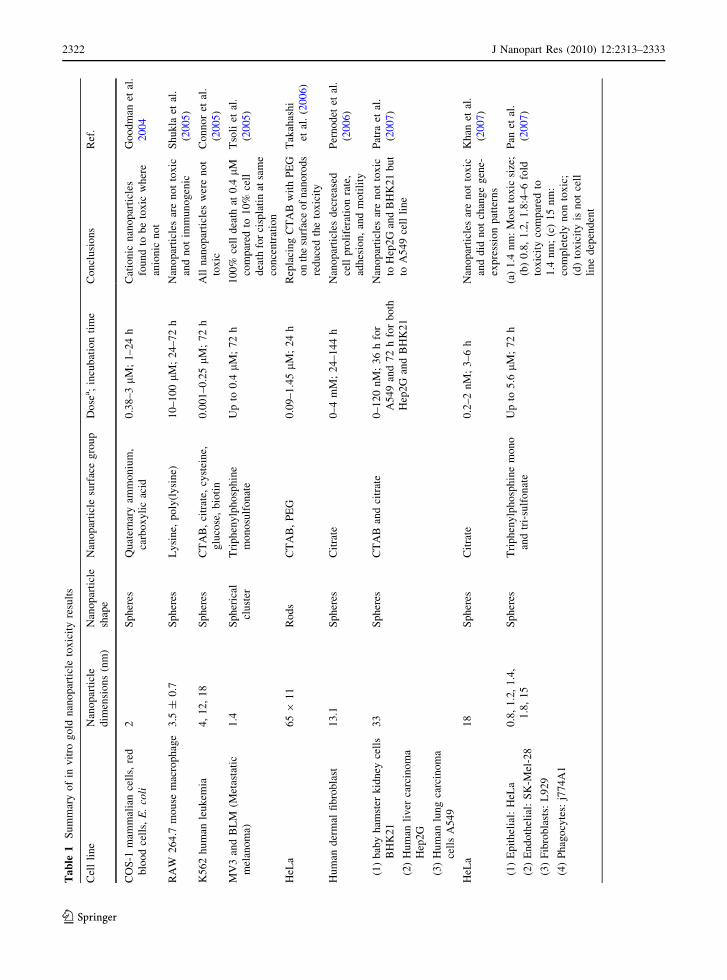
microbial biofilms and clams (filter feeders). The
results of recent gold nanoparticle animal studies in
vivo are summarized in Table 3.
Conclusion and perspective
The available literature reports, both in vitro and in
vivo, vary widely in their methods and conclusions
(Ostrowski et al. 2009). Many reports indicate that
gold nanoparticles are nontoxic; however, others
contradict this finding. To draw a complete conclu-
sion, more studies are needed which:
• Include critical nanoparticle characterization both
prior to and after mixing with the biological
media, with a focus on the change of the physical
properties such as aggregation state, effective
surface charge, degree and identity of protein
adsorption, and desorption of chemicals from the
surface of the nanoparticles.
• Include careful control experiments such as the
discussed ‘‘supernatant control’’ experiment in
Fig. 4 (Alkilany et al. 2009; Bar-Ilan et al. 2009)
• While many studies focus on determining the
lethal dosage of nanoparticles (LD50, dose
required to kill half of the population), little if
any focus on determination of the effective
therapeutic dosage of these nanoparticles (ED50,
dose required to produce therapeutic response in
50% of the population). Determining the ED50
experimentally will help nanotoxicologists to use
more realistic dosing to assess the toxicity of
nanoparticles.
• Most the studies where conducted on simple gold
nanoparticles (citrate or CTAB capped). Efforts
are needed to study the toxicity and pharmacoki-
netics of functionalized gold nanoparticles with
real surface composition (e.g. recognition and
non-fouling molecules) since this surface modifi-
cation can significantly alter the whole story.
• The major administration route in the reported in
vivo studies is intravenous injection. More inves-
tigation is needed to study the toxicity of gold
nanoparticles using different route of exposure
such as inhalation, oral absorption, and dermal
absorption of gold nanoparticles.
Hope for the future
Even though the collective results in the literature
show controversy about the toxicity of gold nano-
particles, we think that the uptake and toxicity of
these nanomaterials are controllable and can be
manipulated. As discussed earlier in this perspective,
researchers have demonstrated the ability to ‘‘detox-
ify’’ and regulate the uptake of these nanoparticles by
functionalizing the surface of the nanoparticles with
smart/benign ligands. In an encouraging recent
example, one single intravenous injection of PEG-
functionalized gold nanorods which showed a long
circulating time (t1/2 17 h) was able to destroy human
xenogaft tumors in mice (von Maltzahn et al. 2009).
In this study, the total dose of nanorods required for
complete photothermal destruction of the tumor was
20 mg/kg (in gold atoms) and did not induce any
toxicity in tumor-free mice (von Maltzahn et al.
2009). As more data are collected, we are optimistic
that proper attention will be paid to surface chemistry
and dosages, so that the full potential of gold
nanoparticles for biomedical applications can be
exploited.
J Nanopart Res (2010) 12:2313–2333 2329
123

References
Aillon KL, Xie YM, El-Gendy N, Berkland CJ, Forrest ML
(2009) Effects of nanomaterial physicochemical proper-
ties on in vivo toxicity. Adv Drug Deliv Rev 61:457–466.
doi:10.1016/j.addr.2009.03.010
Alivisatos AP (1996) Semiconductor clusters, nanocrystals,
and quantum dots. Science 271:933–937
Alkilany AM, Murphy CJ (2009) Gold nanoparticles with a
polymerizable surfactant bilayer: synthesis, polymeriza-
tion, and stability evaluation. Langmuir 25:13874–13879.
doi:10.1021/la901270x
Alkilany AM, Frey RL, Ferry JL, Murphy CJ (2008) Gold
nanorods as nanoadmicelles: 1-naphthol partitioning into
a nanorod-bound surfactant bilayer. Langmuir 24:10235–
10239. doi:10.1021/la8018343
Alkilany AM, Nagaria PK, Hexel CR, Shaw TJ, Murphy CJ,
Wyatt MD (2009) Cellular uptake and cytotoxicity of gold
nanorods: molecular origin of cytotoxicity and surface
effects. Small 5:701–708. doi:10.1002/smll.200801546
Anker JN, Hall WP, Lyandres O, Shah NC, Zhao J, Van Duyne
RP (2008) Biosensing with plasmonic nanosensors. Nat
Mater 7:442–453. doi:10.1038/nmat2162
AshaRani PV, Mun GLK, Hande MP, Valiyaveettil S (2009)
Cytotoxicity and genotoxicity of silver nanoparticles in
human cells. ACS Nano 3:279–290. doi:10.1021/nn8005
96wER
Au L, Zhang Q, Cobley CM et al (2010) Quantifying the
cellular uptake of antibody-conjugated Au nanocages by
two-photon microscopy and inductively coupled plasma
mass spectrometry. ACS Nano 4:35–42. doi:10.1021/
nn901392mER
Ballou B, Ernst LA, Andreko S et al (2007) Sentinel lymph
node imaging using quantum dots in mouse tumor models.
Bioconjug Chem 18:389–396. doi:10.1021/bc060261j
Bar-Ilan O, Albrecht RM, Fako VE, Furgeson DY (2009)
Toxicity assessments of multisized gold and silver nano-
particles in zebrafish embryos. Small 5:1897–1910. doi:
10.1002/smll.200801716
Bastus NG, Sanchez-Tillo E, Pujals S et al (2009) Peptides
conjugated to gold nanoparticles induce macrophage
activation. Mol Immunol 46:743–748. doi:10.1016/
j.molimm.2008.08.277
Cedervall T, Lynch I, Foy M et al (2007) Detailed identifica-
tion of plasma proteins adsorbed on copolymer nanopar-
ticles. Angew Chem Int Ed 46:5754–5756. doi:10.1002/
anie.200700465
Chen YS, Hung YC, Liau I, Huang GS (2009) Assessment of
the in vivo toxicity of gold nanoparticles. Nanoscale Res
Lett 4:858–864. doi:10.1007/s11671-009-9334-6
Chithrani BD, Chan WCW (2007) Elucidating the mechanism
of cellular uptake and removal of protein-coated gold
nanoparticles of different sizes and shapes. Nano Lett
7:1542–1550. doi:10.1021/nl070363y
Chithrani BD, Ghazani AA, Chan WCW (2006) Determining
the size and shape dependence of gold nanoparticle uptake
into mammalian cells. Nano Lett 6:662–668. doi:
10.1021/nl052396o
Cho EC, Xie JW, Wurm PA, Xia YN (2009a) Understanding
the role of surface charges in cellular adsorption versus
internalization by selectively removing gold nanoparticles
on the cell surface with a I2/KI etchant. Nano Lett 9:1080–
1084. doi:10.1021/nl803487r
Cho WS, Cho MJ, Jeong J et al (2009b) Acute toxicity and
pharmacokinetics of 13 nm-sized PEG-coated gold
nanoparticles. Toxicol Appl Pharmacol 236:16–24. doi:
10.1016/j.taap.2008.12.023
Cho EC, Liu Y, Xia Y (2010) A simple spectroscopic method
for differentiating cellular uptakes of gold nanospheres
and nanorods from their mixtures. Angew Chem Int Ed
49:1976–1980. doi:10.1002/anie.200906584
Choi HS, Liu W, Misra P et al (2007) Renal clearance of
quantum dots. Nat Biotechnol 25:1165–1170. doi:
10.1038/nbtl340
Colvin VL (2003) The potential environmental impact of
engineered nanomaterials. Nat Biotechnol 21:1166–1170.
doi:10.1038/nbt875
Conner SD, Schmid SL (2003) Regulated portals of entry
into the cell. Nature 422:37–44. doi:10.1038/nature01
451ER
Connor EE, Mwamuka J, Gole A, Murphy CJ, Wyatt MD
(2005) Gold nanoparticles are taken up by human cells but
do not cause acute cytotoxicity. Small 1:325–327. doi:
10.1002/smll.200400093
De Jong WH, Hagens WI, Krystek P, Burger MC, Sips AJAM,
Geertsma RE (2008) Particle size-dependent organ dis-
tribution of gold nanoparticles after intravenous admin-
istration. Biomaterials 29:1912–1919. doi:10.1016/
j.biomaterials.2007.12.037
Dickerson EB, Dreaden EC, Huang XH et al (2008) Gold
nanorod assisted near-infrared plasmonic photothermal
therapy (PPTT) of squamous cell carcinoma in mice.
Cancer Lett 269:57–66. doi:10.1016/j.canlet.2008.04.026
Dobrovolskaia MA, McNeil SE (2007) Immunological prop-
erties of engineered nanomaterials. Nat Nanotechnol
2:469–478. doi:10.1038/nnano.2007.223
Dobrovolskaia MA, Aggarwal P, Hall JB, McNeil SE (2008)
Preclinical studies to understand nanoparticle interaction
with the immune system and its potential effects on
nanoparticle biodistribution. Mol Pharm 5:487–495. doi:
10.1021/mp800032f
Dobrovolskaia MA, Patri AK, Zheng JW et al (2009) Inter-
action of colloidal gold nanoparticles with human blood:
effects on particle size and analysis of plasma protein
binding profiles. Nanomedicine 5:106–117. doi:10.1016/
j.nano.2008.08.001
Eghtedari M, Oraevsky A, Copland JA, Kotov NA, Conjusteau
A, Motamedi M (2007) High sensitivity of in vivo
detection of gold nanorods using a laser optoacoustic
imaging system. Nano Lett 7:1914–1918. doi:10.1021/
nl070557d
Eghtedari M, Liopo AV, Copland JA, Oraevslty AA, Mota-
medi M (2009) Engineering of hetero-functional gold
nanorods for the in vivo molecular targeting of breast
cancer cells. Nano Lett 9:287–291. doi:10.1021/nl80
2915q
Fako VE, Furgeson DY (2009) Zebrafish as a correlative and
predictive model for assessing biomaterial nanotoxicity.
Adv Drug Deliv Rev 61:478–486. doi:10.1016/j.addr.
2009.03.008
2330 J Nanopart Res (2010) 12:2313–2333
123

Ferrari M (2005) Cancer nanotechnology: opportunities and
challenges. Nat Rev Cancer 5:161–171. doi:10.1038/
nrc1566
Ferry JL, Craig P, Hexel C et al (2009) Transfer of gold
nanoparticles from the water column to the estuarine food
web. Nat Nanotechnol 4:441–444. doi:10.1038/NNANO.
2009.157ER
Finkelstein AE, Walz DT, Batista V, Mizraji M, Roisman F,
Misher A (1976) Auranofin: new oral gold compound for
treatment of rheumatoid-arthritis. Ann Rheum Dis
35:251–257. doi:10.1136/ard.35.3.251
Fischer HC, Chan WCW (2007) Nanotoxicity: the growing
need for in vivo study. Curr Opin Biotechnol 18:565–571.
doi:10.1016/j.copbio.2007.11.008
Gao HJ, Shi WD, Freund LB (2005) Mechanics of receptor-
mediated endocytosis. Proc Natl Acad Sci USA
102:9469–9474. doi:10.1073/pnas.0503879102
Goodman CM, McCusker CD, Yilmaz T, Rotello VM (2004)
Toxicity of gold nanoparticles functionalized with cat-
ionic and anionic side chains. Bioconjug Chem 15:897–
900. doi:10.1021/bc049951i
Griffith LG, Swartz MA (2006) Capturing complex 3D tissue
physiology in vitro. Nat Rev Mol Cell Biol 7:211–224.
doi:10.1038/nrm1858
Grzelczak M, Perez-Juste J, Mulvaney P, Liz-Marzan LM
(2008) Shape control in gold nanoparticle synthesis.
Chem Soc Rev 37:1783–1791. doi:10.1039/b711490g
Gu YJ, Cheng JP, Lin CC, Lam YW, Cheng SH, Wong WT
(2009) Nuclear penetration of surface functionalized gold
nanoparticles. Toxicol Appl Pharmacol 237:196–204. doi:
10.1016/j.taap.2009.03.009
Han G, Ghosh P, Rotello VM (2007) Functionalized gold
nanoparticles for drug delivery. Nanomedicine 2:113–
123. doi:10.2217/17435889.2.1.113
Hauck TS, Ghazani AA, Chan WCW (2008) Assessing the
effect of surface chemistry on gold nanorod uptake, tox-
icity, and gene expression in mammalian cells. Small
4:153–159. doi:10.1002/smll.200700217
Helmus M (2007) The need for rules and regulations. Nat
Nanotechnol 2:333–334. doi:10.1038/nnano.2007.165
Hess H, Tseng Y (2007) Active intracellular transport of
nanopartides: opportunity or threat? ACS Nano 1:390–
392. doi:10.1021/nn700407v
Hillyer JF, Albrecht RM (2001) Gastrointestinal persorption
and tissue distribution of differently sized colloidal gold
nanoparticles. J Pharm Sci 90:1927–1936. doi:10.1002/
jps.1143
Hirsch LR, Stafford RJ, Bankson JA et al (2003) Nanoshell-
mediated near-infrared thermal therapy of tumors under
magnetic resonance guidance. Proc Natl Acad Sci USA
100:13549–13554. doi:10.1073/pnas.2232479100
Jain PK, Huang XH, El-Sayed IH, El-Sayed MA (2008) Noble
metals on the nanoscale: optical and photothermal prop-
erties and some applications in imaging, sensing, biology,
and medicine. Acc Chem Res 41:1578–1586. doi:
10.1021/ar7002804
Jia HY, Liu Y, Zhang XJ et al (2009) Potential oxidative stress
of gold nanoparticles by induced-NO releasing in serum.
J Am Chem Soc 131:40–41. doi:10.1021/ja808033w
Kattumuri V, Katti K, Bhaskaran S et al (2007) Gum arabic as
a phytochemical construct for the stabilization of gold
nanoparticles: in vivo pharmacokinetics and X-ray-con-
trast-imaging studies. Small 3:333–341. doi:10.1002/smll.
200600427
Kelly KL, Coronado E, Zhao LL, Schatz GC (2003) The
optical properties of metal nanoparticles: the influence of
size, shape, and dielectric environment. J Phys Chem B
107:668–677. doi:10.1021/jp026731y
Khan JA, Pillai B, Das TK, Singh Y, Maiti S (2007) Molecular
effects of uptake of gold nanoparticles in HeLa cells.
Chembiochem 8:1237–1240. doi:10.1002/cbic.200700165
Lal S, Clare SE, Halas NJ (2008) Nanoshell-enabled photo-
thermal cancer therapy: impending clinical impact. Acc
Chem Res 41:1842–1851. doi:10.1021/ar800150g
Lee J, Lilly GD, Doty RC, Podsiadlo P, Kotov NA (2009) In
vitro toxicity testing of nanoparticles in 3D cell culture.
Small 5:1213–1221. doi:10.1002/smll.200801788
Leonov AP, Zheng JW, Clogston JD, Stern ST, Patri AK, Wei
A (2008) Detoxification of gold nanorods by treatment
with polystyrenesulfonate. ACS Nano 2:2481–2488. doi:
10.1021/nn800466c
Lewinski N, Colvin V, Drezek R (2008) Cytotoxicity of
nanoparticles. Small 4:26–49. doi:10.1002/smll.2007
00595
Liu YL, Shipton MK, Ryan J, Kaufman ED, Franzen S,
Feldheim DL (2007) Synthesis, stability, and cellular
internalization of gold nanoparticles containing mixed
peptide-poly(ethylene glycol) monolayers. Anal Chem
79:2221–2229. doi:10.1021/ac061578f
Lynch I, Dawson KA (2008) Protein-nanoparticle interactions.
Nano Today 3:40–47. doi:10.1016/S1748-0132(08)70014-8
Lynch I, Cedervall T, Lundqvist M, Cabaleiro-Lago C, Linse
S, Dawson KA (2007) The nanoparticle–protein complex
as a biological entity; a complex fluids and surface science
challenge for the 21st century. Adv Colloid Interface Sci
134–135:167–174. doi:10.1016/j.cis.2007.04.021
Marquis BJ, Love SA, Braun KL, Haynes CL (2009) Analyt-
ical methods to assess nanoparticle toxicity. Analyst
134:425–439. doi:10.1039/b818082b
Maynard AD, Aitken RJ, Butz T et al (2006) Safe handling of
nanotechnology. Nature 444:267–269. doi:10.1038/
444267a
Metz O, Stoll W, Plenert W (1982) Meningosis prophylaxis
with intrathecal Au-198-colloid and methotrexate in
childhood acute lymphocytic-leukemia. Cancer 49:224–
228. doi:10.1002/1097-0142(1982115)49:2\224:AID-CN
CR2820490205[3.0.CO;2-O
Murphy CJ, San TK, Gole AM et al (2005a) Anisotropic metal
nanoparticles: synthesis, assembly, and optical applica-
tions. J Phys Chem B 109:13857–13870. doi:10.1021/
jp0516846
Murphy CJ, Sau TK, Gole A, Orendorff CJ (2005b) Surfactant-
directed synthesis and optical properties of one-dimen-
sional plasmonic metallic nanostructures. MRS Bull
30:349–355
Murphy CJ, Gole AM, Hunyadi SE et al (2008a) Chemical
sensing and imaging with metallic nanorods. Chem
Commun 5:544–557. doi:10.1039/b711069c
Murphy CJ, Gole AM, Stone JW et al (2008b) Gold nano-
particles in biology: beyond toxicity to cellular imaging.
Acc Chem Res 41:1721–1730. doi:10.1021/ar800035u
J Nanopart Res (2010) 12:2313–2333 2331
123

Nativo P, Prior IA, Brust M (2008) Uptake and intracellular
fate of surface-modified gold nanoparticles. ACS Nano
2:1639–1644. doi:10.1021/nn800330a
Nel A, Xia T, Madler L, Li N (2006) Toxic potential of
materials at the nanolevel. Science 311:622–627. doi:
10.1126/science.1114397
Niidome T, Yamagata M, Okamoto Y, Akiyama Y, Takahashi
H, Kawano T, Katayama Y, Niidome Y (2006) PEG-
modified gold nanorods with a stealth character for in vivo
applications. J Control Release 114:343–347. doi:
10.1016/j.jconrel.2006.06.017
Nikoobakht B, El-Sayed MA (2001) Evidence for bilayer
assembly of cationic surfactants on the surface of gold
nanorods. Langmuir 17:6368–6374. doi:10.1021/la010530o
Norman RS, Stone JW, Gole A, Murphy CJ, Sabo-Attwood TL
(2008) Targeted photothermal lysis of the pathogenic
bacteria, Pseudomonas aeruginosa, with gold nanorods.
Nano Lett 8:302–306. doi:10.1021/nl0727056
Oberdorster G, Sharp Z, Atudorei V et al (2004) Translocation
of inhaled ultrafine particles to the brain. Inhal Toxicol
16:437–445. doi:10.1080/08958370490439597
Ostrowski AD, Martin T, Conti J, Hurt I, Harthorn BH (2009)
Nanotoxicology: characterizing the scientific literature,
2000–2007. J Nanopart Res 11:251–257. doi:10.1007/
s11051-008-9579-5
Pan Y, Neuss S, Leifert A et al (2007) Size-dependent cyto-
toxicity of gold nanoparticles. Small 3:1941–1949. doi:
10.1002/smll.200700378ER
Pan Y, Leifert A, Ruau D et al (2009) Gold nanoparticles of
diameter 1.4 nm trigger necrosis by oxidative stress and
mitochondrial damage. Small 5(18):2067–2076. doi:
10.1002/smll.200900466
Parng C (2005) In vivo zebrafish assays for toxicity testing.
Curr Opin Drug Discov Dev 8:100–106
Patra HK, Banerjee S, Chaudhuri U, Lahiri P, Dasgupta AK
(2007) Cell selective response to gold nanoparticles. Nano-
medicine 3:111–119. doi:10.1016/j.nano.2007.03.005
Pernodet N, Fang XH, Sun Y et al (2006) Adverse effects of
citrate/gold nanoparticles on human dermal fibroblasts.
Small 2:766–773. doi:10.1002/smll.200500492ER
Qian XM, Peng XH, Ansari DO, Yin-Goen Q, Chen GZ, Shin
DM, Yang L, Young AN, Wang MD, Nie SM (2008) In
vivo tumor targeting and spectroscopic detection with
surface-enhanced Raman nanoparticle tags. Nat Biotech-
nol 26:83–90. doi:10.1038/nbt1377
Rosi NL, Mirkin CA (2005) Nanostructures in biodiagnostics.
Chem Rev 105:1547–1562. doi:10.1021/cr030067f
Rosi NL, Giljohann DA, Thaxton CS, Lytton-Jean AKR, Han
MS, Mirkin CA (2006) Oligonucleotide-modified gold
nanoparticles for intracellular gene regulation. Science
312:1027–1030. doi:10.1126/science.1125559
Ryan JA, Overton KW, Speight ME et al (2007) Cellular
uptake of gold nanoparticles passivated with BSA-SV40
large T antigen conjugates. Anal Chem 79:9150–9159.
doi:10.1021/ac0715524
Sarin H, Kanevsky AS, Wu HT et al (2008) Effective trans-
vascular delivery of nanoparticles across the blood-brain
tumor barrier into malignant glioma cells. J Transl Med
6:1–15. doi:10.1186/1479-5876-6-80
Sau TK, Murphy CJ (2004) Seeded high yield synthesis of
short Au nanorods in aqueous solution. Langmuir
20:6414–6420. doi:10.1021/la049463z
Shukla R, Bansal V, Chaudhary M, Basu A, Bhonde RR, Sastry
M (2005) Biocompatibility of gold nanoparticles and their
endocytotic fate inside the cellular compartment: a
microscopic overview. Langmuir 21:10644–10654. doi:
10.1021/la0513712
Skrabalak SE, Chen JY, Sun YG et al (2008) Gold nanocages:
synthesis, properties, and applications. Acc Chem Res
41:1587–1595. doi:10.1021/ar800018v
Sonavane G, Tomoda K, Makino K (2008) Biodistribution of
colloidal gold nanoparticles after intravenous administra-
tion: effect of particle size. Colloids Surf B 66:274–280.
doi:10.1016/j.colsurfb.2008.07.004
Stone JW, Sisco PN, Goldsmith EC, Baxter SC, Murphy CJ
(2007) Using gold nanorods to probe cell-induced colla-
gen deformation. Nano Lett 7:116–119. doi:10.1021/
nl062248d
Takahashi H, Niidome Y, Niidome T, Kaneko K, Kawasaki H,
Yamada S (2006) Modification of gold nanorods using
phospatidylcholine to reduce cytotoxicity. Langmuir
22:2–5. doi:10.1021/la0520029
Tervonen T, Linkov I, Figueira JR, Steevens J, Chappell M,
Merad M (2009) Risk-based classification system of
nanomaterials. J Nanopart Res 11:757–766. doi:10.1007/
s11051-008-9546-1
Troutman TS, Barton JK, Romanowski M (2008) Biodegrad-
able plasmon resonant nanoshells. Adv Mater 20:2604–
2608. doi:10.1002/adma.200703026
Tsoli M, Kuhn H, Brandau W, Esche H, Schmid G (2005)
Cellular uptake and toxicity of Au(55) clusters. Small
1:841–844. doi:10.1002/smll.200500104
Turner M, Golovko VB, Vaughan OPH et al (2008) Selective
oxidation with dioxygen by gold nanoparticle catalysts
derived from 55-atom clusters. Nature 454:U31–981. doi:
10.1038/nature07194
Unfried K, Albrecht C, Klotz LO, Von Mikecz A, Grether-
Beck S, Schins RPF (2007) Cellular responses to nano-
particles: target structures and mechanisms. Nanotoxi-
cology 1:52–71. doi:10.1080/00222930701314932
Verma A, Uzun O, Hu YH et al (2008) Surface-structure-
regulated cell-membrane penetration by monolayer-pro-
tected nanoparticles. Nat Mater 7:588–595. doi:10.1038/
nmat2202
Vesaratchanon S, Nikolov A, Wasan DT (2007) Sedimentation
in nano-colloidal dispersions: effects of collective inter-
actions and particle charge. Adv Colloid Interface Sci
134–35:268–278. doi:10.1016/j.cis.2007.04.026
Villiers CL, Freitas H, Couderc R, Villiers MB, Marche PN
(2009) Analysis of the toxicity of gold nanoparticles on
the immune system: effect on dendritic cell functions.
J Nanopart Res 12:55–60. doi:10.1007/s11051-009-9692-0
Von Maltzahn G, Park JH, Agrawal A et al (2009) Computa-
tionally guided photothermal tumor therapy using long-
circulating gold nanorod antennas. Cancer Res 69:3892–
3900. doi:10.1158/0008-5472.CAN-08-4242
Weissleder R (2001) A clearer vision for in vivo imaging. Nat
Biotechnol 19:316–317. doi:10.1038/86684
2332 J Nanopart Res (2010) 12:2313–2333
123

Willets KA, Van Duyne RP (2007) Localized surface plasmon
resonance spectroscopy and sensing. Annu Rev Phys
Chem 58:267–297. doi:10.1146/annurev.physchem.58.
032806.104607
Yamada KM, Cukierman E (2007) Modeling tissue morpho-
genesis and cancer in 3D. Cell 130:601–610. doi:10.1016/
j.cell.2007.08.006
Zhang GD, Yang Z, Lu W, Zhang R, Huang Q, Tian M, Li L,
Liang D, Li C (2009) Influence of anchoring ligands and
particle size on the colloidal stability and in vivo biodis-
tribution of polyethylene glycol-coated gold nanoparticles
in tumorxenografted mice. Biomaterials 30:1928–1936.
doi:10.1016/j.biomaterials.2008.12.038
J Nanopart Res (2010) 12:2313–2333 2333
123