Embed Size (px)
Citation preview
Case ReportPseudovitelliform maculopathy associated with deferoxaminetoxicity: multimodal imaging and electrophysiology of a rare entityKelly M. Bui, MD,
a SriniVas R. Sadda, MD,
a,b and Hani Salehi-Had, MDc
Author affiliations: aUniversity of Southern California Eye Institute, Keck Medical Center of USC, Los Angeles, California;bDoheny Eye Institute, University of California, Los Angeles, California;cAtlantis Eye Care, Huntington Beach, California
SummaryDeferoxamine is a commonly used chelating agent for secondary hemochromatosis. We report a rare retinalmanifestation of deferoxamine toxicity in a 68-year-old man and provide supporting multimodal imagingand electrophysiology. The patient had iron overload related to transfusion-dependent myelodysplastic syn-drome and developed a pseudovitelliform macular lesion related to deferoxamine toxicity. We also describefor the first time the worsening of this maculopathy on deferasirox, an alternative chelating agent. Macularpseudovitelliform lesion is a unique manifestation of deferoxamine toxicity that can be mistaken for patterndystrophy. It is important to recognize this manifestation, because discontinuation of the offending agentmay halt or reverse the toxicity.
Case ReportA 68-year-old man presented to Atlantis Eyecare forevaluation of blurred vision in both eyes. He endorseddark adaptation difficulties for 6 months. A year prior topresentation, he was noted by his general ophthalmolo-gist to have a normal fundus examination. His past med-ical history was notable for myelodysplastic syndrome,for which he received frequent blood transfusions. Hehad been on deferoxamine for 5 years, presently at 16 gper week, for transfusion-related hemochromatosis. Onreview of systems, he reported adult-onset hearing loss.He had no family history of retinal degeneration or otherpertinent history.
On examination, uncorrected Snellen visual acuity was20/25-2 in the right eye and 20/30-2 in the left eye. Ishi-hara plates were 4/15 in the right eye and 1/15 in the lefteye. There was no afferent pupillary defect. He hadbilateral posterior chamber intraocular lenses. Fundusexamination revealed healthy appearing optic nerves,but there was diffuse pigment mottling in both maculae,with pseudovitelliform lesions centrally (Figure 1A).Retinal vessels and periphery were unremarkable.
Fundus autofluorescence (FAF) revealed central hypo-autofluorescence, with surrounding punctuate increasedand decreased autofluorescence in the macula (Figure1B). Wide-field FAF did not reveal any abnormalitiesoutside of the maculae (Figure 1C). Fluorescein angiog-raphy (FA) showed blocked fluorescence centrally, withsurrounding punctate staining and transmission hyper-fluorescence corresponding to the areas of pigment mot-tling (Figure 1D). Indocyanine green (ICG) angiographyshowed increased choroidal vascularity in both maculae(Figure 1E). Optical coherence tomography (OCT) ofthe macula revealed vitreomacular adhesion with centralsubretinal deposits in both eyes (Figure 2A).
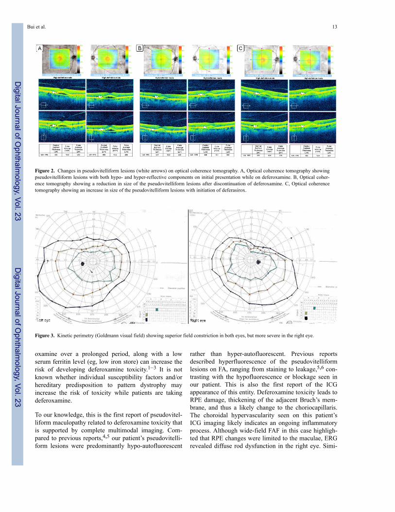
Kinetic perimetry (Goldmann) revealed superior con-striction in both eyes, worse on the right (Figure 3).Electroretinography (ERG) demonstrated a mild-to-moderate reduction in rod function, with relatively pre-served cone function of the right eye and normal rod andcone function of the left eye (Figure 4A and 4B). Theelectro-oculogram (EOG) was markedly abnormal, withan Arden ratio of 1.3 in each eye (Figure 4C).
Given morphologic and functional evidence of retinaltoxicity, deferoxamine was discontinued. Two months
Published February 13, 2017.Copyright ©2017. All rights reserved. Reproduction in whole or in part in any form or medium without expressed written permission of theDigital Journal of Ophthalmology is prohibited.doi:10.5693/djo.02.2016.12.001Correspondence: Hani Salehi-Had, MD, 7777 Edinger Ave, Ste 234, Huntington Beach, CA 92647 (email: [email protected]).
Digital Journal of O
phthalmology, Vol. 23
Digital Journal of O
phthalmology, Vol. 23
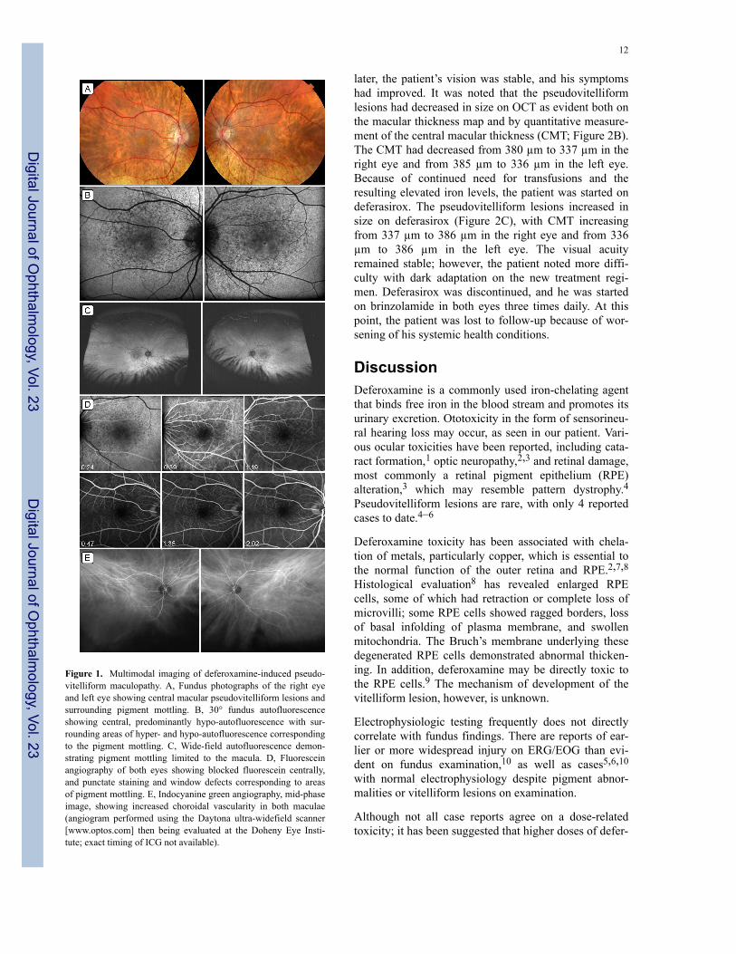
Figure 1. Multimodal imaging of deferoxamine-induced pseudo-vitelliform maculopathy. A, Fundus photographs of the right eyeand left eye showing central macular pseudovitelliform lesions andsurrounding pigment mottling. B, 30° fundus autofluorescenceshowing central, predominantly hypo-autofluorescence with sur-rounding areas of hyper- and hypo-autofluorescence correspondingto the pigment mottling. C, Wide-field autofluorescence demon-strating pigment mottling limited to the macula. D, Fluoresceinangiography of both eyes showing blocked fluorescein centrally,and punctate staining and window defects corresponding to areasof pigment mottling. E, Indocyanine green angiography, mid-phaseimage, showing increased choroidal vascularity in both maculae(angiogram performed using the Daytona ultra-widefield scanner[www.optos.com] then being evaluated at the Doheny Eye Insti-tute; exact timing of ICG not available).
later, the patient’s vision was stable, and his symptomshad improved. It was noted that the pseudovitelliformlesions had decreased in size on OCT as evident both onthe macular thickness map and by quantitative measure-ment of the central macular thickness (CMT; Figure 2B).The CMT had decreased from 380 µm to 337 µm in theright eye and from 385 µm to 336 µm in the left eye.Because of continued need for transfusions and theresulting elevated iron levels, the patient was started ondeferasirox. The pseudovitelliform lesions increased insize on deferasirox (Figure 2C), with CMT increasingfrom 337 µm to 386 µm in the right eye and from 336µm to 386 µm in the left eye. The visual acuityremained stable; however, the patient noted more diffi-culty with dark adaptation on the new treatment regi-men. Deferasirox was discontinued, and he was startedon brinzolamide in both eyes three times daily. At thispoint, the patient was lost to follow-up because of wor-sening of his systemic health conditions.
DiscussionDeferoxamine is a commonly used iron-chelating agentthat binds free iron in the blood stream and promotes itsurinary excretion. Ototoxicity in the form of sensorineu-ral hearing loss may occur, as seen in our patient. Vari-ous ocular toxicities have been reported, including cata-ract formation,1 optic neuropathy,2,3 and retinal damage,most commonly a retinal pigment epithelium (RPE)alteration,3 which may resemble pattern dystrophy.4Pseudovitelliform lesions are rare, with only 4 reportedcases to date.4–6
Deferoxamine toxicity has been associated with chela-tion of metals, particularly copper, which is essential tothe normal function of the outer retina and RPE.2,7,8Histological evaluation8 has revealed enlarged RPEcells, some of which had retraction or complete loss ofmicrovilli; some RPE cells showed ragged borders, lossof basal infolding of plasma membrane, and swollenmitochondria. The Bruch’s membrane underlying thesedegenerated RPE cells demonstrated abnormal thicken-ing. In addition, deferoxamine may be directly toxic tothe RPE cells.9 The mechanism of development of thevitelliform lesion, however, is unknown.
Electrophysiologic testing frequently does not directlycorrelate with fundus findings. There are reports of ear-lier or more widespread injury on ERG/EOG than evi-dent on fundus examination,10 as well as cases5,6,10
with normal electrophysiology despite pigment abnor-malities or vitelliform lesions on examination.
Although not all case reports agree on a dose-relatedtoxicity; it has been suggested that higher doses of defer-
12
Digital Journal of O
phthalmology, Vol. 23
Digital Journal of O
phthalmology, Vol. 23
oxamine over a prolonged period, along with a lowserum ferritin level (eg, low iron store) can increase therisk of developing deferoxamine toxicity.1–3 It is notknown whether individual susceptibility factors and/orhereditary predisposition to pattern dystrophy mayincrease the risk of toxicity while patients are takingdeferoxamine.
To our knowledge, this is the first report of pseudovitel-liform maculopathy related to deferoxamine toxicity thatis supported by complete multimodal imaging. Com-pared to previous reports,4,5 our patient’s pseudovitelli-form lesions were predominantly hypo-autofluorescent
rather than hyper-autofluorescent. Previous reportsdescribed hyperfluorescence of the pseudovitelliformlesions on FA, ranging from staining to leakage,5,6 con-trasting with the hypofluorescence or blockage seen inour patient. This is also the first report of the ICGappearance of this entity. Deferoxamine toxicity leads toRPE damage, thickening of the adjacent Bruch’s mem-brane, and thus a likely change to the choriocapillaris.The choroidal hypervascularity seen on this patient’sICG imaging likely indicates an ongoing inflammatoryprocess. Although wide-field FAF in this case highligh-ted that RPE changes were limited to the maculae, ERGrevealed diffuse rod dysfunction in the right eye. Simi-
Figure 2. Changes in pseudovitelliform lesions (white arrows) on optical coherence tomography. A, Optical coherence tomography showingpseudovitelliform lesions with both hypo- and hyper-reflective components on initial presentation while on deferoxamine. B, Optical coher-ence tomography showing a reduction in size of the pseudovitelliform lesions after discontinuation of deferoxamine. C, Optical coherencetomography showing an increase in size of the pseudovitelliform lesions with initiation of deferasirox.
Figure 3. Kinetic perimetry (Goldmann visual field) showing superior field constriction in both eyes, but more severe in the right eye.
Bui et al. 13
Digital Journal of O
phthalmology, Vol. 23
Digital Journal of O
phthalmology, Vol. 23

larly the EOG revealed diffuse dysfunction of the RPEin both eyes. Of the 4 previously described cases, 3 hadERG data and 2 had EOG data. Two cases describednormal ERG,5,6 and 1 described reduced cone functionwith intact rod function and a normal EOG.6 We believethat the extent or degree of toxicity on the RPE and pho-toreceptor segments can explain the variable findings onERG.
Currently there are three iron-chelating agents approvedby the US Food and Drug Administration: deferoxamine(subcutaneous injection), deferasirox (oral), and deferi-prone (oral). This is the first report of pseudovitelliformmaculopathy that worsened on deferasirox therapy. Thereason for this worsening is unknown; however, it maybe advisable to consider deferiprone therapy for patientswith pseudovitelliform maculopathy from deferoxaminetoxicity.
It is generally accepted that regular screening for defer-oxamine toxicity should be performed for patients onhigh doses or with prolonged exposure to deferoxamine;however, no generally accepted schedule has yet to beestablished. In addition to a dilated fundus examination,helpful ancillary tests include OCT, FA, FAF, ERG,EOG, color vision, and visual field testing. It has beensuggested that a therapeutic index level can be calcula-ted (safe level of <0.025) and may be a meaningful wayto monitor for toxicity as it includes the daily dose perbody weight (mg/kg) and takes into account the serum
ferritin level (mg/L).11 This is important because deple-tion of systemic iron stores may lead to chelation ofother essential metals, such as copper, loss of which cansubsequently lead to retinal toxicity. Discontinuation ofthe chelating agent generally leads to reversal of symp-toms and signs; however, persistent damage has beenreported,4,6 appearing to correlate with prolonged expo-sure. Genead and Fishman12 reported a reduction inpseudovitelliform lesion using brinzolamide, although itis unclear whether the resolution was related to with-drawal of the offending agent or to brinzolamide.
Literature SearchPubMed was searched on August 21, 2015, for English-language articles (1975-present) using the followingcombinations of terms: retina, macula, retinal toxicity,retinopathy, maculopathy, vitelliform, or pseudovitelli-form AND either deferoxamine OR deferasirox.
References1. Taneja R, Malik P, Sharma M, Agarwal MC. Multiple transfused
thalassemia major: ocular manifestations in a hospital-based popula-tion. Indian J Ophthalmol 2010;58:125-30.
2. Olivieri N, Bunci JR, Chew E, et al. Visual and auditory neurotoxic-ity in patients receiving subcutaneous deferoxamine infusions. NEngl J Med 1986;314:869-73.
3. Haimovici R, D’Amico DJ, Gragoudas ES, Sokol S, Deferoxamine
Figure 4. Electroretinogram showing a mild-to-moderate reduction in rod function of the right eye with relatively preserved cone functionand normal rod and cone function of the left eye (a- and b- waves marked; boxes denote the normal limits for each peak). Electrooculogramshowing markedly abnormal Arden ratio of 1.3 in each eye.
14
Digital Journal of O
phthalmology, Vol. 23
Digital Journal of O
phthalmology, Vol. 23
Retinopathy Study Group. The expanded clinical spectrum of defer-oxamine retinopathy. Ophthalmology 2002;109:164-71.
4. Viola F, Barteselli G, Dellʼarti L, et al. Multimodal imaging indeferoxamine retinopathy. Retina 2014;34:1428-38.
5. Genead MA, Fishman GA, Anastasakis A, Lindeman M. Macularvitelliform lesion in desferrioxamine-related retinopathy. Doc Oph-thalmol 2010;121:161-6.
6. Gonzales CR, Lin AP, Engstrom RF, Kreiger AE. Bilateral vitelli-form maculopathy and deferoxamine toxicity. Retina2004;24:464-7.
7. Pall H, Blake DR, Winyard P, et al. Ocular toxicity of desferrioxa-mine—an example of copper promoted auto-oxidative damage? Br JOphthalmol 1989;73:42-7.
8. Rahi AH, Hungerford JL, Ahmed AI. Ocular toxicity of desferrioxa-mine: light microscopic histochemical and ultrastructural findings.Br J Ophthalmol 1986;70:373-81.
9. Klettner A, Koinzer S, Waetzig V, Herdegen T, Roider J. Deferoxa-mine mesylate is toxic for retinal pigment epithelium cells in vitro,and its toxicity is mediated by p38. Cutan Ocul Toxicol2010;2:122-9.
10. Haimovici R, D’Amico DJ, Gragoudas ES, Sokol S. The expandedclinical spectrum of deferoxamine retinopathy. Ophthalmology2002;109:164-71.
11. Porter JB, Jaswon MS, Huehns ER, East CA, Hazell JW. Desfer-rioxamine ototoxicity: evaluation of risk factors in thalassaemicpatients and guidelines for safe dosage. Br J Haematol1989;73:403-9.
12. Genead MA, Fishman GA. Efficacy of brinzolamide ophthalmicsuspension 1% for treatment of a vitelliform macular lesion in apatient with desferrioxamine retinopathy. Ophthalmic Surg LasersImaging 2011;42:e114-7.Online
Bui et al. 15
Digital Journal of O
phthalmology, Vol. 23
Digital Journal of O
phthalmology, Vol. 23