Embed Size (px)
Citation preview

CYANIDE 25
3. HEALTH EFFECTS
3.1 INTRODUCTION
The primary purpose of this chapter is to provide public health officials, physicians, toxicologists, and
other interested individuals and groups with an overall perspective on the toxicology of cyanide. It
contains descriptions and evaluations of toxicological studies and epidemiological investigations and
provides conclusions, where possible, on the relevance of toxicity and toxicokinetic data to public health.
A glossary and list of acronyms, abbreviations, and symbols can be found at the end of this profile.
3.2 DISCUSSION OF HEALTH EFFECTS BY ROUTE OF EXPOSURE
To help public health professionals and others address the needs of persons living or working near
hazardous waste sites, the information in this section is organized first by route of exposure (inhalation,
oral, and dermal) and then by health effect (death, systemic, immunological, neurological, reproductive,
developmental, genotoxic, and carcinogenic effects). These data are discussed in terms of three exposure
periods: acute (14 days or less), intermediate (15–364 days), and chronic (365 days or more).
Levels of significant exposure for each route and duration are presented in tables and illustrated in
figures. The points in the figures showing no-observed-adverse-effect levels (NOAELs) or lowest-
observed-adverse-effect levels (LOAELs) reflect the actual doses (levels of exposure) used in the studies.
LOAELs have been classified into "less serious" or "serious" effects. "Serious" effects are those that
evoke failure in a biological system and can lead to morbidity or mortality (e.g., acute respiratory distress
or death). "Less serious" effects are those that are not expected to cause significant dysfunction or death,
or those whose significance to the organism is not entirely clear. ATSDR acknowledges that a
considerable amount of judgment may be required in establishing whether an end point should be
classified as a NOAEL, "less serious" LOAEL, or "serious" LOAEL, and that in some cases, there will be
insufficient data to decide whether the effect is indicative of significant dysfunction. However, the
Agency has established guidelines and policies that are used to classify these end points. ATSDR
believes that there is sufficient merit in this approach to warrant an attempt at distinguishing between
"less serious" and "serious" effects. The distinction between "less serious" effects and "serious" effects is

CYANIDE 26
3. HEALTH EFFECTS
considered to be important because it helps the users of the profiles to identify levels of exposure at which
major health effects start to appear. LOAELs or NOAELs should also help in determining whether or not
the effects vary with dose and/or duration, and place into perspective the possible significance of these
effects to human health.
The significance of the exposure levels shown in the Levels of Significant Exposure (LSE) tables (Tables
3-1 to 3-3) and figures (Figures 3-1 and 3-2) may differ depending on the user's perspective. Public
health officials and others concerned with appropriate actions to take at hazardous waste sites may want
information on levels of exposure associated with more subtle effects in humans or animals (LOAELs) or
exposure levels below which no adverse effects (NOAELs) have been observed. Estimates of levels
posing minimal risk to humans (Minimal Risk Levels or MRLs) may be of interest to health professionals
and citizens alike.
This section provides information regarding known health effects of cyanide exposure. Exposure to
hydrogen cyanide gas is most common by inhalation. In the discussion below, inhalation exposures are
expressed as ppm hydrogen cyanide for a defined period of time. Exposure to cyanide can also occur by
inhalation of cyanogen gas, a dimer of cyanide. However, cyanogen breaks down in aqueous solution
into cyanide ion (CN–1) and OCN– ions (Cotton and Wilkinson 1980). The rate of the breakdown depends
on pH and is faster in basic media (e.g., hydrogen cyanide is in equilibrium as H+ and CN– in blood with a
pH of 7.38–7.44) than in acidic media (e.g., hydrogen cyanide is the only species in stomach contents at a
pH of 3). The amount of cyanide ion formed within a body tissue or fluid as a result of exposure to
cyanogen has been reported; however, the amount varies with type of body tissue and fluid. Thus, it is
difficult to estimate cyanide levels in body tissues after cyanogen exposure. Therefore, studies regarding
exposure to cyanogen are discussed in the text as ppm cyanogen, but are not included in LSE tables or
figures.
Oral exposure to cyanide usually results from accidental, homicidal, or suicidal ingestion of cyanide salts.
Sodium cyanide and potassium cyanide are the most frequently studied cyanide compounds. Copper
cyanide, potassium silver cyanide, silver cyanide, and calcium cyanide are other compounds that humans
could encounter through oral or dermal exposure; some data for cyanide compounds containing copper or
silver are omitted from the LSE tables and figures because the toxicological effects may have been caused
by the metal, rather than or in addition to CN–. Similarly, toxicological data for ferricyanide compounds
are omitted from the LSE tables and figures because CN– remains tightly bound to iron and is therefore
much less bioavailable than CN– in soluble cyanide compounds. Cassava roots and certain fruit pits

CYANIDE 27
3. HEALTH EFFECTS
contain compounds that can be broken down in the gastrointestinal tract to form cyanide. Cassava roots
form the staple diet of some populations in Africa, Central and South America, and Asia. However, it
must be noted that cassava roots are notoriously deficient in protein and other nutrients and contain many
other compounds, in addition to cyanide, that could be responsible for some of the observed toxic effects.
Thiocyanate, a metabolite of cyanide that is formed in the body after exposure to cyanide compounds, is
responsible for toxic effects to the thyroid gland. When possible, all oral exposures are expressed as mg
CN–/kg/day.
A User's Guide has been provided at the end of this profile (see Appendix B). This guide should aid in
the interpretation of the tables and figures for Levels of Significant Exposure and the MRLs.
3.2.1 Inhalation Exposure
3.2.1.1 Death
Inhalation of sufficient concentrations of hydrogen cyanide gas can rapidly cause death, which has led to
the use of hydrogen cyanide in gas chamber executions (Wexler et al. 1947). An average fatal
concentration for humans was estimated as 546 ppm hydrogen cyanide after a 10-minute exposure (DOA
1976, as cited in Ballantyne 1988). In one case, a worker exposed to 200 ppm hydrogen cyanide in a
silver plating tank became unconscious and eventually died even though he had received antidotal therapy
in a hospital (Singh et al. 1989). In other cases, exposure to 270 ppm hydrogen cyanide led immediately
to death, 181 ppm hydrogen cyanide exposure was fatal after 10 minutes, and 135 ppm hydrogen cyanide
was fatal after 30 minutes in humans (Dudley et al. 1942). Three deep-sea trawler men died when
exposed to toxic fumes (containing lethal concentrations of hydrogen cyanide, carbon dioxide and
hydrogen sulfide) from spoiled fish (Cherian and Richmond 2000); all three men collapsed within
1 minute of exposure, and cyanide exposure was confirmed in one man post-mortem by a blood cyanide
concentration of 0.05 mg/L.
Levels of acute exposure resulting in animal deaths were reported in numerous studies and LC50 (lethal
concentration, 50% death) values were provided for several species. Inhalation LC50 values of hydrogen
cyanide in rats ranged from 143 ppm for 60 minutes to 3,417 ppm for 10 seconds (Ballantyne 1983a).
Five-minute LC50 values of 503 ppm for the rat and 323 ppm for the mouse were reported by DiPasquale
and Davis (1971). At lethal concentrations, animals exhibited hyperactivity and asphyxial convulsions
with death occurring within 20 minutes of exposure; gross pathology findings included pulmonary

CYANIDE 28
3. HEALTH EFFECTS
hemorrhage and congestion of the liver and kidney. Exposure to cyanide resulted in similar LC50 values
in mice (Higgins et al. 1972; Matijak-Schaper and Alarie 1982). LC50 values for hydrogen cyanide in
rabbits ranged from 188 ppm for 30 minutes to 2,200 ppm for 45 seconds (Ballantyne 1983a). Lethal
concentrations were also reported in experiments with dogs exposed for acute (Haymaker et al. 1952) and
intermediate durations (Valade 1952). Both studies used a small number of dogs for different exposure
regimens so that statistical significance could not be evaluated.
The LC50 values in each species and LOAEL values for death in humans in the acute-, and intermediate-
duration category are recorded in Table 3-1 and plotted in Figure 3-1. Lethality data for dogs have been
omitted from Table 3-1 and Figure 3-1 because that species has a relatively low amount of the detoxifying
enzyme rhodanese and is unusually susceptible to cyanide exposure compared to humans or other
mammals (see Sections 3.4.3 and 3.5.3).
3.2.1.2 Systemic Effects
The systemic effects observed in humans and animals after inhalation exposure to cyanide are discussed
below. The highest NOAEL values and all reliable LOAEL values for each systemic effect in each
species and duration category are recorded in Table 3-1 and plotted in Figure 3-1. Systemic toxicity data
for dogs have been omitted from Table 3-1 and Figure 3-1 because that species has a relatively low
amount of the detoxifying enzyme rhodanese and is unusually susceptible to cyanide exposure compared
to humans or other mammals (see Sections 3.4.3 and 3.5.3).
Respiratory Effects. Initially, respiration is stimulated, but later, dyspnea occurs in patients admitted
to a hospital after acute hydrogen cyanide exposure (Chen and Rose 1952; Peden et al. 1986; Potter
1950). The levels of exposure in these accidental poisonings were not provided. Nasal irritation was
reported in volunteers exposed to 16 ppm cyanogen (8 ppm cyanide) for 6–8 minutes (McNerney and
Schrenk 1960). No effects were reported at 8 ppm cyanogen (4 ppm cyanide).
Dyspnea was observed in workers chronically exposed (5–15 years) to 6.4–10.4 ppm of an unspecified
cyanide form evolved from sodium cyanide and copper cyanide during electroplating (El Ghawabi et al.
1975) and in workers exposed to 15 ppm hydrogen cyanide in a silver-reclaiming facility (Blanc et al.
1985). Other complaints included cough, sore throat, altered sense of smell, nasal congestion, epistaxis,
and hemoptysis. However, exposure to other chemicals such as cleaners and cutting oils also occurs in
electroplating operations.

aKey toFigure
Table 3-1 Levels of Significant Exposure to Cyanide - Inhalation
Species(Strain)
LOAEL
CommentsSystemNOAEL
(ppm)Less Serious
(ppm)Serious
(ppm)
ReferenceChemical Form
Exposure/Duration/
Frequency(Route)
ACUTE EXPOSUREDeath1
191
DOA 1976HCN
546 (LC50)546
Human 10 min
299
Singh et al. 1989HCN
M200 (fatal after 3 days)200
Human NS
3
954
AMRL 1971HCN
M503 (5-min LC50)503
Rat(Wistar)
5 min
4
36
Ballantyne 1983aHCN
143 (LC50 in 60 min)143
Rat(NS)
60 min
5
222
Higgins et al. 1972HCN
503 (LC50)503
Rat(Wistar)
5 min
6
955
AMRL 1971HCN
M323 (5-min LC50)323
Mouse(ICR)
5 min
7
223
Higgins et al. 1972HCN
323 (LC50)323
Mouse(ICR)
5 min
8
905
Hume et al. 1995HCN
M400 (90% lethality)400
Mouse(ICR)
3 min
9
161
Matijak-Schaper and Alarie1982HCN
M166 (LC50)166
Mouse(Swiss-Webster)
30 min
10
37
Ballantyne 1983aHCN
188 (LC50 in 35 min)188
Rabbit(NS)
35 min
29

aKey toFigure
(continued)Table 3-1 Levels of Significant Exposure to Cyanide - Inhalation
Species(Strain)
LOAEL
CommentsSystemNOAEL
(ppm)Less Serious
(ppm)Serious
(ppm)
ReferenceChemical Form
Exposure/Duration/
Frequency(Route)
Systemic11
43
Bonsall 1984HCN
Ocular M452 (slight loss of peripheralvision after recovery)
452
Human 13 min
12
228
Purser et al. 1984HCN
Resp 100 (severe dyspnea)100
Monkey(Cynomolgus)
30 min
Cardio 100 (bradycardia, arrhythmia,T-wave abnormalities)
100
13
162
Matijak-Schaper and Alarie1982HCN
Resp M63 (DC50)63
Mouse(Swiss-Webster)
30 min
Neurological14
42
Bonsall 1984HCN
M452 (coma)452
Human 13 min
15
131
Purser et al. 1984HCN
100 (semiconsciousness,disrupted respiration,EEG changes)
100
Monkey(Cynomolgus)
30 min
16
956
Fechter et al. 2002HCN
M5050
Rat(Long- Evans)
3.5 hr
INTERMEDIATE EXPOSURESystemic17
127
O'Flaherty and Thomas 1982HCN
Cardio M200 (increased creatinephosphokinase activity)
200
Rat(Long- Evans)
20 d4 d intervals12.5 min/d
30

aKey toFigure
(continued)Table 3-1 Levels of Significant Exposure to Cyanide - Inhalation
Species(Strain)
LOAEL
CommentsSystemNOAEL
(ppm)Less Serious
(ppm)Serious
(ppm)
ReferenceChemical Form
Exposure/Duration/
Frequency(Route)
CHRONIC EXPOSURESystemic18
49
Blanc et al. 1985HCN
Resp M15 (dyspnea)15
Human NS(occup)
Cardio M15 (palpitations, chest pain)15
Gastro M15 (nausea)15
Endocr M15 (increased mean thyroidstimulating hormonelevels)
15
Dermal M15 (rash)15
Ocular M15 (eye irritation)15
Bd Wt M15 (approximately 8%weight loss)
15
19
55
El Ghawabi et al. 1975NaCN
Resp M6.4 (dyspnea, irritation ofthroat)
6.4
Human 5-15 yr (occup)
Cardio M6.4 (precordial pain)6.4
Gastro M6.4 (vomiting)6.4
Hemato M6.4 (increased hemoglobinand lymphocytes)
6.4
Endocr M6.4 (thyroid enlargement)6.4
Dermal M10.410.4
Ocular M6.4 (lacrimation)6.4
31

aKey toFigure
(continued)Table 3-1 Levels of Significant Exposure to Cyanide - Inhalation
Species(Strain)
LOAEL
CommentsSystemNOAEL
(ppm)Less Serious
(ppm)Serious
(ppm)
ReferenceChemical Form
Exposure/Duration/
Frequency(Route)
Neurological20
48
Blanc et al. 1985HCN
M15 (persistent headache,dizziness, paresthesia)
15
Human NS(occup)
21
a The number corresponds to entries on Figure 3-1.
Bd Wt = body weight; Cardio = cardiovascular; d = day(s); DC50 = concentration that resulted in 50% decrease in average respiratory rate; EEG = electroencephalogram; Endocr =endocrine; F = female; Gastro = gastrointestinal; HCN = hydrogen cyanide; Hemato = hematological; LC50 = lethal concentration, 50%kill; LOAEL = lowest-observed-adverse-effectlevel; M = male; min = minutes; NaCN = sodium cyanide; NOAEL = no-observed-adverse-effect level; NS = not specified; (occup) = occupational; Resp = respiratory; sec =second(s); yr = year(s); x = time(s)
54
El Ghawabi et al. 1975NaCN
M6.4 (confusion, hallucination,headache, dizziness,weakness)
6.4
Human 5-15 yr (occup)
32

10
100
1000
Death
1
2
6m 7m
8m
9m
3r
4r
5r
10h
Respiratory
12k
13m
Cardiovascular
12k
Ocular
11
Neurological
14
15k
16r
ppm
Figure 3-1 Levels of Significant Exposure to Cyanide - InhalationAcute (≤14 days)
c-Catd-Dogr-Ratp-Pigq-Cow
-Humansk-Monkeym-Mouseh-Rabbita-Sheep
f-Ferretj-Pigeone-Gerbils-Hamsterg-Guinea Pig
n-Minko-Other
Cancer Effect Level-Animals LOAEL, More Serious-Animals LOAEL, Less Serious-Animals NOAEL - Animals
Cancer Effect Level-Humans LOAEL, More Serious-Humans LOAEL, Less Serious-Humans NOAEL - Humans
LD50/LC50 Minimal Risk Level for effects other than Cancer
Systemic
33

10
100
1000Cardiovascu
lar
17r
ppm
Figure 3-1 Levels of Significant Exposure to Cyanide - Inhalation (Continued)Intermediate (15-364 days)
c-Catd-Dogr-Ratp-Pigq-Cow
-Humansk-Monkeym-Mouseh-Rabbita-Sheep
f-Ferretj-Pigeone-Gerbils-Hamsterg-Guinea Pig
n-Minko-Other
Cancer Effect Level-Animals LOAEL, More Serious-Animals LOAEL, Less Serious-Animals NOAEL - Animals
Cancer Effect Level-Humans LOAEL, More Serious-Humans LOAEL, Less Serious-Humans NOAEL - Humans
LD50/LC50 Minimal Risk Level for effects other than Cancer
Systemic
34

1
10
100Respiratory
18
19
Cardiovascular
18
19
Gastrointestin
al
18
19
Hematological
19
Endocrine
18
19
Dermal
18
19
Ocular
18
19
Body Weight
18
Neurological
20
21
ppm
Figure 3-1 Levels of Significant Exposure to Cyanide - Inhalation (Continued)Chronic (≥365 days)
c-Catd-Dogr-Ratp-Pigq-Cow
-Humansk-Monkeym-Mouseh-Rabbita-Sheep
f-Ferretj-Pigeone-Gerbils-Hamsterg-Guinea Pig
n-Minko-Other
Cancer Effect Level-Animals LOAEL, More Serious-Animals LOAEL, Less Serious-Animals NOAEL - Animals
Cancer Effect Level-Humans LOAEL, More Serious-Humans LOAEL, Less Serious-Humans NOAEL - Humans
LD50/LC50 Minimal Risk Level for effects other than Cancer
Systemic
CYANIDE
3. HEALTH EFFECTS
35

CYANIDE 36
3. HEALTH EFFECTS
Asphyxia has been observed in rats exposed to 250 ppm cyanogen (125 ppm cyanide) for 7.5–
120 minutes (McNerney and Schrenk 1960), asphyxia and pulmonary edema were observed in dogs
exposed to concentrations ranging from 149 to 633 ppm hydrogen cyanide for 2–10 minutes (Haymaker
et al. 1952), while severe dyspnea was observed in monkeys exposed to 100 ppm hydrogen cyanide for
30 minutes (Purser et al. 1984). Exposure to 63 ppm hydrogen cyanide for 30 minutes resulted in a 50%
decrease in respiratory rate of mice due to depression of the respiratory center (Matijak-Schaper and
Alarie 1982).
In intermediate-duration studies, no respiratory effects were reported in rats exposed to 25 ppm cyanogen
(12.5 ppm cyanide) for 6 months, and a decrease in total lung moisture content was the only finding in
monkeys exposed to 11 ppm cyanogen (5.5 ppm cyanide), also for 6 months (Lewis et al. 1984).
Dyspnea was found in dogs exposed to 45 ppm hydrogen cyanide for 30 minutes a day at 2–8-day
intervals for 28–96 days (Valade 1952).
Cardiovascular Effects. Wexler (1947) reported on four men who were executed by inhalation of
hydrogen cyanide gas (concentration not reported). He reported a distinct slowing of the heart rate within
1–3 minutes of exposure, with further changes in the heart rate, sinus irregularities, and audio-visual
dissociation. Palpitations and hypotension were the most frequently reported cardiovascular effects in
patients after accidental inhalation poisoning with cyanide; however, exact exposure levels were not
known (Peden et al. 1986). Workers occupationally exposed to 6.4–10.4 ppm cyanide for 5–15 years,
which evolved from sodium cyanide and copper cyanide during electroplating, complained of precordial
pain (El Ghawabi et al. 1975). About 14% of workers exposed to 15 ppm hydrogen cyanide in a silver-
reclaiming facility reported palpitations and 31% reported chest pain (Blanc et al. 1985). Exposure to
other chemicals such as cleaners and cutting oils may have also occurred during electroplating operations.
Bradycardia, arrhythmias, and T-wave abnormalities were observed in monkeys exposed to 100 ppm
hydrogen cyanide for 30 minutes (Purser et al. 1984). Increased cardiac-specific creatinine
phosphokinase activity was measured in blood samples from rats 2 hours after 12.5 minutes of exposure
to 200 ppm hydrogen cyanide for 20 days at 4-day intervals (O'Flaherty and Thomas 1982). However, no
treatment-related changes were found in the hearts at histopathology. In addition, no cardiovascular
effects were reported at necropsy in rats and monkeys exposed to 25 ppm cyanogen (12.5 ppm cyanide)
for 6 months (Lewis et al. 1984).

CYANIDE 37
3. HEALTH EFFECTS
Gastrointestinal Effects. Nausea or vomiting was reported in 69% of workers exposed to 15 ppm
hydrogen cyanide in a silver reclaiming facility (Blanc et al. 1985). Vomiting was also reported in
workers exposed to 6.4–10.4 ppm cyanide evolved from sodium cyanide and copper cyanide during
electroplating (El Ghawabi et al. 1975), but exposure to other chemicals such as cleaners and cutting oils
may have also contributed to the effects. The gastrointestinal effects resulting from cyanide exposure are
probably provoked by central nervous system effects and/or by irritation of the gastric mucosa in cases in
which the gas is swallowed during breathing.
Information regarding gastrointestinal effects in animals is limited to a report of vomiting in dogs exposed
to 45 ppm hydrogen cyanide for 28–96 days (Valade 1952).
Hematological Effects. Increased hemoglobin and lymphocyte count were observed in workers
exposed to 6.4–10.4 ppm of an unspecified cyanide form during electroplating (El Ghawabi et al. 1975).
The results were significantly different from controls. Furthermore, punctate basophilia of erythrocytes,
which indicated toxic poisoning, was present in 28 of 36 subjects. However, exposure to copper, a known
hematotoxic agent, also occurred during the electroplating operations. In another study (Kumar et al.
1992), an increase in neutrophil values, an increase in erythrocyte sedimentation rate, and a decrease in
hemoglobin levels were noted in male workers exposed to unspecified concentrations of cyanide for an
unspecified duration during case hardening and electroplating.
In animals, no hematological effects were found in rats and monkeys exposed to 25 ppm cyanogen
(12.5 ppm cyanide) 6 hours/day, 5 days/week, for 6 months (Lewis et al. 1984).
Musculoskeletal Effects. No studies were located regarding musculoskeletal effects in humans after
inhalation exposure to cyanide.
No musculoskeletal effects were observed in rats or monkeys exposed to 25 ppm cyanogen (12.5 ppm
cyanide) for 6 hours/day, 5 days/week for 6 months (Lewis et al. 1984).
Hepatic Effects. An increase in serum alkaline phosphatase was noted in workers exposed to
unspecified levels of cyanide; however, serum bilirubin was found to be within the normal range in these
workers (Kumar et al. 1992).

CYANIDE 38
3. HEALTH EFFECTS
Only one study reported on pathological and histopathological examinations of the liver in animals. No
changes were found in rats and monkeys exposed to 25 ppm cyanogen (12.5 ppm cyanide) for 6 months
(Lewis et al. 1984).
Renal Effects. One study was located regarding renal effects in humans after inhalation exposure to
cyanide. Singh et al. (1989) reported anuria followed by polyuria in a man who was occupationally
exposed to 200 ppm hydrogen cyanide) for an unspecified length of time.
No histopathological changes were observed in kidneys of rats and monkeys exposed to 25 ppm cyanogen
(12.5 ppm cyanide) 6 hours/day, 5 days/week for 6 months (Lewis et al. 1984).
Endocrine Effects. Mean thyroid stimulating hormone (TSH) levels were significantly higher
(although within normal limits) in a group of 36 workers exposed to 15 ppm hydrogen cyanide for an
unspecified duration in a silver-reclaiming facility than in unexposed individuals (p<0.05). T3 levels in
high exposure workers were also elevated relative to unexposed workers (p<0.01). Data for T4 were not
presented, but the investigators indicated that the absence of T4 abnormalities could be accounted for by
the time lapse between exposure and examination (median 10.5 months) (Blanc et al. 1985). Similarly,
thyroid enlargement was present in 20 of 36 workers exposed for 5–15 years to 6.4–10.4 ppm cyanide
evolved from sodium cyanide and copper cyanide. The endocrine effect may be due to formation of
thiocyanate, a metabolite of cyanide. However, exposure to other chemicals such as cleaners and cutting
oils also occurs during electroplating operations. Thyroid 131I uptake was significantly higher when
compared with the control group. This may be due to thiocyanate's ability to block iodine uptake and also
compete with I– as a substrate for the thyroid peroxidase, resulting in less "organification" of I2
(decreasing the iodination of tyrosine to form iodotyrosine) by the thyroid gland. Since the workers were
away from work on the 2 days preceding the test, the results may be explained on the basis of acute
cyanide withdrawal, as with other anti-thyroid agents, where sudden cessation of the drug leads to rapid
accumulation of iodine in the iodine-depleted gland (El Ghawabi et al. 1975).
No studies were located regarding endocrine effects in animals after inhalation exposure to cyanide.
Dermal Effects. Cyanide caused a rash in 42% of workers exposed to 15 ppm hydrogen cyanide
(Blanc et al. 1985). Brick-red chemical burns on the skin were observed in a man who was
occupationally exposed to 200 ppm hydrogen cyanide for an unspecified length of time (Singh et al.

CYANIDE 39
3. HEALTH EFFECTS
1989). No dermatitis was reported in workers exposed to 6.4–10.4 ppm cyanide evolved from sodium
cyanide and copper cyanide (El Ghawabi et al. 1975).
No studies were located regarding dermal effects in animals after inhalation exposure to cyanide.
Ocular Effects. Cyanogen caused eye irritation in volunteers during acute exposure to 16 ppm (8 ppm
cyanide) (McNerney and Schrenk 1960). No effect was observed in those exposed to 8 ppm cyanogen
(4 ppm cyanide). Slight loss of peripheral vision was the only persistent finding from a case report of a
man who had been exposed to 452 ppm hydrogen cyanide (for 13 minutes while cleaning a chemical tank
(Bonsall 1984). During chronic occupational exposure, eye irritation occurred in workers of two
electroplating factories (exposure levels not specified) (Chandra et al. 1988). In other studies, cyanide
caused eye irritation in 58% of workers exposed to 15 ppm hydrogen cyanide (Blanc et al. 1985) and
lacrimation in workers exposed to 6.4 ppm cyanide (El Ghawabi et al. 1975). The eye irritation may not
be due solely to cyanide exposure, as electroplating workers may be exposed to a variety of chemicals
that are irritating to the eyes.
Information regarding ocular effects in animals after inhalation exposure is limited to a report of eye
irritation in rats acutely exposed (7.5–120 minutes) to 250 ppm cyanogen (125 ppm cyanide) (McNerney
and Schrenk 1960).
Body Weight Effects. In an occupational setting, loss of appetite was reported in 58% and weight
loss (approximately 8%) in 50% of workers exposed to 15 ppm hydrogen cyanide (for an unspecified
duration in a silver-reclaiming facility (Blanc et al. 1985).
Decreased body weight was reported in rats exposed to 25 ppm cyanogen (12.5 ppm cyanide)
6 hours/day, 5 days/week for 6 months (Lewis et al. 1984).
3.2.1.3 Immunological and Lymphoreticular Effects
No studies were located regarding immunological effects in humans or animals after inhalation exposure
to cyanide.

CYANIDE 40
3. HEALTH EFFECTS
3.2.1.4 Neurological Effects
The central nervous system is a primary target for cyanide toxicity. Acute exposure of humans to fatal
levels of hydrogen cyanide causes a brief stage of central nervous system stimulation followed by
depression, convulsions, coma with abolished deep reflexes and dilated pupils, paralysis, and in some
cases, death (Bonsall 1984; Chen and Rose 1952; Peden et al. 1986; Potter 1950; Singh et al. 1989).
Though clinical symptoms of cyanide poisoning are well recognized, specific dose-response data are
generally not known. Acute exposure to lower concentrations can cause lightheadedness, breathlessness,
dizziness, numbness, and headaches (Lam and Lau 2000; Peden et al. 1986). Impaired short-term
memory was reported as a delayed effect in a female 1 year after treatment for convulsions following
acute exposure to cyanide gas (Lam and Lau 2000).
Chronic exposure of humans to potassium cyanide and other chemicals may have produced severe
neurological effects such as hemiparesis and hemianopia (Sandberg 1967). During chronic occupational
exposure, workers exposed to 15 ppm hydrogen cyanide for an unspecified duration reported fatigue,
dizziness, headaches, disturbed sleep, ringing in ears, paresthesias of extremities, and syncopes (Blanc et
al. 1985). A dose-effect was demonstrated on high- and low-exposure jobs; however, exact cyanide
concentrations in the air were not known. Neurological effects persisted in some workers even after a
10-month nonexposure period. Similar effects were observed in workers exposed to 6.4 ppm cyanide (El
Ghawabi et al. 1975). Clinical symptoms included headaches, weakness, changes in taste and smell,
dizziness, disturbances of accommodation, and psychosis. Loss of delayed and immediate memory and
decreases in visual ability, psychomotor ability, and visual learning were reported in workers exposed to
unspecified levels of hydrogen cyanide for an unspecified duration (Kumar et al. 1992). In another study,
chronic occupational exposure of workers (5–19 years) to hydrogen cyanide (exposure levels not
specified) resulted in headaches and dizziness in workers (Chandra et al. 1988). Furthermore, when
behavioral functions were tested in this cohort, an alteration of delayed memory and/or visual impairment
was found in 31.5% of workers. However, exposure to other chemicals, such as cleaners and cutting oils,
also occurs during electroplating operations.
The central nervous system is also a primary target for cyanide toxicity in animals. Following acute
exposure, neurological effects before death included restless and panic movements, poor coordination,
tremor, and lethargy in rats exposed to 250 ppm cyanogen (125 ppm cyanide) for 1.5–120 minutes
(McNerney and Schrenk 1960). When rats were exposed to unspecified concentrations of hydrogen
cyanide and kept unconscious for 20–60 minutes, lesions of various degrees developed in the brain

CYANIDE 41
3. HEALTH EFFECTS
(Hirano et al. 1967; Levine 1969; Levine and Stypulkowski 1959a). Necrosis was found mainly in the
mid-sagittal sections of the brain. Demyelination was also reported and morphological signs indicative of
remyelination were reported in rats several months after cyanide intoxication (Hirano et al. 1968), but it
was apparent that this process was slow and incomplete. Acute exposure of dogs for 2–10 minutes, each
to a different concentration ranging from 149 to 633 ppm hydrogen cyanide resulted in motor
incoordination, muscular rigidity, and coma (Haymaker et al. 1952). Extensive necrosis in the grey
matter of the neural system was observed at necropsy. After acute exposure (up to 30 minutes) to 60–
100 ppm hydrogen cyanide, increased delta activity was observed in electroencephalograms of
cynomolgus monkeys, but those exposed at the higher level experienced semiconsciousness within
20 minutes (Purser 1984; Purser et al. 1984). Cyanide exposure levels in most acute-duration studies
were relatively high and usually caused death in some animals. Only transitory behavioral changes were
reported in monkeys exposed to 25 ppm cyanogen (12.5 ppm cyanide) for 6 months (Lewis et al. 1984).
No effects were found at 11 ppm cyanogen (5.5 ppm cyanide) exposure. Exposure of dogs to 45 ppm
hydrogen cyanide for 28–96 days caused tremors, convulsions, and coma (Valade 1952). Vascular and
cellular lesions were found in the central nervous system.
In rats exposed to 10–50 ppm hydrogen cyanide for 3.5 hours, there was no adverse effect 4 weeks later
on hearing or the histology of cochlear hair cells (Fechter et al. 2002). However, co-administration for
2 hours of 100 dB broadband noise (13.6 kHz) caused an hydrogen cyanide-dose-related increase in
auditory compound action potential thresholds compared with noise administered alone that was
statistically significant at 30 ppm hydrogen cyanide; a loss of outer hair cells of the cochlea was noted in
rats exposed to hydrogen cyanide and noise.
The highest NOAEL value and all reliable LOAEL values for neurological effects in each species and
duration category are recorded in Table 3-1 and plotted in Figure 3-1. Neurotoxicity data for dogs have
been omitted from Table 3-1 and Figure 3-1 because that species has a relatively low amount of the
detoxifying enzyme rhodanese and is unusually susceptible to cyanide exposure compared to humans or
other mammals (see Sections 3.4.3 and 3.5.3).

CYANIDE 42
3. HEALTH EFFECTS
No studies were located regarding the following health effects in humans or animals after inhalation
exposure to cyanide:
3.2.1.5 Reproductive Effects
3.2.1.6 Developmental Effects
3.2.1.7 Cancer
3.2.2 Oral Exposure
3.2.2.1 Death
An average fatal dose of 1.52 mg/kg cyanide for humans has been calculated from case report studies of
intentional or accidental poisonings (EPA 1987a). The lowest fatal oral dose reported in humans was
estimated as 0.56 mg/kg cyanide (form not specified) (Gettler and Baine 1938). However, these data
were obtained from the case history; furthermore, analytical measurements of the time lack the precision
of current technology.
Oral LD50 (lethal dose, 50% death) values for sodium cyanide were calculated as 3 mg CN–/kg for
unfasted rats, and 2.7 mg CN–/kg/day for rats that fasted for 24 hours before exposure and for unfasted
rabbits (Ballantyne 1988); initial signs of toxicity appeared sooner in rats (1 minute for both groups) than
in rabbits (4 minutes) and the time to death was shorter in the fasted rats than in the unfasted rats or
rabbits (17 rather than 22 minutes). In rats, the acute LD50 was 8 mg CN–/kg as sodium cyanide, but
22 mg CN–/kg as calcium cyanide (Smyth et al. 1969). Acute LD50 values in rabbits were similar (2.34–
2.7 mg CN–/kg/day) regardless of whether the source was hydrocyanic acid, sodium cyanide, or
potassium cyanide (Ballantyne 1983a). Mortality was 95% in rats and mice that received a single dose of
4 and 6 mg CN–/kg, respectively, in the form of potassium cyanide in a volume of water equivalent to 5%
of body weight (Ferguson 1962); mortality was lower (50% in rats and 35% in mice) when the same
doses were delivered in a volume of water equivalent to 1.25% of body weight. Increased mortality was
observed in rats exposed to 14.5 mg CN–/kg/day as copper cyanide for 90 days (Gerhart 1986) and to
2.6 mg CN–/kg/day as potassium silver cyanide for 90 days (Gerhart 1987); these data are omitted from
Table 3-2 because of the possible confounding effect of the metals. Hemolytic anemia, which probably
resulted from copper toxicity, caused death in rats exposed to copper cyanide (Gerhart 1986). No deaths

CYANIDE 43
3. HEALTH EFFECTS
were reported in male and female rats exposed to 0.2–12.5 mg CN–/kg/day in the drinking water for
13 weeks (NTP 1993).
The LD50 and minimum lethal dose (LDLO) values in each species and all reliable LOAEL values for
death in each species and duration category are recorded in Table 3-2 and plotted in Figure 3-2.
3.2.2.2 Systemic Effects
The systemic effects observed in humans and animals after oral exposure to cyanide are discussed below.
The highest NOAEL values and all reliable LOAEL values for each systemic effect in each species and
duration category are recorded in Table 3-2 and plotted in Figure 3-2. Studies on dogs have been omitted
because that species has a relatively low amount of the detoxifying enzyme rhodanese and is unusually
susceptible to cyanide exposure compared to humans or other mammals (see Sections 3.4.3 and 3.5.3).
Studies involving ingestion of cassava have also been omitted because of the confounding effects of
malnutrition noted in human studies and because of the presence of other toxic compounds besides
cyanogenic glycosides that might contribute to toxicity (e.g., scopoletin; see Section 3.10). Intermediate-
duration studies that employed oral gavage dosing are omitted because bolus administration may
overwhelm detoxification processes in a manner not typical of gradual exposures in drinking water for the
general population.
Respiratory Effects. Breathing irregularities occur after cyanide poisoning through oral exposure.
Stertorous, deep, and rapid breathing was reported in a man who ingested ≈15 mg CN–/kg as potassium
cyanide in a suicide attempt (Liebowitz and Schwartz 1948). Shortness of breath and dyspnea were
observed in two reports of suicide attempts; one man ingested 7.6 mg CN–/kg (Goodhart 1994) and the
other man ingested 0.57 mg CN–/kg (Saincher et al. 1994), both as potassium cyanide. A man admitted to
a hospital after ingesting an unknown amount of sodium cyanide ceased breathing (Grandas et al. 1989).
A woman who ingested an unknown amount of cyanide developed acute respiratory distress syndrome
and arteriolization (elevated oxyhemoglobin saturation) of the ventral venous blood (Martin-Bermudez et
al. 1997). Dyspnea developed in a woman 20 minutes after eating 30 apricot pits (~15 g), resulting in an
estimated cyanide exposure between 0.026 and 0.234 mg CN–/kg (Suchard et al. 1998). Tachypnea was
also reported in children who were poisoned by cyanide after ingesting apricot pits (Lasch and El Shawa
1981).

aKey toFigure
Table 3-2 Levels of Significant Exposure to Cyanide - Oral
Species(Strain)
LOAEL
CommentsSystemNOAEL
(mg/kg/day)Less Serious
(mg/kg/day)Serious(mg/kg/day)
ReferenceChemical Form
Exposure/Duration/
Frequency(Route)
ACUTE EXPOSUREDeath1
91
Ferguson 1962KCN
4 (19/20 died)4
Rat(Sprague-Dawley)
once(GW)
2
92
Smyth et al. 1969Ca(CN)2
22 (LD50)22
Rat(NS)
once(GW)
3
93
Smyth et al. 1969NaCN
8 (LD50)8
Rat(NS)
once(GW)
4
90
Ferguson 1962KCN
6 (19/20 died)6
Mouse(Swiss-Webster)
once(GW)
Systemic5
8
Liebowitz and Schwartz 1948KCN
Resp M15 (hyperventilation)15
Human once(IN)
Cardio M15 (shallow pulse, inaudibleheart sounds, enlargedheart)
15
Gastro M15 (vomiting and nausea)15
Hemato M1515
Musc/skel M15 (generalized muscularrigidity)
15
Renal M15 (albuminuria)15
44

aKey toFigure
(continued)Table 3-2 Levels of Significant Exposure to Cyanide - Oral
Species(Strain)
LOAEL
CommentsSystemNOAEL
(mg/kg/day)Less Serious
(mg/kg/day)Serious(mg/kg/day)
ReferenceChemical Form
Exposure/Duration/
Frequency(Route)
Neurological6
7
Liebowitz and Schwartz 1948KCN
M15 (coma)15
Human once(IN)
7
930
Rosenow et al. 1995KCN
M7.4 (brain lesions,Parkinsonian-like signs,decreased verbal fluencyand speed of informationprocessing)
7.4
Human once(IN)
INTERMEDIATE EXPOSURESystemic8
301
NTP 1993NaCN
Resp 12.512.5
Rat(Fischer- 344)
13 wk(W)
Cardio 12.512.5
Hemato 12.512.5
Hepatic 12.512.5
Renal 12.512.5
Endocr 12.512.5
Bd Wt 12.512.5
9
129
Philbrick et al. 1979KCN
Endocr M30 (decreased plasmathyroxine at 4 months,increased thyroid weightat 11 months)
30
Rat(NS)
11.5 mo(F)
Bd Wt M30 (38% decreased weightgain)
30
45

aKey toFigure
(continued)Table 3-2 Levels of Significant Exposure to Cyanide - Oral
Species(Strain)
LOAEL
CommentsSystemNOAEL
(mg/kg/day)Less Serious
(mg/kg/day)Serious(mg/kg/day)
ReferenceChemical Form
Exposure/Duration/
Frequency(Route)
10
352
Philbrick et al. 1979KSCN
Endocr M67 (decreased plasmathyroxine and thyroxinesecreting rate, increasedthyroid weight at 11 mo)
67
Rat(NS)
11.5 mo(F)
Bd Wt M6767
11
303
NTP 1993NaCN
Cardiob
M24.324.3
F28.828.8
Mouse(B6C3F1)
13 wk(W)
Hematob
M24.324.3
F28.828.8
Hepaticb
M24.324.3
F28.828.8
Renalb
M24.324.3
F28.828.8
Endocr F28.828.8
Bd Wtb
M24.324.3
F28.828.8
46

aKey toFigure
(continued)Table 3-2 Levels of Significant Exposure to Cyanide - Oral
Species(Strain)
LOAEL
CommentsSystemNOAEL
(mg/kg/day)Less Serious
(mg/kg/day)Serious(mg/kg/day)
ReferenceChemical Form
Exposure/Duration/
Frequency(Route)
12
960
Okolie and Iroanya 2003NaCN
Resp M15 (pulmonary edema)15
Rabbit(NewZealand)
4 wk(F)
Hepatic M15 (fatty degneration andnecrosis)
15
Renal M15 (tubular necrosis)15
Bd Wt M1515
13
958
Okolie and Osagie 1999KCN
Hepatic M20 (focal congestion andnecrosis, increasedserum SDH, ALT, AP)
20
Rabbit(NewZealand)
40 wk(F)
Renal M20 (tubular/glomerularnecrosis)
20
Bd Wt M2020
14
959
Okolie and Osagie 2000KCN
Resp M20 (focal pulmonary edemaand necrosis)
20
Rabbit(NewZealand)
40 wk(F)
Cardio M2020
Endocr M2020
Immuno/ Lymphoret15
603
NTP 1993NaCN
12.512.5
Rat(Fischer- 344)
13 wk(W)
47

aKey toFigure
(continued)Table 3-2 Levels of Significant Exposure to Cyanide - Oral
Species(Strain)
LOAEL
CommentsSystemNOAEL
(mg/kg/day)Less Serious
(mg/kg/day)Serious(mg/kg/day)
ReferenceChemical Form
Exposure/Duration/
Frequency(Route)
16
604
NTP 1993NaCN
bM24.3
24.3
F28.828.8
Mouse(B6C3F1)
13 wk(W)
Neurological17
605
NTP 1993NaCN
12.512.5
Rat(Fischer- 344)
13 wk(W)
18
128
Philbrick et al. 1979KCN
M30 (modest myelindegeneration in spinalcord)
30
Rat(NS)
11.5 mo(F)
19
353
Philbrick et al. 1979KSCN
M67 (modest myelindegeneration in spinalcord)
67
Rat(NS)
11.5 mo(F)
20
606
NTP 1993NaCN
bM24.3
24.3
F28.828.8
Mouse(B6C3F1)
13 wk(W)
48

aKey toFigure
(continued)Table 3-2 Levels of Significant Exposure to Cyanide - Oral
Species(Strain)
LOAEL
CommentsSystemNOAEL
(mg/kg/day)Less Serious
(mg/kg/day)Serious(mg/kg/day)
ReferenceChemical Form
Exposure/Duration/
Frequency(Route)
Reproductive21
304
NTP 1993NaCN
b,cM4.5
4.5
F12.512.5
M12.5 (decreased leftepididymal [7%], leftcaudal epididymal [13%],and testes weights [8%],number of spermatidheads per testis [14%],and spermatid count[14%])
12.5
Rat(Fischer- 344)
13 wk(W)
22
a The number corresponds to entries on Figure 3-2.
b Differences in levels of health effects and cancer effects between male and females are not indicated in Figure 3-2. Where such differences exist, only the levels of effect for themost sensitive gender are presented.
c Used to derive intermediate oral minimal risk level (MRL) of 0.05 mg/kg/day; dose divided by an uncertainty factor of 100 (10 for extrapolation from animals to humans, and 10 forhuman variability).
ATP = adenosine triphosphate; Bd Wt = body weight; BUN = blood urea nitrogen; Cardio = cardiovascular; Ca(CN)2 = calcium cyanide; d = day(s); Endocr = endocrine; F = female;(G) = gavage; Gastro = gastrointestinal; Gd = gestation day; (GW) = gavage in water; HCN = hydrogen cyanide; Hemato = hematological; (IN) = ingestion; KAgCN2 = potassiumsilver cyanide; KCN = potassium cyanide; KSCN = potassium thiocyanate; LD50 = lethal dose, 50% kill; Ld = lactation day; LDlo = lowest lethal dose; LOAEL =lowest-observed-adverse-effect level; M = male; mo = month(s); NaCN = sodium cyanide; NOAEL = no-observed-adverse-effect level; NS = not specified; NTP = National ToxicologyProgram; prox = proximal; Resp = respiratory; SDH = sorbital dehydrogenase; T3 = triiodothyronine; T4 = thyroxine; (W) = water; wk = week(s); x = time(s)
305
NTP 1993NaCN
bM8.6
8.6
F28.828.8
M24.3 (10 and 18% decrease inleft epididymus andcaudal epididymusweights)
24.3
Mouse(B6C3F1)
13 wk(W)
49

1
10
100
Death
4m
1r
2r
3r
Respiratory
5
Cardiovascular
5
Gastrointestin
al
5
Hematological
5
Musculoske
letal
5
Renal
5
Neurological
6
7
mg/kg/day
Figure 3-2 Levels of Significant Exposure to Cyanide - OralAcute (≤14 days)
c-Catd-Dogr-Ratp-Pigq-Cow
-Humansk-Monkeym-Mouseh-Rabbita-Sheep
f-Ferretj-Pigeone-Gerbils-Hamsterg-Guinea Pig
n-Minko-Other
Cancer Effect Level-Animals LOAEL, More Serious-Animals LOAEL, Less Serious-Animals NOAEL - Animals
Cancer Effect Level-Humans LOAEL, More Serious-Humans LOAEL, Less Serious-Humans NOAEL - Humans
LD50/LC50 Minimal Risk Level for effects other than Cancer
Systemic
50

0.01
0.1
1
10
100
Respiratory
8r12h14h
Cardiovascular
11m
8r
14h
Hematological
11m
8r
Hepatic
11m
8r12h13h
Renal
11m
8r12h13h
Endocrine
11m
8r
9r
10r
14h
Body Weight
11m
8r
9r
10r
12h13h
Immuno/Lymphor
16m
15r
Neurological
20m
17r
18r
19r
Reproductive
22m
22m
21r
21r
mg/kg/day
Figure 3-2 Levels of Significant Exposure to Cyanide - OralIntermediate (15-364 days
c-Catd-Dogr-Ratp-Pigq-Cow
-Humansk-Monkeym-Mouseh-Rabbita-Sheep
f-Ferretj-Pigeone-Gerbils-Hamsterg-Guinea Pig
n-Minko-Other
Cancer Effect Level-Animals LOAEL, More Serious-Animals LOAEL, Less Serious-Animals NOAEL - Animals
Cancer Effect Level-Humans LOAEL, More Serious-Humans LOAEL, Less Serious-Humans NOAEL - Humans
LD50/LC50 Minimal Risk Level for effects other than Cancer
)
Systemic
(Continued)
CYANIDE
3. HEALTH EFFECTS
51

CYANIDE 52
3. HEALTH EFFECTS
Respiratory effects were also observed in animals exposed to cyanide. Alveolar congestion, pulmonary
edema, and significant decreases in activities of superoxide dismutase, catalase, and alkaline phosphatase
were observed in the lungs of male rabbits that ingested 15 mg/kg/day of cyanide from sodium cyanide in
feed for 4 weeks (Okolie and Iroanya 2003). Labored respiration was reported in rats treated with
4.35 mg CN–/kg/day as copper cyanide by gavage for 90 days (Gerhart 1986). No effects were reported
at 1.45 mg CN–/kg/day. Labored respiration occurred in rats exposed at a lower dose of 0.8 mg CN–
/kg/day when administered in a form of potassium silver cyanide for 90 days (Gerhart 1987). Lung
congestion and hemorrhage seen at necropsy were attributed to asphyxia rather than to a direct effect of
cyanide. In another study, rats were exposed to 0.2–12.5 mg CN–/kg/day as sodium cyanide in the
drinking water for 13 weeks. Changes in absolute lung weight were seen, but they were minor and
sporadic, and the authors did not consider them to be treatment related (NTP 1993). No respiratory
effects were reported in rats exposed to a target dose of 10.4 mg CN–/kg/day as hydrogen cyanide in their
feed for 2 years (Howard and Hanzal 1955). The actual dose, however, may have been considerably
lower than 10.4 mg/kg/day due to evaporation of hydrogen cyanide from the food.
Cardiovascular Effects. Several case studies reported cardiovascular effects in humans after oral
exposure to cyanide. Weak, shallow pulse and inaudible heart sounds were observed in a comatose man
on hospital admission after ingestion of ≈15 mg CN–/kg as potassium cyanide (Liebowitz and Schwartz
1948). Following gastric lavage and glucose infusion, the pulse rate and blood pressure became elevated.
An enlarged heart was noted. No cardiovascular effects were reported during the recovery. In another
study, children poisoned by apricot pits had hypotension upon hospital admission (Lasch and El Shawa
1981).
After intermediate- or chronic-duration oral exposure to inorganic cyanides, cardiovascular effects in
animals, if any, are minimal. No significant histopathological changes were observed in rats exposed to
2.6 or 7.8 mg CN–/kg/day as potassium silver cyanide for 90 days (Gerhart 1987). No treatment-related
effects on heart histopathology or tissue levels of aspartate aminotransferase (AST) or alkaline
phosphatase were observed in male rabbits that ingested 20 mg CN–/kg/day as potassium cyanide via the
diet for 10 months (Okolie and Osagie 2000). Changes in absolute heart weight were seen in male and
female mice exposed to 0.3–28.8 mg CN–/kg/day as sodium cyanide in the drinking water for 13 weeks,
but they were minor and sporadic, and the authors did not consider them to be treatment related (NTP
1993). Dogs fed a diet of cassava ingested an estimated 1.04 mg CN–/kg/day for 14 weeks and exhibited
hemorrhage, pyknotic nuclei, and swelling of muscle fibers in the myocardium, while dogs fed rice to
which 1.04 mg CN–/kg food was added as sodium cyanide did not show any cardiovascular effects

CYANIDE 53
3. HEALTH EFFECTS
(Kamalu 1993). Furthermore, no cardiovascular effects were observed in rats exposed to an estimated
dose of 10.4 mg CN–/kg/day as hydrogen cyanide in their feed for 2 years (Howard and Hanzal 1955).
The actual dose, however, may have differed from 10.4 mg/kg/day due to evaporation of hydrogen
cyanide from the food.
Gastrointestinal Effects. Vomiting was reported in children who ingested a large number of apricot
pits (Lasch and El Shawa 1981) and in a man who ingested 7.6 mg CN–/kg in a suicide attempt (Goodhart
1994). Gastrointestinal spasms were reported in a man who accidentally ingested (and inhaled) an
unknown amount of potassium cyanide (Thomas and Brooks 1970). Gastric surgery for extensive
necrosis had to be performed in a man after he ingested an unknown amount of sodium cyanide (Grandas
et al. 1989). The alkaline properties of solutions of sodium and potassium cyanide cause the corrosive
responses in the stomach following ingestion.
Diarrhea was observed in rats treated orally with 14.5 mg CN–/kg/day copper cyanide for 90 days
(Gerhart 1986). No effects were observed at 4.35 mg/kg/day. However, as the diarrhea was probably due
to the toxicity of copper, these data are omitted from Table 3-2. No gastrointestinal effects were found in
rats exposed to 7.8 mg CN–/kg/day as potassium silver cyanide for 90 days (Gerhart 1987). However,
increased vomiting was reported in pigs in a dose as low as 0.7 mg CN–/kg/day given as potassium
cyanide for 24 weeks by gavage (Jackson 1988). Chronic intestinal inflammation occurred in dogs
exposed to ≥0.27 mg CN–/kg/day for 14.5 months (Hertting et al. 1960).
Hematological Effects. Information regarding hematological effects in humans after oral exposure
to cyanide is limited. No adverse hematologic effects were reported in a man who ingested 15 mg
CN–/kg as potassium cyanide (Liebowitz and Schwartz 1948).
In animals, hematological effects were observed in studies with copper cyanide, potassium cyanide,
potassium silver cyanide, and sodium cyanide. Hemolytic anemia was diagnosed in the group of rats
treated by gavage for 90 days with 14.5 mg CN–/kg/day as copper cyanide (Gerhart 1986). Decreased
erythrocytes were reported together with decreased hemoglobin concentrations and decreased hematocrit.
The diagnosis of anemia was supported by microscopic findings of pigmentation of the spleen and liver,
presence of hemoglobin in the cytoplasm of the renal convoluted tubule epithelium, and by hyperplasia of
hematopoietic tissue (spleen and bone marrow). Decreased hemoglobin was observed also at 4.35 mg
CN–/kg/day after 90 days. Since hemolytic anemia is characteristic of copper toxicity; the hematological
effects can be partially attributed to copper toxicity rather than to cyanide toxicity, and the data are

CYANIDE 54
3. HEALTH EFFECTS
omitted from Table 3-2; other anions of tested cyanide compounds are not known to contribute to
hematological effects. Increased mean corpuscular volume, mean corpuscular hemoglobin concentration,
and spleen weight indicated hematological effects in rats exposed to 7.8 mg CN–/kg/day as potassium
silver cyanide for 90 days by gavage. No effects were found at 2.6 mg CN–/kg/day (Gerhart 1987). The
contribution of silver to the hematological effects is not known. In another study, minimal changes were
observed in hematology in rats and mice exposed to sodium cyanide in the drinking water for 13 weeks
and the authors did not consider them to be treatment related (NTP 1993).
Musculoskeletal Effects. Muscular rigidity was observed in humans after acute cyanide poisoning
(Grandas et al. 1989) and rhabdomyolysis, a clinical syndrome characterized by skeletal muscle injury,
was observed in a man who ingested 0.57 mg CN–/kg as potassium cyanide in a suicide attempt (Saincher
et al. 1994).
No studies were located regarding musculoskeletal effects in animals after oral exposure to cyanide.
Hepatic Effects. Increased serum creatinine and serum creatinine kinase were observed in a man who
ingested 0.57 mg CN–/kg as potassium cyanide in a suicide attempt (Saincher et al. 1994).
In animals, hepatotoxicity was observed after ingestion of copper cyanide. Male rats treated for 90 days
by gavage with 14.5 mg CN–/kg/day as copper cyanide had increased levels of serum glutamic
oxaloacetic transaminase (SGOT) and serum glutamic pyruvic transaminase (SGPT) levels, increased
bilirubin and alkaline phosphatase, and decreased globulin levels in the blood (Gerhart 1986). Liver
necrosis was observed in the group of female rats treated with 4.35 mg CN–/kg/day. However, blood
chemistry did not reveal any changes. The hepatic effects of copper cyanide are possibly due to the
toxicity of copper rather than of cyanide and are therefore omitted from Table 3-2.
Changes in absolute and relative liver weights were reported in rats exposed to 0.2–12.5 mg CN–/kg/day
and mice exposed to 0.3–28.8 mg CN–/kg/day, both as sodium cyanide in the drinking water for
13 weeks, but they were minor and sporadic, and the authors did not consider them to be treatment related
(NTP 1993). Severe cytoplasmic vacuolization of hepatocytes was observed in male rats that ingested
3.6 mg CN–/kg/day as potassium cyanide in drinking water for 15 days (Sousa et al. 2002); hepatic effects
were minimal at 0.36–1.2 mg CN–/kg/day and absent at 0.12 mg CN–/kg/day. In this study, serum AST
was significantly higher than the control (by 21–33%) at the three lower doses, but was lower than the
control at the highest dose. The hepatic toxicity data from Sousa et al. (2002) are omitted from Table 3-2

CYANIDE 55
3. HEALTH EFFECTS
and Figure 3-2 because no incidence data were provided for the lesions and the elevation in AST occurred
with an inverse dose response in treated groups. Liver histopathology (necrosis, fatty degeneration, and
congestion), decreased hepatic enzyme activities (for superoxide dismutase, catalase, and alkaline
phosphatase), and increased serum enzyme activities (lactate dehydrogenase and alanine
aminotransferase) indicative of liver damage were observed in male rabbits that ingested 15 mg
CN–/kg/day from sodium cyanide in feed for 4 weeks (Okolie and Iroanya 2003). In another study,
periportal vacuolation and congestion were observed in the livers of dogs fed 1.04 mg CN–/kg/day, as
cassava, while no hepatic effects were observed in dogs fed rice containing the same concentration of
cyanide, as sodium cyanide, for 14 weeks (Kamalu 1993). Focal congestion and necrosis were observed
in the livers of male rabbits that ingested 20 mg CN–/kg/day as potassium cyanide via the diet for 10
months (Okolie and Osagie 1999). No hepatic effects were reported in rats exposed by gavage to 7.8 mg
CN–/kg/day as potassium silver cyanide for 90 days (Gerhart 1987) or in rats exposed to an estimated
dose of 10.4 mg CN–/kg/day as hydrogen cyanide in their feed for 2 years (Howard and Hanzal 1955).
The actual dose, however, may have differed from 10.4 mg/kg/day due to evaporation of hydrogen
cyanide from the food.
Renal Effects. Information regarding renal effects of cyanide in humans is limited to one report.
Albuminuria was found in a man during the first 2 days after ingestion of 15 mg CN–/kg as potassium
cyanide in a suicide attempt (Liebowitz and Schwartz 1948).
In male rabbits that ingested 15 mg CN–/kg/day from sodium cyanide in feed for 4 weeks, renal
histopathology (tubular and glomerular necrosis) and decreases in renal activities of superoxide dismutase
and catalase were observed (Okolie and Iroanya 2003). In rats, decreased kidney weight was observed in
rats treated with 14.5 mg CN–/kg/day as copper cyanide for 90 days (Gerhart 1986); no changes were
reported at 4.35 mg/kg/day exposure. However, as copper toxicity was probably responsible for the
kidney effects, these data are omitted from Table 3-2. Increased blood urea nitrogen was found at 7.8 mg
CN–/kg/day, but not at 2.6 mg CN–/kg/day, as potassium silver cyanide (Gerhart 1987). The contribution
of silver to this effect is not known. No significant changes indicating renal effects were found on
analysis of blood samples taken at the end of the experiment. Changes in absolute and relative kidney
weights were observed in rats and mice exposed to 0.2–12.5 mg CN–/kg/day and mice exposed to 0.3–
28.8 mg CN–/kg/days sodium cyanide in the drinking water for 13 weeks, but they were minor and
sporadic, and the authors did not consider them to be treatment related (NTP 1993).

CYANIDE 56
3. HEALTH EFFECTS
Histopathological lesions of the kidney have been reported in animals exposed to cyanide. Congestion
and cytoplasmic vacuolization of the proximal tubular epithelium (moderate-to-severe) were observed in
male rats exposed to 1.2–3.6 mg CN–/kg/day as potassium cyanide in drinking water for 15 days (Sousa et
al. 2002); lesions were minimal in severity at 0.3 mg CN–/kg/day and absent at 0.12 mg CN–/kg/day.
However, as no incidence data were provided for these lesions, they are omitted from Table 3-2 and
Figure 3-2. Proliferation of glomerular cells in the kidney was observed in pigs exposed to 0.64 mg
CN–/kg/day in cassava feed for 110 days (Tewe and Maner 1981b). In another study, vacuolation,
swelling, and proximal tubule damage with desquamation of the epithelium and casts were observed in
kidneys of dogs fed 1.04 mg CN–/kg/day as cassava, while increased urinary protein, casts, and some
desquamation, but no damage in proximal tubules, were observed in dogs fed rice with the same
concentration of cyanide, as sodium cyanide, for 14 weeks (Kamalu 1993). Renal tubular and glomerular
necrosis were observed in male rabbits that were exposed to 20 mg CN–/kg/day as potassium cyanide via
the diet for 10 months (Okolie and Osagie 1999). However, no renal effects were observed in rats
exposed to an estimated dose of 10.4 mg CN–/kg/day as hydrogen cyanide in their feed for 2 years
(Howard and Hanzal 1955); in this study, however, the actual dose may have been different due to
evaporation of hydrogen cyanide from the food. Cloudy swelling of epithelial cells of renal tubules was
reported in three dogs; each dog was exposed to a different dose of sodium cyanide (ranging from 0.27 to
1.68 mg CN–/kg/day) for 14.5 months (Hertting et al. 1960).
Endocrine Effects. Cyanide occurs naturally in several plants, such as cassava, soybeans, spinach,
and bamboo shoots, in which it is generated after ingestion from cyanogenic glycosides. Chronic oral
exposure to cyanide in humans who eat cassava as a main carbohydrate source of their diet has been
associated with thyroid toxicity. The effects are probably caused by thiocyanate, a metabolite of cyanide
that reduces iodine uptake by the thyroid. The incidence of endemic goiter correlated with cassava intake
in the Congo, where thyroid uptake of radioiodine was decreased in the goitrous area, compared with the
controls (Delange and Ermans 1971). In another study, altered thyroid hormone parameters were
measured in a village in Mozambique where an epidemic of spastic paraparesis was found, which was
related to ingestion of cassava (Cliff et al. 1986). Increases in thyroid stimulating hormone levels and the
ratio of triiodothyronine to thyroxine (T3/T4) were detected in serum; consistent with these measurements,
the authors calculated a decrease in the index of free thryroxine (FT4I) and an increase in free
triiodothyronine (FT3I). However, the incidence of endemic goiter was not elevated in this village.
Examined individuals also had very high levels of thiocyanate in serum and urine (Cliff et al. 1986).

CYANIDE 57
3. HEALTH EFFECTS
Thyroid effects were also found in animals exposed to cyanide. Dose-related increases in the number of
histological lesions of the thyroid gland (reabsorption vacuoles) were observed in all male rats that
ingested 0.12–3.6 mg CN–/kg/day as potassium cyanide in drinking water for 15 days (Sousa et al. 2002).
These data are omitted from Table 3-2 and Figure 3-2 because plasma levels of T3 and T4 were unaffected
by treatment and longer exposures in other studies found no effect on the thyroid. For example, daily
exposure of male rats by gavage to 0.02–0.24 mg CN–/kg/day as potassium cyanide for 3 months had no
effect on plasma a levels of T3 and T4 or thyroid histology (Soto-Blanco et al. 2002a). Rats fed a diet
containing 30 mg CN–/kg/day as potassium cyanide for 4 months had a significant decrease in plasma
thyroxine levels and thyroxine secretion rates; at 11 months, treated rats showed no significant decreases
in thyroxine concentrations, but had significant increases in relative thyroid weight (Philbrick et al. 1979).
When pigs were fed cassava diets with or without additional potassium cyanide during pregnancy, an
increase in the maternal thyroid weight and thyroid gland hypofunction were observed after ingestion of
11.3 mg CN–/kg/day, but not at 5.6 mg CN–/kg/day (Tewe and Maner 1981b). This study is omitted from
Table 3-2 and Figure 3-2 because it did not include a control group fed a cyanide-free diet. In another
study, no effects on the thyroid gland were noted at 12.5 mg CN–/kg/day in rats given sodium cyanide in
drinking water for 13 weeks or in mice given 24.3–28.8 mg CN–/kg/day (NTP 1993). However, thyroid
effects have been reported at low doses in another study. Decreased thyroid function was found in pigs
exposed to 0.4 mg CN–/kg/day as potassium cyanide for 24 weeks by gavage (Jackson 1988). The
pancreas showed no histopathology in this study. The histology of the pancreas was unaffected in male
rabbits that ingested 20 mg CN–/kg/day as potassium cyanide via the diet for 10 months (Okolie and
Osagie 2000).
Effects on the adrenal gland, including swelling of the adrenal cortex, hemorrhage, and fibrosis, were
observed in dogs fed 1.04 mg CN–/kg/day as cassava, as well as in dogs fed rice with the same
concentration of cyanide, as sodium cyanide, for 14 weeks (Kamalu 1993).
Dermal Effects. No studies were located regarding dermal effects in humans after oral exposure to
cyanide.
During intermediate-duration exposure, discolored inguinal fur was found in rats exposed for 90 days to
14.5 mg CN–/kg/day by gavage as copper cyanide (Gerhart 1986) and to 2.6 mg CN–/kg/day as potassium
silver cyanide (Gerhart 1987). As no dermal effects were described in animals exposed to cyanide
compounds that did not include heavy metals, these data are omitted from Table 3-2 and Figure 3-2.
CYANIDE 58
3. HEALTH EFFECTS
Ocular Effects. Macular degeneration and optic atrophy were reported in 20 West Africans who
ingested cassava over an unspecified period (van Heijst et al. 1994). The mean levels of thiocyanate and
cyanide in these patients were elevated, but were not statistically different from controls (hospital staff).
Individuals with other neurological lesions in addition to ocular effects had significantly elevated serum
levels of thiocyanate and cyanide. The authors indicated that nutritional deficiencies contributed to
neuropathy.
Ocular opacity was noted in rats exposed to 2.6 mg CN–/kg/day as potassium silver cyanide for 90 days
(Gerhart 1987); since it is likely that opacity resulted from deposition of silver, these data are omitted
from Table 3-2 and Figure 3-2. No pathological findings were observed during ophthalmological
examination of rats exposed to 14.5 mg CN–/kg/day as copper cyanide for 90 days (Gerhart 1986).
Body Weight Effects. No studies were located regarding body weight effects in humans after oral
exposure to cyanide.
Decreased body weight gain was cited as one of the effects of exposure to copper cyanide, potassium
cyanide, and potassium silver cyanide. Terminal body weights were significantly reduced by 18% in
male rats that ingested 3.6 mg CN–/kg/day as potassium cyanide in drinking water for 15 days, but were
unaffected at 0.12–1.2 mg CN–/kg/day (Sousa et al. 2002); the effect in high dose rats was significant as
early as the first week of treatment. The significance of these results are uncertain given the lack of body
weight effects in other studies. For example, no effect on body weight gain was observed in male rats
exposed by gavage to 0.02–0.24 mg CN–/kg/day as potassium cyanide for 3 months (Soto-Blanco et al.
2002a). In addition, there was no statistically significant or dose-related effect on body weight gain in
rats exposed to doses as high as 12.5 mg CN–/kg/day or in mice exposed to doses as high as
28.8 mg CN-/kg/day as sodium cyanide in drinking water for 13 weeks (NTP 1993). Reduced body
weight gain was reported in male rats exposed for 90 days to 4.35 mg CN–/kg/day as copper cyanide, but
not in those exposed to 1.45 mg CN–/kg/day for 90 days (Gerhart 1986). Furthermore, decreased weight
gain was found in male rats exposed to 2.6 mg CN–/kg/day as potassium silver cyanide for 90 days
(Gerhart 1987). Since the presence of the copper or silver may have contributed to the observed
decreased body weight, these data are omitted from Table 3-2 and Figure 3-2. Pregnant hamsters fed
1.0 mg CN–/kg/day in cassava for 10 days during gestation had decreased body weight gain (Frakes et al.
1986a). No body weight effects were noted in male rabbits that ingested 15 mg CN–/kg/day from sodium
cyanide in the diet for 4 weeks (Okolie and Iroanya 2003). Body weight gain was significantly reduced in
CYANIDE 59
3. HEALTH EFFECTS
male rabbits that ingested 20 mg CN–/kg/day as potassium cyanide via the diet for 10 months (Okolie and
Osagie 1999).
Metabolic Effects. Yen et al. (1995) reported metabolic acidosis in 67% of patients acutely poisoned
by unknown concentrations of cyanide. Metabolic acidosis was observed in a woman who received an
estimated dose of cyanide between 0.026 and 0.234 mg CN–/kg from ingesting 30 apricot kernels
(approximately 15 g) (Suchard et al. 1998). An apparent attempted homicide victim developed metabolic
acidosis after ingesting an unknown quantity of cyanide (Chin and Calderon 2000).
In one animal study mentioning metabolic effects, decreased serum albumin and lowered calcium and
potassium levels were observed in dogs fed 1.04 mg CN–/kg/day as cassava or sodium cyanide for
14 weeks (Kamalu 1993). Statistically significant reductions in mitochondrial respiratory control ratios
(by 17–22%) and tissue ATP levels (by 23–28%) were reported in the liver and heart, but not the brain of
female rats receiving 3.7 mg CN–/kg/day as potassium cyanide in drinking water for 30 days (Pritsos
1996). As no data were provided to confirm whether these biochemical findings were mirrored by
adverse effects on oxygen usage at the physiological level, these data are omitted from Table 3-2 and
Figure 3-2.
3.2.2.3 Immunological and Lymphoreticular Effects
No studies were located regarding immunological effects in humans after oral exposure to cyanide.
No significant changes in absolute or relative thymus weight were noted in rats and mice exposed to up to
12.5 and 28.8 mg CN–/kg/day as sodium cyanide, respectively, in drinking water for 13 weeks (NTP
1993).
3.2.2.4 Neurological Effects
Neurologic toxicity following cyanide ingestions differs depending on length of exposure and the rate at
which treatment is administered. Neurological effects of cyanide poisoning in humans may correlate with
the amount ingested; however, the exact doses consumed by the victims are usually not known. Tremors
were reported in a patient who accidentally ingested an unknown amount of fluid containing 2.3% silver
cyanide and 6.9% sodium cyanide (Chen and Rose 1952). Children who ingested a large number of

CYANIDE 60
3. HEALTH EFFECTS
apricot pits experienced various neurological effects ranging in severity from headaches to coma (Lasch
and El Shawa 1981). The severity of effects corresponded with the amount of ingested pits. Comatose
patients were admitted to a hospital after ingesting 15 mg CN–/kg (Liebowitz and Schwartz 1948), 7.6 mg
CN–/kg (Goodhart 1994), 114–229 mg CN–/kg (Kasamo et al. 1993), and 5.7 mg CN–/kg (Valenzuela et
al. 1992), all as potassium cyanide. A cancer patient who ingested 3,000 mg of amygdalin soon became
comatose and had two general tonic-clonic seizures (Bromley et al. 2005). Although the dose is generally
nontoxic, hydrolysis would potentially release 180 mg of cyanide. It was suggested that the patient’s high
daily intake of ascorbic acid (4,800 mg/day) may have elevated the rate of hydrolysis in the gut, resulting
in increased release of cyanide. Histopathological effects in the brain were noted in an individual who
died 4 days after being poisoned with potassium cyanide (Riudavets et al. 2005). Effects included
autolysis in several regions of the brain (basal ganglia, thalamus, hypothalamus, and cerebellum), acute
hypoxic/ischemic changes (neuronal necrosis) in the cerebellum (Purkinje and granule cells), basal
ganglia, hypothalamus, and deep cortical layers (manifest as pseudolaminar necrosis), and apoptosis of
glial cells in the white matter.
Several reports were located regarding development of Parkinsonism-like signs in patients after cyanide
ingestion. A man who received emergency treatment for ingestion of 320 mg CN– as potassium cyanide
in a suicide attempt developed extrapyramidal signs in the weeks following his medical discharge,
including drooling, marked micrographia, masked facies, mild intention tremor, and cogwheel rigidity.
Magnetic resonance imaging of the brain revealed bilateral, symmetrical abnormalities of the basal
ganglia, particularly the globus pallidus (Feldman and Feldman 1990). A woman in a light coma had
positive Babinski's sign on the right with slight right hemiparesis and dysphonia within 2 weeks after
acute cyanide poisoning (dose and form of cyanide not reported) (Carella et al. 1988). Within 5 years,
progressive Parkinsonism, dystonia, and apraxia of the right eye opening was present. Atrophy of the
cerebellum and distinct ventricular enlargement in cerebral hemispheres were revealed by computed
tomography and magnetic resonance image examinations. Another female developed Parkinsonian-like
signs (intermittent stiffness with bradykinesia of the limbs) 5 months after ingesting an unknown amount
of an unspecified cyanide compound (Chin and Calderon 2000). In another case, a man went into a coma
after ingesting an unknown amount of sodium cyanide (Grandas et al. 1989). He later regained
consciousness, but was apathetic with reduced speech and a loss of balance; dystonia and severe
Parkinsonism developed during subsequent years. Computed tomography scan revealed bilateral
luciencies in the putamen and external globus pallidus. Similar effects were observed in a 35-year-old
female who was promptly treated after ingestion of a potentially lethal dose of potassium cyanide
(Zaknun et al. 2005). Five days after an initial period of coma, she presented with agitation and

CYANIDE 61
3. HEALTH EFFECTS
involuntary movements of the trunk and extremities, but in the third week, she developed akinetic mutism
and loss of muscle strength; damage to central axonal auditory and somatosensory signal propagation was
detected. Other effects included alterations in signalling in the superior pre-central cortex, disruptions of
the blood-brain barrier, pseudolaminar cortical necrosis, and progressive loss (between days 18 and 54) of
nigro-striatal dopaminergic neurons. Severe Parkinsonism also developed in two men who ingested
≈5.57 mg CN–/kg (Uitti et al. 1985) and 8.57 mg CN–/kg (Rosenberg et al. 1989), respectively, as
potassium cyanide in suicide attempts. Lesions were reported in the globus pallidus and putamen in both
cases. Parkinsonian-like symptoms, including severe bradykinesia, postural instability, delay in initiating
movement, hypokinetic dysarthria, axial cogwheel rigidity, or hand tremor, developed in two men several
weeks after ingesting potassium cyanide—amounts unknown in one and 7.4 mg CN–/kg in the other
(Rosenow et al. 1995). The latter individual exhibited delays in information processing and motor
reactions as well as reduced verbal fluency when examined 5 weeks after the incident. Magnetic
resonance imaging detected lesions of the pallidum, posterior putamen, and substantia nigra in both
patients; deficits in subthalamic nucleus, temporal and occipital cortex, and cerebellum were noted in the
patient who ingested 7.4 mg CN–/kg. Reduced uptake of labeled dopamine in the putamen and caudate
and in glucose metabolism in the temporo-parieto-occipital cortex, cerebellum, and posterior putamen
were detected by positron emission tomography in this patient. In another study, enhanced magnetic
resonance imaging detected lesions in the brain of a woman who became comatose after ingesting an
unknown quantity of an unspecified cyanide compound (Rachinger et al. 2002). Three weeks after the
suicide attempt, pseudolaminar necrosis was detected along the sensorimotor cortex and lesions were
detected in the lentiform nuclei and caudate nuclei. Six weeks after the poisoning incident, hemorrhagic
necrosis was detected in the striatum and globus pallidus, as well as in the basal ganglia and sensorimotor
cortex. It must be noted that these studies do not necessarily demonstrate a true cause and effect
relationship between cyanide exposure and Parkinsonism. However, these nine reports of such a
relationship are indicative of the need for further research on the subject. In addition, other chemicals,
such as manganese and carbon monoxide, and therapy with certain drugs may result in Parkinsonism.
Memory impairment has been reported as a delayed effect in individuals who survived a cyanide
poisoning incident with antidotal treatment. A female developed difficulties with short-term memory
5 months after ingesting an unknown amount of an unspecified cyanide compound (Chin and Calderon
2000).
The effects of chronic oral exposure of humans to cyanogenic glucosides were studied in regions of
Africa with populations that consume a high level of cassava roots (Howlett et al. 1990; Ministry of

CYANIDE 62
3. HEALTH EFFECTS
Health, Mozambique 1984; Monekosso and Wilson 1966; Money 1958; Osuntokun 1968, 1972;
Osuntokun et al. 1969; Tylleskar et al. 1994). In some cases, the diet consisted almost exclusively of
cassava roots, due to failure of other food crops (Howlett et al. 1990). A variety of neuropathies have
been observed in these regions and the findings correlated with increased blood thiocyanate levels, all
collectively termed “tropical ataxic neuropathy” (Osuntokun 1973). Symmetrical hyperreflexia of the
upper limbs, symmetrical spastic paraparesis of the lower limbs, spastic dysarthria, diminished visual
acuity, peripheral neuropathy, cerebellar signs, and deafness were among the clinical findings (Ministry
of Health, Mozambique 1984). Decreased plasma vitamin B12 levels were also detected in affected
individuals (Monekosso and Wilson 1966). Konzo, a distinct upper motor neuron disease characterized
by the sudden onset of varying degrees of symmetric, isolated, nonprogressive spastic paraparesis, has
occurred in rural areas of Africa and has been associated with high dietary cyanide exposure from the
consumption of insufficiently processed bitter cassava (Tylleskar et al. 1994). However, scopoletin, a
potent hypotensive and spasmolytic agent, has also been isolated from cassava roots (Obidoa and Obasi
1991). This substance, which remains in cassava during processing, rather than cyanide, was suggested to
be the etiological agent in the tropical ataxic neuropathy observed among cassava eaters (Obidoa and
Obasi 1991). In addition, protein and vitamin deficiencies may subject people in the tropics who eat
cassava to increased risks of tropical neuropathies (Makene and Wilson 1972; Osuntokun 1972;
Osuntokun et al. 1969). Until it can be shown that scopoletin is the etiological agent, cyanide must be
considered the primary cause of these neuropathies.
The central nervous system is also a primary target of orally administered cyanide in animals. Tremors,
convulsions, recumbency, and lethargy were observed in rats exposed to 7.8 mg CN–/kg/day as potassium
silver cyanide for 90 days by gavage (Gerhart 1987). Since 28 of 40 rats died at this dose level, some of
the effects described may represent nonspecific signs that precede death. Hypoactivity was observed in
all exposed groups starting at a dose of 0.8 mg CN–/kg/day. Similarly, hypoactivity was reported in rats
exposed to ≥0.14 mg CN–/kg/day as copper cyanide for 90 days by gavage (Gerhart 1986). At 4.35 mg
CN–/kg/day, fixed posture occurred, while pronounced lethargy was noted at 14.5 mg CN–/kg/day.
Decreased brain weight was reported at 14.5 mg CN–/kg/day cyanide (Gerhart 1987). The severity of
effects increased as the dose increased in both of these studies and males seemed to be more sensitive to
cyanide toxicity than females.
Male rats receiving 0.24 mg CN–/kg/day as potassium cyanide by gavage for 3 months showed increases
in histopathological lesions of the spinal cord (axonal "spheroids" or swellings), hippocampus (neuronal
loss), and cerebellum (damage to Purkinje cells and loss of white matter) (Soto-Blanco et al. 2002a).

CYANIDE 63
3. HEALTH EFFECTS
Since the original study did not report results quantitatively, this study is omitted from Table 3-2 until
dose-incidence data can be obtained from the study authors. Rats fed a diet containing 30 mg CN–/kg/day
as potassium cyanide and 67 mg CN–/kg/day as potassium thiocyanate for 11.5 months had myelin
degeneration in the spinal cord (Philbrick et al. 1979). The authors mentioned that tissues from exposed
animals were more subject to autolysis, so the strength of the association between neurological
histopathology and cyanide exposure in this study is uncertain; vitamin B12 deficiency was ruled out as a
cause in this study. In a behavioral study, exposure to 0.4 mg CN–/kg/day as potassium cyanide by
gavage for 24 weeks in pigs led to slower reaction time, reduced exploratory behavior, and increased
victimization behavior in pigs (Jackson 1988). In contrast, no neurological effects were reported in rats
fed an estimated dose of 10.4 mg CN–/kg/day as hydrogen cyanide in their feed for 2 years (Howard and
Hanzal 1955). The actual dose, however, may have been considerably lower than 10.4 mg/kg/day due to
evaporation of hydrogen cyanide from the food. No histopathological changes to the brain were noted in
rats and mice exposed to up to 12.5 and 28.8 mg CN–/kg/day, as the sodium salt, respectively, in the
drinking water for 13 weeks (NTP 1993); the spinal cord was not examined for histopathology in this
study. Degenerative changes in ganglion cells were reported in three dogs that were exposed to 0.27–
1.68 mg CN–/kg/day as sodium cyanide in capsules for 14.5 months (Hertting et al. 1960).
The highest NOAEL value and all reliable LOAEL values for neurological effects in each species and
duration category are recorded in Table 3-2 and plotted in Figure 3-2. Studies that employed oral gavage
dosing are omitted because bolus administration may overwhelm detoxification processes in a manner not
typical of gradual exposures in drinking water for the general population
3.2.2.5 Reproductive Effects
No studies were located regarding reproductive effects in humans after oral exposure to cyanide.
Increased early embryonic deaths were reported in rats fed a diet containing 80% cassava powder during
gestation, but no reproductive effects were found in a group fed with 50% cassava powder (Singh 1981).
Furthermore, no changes were observed in the number of implantations or resorptions in hamsters fed a
cassava diet that provided 10.4 mg CN–/kg/day during gestation (Frakes et al. 1986a). Increased gonadal
weight was observed in male rats exposed by oral gavage to 14.5 mg CN–/kg/day as copper cyanide
(Gerhart 1986) or 2.6 mg CN–/kg/day, as potassium silver cyanide, for 90 days (Gerhart 1987). The
NOAEL values were 4.35 mg CN–/kg/day (Gerhart 1986) and 0.8 mg CN–/kg/day (Gerhart 1987),
respectively. No effects were observed in female rats in either study. A reduction in the spermatogenic

CYANIDE 64
3. HEALTH EFFECTS
cycle, testicular germ cell sloughing and degeneration, and occasional abnormal cells were noted in dogs
fed 1.04 mg CN–/kg/day as cassava and in dogs fed rice containing the same concentration of cyanide, as
sodium cyanide, for 14 weeks (Kamalu 1993).
A number of reproductive effects were observed following exposure of rats and mice to sodium cyanide
in the drinking water for 13 weeks (NTP 1993). This study was used as the basis for the intermediate-
duration oral MRL as described in the footnote to Table 3-2 and Appendix A. In male rats, reproductive
effects including decreased left epididymis weight, left cauda epididymis weight, left testis weight,
spermatid heads, and spermatid counts were observed at 12.5 mg CN–/kg/day. In female rats,
significantly more time was spent in proestrus and diestrus stages, and less time was spent in estrus and
metestrus stages in the 4.9 and 12.5 mg CN–/kg/day groups, but these effects were not considered to be
adverse. In male mice, a significant decrease in the left epididymal and caudal epididymal weights was
noted at 24.3 mg CN–/kg/day, but no changes in sperm motility or spermatid head density were observed.
No changes were noted on the estrus cycle length in female mice.
The highest NOAEL value and all reliable LOAEL values for reproductive effects in each species and
duration category are recorded in Table 3-2 and plotted in Figure 3-2. Studies on dogs have been omitted
because that species has a relatively low amount of the detoxifying enzyme rhodanese and is unusually
susceptible to cyanide exposure compared to humans or other mammals (see Sections 3.4.3 and 3.5.3).
Studies involving ingestion of cassava have also been omitted because of the confounding effects of
malnutrition noted in human studies and because of the presence of other toxic compounds besides
cyanogenic glycosides that might contribute to toxicity (e.g., scopoletin; see Section 3.10). Studies that
employed oral gavage dosing are omitted because bolus administration may overwhelm detoxification
processes in a manner not typical of gradual exposures in drinking water for the general population.
3.2.2.6 Developmental Effects
No studies were located regarding developmental effects in humans after oral exposure to cyanide.
Developmental abnormalities (microcephaly with open eyes, limb defects, and growth retardation) were
observed in 28% of the fetuses of rats exposed to feed containing 80% cassava powder during gestation
(Singh 1981). Teratogenic effects (encephalocele and rib abnormalities) were reported in hamsters
exposed to a single oral dose of amygdalin during gestation, but these changes were found only at
maternally toxic doses (Willhite 1982). Fetotoxicity (reduced fetal weight and ossification) were found in

CYANIDE 65
3. HEALTH EFFECTS
the offspring of hamsters fed a cassava diet providing 1.0 mg CN–/kg/day during pregnancy (Frakes et al.
1986a) or to the cyanogenic glucoside linamarin at 120 or 140 mg/kg (Frakes et al. 1985). Blood cyanide
increased to a peak of 110 nmol/mL at 3 hours after such a dose of linamarin or to 140 nmol/mL after
amygdalin (Frakes et al. 1986b). In contrast, no major developmental effects were observed in rats that
were fed a basal cassava diet providing ≈1.2 mg CN–/kg/day or in rats whose cassava feed was
supplemented with potassium cyanide bringing the total dose to 51 mg CN–/kg/day (assuming young
growing rats and pregnant rats consume food each day equivalent to 10% of their body weight) (Tewe
and Maner 1981a). The rats were exposed to cyanide during gestation days 16–20 and then for 21 days
during lactation. When their offspring were exposed to similar diets providing doses of ≈1.2 and 51 mg
CN–/kg/day, decreased growth was observed in the higher dosed weanlings regardless of the exposure in
utero. When pigs were fed a cassava diet alone or one supplemented with potassium cyanide for
110 gestation days, no effects on number of fetuses or upon fetal weight were observed in the 11.3 mg
CN–/kg/day cyanide exposed group (Tewe and Maner 1981b). The rat and pig studies by Tewe and
Maner (1981a, 1981b) are not presented in Table 3-2 or Figure 3-2 because they did not include control
groups fed cyanide-free diets.
The highest NOAEL value and all reliable LOAEL values for developmental effects in each species and
duration category are recorded in Table 3-2 and plotted in Figure 3-2. Studies for dogs have been omitted
because that species has a relatively low amount of the detoxifying enzyme rhodanese and is unusually
susceptible to cyanide exposure compared to humans or other mammals (see Sections 3.4.3 and 3.5.3).
Studies involving ingestion of cassava have also been omitted because of the confounding effects of
malnutrition noted in human studies and because of the presence of other toxic compounds besides
cyanogenic glycosides that might contribute to toxicity (e.g. scopoletin; see Section 3.10). Studies that
employed oral gavage dosing are omitted because bolus administration may overwhelm detoxification
processes in a manner not typical of gradual exposures in drinking water for the general population.
3.2.2.7 Cancer
No studies were located regarding cancer effects in humans or animals after oral exposure to cyanide.

CYANIDE 66
3. HEALTH EFFECTS
3.2.3 Dermal Exposure
Chronic dermal exposure of humans to cyanide can occur in occupational settings. However, the main
route of exposure is considered to be inhalation and, therefore, the occupational exposure studies are
discussed in Section 3.2.1. Studies involving conjunctival exposure are discussed in this section,
although not strictly involving the dermal route.
3.2.3.1 Death
An average LD50 value for dermal exposure of 100 mg CN–/kg as hydrogen cyanide was estimated for
humans (Rieders 1971).
LD50 values calculated for dermal exposure to cyanides in rabbits were 6.7 mg CN–/kg when applied as
hydrogen cyanide, 7.7 mg CN–/kg as sodium cyanide, and 8.9 mg CN–/kg as potassium cyanide
(Ballantyne 1983a). The dermal LD50 of cyanide as sodium cyanide was slightly lowered by moistening
the skin and substantially lowered by abrading the skin (Ballantyne 1988). Similar differences in toxicity
of various chemical forms of cyanide were observed after cyanide was applied to the inferior conjunctival
sac of one eye (Ballantyne 1983a, 1983b, 1988). Transocular LD50 values were 1.0 mg CN–/kg as
hydrogen cyanide, 2.68 mg CN–/kg as sodium cyanide, and 3.2 mg CN–/kg as potassium cyanide. The
deaths occurred within 3–12 minutes. Deaths occurred also in guinea pigs when their skin was exposed to
hydrogen cyanide; however, the doses could not be quantified (Fairley et al. 1934; Walton and
Witherspoon 1926). The LD50 values for death are recorded in Table 3-3.
It should be noted that none of the studies in this section reported the surface area to which the cyanide
was applied.
3.2.3.2 Systemic Effects
No studies were located regarding hematological, musculoskeletal, hepatic, endocrine, or body weight
effects in humans or animals after dermal exposure to cyanide. The systemic effects observed in humans
and animals after dermal exposure to cyanide are discussed below. The highest NOAEL values and all
reliable LOAEL values for each systemic effect in each species and duration category are recorded in
Table 3-3.

Table 3-3 Levels of Significant Exposure to Cyanide - Dermal
Species(Strain)
Exposure/Duration/
Frequency(Route) CommentsSystem NOAEL Less Serious
LOAEL
SeriousReferenceChemical Form
ACUTE EXPOSUREDeath
(NS)Rabbit
18
once7.7 F (dermal LD50)
mg/kgmg/kg
Ballantyne 1983aNaCN
18
(NS)Rabbit
28
once6.7 F (dermal LD50)
mg/kgmg/kg
Ballantyne 1983aHCN
28
(NS)Rabbit
9
once8.9 F (dermal LD50)
mg/kgmg/kg
Ballantyne 1983aKCN
9
(albino)Rabbit
146
once1 F (transocular LD50)
mg/kgmg/kg
Ballantyne 1983a, 1983bHCN
146
(albino)Rabbit
149
once3.2 F (transocular LD50)
mg/kgmg/kg
Ballantyne 1983a, 1983bKCN
149
(NewZealand)
Rabbit
135
once2.4 F (transocular LD50)
mg/kgmg/kg
Ballantyne 1988NaCN
135
(NewZealand)
Rabbit
140
once4.1 F (dermal LD50 -abraded
skin)mg/kg
mg/kg
Ballantyne 1988NaCN
140
(NewZealand)
Rabbit
142
once6.3 F (dermal LD50 -moist
skin)mg/kg
mg/kg
Ballantyne 1988NaCN
142
67

Table 3-3 Levels of Significant Exposure to Cyanide - Dermal (continued)
Species(Strain)
Exposure/Duration/
Frequency(Route) CommentsSystem NOAEL Less Serious
LOAEL
SeriousReferenceChemical Form
SystemicHuman
227
8-10 min Cardio 20000 M (palpitations)
ppmppm
Drinker 1932HCN
227
(albino)Rabbit
148
once Resp 0.9 F (rapid breathing)
mg/kgmg/kg
Ballantyne 1983bHCN
148
Ocular 0.9 F (corneal opacity,keratitis)
mg/kgmg/kg
(albino)Rabbit
151
once Resp 2.5 F (rapid breathing)
mg/kgmg/kg
Ballantyne 1983bKCN
151
Ocular 2.5 F (corneal opacity,keratitis)
mg/kgmg/kg
(albino)Rabbit
154
once Resp 1.69 F
mg/kgmg/kg
2.1 F (rapid breathing)
mg/kgmg/kg
Ballantyne 1983bNaCN
154
Ocular 1.69 F
mg/kgmg/kg
2.1 F (corneal opacity,keratitis)
mg/kgmg/kg
NeurologicalHuman53
8-10 min20000 M (dizziness, weakness,
headache)ppm
ppm
Drinker 1932HCN
53
68

Table 3-3 Levels of Significant Exposure to Cyanide - Dermal (continued)
Species(Strain)
Exposure/Duration/
Frequency(Route) CommentsSystem NOAEL Less Serious
LOAEL
SeriousReferenceChemical Form
(albino)Rabbit
147
once0.9 F (convulsions and loss of
consciousness)mg/kg
mg/kg
Ballantyne 1983bHCN
147
(NewZealand)
Rabbit
150
once2.5 F (convulsions and loss of
consciousness)mg/kg
mg/kg
Ballantyne 1983bKCN
150
(albino)Rabbit
Cardio= cardiovascular; d = day(s); F = female; HCN = hydrogen cyanide; KCN = potassium cyanide; LD50 = lethal dose, 50% kill; LOAEL = lowest-observed-adverse-effect level;M = male; min = minutes; NaCN = sodium cyanide; NOAEL = no-observed-adverse-effect level; NS = not specified; Resp = respiratory
153
once1.7 F
mg/kgmg/kg
2.1 F (convulsions and loss ofconsciousness)
mg/kgmg/kg
Ballantyne 1983bNaCN
153
CY
AN
IDE
3. HE
ALTH
EFFE
CTS
69

CYANIDE 70
3. HEALTH EFFECTS
Respiratory Effects. Breathing irregularities including Cheyne-Stokes respiration developed in two
persons who fell into cisterns containing copper cyanide or potassium cyanide (Dodds and McKnight
1985; Trapp 1970) and one person whose hands were exposed to hydrogen cyanide (Potter 1950). The
effects reflect the central nervous system toxicity of cyanide.
Rapid breathing was reported as the first sign of toxicity in rabbits that received 0.9 mg CN–/kg as
hydrogen cyanide, 1.69 and 2.1 mg CN–/kg as sodium cyanide, and 2.5 mg CN–/kg as potassium cyanide
in their conjunctival sacs (Ballantyne 1983b, 1988). Similarly, labored or rapid breathing preceded coma
and death in guinea pigs exposed dermally to unknown doses of hydrogen cyanide (Fairley et al. 1934;
Walton and Witherspoon 1926).
Cardiovascular Effects. Peripheral vasoconstriction and gross plasma extravasation were reported
in a man who accidentally fell into a cistern with hot copper cyanide (Dodds and McKnight 1985).
Palpitations were recorded in three men who wore respiratory masks while working in an atmosphere
containing 20,000 ppm hydrogen cyanide for 8–10 minutes (Drinker 1932). The masks were reported to
give excellent respiratory protection. Therefore, the effects seen in these men may have been due to
dermal exposure.
No studies were located regarding cardiovascular effects in animals after dermal exposure to cyanide.
Gastrointestinal Effects. No studies were located regarding gastrointestinal effects in humans after
dermal exposure to cyanide.
Acute dermal exposure of guinea pigs to an unknown concentration of hydrogen cyanide resulted in
submucous hemorrhages in the stomach as observed at necropsy (Fairley et al. 1934).
Renal Effects. The information regarding renal effects following dermal exposure to cyanide in
humans is limited to one case report. Transitory oliguria (scanty urination) was observed in a patient who
accidentally fell into a cistern containing 1,000 gallons of hot copper cyanide and remained there for
3 minutes before being rescued (Dodds and McKnight 1985).
No studies were located regarding renal effects in animals after dermal exposure to cyanide.

CYANIDE 71
3. HEALTH EFFECTS
Dermal Effects. No studies were located regarding dermal effects in humans after dermal exposure to
cyanide.
When the skin of rabbits was exposed to 5,000 ppm cyanide as cyanogen for 8 hours, no dermal lesions
were found (McNerney and Schrenk 1960). Vascular congestion was reported in the skin of guinea pigs
after exposure to unknown doses of hydrogen cyanide for 65 minutes (Fairley et al. 1934).
Ocular Effects. No studies were located regarding ocular effects in humans after dermal exposure to
cyanide.
Cyanide toxicity was tested in rabbits by applying 1.69–5.28 mg CN–/kg/day as sodium cyanide to the
inferior conjunctival sac of one eye (Ballantyne 1983b, 1988). Irritation, lacrimation, and conjunctival
hyperemia were present immediately after the treatment. Keratitis developed in some rabbits after a
cyanide application of 0.9 mg CN–/kg as hydrogen cyanide, 2.1 mg CN–/kg as sodium cyanide, and
2.5 mg CN–/kg as potassium cyanide.
3.2.3.3 Immunological and Lymphoreticular Effects
No studies were located regarding immunological effects in humans or animals after dermal exposure to
cyanide.
3.2.3.4 Neurological Effects
Deep coma developed in two persons who accidentally fell into cisterns containing copper cyanide
(Dodds and McKnight 1985) and potassium cyanide, respectively (Trapp 1970). Similarly, a worker,
whose hand was exposed to liquid hydrogen cyanide, fell into a coma, lost deep reflexes, and showed
dilated pupils within 5 minutes (Potter 1950). Men working in an atmosphere containing 20,000 ppm
hydrogen cyanide for 8–10 minutes experienced dizziness, weakness, and headaches (Drinker 1932). The
workers wore masks that were reported to give excellent respiratory protection. However, exposure to
such high concentrations is not safe because the gas is absorbed through the unprotected skin. The effects
seen in these men may have been due to dermal exposure.

CYANIDE 72
3. HEALTH EFFECTS
Weakness and ataxic movements, convulsions, and coma developed in rabbits that received
0.9 mg CN-/kg as hydrogen cyanide, 2.1 mg CN–/kg as sodium cyanide, and 2.5 mg CN–/kg as potassium
cyanide into their conjunctival sacs (Ballantyne 1983b, 1988). Rabbits exposed dermally to 1.92 mg
CN–/kg as hydrogen cyanide, 4.0 mg CN–/kg as potassium cyanide or 2.6 mg CN–/kg as sodium cyanide
exhibited tremors, retrocolic spasms, and convulsions (Ballantyne 1994). Similarly, convulsions and
coma preceded death in guinea pigs exposed dermally to unknown doses of hydrogen cyanide (Fairley et
al. 1934; Walton and Witherspoon 1926).
All reliable LOAEL values for neurological effects in each species for acute duration are recorded in
Table 3-3.
No studies were located regarding the following health effects in humans or animals after dermal
exposure to cyanide:
3.2.3.5 Reproductive Effects
3.2.3.6 Developmental Effects
3.2.3.7 Cancer
3.3 GENOTOXICITY
In vivo. No studies were located regarding genotoxic effects in humans after oral or inhalation exposure
to cyanide.
A single oral dose of 1 mg CN–/kg as potassium cyanide did not inhibit testicular deoxyribonucleic acid
(DNA) synthesis in mice (Friedman and Staub 1976).
Increased DNA fragmentation was electrophoretically detectable in mitochondria isolated from the brains
of male ddy mice that had received a subcutaneous injection of 2.8 mg CN (as potassium cyanide) per kg
(Yamamoto and Mohanan 2002). DNA fragmentation was detected by in situ terminal deoxynucleotide
transferase nick-end labeling (TUNEL) in the parietal and suprrhinal regions of the motor cortex in mice
injected with potassium cyanide (2.4 mg CN/kg/day) for 1–12 days (Mills et al. 1999).

CYANIDE 73
3. HEALTH EFFECTS
In vitro. In vitro genotoxicity studies are summarized in Table 3-4. Cyanide in the form of potassium
cyanide tested negative in Salmonella typhimurium strains TA1535, TA1537, TA1538, TA98, TA100 (De
Flora 1981), TA97, and TA102 (De Flora et al. 1984). A positive mutagenic response was reported for
hydrogen cyanide in strain TA100 without metabolic activation (Kushi et al. 1983). Adding S-9 mix to
the culture decreased the induction of reverse mutations by cyanide to 40% of the nonactivated reaction.
Negative results were also obtained in the DNA repair test in Escherichia coli WP67, CM871, and WP2
with potassium cyanide (De Flora et al. 1984). Cyanide in the form of sodium cyanide tested negative in
S. typhimurium strains TA97, TA98, TA100, and TA1535, with and without metabolic activation (NTP
1993). Potassium cyanide tested at 0.01 or 1.0 mM failed to induce reverse mutations in S. typhimurium
strains TA98 or TA100 with or without metabolic activation (Kubo et al. 2002).
The Vitotox test is a screening assay for genotoxicity and cytotoxicity (Meriläinen and Lampinen 2004).
The genotoxicity component is based on a recombinant S. typhimurium TA104 with a reporter luciferase
operon under control of a recN promoter. In this system, damaged DNA interacts with the recA regulator
protein, initiating the SOS response that derepresses the recN promoter, allowing luciferase expression.
Sodium cyanide tested at concentrations up to 0.8 mM without metabolic activation yielded negative
results for genotoxicity in this system.
Potassium cyanide did not inhibit DNA synthesis in cultured HeLa cells (Painter and Howard 1982).
In cultured A549 human epithelial-like lung carcinoma cells, ≥1 mM potassium cyanide induced dose-
related reductions in cell viability by 8 hours and dose-dependent increases in electrophoretically-
detectable double-strand DNA breaks by 24 hours (Vock et al. 1998). Based on this temporal relationship
and the fact that the induced DNA fragments were smaller than 0.5 Mbp, the authors concluded that the
genotoxic effect of cyanide was indirect and based on the activation of endonucleases by calcium entering
the damaged cells. Dose-related increases in DNA breaks were induced in rat thymocytes treated for
6 hours with ≥1.25 mM potassium cyanide or in baby hamster kidney (BHK-21) cells at 5 mM
(Bhattacharya and Lakshmana Rao 1997). Incubation of cells in calcium-free medium significantly
reduced the level of DNA damage, supporting the hypothesis that a cytotoxic-related calcium-influx
contributes to this fragmentation of DNA.
Storer et al. (1996) evaluated 81 chemicals for genotoxicity in an in vitro alkaline elution assay for DNA
strand breaks in primary cultures of rat hepatocytes. The study included a battery of assays for

CYANIDE 74
3. HEALTH EFFECTS
Table 3-4. Genotoxicity of Cyanide In Vitro
Results Species (test With Without system) End point activation activation Reference Form Prokaryotic organisms:
Salmonella typhimurium
TA82, TA102 Reverse mutation – Not tested De Flora et al. 1984 KCN
S. typhimurium
TA98, TA100, Reverse mutation – – De Flora 1981 KCN TA1535, TA1537, TA1538
S. typhimurium
TA98 Reverse mutation – – Kushi et al. 1983 HCN TA100 (+) +
S. typhimurium
TA98, TA100 Reverse mutation – – Kubo et al. 2002 KCN
Escherichia coli
WP67, CM871, DNA repair test – – De Flora et al. 1984 KCN WP2
S. typhimurium
TA97, TA98, Reverse mutation – – NTP 1993 NaCN TA 100, TA 1535
Eukaryotic organisms:
HeLa cells DNA synthesis – – Painter and Howard KCN inhibition 1982
Human A549 lung DNA breakage +cyt Vock et al. 1998 KCN carcinoma cells
Human TK6 DNA breakage +cyt Henderson et al. 1998 KCN lymphoblastoma cells Rat thymocytes DNA breakage +cyt Bhattacharya and KCN
Lakshmana Rao 1997 Hamster BHK-21 DNA breakage +cyt Bhattacharya and KCN cells Lakshmana Rao 1997 Rat hepatocytes DNA breakage +cyt Storer et al. 1996 KCN
Mitochondrial Mitochondrial DNA + Yamamoto and KCN fraction from brain breakage Mohanan 2002 of male ddy mice
– = negative result; + = positive result; (+) = weakly positive result; +cyt = DNA breakage associated with cytotoxicity; DNA = deoxyribonucleic acid; HCN = hydrogen cyanide; KCN = potassium cyanide; NaCN = sodium cyanide

CYANIDE 75
3. HEALTH EFFECTS
cytotoxicity (including tetrazolium dye reduction, trypan blue dye exclusion after 3 hours of recovery,
ATP content, K+ content, and cell blebbing) to distinguish between genotoxicity and false-positive results
resulting from the loss of membrane integrity in damaged cells. DNA strand breakage following
treatment with ≥6 mM potassium cyanide was determined to be associated with the induction of
endonucleolytic DNA degradation caused by cytotoxicity (ATP content ≤5% of control, increased cell
blebbing). Henderson et al. (1998) electrophoretically detected significant DNA breakage (DNA
migration) in TK6 human lymphoblastoma cells treated with potassium cyanide at 2 mg CN–/mL a
concentration reducing cell survival (as measured by trypan blue exclusion) by 30%.
Dose-related increases in electrophoretically detectable DNA breaks were induced by potassium cyanide
(0.1–2 mM) in a crude mitochondrial fraction isolated from the brains of male ddy mice (Yamamoto and
Mohanan 2002).
3.4 TOXICOKINETICS
Cyanide gas and salts such as sodium cyanide or potassium cyanide are rapidly absorbed following
inhalation or oral exposure, but are more slowly absorbed by dermal exposure. Following inhalation,
cyanide is rapidly distributed throughout the body, with measurable levels detected in all organs studied
to date. Cyanide can be distributed in the body within seconds and death can occur within minutes.
Following oral exposure, the highest levels have been detected in the lungs and blood. Animal studies
have shown that cyanide does not accumulate in the blood and tissues following chronic oral exposure.
Cyanide is transformed to thiocyanate in the body, with a plasma half-life of 20 minutes to 1 hour.
Cyanide metabolites are excreted primarily in the urine, with small amounts excreted through the lungs.
3.4.1 Absorption
3.4.1.1 Inhalation Exposure
Cyanide as hydrogen cyanide is rapidly absorbed (within seconds) following inhalation exposure.
Humans retained 58% of hydrogen cyanide in the lungs after inhaling the gas through normal breathing
(Landahl and Herrmann 1950).
Quantitative data on the absorption of hydrogen cyanide by inhalation were reported in dogs (Gettler and
Baine 1938). During exposure to an unknown concentration of hydrogen cyanide, one dog reportedly

CYANIDE 76
3. HEALTH EFFECTS
absorbed 16.0 mg (1.55 mg/kg); the other dog absorbed 10.1 mg (1.11 mg/kg). These doses were fatal to
the dogs in 15 and 10 minutes, respectively. More recent quantitative data were not available.
3.4.1.2 Oral Exposure
Information regarding the rapid lethal effects following oral intake of cyanide as soluble cyanide salts in
humans indicates that cyanide is rapidly absorbed from the gastrointestinal tract. In a case study, an
80-kg male ingested an estimated 15–25 mg CN–/kg as potassium cyanide in a suicide attempt (Liebowitz
and Schwartz 1948). Based on a concentration of 200 mg hydrogen cyanide/L in the blood 2 hours after
ingestion, it was estimated that the patient had 1.2 g hydrogen cyanide in the blood, with ≈2.3 g CN– in
the body, after 2 hours.
The gastrointestinal absorption of cyanide following ingestion of certain complex iron-containing cyanide
compounds is low because cyanide binds with high affinity to iron. In three volunteers (study authors),
each of whom ingested a capsule containing 500 mg labeled potassium ferric hexacyanoferrate
(KFe[Fe(CN)6]), equivalent to a lethal dose of 3.14–3.64 mg CN–/kg, only 0.03 mg of free CN–/kg were
absorbed (Nielsen et al. 1990). From the mild toxicological effects, minimal absorption of free cyanide
was suspected to have occurred in a woman who attempted suicide by ingesting a coffee spoonful of
potassium ferricyanide (K3Fe(CN)6 or Prussian red) (Hantson et al. 1996). Low bioavailability of cyanide
was deduced in the case of a man who attempted suicide by ingesting an unknown amount of cyanide in
the form of potassium ferrocyanide (Laforge et al. 1999). Despite an initial toxic blood cyanide
concentration of 0.3 mg/100 mL, there were no clinical signs of toxicity and blood chemistry was
otherwise normal. (Since free cyanide absorption and toxicity is unusually low for these iron compounds,
the data are not discussed in Section 3.2.2).
Three dogs were given lethal doses of hydrogen cyanide by gavage. The amount of cyanide absorbed was
determined by the difference between the cyanide given and the cyanide left in the stomach and intestines
(Gettler and Baine 1938). The dogs dosed with 8.4, 4.4, or 1.6 mg HCN/kg, died 8, 21, and 155 minutes
after treatment and had absorbed 17, 24, and 72%, respectively, of the dose given. Rats excreted 47% of
a dose of radioactivity in the urine during 24 hours following gavage treatment with 2 mg CN–/kg as
radiolabeled potassium cyanide (Farooqui and Ahmed 1982), indicating that at least 53% of the cyanide
was absorbed in 24 hours.
CYANIDE 77
3. HEALTH EFFECTS
Sousa et al. (2003) compared the absorption of cyanide in male Wistar rats and Landrace-Large White
pigs that were given a single dose of 1.2 mg CN–/kg as potassium cyanide by aqueous gavage. The peak
blood concentration (Cmax) of cyanide was reached within 15 minutes in rats and by 30 minutes in pigs.
The peak plasma cyanide concentrations were 0.23 and 0.15 mg/100 mL for rats and pigs, respectively.
In this study, the peak blood concentration of thiocyanate was reached within 6 hours in rats and pigs.
The peak plasma thiocyanate concentrations were 42.8 and 58.1 μmol/L for pigs and rats, respectively.
More detail on the mechanism of absorption is provided in Section 3.5.1.
3.4.1.3 Dermal Exposure
No studies were located regarding quantitative absorption in humans after dermal exposure to cyanide
gases or common inorganic salts. Evidence that cyanide can be absorbed through the skin of humans is
provided in case reports of toxic effects in humans after accidental dermal contact with cyanide (see
Section 3.2.3).
Information regarding dermal absorption of cyanide in animals was provided in studies of guinea pigs and
dogs (Walton and Witherspoon 1926). When a small area of the shaved abdomen of guinea pigs was
exposed to hydrogen cyanide vapor for 30–60 minutes, signs of cyanide toxicity observed included rapid
respiration followed by general twitching of muscles, convulsions, and death. In a similar experiment,
shaved and unshaved dogs were placed in a chamber in which their bodies, with the exception of the head
and neck, were exposed to hydrogen cyanide vapor. No signs of toxicity were reported after exposure to
4,975 ppm hydrogen cyanide for 180 minutes. Deaths occurred after exposure to 13,400 ppm hydrogen
cyanide for 47 minutes and suggested dermal absorption. Further indirect evidence regarding dermal
absorption of cyanide as hydrogen cyanide or its salts (Ballantyne 1983a, 1983b, 1988) can be found in
Section 3.2.3.
3.4.2 Distribution
3.4.2.1 Inhalation Exposure
Once cyanide is absorbed, it is rapidly distributed by the blood throughout the body. Tissue levels of
hydrogen cyanide were 0.75, 0.42, 0.41, 0.33, and 0.32 mg/100 g of tissue in the lung, heart, blood,
kidney, and brain, respectively, in a man who died following inhalation exposure to hydrogen cyanide
gas. In one case of death due to cyanide oral exposure, it was estimated that 30 mg of hydrogen cyanide

CYANIDE 78
3. HEALTH EFFECTS
had been ingested and that 3 hours had elapsed before death (Gettler and Baine 1938). In another case,
tissue cyanide levels from a man who died from inhalation of hydrogen cyanide were reported as 0.5 mg
per 100 mL of blood and 0.11, 0.07, and 0.03 mg/100 g in the kidney, brain, and liver, respectively.
Urinary cyanide levels were reported as 0.2 mg/100 mL, and 0.03 mg/100 g were found in the gastric
contents (Finck 1969). Following chronic occupational exposure to 0.19–0.75 ppm hydrogen cyanide,
56.0 and 18.3 μg CN–/100 mL were found in the blood of smokers and nonsmokers, respectively
(Chandra et al. 1980). The cyanide levels in control groups were 4.8 μg/mL for smokers and 3.2 μg/mL
for nonsmokers.
In two dogs exposed to unspecified fatal concentrations of hydrogen cyanide, the highest cyanide levels
were found in the lungs, blood, and heart (Gettler and Baine 1938). Rats exposed to hydrogen cyanide
gas at 356 or 1,180 ppm died within 10 and 5 minutes, respectively (Yamamoto et al. 1982). Samples
taken immediately after respiration stopped showed that the pattern of tissue distribution of cyanide did
not vary with the concentration used. In averaging data for both dose groups, tissue concentrations,
reported as μg/g wet weight (ww), were 4.4 in the lungs, 3.0 in the blood, 2.15 in the liver, 1.4 in the
brain, and 0.68 in the spleen. Thus, the highest cyanide concentrations were observed in the lung.
Rabbits exposed to hydrogen cyanide at 2,714 ppm for 5 minutes had cyanide levels of 170/100 mL in
blood and 48 μg/100 mL in plasma, and tissue levels (in units of μg/100 g) of 0 in the liver, 6 in the
kidney, 50 in the brain, 62 in the heart, 54 in the lung, and 6 in the spleen (Ballantyne 1983a).
3.4.2.2 Oral Exposure
Small but significant levels of cyanide are present in normal blood plasma at concentrations of 0–14 μg %
(Feldstein and Klendshoj 1954). Vitamin B12 contains cyanide, with the source of cyanide attributed to
breakdown of cyanogenic foods by bacteria in the gut.
Cyanide levels in a woman who died 30 minutes after ingesting ≈1,325 mg cyanide as sodium cyanide
were, in mg %: stomach contents, 3.2; brain, 0.7; urine, 0.5; blood, 0.4; kidney, 0.2; stomach wall, 0.2;
and liver, 0.1 (Ansell and Lewis 1970). The mean organ levels of cyanide ion in cases of fatal poisoning
in 17–58 cases were, in mg %: stomach contents, 160; spleen, 3.77; blood, 2.39; liver, 1.62; brain, 1.2;
kidney, 0.61; and urine, 0.06 (Ansell and Lewis 1970). Brain cyanide levels ranged from 0.06 to 1.37 mg
hydrogen cyanide/100 g of tissue in four humans who ingested fatal doses of cyanide (Gettler and Baine
1938). Cyanide levels in the livers of six humans ranged from 0.22 to 0.91 mg hydrogen cyanide/100 g
of tissue. In two cases in which men died from ingestion of unknown quantities of unspecified cyanide

CYANIDE 79
3. HEALTH EFFECTS
salts, cyanide levels were highest in the gastric contents, and next highest in the lungs and blood (Finck
1969).
Combined data from 9 to 10 rats that died 3.3 and 10.3 minutes after gavage doses of 7 or 21 mg CN–/kg
as sodium cyanide showed average tissue concentrations of cyanide in μg/g of: liver, 8.9; lung, 5.8;
blood, 4.9; spleen, 2.1; and brain, 1.5 (Yamamoto et al. 1982). When six rats were treated with 4 mg
CN–/kg as potassium cyanide, signs of central nervous system toxicity were observed (Ahmed and
Farooqui 1982), and cyanide levels 1 hour after exposure were 3,380 μg/g in liver, 748 μg/g in brain, and
550 μg/g in kidney. Forty minutes after male Wistar rats received an oral dose of 1.2 mg CN–/kg as
potassium cyanide, the tissue levels of cyanide were 1.04 μg/mL in blood, 0.54 μg/g in liver, 0.20 μg/g in
brain, 0.29 μg/g in kidney, and 0.07 μg/g in stomach (Saito et al. 2000). Two-fold increases in the
administered dose (2.4 or 4.8 mg CN–/kg) resulted in approximate 2-fold increases in the cyanide content
of these tissues, except for the liver, which showed 3-fold increases. In a study using orally administered
radioactively labeled potassium cyanide, the radioactivity detected in whole blood or plasma decreased
rapidly within 6 hours. Of the low levels of radioactivity detected in red blood cells, about 94% of the
radioactivity recovered was found in the hemolysate, of which 70, 14–25, and 5–10% was detected in the
heme fraction, globin, and cell membranes, respectively (Farooqui and Ahmed 1982). Rabbits treated by
gavage with 11.9–20.3 mg CN–/kg as hydrogen cyanide had cyanide levels of 480 μg/100 mL in blood,
252 μg/100 mL in serum, and tissue levels (μg/100 g wet tissue) of 512 in liver, 83 in kidney, 95 in brain,
105 in the heart, 107 in the lung, and 72 in the spleen at necropsy (Ballantyne 1983a).
Cyanide has not been shown to accumulate in the blood and tissues following chronic oral exposure to
inorganic cyanides. Following the treatment of groups of 10 male and 10 female rats with hydrogen
cyanide in the diet at ≤10.4 mg CN–/kg/day for 2 years, virtually no cyanide was found in plasma or
kidneys (Howard and Hanzal 1955). Low levels were found in erythrocytes (mean of 1.9 μg/100 g).
Levels of thiocyanate, the less toxic primary metabolite of cyanide, increased 3.5-fold in plasma, 3.3-fold
in erythrocytes, 1.3-fold in liver, and 2.5-fold in kidney. Evaporation of hydrogen cyanide from the feed
was thought to have occurred in this study, resulting in lower exposure levels than stated.
3.4.2.3 Dermal Exposure
No studies were located regarding distribution in humans after dermal exposure to cyanide.

CYANIDE 80
3. HEALTH EFFECTS
Six rabbits exposed dermally (area not reported) to 33.75 mg CN–/kg as hydrogen cyanide had blood and
serum cyanide levels of 310 and 144 μg/dL, respectively, and tissue levels (μg/100 g) of 26 in liver, 66 in
kidney, 97 in brain, 110 in heart, 120 in lungs, and 21 in the spleen (Ballantyne 1983a). Cyanide
concentrations were measured immediately after rabbits died, 3–12 minutes after administration of
5.25 mg CN–/kg as hydrogen cyanide, sodium cyanide, or potassium cyanide to their conjunctival sac
(Ballantyne 1983b). Higher cyanide levels were observed in whole blood than in serum in all three
groups. However, blood and serum cyanide levels were significantly lower in sodium cyanide and
potassium cyanide groups than in the hydrogen cyanide group. Hydrogen cyanide-treated rabbits also had
higher concentrations of cyanide in myocardium, lungs, and brain than rabbits from the other two groups.
In all groups, the least amount of cyanide was found in the liver and kidney.
3.4.3 Metabolism
Reports of ingestion of cyanides by humans and reports of occupational exposure (see Section 3.8.1)
indicate that cyanide is transformed into thiocyanate. A plasma half-life of 20 minutes to 1 hour has been
estimated for cyanides in humans after nonlethal exposures (Hartung 1982).
Sousa et al. (2003) conducted a toxicokinetic study in male Wistar rats and Landrace-Large White pigs
that were given a single dose of 1.2 mg CN–/kg as potassium cyanide by aqueous gavage. In this study,
the peak blood concentration of thiocyanate, the main metabolite of cyanide, was reached within 6 hours
in rats and pigs. The peak plasma thiocyanate concentrations were 0.11 and 0.15 mg/100 mL for pigs and
rats, respectively.
The metabolism of cyanide has been studied in animals. The proposed metabolic pathways shown in
Figure 3-3 are (1) the major pathway, conversion to thiocyanate by either rhodanese or 3-mercapto
pyruvate sulfur transferase; (2) conversion to 2-aminothiazoline-4-carboxylic acid (Wood and Cooley
1956); (3) incorporation into a 1-carbon metabolic pool (Boxer and Richards 1952); or (4) combining
with hydroxocobalamin to form cyanocobalamin (vitamin B12) (Ansell and Lewis 1970). Thiocyanate has
been shown to account for 60–80% of an administered cyanide dose (Blakley and Coop 1949; Wood and
Cooley 1956) while 2-aminothiazoline-4-carboxylic acid accounts for about 15% of the dose (Wood and
Cooley 1956). The conversion of cyanide to thiocyanate was first demonstrated in 1894. Conversion of
cyanide to thiocyanate is enhanced when cyanide poisoning is treated by intravenous administration of a
sulfur donor (Smith 1996; Way 1984). The sulfur donor must have a sulfane sulfur, a sulfur bonded to
another sulfur (e.g., sodium thiosulfate). During conversion by rhodanese, a sulfur atom is transferred
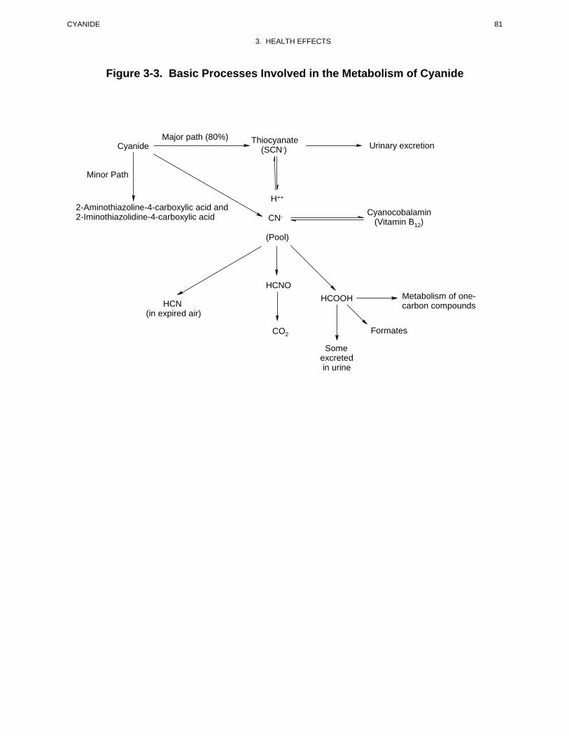
CYANIDE 81
3. HEALTH EFFECTS
Figure 3-3. Basic Processes Involved in the Metabolism of Cyanide
Cyanide Major path (80%) Thiocyanate
(SCN-) Urinary excretion
Cyanocobalamin (Vitamin B12)
2-Aminothiazoline-4-carboxylic acid and 2-Iminothiazolidine-4-carboxylic acid
Minor Path
H++
CN
(Pool)
Metabolism of one-carbon compounds
Formates
HCOOH HCNO
CO2
HCN (in expired air)
Some excreted in urine

CYANIDE 82
3. HEALTH EFFECTS
from the donor to the enzyme, forming a persulfide intermediate. The persulfide sulfur is then transferred
from the enzyme to cyanide, yielding thiocyanate. Thiocyanate is then readily excreted in the urine as the
major metabolite. Once thiocyanate is formed, it is not converted back to cyanide.
Radioisotopic studies showed that albumin interacts with the sulfane pool and that the serum albumin
sulfane sulfur carrier complex can react with cyanide (Schneider and Westley 1969). Higher hepatic
rhodanese and lower serum albumin levels were found in mice fed a protein-free diet for 14 days
compared with mice fed a control diet (Rutkowski et al. 1985). Despite the higher rhodanese levels,
mortality following an intraperitoneal injection of sodium cyanide was higher in mice fed the protein-free
diet both with and without thiosulfate pretreatment. In mice fed the control diet in reduced amounts,
serum albumin levels were higher than controls. Mortality in food-deprived mice was also higher
compared with controls, but only at high cyanide doses when thiosulfate was also administered.
However, the pharmacokinetic studies in dogs, in which thiosulfate administration increased the rate of
elimination of cyanide, suggest that the sulfane sulfur pool may play an important role as the central
compartment for cyanide detoxification (Sylvester et al. 1983; Way 1984).
The species and tissue distribution of rhodanese is highly variable (Himwich and Saunders 1948). In
dogs, the highest activity (conversion of cyanide to thiocyanate) of rhodanese was found in the adrenal
gland, ≈2.5 times greater than the activity in the liver. Monkeys, rabbits, and rats had the highest
rhodanese activity in the liver and kidney, with relatively low levels in the adrenals. It should be noted
that total rhodanese activity in other species was higher than in dogs, which is consistent with the greater
susceptibility of dogs to the acute effects of cyanide. Similar low levels of activity of the enzyme were
found for the brain, testes, lungs, spleen, and muscle among various species.
In vitro studies with rat tissues indicated that rhodanese activity was ≈7 times higher in the nasal mucosa
than in the liver (Dahl 1989). Furthermore, kinetic constants for rhodanese in mitochondria were higher
in nasal than in liver tissue.
Figure 3-4 illustrates the minor pathway for metabolism of cyanide in mammalian systems in which
cyanide chemically combines with the amino acid cystine. This chemical reaction yields cysteine and
β-thiocyanoalanine that is further converted to form 2-aminothiazoline-4-carboxylic acid and its tautomer,
2-iminothiazolidiene-4-carboxylic acid (Wood and Cooley 1955).

CYANIDE 83
3. HEALTH EFFECTS
Figure 3-4. Minor Path for the Removal of Cyanide from the Body
NH2
S C C COOH NH2
H2 H S C C COOH
S C C H
COOH H2 H
H2 CN NH2
Cystine Beta-Thiocyanoalanine
NH2NH CC
N S NHS C C COOH C C COOH H2 H H2 H
2-Aminothiazoline-4-carboxylic acid 2-Imino-4-thiazolidine-carboxylic acid
Source: Ansell and Lewis 1970

CYANIDE 84
3. HEALTH EFFECTS
Reactions of cyanide with the salts or esters of some amino acids (e.g., pyruvate, α-ketoglutarate,
oxaloacetate) lead to formation of cyanohydrin intermediates and their incorporation into intermediary
metabolism.
The ability of cyanide to form complexes with some metallic ions such as cobalt is the basis for the
reaction with hydroxocobalamin that yields cyanocobalamin. Cyanocobalamin (vitamin B12), which
contains cyanide and cobalt, is essential for the health of mammalian organisms.
3.4.4 Elimination and Excretion
3.4.4.1 Inhalation Exposure
Following chronic occupational exposure to 0.19–0.75 ppm hydrogen cyanide, 24-hour urinary levels of
thiocyanate were 6.23 (smokers) and 5.4 μg/mL (nonsmokers) in exposed workers as compared with
3.2 (smokers) and 2.15 μg/mL (nonsmokers) in the controls (Chandra et al. 1980). This study
demonstrates that tobacco smoking contributes to higher thiocyanate levels excreted in the urine. No
studies were located regarding excretion of cyanide in animals after inhalation exposure to cyanide.
3.4.4.2 Oral Exposure
Cyanide metabolites are normally excreted in urine (Vassel et al. 1944) with small amounts eliminated
through the lungs. Urinary excretion of thiocyanate was monitored in a man after ingestion of ≈3–5 g
potassium cyanide (15–25 mg CN–/kg) (Liebowitz and Schwartz 1948). The results indicated that the
patient excreted 237 mg of thiocyanate over a 72-hour period. This quantity was substantially more than
the normal average amount of thiocyanate in urine, which varies between 0.85 and 14 mg/24 hours.
Thirty-one children who had consumed flour made from insufficiently processed cassava had mean
urinary thiocyanate levels of 757 μmol/L, compared with 50 μmol/L in those children who had consumed
sufficiently processed cassava (Tylleskar et al. 1992). In another study (Mlingi et al. 1993), mean urinary
thiocyanate was 490 μmol/L in a village affected by Konzo disease and 350 μmol/L in an unaffected
village, with the villages being comparable in all other respects.
When male Sprague-Dawley rats were given 2 mg CN–/kg [14C] potassium cyanide, urinary excretion of
radioactivity reached 47% of the dose within 24 hours following administration (Farooqui and Ahmed
1982). When [14C] sodium cyanide was injected subcutaneously into rats at a level of 8.3 μmol, no

CYANIDE 85
3. HEALTH EFFECTS
difference in radioactivity eliminated was observed between the group pretreated for 6 weeks with a diet
containing 0.7 mg CN–/kg as potassium cyanide and their matching controls (Okoh 1983). Most of the
radioactivity was detected in the urine (89% by 24 hours). Thiocyanate was the major metabolite. About
4% of the radioactivity was expired, mostly as carbon dioxide.
Sousa et al. (2003) compared toxicokinetic parameters in male Wistar rats and Landrace-Large White
pigs that were given 1.2 mg CN–/kg as potassium cyanide by aqueous gavage. The half-lives of
elimination of cyanide from the blood were 0.54 hours for pigs and 0.64 hours for rats. The half-lives of
elimination of thiocyanate from the blood were 4.95 hours in pigs and 5.8 hours in rats. The overall
clearance of cyanide from the blood was reported as 0.367, and 0.379 mL/minute per kg for pigs and rats,
respectively; the clearance of thiocyanate was reported as 0.135 and 0.061 mL/minute per kg for pigs and
rats, respectively.
Orally administered cyanide and its metabolite thiocyanate were eliminated in the breast milk of lactating
goats (Soto-Blanco and Gorniak 2003). The relevance of the goat data to humans is not established.
3.4.4.3 Dermal Exposure
No studies were located regarding excretion in humans or animals after dermal exposure to cyanide.
3.4.5 Physiologically Based Pharmacokinetic (PBPK)/Pharmacodynamic (PD) Models
Physiologically based pharmacokinetic (PBPK) models use mathematical descriptions of the uptake and
disposition of chemical substances to quantitatively describe the relationships among critical biological
processes (Krishnan et al. 1994). PBPK models are also called biologically based tissue dosimetry
models. PBPK models are increasingly used in risk assessments, primarily to predict the concentration of
potentially toxic moieties of a chemical that will be delivered to any given target tissue following various
combinations of route, dose level, and test species (Clewell and Andersen 1985). Physiologically based
pharmacodynamic (PBPD) models use mathematical descriptions of the dose-response function to
quantitatively describe the relationship between target tissue dose and toxic end points.
PBPK/PD models refine our understanding of complex quantitative dose behaviors by helping to
delineate and characterize the relationships between: (1) the external/exposure concentration and target
CYANIDE 86
3. HEALTH EFFECTS
tissue dose of the toxic moiety, and (2) the target tissue dose and observed responses (Andersen and
Krishnan 1994; Andersen et al. 1987). These models are biologically and mechanistically based and can
be used to extrapolate the pharmacokinetic behavior of chemical substances from high to low dose, from
route to route, between species, and between subpopulations within a species. The biological basis of
PBPK models results in more meaningful extrapolations than those generated with the more conventional
use of uncertainty factors.
The PBPK model for a chemical substance is developed in four interconnected steps: (1) model
representation, (2) model parameterization, (3) model simulation, and (4) model validation (Krishnan and
Andersen 1994). In the early 1990s, validated PBPK models were developed for a number of
toxicologically important chemical substances, both volatile and nonvolatile (Krishnan and Andersen
1994; Leung 1993). PBPK models for a particular substance require estimates of the chemical substance-
specific physicochemical parameters, and species-specific physiological and biological parameters. The
numerical estimates of these model parameters are incorporated within a set of differential and algebraic
equations that describe the pharmacokinetic processes. Solving these differential and algebraic equations
provides the predictions of tissue dose. Computers then provide process simulations based on these
solutions.
The structure and mathematical expressions used in PBPK models significantly simplify the true
complexities of biological systems. If the uptake and disposition of the chemical substance(s) are
adequately described, however, this simplification is desirable because data are often unavailable for
many biological processes. A simplified scheme reduces the magnitude of cumulative uncertainty. The
adequacy of the model is, therefore, of great importance, and model validation is essential to the use of
PBPK models in risk assessment.
PBPK models improve the pharmacokinetic extrapolations used in risk assessments that identify the
maximal (i.e., the safe) levels for human exposure to chemical substances (Andersen and Krishnan 1994).
PBPK models provide a scientifically sound means to predict the target tissue dose of chemicals in
humans who are exposed to environmental levels (for example, levels that might occur at hazardous waste
sites) based on the results of studies where doses were higher or were administered in different species.
Figure 3-5 shows a conceptualized representation of a PBPK model.
No PBPK models were located for cyanide.

CYANIDE 87
3. HEALTH EFFECTS
Figure 3-5. Conceptual Representation of a Physiologically Based Pharmacokinetic (PBPK) Model for a
Hypothetical Chemical Substance
Lungs
L iver
Fat
S low ly perfused tissues
R ich ly perfused tissues
K idney
S kin
V E N O U S
B L O O D
A R T E R I A L
B L O O D
V max K m
Ingestion
Inha led chem ica l E xha led chem ica l
G I Tract
Feces
U rine
C hem ica ls in a ir contacting sk in
Source: adapted from Krishnan et al. 1994
Note: This is a conceptual representation of a physiologically based pharmacokinetic (PBPK) model for a hypothetical chemical substance. The chemical substance is shown to be absorbed via the skin, by inhalation, or by ingestion, metabolized in the liver, and excreted in the urine or by exhalation.

CYANIDE 88
3. HEALTH EFFECTS
3.5 MECHANISMS OF ACTION
This section presents a brief overview of any known mechanisms of metabolism, absorption, distribution,
and excretion including substance reactions or physiological processes that lead to or comprise the
mechanism(s) of toxic effect.
3.5.1 Pharmacokinetic Mechanisms
Absorption. Absorption of cyanide across the gastrointestinal mucosa depends on the pH of the gut
and the pKa and lipid solubility of the particular cyanide compound. Hydrogen cyanide is a weak acid
with a pKa of 9.2 at 25 °C. The acidic environment in the stomach favors the non-ionized form of
hydrogen cyanide and facilitates absorption. Information regarding the rapid lethal effects following oral
intake of cyanide in humans (Gosselin et al. 1984) indicates that cyanide is rapidly absorbed from the
gastrointestinal tract.
Hydrogen cyanide is moderately lipid-soluble, which, along with its small size, allows it to rapidly cross
mucous membranes and to be taken up instantly across the alveolar epithelium of the lung after
inhalation; penetration across the epidermis is less rapid. In addition, some cyanide compounds, such as
potassium cyanide, have a corrosive effect on the skin that can increase the rate of percutaneous
absorption (NIOSH 1976). Information regarding dermal absorption in animals and evidence that cyanide
can be absorbed through the skin of humans is provided in Sections 3.4.1.3 and 3.2.3, respectively.
Distribution. Cyanide is rapidly distributed by the blood throughout the body. In a study using orally
administered radioactively labeled potassium cyanide, radioactivity detected in whole blood or plasma
decreased rapidly within 6 hours. Of the low levels of radioactivity detected in the red blood cells, about
94% of the radioactivity recovered was found in the hemolysate; of which 70% was detected in the heme
fraction, 14–25% in globin, and only 5–10% in cell membranes (Farooqui and Ahmed 1982). Yamamoto
et al. (1982) determined that the pattern of distribution of cyanide did not vary with the concentration
used. Ballantyne (1983b) observed higher cyanide levels in whole blood than in serum in rabbits exposed
dermally to hydrogen cyanide, potassium cyanide, and sodium cyanide. See Section 3.4.2.1 for specific
studies on cyanide tissue distribution.

CYANIDE 89
3. HEALTH EFFECTS
Cyanide is a reactive chemical substance and has the potential to form a variety of adducts in biological
systems. A study of radiolabeled cyanide binding to mouse brain parts revealed that the hypothalamus
accumulated more label than cerebral cortex, hippocampus, or cerebellum (Borowitz et al. 1994).
Similarly, Baskin et al. (1987) found that the left ventricle of the guinea pig heart contained nearly twice
as much as the right ventricle after a brief exposure to cyanide. Binding to certain tissue constituents may
be important for decreasing the actions of cyanide and protecting cells from cyanide toxicity (Devlin et al.
1989b).
Storage. Cyanide does not accumulate in blood and tissues following chronic oral exposure. In a
study with rats administered hydrogen cyanide in the diet at <10.4 mg CN–/kg/day for 2 years, virtually
no cyanide was found in plasma or kidneys (Howard and Hanzal 1955).
Metabolism. See Section 3.4.5.
Excretion. Cyanide metabolites (of which thiocyanate is the major component) are excreted primarily
in urine, with small amounts of the metabolites eliminated through the lungs. When radioactively labeled
cyanide is administered, most of the radioactivity is detected in the urine within 24 hours (Farooqui and
Ahmed 1982; Okoh 1983). Boxer and Richards (1952) were the first to show that cyanide was oxidized
to CO2 and in the Okoh (1983) study, about 4% of the radioactivity was expired, mostly as carbon
dioxide. See Section 3.4.3.4 for information on studies examining elimination and excretion.
Effect of Dose and Duration of Exposure on Toxicity. The severity of neurological effects in
humans and animals after acute oral exposure to cyanide is dose-related (Chen and Rose 1952; Lasch and
El Shawa 1981). Central nervous system effects have been observed following acute-duration exposures
(Levine and Stypulkowski 1959a) and chronic-duration exposures (Hertting et al. 1960), via the
inhalation and oral routes. Necrosis is the most prevalent central nervous system effect following acute-
duration exposure to high concentrations of cyanide, whereas demyelination is observed in animals that
survive repeated exposure protocols (Bass 1968; Ibrahim et al. 1963).
Increased duration of exposure to inhaled cyanide in mice resulted in lower LC50 values (Higgins et al.
1972; Matijak-Schaper and Alarie 1982). Additionally, cyanide toxicity was influenced by dilution of the
gavage dose. Greater dilution resulted in higher mortality for the same total dose (Ferguson 1962).

CYANIDE 90
3. HEALTH EFFECTS
Tylleskar et al. (1992) studied a population in rural Zaire that was affected with Konzo. Konzo is
characterized by symmetric isolated bilateral involvement of upper motor neurons of abrupt onset; the
damage is permanent, but not progressive. The Konzo patients had serum thiocyanate concentrations
below those of the controls. The authors suggest that the combination of high exposure and a decreased
conversion rate because of a deficiency in suitable sulfur substrates might explain this difference. Thus,
daily exposure and decreased conversion rates may lead to high blood concentrations of cyanide that may
then lead to upper motor neuron damage. It has been suggested that defects in the metabolic conversion
of cyanide to thiocyanate, as well as nutritional deficiencies of protein and vitamin B12, play a role in the
development of central nervous system disorders such as tropical ataxic neuropathy, tobacco amblyopia,
and Leber's hereditary optic atrophy.
Route-Dependent Toxicity. A great similarity exists among cyanide-induced effects following
inhalation, oral, and dermal exposure. Signs of toxicity in target organs from acute cyanide exposure
(primarily central nervous system and heart), and chronic exposure (including central nervous system and
thyroid gland), are similar in both humans and animals regardless of route. In general, the latency of
effects is shortest by the inhalation route, similar for the oral route, but longer for the dermal route, since
the skin is a thicker barrier to penetration. The rate of cyanide absorption and, therefore, latency of toxic
effects is decreased in fasting animals.
3.5.2 Mechanisms of Toxicity
Effects of Metabolism on Toxicity. Cyanide (as hydrogen cyanide), originating in vivo by dissociation of
potassium cyanide, sodium cyanide, and other cyanogenic compounds or arising from catabolism of
cyanogenic glycosides, exerts its acute toxic effects by complexing with the ferric iron atom in
metalloenzymes, resulting in histotoxic anoxia through inhibition of cytochrome c oxidase (Rieders 1971;
Way 1984), metalloenzymes that function as the terminal oxidase of the inner mitochondrial membrane
respiratory chain. A two-step process has been proposed: cyanide as hydrogen cyanide first penetrates a
protein crevice of cytochrome c oxidase and binds to the protein (Stannard and Horecker 1948).
Hydrogen cyanide then binds to the trivalent iron ion of the enzyme, forming a relatively stable (but
reversible) coordination complex. One mole of hydrogen cyanide is bound to one mole of cytochrome c
oxidase (Van Buuren et al. 1972). As a result, the enzyme becomes unable to catalyze the reactions in
which electrons would be transferred from reduced cytochrome to oxygen. Cellular oxygen utilization is
thus impaired, with resultant reduction in or cessation of aerobic metabolism (Rieders 1971; Way 1984).
Glucose catabolism then shifts from the aerobic pathway to anaerobic metabolism including the pentose

CYANIDE 91
3. HEALTH EFFECTS
phosphate pathway, resulting in increased blood glucose, pyruvic acid, lactic acid, and nicotinamide
adenine dinucleotide (NADPH) levels, and a decrease in the adenosine triphosphate/adenosine
diphosphate (ATP/ADP) ratio (Rieders 1971; Way 1984). Wilson et al. (1994) suggest that it is the
binding of cyanide to oxidized CuB, the copper ion that is part of the dioxygen binding-site that leads to
the inhibition of cytochrome c oxidase.
The inhibition of oxygen use by cells (termed histoxic hypoxia) causes oxygen tensions to rise in
peripheral tissues (Smith 1996). This results in a decrease in the unloading gradient for oxyhemoglobin;
thus, oxyhemoglobin is carried in the venous blood (Rieders 1971). Inhibition of oxygen utilization is
thought to occur rapidly after cyanide exposure. Tadic (1992) determined that inhibition of cytochrome c
oxidase activity in rat brains was most pronounced between 15 and 20 minutes after administration of
sodium cyanide (12 mg/kg or 1.3xLD50). In addition to binding to cytochrome c oxidase, cyanide also
binds to catalase, peroxidase, methemoglobin, hydroxocobalamin, phosphatase, tyrosinase, ascorbic acid
oxidase, xanthine oxidase, and succinic dehydrogenase. These reactions may also contribute to the
classic signs of cyanide toxicity (Ardelt et al. 1989; Rieders 1971). Information on mechanisms of
toxicity in target organs is presented below.
Target Organ Toxicity. The central nervous system is the primary target for cyanide toxicity in humans
and animals. Acute inhalation of high concentrations of cyanide provokes a brief central nervous system
stimulation followed by depression, convulsions, coma, and death in humans (Bonsall 1984; Chen and
Rose 1952; Peden et al. 1986; Potter 1950; Singh et al. 1989) and in animals (Haymaker et al. 1952;
McNerney and Schrenk 1960; Purser et al. 1984). The effects are probably due to rapid biochemical
changes in the brain, such as changes in ion flux, neurotransmitter release, and possibly peroxide
formation (Johnson and Isom 1985; Kanthasamy et al. 1991a, 1994; Persson et al. 1985). In both in vivo
and in vitro studies using brain tissue, the sensitivity of mitochondrial cytochrome c oxidase activity to
inhibition by cyanide was greater than the inhibition of mitochondrial respiratory activity. Only after
cytochrome c oxidase activity was depressed by >50% was a large decrease in respiratory activity
detected, suggesting that a large portion of cytochrome c oxidase may serve as a functional reserve.
Cyanide poisoning likely involves mechanisms in addition to inhibition of cytochrome c oxidase activity
(Pettersen and Cohen 1993). Cyanide is a strong nucleophile with multiple effects including release of
secondary neurotransmitters, release of catecholamines from adrenal glands and adrenergic nerves, and
inhibition of antioxidant enzymes in the brain (Smith 1996). However, the extremely low concentration
of cyanide required to inhibit the oxidase, the rapid interaction of hydrogen cyanide with the enzyme, and
the key role of cytochrome c oxidase in aerobic metabolism all combine to make cyanide inhibition of the

CYANIDE 92
3. HEALTH EFFECTS
terminal step of electron transport (Chance and Erecinsk 1971; Gibson and Greenwood 1963), the key
molecular target in cyanide poisoning.
Inhalation and oral studies in animals have shown that acute or chronic cyanide exposure leads to
encephalopathy in both white and gray matter. In particular, damage has been observed in regions such
as the deep cerebral white matter, corpus callosum, hippocampus, corpora striata, pallium, and substantia
nigra. White matter may be more sensitive because of its relatively low cytochrome c oxidase content.
Rats injected subcutaneously with daily maximal doses between >3.7 and 9.2 mg CN–/kg/day (not
averaged) 3 days/week for 3 months developed necrotic lesions of the corpus callosum and optic nerve,
but there was not a consistent dose-response (Lessell 1971); this may reflect variability in diffusion of
cyanide into the systemic circulation by the subcutaneous injection route. High mortality was observed
among exposed animals. These effects have been observed following acute-duration exposures (Levine
and Stypulkowski 1959a) and chronic-duration exposures (Hertting et al. 1960). Necrosis is a prevalent
central nervous system effect following acute exposure to high concentrations of cyanide, whereas
demyelination is observed in animals that survive repeated exposure protocols (Bass 1968; Ibrahim et al.
1963). The mechanism of cyanide-induced demyelination is not completely understood, but the evidence
suggests that a direct effect of cyanide on white matter may not be necessary. It has been suggested that
local edema affecting the oligodendrocytes and caused by vascular changes triggered by cyanide
represent a primary event in demyelination (Bass 1968; Ibrahim et al. 1963). Aitken and Braitman (1989)
determined that cyanide has a direct effect on neurons not mediated by its inhibition of metabolism.
Consistent with the view that cyanide toxicity is due to the inability of tissue to utilize oxygen is a report
that in cyanide-intoxicated rats, arterial pO2 levels rose, while carbon dioxide levels fell (Brierley et al.
1976). The authors suggested that the low levels of carbon dioxide may have led to vasoconstriction and
reduction in brain blood flow; therefore, brain damage may have been due to both histotoxic and anoxic
effects. Partial remyelination after cessation of exposure has been reported, but it is apparent that this
process, unlike that in the peripheral nervous system, is slow and incomplete (Hirano et al. 1968). The
topographic selectivity of cyanide-induced encephalopathy may be related to the depth of acute
intoxication and distribution of blood flow, which may result in selected regions of vascular insufficiency
(Levine 1969).
Several studies have suggested that a disruption in neuronal calcium regulation may be an important
factor in the manifestation of cyanide-induced neurotoxic events following acute exposure. The
predominance of anaerobic metabolism in a cyanide-poisoned cell decreases the ATP/ADP ratio, or
energy charge (Isom et al. 1975), and thus alters energy-dependent processes such as cellular calcium

CYANIDE 93
3. HEALTH EFFECTS
homeostasis (Johnson et al. 1986). Elevated levels of intracellular calcium in a cyanide-exposed,
presynaptic squid neuron were observed in an in vitro study (Adams et al. 1985). Elevated levels of
neuronal calcium may initiate release of neurotransmitters from the presynaptic terminal, which can
activate the nervous system (Maduh et al. 1990a). Levels of whole-brain calcium increased when
potassium cyanide was administered subcutaneously to mice. These increases were correlated with
cyanide-induced tremors (Johnson et al. 1986). Brain injury may be associated with cyanide-induced
endogenous glutamate release, mediated by both calcium dependent and independent mechanisms, which
in turn produce excitotoxic responses in select brain areas (Patel et al. 1991, 1992, 1993). In examining
receptor subtypes involved in mediating cyanide-induced toxicity, sodium cyanide-induced cytotoxicity
was found to be mediated primarily by activation of the N-methyl-D aspartate (excitatory amino acid)
receptor. Sturm et al. (1993) examined the ability of adenosine to attenuate the excitotoxicity secondary
to glutamate receptor activation following potassium cyanide exposure in hippocampal neuronal cell
cultures. The authors concluded that neuronal cell death was mediated at least in part by glutamate and
that the cell death was attenuated by adenosine via the A1-specific receptor. Increases in intracellular
calcium have also been associated with cyanide-induced effects on vascular smooth muscle and cardiac
muscle, possibly inducing cell damage (Allen and Smith 1985; Robinson et al. 1985a). These effects may
result from ischemia-induced increases in extracellular potassium, which in turn enhance cellular
permeabilities to calcium (Robinson et al. 1985b). Furthermore, changes in cytosolic pH and dysfunction
of hydrogen ion handling mechanisms were observed in neuronal cells exposed in vitro to cyanide
(Maduh et al. 1990b). Pazdernik et al. (1994) reported an increase of local cerebral glucose utilization
(LCGU) in many regions of the brain within a minute after sublethal exposure to 2.7–5 mg CN–/kg as
sodium cyanide by controlled intravenous infusion over 1 hour. However, by 1 hour, there was a global
increase in LCGU in almost every region of the brain. LCGU values returned to normal in all regions
except the choroid plexus by 6 hours and in that region as well by 24 hours. These results support the
expectation that cyanide causes a shift from aerobic to anaerobic metabolism, as illustrated by increases in
extracellular lactate and pyruvate and in LCGU.
When cyanide blocks oxidative metabolism in mitochondria, cells shift their metabolism and enhanced
glucose utilization occurs. One consequence of this altered metabolic pattern is accumulation of
nicotinamide adenine dinucleotide (NADH), which is a powerful stimulant of calcium mobilization from
cell stores through "inositol triphosphate receptors." Elevated calcium damages cells. Increase in cellular
NADH, therefore, is an important event in the toxic action of cyanide (Kaplin et al. 1996).

CYANIDE 94
3. HEALTH EFFECTS
Studies have shown that cyanide releases catecholamines from rat pheochromocytoma cells and brain
slices (Kanthasamy et al. 1991b), from isolated bovine adrenal glands (Borowitz et al. 1988), and from
adrenals of mice following subcutaneous injection of high doses of potassium cyanide (Kanthasamy et al.
1991b). Thus, it was proposed that the cardiac and peripheral autonomic responses to cyanide are
partially mediated by an elevation of plasma catecholamines (Kanthasamy et al. 1991b). Dopamine levels
in potassium cyanide-treated animals were significantly decreased in striatum and hippocampus, and
somewhat decreased in cerebral cortex of mice (Kanthasamy et al. 1994), while extracellular levels of
dopamine and homovanillic acid were increased in the brain of rats treated with sodium cyanide (Cassel
et al. 1995). Kiuchi et al. (1992) suggested that suppression of ATP production by sodium cyanide
induces an abrupt and remarkable increase in dopamine release from the nerve terminal in the striatum.
Kanthasamy et al. (1994) also observed that in striatal and hippocampal tissues, but not in cerebral cortex,
malondialdehyde levels increased indicating the occurrence of lipid peroxidation in these brain regions.
In addition, reduced numbers of tyrosine hydroxylase (TH) positive cells indicated a loss of dopaminergic
neurons (Kanthasamy et al. 1994). Behavioral effects seen in the mice were reversed by administration of
l-DOPA (treatment for dopamine-deficiency). Ardelt et al. (1994) also evaluated hydroperoxide
generation as a potential mechanism of cyanide neurotoxicity. Increased lipid peroxidation was observed
in brain and kidney, but not in liver or heart. It was also determined that calcium plays a critical role in
lipid peroxidation in neuronal cells. Subcellular fractionation of brain tissue showed an increase in lipid
peroxidation in the microsomal but not mitochondrial fraction. Matsumoto et al. (1993) evaluated the
involvement of extracellular calcium in dopamine release from rat striatum resulting from cyanide
exposure. A gradual increase in intracellular calcium was observed during incubation of sodium cyanide
with striatal slices. The excessive influx of extracellular calcium during sodium cyanide perfusion may
contribute to the changes in dopamine levels in striatum and to the observed suppression of dopamine
release in response to high potassium stimulation. Release of dopamine was not suppressed by perfusion
with a calcium-free solution; thus, additional mechanisms other than the opening of calcium channels
must also be involved in dopamine release by cyanide. Decreased dopamine uptake has been suggested
as an explanation for this increase in dopamine, since dopamine uptake is driven by a sodium gradient
that is maintained by the Na/K ATPase and could be reduced if ATP is depleted. Cyanide did not affect
monamine oxidase or catechol-o-methyl transferase, suggesting that a disturbance in dopamine
metabolism did not lead to extracellular dopamine elevation (Matsumoto et al. 1993).
Mills et al. (1999) reported that there is more than one mode of cell death operating in the brains of mice
injected with potassium cyanide. Extensive DNA fragmentation, pyknosis, and chromosome
condensation, all characteristics of apoptosis, were observed in the parietal and suprarhinal regions of the
CYANIDE 95
3. HEALTH EFFECTS
motor cortex of treated mice. However, necrotic lesions with astrocytic gliosis were found in the
substantia nigra. Pretreatment with the antioxidant alpha-phenyl-tert-butyl nitrone reduced cortical DNA
fragmentation, but had no effect on the necrotic lesions produced in the substantia nigra.
Prabhakaran et al. (2002) similarly reported different modes of death induced by cyanide in primary
cultures of rat cortical or mesencephalic neurons; the mode of cell death and the reactive oxygen species
generated differed in the two kinds of cells. Cortical neurons exhibited apoptosis, with increases in
hydrogen peroxide and superoxide, and a moderate change in mitochondrial membrane potential, leading
to release of cytochrome c and activation of caspase-3-like protease (a cysteine protease associated with
apoptosis). Mesencephalic neurons exhibited necrosis involving excess nitric oxide and superoxide, with
a more pronounced reduction in mitochondrial membrane potential. Additional studies demonstrated that
necrosis of exposed mesencephalic cells or cortical neurons exposed to 0.5–0.6 mM KCN was induced by
the upregulation of uncoupling protein 2 (UCP-2), a protein of the inner mitochondrial membrane (Li et
al. 2005; Prabhakaran et al. 2005). UCP-2 increases proton leak across the inner mitochondrial
membrane, dissociating respiration from ATP synthesis. In experiments preventing the expression of
UCP-2, the necrotic death of cultured mesencephalic cells exposed to cyanide was also prevented
(Prabhakaran et al. 2005).
The mediation of cyanide-induced apoptosis has been studied in cultured cortical neurons exposed to
0.3 mM cyanide (Shou et al. 2002, 2003). Treatment with cyanide activated p38 mitogen-activated
protein (MAP) kinase within 30 minutes, an upstream event necessary for the translocation of Bax protein
from the cytosol to mitochondria 2.5 hours later (Shou et al. 2003). Translocation of Bax protein to
mitochondria is a required step in the release of cytochrome c from mitochondria as well as the caspase
cascade that regulates apoptosis. Cyanide treatment of cortical neurons also results in the activation of
the redox-sensitive transcription factor NF-κB, and its translocation to the nucleus, where it upregulates
expression of the pro-apoptotic proteins Bax and Bcl-XS (Shou et al. 2002). Increased cytosolic calcium
levels also contribute to apoptosis of cyanide-treated cortical neurons (Shou et al. 2004). Increased
calcium activates cellular calcineurin, which stimulates the activation of the protein known as BAD (Bcl
2/Bcl-XL-antagonist, causing cell death) and its translocation to mitochondria within 1 hour of treatment
with cyanide. The net effect of BAD is to selectively inhibit proteins (Bcl-1/Bcl-XL) that are antagonists
to apoptosis (Shou et al. 2004).
It has been noted that survivors of cyanide poisoning incidents may develop Parkinsonian-like signs, with
lesions in the substantia nigra, a dopaminergic center, confirmed by MRI (Carella et al. 1988; Chin and

CYANIDE 96
3. HEALTH EFFECTS
Calderon 2000; Grandas et al. 1989; Feldman and Feldman 1990; Rachinger et al. 2002; Rosenberg et al.
1989; Rosenow et al. 1995; Uitti et al. 1985; Zaknun et al. 2005). Jones et al. (2000, 2003) have
presented evidence based on experiments on PC12 cells (a pheochromocytoma cell line that can be
induced to differentiate as neurons) and fetal rat mesencephalic cells indicating that cyanide toxicity is
exacerbated by the oxidation of dopamine. Increases in apoptosis and reactive oxygen species occurred at
higher levels in PC12 cells incubated in dopamine plus potassium cyanide compared to those incubated in
either chemical separately; concentrations of potassium cyanide that had no effect on fetal rat midbrain
cells significantly increased the adverse effects of added dopamine. Toxicity in one or both systems was
reduced by preincubation with antioxidants (superoxide dismutase, glutathione catalase), an inhibitor to
nitric oxide synthase (Nomega-nitro-L-arginine methyl ester), and the peroxynitrite scavenger uric acid.
The authors suggest that the inactivation of antioxidant enzymes by cyanide as described by Ardelt et al.
(1989) may render neurons more vulnerable to the adverse effects of dopamine oxidation. Dopaminergic
brain centers would therefore be more sensitive to cyanide neurotoxicity. In cultured cerebellar granule
cells taken from 8-day-old rat pups, cyanide treatment generated nitric oxide and reactive oxygen species
concurrently, resulting in lipid peroxidation (Gunasekar et al. 1996).
Chao et al. (1996) investigated the possibility that cyanide had an effect on motor neurons that was
independent of respiratory impairment. In mouse triangularis sterni and diaphragm nerve-muscle
preparations under glucose-free conditions, 10 μM sodium cyanide increased spontaneous transmitter
release. This was correlated with a depression of ATP-sensitive potassium currents, an effect that was
antagonized by diazoxide, an opener of ATP-sensitive K+ channels. The authors suggest that cyanide
causes depolarization of motor nerve terminals via its effect on the ATP-sensitive K+ channels. Cassel et
al. (1994) examined the in vitro effects of sodium cyanide on two forms of monoamine oxidase (MAO),
an enzyme important in regulation of biogenic amines in the brain and peripheral tissue. In striatal tissue,
cyanide produced a dose-dependent increase in the activity of MAO-A but not MAO-B. Greer and Carter
(1995) investigated the effects of hydrogen cyanide on the neural mechanisms controlling breathing.
Cyanide, at concentrations considered lethal in vivo, caused a modest depression of the frequency and
amplitude of inspiratory rhythmic discharge. The neuronal network underlying respiration continued to
function for hours in the presence of very high concentrations of cyanide. The authors hypothesized that
the rapid suppression of breathing caused by cyanide in vivo is due to changes in neuronal excitability in
respiratory centers in the central nervous system.
Results of in vitro studies suggest an interaction between calcium ions and cyanide in cardiovascular
effects (Allen and Smith 1985; Robinson et al. 1985a). It has been demonstrated that exposure to cyanide

CYANIDE 97
3. HEALTH EFFECTS
in metabolically depleted ferret papillary muscle eventually results in elevated intracellular calcium
levels, but only after a substantial contracture develops (Allen and Smith 1985). The authors proposed
that intracellular calcium may precipitate cell damage and arrhythmias. The mechanism by which
calcium levels are raised was not determined. Franchini and Krieger (1993) produced selective
denervation of the aortic and carotid bifurcation areas, and confirmed the carotid body chemoreceptor
origin of cardiovascular, respiratory, and certain behavioral responses to cyanide in rats. Bradycardia and
hyperventilation induced by cyanide are typical responses evoked by carotid body chemoreceptor
stimulation (Franchini and Krieger 1993).
The respiratory effects of cyanide include dyspnea, asphyxia, and a decrease in respiratory rate (Blanc et
al. 1985; Matijak-Schaper and Alarie 1982; McNerney and Schrenk 1960). A recent study (Bhattacharya
et al. 1994) demonstrated an initial increased air flow, transthoracic pressure, and tidal volume
accompanied by a significant decrease in pulmonary phospholipids following inhalation of hydrogen
cyanide in rats. This study also showed that hydrogen cyanide exhibited a direct effect on pulmonary
cells in rats.
Cyanide-induced effects on the thyroid gland are particularly important in chronic cyanide exposures and
are discussed in several studies. Thiocyanate markedly inhibits accumulation of iodine by the thyroid
gland, thus decreasing the ability of the gland to maintain a concentration of iodine above that of blood
(VanderLaan and Bissell 1946). In addition, thiocyanate may inhibit the iodination process, thus
interfering with the binding of glandular iodine and reducing the formation of thyroxine (Ermans et al.
1972). Changes in thyroid chemistry reported in individuals chronically exposed to cyanide have not
been accompanied by manifestations of hypothyroidism. Fukayama et al. (1992) studied the antithyroid
action of thiocyanate in a culture system of thyroid follicles. Thiocyanate concentrations equivalent to
serum levels in smokers showed three independent antithyroid actions, including inhibition of iodide
transport, inhibition of binding of iodide in the thyroid, and increased iodide efflux. The discrepancy in
the potency of the antithyroid activity of thiocyanate in vivo and in vitro appears to be due to the presence
of iodide and moieties such as the perchlorate ion, which is known to alter the effect of thiocyanate on the
thyroid (Van Middlesworth 1986).
Persons with a metabolic disturbance in the conversion of cyanide to thiocyanate may be at greater risk
from the toxic effect of cyanide. A defect in the rhodanese system and vitamin B12 deficiency have been
noted in persons with tobacco amblyopia and Leber's hereditary optic atrophy exposed to tobacco smoke
which contains cyanide (Wilson 1983). Iodine deficiency, along with excess chronic exposure to cyanide,

CYANIDE 98
3. HEALTH EFFECTS
may, in certain cases, be involved in the etiology of such thyroid disorders as goiter and cretinism
(Delange and Ermans 1971; Ermans et al. 1972). Also, protein deficiencies and vitamin B12 and
riboflavin, and other deficiencies may subject people who eat foods high in cyanogenic glycosides to
increased risk of neuropathies (Makene and Wilson 1972; Osuntokun 1972; Osuntokun et al. 1969).
Patients with motor neuron disease (amyotrophic lateral sclerosis) possess a disorder in cyanide
metabolism that may result in higher susceptibility to cyanide (Kato et al. 1985).
Carcinogenesis. No studies were located regarding carcinogenic effects of cyanide exposure in humans
or animals following any route of exposure. Therefore, no mechanism of carcinogenesis can be
discussed.
Caveat Regarding in vitro Studies. During a study of the effect of sodium cyanide on cultured SH-SY5Y
human neuroblastoma cells, Arun et al. (2005) observed that no significant toxicity was observed up to
concentrations of 10 mM and conducted further experiments to determine the cause of the apparent
resistance to cyanide. Culturing at 37 °C for 2 hours resulted in variable depletion of cyanide from the
medium, depending on the type of vessel: by 3.9% using 15-mL capped polypropylene tubes, by 22.4%
using vented cap 25-cm2 culture flasks, and by 57.2–86.7% using unsealed multiwell plates (12-, 24-, or
96-well). Cyanide loss from standard Dulbecco’s monified Eagle’s/Ham’s F12 medium (DMEM) with
10% fetal bovine serum (FBS) was compared to the loss from individual constituents of medium tested
after 2-hour culturing at room temperature. Cyanide loss from solution was 68.6% from an amino acid
mixture, 47.9% from glucose solution, 36.3% from phenol red (typically included as a pH indicator), and
81.1% from standard DMEM plus 10% FBS. Arun et al. (2005) measured a 10% loss of sodium cyanide
from deionized water (0.2 mL volume) over a 2-hour period at room temperature and higher losses when
a protein donor (alanine) was present. Release of HCN by outgassing accompanied cyanide depletion
from the medium. The results of these experiments indicate there is measurable loss of cyanide from
solution where vessels are not sealed. This may result in an underestimate of the toxicity of cyanide in in
vitro culture experiments.
3.5.3 Animal-to-Human Extrapolations
Biological effects of cyanide in humans have been demonstrated (Smith 1996; Wexler et al. 1947).
However, no studies directly comparing the cytotoxicity of similar animal and human cells were
available. However, a difference in species susceptibility to cyanide poisoning was indicated by slightly
lower lethal concentrations in rabbits compared to rats (Ballantyne 1983a). Additionally, mortality from
cyanides applied dermally varied depending on the cyanide compound used. In the Ballantyne (1983a)

CYANIDE 99
3. HEALTH EFFECTS
study, dermal application resulted in cyanide levels in blood and serum that were lower after topical
sodium cyanide and potassium cyanide exposure than from hydrogen cyanide; however, oral exposure in
rabbits produced an LD50 of 2.3–2.7 mg CN–/kg/day, regardless of whether the source was hydrocyanic
acid, sodium cyanide, or potassium cyanide (Ballantyne 1983a).
Species and tissue distribution of rhodanese (thiosulfate sulfurtransferase), an enzyme important in
metabolizing cyanide, is highly variable (Drawbaugh and Marrs 1987; Himwich and Saunders 1948). In
dogs, the highest activity of rhodanese was found in the adrenal gland, ≈2.5 times greater than the activity
in the liver (Himwich and Saunders 1948). Monkeys, rabbits, and rats had the highest rhodanese activity
in liver and kidney, with relatively low levels in adrenals. It should be noted that total rhodanese activity
in other species was higher than in dogs, which is consistent with the greater susceptibility of dogs to the
acute effects of cyanide. Thus, dogs may not be a good model from which to extrapolate the toxicity of
cyanide to humans. Similar activities of the enzyme among the species were found for the brain, testes,
lungs, spleen, and muscle. Plasma activities of rhodanese in rats, hamsters, rabbits, and guinea pigs
ranged from 14 to 20 Units/mL compared to 31 Units/mL for Beagle dogs (Drawbaugh and Marrs 1987).
In an effort to identify appropriate animal models for testing the efficacy of methemoglobin-forming
cyanide antidotes, Rockwood et al. (2003) compared the endogenous activities of the erythrocyte NADH-
dependent enzyme methemoglobin reductase (ferricyanide reductase) in several species. Two strains of
beagles had enzyme activities roughly 40–50% lower than the mean for humans and with no overlap to
the range for the human data. The enzyme activities of the other tested species had higher means than the
human, but the ranges for the Rhesus and Aotus monkeys were similar to the human, indicating that these
would be appropriate models. Data for the marmoset, Cynomolgus monkey, and African green monkey
showed less overlap to the human data, whereas data for the ferret, chimpanzee, and baboon showed no
overlap.
3.6 TOXICITIES MEDIATED THROUGH THE NEUROENDOCRINE AXIS
Recently, attention has focused on the potential hazardous effects of certain chemicals on the endocrine
system because of the ability of these chemicals to mimic or block endogenous hormones. Chemicals
with this type of activity are most commonly referred to as endocrine disruptors. However, appropriate
terminology to describe such effects remains controversial. The terminology endocrine disruptors,
initially used by Thomas and Colborn (1992), was also used in 1996 when Congress mandated the EPA to
develop a screening program for “...certain substances [which] may have an effect produced by a

CYANIDE 100
3. HEALTH EFFECTS
naturally occurring estrogen, or other such endocrine effect[s]...”. To meet this mandate, EPA convened a
panel called the Endocrine Disruptors Screening and Testing Advisory Committee (EDSTAC), and in
1998, the EDSTAC completed its deliberations and made recommendations to EPA concerning endocrine
disruptors. In 1999, the National Academy of Sciences released a report that referred to these same types
of chemicals as hormonally active agents. The terminology endocrine modulators has also been used to
convey the fact that effects caused by such chemicals may not necessarily be adverse. Many scientists
agree that chemicals with the ability to disrupt or modulate the endocrine system are a potential threat to
the health of humans, aquatic animals, and wildlife. However, others think that endocrine-active
chemicals do not pose a significant health risk, particularly in view of the fact that hormone mimics exist
in the natural environment. Examples of natural hormone mimics are the isoflavinoid phytoestrogens
(Adlercreutz 1995; Livingston 1978; Mayr et al. 1992). These chemicals are derived from plants and are
similar in structure and action to endogenous estrogen. Although the public health significance and
descriptive terminology of substances capable of affecting the endocrine system remains controversial,
scientists agree that these chemicals may affect the synthesis, secretion, transport, binding, action, or
elimination of natural hormones in the body responsible for maintaining homeostasis, reproduction,
development, and/or behavior (EPA 1997). Stated differently, such compounds may cause toxicities that
are mediated through the neuroendocrine axis. As a result, these chemicals may play a role in altering,
for example, metabolic, sexual, immune, and neurobehavioral function. Such chemicals are also thought
to be involved in inducing breast, testicular, and prostate cancers, as well as endometriosis (Berger 1994;
Giwercman et al. 1993; Hoel et al. 1992).
Nishihara et al. (2000) screened a large number of industrial chemicals for estrogenic activity in vitro
using a two-hybrid expression system: Saccharomyces cerivisiae Y190 containing expression plasmids
for the estrogen receptor alpha and the coactivator TIF-2. At concentrations as high as 1 mM, potassium
cyanide did not induce estrogenic expression in this assay (defined as 10% of the agonist activity of
10-7 M 17-beta-estradiol). No other reports of cyanides modulating estrogen hormonal levels were
located.
3.7 CHILDREN’S SUSCEPTIBILITY
This section discusses potential health effects from exposures during the period from conception to
maturity at 18 years of age in humans, when all biological systems will have fully developed. Potential
effects on offspring resulting from exposures of parental germ cells are considered, as well as any indirect

CYANIDE 101
3. HEALTH EFFECTS
effects on the fetus and neonate resulting from maternal exposure during gestation and lactation.
Relevant animal and in vitro models are also discussed.
Children are not small adults. They differ from adults in their exposures and may differ in their
susceptibility to hazardous chemicals. Children’s unique physiology and behavior can influence the
extent of their exposure. Exposures of children are discussed in Section 6.6, Exposures of Children.
Children sometimes differ from adults in their susceptibility to hazardous chemicals, but whether there is
a difference depends on the chemical (Guzelian et al. 1992; NRC 1993). Children may be more or less
susceptible than adults to health effects, and the relationship may change with developmental age
(Guzelian et al. 1992; NRC 1993). Vulnerability often depends on developmental stage. There are
critical periods of structural and functional development during both prenatal and postnatal life, and a
particular structure or function will be most sensitive to disruption during its critical period(s). Damage
may not be evident until a later stage of development. There are often differences in pharmacokinetics
and metabolism between children and adults. For example, absorption may be different in neonates
because of the immaturity of their gastrointestinal tract and their larger skin surface area in proportion to
body weight (Morselli et al. 1980; NRC 1993); the gastrointestinal absorption of lead is greatest in infants
and young children (Ziegler et al. 1978). Distribution of xenobiotics may be different; for example,
infants have a larger proportion of their bodies as extracellular water, and their brains and livers are
proportionately larger (Altman and Dittmer 1974; Fomon 1966; Fomon et al. 1982; Owen and Brozek
1966; Widdowson and Dickerson 1964). The infant also has an immature blood-brain barrier (Adinolfi
1985; Johanson 1980) and probably an immature blood-testis barrier (Setchell and Waites 1975). Many
xenobiotic metabolizing enzymes have distinctive developmental patterns. At various stages of growth
and development, levels of particular enzymes may be higher or lower than those of adults, and
sometimes unique enzymes may exist at particular developmental stages (Komori et al. 1990; Leeder and
Kearns 1997; NRC 1993; Vieira et al. 1996). Whether differences in xenobiotic metabolism make the
child more or less susceptible also depends on whether the relevant enzymes are involved in activation of
the parent compound to its toxic form or in detoxification. There may also be differences in excretion,
particularly in newborns who all have a low glomerular filtration rate and have not developed efficient
tubular secretion and resorption capacities (Altman and Dittmer 1974; NRC 1993; West et al. 1948).
Children and adults may differ in their capacity to repair damage from chemical insults. Children also
have a longer remaining lifetime in which to express damage from chemicals; this potential is particularly
relevant to cancer.

CYANIDE 102
3. HEALTH EFFECTS
Certain characteristics of the developing human may increase exposure or susceptibility, whereas others
may decrease susceptibility to the same chemical. For example, although infants breathe more air per
kilogram of body weight than adults breathe, this difference might be somewhat counterbalanced by their
alveoli being less developed, which results in a disproportionately smaller surface area for alveolar
absorption (NRC 1993).
From the few oral studies available, the effects of cyanide on children appear to be like those of similarly
exposed adults. This is expected based on cyanide's inhibition of mitochondrial respiration in all cells.
Neurological (headache and coma), respiratory (tachypnea), cardiovascular (hypotension), and
gastrointestinal effects (vomiting) have been reported in children who have been poisoned by eating
apricot pits (Lasch and El Shawa 1981). Congenital hypothyroidism has been observed in some children
who were exposed to increased thiocyanate levels because of the maternal cassava diet during pregnancy
(Ermans et al. 1980).
Developmental studies in animals (rats or hamsters) orally exposed to potassium cyanide, cassava diets,
or one of the cyanogenic glycosides (amygdalin, linamarin) reported fetal toxicity (reduced fetal weight,
delayed ossification) and developmental anomalies (microcephaly, limb defects, encephalocele, and rib
abnormalities) in offspring (Frakes et al. 1986a; Singh 1981; Tewe and Maner 1981a; Willhite 1982).
These effects occurred at exposure levels that were toxic to the dam. A developmental study in pigs
indicates that this species is less sensitive than rodents to gestational exposure to cyanide (Tewe and
Maner 1981b). Results of a study in lactating goats indicate that cyanide and thiocyanate can be
transferred through milk to nursing offspring (Soto-Blanco and Gorniak 2003).
It is likely that the observed adverse effects on fetal growth and the timing of ossification following
maternal exposure to cyanide has multiple sources. In addition to a direct action of cyanide on
mitochondrial respiration in fetal cells, the reduction in oxygen-carrying capacity of the maternal blood
would also be expected to result in lower metabolic rates in fetuses. Furthermore, hypothyroidism,
characterized by reductions in thyroid hormone levels, would also adversely affect rates of cell
metabolism.
In goats, maternal co-administration of sodium thiocyanate prevented the rise in erythrocyte cyanide
levels caused by sodium nitroprusside (Curry et al. 1997). Sodium nitroprusside is infused intravenously
as a vasodilator for the treatment of hypertensive emergencies (Agarwal and Kumari 2003; Curry et al.
1997; Przybylo et al. 1995; Randall and St. Louis 1996; Sipe et al. 2001). In the blood, sodium

CYANIDE 103
3. HEALTH EFFECTS
nitroprusside nonenzymatically receives one electron from oxyhemoglobin, forming the nitroprusside
radical, which dissociates to nitric oxide (the vasodilator) and five cyanide ions (Przybylo et al. 1995). In
practice, sodium thiocyanate is co-administered to prevent cyanide toxicity. Curry et al. (1997) infused
sodium nitroprusside into gravid ewes, resulting in elevations of erythrocyte cyanide concentrations that
caused the death of one ewe and one fetus from cardiac toxicity. Co-administration of sodium
thiocyanate to gravid ewes prevented the elevation in erythrocyte cyanide levels in ewes and fetuses.
Curry et al. (1997) conclude that sodium thiocyanate, like cyanide and sodium nitroprusside, cross the
placenta in goats. The relevance of the goat study to humans is not known.
3.8 BIOMARKERS OF EXPOSURE AND EFFECT
Biomarkers are broadly defined as indicators signaling events in biologic systems or samples. They have
been classified as markers of exposure, markers of effect, and markers of susceptibility (NAS/NRC
1989).
Due to a nascent understanding of the use and interpretation of biomarkers, implementation of biomarkers
as tools of exposure in the general population is very limited. A biomarker of exposure is a xenobiotic
substance or its metabolite(s) or the product of an interaction between a xenobiotic agent and some target
molecule(s) or cell(s) that is measured within a compartment of an organism (NAS/NRC 1989). The
preferred biomarkers of exposure are generally the substance itself, substance-specific metabolites in
readily obtainable body fluid(s), or excreta. However, several factors can confound the use and
interpretation of biomarkers of exposure. The body burden of a substance may be the result of exposures
from more than one source. The substance being measured may be a metabolite of another xenobiotic
substance (e.g., high urinary levels of phenol can result from exposure to several different aromatic
compounds). Depending on the properties of the substance (e.g., biologic half-life) and environmental
conditions (e.g., duration and route of exposure), the substance and all of its metabolites may have left the
body by the time samples can be taken. It may be difficult to identify individuals exposed to hazardous
substances that are commonly found in body tissues and fluids (e.g., essential mineral nutrients such as
copper, zinc, and selenium). Biomarkers of exposure to cyanide are discussed in Section 3.8.1.
Biomarkers of effect are defined as any measurable biochemical, physiologic, or other alteration within an
organism that, depending on magnitude, can be recognized as an established or potential health
impairment or disease (NAS/NRC 1989). This definition encompasses biochemical or cellular signals of
tissue dysfunction (e.g., increased liver enzyme activity or pathologic changes in female genital epithelial

CYANIDE 104
3. HEALTH EFFECTS
cells), as well as physiologic signs of dysfunction such as increased blood pressure or decreased lung
capacity. Note that these markers are not often substance specific. They also may not be directly
adverse, but can indicate potential health impairment (e.g., DNA adducts). Biomarkers of effects caused
by cyanide are discussed in Section 3.8.2.
A biomarker of susceptibility is an indicator of an inherent or acquired limitation of an organism's ability
to respond to the challenge of exposure to a specific xenobiotic substance. It can be an intrinsic genetic or
other characteristic or a preexisting disease that results in an increase in absorbed dose, a decrease in the
biologically effective dose, or a target tissue response. If biomarkers of susceptibility exist, they are
discussed in Section 3.10, Populations That Are Unusually Susceptible.
3.8.1 Biomarkers Used to Identify or Quantify Exposure to Cyanide
Methods are available to measure levels of cyanide and its metabolite, thiocyanate, in blood and urine.
High blood cyanide levels of 250–300 μg/100 mL were reported in cases of death from cyanide poisoning
(Vogel et al. 1981). The relationship between increased exposure and increased urine levels of
thiocyanate was demonstrated in workers exposed occupationally to 6.4–10.3 ppm cyanide in air (El
Ghawabi et al. 1975). In another study, blood cyanide concentrations varied from 0.54 to
28.36 μg/100 mL in workers exposed to ≈0.2–0.8 ppm cyanide in air and from 0.0 to 14.0 μg/100 mL in
control workers (Chandra et al. 1988). Correspondingly, blood thiocyanate concentrations were 0.05–
2.80 mg/100 mL in exposed workers and 0.02–0.88 mg/100 mL in control workers, respectively. Data
obtained from the controls indicate that cyanide can be detected in populations exposed to low cyanide
levels in the environment. Cyanide-containing food, metabolism of certain drugs, and combustion of
nitrogenous polymers are among several sources of cyanide exposure. Furthermore, industrially polluted
air, soil, and water may contribute to higher environmental cyanide levels.
Several studies showed increased cyanide and thiocyanate levels in body fluids of smokers. The
difference between smokers and nonsmokers can be quite distinct (Maliszewski and Bass 1955). Mean
thiocyanate levels in smokers and nonsmokers, respectively, were found to be 7.1 and 2.0 μg/mL in
plasma, 75.7 and 20.3 μg/mL in saliva, and 12.3 and 2.1 μg/mL in urine. A more recent study also
reported on mean thiocyanate levels in smokers and nonsmokers, respectively (Jarvis 1989). Levels
reported were 7.1 and 2.9 μg/mL in plasma, 142 and 76 μg/mL in saliva, and 9.0 and 5.8 μg/mL in urine.
Another study found a correlation between the number of cigarettes smoked per day and the thiocyanate
levels in plasma and in saliva (Yamanaka et al. 1991). Based on changes in salivary thiocyanate in six

CYANIDE 105
3. HEALTH EFFECTS
smokers who stopped smoking, this study estimated the half-life of salivary thiocyanate to be 9.5 days. In
addition, infants living in homes with family members who smoked heavily were found to have
significantly higher serum thiocyanate levels than those infants who were not exposed to cigarette smoke
in the home (Chen et al. 1990). It is unclear whether passive smoking (exposure of a nonsmoker to air
contaminated with tobacco smoke) is a factor in elevated fetal serum thiocyanate levels. In one study,
fetal thiocyanate levels were increased in association with passive smoking in the home (Bottoms et al.
1982), while another study did not report an association (Hauth et al. 1984).
Whether it is more appropriate to use whole blood or plasma for measuring cyanide concentrations has
been the subject of several reports. Cyanide plasma levels are usually about one-third to one-half,
depending on the species, those found in whole blood (Ballantyne 1983a). However, they can more
closely reflect the actual tissue dose. Furthermore, cyanide was found to attach more readily to plasma
albumin than to hemoglobin (McMillan and Svoboda 1982). It was suggested that hemoglobin in
erythrocytes binds cyanide molecules, but does not play any role in their metabolism. Some authors
argue that cyanide in red blood cells may be biologically active (Way 1984). In addition, it is known that
cyanide rapidly leaves serum and plasma, especially in the first 20 minutes. It may be appropriate to
measure cyanide in both whole blood and plasma. Whole blood samples can be stored at 4 °C for several
weeks with little change in cyanide content.
In cyanide-poisoning cases, any blood levels of cyanide >0.02 mg/100 mL indicate a toxic situation
(Berlin 1977). However, because cyanide binds tightly to cytochrome c oxidase, serious effects can also
occur at lower levels; therefore, the clinical condition of the patient should be considered when
determining proper therapy. Linden and Lovejoy (1998) presented a rough estimate of blood cyanide
levels at which symptoms appear: flushing and tachycardia at 0.05–0.1 mg/100 mL, obtundation (dulled
sensibility) at 0.1–0.25 mg/100 mL, coma and respiratory depression at 0.25–0.3 mg/100 mL, and death
at >0.3 mg/100 mL.
An almond-like smell in the breath of a poisoned patient can warn a physician that the individual may be
suffering from cyanide poisoning. Approximately 60–70% of the population can detect the bitter almond
odor of hydrogen cyanide. The odor threshold for those sensitive to the odor is estimated to be 1–5 ppm
in the air. However, even at high toxic concentrations, up to 20% of all individuals are genetically unable
to smell hydrogen cyanide (Snodgrass 1996). Some effects of cyanide that can also be used to monitor
exposure are discussed in Section 3.8.2.

CYANIDE 106
3. HEALTH EFFECTS
3.8.2 Biomarkers Used to Characterize Effects Caused by Cyanide
Cyanide can inhibit enzymatic activity by binding to some metallic moieties in metalloenzymes (Ardelt et
al. 1989; Way 1984) and cytochrome c oxidase is especially sensitive to cyanide inhibition. Dose-related
reductions in cytochrome c oxidase activity were detected in various organs of rats exposed to oral doses
of potassium cyanide (Ikegaya et al. 2001); this marker was suggested as a method of diagnosis for
samples taken within 2 days post-mortem. Consequent to the inhibition of cytochrome c oxidase,
theoretically, oxygen cannot be used and histotoxic anoxia occurs. Elevated plasma lactate
concentrations, resulting from the shift to anaerobic metabolism, have been used to assess the severity of
cyanide poisoning in humans (Baud et al. 1996, 2002). Death is caused by respiratory failure. Dyspnea,
palpitations, hypotension, convulsions, and vomiting are among the first effects of acute cyanide
poisoning (see Section 3.2). Ingestion of amounts ≥50–100 mg sodium or potassium cyanide may be
followed by almost instantaneous collapse and cessation of respiration (Hartung 1982). Data summarized
by Hartung (1982) indicate that exposure to a concentration in air of 270 ppm causes immediate death;
concentrations of 181 and 135 ppm are fatal after 10 and 20 minutes of exposure, respectively;
concentrations between 45 and 55 ppm can be tolerated for 30–60 minutes with immediate or late effects;
and 18–36 ppm may produce slight symptoms after several hours of exposure. Following chronic
exposure, cyanide has been associated with the development of tropical neuropathy, tobacco amblyopia,
and Leber's hereditary optic atrophy (Wilson 1965). Chronic exposure to cyanide arising from
consumption of cyanogenic plant foods has also been connected with the occurrence of endemic goiter
(Delange and Ermans 1971).
Neuropathological sequelae of acute cyanide poisoning have been detected in the brain by magnetic
resonance imaging (MRI) and positron emission tomography (PET). MRI techniques identified brain
lesions that developed in the weeks following a poisoning event, typically in the globus pallidus,
putamen, substantia nigra, and cerebellum (Rosenberg et al. 1989; Rosenow et al. 1995). PET has been
used to localize deficiencies in dopa uptake in the striatum and reduced glucose metabolism in the
cerebral cortex and other brain regions affected by cyanide (Rosenow et al. 1995). These imaging
methods cannot determine that cyanide was the cause of the lesions, but provide a means of monitoring
changes in the extent of brain lesions following cyanide exposure.
In the development of antidotes to cyanide, the following neurochemical biomarkers of cyanide toxicity
have been considered (Isom and Borowitz 1995): inhibition of cytochrome c oxidase, activation of
voltage sensitive calcium channels, activation of receptor operated calcium channels, elevation of

CYANIDE 107
3. HEALTH EFFECTS
cytosolic free Ca2+, activation of intracellular calcium cascades, inhibition of antioxidant enzymes
(superoxide dismutase, catalase, and glutathione peroxidase), peroxidation of membrane lipids, and
generation of reactive oxygen species.
Genetic markers for cyanide-induced hypoxia have been identified in several human cell lines (human
intestinal epithelial T84 cells and Jurkat T cells) exposed to sodium cyanide in vitro (Kiang et al. 2003).
Cyanide treatment upregulated the expression of inducible nitric oxide synthase (iNOs) and heat shock
protein-70 (HSP-70) in both cell types, p53 in T84 cells and the protooncogene Bcl-2 in Jurkat T cells.
Cellular caspase-3 activity, indicative of apoptosis, was also significantly increased in both cell types. An
inhibitor to iNOs (Nomega-nitro-L-arginine or LNNA) abolished the cyanide-induced increase in iNOs,
HSP-70, and Bcl-2 and the increase in caspase-3 activity. These studies indicate genetic responses to
cyanide exposure in vitro and could provide a strategy for comparing tissue-specific responses to cyanide
and developing therapeutic interventions following cyanide exposure in vivo.
For more information on biomarkers for renal and hepatic effects of chemicals see ATSDR/CDC
Subcommittee Report on Biological Indicators of Organ Damage (1990) and for information on
biomarkers for neurological effects see OTA (1990).
3.9 INTERACTIONS WITH OTHER CHEMICALS
Interactions in the context of this profile refer to modifications in toxic responses when an organism is
exposed to another compound in addition to cyanide. A number of compounds act in synergy with
cyanide to produce toxic effects. In smoke, both hydrogen cyanide and carbon monoxide would
potentially increase central nervous system effects in exposed individuals (Birky and Clarke 1981). High
blood cyanide levels were found in fire victims; however, the carboxyhemoglobin levels were also high.
Thus, it is difficult to assess the relative significance of hydrogen cyanide in the toxicity from smoke
inhalation.
In an investigation to examine toxicological interactions of the primary fire gases, the additive,
synergistic, or antagonistic effects of combinations of hydrogen cyanide with carbon monoxide or with
carbon dioxide on the 30-minute LC50 value for hydrogen cyanide alone were determined in rats (Levin et
al. 1987). Co-exposure of rats to hydrogen cyanide (LC50=110 ppm) and carbon monoxide
(LC50=4,600 ppm) resulted in lethal effects of these two gases that were additive. In contrast, co
exposure to hydrogen cyanide and 5% carbon dioxide (not lethal by itself) resulted in an increase in

CYANIDE 108
3. HEALTH EFFECTS
lethality of hydrogen cyanide, reflected as a decrease of the hydrogen cyanide LC50 value to 75 ppm.
Dodds et al. (1992) also investigated the effect of simultaneous exposure to cyanide and carbon monoxide
in rats, and found an additive effect on certain parameters, including lactate elevation and neurologic
index. Norris et al. (1986) reported a synergistic effect on lethality in mice that were injected with
potassium cyanide and exposed to carbon monoxide atmospheres.
Synergism has also been observed between cyanide and ascorbic acid. Guinea pigs exhibited increased
toxic effects when treated with ascorbic acid prior to oral administration of potassium cyanide (Basu
1983). When guinea pigs were treated solely with 3.2 mg CN–/kg as potassium cyanide, 38% exhibited
slight tremors, whereas 100% of those pre-treated on 3 consecutive days with 1.3 g/kg ascorbic acid
before potassium cyanide was administered exhibited severe tremors, ataxia, muscle twitches, paralysis,
and convulsions. It has been suggested that this synergistic effect results from the ability of ascorbic acid
to compete with cyanide for cysteine, thus diminishing the detoxication of cyanide.
Antidotes for cyanide poisoning have been intensively studied and reviewed (Way 1984). Cyanide
antagonists can be classified into two general groups: those that act as sulfane sulfur donors for
rhodanese-catalyzed cyanide detoxification and those that induce chemical binding of cyanide. Sulfur
donors include sodium thiosulfate, polythionates, and thiosulfates. Sodium thiosulfate has been
successfully used as an antidote against cyanide poisoning in humans for decades (Way 1984). A
pharmacokinetic study in dogs demonstrated that intravenous administration of thiosulfate increased the
detoxification rate of intravenously given cyanide to thiocyanate over 30 times (Sylvester et al. 1983). In
this study, pretreatment with thiosulfate decreased the biological half-life of cyanide from ≈39 to
≈15 minutes and also decreased the volume of distribution of cyanide from 498 to 204 mL/kg.
Thiosulfate pretreatment had prophylactic effects in guinea pigs exposed to cyanide by intravenous
infusion (Mengel et al. 1989). The protection lasted for several hours depending on the dose of
thiosulfate administered.
Antagonists that induce the chemical binding of cyanide to sites other than cytochrome c oxidase include
sodium nitrite, amyl nitrite, and hydroxylamine. These compounds generate methemoglobin, which
competes with cytochrome c oxidase for cyanide to form cyanmethemoglobin (Way 1984). Sodium
nitrite has been effectively used in the therapy of cyanide intoxication in humans especially in
combination with sodium thiosulfate (Smith 1996; Way 1984). Studies in mice demonstrated that
intraperitoneal pretreatment with sodium nitrite more than doubled the LD50 value of intraperitoneally
administered sodium cyanide from 3.18 to 7.95 mg CN–/kg (Kruszyna et al. 1982). Peak

CYANIDE 109
3. HEALTH EFFECTS
methemoglobinemia was 35% at 40 minutes. Other methemoglobin generating agents seemed to be less
effective. 4-Dimethylaminopropiophenol enhanced the LD50 value to 6.36 mg CN–/kg and
hydroxylamine to 4.66 mg CN–/kg with peak methemoglobinemia being 40 and 36%, respectively, at
7 minutes. The data suggested that sodium nitrite, a slow methemoglobin former, gave prolonged
protection against cyanide, while animals treated with fast methemoglobin formers died later on, probably
due to the cyanide release from the cyanmethemoglobin pool. An improvement of cyanide-altered
cerebral blood flow was observed in dogs treated with sodium nitrite or 4-dimethylaminophenol
following intravenous injection of hydrogen cyanide (Klimmek et al. 1983). However, neither treatment
prevented the progression of lactic acidosis.
Cobalt-containing compounds may also function as binders by forming a stable complex with cyanide. A
dramatic antagonism of the lethal effects of potassium cyanide was reported when cobaltous chloride was
administered to mice along with sodium thiosulfate (Isom and Way 1973). The authors suggested that
this synergistic antidotal effect of cobaltous chloride may be associated with the physiological disposition
of the cobaltous ion and its ability to chelate both thiocyanate and cyanide ions. This ability is also
utilized when dicobalt ethylenediamine tetra-acetate acid (Co2EDTA) is used as a cyanide antidote. An
improvement of cerebral aerobic metabolism and blood flow was observed in dogs treated with 10 mg/kg
Co2EDTA intravenously following intravenous application of 1.6 mg CN–/kg as potassium cyanide
(Klimmek et al. 1983). A lower molecular weight porphyrin cobalt compound than hydroxocobalamin
(CoTPPS) was used as an antidote to the lethal effects of cyanide (McGuinn et al. 1994). The interaction
with hydroxocobalamin (see Section 3.4.3) was also proposed as a mechanism for cyanide detoxification
in cases of acute poisoning. It was demonstrated that intravenous administration of hydroxocobalamin
(50–250 mg/kg) prior to or after intraperitoneal injection of potassium cyanide prevented lethality and
decreased cyanide-induced toxic effects in mice (Mushett et al. 1952).
Pretreatment of rats with chlorpromazine (10 mg/kg intramuscularly) and sodium thiosulfate
(1,000 mg/kg intraperitoneally) greatly decreased or abolished the increase in plasma creatine kinase
observed in rats exposed to hydrogen cyanide at 200 ppm for 12.5 minutes (O'Flaherty and Thomas
1982). In an in vitro study, chlorpromazine and 4,4'-diisothiocyano-2,2'-stilbene disulfonic acid reduced
cyanide-induced contractions in vascular smooth muscle (Robinson et al. 1985a). It was suggested that
chlorpromazine prevents cyanide-induced calcium influx and reduces peroxidation of membrane lipids
(Maduh et al. 1988).

CYANIDE 110
3. HEALTH EFFECTS
The ability of cyanide to combine with carbonyl groups of some intermediary metabolites (e.g., sodium
pyruvate, α-ketoglutarate) to form cyanohydrin has been used for antidotal purposes. Pretreatment of
mice with 1 g/kg sodium pyruvate intraperitoneally prior to subcutaneous injection of potassium cyanide
caused a statistically significant increase in the LD50 values from 3.1 to 5 mg CN–/kg (Schwartz et al.
1979). Sodium pyruvate also prevented the development of convulsions in cyanide-exposed mice.
Similarly, intraperitoneal pretreatment of mice with 2 g/kg α-ketoglutarate before the intraperitoneal
injection of potassium cyanide increased the LD50 value from 2.68 to 13.32 mg CN–/kg (Moore et al.
1986). It was further demonstrated that both sodium pyruvate and α-ketoglutarate enhanced the antidotal
effects of other cyanide antagonists (e.g., sodium thiosulfate, sodium nitrite) (Moore et al. 1986; Schwartz
et al. 1979).
A striking protection against cyanide can be elicited by a new conceptual approach, employing carrier
erythrocytes containing highly purified rhodanese (thiosulfate sulfur transferase). Several studies have
shown that resealed erythrocytes containing rhodanese and sodium thiosulfate rapidly metabolize cyanide
to the less toxic thiocyanate (Cannon et al. 1994; Petrikovic et al. 1995). Maduh and Baskin (1994)
showed that rhodanese may be regulated by protein phosphorylation and treatments that alter the
phosphorylation state of rhodanese may affect cyanide detoxification via formation of thiocyanate.
Several papers discuss the effects of oxygen alone or with other compounds on cyanide toxicity. Oxygen
alone results in minimal antagonism in mice injected with potassium cyanide and only slightly enhances
the antagonistic effects of sodium nitrite on cyanide (Sheehy and Way 1968). The antidotal effect of
sodium thiosulfate alone or in combination with sodium nitrite, was enhanced by oxygen. Oxygen-
treated mice did not show behavioral signs of cyanide intoxication below doses of 2.4 mg CN–/kg as
potassium cyanide, whereas air-treated animals showed effects such as gasping, irregular breathing, and
convulsions at levels as low as 1.2 mg CN–/kg as potassium cyanide (Isom et al. 1982). When mice were
pretreated with sodium nitrite and sodium thiosulfate and either air or oxygen, the dose of potassium
cyanide needed to cause a 59% inhibition of brain cytochrome c oxidase more than doubled in mice in an
oxygen atmosphere; all points on the oxygen curve differed significantly from the air-treatment curve.
A striking enhancement of the oxidation of glucose to carbon dioxide was observed when oxygen, sodium
nitrite, and sodium thiosulfate were given to mice dosed at 18 mg CN–/kg as potassium cyanide; no
enhancement was noticed at 4 or 6 mg CN–/kg as potassium cyanide (Isom and Way 1974). These studies
indicate that oxygen can be used in supporting classical cyanide antagonists in the therapy of cyanide
poisoning, but even hyperbaric oxygen alone had no effect on cyanide poisoning in mice (Way et al.

CYANIDE 111
3. HEALTH EFFECTS
1972). The mechanism of the action is not known, since cyanide inhibits the cellular utilization of
oxygen through inhibiting cytochrome c oxidase and, theoretically, the administration of oxygen should
have no effect or useful purpose (Smith 1996).
Propargylglycine, which is an inhibitor of the enzyme cystathionine gamma-lyase, significantly lowered
the LD50 for sodium cyanide intraperitoneally injected into rats (Porter et al. 1996). The authors
suggested that the enzyme contributes to cyanide detoxification, possibly through the pathway that
provides sulfur donors for the enzyme rhodanese.
3.10 POPULATIONS THAT ARE UNUSUALLY SUSCEPTIBLE
A susceptible population will exhibit a different or enhanced response to cyanide than will most persons
exposed to the same level of cyanide in the environment. Reasons may include genetic makeup, age,
health and nutritional status, and exposure to other toxic substances (e.g., cigarette smoke). These
parameters result in reduced detoxification or excretion of cyanide, or compromised function of organs
affected by cyanide. Populations who are at greater risk due to their unusually high exposure to cyanide
are discussed in Section 6.7, Populations with Potentially High Exposures.
Persons with a metabolic disturbance in the conversion of cyanide to thiocyanate may be at greater risk.
A defect in the rhodanese system and vitamin B12 deficiency have been associated with tobacco
amblyopia and Leber's hereditary optic atrophy in persons exposed to cyanide in tobacco smoke (Wilson
1983). Individuals with preterminal chronic renal failure have elevated serum thiocyanate levels because
of impaired clearance of thiocyanate, increasing their vulnerability to cyanide exposure (Koyama et al.
1997).
A number of dietary deficiencies may increase the risk of deleterious cyanide effects. Iodine deficiency is
involved in the etiology of such thyroid disorders as goiter and cretinism. These disorders may be
exacerbated by excess exposure to cyanide (Delange and Ermans 1971; Ermans et al. 1972). Protein
deficiencies and vitamin B12, riboflavin, and other vitamin and elemental deficiencies may subject people
in the tropics who eat cassava to increased risks of tropical neuropathies (Makene and Wilson 1972;
Osuntokun 1972; Osuntokun et al. 1969). However, a recent study reported that scopoletin, a potent
hypotensive and spasmolytic agent found in cassava roots, may be the etiological agent in the tropical
neuropathies observed among cassava eaters, rather than cyanide (Obidoa and Obasi 1991). Furthermore,
children and women seem to be more susceptible to the endemic spastic paraparesis in the cassava

CYANIDE 112
3. HEALTH EFFECTS
consumption regions (Rosling 1988). Studies that have uncovered more severe effects from cyanides in
nutritionally deprived animals provide support to the observations in humans (Kreutler et al. 1978;
Rutkowski et al. 1985).
In areas where cassava is a staple food, congenital hypothyroidism is present in 15% of newborns
(Ermans et al. 1980), indicating that fetuses may be at a higher risk. Animal studies provide further
evidence that fetuses may be at a higher risk than the general population. Developmental toxicity has
been observed in rodents following inhalation, oral, and parenteral exposure to cyanide-containing
compounds (Doherty et al. 1982, 1983; Frakes et al. 1985, 1986a; Singh 1981; Willhite 1982).
One group of people who may be at greater risk are those who are exposed to cyanide but are unable to
smell the chemical (Kirk and Stenhouse 1953; Snodgrass 1996). Patients with motor neuron disease
(amyotrophic lateral sclerosis) possess a disorder in cyanide detoxification that may result in their higher
susceptibility to cyanide (Kato et al. 1985).
3.11 METHODS FOR REDUCING TOXIC EFFECTS
This section will describe clinical practice and research concerning methods for reducing toxic effects of
exposure to cyanide. However, because some of the treatments discussed may be experimental and
unproven, this section should not be used as a guide for treatment of exposures to cyanide. When specific
exposures have occurred, poison control centers and medical toxicologists should be consulted for
medical advice. The following texts provide specific information about treatment following exposures to
cyanide:
Ellenhorn MJ, Barceloux DG. 1997. Medical toxicology, diagnosis and treatment of human poisoning. New York, NY: Elsevier Publishing.
Goldfrank LR, Flomenbaum NE, Lewin NA, et al., eds. 1998. Goldfranks toxicologic emergencies. 6th
edition. Stamford, CT: Appleton and Lange, 1569-1585.
Gosselin RE, Smith RP, Hodge HC. 1984. Clinical toxicology of commercial products. 5th edition. III123-130. Baltimore, MD: Williams and Wilkins.
IPCS/CEC. 1993. Evaluation of antidotes series. Vol. 2. Antidotes for poisoning by cyanide. International Program on Chemical Safety/Commission of the European Communities http://www.inchem.org/documents/antidote/antidote/ant02.htm. June 16, 2004.

CYANIDE 113
3. HEALTH EFFECTS
3.11.1 Reducing Peak Absorption Following Exposure
Human exposure to cyanide may occur by inhalation, ingestion, or by dermal contact, but the general
population is more likely to be exposed by inhaling air or ingesting food or water contaminated with
cyanide. General recommendations for reducing absorption of cyanide from inhalation and dermal
exposure include removing the exposed individual from the contaminated area and removing the
contaminated clothing (Ellenhorn and Barceloux 1997; Goldfrank et al. 1990; Stutz and Janusz 1988). If
the eyes and skin are exposed, they should be flushed with water. However, in order not to become
secondary victims, rescuers may enter potentially contaminated areas only with self-contained breathing
apparatus and protective clothing. Speed is essential during a rescue operation.
In order to reduce absorption of ingested cyanide, gastric lavage with activated charcoal may be
performed immediately after ingestion. Individuals exposed by any route are commonly administered
100% oxygen and assisted ventilation, including endotracheal intubation, as needed. Hyperbaric oxygen
has been advocated when patients do no respond to standard therapy (Litovitz et al. 1983); however,
studies in laboratory animals suggest that hyperbaric oxygen is no more effective than normobaric oxygen
(Way 1984). An antidotal combination of inhaled amyl nitrate and intravenous sodium nitrite and sodium
thiosulfate are often indicated. The IPCS/CEC (1993) review of antidotes for cyanide poisoning noted
that individuals deficient in glucose-6-phosphate-dehyrogenase are at risk of severe hemolysis from
sodium nitrite therapy because of the resulting high plasma methemoglobin concentrations. Monitoring
for metabolic acidosis, cardiac dysrhythmias, and possible pulmonary edema is suggested.
3.11.2 Reducing Body Burden
The primary target for cyanide toxicity is the central nervous system following both acute and chronic
exposure. Exposure to high doses of cyanide can rapidly lead to death (see Section 3.2). Cyanide is not
stored in the organism and one study indicates that, under the stated parameters, >50% of the received
dose can be eliminated within 24 hours (Okoh 1983). However, because of the rapid toxic action of
cyanide, therapies that enhance metabolism and elimination of cyanide are warranted immediately.
Cyanide is metabolized in the body by two metabolic pathways that have been identified (Ansell and
Lewis 1970). The first and major metabolic pathway involves the transfer of sulfane sulfurs from a donor
to cyanide to yield thiocyanate (see Section 3.4). The reaction employs the enzyme rhodanese as a
catalyst. Thiocyanate is a fairly stable compound and is excreted predominately in urine. Serum proteins

CYANIDE 114
3. HEALTH EFFECTS
(especially albumin) are a major internal pool of sulfane sulfurs. Their protective role against cyanide
toxicity was confirmed in tests with laboratory animals (Rutkowski et al. 1985; Tewe and Maner 1980,
1982). Cyanide antagonists help convert cyanide to thiocyanate. Sodium thiosulfate is commonly used in
cases of cyanide poisoning (Bonsall 1984; Mengel et al. 1989; Schubert and Brill 1968; Sylvester et al.
1983). Sodium thiocyanate is also used to prevent toxicity resulting from the cyanide released from
sodium nitroprusside during infusion therapy for hypertensive emergencies (see Section 3.7) (Agarwal
and Kumari 2003; Curry et al. 1997; Przybylo et al. 1995; Randall and St. Louis 1996; Sipe et al. 2001).
This usage has been shown to be effective in preventing cyanide toxicity in the fetuses of gravid ewes
infused with sodium nitroprusside (Curry et al. 1997). An increase in antidotal effect was noted when
rhodanese was combined with thiosulfate (Frankenberg 1980). Similarly, other sulfane sulfur donors and
disulfides such as 2-aminoethyl-4-methoxyphenyl disulfide hydrochloride have protective effects against
cyanide toxicity (Baskin et al. 1999; Petrikovics et al. 1995; Ternay et al. 2000).
The second and minor metabolic pathway consists of the reaction of cyanide with cystine to yield cysteine
and β-thiocyanoalanine (Wood and Cooley 1955). The latter is then converted to 2-imino-4-thiazolidine
carboxylic acid and excreted in urine. Cystine has not been used for the purpose of mitigation of cyanide
effects because its contribution to detoxification via this pathway is minor.
3.11.3 Interfering with the Mechanism of Action for Toxic Effects
The mechanism of acute cyanide toxicity is well understood (see Section 3.5). Cyanide inhibits the
activity of some enzymes by binding to their metallic moiety. By blocking the action of cytochrome c
oxidase, histotoxic hypoxia/anoxia develops rapidly in exposed organisms (Smith 1996). The ability of
cyanide to bind to some metallic ions is utilized with antidotes that induce methemoglobinemia in
exposed organisms. Cyanide binds to the ferric ion of methemoglobin to form inactive cyanmethemo
globin (see Section 3.9). Antidotes utilized for this purpose either clinically or experimentally include
amyl nitrite, sodium nitrite, hydroxylamine, p-aminopropiophenone, p-aminoheptanophenone, 8-[(4
amino-1-methylbutyl)amino]-5-1-hexyloxy)-6-methoxy-4-methylquinoline-DL-tartrate (WR242511),
4-dimethylaminophenol, and primaquine (Bhattacharya 1995; Bhattacharya et al. 1991, 1993; Bright and
Marrs 1987; Hall et al. 1987; Kampe et al. 2000; Kruszyna et al. 1982; Menton et al. 1996, 1997; Scharf
et al. 1992; Schubert and Brill 1968; USAMRICD 1994; Vick and Von Bredow 1996). The disadvantage
of these antidotes is that the methemoglobinemia further aggravates the depletion of oxygen from tissues;
therefore, antidote-induced methemoglobin levels need to be closely followed in clinical practice.
Prophylactic administration of stroma-free methemoglobin preserved cardiovascular and metabolic

CYANIDE 115
3. HEALTH EFFECTS
function in dogs exposed to cyanide intravenously (Breen et al. 1996); the additional methemoglobin
traps cyanide in the blood, thereby protecting tissues. Experimentally, the antagonistic effect of sodium
nitrite is improved by co-administration with atropine, an effect attributed to the suppression of
bradycardia (Vick and Von Bredow 1996; Yamamoto 1995). A complex of diethylamine/nitric oxide
reduced the toxicity of cyanide in mice (Baskin et al. 1996); the beneficial effect was attributed to
methemoglobin-forming activity at higher doses and the vasodilation activity of nitric oxide.
Cyanide's binding to metallic ions is also employed in a reaction with cobalt-containing compounds that
yields cyanocobalamin (see Section 3.9). Cobalt compounds generally are not used because of their
toxicity; however, Co2EDTA (Klimmek et al. 1983) and hydroxocobalamin (Benabid et al. 1987;
Mannaioni et al. 2002; Mengel et al. 1989; Mushett et al. 1952) have been used as antidotes both in
clinical and laboratory trials. Cardiac toxicity from Co2EDTA use under clinical conditions has raised
caution in its clinical use, as the cardiac toxicity of cobalt is well known (Way 1984). Both of these
antidotes have the advantage of not inducing methemoglobinemia. One study (McGuinn et al. 1994) used
a lower molecular weight cobalt porphyrin compound (CoTPPS) as an antidote to the lethal effects of
cyanide. This compound was found to have a high affinity for cyanide due to its low molecular weight,
and it allows administration in 3-fold molar excess of binding sites over a lethal dose of cyanide.
Similarly, cyanide forms stable complexes with selenite (Palmer and Olson 1979).
In an effort to find additional antidotes that would not produce methemoglobinemia, compounds such as
sodium pyruvate, dihydroxyacetone, α-ketoglutarate (Niknahad and O'Brien 1996), oxaloacetate,
pyridoxal 5'-phosphate, chlorpromazine, and naloxone (Way 1984) have been introduced (see
Section 3.9). Interactions of cyanide with carbonyl groups of these compounds lead to formation of inert
cyanohydrin intermediates (Bhattacharya and Vijayaraghavan 2002; Hume et al. 1995; Keniston et al.
1987; Moore et al. 1986; Schwartz et al. 1979; Yamamoto 1989). Niknahad et al. (1994) demonstrated
that dihydroxyacetone and glyceraldehyde are much more effective than pyruvate and α-ketoglutarate as
cyanide antagonists. Dihydroxyacetone and sodium thiosulfate together had synergistic effects against
potassium cyanide (Niknahad and Ghelichkani 2002); a combination of pretreatment with
dihydroxyacetone and post-treatment with sodium thiosulfate increased the LD50 of potassium cyanide
(subcutaneous) in mice by nearly 10-fold. In rabbits injected (subcutaneous) with high doses of
potassium cyanide, the beneficial effect of dihydroxyacetone and sodium thiosulfate diminished after
1 hour, which the authors attributed to metabolism of dihydroxyacetone with concomitant release of
bound cyanide; additional treatment with dihydroxyacetone was needed to prevent the death of the
animals. Several studies have shown that α-ketoglutaric acid has a synergistic antidotal effect against

CYANIDE 116
3. HEALTH EFFECTS
hydrogen cyanide, sodium cyanide, or potassium cyanide when administered with sodium nitrite and/or
sodium thiosulfate. These studies did not address the problem of lactic acidosis that follows cyanide
exposure.
Pharmacological approaches to finding antidotes for cyanide are also under investigation. Maduh et al.
(1995) examined the effects of a protein kinase C inhibitor, 1-5-(isoquinolinesulfonyl)-2 methylpiperazine
(H-7), on cellular energy depletion caused by sodium cyanide. They reported that H-7 partially prevented
cellular energy depletion and increased the number of surviving cells.
Neurological damage following exposure to cyanide has been associated with an influx of calcium ions
and the subsequent release of biogenic amines. Accordingly, calcium channel blockers have been tested
for their efficacy in preventing typical cyanide-induced changes. Diltiazem pretreatment, but not co
treatment prevented a cyanide-induced decrease in dopamine (and increase in L-dopa) in the corpus
striatum of rats (Mathangi and Namasivayam 2004b). The calcium channel blockers procaine (also an
anesthetic) and verapamil antagonized the toxicity of potassium cyanide in mice (Jiang et al. 1998). Both
compounds extended the time to death of a lethal dose of potassium cyanide and prevented the cyanide-
induced rise in total brain calcium. Procaine increased the LD50 for potassium cyanide and its protective
effect was synergistic in mice treated with sodium nitrite and/or sodium thiosulfate. The antioxidants,
Trolox® (6-hydroxy-2,5,7,8-tetramethylchroman 2-carboxylic acid) and EGTA, and the Ca2+/Mg2+
dependent endonuclease inhibitor, aurintricarboxylic acid, all increased the LD50 for potassium cyanide
subcutaneously injected into mice (Bhattacharya and Lakshamana Rao 2001). However, mean survival
time was not significantly increased compared to mice receiving a lethal dose (LD50x8).
Several antioxidants have been shown to reduce some effects of cyanide toxicity. Dietary
supplementation with antioxidant vitamins A, C, and E partially antagonized cyanide-induced reductions
in superoxide dismutase in the liver, kidney, and lung and catalase in the kidney and lung of rabbits
(Okolie and Iroanya 2003). Cyanide-induced histopathology was ameliorated by vitamin treatment;
vitamin supplementation eliminated hepatic congestion in the liver (but not necrosis or fatty
degeneration), eliminated glomerular, but not tubular necrosis in the kidney, and eliminated alveolar
congestion and pulmonary edema in cyanide-treated rabbits. These results, along with the study by Basu
(1983, see Section 3.9) that employed "megadoses" of vitamin C, suggest that the effect of vitamins on
cyanide toxicity may be sensitive to dose and may be tissue-specific. Vitamin C reduced peroxide
accumulation and cell death in rat pheochromocytoma cells (PC12 cells) exposed to potassium cyanide
(Kanthasamy et al. 1997). Ibuprofen (Lambat et al. 2000) and aspirin (Daya et al. 2000) reduced the

CYANIDE 117
3. HEALTH EFFECTS
production of superoxide radicals in rat brain homogenates treated with potassium cyanide. Melatonin
and 6-hydroxymelatonin protect against cyanide-induced neurotoxicity (seizures, neuronal cell death) by
suppressing the formation of superoxide anion radicals and lipid peroxidation (Choi and Han 2002;
Maharaj et al. 2003; Yamamoto and Tang 1996a, 1996b, 1996c, 1998).
Some antidotes have been tested in cultured hepatocytes. Glycine reduces cyanide-induced mortality of
hepatocytes in vitro by countering the influx of sodium ions that occurs from metabolic acidosis as ATP
is depleted by mitochondrial poisoning (Carini et al. 1997); sodium overload can lead to irreversible cell
injury from osmotic swelling. L- and D-cysteine reduce the toxicity of cyanide to hepatocytes by
increasing the pool of thiosulfate available for thiocyanate formation (Huang et al. 1998).
Dexamethasone retarded hepatocyte toxicity by reducing the hydrolysis of membrane phospholipids
induced by cyanide (Pastorino et al. 1995).
Sun et al. (1995) reported that the nitric oxide generator, isosorbide dinitrate, is an effective cyanide
antidote in mice. They showed that the mechanism does not involve methemoglobin formation and
suggested that nitric oxide might antagonize the respiratory depressant effects of cyanide. Other more
efficient nitric oxide generators may be very useful cyanide antidotes.
Myers et al. (1995) investigated the effect of transfection with the protooncogene Bcl-2 on survival of
GT1-7 hypothalamic tumor cells exposed to potassium cyanide under aglycemic conditions in vitro.
Transfectants were protected against delayed (24–72-hour) cell death, ATP depletion, lipid oxidation, and
impaired mitochondrial respiration. The authors suggest that Bcl-2 may operate by an antioxidant
mechanism.
Fructose, but not glucose, protected primary cultures of rat hepatocytes against time-dependent toxicity of
2.5 mM sodium cyanide for up to 4 hours (Thompson et al. 2003). The difference in efficacy between the
two glycolytic substrates was attributed to fact that fructokinase has a low Km for the phosphorylation of
fructose compared to the relatively high Km for hepatic glucokinase. Therefore, fructose, but not glucose,
provides an alternate source of ATP during cyanide exposure in vitro.
In addition, other chemicals such as α-adrenergic blocking agents like chlorpromazine (O'Flaherty and
Thomas 1982; Way and Burrows 1976) or oxygen (Burrows et al. 1973; Sheehy and Way 1968; Way et
al. 1966) may be used to enhance the protective action of other antidotes. However, the mechanism of

CYANIDE 118
3. HEALTH EFFECTS
their action is not well understood. Further research for a potent and safe antidote to mitigate cyanide
toxicity is desirable, particularly among smoke inhalation victims who have carbon monoxide poisoning.
In summary, the efficacy and safety of experimental treatments discussed in this section have not been
compared systematically and therefore, do not replace the current therapeutic practice. It must be stressed
that the therapeutic value of the antidotes mentioned above is heavily dependent on the time lapse
between exposure and their use, since the usual course of inorganic cyanide poisoning is acute and
proceeds at very high rates.
3.12 ADEQUACY OF THE DATABASE
Section 104(I)(5) of CERCLA, as amended, directs the Administrator of ATSDR (in consultation with the
Administrator of EPA and agencies and programs of the Public Health Service) to assess whether
adequate information on the health effects of cyanide is available. Where adequate information is not
available, ATSDR, in conjunction with the National Toxicology Program (NTP), is required to assure the
initiation of a program of research designed to determine the health effects (and techniques for developing
methods to determine such health effects) of cyanide.
The following categories of possible data needs have been identified by a joint team of scientists from
ATSDR, NTP, and EPA. They are defined as substance-specific informational needs that if met would
reduce the uncertainties of human health assessment. This definition should not be interpreted to mean
that all data needs discussed in this section must be filled. In the future, the identified data needs will be
evaluated and prioritized, and a substance-specific research agenda will be proposed.
3.12.1 Existing Information on Health Effects of Cyanide
The existing data on health effects of inhalation, oral, and dermal exposure of humans and animals to
cyanide are summarized in Figure 3-6. The purpose of this figure is to illustrate the existing information
concerning the health effects of cyanide. Each dot in the figure indicates that one or more studies provide
information associated with that particular effect. The dot does not necessarily imply anything about the
quality of the study or studies, nor should missing information in this figure be interpreted as a “data
need”. A data need, as defined in ATSDR’s Decision Guide for Identifying Substance-Specific Data
Needs Related to Toxicological Profiles (Agency for Toxic Substances and Disease Registry 1989b), is

CYANIDE 119
3. HEALTH EFFECTS
Figure 3-6. Existing Information on Health Effects of Cyanide
Death
Acute
Interm
ediat
e
Chronic
Immun
ologic
/Lymph
oretic
Neurol
ogic
Reprod
uctive
Develop
mental
Genoto
xic
Cancer
Systemic
Human
Inhalation
Oral
Dermal
Death
Acute
Interm
ediate
Chronic
Immuno
logic/L
ymph
oretic
Neurolo
gic
Reprod
uctive
Develo
pmenta
l
Genoto
xic
Cance
r
Systemic
Animal
Inhalation
Oral
Dermal
Existing Studies

CYANIDE 120
3. HEALTH EFFECTS
substance-specific information necessary to conduct comprehensive public health assessments.
Generally, ATSDR defines a data gap more broadly as any substance-specific information missing from
the scientific literature.
In the section that follows, data needs are identified for cyanide forms for which toxicity data were
available and were, therefore, summarized in Section 3.2. These forms include primarily sodium cyanide,
potassium cyanide, and hydrogen cyanide. As seen from Figure 3-6, information is available regarding
death, systemic effects of acute exposure, and neurological effects in humans after inhalation, oral, and
dermal exposure to cyanide. In addition, information is available regarding chronic systemic effects in
humans after inhalation and oral exposure.
Data regarding death, systemic effects of acute exposure, and neurological effects were obtained for
animals following inhalation, oral, and dermal exposure to cyanide. Furthermore, information was
obtained regarding systemic effects after intermediate-duration inhalation and oral exposure, and chronic
oral exposure. In addition, information exists regarding developmental and reproductive effects after oral
exposure of animals to cyanide. Studies involving cassava are omitted from consideration in this figure
because they do not provide quantitative dose-response information for cyanide.
3.12.2 Identification of Data Needs
Acute-Duration Exposure. The target organs of acute cyanide exposure are the central nervous
system, respiratory system, and cardiovascular system. Exposure to high levels of cyanide leads rapidly
to death. Lethality data are available in humans for acute inhalation (Dudley et al. 1942; Singh et al.
1989), oral (Gettler and Baine 1938), and dermal (Rieders 1971) exposures to hydrogen cyanide;
however, specific exposure levels are often not available. Neurological sequelae (see Neurotoxicity
below) were reported as long-term, sometimes delayed effects such as Parkinsonism in survivors of acute
poisoning incidents following inhalation (Lam and Lau 2000) or oral exposure (Carella et al. 1988; Chin
and Calderon 2000; Rachinger et al. 2002; Rosenberg et al. 1989; Rosenow et al. 1995). Lethality studies
were performed in several animal species, and LC50 and LD50 values were derived for inhalation
(hydrogen cyanide and cyanogen) (Ballantyne 1983a), oral (potassium cyanide and sodium cyanide)
(Ballantyne 1983a, 1988), and dermal (hydrogen cyanide, potassium cyanide, and sodium cyanide)
(Ballantyne 1983a, 1988) exposures. The systemic effects observed in animals included serious
impairments in the central nervous system (semiconsciousness), lung (dyspnea), and heart (arrhythmia).
These effects were also seen in humans regardless of route of cyanide exposure. Since most of the animal

CYANIDE 121
3. HEALTH EFFECTS
studies only reported lethality as an end point and the only other effects were serious, there is no suitable
NOAEL or LOAEL values to serve as the basis for acute MRLs. Additional acute studies by all routes
using several dose levels and examining comprehensive end points would help to determine thresholds for
known target organs and for any new target organs that might be identified. The information would be
useful to populations living near hazardous waste sites that can be exposed to cyanide in contaminated
water or soil for a short time.
Intermediate-Duration Exposure. No intermediate-duration studies were located regarding
cyanide effects in humans. A few inhalation (Valade 1952) and oral (Jackson 1988; Kamalu 1993;
Okolie and Osagie 1999; Philbrick et al. 1979; Sousa et al. 2002; Tewe and Maner 1981b) studies in
animals indicated the central nervous system is an important target organ of intermediate-duration
exposure to cyanide toxicity (potassium cyanide and hydrogen cyanide). In addition, hematological,
hepatic, renal, and reproductive effects may be caused by oral exposure. Studies on cyanide compounds
containing metals such as copper and silver (Gerhart 1986, 1987) are inappropriate for establishing dose-
responses for cyanide because the metals may contribute to toxicity. An oral rat study by Soto-Blanco et
al. (2002a), which employed the lowest exposure levels, did not report dose-response results in sufficient
detail to serve as a basis for an MRL. The extensive oral study on sodium cyanide by NTP (1993) did not
evaluate neurohistopathology in the spinal cord. No intermediate-duration dermal studies were available.
It is known, however, that cyanides can rapidly penetrate the skin and similar toxic effects are presumed.
No intermediate-duration inhalation MRL could be derived because of the lack of data. An intermediate
oral MRL of 0.05 mg/kg/day was derived from a study showing reproductive effects in rats exposed in
drinking water to 12.5 mg CN–/kg/day as sodium cyanide for 3 months (NTP 1993). This study is further
described in the Reproductive Toxicity section below. Additional intermediate-duration inhalation
studies using several dose levels would be useful to determine threshold levels for neurotoxicity. The
information would be useful to populations living near hazardous waste sites that can be repeatedly
exposed to cyanide in contaminated water or soil for periods of <1 year.
Chronic-Duration Exposure and Cancer. Some reports of occupationally exposed workers
indicated that low concentrations of hydrogen cyanide may have caused neurological, respiratory, and
cardiovascular effects (Blanc et al. 1985; Chandra et al. 1980, 1988; El Ghawabi et al. 1975; Kumar et al.
1992). The route of exposure was predominantly inhalation, although dermal exposure can also occur in
the work place. The studies, however, lacked either information about exposure levels or used small
cohorts of workers. Studies in populations that used cassava roots as a main source of their diet described
the neurological effects of cyanide consumption (Osuntokun 1972, 1980). However, these effects may be

CYANIDE 122
3. HEALTH EFFECTS
due to a recently identified substance, scopeletin, rather than to cyanide (Obidoa and Obasi 1991). For
chronic exposure in animals, only one oral study in rats (hydrogen cyanide) was located (Howard and
Hanzal 1955). However, the reliability of this study is low because of the unstable cyanide levels in their
feed throughout the experiment due to evaporation of cyanide. Furthermore, no effects were found in the
study besides nondose-related changes in weight gain in female rats, but not in male rats. No chronic
studies in animals were located for the inhalation and dermal routes. Therefore, data are not sufficient to
derive MRL values for chronic exposure. Additional chronic-duration studies in animals would be
helpful to determine thresholds for target organs.
No studies were located regarding carcinogenicity of cyanide in humans or animals. The chronic toxicity
studies suggested above should be designed to also analyze the carcinogencity of cyanide.
The results of chronic toxicity and carcinogenicity studies would be useful to populations living near
hazardous waste sites that can be repeatedly exposed to cyanide in contaminated water or soil for periods
exceeding 1 year.
Genotoxicity. No human data are available on the genotoxicity of cyanide. No genotoxicity was
found in one in vivo study in mice exposed orally to potassium cyanide (Friedman and Staub 1976).
However, DNA fragmentation has been detected in DNA from the brains of mice injected with potassium
cyanide (Mills et al. 1999; Yamamoto and Mohanan 2002). The relevance of this fragmentation to
genotoxicity is not known. In vitro studies with cyanide in the form of potassium cyanide did not show
any mutagenic activity in S. typhimurium or E. coli (De Flora 1981; De Flora et al. 1984; Kubo et al.
2002), and cyanide in the form of sodium cyanide tested negative in Salmonella strains TA97, TA98,
TA100, and TA1535, with and without metabolic activation (NTP 1993). One study in S. typhimurium
suggested that hydrogen cyanide may be mutagenic (Kushi et al. 1983); an increase in the induction of
reverse mutations was noted without metabolic activation. A number of in vitro studies on cultured cells,
including A549 human epithelial-like lung carcinoma cells, as well as various animal cell types, reported
DNA fragmentation following exposure to potassium cyanide (Bhattacharya and Lakshmana Rao 1997;
Henderson et al. 1998; Storer et al. 1996; Vock et al. 1998). These studies indicate that the DNA
fragmentation is secondary to the general cytotoxicity of cyanide, which results in the release of
endonucleases by the dying cells. As there are no structural reasons to suggest that cyanide may be
genotoxic and fragmentation is secondary to cytotoxicity, it does not appear that further genotoxicity
studies are needed at this time, until the Kushi reverse mutation data can be replicated independently.

CYANIDE 123
3. HEALTH EFFECTS
Reproductive Toxicity. No data were located regarding reproductive effects of cyanide in humans.
One animal study reported increased resorptions in rats following oral exposure to a cassava diet (Singh
1981). Because some human populations use cassava roots as the main source of their diet, further
information regarding this observation would be useful for these populations, but this is probably not a
concern for people living in the United States. Increased gonadal weight was found in male rats in 90-day
oral studies of copper cyanide and potassium silver cyanide (Gerhart 1986, 1987), but the possible
contribution of the metals to the dose-response cannot be discounted. A number of reproductive effects,
including decreases in left cauda epididymal weight, left testis weight, spermatid heads, and spermatid
counts were noted in rats exposed to sodium cyanide in the drinking water for 13 weeks (NTP 1993).
This study was used as the basis for the intermediate oral MRL (see Intermediate-Duration Exposure
above and Appendix A). Thus, it appears that only limited value would be associated with further
reproductive studies at this time.
Developmental Toxicity. No studies were located regarding teratogenic effects in humans exposed
to cyanide by any route, although hypothyroidism, attributed to elevated thiocyanate levels, has been
observed in offspring as a result of maternal dietary consumption of cassava during pregnancy (Ermans et
al. 1980). Developmental studies in animals were performed only following oral exposure and
contradictory results were obtained. Teratogenic effects of cyanide exposure were observed in rats and
hamsters fed a cassava diet (Frakes et al. 1986a; Singh 1981), while no effects were found in rats and pigs
fed cassava diets alone or supplemented with potassium cyanide (Tewe and Maner 1981a, 1981b).
However, the latter studies are flawed in that they did not include a control group not exposed to cyanide.
Furthermore, growth retardation was the only effect in weanling rats in the second generation of a two-
generation oral exposure study with potassium cyanide. More data regarding developmental toxicity in
experimental animals would be useful to identify the possible risk for humans. Studies on developmental
neurotoxiocology, including postnatal behavior analysis, would provide significant information relative to
child development for populations living near hazardous waste sites containing cyanide.
Immunotoxicity. No data were located regarding immunological effects in humans or animals after
inhalation, oral, or dermal exposure to cyanide. A battery of immune function tests has not been
performed in humans or animals; testing in animals under low-level exposure conditions would be useful
to clarify whether cyanide is an immunotoxicant.
Neurotoxicity. The central nervous system is an important target for cyanide toxicity in humans and
animals following exposure by all three routes. Acute inhalation exposure to high levels of cyanide,

CYANIDE 124
3. HEALTH EFFECTS
regardless of the form, leads quickly to death that is preceded by dyspnea, convulsions, and central
nervous system depression (Bonsall 1984; Chen and Rose 1952; Peden et al. 1986; Potter 1950; Singh et
al. 1989). Neurological and behavioral effects were observed in humans after chronic inhalation exposure
to hydrogen cyanide in the workplace (Blanc et al. 1985; Chandra et al. 1988; El Ghawabi et al. 1975;
Lam and Lau 2000). Oral exposure to cyanide led to the development of severe peripheral neuropathies,
and hearing and visual problems in those who used cassava as a staple in the diet (Osuntokun 1980).
However, these effects may be due to a recently identified substance, scopeletin, rather than due to
cyanide (Obidoa and Obasi 1991). Some neurological effects (memory loss and a Parkinsonian-type
syndrome have been reported as delayed effects following accidental acute ingestion of soluble cyanide
compounds (Chin and Calderon 2000; Grandas et al. 1989; Rachinger et al. 2002; Rosenberg et al. 1989;
Rosenow et al. 1995; Uitti et al. 1985). Experimental studies in animals exposed to hydrogen cyanide or
cyanide compounds by the inhalation (Purser et al. 1984; Valade 1952), oral (Philbrick et al. 1979), or
dermal routes (Ballantyne 1983b), have found neurological effects similar to those seen in humans.
Behavioral changes were reported in pigs after oral exposure to potassium cyanide (Jackson 1988).
Additional studies for neurological effects for all routes and durations would be useful for determining the
NOAEL values for this most sensitive end point. Of particular value would be studies in animals that
correlate morphological changes, such as demyelination, with changes in higher functions, such as
learning and memory.
Epidemiological and Human Dosimetry Studies. Human exposure to low levels of cyanide is
quite common. Cigarette and fire smoke contain cyanide (EPA 1981e); cyanide is used as a postharvest
pesticide fumigant (Jenks 1979) and can be detected at low levels in drinking water supplies (EPA
1981e). Workers are exposed to cyanide in several industries, but usually only when not using personal
protective gear (Blanc et al. 1985). Although several studies reported neurological and thyroid effects in
workers chronically exposed occupationally, dose relationships of these effects are not known, and the
effects may have been confounded by simultaneous exposure to other chemicals. Similarly, exact
correlations between environmental exposures and cyanide levels in blood or urine were not established.
Therefore, occupational and environmental studies that would provide data on exposure levels and
concentrations found in body fluids would be useful. These studies might be useful for establishing
cause/effect relationships that might lead to future monitoring of populations exposed to low levels of
cyanide from dietary sources or contaminated waste sites. Furthermore, studies regarding the health
status, including significant elevations in urinary thiocyanate as a biomarker, of such populations would
be informative. Studies examining exposure to cyanide via cassava consumption would not be useful,

CYANIDE 125
3. HEALTH EFFECTS
since cassava is not normally consumed in the United States and it contains another substance rather than
cyanide which may contribute to neurotoxicity (Obidoa and Obasi 1991).
Biomarkers of Exposure and Effect.
Exposure. Concentrations of cyanide can be measured in the blood, urine, and tissues, and the metabolite
thiocyanate can be measured in blood and urine (Ballantyne 1983a; Berlin 1977; Chandra et al. 1988; El
Ghawabi et al. 1975; Jarvis 1989; Maliszewski and Bass 1955; Vogel et al. 1981; Yamanaka et al. 1991).
Since certain amounts of cyanide can always be found in the human tissues, urine, and expired air, only
exposure to high doses can be detected by this way. Cyanide is metabolized in the body to thiocyanate in
a reaction that is catalyzed by the enzymes rhodanese and mercaptopyruvate sulfur transferase (Ansell
and Lewis 1970). Significant elevations in thiocyanate levels have been detected in cassava-eating
populations (Ermans et al. 1980; Mlingi et al. 1993; Tylleskar et al. 1992) and in animals (Blakely and
Coop 1949; Himwich and Saunders 1948; Howard and Hanzal 1955; Okoh 1983; Smith 1996; Sousa et
al. 2003; Way 1984; Wood and Cooley 1956) and can serve as a reasonable marker of exposure. Since
cyanide is eliminated from the body relatively rapidly, but thiocyanate levels are sustained for longer
periods, other biomarkers of low-level exposure would be useful.
Effect. The target organs of cyanide toxicity are the central nervous system and the cardiovascular
system, but exposure to other chemicals may have similar effects. Reductions in cytochrome c oxidase
activity in specific organs and elevations in plasma lactate concentrations have been used as measures of
cyanide toxicity following acute exposure (Baud et al. 1996, 2002; Ikegaya et al. 2001). Imaging
techniques, such as MRI and PET, have been use to follow the course of brain injury or monitor changes
in glucose utilization by specific brain regions, respectively, following acute exposure to cyanide (Carella
et al. 1988; Chin and Calderon 2000; Grandas et al. 1989; Feldman and Feldman 1990; Rachinger et al.
2002; Rosenberg et al. 1989; Rosenow et al. 1995; Uitti et al. 1985; Zaknun et al. 2005). The features
examined in these studies are not specific to cyanide exposure. Thus, there is a need for studies
evaluating characteristic changes in the brain following exposure to cyanide under different exposure
conditions (routes of exposure, dose levels, frequency, durations, and form administered). Evaluating
differences in the effect of metal cyanide compounds (copper cyanide or silver cyanide) versus the
soluble cyanides would help evaluate the contribution of the metal to toxicity. These kinds of studies
could also serve as a basis for evaluating the efficacy of antidotes.

CYANIDE 126
3. HEALTH EFFECTS
Some genetic markers for cyanide-induced hypoxia have been identified in some human cell lines with or
without the use of biologically relevant inhibitors (Kiang et al. 2003). These kinds of studies could be
expanded to evaluate tissue-specific (cell-type-specific) differences in responses to cyanide exposure.
More studies to identify subtle biochemical changes to serve as biomarkers of effects of low cyanide
exposure would be useful and could also serve as a platform for the development of new antidotes.
Absorption, Distribution, Metabolism, and Excretion. Hydrogen cyanide, sodium cyanide, and
potassium cyanide are readily absorbed following inhalation, oral, and dermal exposures (Ballantyne
1983a; Sousa et al. 2003). Inhalation exposure provides the most rapid route of entry. Cyanide is
distributed throughout the body and detoxified by a mitochondrial enzyme, rhodanese (Ansell and Lewis
1970). Other minor detoxification pathways include spontaneous reaction with cystine and the reaction
with hydroxo-cobalamin (Ansell and Lewis 1970). The severity and rapidity of the onset of effects
depend on the route, dose, duration of exposure, and the cyanide compound administered. Certain iron-
containing cyanide compounds exhibit very low bioavailability by the oral route (Nielsen et al. 1990) as
suggested by the absence of toxicity among attempted suicides of people who ingested these compounds
(Hantson et al. 1996; Laforge et al. 1999). Once cyanides have been absorbed, excretion is similar in
humans (Chandra et al. 1980; Liebowitz and Schwartz 1948) and animals (Farooqui and Ahmed 1982;
Okoh 1983; Sousa et al. 2003). Cyanide metabolites are excreted primarily in urine, and small amounts
of hydrogen cyanide are eliminated through the lungs (Farooqui and Ahmed 1982; Okoh 1983).
Additional quantitative data on the toxicokinetics of cyanide would be useful, because there are few
studies available that quantitate absorption, distribution and excretion following acute inhalation
exposure. The only studies reporting the transfer of cyanide or thiocyanate into breast milk or across the
placenta were conducted in goats (Soto-Blanco and Gorniak 2003), which are not an appropriate animal
model for humans. Such studies need to be conducted in a more appropriate animal model. No data were
found that dealt with saturation kinetics in cyanide metabolism, since cyanide is fatal long before
saturation is reached.
Comparative Toxicokinetics. Several studies on cyanide lethality and toxicity indicate that the
central nervous system (Blanc et al. 1985; Bonsall 1984; Chandra et al. 1988; Chen and Rose 1952;
Dodds and McKnight 1985; El Ghawabi et al. 1975; Fairley et al. 1934; Haymaker et al. 1952; Hirano et
al. 1967; Jackson 1988; Kumar et al. 1992; Lasch and El Shawa 1981; Levine 1969; Levine and
Stypulkowski 1959a; Lewis et al. 1984; McNerney and Schrenk 1960; Peden et al. 1986; Potter 1950;
Purser et al. 1984; Singh et al. 1989; Trapp 1970; Valade 1952; Walton and Witherspoon 1926), the
reproductive system (Kamalu 1993; NTP 1993; Singh 1981), and the thyroid gland (Blanc et al. 1985; El

CYANIDE 127
3. HEALTH EFFECTS
Ghawabi et al. 1975; Gerhart 1986, 1987; Jackson 1988; Philbrick et al. 1979; Soto-Blanco et al. 2002a;
Tewe and Maner 1981a, 1981b) are target organs in both humans and animals. Toxicokinetic studies
cannot be performed in humans; however, data regarding cyanide distribution have been obtained during
autopsies in several lethal cases of poisoning following inhalation or oral exposure to hydrogen cyanide,
sodium cyanide, or potassium cyanide (Finck 1969; Gettler and Baine 1938). A large proportion of the
toxicokinetic studies in animals was published between 1935 and 1965 (Blakely and Coop 1949; Boxer
and Rickards 1952; Gettler and Baine 1938; Howard and Hanzal 1955; Walton and Witherspoon 1926;
Wood and Cooley 1956). As a result, much of the information is descriptive rather than quantitative, and
the quantitative data presented were generated with inaccurate analytical equipment and methodologies.
However, more recent studies in rats with hydrogen cyanide, sodium cyanide, and potassium cyanide
indicate a pattern of distribution that is similar to that in humans (Ballantyne 1983a, 1983b; Sousa et al.
2003; Yamamoto et al. 1982). Furthermore, a study regarding transocular exposure showed that tissue
concentrations of cyanide in rabbits varied depending on the cyanide compound used (Ballantyne 1983a,
1983b). Detailed pharmacokinetic studies on cyanide and its interaction with thiosulfate have been
conducted in dogs (Sylvester et al. 1983). A comparative quantitative toxicokinetic study in male rats and
pigs exposed to a single dose of potassium cyanide focused on the plasma concentration of cyanide and
thiocyanate (Sousa et al. 2003). Additional toxicokinetic data in several species would be needed to
identify the best model for assessing human risk. On account of the relatively low hepatic content of the
detoxifying enzyme rhodanese compared to other species (Drawbaugh and Marrs 1987; Himwich and
Saunders 1948), dogs do not appear to be the optimal model species for extrapolation to humans.
Methods for Reducing Toxic Effects. This discussion presumes that cyanide exposure was not
prevented by the use of protective gear, which, if possible, should be the major strategy for avoiding toxic
effects. The mechanism by which cyanide enters the blood stream in humans is not known, but due to the
relatively small size of the molecule, it is likely that cyanide simply follows a concentration gradient.
Some of the mechanisms of toxic action of cyanide are known: the compound inhibits the activity of
various enzymes by binding to their metallic moiety. Cyanide antagonists, such as sodium thiosulfate,
have been used as antidotes to cyanide poisoning by aiding in the conversion of the cyanide ion to
thiocyanate (Bonsall 1984; Mengel et al. 1989; Schubert and Brill 1968; Sylvester et al. 1983). Other
antidotes such as amyl nitrite, sodium nitrite, hydroxylamine, p-aminopropiophenone, 4-dimethyl
aminophenol, and primaquine work by binding to iron ions and increase the levels of methemoglobin to
which cyanide can bind (Bright and Marrs 1987; Kruszyna et al. 1982; Schubert and Brill 1968). In
practice, antidote therapy is continued until serum parameters (blood oxygen and serum pH) indicate that
no additional cyanide is impairing mitochondrial function. Additional research has been carried out on

CYANIDE 128
3. HEALTH EFFECTS
antidotes that would not produce methemoglobinemia (Bhattacharya and Vijayaraghavan 2002; Keniston
et al; 1987; Moore et al. 1986; Klimmek et al. 1983; Niknahad and O'Brien 1996; Schwartz et al. 1979;
Yamamoto 1989). Synergistic effects of supplemental therapies such as alpha-ketoglutaric acid along
with conventional antidotes have been examined (Hume et al. 1995; Niknahad et al. 1994).
Pharmacological approaches have been employed to find antidotes for cyanide (Isom and Borowitz 1995;
Maduh et al. 1995). Other types of compounds that have been tested include calcium-channel blockers
(Jiang et al. 1998; Mathangi and Namisivayam 2004a), antioxidants (Choi and Han 2002; Kanthasamy et
al. 1997; Maharaj et al. 2003; Okolie and Iroanya 2003; Yamamoto and Tang 1996a, 1996b, 1996c,
1998), anti-inflammatory drugs (Daya et al. 2000; Lambat et al. 2000), and certain amino acids (Carini et
al. 1997). Evaluations of antidotes need to consider the progression of lactate acidosis.
Children’s Susceptibility. Data needs relating to both prenatal and childhood exposures, and
developmental effects expressed either prenatally or during childhood, are discussed in detail in the
Developmental Toxicity subsection above.
There is some evidence from the cassava-eating populations that hypothyroidism may occur from
gestational exposure to cyanide (Ermans et al. 1980) and from lactating ewes that cyanide can be
transferred in milk of exposed goats (Soto-Blanco and Gorniak 2003). In general, the effects in children
are not expected to differ from adults. However, there is no study that has yet examined possible
neurological or neurobehavioral deficits in offspring following gestational exposure to cyanide. This
would appear to be a significant issue, given the report suggesting that neurohistopathology is the most
sensitive effect in rats (Soto-Blanco et al. 2002a). Studies evaluating the different sensitivity of young
organisms to side effects of cyanide antidotes would be useful in establishing suitable dose levels of
antidotes for children.
Child health data needs relating to exposure are discussed in Section 6.8.1, Identification of Data Needs:
Exposures of Children.
3.12.3 Ongoing Studies
A number of ongoing studies concerning health effects and mechanisms of action associated with cyanide
have been identified in the 2005 version of the Federal Research In Progress (FEDRIP) database
(FEDRIP 2005).

CYANIDE 129
3. HEALTH EFFECTS
In a project supported by the National Institute for Occupational Safety and Health, Dr. Laurence D.
Fechter, of the Loma Linda Veterans Association, Redlands, California, is investigating the effect of
hydrogen cyanide exposure on noise-induced hearing loss in rats.
Dr. Gary E. Isom of Purdue University, West Lafayette, Indiana, is evaluating necrotic and apoptopic
mechanisms of neuronal cell death caused by cyanide in a project supported by the National Institute of
Environmental Health Sciences.
Under the auspices of the National Cancer Institute, Dr. Timothy R. Fennell, of Research Triangle
Institute, North Carolina, is investigating methods for the analysis of adducts formed between hemoglobin
and reactive chemicals, including cyanide. The study aims to understand the basis for selective
vulnerability of specific brain regions to cyanide.
Dr. Julia A. Kovacs of the University of Washington, Seattle, Washington, is evaluating the inhibitory
effect of cyanide on superoxide reductases as part of a project on structure-reactivity relationships for
metalloenzymes; this project is supported by the National Institute of General Medical Sciences.
Dr. Herbert T. Nagasawa of the Department of Veterans Affairs, Medical Center, Minneapolis,
Minnesota, is evaluating the efficacy of derivatives of 2-mercaptopyruvic acid as antidotes for cyanide in
mice.
Dr. Arne Slungaard of the University of Minnesota, is investigating the effect of thiocyanate on
eosinophil peroxidase-mediated oxidative damage, inflammation, and apoptosis. The study is supported
by the National Heart, Lung and Blood Institute.
Dr. Patricia Sonsalla of the University of Medicine and Dentistry of New Jersey, R.W. Johnson Medical
School, is investigating a number of mitochondrial toxins, including cyanide, to establish their effect on
dopamine homeostasis, the function of dopamine vesicles, and the role of dopamine in modulating
neurodegeneration. This study is supported by the National Institute of Neurological Disorders and
Stroke.